Embed Size (px)
Citation preview

of July 14, 2018.This information is current as
in Immune FunctionThe Role of the Transcription Factor CREB
Andy Y. Wen, Kathleen M. Sakamoto and Lloyd S. Miller
http://www.jimmunol.org/content/185/11/6413doi: 10.4049/jimmunol.1001829
2010; 185:6413-6419; ;J Immunol
Referenceshttp://www.jimmunol.org/content/185/11/6413.full#ref-list-1
, 33 of which you can access for free at: cites 69 articlesThis article
average*
4 weeks from acceptance to publicationFast Publication! •
Every submission reviewed by practicing scientistsNo Triage! •
from submission to initial decisionRapid Reviews! 30 days* •
Submit online. ?The JIWhy
Subscriptionhttp://jimmunol.org/subscription
is online at: The Journal of ImmunologyInformation about subscribing to
Permissionshttp://www.aai.org/About/Publications/JI/copyright.htmlSubmit copyright permission requests at:
Email Alertshttp://jimmunol.org/alertsReceive free email-alerts when new articles cite this article. Sign up at:
Print ISSN: 0022-1767 Online ISSN: 1550-6606. All rights reserved.1451 Rockville Pike, Suite 650, Rockville, MD 20852The American Association of Immunologists, Inc.,
is published twice each month byThe Journal of Immunology
by guest on July 14, 2018http://w
ww
.jimm
unol.org/D
ownloaded from
by guest on July 14, 2018
http://ww
w.jim
munol.org/
Dow
nloaded from

The Role of the Transcription Factor CREB in ImmuneFunctionAndy Y. Wen,* Kathleen M. Sakamoto,† and Lloyd S. Miller‡
CREB is a transcription factor that regulates diversecellular responses, including proliferation, survival,and differentiation. CREB is induced by a variety ofgrowth factors and inflammatory signals and subse-quently mediates the transcription of genes containinga cAMP-responsive element. Several immune-relatedgenes possess this cAMP-responsive element, includ-ing IL-2, IL-6, IL-10, and TNF-a. In addition, phos-phorylated CREB has been proposed to directly in-hibit NF-kB activation by blocking the binding ofCREB binding protein to the NF-kB complex, therebylimiting proinflammatory responses. CREB also inducesan antiapoptotic survival signal in monocytes and mac-rophages. In T and B cells, CREB activation promotesproliferation and survival and differentially regulates Th1,Th2, and Th17 responses. Finally, CREB activation is re-quired for the generation and maintenance of regulatoryT cells. This review summarizes current advances involv-ing CREB in immune function—a role that is continuallybeing defined. The Journal of Immunology, 2010, 185:6413–6419.
CREB is a transcription factor that is known for its rolein cell proliferation, differentiation, and survival (1–3). However, emerging evidence has revealed specific
functions of CREB in immune responses, including inhibit-ing NF-kB activation, inducing macrophage survival, andpromoting the proliferation, survival, and regulation of Tand B lymphocytes. In this brief review, we summarize ourunderstanding to date of how this key transcription factorplays a role in the regulation of immune responses.
CREB
CREB is one of the best understood phosphorylation-depen-dent transcription factors (1–3). Several different serine-threoninekinases have been shown to promote phosphorylation of CREB
at its transcription activating site, serine 133, including 1)a cAMP-dependent protein kinase A (PKA); 2) protein kinaseC (PKC; including PKC«); 3) calmodulin kinases (CaMKs;e.g., CaMK-IV) that respond to calcium fluxes from the extra-cellular environment or from intracellular calcium stores; and4) pp90 ribosomal S6 kinase (pp90 RSK; also known as RSK2)(1–6). Once serine 133 of CREB is phosphorylated, CREBinteracts with its coactivator protein, CREB-binding protein(CBP), or p300 to initiate transcription of CREB-responsivegenes (1–3). CBP is a cofactor for many other transcriptionfactors and helps to stimulate transcription by modulatingchromatin through histone acetylation and recruiting factorsrequired for RNA polymerization (1–3). CREB has beenshown to be involved in a variety of cellular processes, includingcell proliferation, survival, differentiation, adaptive responses,glucose homeostasis, spermatogenesis, circadian rhythms, andsynaptic plasticity associated with memory (1–3). However,emerging evidence over the past decade has demonstrated thatCREB plays an important role in immune responses.
The CREB family of transcription factors and their structuralcomponents
The CREB family of transcription factors consists of cAMP-responsive activators in mammalian systems including CREB,cAMP response element modulator, and activating transcrip-tion factor 1 (1–3). The CREB family is composed of specificstructural components characterized by a transactivation do-main that consists of a kinase inducible domain (KID) anda constitutively active glutamine-rich domain (Q2) that syn-ergize in response to cAMP stimulation (1–3). All CREBfamily members have a basic region leucine zipper dimeriza-tion domain located at the carboxy-terminal end, and theybind to DNA target sequences, such as the cAMP-respon-sive element (CRE), by dimerization through a leucine zipper(1–3). The target sequence CRE exists as both an eight-base-pair palindrome (59-TGACGTCA-39) and also as a less ac-tive half-site motif (59-CGTCA-39) (1–3). In addition, trans-ducers of regulated CREB activity enhance CRE-dependent
*Division of Pediatric Critical Care and †Division of Pediatric Hematology and Oncol-ogy, Department of Pediatrics and ‡Division of Dermatology, Department of Medicine,David Geffen School of Medicine, University of California Los Angeles, Los Angeles,CA, 90095
Received for publication August 9, 2010. Accepted for publication September 21, 2010.
This work was supported by National Institutes of Health Grants T32 HD07512 (toA.Y.W.); R01 HL83077, HL78526, and HL097561 (to K.M.S.); and R01 AI078910and R03 AR054534 (to L.S.M.). K.M.S. is also a Scholar of the Leukemia and Lym-phoma Society of America. K.M.S. has received research support from Abbott Labora-tories, Inc.
Address correspondence and reprint requests to: Lloyd S. Miller, Division of Derma-tology, Department of Medicine, University of California Los Angeles, 52-121 Center
for Health Sciences, 10833 Le Conte Avenue, Los Angeles, CA 90095. E-mail address:[email protected]
Abbreviations used in this paper: Bfl-1/A1, B cell lymphoma-2–related gene expressed infetal liver-1/A1; CaMK, calmodulin kinase; CBP, CREB-binding protein; CRE, cAMP-responsive element; GSK-3b, glycogen synthase kinase-3b; PAI-2, plasminogen activatorinhibitor-2; PKA, protein kinase A; PKC, protein kinase C; pp90 RSK, pp90 ribosomalS6 kinase; PRR, pattern recognition receptor; Treg, regulatory T cell; TSDR, Treg-specific demethylated region.
Copyright� 2010 by TheAmerican Association of Immunologists, Inc. 0022-1767/10/$16.00
www.jimmunol.org/cgi/doi/10.4049/jimmunol.1001829
by guest on July 14, 2018http://w
ww
.jimm
unol.org/D
ownloaded from
transcription by interacting with the transcription factor II D,which is a key general transcription factor that contains theTATA box binding protein (7). In 2004, a genome-wideanalysis of rat PC12 cells (a cell line established from a ratpheochromocytoma that is a common cell type used for stud-ies of CREB function) identified all genes that are targets ofCREB, termed the CREB regulon (8). This analysis resulted inboth the confirmation of known targets and the identificationof additional targets of CREB that were grouped into thefollowing categories: transcription factors, signaling molecules,neuron-associated molecules, metabolic factors, and factors in-volved in cell cycle and proliferation (8). These many and vastlydifferent targets of CREB help to explain the numerous func-tional cellular processes that CREB has been shown to regu-late. However, because this study was performed in a ratpheochromocytoma cell line, this list of CREB targets maynot reflect the behavior of CREB in all cell types. Further-more, CREB has been shown to induce transcription of im-mune-related genes that possess a CRE element, includingIL-2, IL-6, IL-10, TNF-a, cyclooxygenase-2, and macrophagemigration-inhibitory factor (2, 9, 10).
CREB and NF-kB signaling
The innate immune system uses various types of pattern rec-ognition receptors (PRRs) to recognize components of bacte-ria, fungi, and viruses called pathogen-associated molecular pat-terns (11, 12). These PRRs include TLRs, nucleotide-bindingoligomerization domain-like receptors, retinoic acid induci-ble gene I-like receptors, and C-type lectin receptors (11, 12).In particular, TLRs are PRRs that mediate cellular responsesto lipopeptides (TLRs 1, 2, and 6), LPS (TLR4), flagellum(TLR5), and microbial RNA and DNA nucleotide sequences(TLRs 3, 7, 8, and 9) (11, 12). PRRs, including TLRs, initiatevarious signaling cascades that lead to the transcription ofproinflammatory mediators that promote innate immune re-sponses, including cytokines, chemokines, adhesion molecules,and antimicrobial peptides (11, 12). One critical pathway thatis triggered by PRRs is the NF-kB pathway (13, 14). TheNF-kB family of transcription factors includes RelA (p65),c-Rel, RelB, NF-kB1 (p50 and p105), and NF-kB2 (p52and p100) (13, 14). RelA, c-Rel, and RelB contain transcrip-tionally active domains in their C-terminal halves and mediatethe majority of NF-kB–mediated gene transcription, whereasp50 and p52 do not contain transactivation domains but canmodulate NF-kB activity by forming heterodimers with RelA,c-Rel, and RelB (13, 14). Prototypical activators of NF-kB areTLRs, which initiate a signaling cascade beginning with TLRadapter molecules MyD88 and TRIF, resulting in NF-kBpathway activation and the triggering of other important sig-naling cascades through IFN regulatory factors and MAPKs,including ERK1/2, JNKs, and p38 isoforms (11, 12). In addi-tion to PRRs, the NF-kB pathway can also be activated byproinflammatory cytokines, such as IL-1b and TNF-a (13,14). Under resting conditions, the NF-kB transcription fac-tors are sequestered in the cytoplasm associated with inhibitormolecule IkB, which preventsNF-kB activation (13, 14).WhenNF-kB is activated, a cascade of signaling events occur thatultimately lead to IkB degradation, which allows the releaseof NF-kB and facilitates the translocation of NF-kB to thenucleus, where it promotes the transcription of genes involvedin proinflammatory immune responses (13, 14).
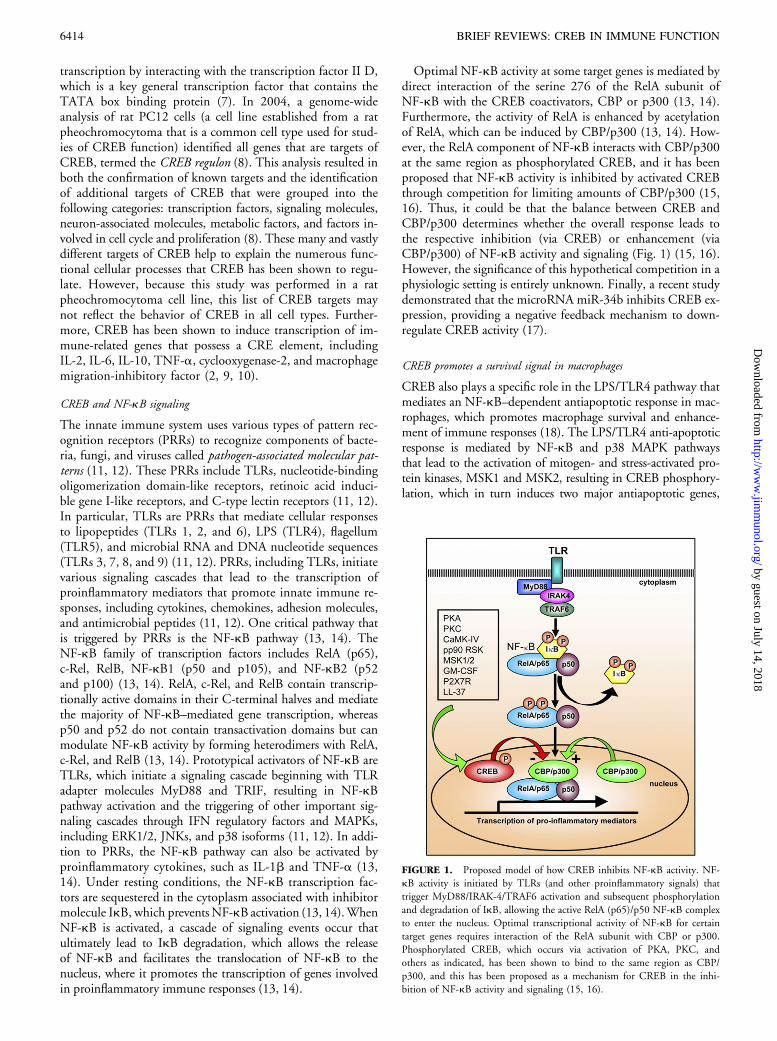
Optimal NF-kB activity at some target genes is mediated bydirect interaction of the serine 276 of the RelA subunit ofNF-kB with the CREB coactivators, CBP or p300 (13, 14).Furthermore, the activity of RelA is enhanced by acetylationof RelA, which can be induced by CBP/p300 (13, 14). How-ever, the RelA component of NF-kB interacts with CBP/p300at the same region as phosphorylated CREB, and it has beenproposed that NF-kB activity is inhibited by activated CREBthrough competition for limiting amounts of CBP/p300 (15,16). Thus, it could be that the balance between CREB andCBP/p300 determines whether the overall response leads tothe respective inhibition (via CREB) or enhancement (viaCBP/p300) of NF-kB activity and signaling (Fig. 1) (15, 16).However, the significance of this hypothetical competition in aphysiologic setting is entirely unknown. Finally, a recent studydemonstrated that the microRNA miR-34b inhibits CREB ex-pression, providing a negative feedback mechanism to down-regulate CREB activity (17).
CREB promotes a survival signal in macrophages
CREB also plays a specific role in the LPS/TLR4 pathway thatmediates an NF-kB–dependent antiapoptotic response in mac-rophages, which promotes macrophage survival and enhance-ment of immune responses (18). The LPS/TLR4 anti-apoptoticresponse is mediated by NF-kB and p38 MAPK pathwaysthat lead to the activation of mitogen- and stress-activated pro-tein kinases, MSK1 and MSK2, resulting in CREB phosphory-lation, which in turn induces two major antiapoptotic genes,
FIGURE 1. Proposed model of how CREB inhibits NF-kB activity. NF-
kB activity is initiated by TLRs (and other proinflammatory signals) that
trigger MyD88/IRAK-4/TRAF6 activation and subsequent phosphorylation
and degradation of IkB, allowing the active RelA (p65)/p50 NF-kB complex
to enter the nucleus. Optimal transcriptional activity of NF-kB for certain
target genes requires interaction of the RelA subunit with CBP or p300.
Phosphorylated CREB, which occurs via activation of PKA, PKC, and
others as indicated, has been shown to bind to the same region as CBP/
p300, and this has been proposed as a mechanism for CREB in the inhi-
bition of NF-kB activity and signaling (15, 16).
6414 BRIEF REVIEWS: CREB IN IMMUNE FUNCTION
by guest on July 14, 2018http://w
ww
.jimm
unol.org/D
ownloaded from

plasminogen activator inhibitor-2 (PAI-2) and B cell lymphoma-2–related gene expressed in fetal liver-1/A1 (Bfl-1/A1) (18, 19).Thus, CREB promotes an antiapoptotic survival signal inmacro-phages, leading to enhanced host immune responses. This actionof CREB is important because certain microbes, such as Salmo-nella spp., Shigellae spp., and Yersiniae spp., inhibit this survivalsignal and induce the apoptosis, or killing, of macrophages asamechanism to evade host immune responses (19). Furthermore,the lethal toxin of Bacillus anthracis has been shown to directlyinhibit the CREB-dependent macrophage antiapoptotic signal(18, 19). In addition to these findings, an adenylate cyclase genein Mycobacterium tuberculosis (Rv0386) facilitated delivery of abacteria-derived cAMP into themacrophage cytoplasm,which re-sulted in TNF-a production that depended on CREB phos-phorylation (20). This CREB-mediated TNF-a production in-creased survival of M. tuberculosis within macrophages, repre-senting an additional mechanism by which CREB promotes bac-terial pathogenesis (20).
Potential factors that may inhibit NF-kB signaling through activationof CREB
Several different factors may inhibit NF-kB activity in mono-cytes/macrophages through the induction of CREB. For ex-ample, GM-CSF signaling results in CREB phosphoryla-tion (21, 22). Phosphorylaton of CREB in this setting invo-lves activation of pp90 RSK through an MEK-dependent sig-naling pathway (21). Because GM-CSF can induce activation ofCREB, this may be anothermechanismwherebyCREB inhibitsNF-kB activity, thus decreasing proinflammatory responses assuggested in the proposed model (Fig. 1).P2X7R is a ligand-gated cation channel nucleotide receptor
that is present on monocytes and macrophages and is activatedby extracellular ATP after tissue injury or infection (23). Inparticular, P2X7R triggers activation of the nucleotide-bindingoligomerization domain-like receptor, nucleotide-binding olig-omerization domain-like receptor pyrin domain-containing 3inflammasome, which is a cytoplasmic protein complex requiredto activate caspase-1 (23). Because caspase-1 cleaves pro–IL-1b and pro–IL-18 into their active forms, P2X7R is one ofthe key receptors that are important for promoting IL-1b andIL-18 responses (23). Upon ligand binding, P2X7R has beenshown to result in the activation of CREB and other transcrip-tion factors, including MAPKs p38 and ERK1/2, and it is alsoinvolved in the production of reactive oxygen species and IL-1isoforms as well as the creation of nonspecific cell membranepores (24, 25). The phosphorylation and activation of CREBby P2X7R includes a CREB/CBP complex formation and ispartly mediated via an extracellular calcium influx and theMEK/ERK system (24). Although the significance of the acti-vation of CREB by the P2X7R in immune responses is not en-tirely clear, one study found that P2X7R stimulation decreasedLPS-stimulated inducible NO synthase and cyclooxygenase-2expression and reduced NO release in microglia in a mechanisminvolving activation of CREB (26). Thus, activation of CREBby P2X7R could promote CREB inhibition of NF-kB activityas suggested in the proposed model (Fig. 1).The antimicrobial peptide cathelidicin (also known as LL-
37) has been demonstrated to have broad-spectrum microbi-cidal activity while also having immunomodulatory activity,which is dependent on activation of MAPKs (ERK1/2 andp38), Elk-1, and NF-kB (27, 28). When PBMCs were stimu-
lated with LL-37 and IL-1b, there was an increase in CREBphosphorylation, suggesting that LL-37 also regulates CREBactivity, potentially inducing the proposed CREB-mediated in-hibition of NF-kB activity (Fig. 1) (28).
CREB induces IL-10 production
IL-10 is a potent anti-inflammatory cytokine that plays a keyrole in mediating a feedback inhibition loop that limits inflam-mation and prevents unwanted tissue damage (29). In macro-phages, IL-10 is produced in response to activation of TLRs2, 3, 4, 7, and 9 (30, 31). TLR signaling via MyD88 or TRIFresults in activation of NF-kB and MAPK pathways (ERK1/2and p38), which subsequently induces production of proin-flammatory cytokines such as TNF-a, IL-1b, IL-6, and IL-10 (29). MAPKs activate MSK1 and MSK2 that phosphor-ylate the transcription factors CREB and AP-1 (e.g., c-Fos,c-Jun, JunB), which subsequently bind to the IL-10 promoterto induce transcription (Fig. 2) (32). This pathway is also re-quired for the transcription of the dual-specificity proteinphosphatase 1, which provides a negative feedback signal byinhibiting p38 (32). Thus, CREB plays an essential role inthe production of IL-10, which in turn inhibits TLR-inducedinflammation and prevents tissue damage (29). It should benoted that IL-10 can also regulate the pathway for TLR-inducedNF-kB activation through the production of glycogen synthasekinase-3b (GSK-3b), which increases the binding of CREB anddecreases the binding of CBP/p300 to the RelA subunit of NF-
FIGURE 2. CREB-induced IL-10 production is regulated by IFN-g. TLR
signaling results in the activation of NF-kB and MAPKs (ERK1/2 and p38),
which induces production of proinflammatory cytokines (e.g., TNF-a, IL-1b,
IL-6) and the anti-inflammatory cytokine IL-10 (29). MAPKs activate MSK1
and MSK2 to directly phosphorylate CREB and AP-1, which bind to the
IL-10 promoter and initiate transcription (32). This same pathway also indu-
ces dual-specificity protein phosphatase 1, which feeds back to inhibit p38
(32). Dectin-1, which recognizes zymosan, and the HIV-1 Tat protein also
induce IL-10 production by activating CREB, Sp1, and Ets-1 through cal-
modulin, CaMK-II, and MAPKs (37–40). Finally, IFN-g inhibits IL-10
production by 1) interfering with the PI3K-AKT pathway, thereby releasing
GSK-3b, which subsequently downregulates the activation and transcrip-
tional activity of CREB and AP-1 proteins that induce IL-10 production;
and 2) directly inhibiting MAPKs (45, 55).
The Journal of Immunology 6415
by guest on July 14, 2018http://w
ww
.jimm
unol.org/D
ownloaded from

kB (Fig. 2) (33). This mechanism utilizing GSK-3b is uniquefrom other cytokines and is regulated by the constitutively activetranscription factors Sp1 and Sp3 (34–36). Finally, recognitionof the yeast particle zymosan by dectin-1 induces productionof IL-10 by initiating CaMK-II- and Pyk2-transduced calciumsignals that activate the ERK1/2 pathway and CREB in a path-way involving reactive oxygen species (37). Thus, in addition toTLRs, dectin-1 provides another mechanism that utilizes CREBto produce IL-10 (37).
HIV-1 Tat induces IL-10 via CREB
Interestingly, the HIV-1 transactivator protein Tat, which isrequired for viral replication and progression of HIV-1 disease,has been shown to induce IL-10 transcription in monocytesand macrophages by activating CREB, Sp1, and Ets-1 throughcalmodulin, CaMK-II, and MAPKs (ERK1/2 and p38) (38–40). Thus, during HIV-1 disease, Tat utilizes CREB to pro-mote IL-10 production. Although the significance of this re-garding HIV pathogenesis is not entirely clear, IL-10 can in-hibit HIV-1 replication in monocytes and macrophages (41),suggesting that Tat/CREB-induced IL-10 production pro-vides a negative feedback signal to prevent excess HIV-1 rep-lication. In addition, because IL-10 can also prevent apoptosisof monocytes and macrophages (42), Tat/CREB-induced IL-10 may allow infected monocytes and macrophages to serve ascellular viral reservoirs.
IFN-g regulates IL-10 production through multiple mechanismsinvolving CREB
In contrast to the anti-inflammatory effects of IL-10, IFN-gis a potent macrophage stimulating factor that promotes vari-ous immune functions, including Ag presentation, cytokineproduction, and antimicrobial activity (43). In addition,IFN-g inhibits IL-10 production and consequently blocksthe immune inhibitory actions of IL-10, resulting in an en-hancement of macrophage activation and immune responses(43). In particular, IFN-g can downregulate the TLR-inducedIL-10 inhibitory response (43). In addition, IFN-g can suppressIL-10 production by two distinct mechanisms: 1) IFN-g caninterfere with the phosphoinositide 3-kinase–AKT pathway,releasing GSK-3b, which inhibits the activation and transcrip-tional activity of CREB and AP-1 proteins (including c-Fos, cJun, and JunB) that promote IL-10 production; and 2) IFN-gdirectly inhibits MAPKs (ERK1/2, JNK, and p38), whichresults in diminished CREB phosphorylation and AP-1 tran-scriptional activity (Fig. 2) (44, 45). Therefore, IFN-g can in-hibit IL-10 production through a number of different mecha-nisms that involve CREB (29).
CREB and T cells, including Th1, Th2, and Th17 cells
CRE elements have been identified in the promoters and en-hancers of many T cell-specific genes, including TCRa, TCRVb, CD3d, CD8a, IL-2, CD25/IL-2Ra, and IL-2Rg, sug-gesting that CREB plays a role in T cell function (10, 46–51). To analyze the specific function of CREB in T cells, Bar-ton et al. (52) engineered a transgenic mouse strain expressinga dominant negative form of CREB under the control of theT cell-specific CD2 promoter. This dominant negative CREBmutation (serine 133 to alanine 133) retained DNA-bindingactivity but was rendered transcriptionally inactive (52). These
CREB mutant mice had normal T cell development in thethymus (52). However, activated T cells from this mouse strainhad a marked defect in proliferation and IL-2 production,resulting in G1 cell-cycle arrest and apoptotic cell death (52).Another group generated a transgenic mouse strain that had thesame CREB dominant-negative mutation, but under the con-trol of the Lck promoter (53). In this case, T cells from thismouse strain did not have any defects in proliferation or IL-2production, but instead had impaired Th cell function (53).Specifically, the CD4+ T cells were defective in their ability toproduce Th-1 (IFN-g) and Th-2 (IL-4) effector cytokines, andthemice failed to produce an Ag-specific IgM and IgG humoralimmune response (53). The differences in the findings betweenthese two studies is likely explained by the differential expres-sion of the mutant CREB by the CD2 versus Lck promoters,but taken together these studies demonstrate that CREB playsa role in T cell proliferation and function. CREB activation alsoplays an important role in governing the cytokines produced byTh1 (IL-2 and IFN-g) and Th2 cells (IL-4 and IL-13) throughthe regulation of IFN-g production. One study found that theIFN-g promoter is hypomethylated in Th1 cells, allowingCREB to induce IFN-g production and promote Th1 re-sponses, whereas the IFN-g promoter is hypermethylated inTh2 cells, resulting in chromatin condensation and preven-tion of CREB-mediated production of IFN-g (54). Finally,IL-17, which is produced by Th-17 cells, downregulates CREBphosphorylation and ensuing IFN-g production in T cells ob-tained from patients with tuberculosis, suggesting that IL-17can inhibit CREB-mediated IFN-g production (55).CREB phosphorylation in T cells has been shown to involve
several signaling molecules and pathways, including PKA,PKC (including PCKa and PKCu), Ras, ERK1/2 MAPKs,and pp90 RSK (10, 56, 57). In addition, the costimula-tory molecule CD28 was shown to optimally activate CREBthrough p38 and CaMK-IV (58, 59). Although these differ-ent studies suggest that several signaling pathways lead toCREB phosphorylation in T cells, a large microarray analysisof con A/anti-CD28 stimulated T cells revealed a greater than100-fold increase in phosphorylated CREB, which was re-duced 50% when the cells were treated with a PKC inhibitorand completely blocked with a PKA inhibitor, suggesting thatPKC and PKA are the major pathways that promote CREBphosphorylation (10). More recently, Kaiser et al. (60) dem-onstrated that MSK1/2 directly phosphorylated CREB inT cells and led to T cell proliferation and production of IL-2in a pathway that was downstream of both ERK1/2 and p38.Collectively, CREB phosphorylation in T cells is mediated bytriggering PKA and PKC, resulting in activation of MAPKsERK1/2 and p38 and subsequent MSK1/2-mediated CREBphosphorylation that promotes T cell activation and prolifer-ation.
CREB and FoxP3 regulatory T cells
Recently, there has been an intense interest on the role thatCD4+CD25+ regulatory T cells (Tregs) play in promotingperipheral tolerance and downregulating pathogenic T cellresponses, including autoimmune responses and allograft re-jection (61). The function of Tregs is dependent on TGF-b–dependent expression of the transcription factor FoxP3,which is required for the development and function of Tregs(61). Emerging evidence has suggested a role for CREB, which
6416 BRIEF REVIEWS: CREB IN IMMUNE FUNCTION
by guest on July 14, 2018http://w
ww
.jimm
unol.org/D
ownloaded from

is activated by TCR activation, in TGF-b/FoxP3–dependentTreg generation and maintenance (Fig. 3). Several studieshave defined a Treg-specific demethylated region (TSDR) inthe FoxP3 locus, which contains a CREB-activating transcrip-tion factor site overlapping a CpG island (61–64). The CpGisland is found demethylated in Tregs and methylated inconventional T cells (61–64). TGF-b, as well as treatmentwith azacytidine, can induce demethylation of this locus,allowing CREB to stabilize FoxP3 expression, thus promotingand maintaining the Treg phenotype (61–64). In addition,a recent study demonstrated that transcription of FoxP3 anddevelopment of Tregs depends on the formation of the “c-Relenhanceosome” at the FoxP3 promoter, which, in addition to
CREB, contains c-Rel, p65,NFAT, and Smad3 (65). Together,the CREB-induced generation and maintenance of Tregs rep-resents an additional example of how CREB inhibits immuneresponses.
CREB and B cells
Previous studies in mature and immature B cells have dem-onstrated that BCR stimulation, which promotes B cell acti-vation and proliferation, involved ERK1/2- and Elk-1–inducedCREB phosphorylation (66). Furthermore, BCR induction ofCREB phosphorylation was dependent on PKCd and pp90RSK signaling pathways (67). Thus, B cell activation andproliferation through the BCR involve signaling pathways
FIGURE 3. CREB promotes TGFb-mediated generation and maintenance of FoxP3 Tregs. The TSDR in the FoxP3 locus is found methylated in conventional
T cells. A, The methylation prevents phosphorylated CREB, which is induced by TCR activation, from binding to this region. B, TGF-b induces demethylation
of the TSDR. C, Demethylation of the TSDR allows phosphylated CREB to bind to the FoxP3 locus to promote FoxP3 expression and the development and
stabilization of Tregs (62–64). Transcription of FoxP3 also involves the formation of a “c-Rel enhanceosome”, which contains c-Rel, p65, NFAT, and Smad3 in
addition to CREB (65).
Table I. Summary of the many different roles of CREB in immune function
Role of CREB Cell Type Associated Factors Overall Role in Immune Function References
Inhibition of NF-kBsignaling
Monocytes andendothelial cells
CREB interacts with the RelA(p65) component of NF-kB at the
same region as CBP/p300
Anti-inflammatory (may beprotective or pathogenic depending
on the context)
15, 16
Antiapoptotic survivalsignal
Macrophages TLR4 triggers NF-kB/MAPKs andMSK1/2, which activate PAI-2 andBfl-1/A1 to phosphorylate CREB
Pathogenic (mechanism forpathogens to evade host immune
responses)
18, 19
TNF-a–mediatedsurvival signal
Macrophages M. tuberculosis-derived adenylatecylcase (Rv0386) phosphorylates
CREB
Pathogenic (promotes survival ofM. tuberculosis in macrophages)
20
Induction of IL-10 Macrophages andmyeloid dendritic cells
MAPKs (ERK1/2 and p38) triggerMSK1/2 to activate CREB
Anti-inflammatory (may beprotective or pathogenic depending
on the context)
29–32
Induction of IL-10 Macrophages TLR2 activation by Candida albicanstriggers IL-10 production
Pathogenic (promotes survival ofTregs, leading to disseminated
candidiasis)
31
Induction of IL-10 byHIV-1 Tat
Macrophages Sp1, Ets-1, calmodulin, CaMK-II,and MAPKs
Unknown, may be pathogenic(macrophages could act as cellular
viral reservoirs)
38–40
Promotes T cell activationand proliferation andTh cell activity
T cells PKA, PKC, Ras, ERK1/2, pp90RSK and CD28 (via p38 and
CaMK-IV)
May be protective or pathogenic(depending on the Ag)
52, 53, 56–59
Promotes IFN-gproduction
T cells IFN-g promoter is hypomethylatedin Th1 cells, allowing CREB
binding
Protective (promotes Th1 responsesthat are protective in tuberculosis)
54, 55
Induction of FoxP3,promoting Treggeneration andmaintenance
T cells TSDR in the FoxP3 locus, whichallows CREB to promote FoxP3
expression
Anti-inflammatory (may bepathogenic relating to infections and
tumor immunosurveillance andprotective relating to autoimmunity)
61–65
Promotes B cell activationand proliferation
B cells ERK1/2, E1K-1, PKCd, pp90 RSK May be protective or pathogenic(depending on the Ag)
66, 67
The Journal of Immunology 6417
by guest on July 14, 2018http://w
ww
.jimm
unol.org/D
ownloaded from

that include PKCd and pp90 RSK, resulting in ERK1/2- andElk-1–dependent activation of CREB (66, 67).
ConclusionsCREB plays many different roles in immune function(Table I). CREB often promotes anti-inflammatory immuneresponses, such as through the inhibition of NF-kB activity,the induction of IL-10, and the generation of Tregs. Theseanti-inflammatory responses could be protective by inhibitingunwanted inflammation, tissue damage, and autoimmune re-sponses, or they could be pathogenic in the context of in-fection and tumor immunosurveillance. However, CREB alsopromotes activation and proliferation of T and B cells anddifferentially regulates Th1, Th2, and Th17 responses. Futureinvestigation into the role of CREB in immune function willlead to an increased understanding of how this transcriptionfactor regulates specific immune responses. One challenge is toapply innovative technology to uncover the roles of CREB inimmune responses, such as chromatin immunoprecipitationcoupled to massively parallel sequencing to monitor histonemodifications and nucleosome dynamics to characterize theevents required for CREB binding and activation of immune-related genes (68, 69). Although CREB appears to be involvedin an amazingly diverse range of processes in immune cellsand other cell types, further research could reveal signalingpathways or collaborating proteins that are selectively in-volved in its proinflammatory or its anti-inflammatory func-tions. Therefore, a better understanding of the activationmechanisms and mechanisms of action of CREB in differentcell types and settings could suggest therapeutic strategies forselective manipulation of immune responses.
DisclosuresThe authors have no financial conflicts of interest.
References1. Shaywitz, A. J., and M. E. Greenberg. 1999. CREB: a stimulus-induced transcrip-
tion factor activated by a diverse array of extracellular signals. Annu. Rev. Biochem.68: 821–861.
2. Mayr, B., and M. Montminy. 2001. Transcriptional regulation by the
phosphorylation-dependent factor CREB. Nat. Rev. Mol. Cell Biol. 2: 599–609.3. Sakamoto, K. M., and D. A. Frank. 2009. CREB in the pathophysiology of cancer:
implications for targeting transcription factors for cancer therapy. Clin. Cancer Res.15: 2583–2587.
4. Brindle, P., S. Linke, and M. Montminy. 1993. Protein-kinase-A-dependent acti-
vator in transcription factor CREB reveals new role for CREM repressors. Nature364: 821–824.
5. Enslen, H., H. Tokumitsu, and T. R. Soderling. 1995. Phosphorylation of CREB
by CaM-kinase IV activated by CaM-kinase IV kinase. Biochem. Biophys. Res. Com-mun. 207: 1038–1043.
6. Gubina, E., X. Luo, E. Kwon, K. Sakamoto, Y. F. Shi, and R. A. Mufson. 2001.
betac cytokine receptor-induced stimulation of cAMP response element binding
protein phosphorylation requires protein kinase C in myeloid cells: a novel cytokine
signal transduction cascade. J. Immunol. 167: 4303–4310.7. Conkright, M. D., G. Canettieri, R. Screaton, E. Guzman, L. Miraglia,
J. B. Hogenesch, and M. Montminy. 2003. TORCs: transducers of regulated CREB
activity. Mol. Cell 12: 413–423.8. Impey, S., S. R. McCorkle, H. Cha-Molstad, J. M. Dwyer, G. S. Yochum,
J. M. Boss, S. McWeeney, J. J. Dunn, G. Mandel, and R. H. Goodman. 2004.
Defining the CREB regulon: a genome-wide analysis of transcription factor regu-
latory regions. Cell 119: 1041–1054.9. Brenner, S., S. Prosch, K. Schenke-Layland, U. Riese, U. Gausmann, and C. Platzer.
2003. cAMP-induced Interleukin-10 promoter activation depends on CCAAT/
enhancer-binding protein expression and monocytic differentiation. J. Biol. Chem.278: 5597–5604.
10. Hughes-Fulford, M., E. Sugano, T. Schopper, C. F. Li, J. B. Boonyaratanakornkit,
and A. Cogoli. 2005. Early immune response and regulation of IL-2 receptor
subunits. Cell. Signal. 17: 1111–1124.11. Takeuchi, O., and S. Akira. 2010. Pattern recognition receptors and inflammation.
Cell 140: 805–820.
12. Medzhitov, R. 2010. Inflammation 2010: new adventures of an old flame. Cell 140:771–776.
13. Ghosh, S., and M. S. Hayden. 2008. New regulators of NF-kappaB in inflamma-tion. Nat. Rev. Immunol. 8: 837–848.
14. Medzhitov, R., and T. Horng. 2009. Transcriptional control of the inflammatoryresponse. Nat. Rev. Immunol. 9: 692–703.
15. Ollivier, V., G. C. Parry, R. R. Cobb, D. de Prost, and N. Mackman. 1996. Ele-vated cyclic AMP inhibits NF-kappaB-mediated transcription in humanmonocytic cells and endothelial cells. J. Biol. Chem. 271: 20828–20835.
16. Parry, G. C., and N. Mackman. 1997. Role of cyclic AMP response element-binding protein in cyclic AMP inhibition of NF-kappaB-mediated transcription.J. Immunol. 159: 5450–5456.
17. Pigazzi, M., E. Manara, E. Baron, and G. Basso. 2009. miR-34b targets cyclicAMP-responsive element binding protein in acute myeloid leukemia. Cancer Res.69: 2471–2478.
18. Park, J. M., F. R. Greten, A. Wong, R. J. Westrick, J. S. Arthur, K. Otsu,A. Hoffmann, M. Montminy, and M. Karin. 2005. Signaling pathways and genesthat inhibit pathogen-induced macrophage apoptosis—CREB and NF-kappaB askey regulators. Immunity 23: 319–329.
19. Hsu, L. C., J. M. Park, K. Zhang, J. L. Luo, S. Maeda, R. J. Kaufman, L. Eckmann,D. G. Guiney, and M. Karin. 2004. The protein kinase PKR is required for mac-rophage apoptosis after activation of Toll-like receptor 4. Nature 428: 341–345.
20. Agarwal, N., G. Lamichhane, R. Gupta, S. Nolan, and W. R. Bishai. 2009. CyclicAMP intoxication of macrophages by a Mycobacterium tuberculosis adenylate cyclase.Nature 460: 98–102.
21. Cheng, J. C., K. Kinjo, D. R. Judelson, J. Chang, W. S. Wu, I. Schmid,D. B. Shankar, N. Kasahara, R. Stripecke, R. Bhatia, et al. 2008. CREB is a criticalregulator of normal hematopoiesis and leukemogenesis. Blood 111: 1182–1192.
22. Kwon, E. M., M. A. Raines, J. Blenis, and K. M. Sakamoto. 2000. Granulocyte-macrophage colony-stimulating factor stimulation results in phosphorylation ofcAMP response element-binding protein through activation of pp90RSK. Blood95: 2552–2558.
23. Tschopp, J., and K. Schroder. 2010. NLRP3 inflammasome activation: The con-vergence of multiple signalling pathways on ROS production? Nat. Rev. Immunol.10: 210–215.
24. Gavala, M. L., Z. A. Pfeiffer, and P. J. Bertics. 2008. The nucleotide receptorP2RX7 mediates ATP-induced CREB activation in human and murine monocyticcells. J. Leukoc. Biol. 84: 1159–1171.
25. Potucek, Y. D., J. M. Crain, and J. J. Watters. 2006. Purinergic receptors modulateMAP kinases and transcription factors that control microglial inflammatory geneexpression. Neurochem. Int. 49: 204–214.
26. Brautigam, V. M., C. Frasier, M. Nikodemova, and J. J. Watters. 2005. Purinergicreceptor modulation of BV-2 microglial cell activity: potential involvement of p38MAP kinase and CREB. J. Neuroimmunol. 166: 113–125.
27. Bowdish, D. M., D. J. Davidson, D. P. Speert, and R. E. Hancock. 2004. Thehuman cationic peptide LL-37 induces activation of the extracellular signal-regulated kinase and p38 kinase pathways in primary human monocytes. J. Immu-nol. 172: 3758–3765.
28. Yu, J., N. Mookherjee, K. Wee, D. M. Bowdish, J. Pistolic, Y. Li, L. Rehaume, andR. E. Hancock. 2007. Host defense peptide LL-37, in synergy with inflammatorymediator IL-1beta, augments immune responses by multiple pathways. J. Immunol.179: 7684–7691.
29. Saraiva, M., and A. O’Garra. 2010. The regulation of IL-10 productionby immune cells. Nat. Rev. Immunol. 10: 170–181.
30. Boonstra, A., R. Rajsbaum, M. Holman, R. Marques, C. Asselin-Paturel,J. P. Pereira, E. E. Bates, S. Akira, P. Vieira, Y. J. Liu, et al. 2006. Macrophagesand myeloid dendritic cells, but not plasmacytoid dendritic cells, produce IL-10 inresponse to MyD88- and TRIF-dependent TLR signals, and TLR-independentsignals. J. Immunol. 177: 7551–7558.
31. Netea, M. G., R. Sutmuller, C. Hermann, C. A. Van der Graaf, J. W. Van derMeer, J. H. van Krieken, T. Hartung, G. Adema, and B. J. Kullberg. 2004. Toll-likereceptor 2 suppresses immunity against Candida albicans through induction ofIL-10 and regulatory T cells. J. Immunol. 172: 3712–3718.
32. Ananieva, O., J. Darragh, C. Johansen, J. M. Carr, J. McIlrath, J. M. Park,A. Wingate, C. E. Monk, R. Toth, S. G. Santos, et al. 2008. The kinases MSK1 andMSK2 act as negative regulators of Toll-like receptor signaling. Nat. Immunol. 9:1028–1036.
33. Martin, M., K. Rehani, R. S. Jope, and S. M. Michalek. 2005. Toll-like receptor-mediated cytokine production is differentially regulated by glycogen synthase kinase3. Nat. Immunol. 6: 777–784.
34. Brightbill, H. D., S. E. Plevy, R. L. Modlin, and S. T. Smale. 2000. A prominentrole for Sp1 during lipopolysaccharide-mediated induction of the IL-10 promoterin macrophages. J. Immunol. 164: 1940–1951.
35. Chan, M. M., B. K. Cheung, J. C. Li, L. L. Chan, and A. S. Lau. 2009. A role forglycogen synthase kinase-3 in antagonizing mycobacterial immune evasion by neg-atively regulating IL-10 induction. J. Leukoc. Biol. 86: 283–291.
36. Tone, M., M. J. Powell, Y. Tone, S. A. Thompson, and H. Waldmann. 2000. IL-10gene expression is controlled by the transcription factors Sp1 and Sp3. J. Immunol.165: 286–291.
37. Kelly, E. K., L. Wang, and L. B. Ivashkiv. 2010. Calcium-activated pathways andoxidative burst mediate zymosan-induced signaling and IL-10 production inhuman macrophages. J. Immunol. 184: 5545–5552.
38. Gee, K., J. B. Angel, S. Mishra, M. A. Blahoianu, and A. Kumar. 2007. IL-10regulation by HIV-Tat in primary human monocytic cells: involvement ofcalmodulin/calmodulin-dependent protein kinase-activated p38 MAPK and Sp-1and CREB-1 transcription factors. J. Immunol. 178: 798–807.
6418 BRIEF REVIEWS: CREB IN IMMUNE FUNCTION
by guest on July 14, 2018http://w
ww
.jimm
unol.org/D
ownloaded from

39. Gee, K., J. B. Angel, W. Ma, S. Mishra, N. Gajanayaka, K. Parato, and A. Kumar.2006. Intracellular HIV-Tat expression induces IL-10 synthesis by the CREB-1transcription factor through Ser133 phosphorylation and its regulation by theERK1/2 MAPK in human monocytic cells. J. Biol. Chem. 281: 31647–31658.
40. Li, J. C., and A. S. Lau. 2007. A role for mitogen-activated protein kinase and Ets-1in the induction of interleukin-10 transcription by human immunodeficiencyvirus-1 Tat. Immunology 121: 337–348.
41. Akridge, R. E., L. K. Oyafuso, and S. G. Reed. 1994. IL-10 is induced duringHIV-1 infection and is capable of decreasing viral replication in human macro-phages. J. Immunol. 153: 5782–5789.
42. Zhou, J. H., S. R. Broussard, K. Strle, G. G. Freund, R. W. Johnson, R. Dantzer,and K. W. Kelley. 2001. IL-10 inhibits apoptosis of promyeloid cells by activatinginsulin receptor substrate-2 and phosphatidylinositol 39-kinase. J. Immunol. 167:4436–4442.
43. Schroder, K., P. J. Hertzog, T. Ravasi, and D. A. Hume. 2004. Interferon-gamma:an overview of signals, mechanisms and functions. J. Leukoc. Biol. 75: 163–189.
44. Hu, X., P. K. Paik, J. Chen, A. Yarilina, L. Kockeritz, T. T. Lu, J. R. Woodgett, andL. B. Ivashkiv. 2006. IFN-gamma suppresses IL-10 production and synergizes withTLR2 by regulating GSK3 and CREB/AP-1 proteins. Immunity 24: 563–574.
45. Grimes, C. A., and R. S. Jope. 2001. CREB DNA binding activity is inhibited byglycogen synthase kinase-3 beta and facilitated by lithium. J. Neurochem. 78: 1219–1232.
46. Solomou, E. E., Y. T. Juang, and G. C. Tsokos. 2001. Protein kinase C-thetaparticipates in the activation of cyclic AMP-responsive element-binding proteinand its subsequent binding to the -180 site of the IL-2 promoter in normalhuman T lymphocytes. J. Immunol. 166: 5665–5674.
47. Yeh, J. H., P. Lecine, J. A. Nunes, S. Spicuglia, P. Ferrier, D. Olive, and J. Imbert.2001. Novel CD28-responsive enhancer activated by CREB/ATF and AP-1 familiesin the human interleukin-2 receptor alpha-chain locus. Mol. Cell. Biol. 21: 4515–4527.
48. Mayall, T. P., P. L. Sheridan, M. R. Montminy, and K. A. Jones. 1997. Distinctroles for P-CREB and LEF-1 in TCR alpha enhancer assembly and activation onchromatin templates in vitro. Genes Dev. 11: 887–899.
49. Anderson, S. J., S. Miyake, and D. Y. Loh. 1989. Transcription from a murineT-cell receptor V beta promoter depends on a conserved decamer motif similar tothe cyclic AMP response element. Mol. Cell. Biol. 9: 4835–4845.
50. Gao, M. H., and P. B. Kavathas. 1993. Functional importance of the cyclic AMPresponse element-like decamer motif in the CD8 alpha promoter. J. Immunol. 150:4376–4385.
51. Gupta, A., and C. Terhorst. 1994. CD3 delta enhancer. CREB interferes with thefunction of a murine CD3-delta A binding factor (M delta AF). J. Immunol. 152:3895–3903.
52. Barton, K., N. Muthusamy, M. Chanyangam, C. Fischer, C. Clendenin, andJ. M. Leiden. 1996. Defective thymocyte proliferation and IL-2 production intransgenic mice expressing a dominant-negative form of CREB. Nature 379: 81–85.
53. Zhang, F., M. Rincon, R. A. Flavell, and T. M. Aune. 2000. Defective Th functioninduced by a dominant-negative cAMP response element binding protein mutationis reversed by Bcl-2. J. Immunol. 165: 1762–1770.
54. Yano, S., P. Ghosh, H. Kusaba, M. Buchholz, and D. L. Longo. 2003. Effect ofpromoter methylation on the regulation of IFN-gamma gene during in vitro differ-
entiation of human peripheral blood T cells into a Th2 population. J. Immunol. 171:2510–2516.
55. Pasquinelli, V., J. C. Townsend, J. O. Jurado, I. B. Alvarez, M. F. Quiroga,P. F. Barnes, B. Samten, and V. E. Garcıa. 2009. IFN-gamma production duringactive tuberculosis is regulated by mechanisms that involve IL-17, SLAM, andCREB. J. Infect. Dis. 199: 661–665.
56. Grady, G. C., S. M. Mason, J. Stephen, J. C. Zuniga-Pflucker, and A. M. Michie.2004. Cyclic adenosine 59-monophosphate response element binding protein playsa central role in mediating proliferation and differentiation downstream of the pre-TCR complex in developing thymocytes. J. Immunol. 173: 1802–1810.
57. Muthusamy, N., and J. M. Leiden. 1998. A protein kinase C-, Ras-, and RSK2-dependent signal transduction pathway activates the cAMP-responsive element-binding protein transcription factor following T cell receptor engagement. J. Biol.Chem. 273: 22841–22847.
58. Yu, C. T., H. M. Shih, and M. Z. Lai. 2001. Multiple signals required for cyclicAMP-responsive element binding protein (CREB) binding protein interaction in-duced by CD3/CD28 costimulation. J. Immunol. 166: 284–292.
59. Hsueh, Y. P., H. E. Liang, S. Y. Ng, and M. Z. Lai. 1997. CD28-costimulationactivates cyclic AMP-responsive element-binding protein in T lymphocytes. J.Immunol. 158: 85–93.
60. Kaiser, M., G. R. Wiggin, K. Lightfoot, J. S. Arthur, and A. Macdonald. 2007.MSK regulate TCR-induced CREB phosphorylation but not immediate early genetranscription. Eur. J. Immunol. 37: 2583–2595.
61. Sakaguchi, S., M. Miyara, C. M. Costantino, and D. A. Hafler. 2010. FOXP3+regulatory T cells in the human immune system. Nat. Rev. Immunol. 10: 490–500.
62. Polansky, J. K., K. Kretschmer, J. Freyer, S. Floess, A. Garbe, U. Baron, S. Olek,A. Hamann, H. von Boehmer, and J. Huehn. 2008. DNA methylation controlsFoxp3 gene expression. Eur. J. Immunol. 38: 1654–1663.
63. Polansky, J. K., L. Schreiber, C. Thelemann, L. Ludwig, M. Kruger, R. Baumgrass,S. Cording, S. Floess, A. Hamann, and J. Huehn. 2010. Methylation matters:binding of Ets-1 to the demethylated Foxp3 gene contributes to the stabilization ofFoxp3 expression in regulatory T cells. J. Mol. Med. 88: 1029–1040 (epub ahead ofprint).
64. Kim, H. P., and W. J. Leonard. 2007. CREB/ATF-dependent T cell receptor-induced FoxP3 gene expression: a role for DNA methylation. J. Exp. Med. 204:1543–1551.
65. Ruan, Q., V. Kameswaran, Y. Tone, L. Li, H. C. Liou, M. I. Greene, M. Tone, andY. H. Chen. 2009. Development of Foxp3(+) regulatory t cells is driven by thec-Rel enhanceosome. Immunity 31: 932–940.
66. Yasuda, T., H. Sanjo, G. Pages, Y. Kawano, H. Karasuyama, J. Pouyssegur,M. Ogata, and T. Kurosaki. 2008. Erk kinases link pre-B cell receptor signaling totranscriptional events required for early B cell expansion. Immunity 28: 499–508.
67. Blois, J. T., J. M. Mataraza, I. Mecklenbrauker, A. Tarakhovsky, and T. C. Chiles.2004. B cell receptor-induced cAMP-response element-binding protein activation inB lymphocytes requires novel protein kinase Cdelta. J. Biol. Chem. 279: 30123–30132.
68. Barski, A., S. Cuddapah, K. Cui, T. Y. Roh, D. E. Schones, Z. Wang, G. Wei,I. Chepelev, and K. Zhao. 2007. High-resolution profiling of histone methylationsin the human genome. Cell 129: 823–837.
69. Smale, S. T. 2010. Seq-ing LPS-induced enhancers. Immunity 32: 296–298.
The Journal of Immunology 6419
by guest on July 14, 2018http://w
ww
.jimm
unol.org/D
ownloaded from