Embed Size (px)
Citation preview

The role of the Low-Density Lipoprotein Receptor Family in the pathology of the
Antiphospholipid Syndrome
Menno van Lummel

Cover: Electrostatic potential of the fi fth domain of β2GPI
Printing: Febodruk, Enschede
Lay-out: Febodruk, Enschede
ISBN-10: 90-393-4330-6ISBN-13: 978-90-393-4330-2

The role of the Low-Density Lipoprotein Receptor Family in the pathology of the
Antiphospholipid Syndrome
Rol van Low-Density Lipoproteine Receptoren in de pathologie van het
Antifosfolipiden Syndroom
(met een samenvatting in het Nederlands)
Proefschrift
ter verkrijging van de graad van doctor
aan de universiteit Utrecht
op gezag van de rector magnifi cus, prof. dr. W.H. Gispen
in gevolge het besluit van het college voor promoties
in het openbaar te verdedigen
op donderdag 7 september 2006 des ochtends te 10:30 uur
door
Menno van Lummel
Geboren op 26 januari 1978, te Maassluis
Promotor:Prof. dr. Ph. G. de Groot
Co-promotor:Dr. R.H.W.M Derksen
Manuscriptcommissie:Prof. dr. F. MiedemaProf. dr. G.J.A.M. StrousProf. dr. G. PasterkampProf. dr. J.W. Cohen Tervaert
The studies described in this thesis were supported by a grant from the Nether-lands Institute for Health Research and Development (Zon-MW: 902-26-290)
Financial support of NWO and Universiteit Utrecht for the publication of this the-sis is gratefully acknowledged.
Additional fi nancial support for the publication of this thesis by Dr. Ir. van de Laar Stichting, Pfi zer and Lancyr Totaalbeheer is gratefully acknowledged.

Contents
Abbreviations 7
Chapter 1 General Introduction 9
Chapter 2 Beta2-glycoprotein I and LDL-receptor family members. 51
Chapter 3 The binding site in beta2-glycoprotein I for ApoER2’ on
platelets is located in domain V. 63
Chapter 4 Interaction of beta2-glycoprotein I with members of
the low-density lipoprotein receptor family. 89
Chapter 5 Molecular characterization of the apolipoprotein
E receptor 2’- binding site of beta2-glycoprotein I
by site-directed mutagenesis. 115
Chapter 6 Pathogenic anti-beta2-glycoprotein I antibodies
recognize domain I of beta2-glycoprotein I only after
a conformational change. 137
Chapter 7 General Discussion 165
Chapter 8 Summary 185
Chapter 9 Nederlandse Samenvatting 193
Chapter 10 Appendices 201
Bibliography 202
Presentations 203
Curriculum Vitae 205
Dankwoord 206


7
Abbreviations
α2AP alpha2-antiplasmin
β2GPI β2-glycoprotein I
aCL anti-cardiolipin antibodies
AD Alzheimer disease
ADP adenosine diphosphate
APC Activated Protein C
aPL antiphospholipid antibodies
apoB apolipoprotein B
apoE apolipoprotein E
apoER2’ apolipoprotein E receptor 2’
APS Antiphospholipid syndrome
APTT activated partial thromboplastin time
AT antithrombin
ATP adenosine triphosphate
BCA bicinchoninic acid protein assay
BHK baby hamster kidney
BSA bovine serum albumin
CCP complement control protein
CHAPS 3-[(3-cholamidopropyl) dimethylammonio]-1-
propanesulfonic acid
CK-MB creatine kinase myoglobin class A repeats
CR complement type repeat
Dab1 Disabled 1
DEA diethynolamine
DM domain deletion mutants
dRVVT dilute Russell’s viper venom time
DTS dense tubular system
DVT deep venous thrombosis
EC endothelial cells
EDTA ethylenediaminetetraacetic acid
EGF epidermal growth factor
ELISA enzyme linked immunosorbent assay
FII prothrombin
FV factor V
FVa activated factor V
FVII factor VII
FVIIa activated factor VII
Abbreviations
FVIII factor VIII
FVIIIa activated factor VIII
FIX factor IX
FIXa activated factor IX
FX factor X
FXa activated factor X
FXI factor XI
FXIa activated factor XI
FXII factor XII
FXIIa activated factor XII
FXIII factor XIII
FXIIIa activated factor XIII
FDP fi brin degradation products
FH familial hypercholesterolemia
FPLC fast performance liquid
chromatography
GP glycoprotein
HMWK high molecular weight kininogen
IE/ml international units per milliliter
ISTH International Society of Thrombosis
and Haemostasis
KCT kaolin clotting time
kDa kiloDalton
KD equilibrium dissociation constant
LA LDL-R type A
LAC lupus anticoagulants
LDL low-density lipoprotein
LDL-R low-density lipoprotein receptor
Leu leucine
LLD lupus-like disease
LMWH low molecular weight heparin
LRP LDL-R related protein
MegBP megalin binding protein
MES 2-(N-Morpholino) ethanesulfonic acid
μg microgram
μm micro molar
ml milliLiter

8
mM milliMolar
MTS microtubular system
nM nanoMolar
NO nitric oxide
NPP normal pooled plasma
OCS open canalicular system
OPD orthophenylenediamine
oxLDL oxidized LDL
PAI-1 plasminogen activator inhibitor-1
PC phosphatidylcholine
PE phosphotidylethanolamine
PEG polyethylene glycol
PF4 platelet factor 4
PGI2 prostaglandin I2PL phospholipids
PnPP para-nitrophenyl phosphate
PS phosphatidylserine
PT prothrombin time
PTC proximal tubule cells
RAMPO rabbit anti-mouse antibody peroxidase
RAP receptor associated protein
Req Response at equilibrium
SCR short consensus repeats
SDS-PAGE sodium dodecyl sulfate-polyacryl amide gel electrophoresis
Ser serine
SLE systemic lupus erythematosus
SMC smooth muscle cells
SorLA sorting protein-related receptor containing LDL-R
SPR surface plasmon resonance
SSC Scientifi c and standardization Committee
SWARPO swine anti-rabbit antibody peroxidase
TAFI thrombin-activatable fi brinolysis inhibitor
TBST TBS Tween 20
TF tissue factor
TFPI tissue factor pathway inhibitor
TIA transient ischemic attacks
TLR toll-like receptor
TM transmembrane region
tPA tissue-type plasminogen activator
Trp tryptophan
Abbreviations
TX thromboxane
TxA2 thromboxane A2
Tyr tyrosine
U Units
UFH unfractionated heparin
uPA urokinase-type plasminogen
activator
VKA vitamin K antagonists
VLDL very low-density lipoprotein
VLDL-R very low-density lipoprotein
receptor
vWF von Willebrand factor

1GENERAL INTRODUCTION
The antiphospholipid syndrome 11
Antiphospholipid antibodies 12
The haemostatic process 13
Platelet Biochemistry 14
Platelets and Haemostasis 16
Coagulation 17
Inhibition of coagulation 19
Fibrinolysis 19
Beta2-glycoprotein I 20
Protein Chemistry 20
Physiological function of β2GPI 22
β2GPI and cellular activation 23
Multiligand Receptors 25
The Low-Density Lipoprotein Receptor Family 25
Common structure of the LDL-R family 27
LDL Receptor 28
VLDL Receptor 29
Low-density lipoprotein Receptor-related Protein 30
Megalin 30
Apolipoprotein Receptor 2’ 31
Outline of this thesis 33
References 35


11
General Introduction
The Antiphospholipid SyndromeThe antiphospholipid syndrome (APS) is a non-infl ammatory autoimmune disease that is defi ned by both clinical and laboratory criteria. The clinical conditions are characterized by the presence of venous and/or arterial thrombosis and/or pregnancy complications 1;2. Venous or arterial thrombosis includes deep venous thrombosis (DVT), transient ischemic attacks (TIA) or myocardial ischemia or infarction 3;4. The syndrome is diagnosed when one of the clinical criteria is present in combination with the presence of antiphospholipid antibodies (aPL) (Table 1) 1;5,220. aPL are also associated with other clinical features including heart valve abnormalities, livedo reticularis, chorea, nephropathy, thrombocytopenia and haemolytic anaemia 6. These clinical manifestations, which are not part of the inclusion criteria, are frequently found in patients with APS. aPL is a general term that describes a variety of closely related antibodies; lupus anticoagulants (LAC), anti-cardiolipin antibodies (aCL) and anti-β2-glycoprotein I antibodies. The presence of aPL in plasma are detected by a phospholipid-dependent prolongation of a clotting assay (LAC) or by solid phase immune assays, the anticardiolipin- and the anti-β2-glycoprotein I enzyme linked immunosorbent assay (ELISA). Autoantibodies that cause LAC and aCL are closely related, but not per se identical autoantibodies. This raises the question which antibody is the most relevant. Several studies show that antibodies inducing a LAC correlate best with a history of thrombotic complications 7;8. The paradoxical association between in vitro prolongation of clotting assays (LAC) and in vivo observed thrombosis, stimulated scientists to search for the real antigen to which the antibodies are directed. Until the early nineties, it was thought that aPL are directed against anionic phospholipids directly. In 1990, three groups independently discovered a phospholipid-binding plasma protein to which the antibodies are directed; β2-glycoprotein I (β2GPI) 9-11. Since 1991, a second cofactor was discovered in APS, prothrombin, another plasma protein with affi nity for anionic phospholipids that could also be a target for aPL 12. From that moment on a search for other phospholipid binding plasma proteins started. At present, several proteins have been identifi ed that may serve as antigen for aPL, including β2GPI, Protein S, Protein C, Protein Z, annexin A5, thrombomodulin, high molecular weight kininogen (HMWK), tissue factor pathway inhibitor (TFPI) and prothrombin 13. Despite the presence of these proteins as possible target of aPL, β2GPI is the main cofactor with clinical signifi cance in APS 7;8;14. For example, LAC positive anti-β2GPI antibodies correlate best with thromboembolic complications observed in APS patients. Interaction of autoantibodies with β2GPI seems very important in the pathophysiology of APS.

12
Chapter 1
Table 1. Revised classifi cation criteria for the antiphospholipid syndrome
Clinical criteria 1. One or more clinical episodes of objectively verifi ed arterial, venous or small vessel thrombosis. Thrombosis must be present without indications of infl ammation. 2. Pregnancy morbidity: - One or more unexplained deaths of a morphologically normal foetus at or beyond the 10th week of gestation - One or more premature births of a morphologically normal neonate before the 34th week of gestation - Three or more unexplained consecutive spontaneous abortions before the 10th week of gestation
Laboratory criteria 1. Positive test for anti- β2GPI dependent anticardiolipin antibodies on two or more occasions at least 6 weeks apart, measured by ELISA 2. Prolongation of at least one phospholipid dependent coagulation assay measured in plasma, on two or more occasions at least 6 weeks apart.A patient is diagnosed with the antiphospholipid syndrome when the patient meets one or more of the clinical criteria in the combination with one or more of the serological criteria.
Antiphospholipid AntibodiesaPL are classifi ed according to their in vitro method of detection; ELISA’s measure anti-β2GPI antibodies and clotting assays detect LAC. LAC are immunoglobulins of the class IgG or IgM that interfere with in vitro phospholipid-depending coagulation assays, such as the prothrombin time (PT), activated partial thromboplastin time (APTT), kaolin clotting time (KCT) or dilute Russell’s viper venom time (dRVVT). The APTT and the KCT measure coagulation activated via the intrinsic pathway (Figure 1). This pathway starts with the activation of factor XII. The PT measures coagulation activated via factor VII. In the dRVVT, coagulation starts via the direct activation of factor X. These LAC assays must be performed in accordance with the recommendation of the Scientifi c and Standardization Committee of the International Society of Thrombosis and Haemostasis (SSC of ISTH) 15. The laboratory criteria for APS require that aPL (anti-β2GPI antibodies and/or aCL) tests are positive on at least two occasions with blood samples taken more than 12 weeks apart, to avoid

13
General Introduction
positive aPL associated with acute infectious diseases. β2GPI and prothrombin are the main targets for aPL causing LAC. Antiprothrombin antibodies are responsible for prothrombin-dependent LAC and anti-β2GPI antibodies for β2GPI-dependent LAC. Specifi c neutralization with cardiolipin enables differentiation between anti-β2GPI antibodies or antiprothrombin antibodies dependent LAC 16. The addition of cardiolipin neutralizes β2GPI-dependent LAC but not prothrombin-dependent LAC in an APTT based assay. aPL directed against a specifi c epitope in domain I of β2GPI correlates best with thrombosis (Gly40-Arg43) 17. This epitope is shield off when β2GPI is circulating free in plasma. Only after a conformational change, such as that induced by binding of β2GPI to phospholipids, this epitope is exposed.
Figure 1. Coagulation cascade and fi brin formation by the intrinsic and extrinsic pathways. The initiation of the coagulation occurs following vascular injury and the exposure of TF to the blood. This triggers the extrinsic pathway. The intrinsic pathway can be triggered when thrombin is generated, leading to the activation of factor XI. The two pathways converge by the formation of factor Xa. PL refers to phospholipid.
The Haemostatic ProcessNormal vascular endothelial cells maintain blood fl uidity by inhibiting coagulation and platelet aggregation. These cells also prevent platelets and coagulation factors from exposure to reactive and pro-thrombotic subendothelium. The subendothelium
14
Chapter 1
contains components like collagen, fi bronectin and von Willebrand factor (vWF), which promote platelet adhesion. In addition, the subendothelium also exposes a membrane protein called tissue factor (TF) that triggers blood coagulation. After injury of the endothelial layer, these pro-thrombotic components are exposed to the fl owing blood. This induces the formation of the haemostatic plug by promoting platelet adhesion and aggregation and by activating coagulation. Haemostasis is composed of four major events that occur in a sequence following the loss of vascular integrity 18. The completion of each step activates another coagulation factor in a chain reaction that leads to the conversion of fi brinogen to fi brin:
I. The initial phase of the process is vascular constriction in order to limit the blood fl ow to the site of injury.II. Next, platelets adhere to the exposed subendothelium followed by platelet aggregation at the site of injury, forming a temporary, soft platelet plug. Fibrinogen and vWF are primarily responsible for stimulating platelet clumping. Upon activation, platelets release among others ADP and thromboxane (TX) A2 (which activate additional platelets), serotonin, factor V and other proteins important for the coagulation cascade. In addition, activated platelets expose anionic phospholipids on their cellular surface to accommodate the formation of the plug.III. To stabilize the initially loose platelet plug, the coagulation cascade converts fi brinogen to fi brin that replaces the platelet plug. IV. The clot is dissolved in time to enable tissue repair by a process called fi brinolysis.
Although the haemostatic process is presented here as a sequence of distinct events, the actual process involves a very complex interplay between these subprocesses.
Platelet BiochemistryIn 1882, Bizzozzero described the identifi cation of platelets as a class of blood corpuscles, whereas in 1888 Eberth and Schimmelbusch fi rst reported the importance of platelets for the formation of a haemostatic plug. Another highlight in platelet research was placed by Aschoff in 1925, who’s opinion still provides two keys to the understanding of thrombogenesis: i. ‘...aggregations of platelets as they are present in a thrombus can only be sedimented as long as the blood is fl owing’ and

15
General Introduction
ii. ‘...formation of fi brin is not a primary event in thrombosis, but is preceded by important changes of the corpuscular elements of the blood’. To understand the mechanism of arterial thrombosis the latter changes have to be understood. Platelets are the smallest corpuscular components of human blood (diameter 2 − 4 μm) - the physiological number varies from 150.000 to 450.000/μL blood. Platelets are no complete cells, as they do not have a nucleus. The origin of platelets is the bone marrow, where megakaryocytes (diameter 40 − 50 μm) liberate platelets as the endproduct of protrusion of their membranes 19. The typical shape of resting platelets is discoid. Upon activation, they undergo a shape change to a globular form with pseudopodia (up to 5 μm long) (Figure 2). Platelets maintain their disc shape by a circumferential coil of microtubules and by actin and myosin 20. Actin and myosin assemble into microfi laments upon platelet activation, providing the contractile force necessary for shape change and pseudopod formation. A second two-dimensional network of shorter actin fi bres serves as a membrane skeleton, responsible for the discoid shape of the resting platelet. In the periphery, close to the microtubular system (MTS) a membrane system is located called the dense tubular system (DTS). The DTS serves as a pool for platelet calcium and is the major compartment of arachidonate accumulation and thromboxane synthesis 21. The open canalicular system (OCS) surrounds the organelle zone and offers additional membrane capacity during platelet activation. The majority of actin and cytoskeletal-associated proteins and fi laments are distributed throughout the platelet cytoplasm. These cytoskeleton proteins may be involved in traffi c of organelles, proteins and intracellular signaling molecules 22;23.
Figure 2. Platelet shape after activation. Resting platelets (Panel A) are normally discoid shaped and undergo a shape change to a globular form with pseudopodia upon activation (Panel B). Dr. Harry Heynen, the Netherlands, kindly provided these fi gures.
16
Chapter 1
Platelets and HaemostasisPlatelets play a critical role in haemostasis. The reactions involved include platelet adhesion to the injured vessel wall, spreading, secretion of stored platelet constituents and formation of platelet aggregates (Figure 3) 18. Under physiological conditions, platelets do not adhere to the vascular endothelium. Synthesis and secretion of nitric oxide (NO) and prostaglandin I2 (PGI2) by endothelial cells support the non-thrombotic nature of the endothelium 24;25. After damage of the endothelium, platelets adhere to the exposed subendothelium. This occurs in two steps; binding of subendothelial von Willebrand factor (vWF) to glycoprotein (GP) Ib on the platelet surface is required to slow down platelets followed by the binding of platelet receptors α2β1 and GPVI to the exposed collagen. GPVI and α2β1 play important roles to activate platelets in the early stage and they cooperate with GPIb to fully activate platelets to form thrombi 26. A critical event in primary haemostasis is a change in the conformation of GPIIb/IIIa, a platelet surface receptor that binds to fi brinogen, as well vWF, fi bronectin and vitronectin 27. Fibrinogen is a symmetric protein with two identical sites. It enables platelet-platelet interaction by binding to two GPIIb/IIIa on to two different platelets. In addition, thrombospondin, a plasma glycoprotein that potentiates platelet activation and stabilizes platelet aggregates, can also interact with glycoproteins on the platelet surface 28. Platelets contain several classes of granules in which intracellular constituents are stored, including dense bodies (containing serotonin, ADP, ATP, pyrophosphate and calcium), α-granules (containing fi brinogen, vWF, coagulation factor V, PF4 and platelet-derived growth factor (PF4)) and lysosomes (containing acid hydrolases) 29. These constituents, released after platelet activation, are involved in haemostasis and further platelet activation.

17
General Introduction
Figure 3. Schematic representation platelet adhesion and subsequent thrombus formation. A) Rolling of platelets over von Willebrand factor and collagen mediated by glycoprotein (GP) Ib on the platelet surface. B) Platelet arrest mediated by binding of α2β1 and GPVI to collagen, and by binding of αIIbβ3 to collagen-bound vWF. C) Platelet activation, spreading and secretion. D) Platelet-platelet interaction mediated by binding of α2β1 to vWF, and by binding of αIIbβ3 to fi brinogen.
CoagulationThe coagulation cascade is the process of fi brin formation and encompasses the intrinsic- and extrinsic pathway. These pathways involve components from both the blood stream and a procoagulant surface expressing anionic phospholipids. The extrinsic pathway is the principal initiation pathway of in vivo blood coagulation 30. This pathway is initiated at the site of injury in response to the exposure of TF, a cofactor in the factor VIIa-catalyzed activation of factor X (Figure 1). The formation of a complex between factor VIIa and TF is the fi rst step in the overall clotting

18
Chapter 1
cascade. Monocytes and endothelial cells, exposed to endotoxins or cytokines, synthesize TF 31-33. In addition, a cryptic form of TF can circulate in the vasculature under normal conditions 34;35. The intrinsic pathway might become important in pathological conditions like sepsis 36;37.The fi rst plasma component of the extrinsic pathway is factor VII, a vitamin-K dependent serine protease that is activated into factor VIIa by proteolytic cleavage. Factor Xa is the physiological activator for factor VII bound to TF during injury 38;39. Vitamin K-dependent proteases contain Gla-residues that require vitamin K for proper synthesis. This modifi cation is crucial for calcium binding, a necessity for vitamin-K dependent coagulation factors to bind to phospholipids 40-42. Factor VII circulates in the blood in both an inactive and an active form; approximately 1% of total factor VII is in its active form 43. Factor VIIa does not have any relevant enzymatic activity by itself. Only in complex with TF and in the presence of phospholipids (PL), factor VIIa can activate its substrates. Formation of this TF/factor VIIa complex increases the catalytic effi ciency of the enzyme by four orders of magnitude when compared with factor VIIa alone. The assembly of complexes between cofactors, enzymes and substrates is a common theme in coagulation, as it increases effi ciency and speed of the reactions. When bound to TF, further activation of factor VII proceeds. The TF/FVIIa complex cleaves factor IX and factor X to their activated forms, which remain attached to the membrane surface. The required cofactor for factor IXa to convert factor X into factor Xa is the nonenzymatic factor VIII. The cofactor for factor Xa to convert prothrombin into thrombin is the nonenzymatic factor V. A part of factor V that participates in the prothrombinase complex is the result of release from the platelets α-granula 44. The concentration of TF directs the effi ciency of activation of factor IX and factor X; low concentrations of TF activate factor IX, whereas high TF concentrations predominantly activate factor X 45-47. The intrinsic pathway leads to the activation of factor IX, a vitamin K-dependent serine protease, by factor XIa. Activation of factor XI requires the factor XIIa/activated height-molecular weight kininogen (HMWK) complex. The physiological role of the intrinsic pathway is uncertain; only a defi ciency in factor XI is associated with hemorrhagic abnormalities 48. Moreover, factor XI defi ciency results in only a mild disorder of haemostasis in half of the affected individuals. Instead, these coagulation factors contribute in the initiation of infl ammatory responses (sepsis) 37;49, complement activation 50 and fi brinolysis 51. Currently, it is now thought that thrombin is the physiological activator of factor XI 52. After conversion of factor X to factor Xa by either the extrinsic- or the intrinsic

19
General Introduction
pathway, factor Xa converts prothrombin (factor II) to thrombin (factor IIa). After cleavage of prothrombin by factor Xa, the N-terminal Gla-domain is removed and thrombin dissociates from the phospholipid surface. By activating factor V (increasing tenase activity), factor VIII (increasing prothrombinase activity) and factor XI, thrombin amplifi es its own generation. This results in an increase of thrombin activity and fi brin formation. A relative small amount is necessary to cleave fi brinogen into fi brin. A larger amount of thrombin activates factor XIII and TAFI, which induce fi brin cross-linking, and inhibition of fi brinolysis, respectively.
Inhibition of coagulation Generation of thrombin is under control of a number of inhibitory mechanisms, including proteolytic feedback by thrombin 53, inhibition by protease activity (C1-inhibitor, antithrombin (AT)), tissue factor pathway inhibitor (TFPI)) 54, α1-antitrypsin 55, α2-macroglobulin 56 and the Activated Protein C (APC) pathway 57. The protein C pathway provides important control on coagulation by regulating the activities of the tenase- and prothrombinase complex 57;58. Thrombin can bind to the endothelial cell surface receptor thrombomodulin thereby converting Protein C to Protein Ca. Together with the cofactor Protein S, protein Ca degrades factors Va and VIIIa, thereby limiting the activity of these two factors in the coagulation cascade. TFPI is a Kunitz-type plasma proteinase inhibitor that regulates tissue factor-induced coagulation 59. TFPI directly inhibits activated factor X and, in a factor Xa-dependent fashion, produces feedback inhibition of the factor VIIa/tissue factor catalytic complex. AT inhibits a large number of coagulation factors including thrombin and factors IXa, Xa, XIa and XIIa 60. Therefore AT plays an important role in the inhibition of coagulation.
FibrinolysisA fi brin clot must be removed in order to allow new tissue growth to establish the integrity of the blood fl ow. The process involved is called fi brinolysis. Fibrinolysis is initiated by the conversion of the plasma protein plasminogen into plasmin (Figure 4). The major in vivo activator of plasminogen is tissue-type plasminogen activator (tPA), a serine protease produced by endothelial cells 61. Urokinase or urinary type plasminogen activator (uPA) can also activate plasminogen. The importance of uPA is predominantly in tissues where it plays a role in the degradation of the extracellular matrix. This facilitates the migration of cells, a process that is important in wound healing and in tumor invasion and metastasis 62. A small amount of fi brin bound plasminogen is required to activate fi brinolysis.

20
Chapter 1
Activation of fi brinolysis is a balanced process as α2-antiplasmin, crosslinked to fi brin by factor XIII, inhibits the rate of fi brinolysis. Thrombin activates thrombin-activatable fi brinolysis inhibitor (TAFI) thus modulating fi brinolysis 63. Its activity leads to removal of C-terminal lysines from partially degraded fi brin. This leads to an impairment of plasminogen activation, thereby reducing the rate of the fi brinolytic process. Activation of fi brinolysis leads to the degradation of fi brin into fi brin degradation products (FDP). Once the fi brin clot is degraded, formation of normal endothelium takes place maintaining normal vessel wall integrity.
Figure 4. Fibrinolytic pathway. Schematic representation of fi brinolysis. Arrows indicate activation pathways and blunt ended arrows indicate inhibitory pathways. PAI-1: plasminogen activator inhibitor, tPA: tissue-type plasminogen activator, uPA: Urokinase-type PA, TAFI: thrombin activatable fi brinolysis inhibitor, β2AP: alpha2-antiplasmin, FDP: fi brin degradation products.
β2-Glycoprotein I
Protein ChemistryThe main protein involved in APS, β2GPI is a monomeric, soluble glycoprotein present in plasma at concentrations of 10 — 200 μg/mL (0.25 — 5 μM) of which 4 — 13% is associated with lipoproteins 64. The molecule was fi rst classifi ed as an apolipoprotein and originally termed apolipoprotein H 65. Messenger RNA of β2GPI is found in endothelial cells (EC) 66, placenta 67, central nervous system 66, astrocytes 68 and hepatocytes 69;70, but its major source of synthesis is the liver. The gene

21
General Introduction
consists of eight exons 71. Four alleles are located on a single locus on chromosome 17q23 72. A wide variation in plasma β2GPI levels in different ethnic populations indicates that expression of β2GPI might be under genetic regulation 73. β2GPI is synthesized as a single chain polypeptide consisting of 326 amino acids with a calculated molecular mass of 36.3 kDa. It contains four potential N-glycosylation motifs that account for approximately 20% (w/w) of its total molecular weight of 45 kDa determined by SDS-PAGE 74-76. The mature sequence of β2GPI consists of fi ve consecutive homologous segments referred to as short consensus repeats (SCR), sushi domains or complement control protein (CCR) repeats 77. The fi rst four domains structurally resemble each other and consist of 60 amino acids residues and four cysteines each. The fi fth domain is aberrant and consists of 82 amino acids with two additional cysteines that are involved in intramolecular disulfi de bonds. A 2000 Å2 large positively charged area of 14 cationic amino acids in domain V is implicated in membrane binding (Figure 5). It includes four lysines from the region defi ned by Cys281-Cys288 in domain V and lysines 308 and 324 that are important in phospholipid binding 77;78. A fl exible loop Ser311-Lys317 in the middle of this charged region is essential for phospholipid binding as it serves as a membrane insertion loop 79;80. In addition to the phospholipid-binding region in domain V, a secondary interaction between domain I and phospholipids may occur at low ionic strength 81. Therefore, β2GPI binding to anionic phospholipids could result either from the combined interaction of the lysine-rich region with the hydrophobic loop or from a two-step process involving domain I and domain V. In summary, the structure of β2GPI suggests a membrane insertion loop in domain V. Domain III and domain IV are protected from proteolysis by the presence of potential glycosylation sites. Two domains (domain I and II) point away from the membrane surface, thus are able to interact with other proteins, antibodies or cellular receptors. Both hydrophobic as electrostatic interactions are important for the binding of β2GPI to anionic phospholipids. However, β2GPI does not bind to nonpolar phospholipids, such as phosphatidylcholine, emphasizing the importance of electrostatic interactions.

22
Chapter 1
VIV
III
COOH
II
I
NH2
310
318
Figure 5. Structural representation of human β2-glycoprotein I. Ribbon drawing of β2GPI showing the fi ve consecutive domains labeled I to V. The N-linked glycans are shown in blue as ball-and-stick model. α-Strands are displayed in green and α-helices in red. The phospholipid insertion loop in domain V is labeled 310-318. Adapted from Bouma et al. EMBO J, 1999.
Physiological function of β2GPI The physiological function of β2GPI is not known. Studies in mice show that defi ciency of β2GPI does not result in clinical manifestations 82;83. In addition, persons defi cient of β2GPI seems to be completely healthy. Based on in vitro experiments, the protein is implicated in a variety of physiological processes, including haemostasis 84;85, atherosclerosis 86;87, autoimmune diseases, platelet-depending thrombosis 88-90 and apoptosis 91-93. However, the in vivo data in humans and mice do not support the claimed in vitro functions of β2GPI.The role of β2GPI in haemostasis in vitro is diffi cult to understand, because the protein can exert both pro- and anticoagulant properties in vitro. Several hypotheses are put forward by which β2GPI can infl uence haemostasis. One of the hypotheses is that binding of β2GPI to anionic phospholipids results in competition between β2GPI and coagulation factors for phospholipid surfaces 94. The affi nity

23
General Introduction
of β2GPI for anionic phospholipids is rather low and therefore the observed in vitro activities cannot merely be explained by its affi nity for phospholipids. The affi nity of β2GPI for anionic phospholipids increases in the presence of aPL (more then thousand times increased affi nity) 95, suggesting that the observed activities of β2GPI is the result of formation of bivalent complexes. Procoagulant activities of β2GPI/anti-β2GPI antibody complexes also include the prevention of factor Va degradation by activated protein C (APC) 96. Anticoagulant activities of β2GPI include the inhibition of ADP mediated platelet aggregation 97 and impairment of thrombin generation 84. β2GPI inhibits activation of factor XII in the presence of anionic phospholipids, which in turn inhibits the activation of the intrinsic pathway 98;99. Furthermore, β2GPI can bind to factor XI thereby preventing the formation of factor XIa by thrombin and FXIIa 100. β2GPI not only interacts in vitro with factors of the coagulation cascade and platelets, but it also participates in the regulation of fi brinolysis. Monoclonal aCL, established from a patient with APS inhibit the fi brinolytic activity by an elevation in PAI-I activity 101. Furthermore, plasmin cleaves β2GPI in domain V (nicked β2GPI) 100. Nicked β2GPI binds to plasminogen and suppresses plasmin formation in the presence of fi brin or tissue plasminogen activator (tPA) 102. The conversion of plasminogen to plasmin by tPA is a key event in fi brinolysis.
β2GPI and cellular activationThe pathophysiological mechanisms explaining the clinical manifestations induced by aPL are not understood. Several hypotheses have been proposed. aPL can bind to and activate endothelial cells and platelets 103;104, interfere with haemostasis, disrupt annexin V binding to anionic phospholipids 72 and interfere with the fi brinolytic system 105. Still, the physiological relevance of each of these mechanisms is not proven. A selection of the mechanism is discussed below. aPL activate a variety of cells. After binding of aPL to the cellular surface in the presence of β2GPI, endothelial cells upregulate several adhesion molecules (ICAM-1, VCAM-1) and secrete cytokines thereby promoting prothrombotic properties 106;107. After infusion of aPL, ICAM-1 knockout mice are unable to produce thrombus formation seen in their wild type counterparts suggesting that the expression of adhesion molecules is essential for aPL to create a hypercoagulable condition. In addition, IgG aPL (isolated from APS patients positive for anti-β2GPI antibodies) are able to induce TF due to activation of endothelial cells via a p38 MAPK dependent mechanism 108. These fi ndings advocate a pleiotropic effect of aPL on endothelial cell functions, responsible for the eventual procoagulant endothelial phenotype in

24
Chapter 1
APS. As mentioned before, tissue factor (TF), formerly known as thromboplastin, is the key initiator of the coagulation cascade. Isolated IgG form APS patients induce TF expression on isolated peripheral blood monocytes in a p38 and ERK-1/2 MAPK dependent manner 109. Increased tissue factor activity might be a signifi cant contributor towards the hypercoagulability associated with APS 110. F(ab’)2 fragments of purifi ed IgG from patients effectively increased the phosphorylation of platelet p38 MAPK and calcium-dependent cytosolic phospholipase A2 111. Furthermore, aPL treated platelets demonstrate increased phosphorylation of p38 MAPK and production of thromboxane B2 112. Platelets, sensitized by β2GPI/anti-β2GPI complexes show an increase in deposition on a collagen-containing surface 113. In this study, interaction between recombinant dimeric β2GPI and apolipoprotein E receptor 2’ (apoER2’) on platelets was reported. Binding of oxidized LDL (oxLDL) to macrophages is a key role in early atherogenesis. This results in foam cell formation with subsequent fatty streak formation. aPL can recognize oxLDL, suggesting their involvement in the pathogenesis of atherosclerosis 114. In addition, complexes of oxLDL and β2GPI are detected in plasma of APS patients 115. Antibodies directed against oxLDL/β2GPI complexes are present in these APS patients. These data clearly show that aPL activate cells involved in coagulation and atherosclerosis leading to subsequent intracellular signaling. These fi ndings are important to understand the pathophysiology of APS.Cellular activation is not the result of β2GPI binding to anionic phospholipids, but the result of binding to specifi c cellular receptors. Several receptors are suggested, among others apolipoprotein E receptor 2’ (apoER2’), a member of the low-density lipoprotein receptor (LDL-R) family, on the platelet surface 113, annexin A2 (also known as annexin II) on monocytes 116, toll like receptors (TLR) on endothelial cells 117 and the scavenger receptor on macrophages 118. Binding of β2GPI/anti-β2GPI antibody complexes to apoER2’ is not unique for this receptor. Involvement of the low-density lipoprotein receptor (LDL-R) in the pathogenesis of APS is described in animal models 119. Thus, other members of the LDL-R family might be involved, including megalin, the low-density lipoprotein receptor (LDL-R), the very low-density lipoprotein receptor (VLDL-R) and the LDL-R related protein (LRP). A variety of cells and tissues involved in the clinical aspects of APS express these receptors, such as platelets, monocytes, endothelial cells, neuronal cells, Kupffer cells, placenta and kidney. Further studies should elucidate the specifi c role of different receptors in the pathogenesis of APS.

25
General Introduction
Multiligand Receptors
After ligand binding, cell surface receptors can transduce intracellular signals to the nucleus. The general principle ‘one lock-one key’ for the interaction between receptors and their ligands was assumed for a long time. However, due to the discovery of the so-called multiligand receptors this original idea was adjusted. Some receptors are capable to bind a range of dissimilar ligands. The multiligand receptor family is a family of membrane-bound receptors that mediate opsonization and endocytosis of ligands. One of these multiligand-binding receptors is the low-density lipoprotein receptor (LDL-R) family. Originally, these receptors were considered to be involved only in the uptake of lipoproteins. However, it is now clear that members of the LDL-R family are involved in binding a variety of ligands followed by induction of intracellular signaling cascades 120. Therefore, some members of the LDL-R family (LRP, megalin) are designated as scavenger receptors.
The Low-Density Lipoprotein Receptor FamilyTraditionally, all members of the LDL-R family are cell surface receptors involved in endocytosis. They function in delivering ligands from the extracellular space to lysosomes for degradation. At present, twelve members are identifi ed in mammals; LDL-R 132, LRP (also known as LRP1) 133, megalin (also known as gp330 or LRP2) 134, LRP1B 135, LRP3 136, LRP5 137, LRP6 138, apoER2 (also known as LRP8) 139, VLDL-R or LR8 140, a multiple epidermal growth factor (EGF) repeat containing protein, MEGF7 130, sorting protein-related receptor containing LDL-R class A repeats (SorLA)/LDL-R relative (LR11) 141;142 and LRP12 (formerly known as ST7) 143. All these members consist of the same structural motifs that resemble the structure of the LDL-R and are shown in Figure 6.The general vision has been that endocytic receptors mainly regulate the concentration of extracellular ligands and provide these ligands to cells in need of these metabolites. Recent evidence suggests that members of the LDL-R gene family are also involved in signal transduction. The VLDL-R and apoER2’ are involved in neuronal development 121, the cytoplasmic tail of LRP serves as a docking site for several adapter proteins involved in signaling 122 and the intracellular domain of megalin interacts with adapter proteins involved in the regulation of endocytosis (Dab2, ARH and megalin binding protein; MegBP) 123;124. The LDL-R family is classifi ed as a multiligand receptor family as one member can bind several structurally and functionally unrelated ligands. The receptors can bind to proteases 125, protease

26
Chapter 1
inhibitors 126, signaling molecules 127, heat-shock proteins 128, steroid carriers 129, toxins and antibiotics 130 and lipoproteins 131. Another interesting feature of this receptor family is the interaction with other cell-surface proteins, such as seven transmembrane-spanning receptors, ion-channels, glycosylphos-phatidylinositol (GPI)-anchored proteins or adhesion molecules. Therefore, members of the LDL-R family obtain functions unusual for endocytic receptors. In addition, members of LDL-R family share many functional properties, such as clustering in so-called clathrin-coated pits mediated by adapter-proteins, a pH-sensitive ligand-uncoupling mechanism and recycling of the receptors back to the cell surface after ligand-dissociation. In this section, fi ve members of the LDL-R family are discussed in detail. Table 2 summarizes cell expression and examples of ligands with respect to several members of the LDL-R family.
Figure 6. Structural representation of members of the LDL receptor family. Members of the LDL-R family share common structural motifs, including a single membrane anchor, complement-type repeats that constitute the ligand-binding domains, and epidermal growth factor (EGF) precursor homology domains required for ligand release into endosomes. NPxY designates the four amino-acid motif, Asn-Pro-X-Tyr, that mediates clustering of the receptors in coated pits. O-linked glycans are found in some of the receptors.

27
General Introduction
Table 2. Expression and ligands of the LDL-R family members
Receptor Expression Ligands Biological function ReferencesLDL receptor
brain, lung, liver, heart, skeletal muscle, kidney, megakaryocytes
RAP, apoB/E- containing lipoproteins
Cholesterol homeostasis
158-160,188, 218
VLDL receptor
brain, liver, heart, muscle, adipose tissue
RAP, apoE-containing lipoproteins, pro-uPA, uPA/PAI-1 complex, TFPI, Reelin
Neuronal migration, synaptic transmission
167;168,188, 218
LRP brain, lung, liver, adipose tissue, placenta, smooth muscle cells, monocytes, fi broblasts
RAP, apoE-containing lipoproteins, lipases, tPA/PAI-1, TAT, factor VII, lactoferrin
Endocytosis, embryonic development, cell signaling
174;175,188, 218;219
Megalin kidney, ileum RAP, apoB/E-containing lipoproteins, β2GPI, vitamin D-BP, LDL, VLDL, β2-macroglobulin
vitamin/nutrient absorption by intestine and kidney, transport across blood-brain barrier
175, 188;189, 217;218
ApoER2 brain, platelets, megakaryocytes, testis
RAP, apoE-containing lipoproteins, lipases, Reelin, LDL, VLDL,β2-macroglobulin
neuronal migration, synaptic transmission, male fertility
139, 188,212-215, 218
Common structure of the LDL-R familyThe LDL-R family is a family of transmembrane receptors that reside on the cellular surface. The members all consist of the same structural motifs that make up the LDL-R. The modular structures of the extracellular domains of the individual receptors are very similar and remarkably conserved throughout evolution. There are a number of characteristic features of the LDL-R family which include the following 144: I, extracellular ligand-binding domain consisting of complement-type repeats (requirement of calcium for ligand-binding), II, epidermal growth factor (EGF) precursor homology domain containing YWTD (Tyr-Trp-Thr-Asp) repeats required for pH dependent ligand release in endosomes 145, III, single membrane-spanning anchor. O-linked sugars are present in some of the receptors 146. The complement-type repeats of approximately 40 amino acids containing six cysteines residues per repeat constitute a ligand-binding motif. These cysteines contribute to the formation of a stable, functional motif. Clusters of repeats constitute a ligand-binding domain and differential clustering of these repeats within a ligand-

28
Chapter 1
binding domain can reveal specifi city with regard to ligand binding. In several of the receptors, O-linked sugars separate the ligand-binding domains and the EGF precursor homology domains. The O-linked glycosylation domains keep the ligand away from the cellular surface. All members include a single transmembrane domain consisting of hydrophobic residues that anchors the receptors in the membrane. In contrast to the large extracellular part of the receptors, the shorter intracellular part share very little sequence similarity, with the exception of a short amino acid motif characterized by the consensus sequence NPxY (Asn-Pro-X-Tyr where the X is any amino-acid). This motif mediates clustering of the LDL-R in coated pit before endocytosis 147.
LDL ReceptorAfter the discovery in 1974 of a cellular pathway for traffi cking plasma LDL into cells, the LDL-R was identifi ed in 1986 132. This receptor, known as the apoB/E receptor, regulates cholesterol homeostasis by receptor-mediated endocytosis of lipoprotein particles (apolipoprotein E (apoE) and apoB). The LDL-R is also termed the apoB/E receptor referring to its ability to bind both apoB- and apoE containing lipoproteins 131;148;149. The gene encoding for the LDL-R is located on chromosome 19p13 150. Mutations in the gene encoding for the LDL-R in humans, rabbits or mice result in increased plasma LDL particles (familial hypercholesterolemia; FH) and the development of atherosclerotic lesions at an early age 151-153. The mature LDL-R is a cell-surface, transmembrane receptor consisting of 839 amino acids (160 kDa) 154. The receptor contains three types of extracellular protein modules. A stretch of 292 amino acids at the N-terminal part of the ligand-binding domain is characterized by seven cysteine-rich, complement type repeats (each consisting of 40 residues) which vary in some amino acids. These modules are known as LDL-R type A (LA). Proper folding of the LA modules requires the presence of calcium ions 155;156. Complement type repeats 1 and 2 and EGF precursor repeat B are not required for binding of apoB/E to the receptor, whereas complement type repeats 3-7 and EGF precursor repeat A are required for optimal binding of apoB 144. Complement type repeat 5 is crucial for apoE binding. The negative charges at the carboxy terminus of each of the seven repeats participate in the binding of lipoprotein(s) via cationic residues on apoB/E 157. The receptor also contains a 400-residue region immediately after the LA modules. This part of the receptor consists of two endothelial growth factor (EGF) precursor homology domains, followed by an YWTD motif and a third O-linked glycosylated EGF precursor homology. This is the least conserved domain of the receptor and is not involved in receptor function.

29
General Introduction
The transmembrane segment is followed by a cytoplasmic tail of 50 amino acids that also functions in the sorting of the receptor to the basolateral membrane in polarized cells. The LDL-R is expressed in most of the tissues in the human body, with the highest expression on liver cells (Kupffer cells and parenchymal cells) 158;159 and cells of the adrenal gland 160. In addition, LDL uptake by the LDL-R is also observed in the kidney, spleen and heart 160. Human platelets do not express the LDL-R since i. binding of LDL to platelets is independent of divalent cations 161 and ii. an antibody directed against the ligand-binding domain of the LDL-R does not infl uence binding of LDL to platelets 162. In contrast, promegakaryoblasts do express the LDL-R 163. Apart from regulating the plasma concentrations of apoB/E it is proposed that the LDL-R acts in concert with the LDL-R related protein (LRP), another member of the LDL-R family, in regulating plasma levels of factor VIII in vivo 164. This represents a previously unrecognized link between LDL-R and haemostasis.
VLDL ReceptorIn 1992, Takahashi et al. fi rst cloned the VLDL-R by low stringency hybridization from rabbit heart VLDL-R-subtracted cDNA library by 165. This receptor is remarkably similar to the LDL-R. Not only the cDNA but also the gene structures are very homologous. In 1994, the same group cloned human VLDL-R cDNA from the THP-1 cDNA library 166. The gene encoding for the VLDL-R is located on chromosome 9p24. The human VLDL-R contains 19 exons spanning approximately 40 kb. A signifi cant difference is the addition of an extra exon that encodes an additional complement-type repeat in the ligand-binding domain of the VLDL-R. The VLDL-R therefore contains an 8-fold repeat, whereas the LDL-R includes a 7-fold repeat. Two variants of the receptor exist 166. The polymorphisms result from the presence/absence of an 84 bp region coding for the serine/threonine-rich O-linked glycosylation motif. This difference only affects the size of the receptor and not its function. VLDL-R mRNA tissue distribution is rather different from the distribution of LDL-R mRNA; highly abundant in extrahepatic tissues, e.g. the heart (myocardium), muscle, adipose tissue and developing adult brain, but barely detectable in the liver 167. Platelets do not express the VLDL-R. In human and rabbit atherosclerotic lesions the VLDL-R is expressed in macrophages and smooth muscle cells (SMC) 168. Thus, the VLDL-R may function in the uptake of lipoproteins in tissues that are active in the metabolism of fatty acids. However, VLDL-R knockout mice did not show any lipoprotein abnormality although these mice showed a reduction in adipose tissue mass 169. The VLDL-R might be involved in neuronal development. The VLDL-R can
30
Chapter 1
bind Reelin, a large extracellular glycoprotein on their extracellular domains that subsequently induces phosphorylation of Disabled 1 (Dab1) 170.
Low-density lipoprotein Receptor-related ProteinThe fi rst identifi ed LDLR-related protein was LRP1. It was cloned in 1988 and identifi ed as a receptor for α2-macroglobulin 133. LRP1 is a very large hetero-dimeric cell surface protein synthesized as a single polypeptide precursor protein of 4525 amino acids, consisting of 515- and 85 kDa subunits 171. Both subunits serve different functions 172. The precursor protein is cleaved in the trans-Golgi-apparatus by a furin-like protease into a α-chain (3925 amino acids) and a β-chain (600 amino acids). The α-chain contains several ligand-binding sites and is entirely extracellular. The β-chain contains an extracellular domain, the transmembrane domain, and a cytoplasmic domain containing the NPXY motifs. The ligand-binding sites in the α-chain are similar to those found in the LDL-R and are clustered as 2, 8, 10 and 11 complement-type repeats in four domains and also includes O-linked glycosylation sites. Ligand binding to the extracellular domain activates LRP due to dimerization 173. In this process, the cytoplasmic domain performs a catalytic function. LRP is abundantly present in various tissues such as liver, placenta, lung, brain and embryonic tissues, and is expressed in an array of cell types, including parenchymal cells, Kupffer cells, neurons, astrocytes, smooth muscle cells, monocytes, adipocytes, and fi broblasts 174;175. Ligand binding, as for the LDL-R, depends on the presence of calcium ions 133;176. The receptor is involved in the catabolism of several unrelated proteins, proteinase-inhibitor complexes 125;177;178, Kunitz-type inhibitors 179 and matrix proteins 180;181. This extensive range of ligands and properties suggests a role for the receptor in many physiological- and pathophysiological processes ranging from lipoprotein metabolism 182, fi brinolysis 182;183, cell growth and migration 184 to atherosclerosis 185. Furthermore, LRP has a function in haemostasis, as coagulation factor VIII binds to the receptor 186. Furthermore, plasminogen activator inhibitor-1 (PAI-1) promotes the clearance of thrombin by LRP at sites of endothelial injury 187.
MegalinMegalin is an endocytic receptor on the apical plasma membranes of proximal tubule cells (PTC) in the kidney 188;189. The receptor is involved in the reabsorption and metabolism of various proteins fi ltered by glomeruli. In addition, the thyroid and parathyroid gland 190;191, visceral yolk sac 192 and the neuro-ectoderm 193 express megalin. Megalin was fi rst identifi ed as a target antigen for antibodies in Heymann
31
General Introduction
nephritis 194. Like LRP, megalin is, for its ability to bind a variety of ligands, a scavenger receptor. The protein is expressed as a single polypeptide of 4660 amino acids (600 kDa) containing 36 cysteine-rich ligand-binding domains, 16 EGF precursor homology domains and 40 YWTD repeats in the extracellular domain. Megalin is cloned from rat 134 and human 195 cDNA, where it is located on chromosome 2. Like LRP, there are four clusters of ligand-binding domains consisting of 7, 8, 10 and 11 complement-type repeats that recognize several groups of ligands. In addition, the extracellular domain interacts with the large glycoprotein cubulin that is important for the megalin-cubulin dependent uptake of vitamin B12 and vitamin D by the proximal tubule cells 196-198. The intracellular domain of megalin contains three NPXY motifs, which can interact with signaling adapter molecules known to be involved in endocytosis (including Dab2, ARH and MegBP) 199-201. The overall sequence of the intracellular domain is different from that of other members of the LDL-R family. It contains Src-homology binding sites, casein-kinase sites and protein kinase phosphorylation sites, indicating that megalin may be involved in signaling 120;202. One of the best-characterized functions of megalin is the proximal-tubular reuptake of low-molecular weight proteins, such as vitamin D-binding protein from the glomerular fi ltrate. Megalin knockout mice develop severe proteinuria and vitamin-D defi ciency, demonstrating an in vivo role for megalin in the uptake of proteins by the kidney. Megalin is also involved in drug transport 203. Megalin is also implicated in binding β2GPI 204.
Apolipoprotein E Receptor 2The gene coding for the apolipoprotein E receptor 2 (apoER2) was cloned from cDNA library of human placenta in 1995 139. The amino acid sequence of the cDNA revealed that human apoER2 consists of fi ve functional domains resembling the LDL- and VLDL receptor. The gene encodes a protein of 963 amino acids with a molecular weight of 105 kDa and is located on chromosome 1p34 205. The N-terminal 294 amino acid sequence of human apoER2 is composed of a 40-amino acid sequence repeated 7 times, with characteristics similar to those of the complement-type, cysteine-rich repeats in the ligand-binding domains of the LDL-R and VLDL-R. Approximately 50% of the amino acids in each repeat in the ligand-binding domain of human apoER2 are identical to that in the human LDL-R and VLDL-R. Despite that human apoER2 and the LDL-R contain the same number of complement-type repeats, the ligand-binding domain of apoER2 is closer related to that of the VLDL-R than the LDL-R, regardless of an extra repeat in the VLDL-R. The EGF precursor homology domain consists of three growth factor repeats and
32
Chapter 1
55% of the amino acids are identical to those in the LDL-R and VLDL-R. The amino-acid sequence in the O-linked sugar domain in apoER2’ is for approximately 21% and 27% identical with the corresponding domains in the VLDL-R and LDL-R. The amino acid sequence in the transmembrane domain of apoER2 is poorly conserved compared to other members of the LDL-R family. Approximately 35% of the amino acids in this domain are similar to those found in the LDL-R and the VLDL-R. The cytoplasmic domain of apoER2 includes 115 amino acids, which is about 2-fold more than the amount of amino acids in the cytoplasmic domain of the LDL- and VLDL-R (50 and 54 amino acids, respectively). A unique insertion of 59 amino acids in the cytoplasmic domain of apoER2 may comprise a signal for specifi c localization of the receptor in cholesterol-rich microdomains in the neural cell membrane 206. The amino-terminus of apoER2 is very similar to the amino acid sequence surrounding the coated pit signal of the LDL-R and the VLDL-R. This may imply clustering of apoER2 in coated pits thereby mediating its ligand internalization 207. Several studies show the existence of tissue- and species-specifi c splice variants of apoER2. In chicken, two splice variants of the ligand-binding domain with seven or eight LA repeats are identifi ed 208. However, in mice three splice variants of which none contains repeats 4 — 6 exist 209. In addition, two human splice variants are reported; one containing only repeat 1 — 3 and a second splice variant which lacks repeat 4 — 6, but contains an additional LA repeat (that comprise the ligand-binding domains) and a unique cysteine-rich domain present 210;211. The human splice variant of apoER2 lacking repeat 4 — 6 is also identifi ed on human platelets and is designated as apoER2’ 212.Ligand degradation by apoER2 is low, compared to the other LDL-R family members. This suggests a different function than endocytosis. Indeed, apoER2 has an important function in neuronal development, as Reelin can bind apoER2 expressed by neurons 213-215. Consequently, intracellular Dab1, which binds to the cytoplasmic domains of apoER2, becomes tyrosine phosphorylated. In addition, deletion of the gene encoding apoER2 results in cortical layering defects. Binding of Reelin to apoER2 clusters apoER2 on the cell surface, subsequently followed by Dab1 dimerization/oligomerization 207. This process is suffi cient to transmit the signal and does not appear to require any co-receptor. Brain and placenta mainly express apoER2, but also vascular cells and platelets express the receptor. This abundant expression in the brain supports the role of apoER2 in brain development. Studies using experimental animal models do not report abnormalities with respect to thrombosis or atherosclerosis. The contribution to lipid metabolism is probably limited 216.

33
General Introduction
Outline of this thesis
The pathological mechanisms responsible for the heterogeneous clinical manifestations observed in the antiphospholipid syndrome (APS) are largely unknown. We hypothesize that the low-density lipoprotein receptor (LDL-R) family is involved in the pathology of APS. The platelet receptor apolipoprotein E receptor 2’ (apoER2’) can bind to our recombinant form of dimeric β2GPI. Binding of this recombinant protein to apoER2’ results in platelet activation and downstream signaling, involving p38 MAPK phosphorylation and thromboxane synthesis. To understand the pathophysiology of APS, it is of importance to understand the molecular interaction between cellular receptors and their ligand(s).The aim of this thesis is to investigate the interaction between recombinant dimeric β2GPI and members of the LDL-R family on a molecular level. This objective will be addressed following a review of the interaction between β2GPI and LDL-R family members and their role in the antiphospholipid syndrome (Chapter 2). The following questions are addressed:
1. As apoER2’ is the only member of the LDL-R family present on platelets, our interest is focussed on this receptor (Chapter 3). Is dimerization of β2GPI by anti-β2GPI antibodies a prerequisite for binding of β2GPI to apoER2’? Which domain of dimeric β2GPI is recognized by apoER2’?
2. Are other members of the low-density lipoprotein receptor family able to bind dimeric β2GPI (Chapter 4)? Is binding of dimeric β2GPI to the LDL-R family members characterized by differences in affi nity?
3. Which epitope in domain V of β2GPI is recognized by LDL-R homologues (Chapter 5)? Does this epitope coincide with the phospholipid binding site in domain V of β2GPI?
4. Is a conformational change within β2GPI necessary to induce binding of β2GPI to a surface (Chapter 6)?

34
Chapter 1
Finally, the fi ndings of these investigations are discussed in relation to published data on the LDL-R family in order to propose a mechanism by which β2GPI/anti-β2GPI antibody complexes cause the clinical manifestations in the antiphospholipid
syndrome (Chapter 7).

35
General Introduction
References
1. Wilson WA, Gharavi AE, Koike T et al. International consensus statement on preliminary
classifi cation criteria for defi nite antiphospholipid syndrome: report of an
international workshop. Arthritis Rheum. 1999;42:1309-1311.
2. Roubey RA. Treatment of the antiphospholipid syndrome. Curr.Opin.Rheumatol.
2002;14:238-242.
3. Cervera R, Piette JC, Font J et al. Antiphospholipid syndrome: clinical and immunologic
manifestations and patterns of disease expression in a cohort of 1,000 patients.
Arthritis Rheum. 2002;46:1019-1027.
4. Cervera R. Coronary and valvular syndromes and antiphospholipid antibodies.
Thromb.Res. 2004;114:501-507.
5. Galli M, Barbui T. Antiphospholipid syndrome: clinical and diagnostic utility of
laboratory tests. Semin.Thromb.Hemost. 2005;31:17-24.
6. Asherson RA, Cervera R. Unusual manifestations of the antiphospholipid syndrome.
Clin.Rev.Allergy Immunol. 2003;25:61-78.
7. Petri M, Rheinschmidt M, Whiting-O’Keefe Q, Hellmann D, Corash L. The frequency of
lupus anticoagulant in systemic lupus erythematosus. A study of sixty consecutive
patients by activated partial thromboplastin time, Russell viper venom time, and
anticardiolipin antibody level. Ann.Intern.Med. 1987;106:524-531.
8. Derksen RH, Bouma BN, Kater L. The prevalence and clinical associations of the
lupus anticoagulant in systemic lupus erythematosus. Scand.J.Rheumatol.
1987;16:185-192.
9. Galli M, Comfurius P, Maassen C et al. Anticardiolipin antibodies (ACA) directed not
to cardiolipin but to a serum protein factor. Lancet 1990;335:1544-1547.
10. McNeil HP, Simpson RJ, Chesterman CN, Krilis SA. Antiphospholipid antibodies are
directed against a complex antigen that includes a lipid binding inhibitor of
coagulation: beta2-glycoproteinI (apolipoprotein H). Proc.Natl.Acad.Sci.USA
1990;87:4120-4124.
11. Matsuura E, Igarashi Y, Fujimoto M, Ichikawa K, Koike T. Anticardiolipin cofactor(s)
and differential diagnosis of autoimmune disease. Lancet 1990;336:177-178.
12. Bevers EM, Galli M, Barbui T, Comfurius P, Zwaal RFA. Lupus anticoagulant IgG’s
(LA) are not directed to phospholipids only, but to a complex of lipid-bound human
prothrombin. Thromb.Haemost. 1991;66:629-632.
13. De Groot PhG, Horbach DA, Derksen RHWM. Protein C and other cofactors involved
in the binding of antiphospholipid antibodies: relation to the pathogenesis of
thrombosis. Lupus 1996;5:488-493.
14. de Laat HB, Derksen RH, Urbanus RT, Roest M, De Groot PG. beta2-glycoprotein I-
dependent lupus anticoagulant highly correlates with thrombosis in the
antiphospholipid syndrome. Blood. 2004;104:3598-3602.

36
Chapter 1
15. Brandt JT, Barna LK, Triplett DA. Laboratory identifi cation of lupus anticoagulants:
Results of the Second International Workshop for identifi cation of lupus anticoagulants
- On behalf of the subcommittee on lupus anticoagulants antiphospholipid antibodies
of the ISTH. Thromb.Haemost. 1995;74:1597-1603.
16. Simmelink MJ, Derksen RH, Arnout J, De Groot PG. A simple method to discriminate
between beta2-glycoprotein I- and prothrombin-dependent lupus anticoagulants.
J.Thromb.Haemost. 2003;1:740-747.
17. de Laat B, Derksen RH, van LM, Pennings MT, De Groot PG. Pathogenic anti-
beta2-glycoprotein I antibodies recognize domain I of beta2-glycoprotein I only after
a conformational change. Blood. 2006;107:1916-1924.
18. Ruggeri ZM, Dent JA, Saldivar E. Contribution of distinct adhesive interactions to
platelet aggregation in fl owing blood. Blood. 1999;94:172-178.
19. Wright JH. The origin and nature of blood platelets [abstract]. Boston Med.Surg.J.
1906;154:643.
20. White JG, Rao GH. Microtubule coils versus the surface membrane cytoskeleton in
maintenance and restoration of platelet discoid shape. Am.J.Pathol. 1998;152:597-
609.
21. Gerrard JM, White JG, Rao GH, Townsend D. Localization of platelet prostaglandin
production in the platelet dense tubular system. Am.J.Pathol. 1976;83:283-298.
22. Huby RD, Carlile GW, Ley SC. Interactions between the protein-tyrosine kinase ZAP-
70, the proto-oncoprotein Vav, and tubulin in Jurkat T cells. J.Biol.Chem.
1995;270:30241-30244.
23. Pletjushkina OJ, Belkin AM, Ivanova OJ et al. Maturation of cell-substratum focal
adhesions induced by depolymerization of microtubules is mediated by increased
cortical tension. Cell Adhes.Commun. 1998;5:121-135.
24. Cines DB, Pollak ES, Buck CA et al. Endothelial cells in physiology and in the
pathophysiology of vascular disorders. Blood 1998;91:3527-3561.
25. de Graaf JC, Banga JD, Moncada S et al. Nitric oxide functions as an inhibitor of
platelet adhesion under fl ow conditions. Circulation 1992;85:2284-2290.
26. Pytela R, Pierschbacher MD, Ginsberg MH, Plow EF, Ruoslahti E. Platelet membrane
glycoprotein IIb/IIIa: member of a family of Arg-Gly-Asp--specifi c adhesion receptors.
Science. 1986;231:1559-1562.
27. Shattil SJ, Kashiwagi H, Pampori N. Intergrin signaling: the platelet paradigm. Blood
1998;91:2645-2657.
28. Shattil SJ, Hoxie JA, Cunningham M, Brass LF. Changes in the platelet membrane
glycoprotein IIb.IIIa complex during platelet activation. J.Biol.Chem. 1985;260:11107-
11114.
29. Rendu F, Brohard-Bohn B. The platelet release reaction: granules’ constituents,
secretion and functions. Platelets. 2001;12:261-273.

37
General Introduction
30. Rapaport SI, Rao LV. The tissue factor pathway: how it has become a “prima
ballerina”. Thromb.Haemost. 1995;74:7-17.
31. Edwards RL, Rickles FR, Bobrove AM. Mononuclear cell tissue factor: cell of origin
and requirements for activation. Blood. 1979;54:359-370.
32. Edwards RL, Rickles FR. The role of human T cells (and T cell products) for monocyte
tissue factor generation. J.Immunol. 1980;125:606-609.
33. Bevilacqua MP, Pober JS, Majeau GR, Cotran RS, Gimbrone MA, Jr. Interleukin 1
(IL-1) induces biosynthesis and cell surface expression of procoagulant activity in
human vascular endothelial cells. J.Exp.Med. 1984;160:618-623.
34. Giesen PLA, Rauch U, Bohrmann B et al. Blood-borne tissue factor: another view of
thrombosis. Proc Natl Acad Sci USA 1999;96:2311-2315.
35. Osterud B, Bjorklid E. Sources of tissue factor. Semin.Thromb.Hemost. 2006;32:11-
23.
36. Herwald H, Morgelin M, Olsen A et al. Activation of the contact-phase system on
bacterial surfaces--a clue to serious complications in infectious diseases. Nat.Med.
1998;4:298-302.
37. Hack CE, Wagstaff J, Strack van Schijndel RJ et al. Studies on the contact system of
coagulation during therapy with high doses of recombinant IL-2: implications for
septic shock. Thromb.Haemost. 1991;65:497-503.
38. Rao LVM, Williams T, Rapaport SI. Studies of the activation of factor VII bound to
tissue factor. natio 1996;87:3738-3748.
39. Romisch J, Feussner A, Vermohlen S, Stohr HA. A protease isolated from human
plasma activating factor VII independent of tissue factor. Blood Coagul.Fibrinolysis.
1999;10:471-479.
40. Stafford DW. The vitamin K cycle. J.Thromb.Haemost. 2005;3:1873-1878.
41. Stenfl o J. Contributions of Gla and EGF-like domains to the function of vitamin K-
dependent coagulation factors. Crit Rev.Eukaryot.Gene Expr. 1999;9:59-88.
42. Sunnerhagen M, Drakenberg T, Forsen S, Stenfl o J. Effect of Ca2+ on the structure of
vitamin K-dependent coagulation factors. Haemostasis. 1996;26 Suppl 1:45-53.:45-
53.
43. Morrissey JH, Macik BG, Neuenschwander PF, Comp PC. Quantitation of activated
factor VII levels in plasma using a tissue factor mutant selectively defi cient in
promoting factor VII activation. Blood. 1993;81:734-744.
44. Vicic WJ, Lages B, Weiss HJ. Release of human platelet factor V activity is induced
by both collagen and ADP and is inhibited by aspirin. Blood. 1980;56:448-455.
45. Morrison SA, Jesty J. Tissue factor-dependent activation of tritium-labeled factor IX
and factor X in human plasma. Blood. 1984;63:1338-1347.
46. Marlar RA, Kleiss AJ, Griffi n JH. An alternative extrinsic pathway of human blood
coagulation. Blood. 1982;60:1353-1358.

38
Chapter 1
47. Jesty J, Silverberg SA. Kinetics of the tissue factor-dependent activation of
coagulation Factors IX and X in a bovine plasma system. J.Biol.Chem. 1979;254:12337-
12345.
48. Gailani D. Advances and dilemmas in factor XI. Curr.Opin.Hematol. 1994;1:347-
353.
49. Herwald H, Morgelin M, Bjorck L. Contact activation by pathogenic bacteria: a
virulence mechanism contributing to the pathophysiology of sepsis. Scand.J.Infect.
Dis. 2003;35:604-607.
50. Hack CE, Nuijens JH, Strack van Schijndel RJ et al. A model for the interplay of
infl ammatory mediators in sepsis--a study in 48 patients. Intensive Care Med. 1990;16
Suppl 3:S187-91.:S187-S191.
51. Bouma BN, Meijers JC. Role of blood coagulation factor XI in downregulation of
fi brinolysis. Curr.Opin.Hematol. 2000;7:266-272.
52. Gailani D, Broze GJ, Jr. Factor XI activation in a revised model of blood coagulation.
Science. 1991;253:909-912.
53. Esmon CT. The roles of protein C and thrombomodulin in the regulation of blood
coagulation. J.Biol.Chem. 1989;264:4743-4746.
54. Broze GJ, Jr., Likert K, Higuchi D. Inhibition of factor VIIa/tissue factor by
antithrombin III and tissue factor pathway inhibitor. Blood 1993;82:1679-1680.
55. Scully MF, Toh CH, Hoogendoorn H et al. Activation of protein C and its distribution
between its inhibitors, protein C inhibitor, a1-antitrypsin and a2- macroglobulin,
in patients with disseminated intravascular coagulation. Thromb.Haemost.
1993;69:448-453.
56. Machovich R, Borsodi A, Blasko G, Orakzai SA. Inactivation of alpha- and beta-
thrombin by antithrombin-III, alpha 2-macroglobulin and alpha 1-proteinase inhibitor.
Biochem.J. 1977;167:393-398.
57. Dahlback B, Villoutreix BO. Regulation of blood coagulation by the protein C
anticoagulant pathway: novel insights into structure-function relationships and
molecular recognition. Arterioscler.Thromb.Vasc.Biol. 2005;25:1311-1320.
58. Dahlback B, Villoutreix BO. The anticoagulant protein C pathway. FEBS Lett.
2005;579:3310-3316.
59. Broze GJ, Jr. Tissue factor pathway inhibitor and the revised theory of coagulation.
Annu.Rev.Med. 1995;46:103-112.
60. Pike RN, Buckle AM, le Bonniec BF, Church FC. Control of the coagulation system by
serpins. Getting by with a little help from glycosaminoglycans. FEBS J. 2005;272:4842-
4851.
61. Saksela O. Plasminogen activation and regulation of pericellular proteolysis. Biochim.
Biophys.Acta 1985;823:35-65.

39
General Introduction
62. Myohanen H, Vaheri A. Regulation and interactions in the activation of cell-associated
plasminogen. Cell Mol.Life Sci. 2004;61:2840-2858.
63. Bouma BN, Meijers JC. Thrombin-activatable fi brinolysis inhibitor (TAFI, plasma
procarboxypeptidase B, procarboxypeptidase R, procarboxypeptidase U). J.Thromb.
Haemost. 2003;1:1566-1574.
64. Fogelson AL, Tania N. Coagulation under fl ow: the infl uence of fl ow-mediated
transport on the initiation and inhibition of coagulation. Pathophysiol.Haemost.
Thromb. 2005;34:91-108.
65. Steinkasserer A, Estaller C, Weiss EH, Sim RB, Day AJ. Complete nucleotide and
deduced amino acid sequence of human beta 2-glycoprotein I. Biochem.J.
1991;277:387-391.
66. Caronti B, Calderaro C, Alessandri C et al. Beta2-glycoprotein I (beta2-GPI) mRNA
is expressed by several cell types involved in anti-phospholipid syndrome-related
tissue damage. Clin.Exp.Immunol. 1999;115:214-219.
67. Chamley LW, Allen JL, Johnson PM. Synthesis of beta2 glycoprotein 1 by the
human
placenta. Placenta 1997;18:403-410.
68. Avery VM, Adrian DL, Gordon DL. Detection of mosaic protein mRNA in human
astrocytes. Immunol.Cell Biol. 1993;71:215-219.
69. Alvarado-de la Barrera C, Bahena S, Llorente L et al. β2-glycoprotein-I mRNA
transcripts are expressed by hepatocytes but not by resting or activated human
endothelial cells. Thromb.Res. 1998;90:239-243.
70. Day JR, O’Hara PJ, Grant FJ et al. Molecular cloning and sequence analysis of
the cDNA encoding human apolipoprotein H (beta 2-glycoprotein I). Int.J.Clin.Lab
Res. 1992;21:256-263.
71. Chen Q, Kamboh MI. Complete DNA sequence variation in the apolipoprotein H (beta-
glycoprotein I) gene and identifi cation of informative SNPs. Ann.Hum.Genet.
2006;70:1-11.
72. Steinkasserer A, Cockburn DJ, Black DM et al. Assignment of apolipoprotein H (APOH:
beta-2-glycoprotein I) to human chromosome 17q23----qter; determination of the
major expression site. Cytogenet.Cell Genet. 1992;60:31-33.
73. Mehdi H, Aston CE, Sanghera DK, Hamman RF, Kamboh MI. Genetic variation
in the apolipoprotein H (beta2-glycoprotein I) gene affects plasma apolipoprotein H
concentrations. Hum.Genet. 1999;105:63-71.
74. Lozier J, Takahashi N, Putnam FW. Complete amino acid sequence of human plasma
beta 2-glycoprotein. Proc.Natl.Acad.Sci.USA 1984;81:3640-3644.
75. Gambino R, Ruiu G, Pagano G, Cassader M. Study of the glycosylation of apolipoprotein
H. Chem.Phys.Lipids. 1999;103:161-174.

40
Chapter 1
76. Galazka M, Keil LB, Kohles JD et al. A stable, multi-subunit complex of β2-glycoprotein
I. Thromb.Res. 1998;90:131-137.
77. Bouma B, De Groot PhG, van den Elsen JMH et al. Adhesion mechanism of human
β2-glycoprotein I to phospholipids based on its crystal structure. EMBO J.
1999;18:5166-5174.
78. Sheng YH, Sali A, Herzog H, Lahnstein J, Krilis SA. Site-directed mutagenesis of
recombinant human b2-glycoprotein I identifi es a cluster of lysine residues that
are critical for phospholipid binding and anti-cardiolipin antibody activity. J.Immunol.
1996;157:3744-3751.
79. Hammel M, Schwarzenbacher R, Gries A et al. Mechanism of the interaction of
beta(2)-glycoprotein I with negatively charged phospholipid membranes. Biochemistry
2001;40:14173-14181.
80. Schwartenbacher R, Zeth K, Diederichs K et al. Crystal structure of human β2-
glycoprotein I: implication for phospholipid binding and the antiphospholipid
syndrome. The EMBO journal 1999;18:6228-6239.
81. Hagihara Y, Goto Y, Kato H, Yoshimura T. Role of the N- and C-terminal domains
of bovine b2- glycoprotein I in its interaction with cardiolipin. J.Biochem.(Tokyo)
1995;118:129-136.
82. Robertson SA, Roberts CT, van BE et al. Effect of beta2-glycoprotein I null mutation
on reproductive outcome and antiphospholipid antibody-mediated pregnancy
pathology in mice. Mol.Hum.Reprod. 2004;10:409-416.
83. Shoenfeld Y, Ziporen L. Lessons from experimental APS models. Lupus 1998;7:S158-
S161.
84. Sheng Y, Reddel SW, Herzog H et al. Impaired thrombin generation in beta 2-
glycoprotein I null mice. J Biol Chem. 2001;276:13817-13821.
85. Miyakis S, Giannakopoulos B, Krilis SA. Beta 2 glycoprotein I--function in health and
disease. Thromb.Res. 2004;114:335-346.
86. Shoenfeld Y, Sherer Y, George J, Harats D. Autoantibodies associated with
atherosclerosis. Ann.Med. 2000;32 Suppl 1:37-40.:37-40.
87. Matsuura E, Kobayashi K, Yadsuda T, Koike T. Antiphospholipid antibodies and
atherosclerosis. Lupus 1998;7:S135-S139.
88. Arfors L, Vesterqvist O, Johnsson H, Green K. Increased thromboxane formation in
patients with antiphospholipid syndrome. Eur.J.Clin.Invest 1990;20:607-612.
89. Fischetti F, Durigutto P, Pellis V et al. Thrombus formation induced by antibodies
to beta2-glycoprotein I is complement dependent and requires a priming factor.
Blood. 2005;106:2340-2346.
90. Levy Y, Shenkman B, Tamarin I et al. Increased platelet deposition on extracellular
matrix under fl ow conditions in patients with antiphospholipid syndrome who
experience thrombotic events. Arthritis Rheum. 2005;52:4011-4017.
41
General Introduction
91. Bengtsson AA, Sturfelt G, Gullstrand B, Truedsson L. Induction of apoptosis in
monocytes and lymphocytes by serum from patients with systemic lupus
erythematosus - an additional mechanism to increased autoantigen load? Clin.Exp.
Immunol. 2004;135:535-543.
92. Zwaal RF, Comfurius P, Bevers EM. Surface exposure of phosphatidylserine in
pathological cells. Cell Mol.Life Sci. 2005;62:971-988.
93. Manfredi AA, Rovere P, Heltai S et al. Apoptotic cell clearance in systemic lupus
erythematosus II. Role of b2-glycoprotein I. Arthritis Rheum 1998;41:215-223.
94. Mukherjee M, Dawson G, Sembhi K, Kakkar VV. Triglyceride dependence of factor VII
coagulant activity in deep venous thrombosis. Thromb.Haemostas 1996;76:500-
501.
95. Arnout J, Wittevrongel C, Vanrusselt M, Hoylaerts M, Vermylen J. Beta-2-glycoprotein
I dependent lupus anticoagulants form stable bivalent antibody beta-2-glycoprotein
I complexes on phospholipid surfaces. Thromb.Haemost. 1998;79:79-86.
96. Mori T, Takeya H, Nishioka J, Gabazza EC, Suzuki K. β2-glycoprotein I modulates
the anticoagulant activity of activated protein C on the phospholipid surface.
Thromb.Haemostas 1996;75:49-55.
97. Nimpf J, Wurm H, Kostner GM. Interaction of beta 2-glycoprotein-I with human blood
platelets: infl uence upon the ADP-induced aggregation. Thromb.Haemost.
1985;54:397-401.
98. Schousboe I, Rasmussen MS. Synchronized inhibition of the phospholipid mediated
autoactivation of factor XII in plasma by β2-glycoprotein I and anti- β2-glycoprotein
I. Thromb.Haemost. 1995;73:798-804.
99. Schousboe I. β2-glycoproteine I: A plasma inhibitor of the contact activation of the
intrinsic blood coagulation pathway. Blood 1985;66:1086-1091.
100. Shi T, Iverson GM, Qi JC et al. Beta 2-Glycoprotein I binds factor XI and inhibits
its activation by thrombin and factor XIIa: loss of inhibition by clipped beta 2-
glycoprotein I. Proc.Natl.Acad.Sci.U.S.A 2004;101:3939-3944.
101. Ieko M, Ichikawa K, Atsumi T et al. Effects of beta2-glycoprotein I and monoclonal
anticardiolipin antibodies on extrinsic fi brinolysis. Semin.Thromb.Hemost.
2000;26:85-90.
102. Yasuda S, Atsumi T, Ieko M, Koike T. Beta2-glycoprotein I, anti-beta2-glycoprotein
I, and fi brinolysis. Thromb.Res. 2004;114:461-465.
103. Del Papa N, Guidali L, Sala A et al. Endothelial cells as target for antiphospholipid
antibodies - Human polyclonal and monoclonal anti-β2-glycoprotein I antibodies react
in vitro with endothelial cells through adherent β2-glycoprotein I and induce
endothelial activation. Arthritis Rheum. 1997;40:551-561.
42
Chapter 1
104. Lutters BCH, Meijers JCM, Derksen RHWM, Arnout J, De Groot PG. Dimers of
β2-glycoprotein I mimic the in vitro effects of β2-glycoprotein I-anti-β2-glycoprotein
I antibody complexes. The journal of biological chemistry 2001;276:3060-3067.
105. Yasuda S, Bohgaki M, Atsumi T, Koike T. Pathogenesis of antiphospholipid
antibodies: impairment of fi brinolysis and monocyte activation via the p38 mitogen-
activated protein kinase pathway. Immunobiology. 2005;210:775-780.
106. Simantov R, Lo SK, Gharavi A et al. Antiphospholipid antibodies activate vascular
endothelial cells. Lupus 1996;5:440-441.
107. De Groot PhG. VOVOL-cursus Stollingsonderzoek. niet van toepassing 19961-49.
108. Vega-Ostertag M, Casper K, Swerlick R et al. Involvement of p38 MAPK in the up-
regulation of tissue factor on endothelial cells by antiphospholipid antibodies.
Arthritis Rheum. 2005;52:1545-1554.
109. Lopez-Pedrera C, Buendia P, Cuadrado MJ et al. Antiphospholipid antibodies from
patients with the antiphospholipid syndrome induce monocyte tissue factor
expression through the simultaneous activation of NF-kappaB/Rel proteins via the
p38 mitogen-activated protein kinase pathway, and of the MEK-1/ERK pathway.
Arthritis Rheum. 2006;54:301-311.
110. Cuadrado MJ, López-Pedrera C, Khamashta MA et al. Thrombosis in primary
phospholipid syndrome. A pivotal role for monocyte tissue factor expression. Arthritis
Rheum. 1997;40:834-841.
111. Vega-Ostertag M, Harris EN, Pierangeli SS. Intracellular events in platelet
activation induced by antiphospholipid antibodies in the presence of low doses of
thrombin. Arthritis Rheum. 2004;50:2911-2919.
112. Pierangeli SS, Vega-Ostertag M, Harris EN. Intracellular signaling triggered by
antiphospholipid antibodies in platelets and endothelial cells: a pathway to targeted
therapies. Thromb.Res. 2004;114:467-476.
113. Lutters BC, Derksen RH, Tekelenburg WL et al. Dimers of beta 2-glycoprotein
I increase platelet deposition to collagen via interaction with phospholipids and the
apolipoprotein E receptor 2’. J.Biol.Chem. 2003;278:33831-33838.
114. Matsuura E, Kobayashi K, Inoue K, Lopez LR, Shoenfeld Y. Oxidized LDL/beta2-
glycoprotein I complexes: new aspects in atherosclerosis. Lupus. 2005;14:736-741.
115. Kobayashi K, Kishi M, Atsumi T et al. Circulating oxidized LDL forms complexes
with beta2-glycoprotein I: implication as an atherogenic autoantigen. J.Lipid Res.
2003;44:716-726.
116. Ma K, Hajjar KA, Simantov R. High affi nity binding of beta2-GP1 to human
endothelial cells is mediated by annexin 2. The journal of biological chemistry
2000;275:15541-15548.

43
General Introduction
117. Raschi E, Testoni C, Bosisio D et al. Role of the MyD88 transduction signaling
pathway in endothelial activation by antiphospholipid antibodies. Blood
2003;101:3495-3500.
118. Hasunuma Y, Matsuura E, Makita Z et al. Involvement of b2-glycoprotein I
and anticardiolipin antibodies in oxidatively modifi ed low-density lipoprotein
uptake by macrophages Involvement of b2-glycoprotein I and anticardiolipin
antibodies in oxidatively modifi ed low-density lipoprotein uptake by macrophages.
Clin.Exp.Immunol. 1997;107:569-573.
119. Shoenfeld Y, Harats D, George J. Atherosclerosis and the antiphospholipid
syndrome: a link unravelled? Lupus. 1998;7 Suppl 2:S140-3.:S140-S143.
120. May P, Herz J, Bock HH. Molecular mechanisms of lipoprotein receptor signalling.
Cell Mol.Life Sci. 2005;62:2325-2338.
121. Hussain MM. Structural, biochemical and signaling properties of the low-density
lipoprotein receptor gene family. Frontiers in Bioscience 2001;6:417-428.
122. May P, Reddy YK, Herz J. Proteolytic processing of low density lipoprotein
receptor-related protein mediates regulated release of its intracellular domain.
J.Biol.Chem. 2002;277:18736-18743.
123. Oleinikov AV, Zhao J, Makker SP. Cytosolic adaptor protein Dab2 is an intracellular
ligand of endocytic receptor gp600/megalin. Biochem.J. 2000;347 Pt 3:613-21.:613-
621.
124. Morris SM, Tallquist MD, Rock CO, Cooper JA. Dual roles for the Dab2 adaptor
protein in embryonic development and kidney transport. EMBO J. 2002;21:1555-
1564.
125. Andreasen PA, Sottrup-Jensen L, Kjoller L et al. Receptor-mediated endocytosis
of plasminogen activators and activator/inhibitor complexes. FEBS Lett.
1994;338:239-245.
126. Moestrup SK, Nielsen S, Andreasen P et al. Epithelial glycoprotein-330 mediates
endocytosis of plasminogen activator-plasminogen activator inhibitor type-1
complexes. The journal of biological chemistry 1993;268:16564-16570.
127. Li Y, Cam J, Bu G. Low-density lipoprotein receptor family: endocytosis and
signal transduction. Mol.Neurobiol. 2001;23:53-67.
128. Afek A, George J, Gilburd B et al. Immunization of low-density lipoprotein
receptor defi cient (LDL-RD) mice with heat shock protein 65 (HSP-65) promotes
early atherosclerosis. J.Autoimmun. 2000;14:115-121.
129. Willnow TE, Nykjaer A. Pathways for kidney-specifi c uptake of the steroid
hormone 25-hydroxyvitamin D3. Curr.Opin.Lipidol. 2002;13:255-260.
130. Chung NS, Wasan KM. Potential role of the low-density lipoprotein receptor
family as mediators of cellular drug uptake. Adv.Drug Deliv.Rev. 2004;56:1315-
1334.

44
Chapter 1
131. Chait A. The role of lipoprotein receptors in lipid transport and in the pathogenesis
of the hyperlipoproteinemias. Spec.Top.Endocrinol.Metab. 1983;5:1-53.:1-53.
132. Brown MS, Goldstein JL. A receptor-mediated pathway for cholesterol homeostasis.
Science. 1986;232:34-47.
133. Herz J, Hamann U, Rogne S et al. Surface location and high affi nity for calcium of a
500-kd liver membrane protein closely related to the LDL-receptor suggest a
physiological role as lipoprotein receptor. EMBO J. 1988;%20;7:4119-4127.
134. Saito A, Pietromonaco S, Loo AK, Farquhar MG. Complete cloning and sequencing
of rat gp330/"megalin," a distinctive member of the low density
lipoprotein receptor gene family. Proc.Natl.Acad.Sci.U.S.A. 1994;91:9725-9729.
135. Li Y, Knisely JM, Lu W et al. Low density lipoprotein (LDL) receptor-related
protein 1B impairs urokinase receptor regeneration on the cell surface and inhibits
cell migration. J.Biol.Chem. 2002;277:42366-42371.
136. Ishii H, Kim DH, Fujita T et al. cDNA cloning of a new low-density lipoprotein
receptor-related protein and mapping of its gene (LRP3) to chromosome bands
19q12-q13. 2. Genomics. 1998;51:132-135.
137. Hey PJ, Twells RC, Phillips MS et al. Cloning of a novel member of the low-density
lipoprotein receptor family. Gene. 1998;216:103-111.
138. Brown SD, Twells RC, Hey PJ et al. Isolation and characterization of LRP6, a
novel member of the low density lipoprotein receptor gene family. Biochem.Biophys.
Res.Commun. 1998;248:879-888.
139. Kim DH, Iijima H, Goto K et al. Human apolipoprotein E receptor 2. A novel
lipoprotein receptor of the low density lipoprotein receptor family predominantly
expressed in the brain. The journal of biological chemistry 1996;271:8373-8380.
140. Nimpf J, Schneider WJ. The VLDL receptor: an LDL receptor relative with eight
ligand binding repeats, LR8. Atherosclerosis. 1998;141:191-202.
141. Morwald S, Yamazaki H, Bujo H et al. A novel mosaic protein containing LDL
receptor elements is highly conserved in humans and chickens. Arterioscler.Thromb.
Vasc.Biol. 1997;17:996-1002.
142. Yamazaki H, Bujo H, Saito Y. A novel member of the LDL receptor gene family
with eleven binding repeats is structurally related to neural adhesion molecules and
a yeast vacuolar protein sorting receptor. J.Atheroscler.Thromb. 1997;4:20-26.
143. Battle MA, Maher VM, McCormick JJ. ST7 is a novel low-density lipoprotein
receptor-related protein (LRP) with a cytoplasmic tail that interacts with proteins
related to signal transduction pathways. Biochemistry. 2003;42:7270-7282.
144. Russell DW, Brown MS, Goldstein JL. Different combinations of cysteine-rich
repeats mediate binding of low density lipoprotein receptor to two different
proteins. J.Biol.Chem. 1989;264:21682-21688.
45
General Introduction
145. Davis CG, Goldstein JL, Sudhof TC et al. Acid-dependent ligand dissociation
and recycling of LDL receptor mediated by growth factor homology region. Nature.
1987;326:760-765.
146. Magrane J, Casaroli-Marano RP, Reina M, Gafvels M, Vilaro S. The role of O-linked
sugars in determining the very low density lipoprotein receptor stability or release
from the cell. FEBS Lett. 1999;451:56-62.
147. Chen WJ, Goldstein JL, Brown MS. NPXY, a sequence often found in cytoplasmic
tails, is required for coated pit-mediated internalization of the low density
lipoprotein receptor. J.Biol.Chem. 1990;265:3116-3123.
148. Dergunov AD, Novoselov AV, Visvikis S et al. The composition, structural properties
and binding of very-low-density and low-density lipoproteins to the LDL receptor in
normo- and hypertriglyceridemia: relation to the apolipoprotein E phenotype. Biol.
Chem. 2005;386:441-452.
149. Boren J, Lee I, Zhu W et al. Identifi cation of the low density lipoprotein receptor-
binding site in apolipoprotein B100 and the modulation of its binding activity by the
carboxyl terminus in familial defective apo-B100. J.Clin.Invest. 1998;101:1084-
1093.
150. Francke U, Brown MS, Goldstein JL. Assignment of the human gene for the low
density lipoprotein receptor to chromosome 19: synteny of a receptor, a ligand, and
a genetic disease. Proc.Natl.Acad.Sci.U.S.A. 1984;81:2826-2830.
151. Hobbs HH, Brown MS, Russell DW, Davignon J, Goldstein JL. Deletion in the gene
for the low-density-lipoprotein receptor in a majority of French Canadians with
familial hypercholesterolemia. N.Engl.J.Med. 1987;317:734-737.
152. Tanzawa K, Shimada Y, Kuroda M et al. WHHL-rabbit: a low density lipoprotein
receptor-defi cient animal model for familial hypercholesterolemia. FEBS Lett.
1980;118:81-84.
153. Ishibashi S, Brown MS, Goldstein JL et al. Hypercholesterolemia in low density
lipoprotein receptor knockout mice and its reversal by adenovirus-mediated gene
delivery. J.Clin.Invest. 1993;92:883-893.
154. Yamamoto T, Davis CG, Brown MS et al. The human LDL receptor: a cysteine-rich
protein with multiple Alu sequences in its mRNA. Cell. 1984;39:27-38.
155. bdul-Aziz D, Fisher C, Beglova N, Blacklow SC. Folding and binding integrity of
variants of a prototype ligand-binding module from the LDL receptor possessing
multiple alanine substitutions. Biochemistry. 2005;44:5075-5085.
156. Guo Y, Yu X, Rihani K, Wang QY, Rong L. The role of a conserved acidic residue
in calcium-dependent protein folding for a low density lipoprotein (LDL)-A module:
implications in structure and function for the LDL receptor superfamily. J.Biol.Chem.
2004;279:16629-16637.

46
Chapter 1
157. Lund-Katz S, Laplaud PM, Phillips MC, Chapman MJ. Apolipoprotein B-100
conformation and particle surface charge in human LDL subspecies: implication for
LDL receptor interaction. Biochemistry. 1998;37:12867-12874.
158. Huettinger M, Schneider WJ, Ho YK, Goldstein JL, Brown MS. Use of monoclonal
anti-receptor antibodies to probe the expression of the low density lipoprotein
receptor in tissues of normal and Watanabe heritable hyperlipidemic rabbits. J.Clin.
Invest. 1984;74:1017-1026.
159. Gherardi E, Brugni N, Bowyer DE. Purifi cation of low density lipoprotein receptor
from liver and its quantifi cation by anti-receptor monoclonal antibodies. Biochem.
J. 1988;253:409-415.
160. Gherardi E, Bowyer DE, Fitzsimmons C et al. Probing of the expression of the low-
density lipoprotein receptor in vivo using an anti-receptor monoclonal antibody.
Biochem.J. 1991;280:1-7.
161. Curtiss LK, Plow EF. Interaction of plasma lipoproteins with human platelets.
Blood. 1984;64:365-374.
162. Pedreno J, de CC, Cullare C et al. LDL binding sites on platelets differ from the
"classical" receptor of nucleated cells. Arterioscler.Thromb.
1992;12:1353-1362.
163. Budd TJ, Hemming FW, Middleton B. Characterization of the LDL receptor in rat
promegakaryoblasts in culture. Biosci.Rep. 1991;11:15-21.
164. Bovenschen N, Mertens K, Hu L, Havekes LM, van Vlijmen BJ. LDL receptor
cooperates with LDL receptor-related protein in regulating plasma levels of
coagulation factor VIII in vivo. Blood. 2005;106:906-912.
165. Takahashi S, Kawarabayasi Y, Nakai T, Sakai J, Yamamoto T. Rabbit very low
density lipoprotein receptor: a low density lipoprotein receptor-like protein with
distinct ligand specifi city. Proc.Natl.Acad.Sci.U.S.A. 1992;89:9252-9256.
166. Sakai J, Hoshino A, Takahashi S et al. Structure, chromosome location, and
expression of the human very low density lipoprotein receptor gene. J.Biol.Chem.
1994;269:2173-2182.
167. Takahashi S, Sakai J, Fujino T et al. The very low-density lipoprotein (VLDL)
receptor: characterization and functions as a peripheral lipoprotein receptor.
J.Atheroscler.Thromb. 2004;11:200-208.
168. Jaakkola O, Kallioniemi OP, Nikkari T. Lipoprotein metabolism of human and
rabbit arterial cells in primary culture. Eur.Heart J. 1990;11 Suppl E:128-33.:128-
133.
169. Frykman PK, Brown MS, Yamamoto T, Goldstein JL, Herz J. Normal plasma
lipoproteins and fertility in gene-targeted mice homozygous for a disruption in
the gene encoding very low density lipoprotein receptor. Proc.Natl.Acad.Sci.U.S.A.
1995;92:8453-8457.

47
General Introduction
170. Benhayon D, Magdaleno S, Curran T. Binding of purifi ed Reelin to ApoER2 and VLDLR
mediates tyrosine phosphorylation of Disabled-1. Brain Res.Mol.Brain Res.
2003;112:33-45.
171. Herz J, Kowal RC, Goldstein JL, Brown MS. Proteolytic processing of the 600 kd low
density lipoprotein receptor-related protein (LRP) occurs in a trans-Golgi
compartment. EMBO J. 1990;9:1769-1776.
172. Krieger M, Herz J. Structures and functions of multiligand lipoprotein receptors:
macrophage scavenger receptors and LDL receptor-related protein (LRP). Annu Rev
Biochem 1994;63:601-637.
173. Bacskai BJ, Xia MQ, Strickland DK, Rebeck GW, Hyman BT. The endocytic receptor
protein LRP also mediates neuronal calcium signaling via N-methyl-D-aspartate
receptors. Proc.Natl.Acad.Sci.U.S.A. 2000;97:11551-11556.
174. Moestrup SK, Gliemann J, Pallesen G. Distribution of the alpha 2-macroglobulin
receptor/low density lipoprotein receptor-related protein in human tissues. Cell
Tissue Res. 1992;269:375-382.
175. Moestrup SK, Kaltoft K, Petersen CM et al. Immunocytochemical identifi cation
of the human alpha 2-macroglobulin receptor in monocytes and fi broblasts:
monoclonal antibodies defi ne the receptor as a monocyte differentiation antigen.
Exp.Cell Res. 1990;190:195-203.
176. Simonovic M, Dolmer K, Huang W et al. Calcium coordination and pH dependence
of the calcium affi nity of ligand-binding repeat CR7 from the LRP. Comparison with
related domains from the LRP and the LDL receptor. Biochemistry. 2001;40:15127-
15134.
177. Andersen OM, Petersen HH, Jacobsen C et al. Analysis of a two-domain binding
site for the urokinase-type plasminogen activator-plasminogen activator inhibitor-1
complex in low-density-lipoprotein-receptor-related protein. Biochem.J.
2001;357:289-296.
178. Kounnas MZ, Church FC, Argraves WS, Strickland DK. Cellular internalization
and degradation of antithrombin III-thrombin, heparin cofactor II-thrombin, and
alpha 1-antitrypsin-trypsin complexes is mediated by the low density lipoprotein
receptor-related protein. J.Biol.Chem. 1996;271:6523-6529.
179. Warshawsky I, Broze GJ, Jr., Schwartz AL. The low density lipoprotein receptor-
related protein mediates the cellular degradation of tissue factor pathway inhibitor.
Proc.Natl.Acad.Sci.U.S.A. 1994;91:6664-6668.
180. Gaultier A, Salicioni AM, Arandjelovic S, Gonias SL. Regulation of the Composition
of the Extracellular Matrix by Low Density Lipoprotein Receptor-related Protein-1:
ACTIVITIES BASED ON REGULATION OF mRNA EXPRESSION. J.Biol.Chem. 2006;281:7332-
7340.

48
Chapter 1
181. Fears CY, Grammer JR, Stewart JE, Jr. et al. Low-density lipoprotein receptor-
related protein contributes to the antiangiogenic activity of thrombospondin-2 in a
murine glioma model. Cancer Res. 2005;65:9338-9346.
182. Strickland DK, Ranganathan S. Diverse role of LDL receptor-related protein in the
clearance of proteases and in signaling. J.Thromb.Haemost. 2003;1:1663-1670.
183. Noguchi T, Noguchi M, Masubuchi H, Seki T, Ariga T. IL-1beta down-regulates
tissue-type plasminogen activator by up-regulating low-density lipoprotein receptor-
related protein in AML 12 cells. Biochem.Biophys.Res.Commun. 2001;%19;288:42-
48.
184. Lillis AP, Mikhailenko I, Strickland DK. Beyond endocytosis: LRP function in cell
migration, proliferation and vascular permeability. J.Thromb.Haemost. 2005;3:1884-
1893.
185. Handschug K, Schulz S, Schnurer C et al. Low-density lipoprotein receptor-related
protein in atherosclerosis development: up-regulation of gene expression in patients
with coronary obstruction. J.Mol.Med. 1998;76:596-600.
186. Saenko EL, Yakhyaev AV, Mikhailenko I, Strickland I, Sarafanov AG. Role of the low
density lipoprotein-related protein receptor in mediation of factor VIII catabolism.
The journal of biological chemistry 1999;274:37685-37692.
187. Stefansson S, Lawrence DA, Argraves WS. Plasminogen activator inhibitor-1 and
vitronectin promote the cellular clearance of thrombin by low density lipoprotein
receptor-related proteins 1 and 2. J.Biol.Chem. 1996;271:8215-8220.
188. Hussain MM, Strickland DK, Bakillah A. The mammalian low-density lipoprotein
receptor family. Annu.Rev.Nutr. 1999;19:141-172.
189. Lundgren S, Carling T, Hjalm G et al. Tissue distribution of human gp330/megalin,
a putative Ca(2+)-sensing protein. J.Histochem.Cytochem. 1997;45:383-392.
190. Marino M, Zheng G, McCluskey RT. Megalin (gp330) is an endocytic receptor for
thyroglobulin on cultured fi sher rat thyroid cells. J.Biol.Chem. 1999;274:12898-
12904.
191. Assemat E, Chatelet F, Chandellier J et al. Overlapping expression patterns of
the multiligand endocytic receptors cubilin and megalin in the CNS, sensory organs
and developing epithelia of the rodent embryo. Gene Expr.Patterns. 2005;6:69-78.
192. Christensen EI, Verroust PJ. Megalin and cubilin, role in proximal tubule function
and during development. Pediatr.Nephrol. 2002;17:993-999.
193. Drake CJ, Fleming PA, Larue AC et al. Differential distribution of cubilin and megalin
expression in the mouse embryo. Anat.Rec.A Discov.Mol.Cell Evol.Biol. 2004;277:163-
170.
194. Kerjaschki D, Farquhar MG. The pathogenic antigen of Heymann nephritis is a
membrane glycoprotein of the renal proximal tubule brush border. Proc.Natl.Acad.
Sci.U.S.A. 1982;79:5557-5561.

49
General Introduction
195. Raychowdhury R, Niles JL, McCluskey RT, Smith JA. Autoimmune target in Heymann
nephritis is a glycoprotein with homology to the LDL receptor. Science. 1989;244:1163-
1165.
196. Birn H, Willnow TE, Nielsen R et al. Megalin is essential for renal proximal tubule
reabsorption and accumulation of transcobalamin-B(12). Am.J.Physiol Renal Physiol.
2002;282:F408-F416.
197. Verroust P. [Role and physiologic importance of megalin in the internalization of
vitamin D by endocytosis at the luminal pole of the proximal convoluted tubule]
Role et importance physiologique de la megaline dans l’internalization de la
vitamine D par endocytose au pole luminal du tubule contourne proximal.
Nephrologie 1999;20:390-391.
198. Nykjaer A, Dragun D, Walther D et al. An endocytic pathway essential for renal
uptake and activation of the steroid 25-(OH) vitamin D3. Cell 1999;96:507-515.
199. Maurer ME, Cooper JA. Endocytosis of megalin by visceral endoderm cells requires
the Dab2 adaptor protein. J.Cell Sci. 2005;118:5345-5355.
200. Nagai M, Meerloo T, Takeda T, Farquhar MG. The adaptor protein ARH escorts
megalin to and through endosomes. Mol.Biol.Cell. 2003;14:4984-4996.
201. Petersen HH, Hilpert J, Militz D et al. Functional interaction of megalin with the
megalin-binding protein (MegBP), a novel tetratrico peptide repeat-containing
adaptor molecule. J.Cell Sci. 2003;116:453-461.
202. Zou Z, Chung B, Nguyen T et al. Linking receptor-mediated endocytosis and cell
signaling: evidence for regulated intramembrane proteolysis of megalin in proximal
tubule. J.Biol.Chem. 2004;279:34302-34310.
203. Sousa MM, Norden AG, Jacobsen C et al. Evidence for the role of megalin in renal
uptake of transthyretin. J.Biol.Chem. 2000;275:38176-38181.
204. Moestrup SK, Schousboe I, Jacobsen C et al. beta2-glycoprotein-I (apolipoprotein H)
and beta2-glycoprotein-I-phospholipid complex harbor a recognition site for the
endocytic receptor megalin. J.Clin.Invest 1998;102:902-909.
205. Kim DH, Magoori K, Inoue TR et al. Exon/intron organization, chromosome
localization, alternative splicing, and transcription units of the human apolipoprotein
E receptor 2 gene. J.Biol.Chem. 1997;272:8498-8504.
206. Riddell DR, Sun XM, Stannard AK, Soutar AK, Owen JS. Localization of apolipoprotein
E receptor 2 to caveolae in the plasma membrane. Journal of Lipid Research
2001;42:998-1002.
207. Strasser V, Fasching D, Hauser C et al. Receptor clustering is involved in Reelin
signaling. Mol.Cell Biol. 2004;24:1378-1386.
208. Brandes C, Novak S, Stockinger W et al. Avian and murine LR8B and human
apolipoprotein E receptor 2: differentially spliced products from corresponding
genes. Genomics. 1997;42:185-191.

50
Chapter 1
209. Brandes C, Kahr L, Stockinger W et al.: Alternative splicing in the ligand binding
domain of mouse ApoE receptor-2 produces receptor variants binding reelin but not
a2-macroglobulin. J.Biol.Chem. 2001;276(25):22160-9
210. Koch S, Strasser V, Hauser C et al. A secreted soluble form of ApoE receptor 2 acts as
a dominant-negative receptor and inhibits Reelin signaling. EMBO J. 2002;21:5996-
6004.
211. Sun XM, Soutar AK. Expression in vitro of alternatively spliced variants of the
messenger RNA for human apolipoprotein E receptor-2 identifi ed in human tissues
by ribonuclease protection assays. European Journal of Biochemistry 1999;262:230-
239.
212. Riddell DR, Vinogradov DV, Stannard AK, Chadwick N, Owen JS. Identifi cation and
characterization of LRP8 (apoER2) in human blood platelets. Journal of Lipid
Research 1999;40:1925-1930.
213. Beffert U, Weeber EJ, Durudas A et al. Modulation of synaptic plasticity and memory
by Reelin involves differential splicing of the lipoprotein receptor Apoer2. Neuron.
2005;47:567-579.
214. D’Arcangelo G. Apoer2: a reelin receptor to remember. Neuron. 2005;47:471-473.
215. Deguchi K, Inoue K, Avila WE et al. Reelin and disabled-1 expression in developing
and mature human cortical neurons. J.Neuropathol.Exp.Neurol. 2003;62:676-684.
216. Trommsdorff M, Gotthardt M, Hiesberger T et al. Reeler/disabled-like disruption of
neuronal migration in knockout mice lacking the VLDL receptor and apoE receptor
2. Cell 1999;97:689-701.
217. Barth JL, Argraves S. Cubulin and megalin: partners in lipoprotein and vitamin
metabolism. TCM. 2001;11;26-31
218. Andersen OM, Petersen HH. New light on a long-known protein family. Chem Bio
Chem. 2003;4;1137-1146
219. Herz J, Strickland DK. LRP; a multifunctional scavenger and signaling receptor.
J.Clin.Invest. 2001;108:779-784
220. Miyakis S, Lockshin M.D., Atsumi T. International consensus statement on an update
of the classifi cation criteria for defi nite antiphospholipid syndrome (APS). Journal
of Thrombosis and Heamostasis 2006;4:295-310

Philip G. de Groot, Menno van Lummel, Maarten Pennings, Rolf T. Urbanus, Bas de
Laat, Peter J. Lenting and Ronald H.W.M Derksen
Contribution to the Conference XIth International Congress on Antiphospholipid
Antibodies, 14-18th November 2004, Sydney Australia.
Thrombosis Research 2004; 114 (5-6): 455-9
2BETA2-GLYCOPROTEIN I AND
LDL-RECEPTOR FAMILY MEMBERS


Beta2-glycoprotein and LDL-R family members; Review
IntroductionThe antiphospholipid syndrome (APS) is a non-infl ammatory disease characterized by the presence of antiphospholipid antibodies (aPL) in the plasma of patients with venous and/or arterial thrombosis or pregnancy morbidity 1;2. Despite more than 20 years of active research, we do not understand the causal connection between the presence of antiphospholipid antibodies and increased thrombogenicity. A large number of possible pathophysiological explanations have been suggested during these years, however, none of them survived when large cohorts of patient samples were tested 3. Since the discovery of aPL it is recognized that the term aPL comprises a heterogeneous population of antibodies. It took us, however, rather long to recognize that not all aPL are pathological. There is now fi rm evidence that only subpopulations of aPL are relevant for the clinical complications that go along with APS. Most studies performed till now on the clinical relevance of possible pathological mechanisms of aPL were polluted by including in the evaluation non-pathological anti-phospholipid antibodies, which might explain the disappointing results of most of these studies. New studies on the correlation between clinical manifestations and the presence of certain subpopulations of aPL suggested that specially anti-β2GPI antibodies that were able to prolong clotting times are clinical relevant 4. By selection of only plasma samples with aPL that are relevant, by using new tools and by following new leads how these antibodies might function, we now fi nally seem to come to the bottom of the mysterious correlation between the presence of aPL and thrombosis. This review will discuss the progress we have made on the pathophysiology of the syndrome so far.
Beta2-glycoprotein and LDL-R family members; Review
53

54
Chapter 2
The relevant antibodiesThe presence of aPL in plasma of patients can be detected by either prolongation of phospholipid dependent coagulation tests (lupus anticoagulants, LAC), or by solid phase immune assays. Originally, it was thought that aPL were directed against negatively charged phospholipids and therefore cardiolipin was the antigen of choice in the ELISAs. We now know that plasma proteins, in particular β2-Glycoprotein I (β2GPI) and prothrombin, are the antigens to which aPL antibodies are directed 5-8. Antiphospholipid antibodies that cause LAC activity and those resulting in a positive anticardiolipin ELISA are closely related but not identical antibodies. LAC can be caused by anti-prothrombin or anti-β2GPI antibodies, while detection of antibodies with the anticardiolipin ELISA should be directed against β2GPI. Meta analyses on the predictive value of the different types of aPL antibodies showed that antibodies that induce LAC activity correlate best with a history of thrombo-embolic complication 9. Apparently, the antibodies that inhibit a biological activity are more relevant than antibodies that only recognize the protein. In a recent study we have shown that patients with a β2GPI-dependent LAC activity have a very high correlation with a history of thrombo-embolic complications whereas for patients with a LAC not dependent on β2GPI hardly any correlation was found 4. Thus, anti-β2GPI antibodies that cause LAC activity are the best candidates to explain the increased thrombogenicity of patients with the antiphospholipid syndrome. The intriguing question, however, is why this particular set of antibodies is the thrombogenetic one.
A long puzzleIn a LAC assay, the presence of aPL is detected by a prolongation of a clotting times, an assay normally used to detect a bleeding tendency. It is diffi cult to envision how these antibodies that are detected with assays for a bleeding tendency can cause thrombosis. Is there a link between the way the antibodies induce a prolongation of an in vitro clotting assay and the pathophysiology seen in patients with these antibodies circulating in their blood? Now we do know why the antibodies induce a prolongation of clotting tests. The antibodies dimerize β2GPI or prothrombin, thereby increasing their affi nity for negatively charged phospholipids 10;11. Negatively charged phospholipids are essential cofactors in normal coagulation 12. Dimerization of β2GPI or prothrombin increases the affi nity for phospholipids to such an extent that binding competes with that of clotting factors, and coagulation is inhibited. An important observation was that β2GPI, dimerized after binding of antibodies, not only has an increased affi nity for phospholipids but also for

55
Beta2-glycoprotein and LDL-R family members; Review
cellular surfaces. The increased affi nity of the complex results in binding of β2GPI to cells. In particular increased binding of β2GPI to endothelial cells, monocytes and platelets have been shown after interaction with anti-β2GPI antibodies 13;14. Important progress in unravelling the apparent contradiction between in vitro testing and in vivo clinical observations was the notion that binding of β2GPI to cells resulted in activation of these cells 15;16. Activation of endothelial cells, monocytes or platelets results in a shift towards a more thrombotic phenotype. Anti-prothrombin antibodies also induce dimerization of prothrombin and an increased affi nity to negatively charged phospholipids and cells. However, in contrast to β2GPI, binding of prothrombin-antibody complexes to platelets does not result in activation of platelets (unpublished results). Thus, binding of antibody complexes to platelet surfaces is not enough to activate them. There must be an additional interaction of β2GPI–antibody complexes at the platelet membrane to explain why these complexes can activate platelets
Platelet activationThe central importance of platelets in the development of arterial thrombosis and cardiovascular disease is well established 17. An increased tendency of platelets to aggregate is an important risk factor for the development of cardiovascular disease and inhibition of platelet function is the therapeutic option in the management of high-risk patient populations. The measurement of possible platelet activation in vivo is very diffi cult due to the fact that collecting of blood by itself can already activate platelets. A reliable assay to measure in vivo platelet activation is the measurement of thromboxane metabolites present in urine 18. Forastiero et al. showed that patients with the antiphospholipid syndrome have increased levels of 2,3 dinorthromboxane B2 in their urine, an important indication that activated platelets do circulate in this patient group 19. There are also several in vivo studies that show that antiphospholipid antibodies can activate platelets 20-22.It is diffi cult to envision that mere binding of β2GPI to negatively charged phospholipids on the surface of cells activates these cells. Other proteins with high affi nity for negatively charged phospholipids such as annexin A5 and prothrombin have been shown to bind to platelets without activating them. However, we and others have clearly shown that binding of β2GPI/anti-β2GPI-antibody complexes to platelets resulted in activation of these cells 13;22. In general, cells are activated after binding of a ligand to a receptor. Therefore, we started a search for platelet membrane receptor proteins that can bind β2GPI or β2GPI-antibody complexes. Major progress was made when we found that RAP (receptor associated protein),
56
Chapter 2
a universal inhibitor protein that prevents the binding of all ligands to every member of the LDL-receptor family, was able to inhibit the platelet activation induced by β2GPI-antibody complexes. As apo-ER2’ is the only member of the LDL-receptor family on platelets, our interest was focussed on this receptor protein 23. We have now shown that β2GPI-antibody complexes activate platelets and induce thromboxane synthesis in an apo-ER2’ dependent way. We have proposed a mechanism by which the antibody complexes pre-activate platelets to such an extent that they become more sensitive for other agonists 15 (see below). Apart from apoER2’, two other proteins have been suggested as possible receptors for β2GPI, annexin A2 on monocytes and a Toll-like receptor on endothelial cells 16;24. Further studies should elucidate the respective roles of the different receptors. However, there is evidence that the binding of β2GPI antibody complexes to LDL-receptor family members is not unique for apoER2. It is now recognised that β2GPI-antibody complexes and for some members also β2GPI alone can bind to all of the members of the LDL-receptor family tested so far (Megalin, apo-ER2’, LRP-1 and VLDL). These other family members are expressed on a number of other cells (e.g. endothelial cells, monocytes, neuronal cells, smooth muscle cells) that are thought to be involved in various clinical aspects of the antiphospholipid syndrome. Inhibition of binding of β2GPI-antibody complexes to endothelial cells by RAP also inhibits the induction of tissue factor expression.
LDL-receptor familyCell surface receptors can transduce signals from the extracellular environment to the nucleus of cells, so that the cells can adapt themselves to a changing environment 25. It has been assumed that this process was very specifi c and a ‘one ligand-one receptor’ notion was a general rule. However, this concept is due to revision with the discovery of so-called multiliga family members. The fi rst identifi ed member was the low-density lipoprotein receptor (LDLR). The family members contain a characteristic set of structural domains that include ligand binding repeats composed of cysteine rich, small complement type (CR) domains, EGF-like domains, six-bladed β-propellor domains with multiple copies of a YWTD consensus sequence, a single membrane spanning domain and a cytoplasmic domain that harbours NPxY motifs. The presence of repetitive negative charges in the CR domains is responsible for the binding of the ligands. Two CR domains are suffi cient for high affi nity binding. Some ligands bind to identical or partially overlapping sites, while other ligands have unique binding sites. The binding domain(s) for β2GPI is/are unknown. Not all family members have the same number of CR domains and

57
Beta2-glycoprotein and LDL-R family members; Review
it has been shown that besides the CR domains, the other domains can participate in the binding of ligands. A number of different natural occurring variants of the apoER2’ have been described and some of them seem to be cell and tissue specifi c 27. ApoER2 present in plates lacks binding repeats 4-6 (apoER2delta4-6) but contains the full-length cytoplasmic tail. It has been shown that apoER2 in platelets is involved in cellular signalling and induction of the synthesis of NO. Interestingly, in the fi rst publication on the role of apoER2 in platelets, it was proposed that this receptor might be involved in inhibition of platelet function 23. In the two publications that followed shortly, apoER2 was suggested to be responsible for enhanced platelet aggregability 15;28.
A model of platelet activation in the presence of anti-ß2GPI antibodiesThe binding of β2GPI-antibody complexes to the phospholipids of the cellular membrane is a prerequisite before physiological concentrations of β2GPI can bind to apoER2’. Thermodynamically this can be understood because binding to a membrane reduces the entropy of the receptor-ligand interaction, allowing also low affi nity interactions. Alternatively, due to mass action effects the local concentration of β2GPI is high enough for this interaction. A third explanation is that binding of the antibodies to β2GPI induces a conformational change into β2GPI, exposing a neo-epitope that is involved in the interaction with the receptor 29. Thus, the binding of β2GPI-antibody complexes to negative charges on cellular surfaces positions β2GPI optimally for a subsequent interaction with apoER2’. Of importance, the negative charges on a cellular surface do not necessarily be provided by phospholipids as heparan sulphates might also provide negative charge 30. The role of the anti-β2GPI antibodies is that it supplies β2GPI with enough affi nity to bind to cells.After binding to the cellular surface, β2GPI interacts with apoER2’ (Figure 1). Both domains I and domain V of β2GPI are able to interact with members of the LDL-receptor family. Although the affi nity of domain V for LDL family receptors is much higher than the affi nity for domain I, a monoclonal antibody directed against domain I inhibits platelet activation completely, pointing to an important role of domain I in this interaction. The interaction of β2GPI to apoER2’ resulted in phosphorylation of apoER2’ followed by phosphorylation of p38 MAP kinase and synthesis of thromboxane A2. Thromboxane A2 mediates further activation of the platelets. The activation of cells by antiphospholipid antibodies is normally weak and not enough to fully activate the cell. Antiphospholipid antibodies make the cells more sensible for other activators. Alternatively, it enables that other activators can

58
Chapter 2
activate cells at lower concentration. A second hit always seems necessary. This explains why although the antibodies are permanently present in the plasma of patients, the patients do not suffer continuously from thrombotic complications.
Only the risk to develop thrombosis is increased.
Figure 1. A model describing how anti-ß2GPI antibodies could sensitise platelets.

59
Beta2-glycoprotein and LDL-R family members; Review
References
1. Roubey RA. Update on antiphospholipid antibodies. Curr Opin Rheumatol. 2000;12:
74-8
2. Arnout J, Vermylen J. Current status and implications of autoimmune antiphospholipid
antibodies in relation to thrombotic disease. J Thromb Haemost. 2003; 1: 931-42
3. Espinosa G, Cervera R, Font J, Shoenfeld Y. Antiphospholipid syndrome: pathogenic
mechanisms. Autoimmun Rev 2003; 2:86-93.
4. de Laat HB, Derksen RHWM, Urbanus RT, Roest M, de Groot PG. ß2-glycoprotein I
dependent lupus anticoagulant highly correlates with thrombosis in the
Antiphospholipid Syndrome. Blood. 2004 Dec 1;104(12):3598-602
5. McNeil HP, Simpson RJ, Chesterman CN, Krilis SA. Anti-phospholipid antibodies are
directed against a complex antigen that includes a lipid-binding inhibitor of
coagulation: ß2glycoprotein I. Proc Natl Acad Sci USA 1990; 87:4120-4
6. Galli M, Comfurius P, Maassen C, Hemker HC, De Baets MH, Van Breda-Vriesman PJC
et al. Anticardiolipin antibodies (ACA) are directed not to cardiolipin but to a plasma
cofactor. Lancet 1990:335:1544-47
7. Oosting JD, Derksen RHWM, Entjes TI, Bouma BN & de Groot PhG. Lupus anticoagulant
is frequently dependent on the presence of ß2-Glycoproteine I. Thromb Haemostas
1992: 67; 499-502.
8. Bevers EM, Galli M, Barbui T, Comfurius P, Zwaal RF. Lupus anticoagulant IgG’s (LA) are
not directed to phospholipids only, but to a complex of lipid-bound human
prothrombin. Thromb Haemost. 199: 66: 629-32
9. Galli M, Luciani D, Bertolini G, Barbui T. Lupus anticoagulants are stronger risk
factors for thrombosis than anticardiolipin antibodies in the antiphospholipid
syndrome: a systematic review of the literature. Blood 2003; 101: 1827-32
10. Willems GM, Janssen MP, Pelsers MAL, Comfurius P, Galli M, Zwaal RFA, Bevers E.
Role of divalency in the high affi nity binding of cardiolipin antibody-ß2-glycoprotein
I complexes to lipid membranes. Biochemistry 1996; 35:13833-42
11. Simmelink MJ, Horbach DA, Derksen RH, Meijers JC, Bevers EM, Willems GM, et al.
Complexes of anti-prothrombin antibodies and prothrombin cause lupus anticoagulant
activity by competing with the binding of clotting factors for catalytic phospholipid
surfaces. Br J Haematol 2001; 113: 621-9
12. Esmon CT. Regulation of blood coagulation. Biochim Biophys Acta 2000;1477:349-60
13. Lutters BC, Meijers JC, Derksen RHWM, Arnout J, De Groot PhG. Dimers of ß2-
glycoprotein I mimic the in vitro effects of ß2-glycoprotein I-anti-ß2-glycoprotein I
antibody complexes. J Biol Chem 200;276:3060-7
14 Ma K, Simantov R, Zhang JC, Silverstein R, Hajjar KA, McCrae KR. High affi nity
binding of beta2-glycoprotein I to human endothelial cells is mediated by annexin II.
J Biol Chem 2000; 275: 15541-8

60
Chapter 2
15. Lutters BCH, Derksen RHWM, Tekelenburg WL, Lenting PJ, Arnout J, De Groot PG.
Dimers of ß2-glycoprotein I increase platelet deposition to collagen via interaction
with phospholipids and the apolipoprotein E receptor 2’. J. Biol Chem. 2003; 278:
33831-8
16. Raschi E, Testoni C, Bosisio D, Borghi MO, Koike T, Mantovani A, Meroni PL. Role of
the MyD88 transduction signaling pathway in endothelial activation by antiphospholipid
antibodies. Blood 2003; 101: 3495-500
17. Jackson SP, Schoenwaelder SM. Antiplatelet therapy: in search of the ‘magic bullet’.
Nat Rev Drug Discov. 2003; 2: 775-89.
18. FitzGerald GA, Austin S, Egan K, Cheng Y, Pratico D. Cyclo-oxygenase products and
atherothrombosis. Ann Med. 2000; 32 Suppl 1:21-6.
19. Forastiero R, Martinuzzo M, Carreras LO, Maclouf J. Anti-beta2 glycoprotein I
antibodies and platelet activation in patients with antiphospholipid antibodies:
association with increased excretion of platelet-derived thromboxane urinary
metabolites. Thromb Haemost. 1998; 79: 42-5.
20. Martinuzzo ME, Maclouf J, Carreras LO, Levy-Toledano S Antiphospholipid antibodies
enhance thrombin-induced platelet activation and thromboxane formation. Thromb
Haemost. 1993; 70: 667-71
21. Joseph JE, Donohoe S, Harrison P, Mackie IJ, Machin SJ. Platelet activation and
turnover in the primary antiphospholipid syndrome. Lupus. 1998; 7: 333-40
22. Jankowski M, Vreys I, Wittevrongel C, Boon D, Vermylen J, Hoylaerts MF, Arnout
J Thrombogenicity of beta 2-glycoprotein I-dependent antiphospholipid antibodies
in a photochemically induced thrombosis model in the hamster. Blood. 2003; 101:
157-62
23. Riddell DR, Vinogradov DV, Stannard AK, Chadwick N, Owen JS. Identifi cation and
characterization of LRP8 (apoER2) in human blood platelets. J Lipid Res. 1999; 40:
1925-30.
24. Ma K, Simantov R, Zhang JC, Silverstein R, Hajjar KA, McCrae KR. High affi nity
binding of beta 2-glycoprotein I to human endothelial cells is mediated by annexin
II. J Biol Chem 2000;275:15541-8
25. Krieger M, Stern DM. Series introduction: multiligand receptors and human disease.
J Clin Invest. 2001; 108: 645-7.
26. Herz J, Strickland DK. LRP: a multifunctional scavenger and signaling receptor. J Clin
Invest. 2001; 108: 779-84
27. Schneider WJ, Nimpf J. LDL receptor relatives at the crossroad of endocytosis and
signaling. Cell Mol Life Sci. 2003; 60: 892-903.
28. Relou AM, Gorter G, van Rijn HJ, Akkerman JW. Platelet activation by the apoB/E
receptor-binding domain of LDL Thromb Haemost. 2002 ; 87: 880-7

61
Beta2-glycoprotein and LDL-R family members; Review
29. Guerin J, Sheng Y, Reddel S, Iverson GM, Chapman MG, Krilis SA Heparin inhibits
the binding of beta 2-glycoprotein I to phospholipids and promotes the plasmin-
mediated inactivation of this blood protein. Elucidation of the consequences of the
two biological events in patients with the anti-phospholipid syndrome. J Biol Chem.
2002; 277: 2644-9.
30. Wang SX, Sun YT & Sui SF. Membrane induced conformational changes in human
apolipiprotein H. Biochem J 2000;348:103-6


Menno van Lummel, Maarten T.T. Pennings, Ronald H.W.M. Derksen, Rolf T. Urbanus,
Bianca C.H. Lutters, Niels Kaldenhoven and Philip G. de Groot
Journal of Biological Chemistry 2005 Nov 4; 280 (44): 36729-36
3THE BINDING SITE IN BETA2-
GLYCOPROTEIN I FOR APOER2’ ON
PLATELETS IS LOCATED IN DOMAIN V


65
Beta2-glycoprotein I and platelets
65
AbstractThe antiphospholipid syndrome (APS) is caused by auto-antibodies directed against β2-glycoprotein I (β2GPI). Dimerization of β2GPI results in an increased platelet deposition to collagen. We found that apolipoprotein E receptor 2’ (apoER2’), a member of the LDL receptor family, is involved in activation of platelets by dimeric β2GPI. To identify which domain of dimeric β2GPI inter-acts with apoER2’, we have constructed domain deletion mutants of dimeric β2GPI, lacking domain I (delta I), II (delta II) or V (delta V), and a mutant with a Trp316Ser substitution in the PL-insertion loop of domain V. Delta I and delta II prolonged the clotting time, as did full length dimeric β2GPI; delta V had no effect on the clotting time. Secondly, delta I and delta II bound to anionic PL, comparable to full length dimeric β2GPI. Delta V and the Trp316Ser mutant bound with decreased affi nity to anionic PL. Platelet adhesion to collagen in-creased signifi cantly when full length dimeric β2GPI, delta I or delta II (mean increase 150%) was added to whole blood. No increase was found with plasma β2GPI, delta V or the Trp316Ser mutant. Immunoprecipitation indicated that full length dimeric β2GPI, delta I, delta II and the Trp316Ser mutant can in-teract with apoER2’ on platelets. Delta V did not associate with apoER2’. We conclude that domain V is involved in both binding β2GPI to anionic PL and in interaction with apoER2’ and subsequent activation of platelets. The binding site in β2GPI for interaction with apoER2’ does not overlap with the hydropho-bic insertion loop in domain V.
Beta2-glycoprotein I and platelets

66
Chapter 3
IntroductionThe antiphospholipid syndrome (APS) is a non-infl ammatory autoimmune disease associated with a wide variety of clinical symptoms. The main clinical features are arterial, venous or small vessel thrombosis, both early and late pregnancy losses and pre-eclampsia 1 2 3 4. The syndrome is diagnosed when one of the above clinical criteria is accompanied by the persistent presence of antiphospholipid antibod-ies (aPL; lupus anticoagulants (LAC) and anticardiolipin antibodies (aCL)) in the plasma of patients. These aPL are a heterogeneous group of antibodies directed to plasma proteins with affi nity for anionic phospholipids (PL). We now know that the most important plasma protein, to which the aPL are directed, is beta2-glycopro-tein I (β2GPI or apolipoprotein H) 5 6. Beta2-glycoprotein I is abundantly present in plasma (approximately 200 μg/mL) and is mainly synthesized in the liver, although m-RNA coding for β2GPI has been found in a variety of cells such as trophoblasts, placental cells, endothelial cells and neurons 7 8 9. The mature sequence of human β2GPI consists of 326 (44 kDa) ami-no acids (aa) with four N-linked glycosylation sites. It is composed of fi ve repeating units that belong to the complement control protein (CCP) family. The fi rst four domains have approximately 60 aa residues and 4 cysteines each, with potential disulphide bridges joining the fi rst to third and the second to fourth cysteines to contribute to a ‘looped-back’ structure, called Sushi domains. The fi fth domain is aberrant, having 82 aa and three disulphide bridges. A positively charged (multiple lysine) region between cys281-cys288 in domain V is highly conserved and a criti-cal phospholipid-binding site 10 11 12 13. The fl exible loop Ser311-Lys317, containing Trp316 that is essential for phospholipid binding 14, is located in the middle of this charged region. Domain V has also been described to interact with anionic hydro-phobic ligands 15. Domain I of β2GPI harbors another cationic region. Involvement of this region in binding to PL has also been described 16. Apolipoprotein E receptor 2 (also known as apoER2 or LRP8) is a member of the low density lipoprotein (LDL) receptor family. It has been identifi ed by Kim et al. in 1996 and shares structural homology with the LDL- and the very low density lipoprotein (VLDL) -receptors 17. With respect to restricted tissue expression (brain, testis and placenta) and struc-tural homology, apoER2 is closer to the VLDL receptor (45-63% aa homology) than to the LDL receptor 18. The apoER2 cDNA encodes a cluster of eight complement type repeat (CR) domains, not all of which are translated into the polypeptide chain. Due to alternative splicing, receptors with either four or fi ve CR-domains are pro-duced 19. Spatial and temporal differences in the expression pattern of these pro-teins suggest different physiological functions for individual receptor species. It
Chapter 3

67
Beta2-glycoprotein I and platelets
seems that apoER2 has an alternative physiological function in vivo, as there is fi rm support that this protein is involved in signaling processes 20 21 22 23. Recently, a splice variant of apolipoprotein E Receptor 2 (apoER2’ or apoER2Δ5) was identifi ed in platelets and megakaryocytic cell lines, as a member of the LDL receptor family 24. Platelet apoER2’ mRNA encodes a 130 kDa protein including the LDL receptor class A repeats, epidermal growth factor (EGF) homology repeats, O-linked sugar domain, a cytoplasmic domain that contains one internalization signal and a single transmembrane region (TM). In recent publications it has been shown that LDL and dimeric β2GPI can interact with apoER2’on platelets 25 22. Until now, little is known about the interaction between (dimeric) β2GPI and platelets. As for phospholipid binding, cationic patches might play an important role, because lig-and-binding to apoER2’ is dependent on electrostatic interactions 26. Beta2-glyco-protein I contains two cationic regions, located in domain I (including the interface between domain I and II) and domain V. The largest cationic patch is in domain V. One may speculate that these domains play a role in binding of dimeric β2GPI to apoER2’on platelets. To understand the mechanism of the interaction between apoER2’ and dimeric β2GPI it is essential to know; i. which domain(s) and ii. what structures in these domains are involved in interaction with apoER2’.
Beta2-glycoprotein I and platelets
68
Chapter 3
Experimental procedures
Construction of dimeric constructs of β2GPI The dimer apple 4 − β2GPI and the apple 2 − β2GPI, which is not able to form dim-ers, were constructed as described previously 27. To exclude the possibility that apple 4 − β2GPI binds via the dimerization domain of factor XI (apple 4), dimer apple 4 was constructed. The sequence of dimeric apple 4 was amplifi ed from the vector apple4 − tissue − type plasminogen activator (tPA)-S478A with the primers apple 4-Bgl II (GCC AGA TCT TTC TGC CAT TCT TCA) and apple 4-Xba I (GGT CTA GAC TCG AGT CCC TCC TTT GAT GCG TG). The PCR product was subcloned into the vector pCR®2.1-TOPO® (Invitrogen, Breda, The Netherlands), and cloned into the vector apple 4 − tPA − S478A with Bgl II and Xba I (underlined in apple 4-Bgl II and apple 4-Xba I, respectively). The starting point for the construction of the domain deletion mutants (DM) was the full-length cDNA of apple 4 − C321S − β2GPI (in short apple 4 − β2GPI) cloned into the vector apple 4 − C321S − tPA − S478A. The domain I deletion was constructed with the primers domain II β2GPI-Xho I forward (CC CTC GAG AT CCC AGA GTA TGT CCT TTT GCT G) and β2GPI-Xba I reverse (GC TCT AGA AAA CAA GTG TGA CAT TTT ATG TGG A). For the construction of the domain II deletion a set of two primers was used; for domain I amplifi cation the primers β2GPI-Xho I forward (C CCT CGA GGA CGG ACC TGT CCC AAG CC) and domain I β2GPI reverse (TGT ACA TTT CAG AGT GTT GAT G) and for domain III-V amplifi cation the primers domain III β2GPI forward (ACT CTG AAA TGT ACA CCC ATC ATC TGC CCT CCA CCA) and β2GPI-Xba I reverse. These two products served as a template in a second PCR to amplify the full-length domain I deletion using β2GPI-Xho I forward and β2GPI-Xba I reverse. The domain V deletion was constructed with the primers β2GPI-Xho I forward and domain IV β2GPI-Xba I reverse (TCT AGA TCA TTT ACA ACT TGG CAT GGC AGA CCA). To construct fusion proteins of apple 4 and the domain deletion mutants of β2GPI, the PCR product was cloned with Xho I and Xba I into the vector apple 4 − C321S − tPA − S478A. In this way DM of apple 4 − β2GPI were constructed. Sequence analyse were performed to confi rm correct amplifi cation of the cDNAs.
Construction of soluble human apoER2’ Mature megakaryocytes were cultured from citrated umbilical cord blood as de-scribed by Den Dekker et al. 28. cDNA was synthesized from mRNA of mature meg-akaryocytes using superscript II reverse transcriptase (Invitrogen) according to the manufacturer’s instructions. Soluble human apoER2’ (sh-apoER2’) was then cloned from this cDNA using Phusion DNA polymerase (Finnzymes). Primer design
Chapter 3
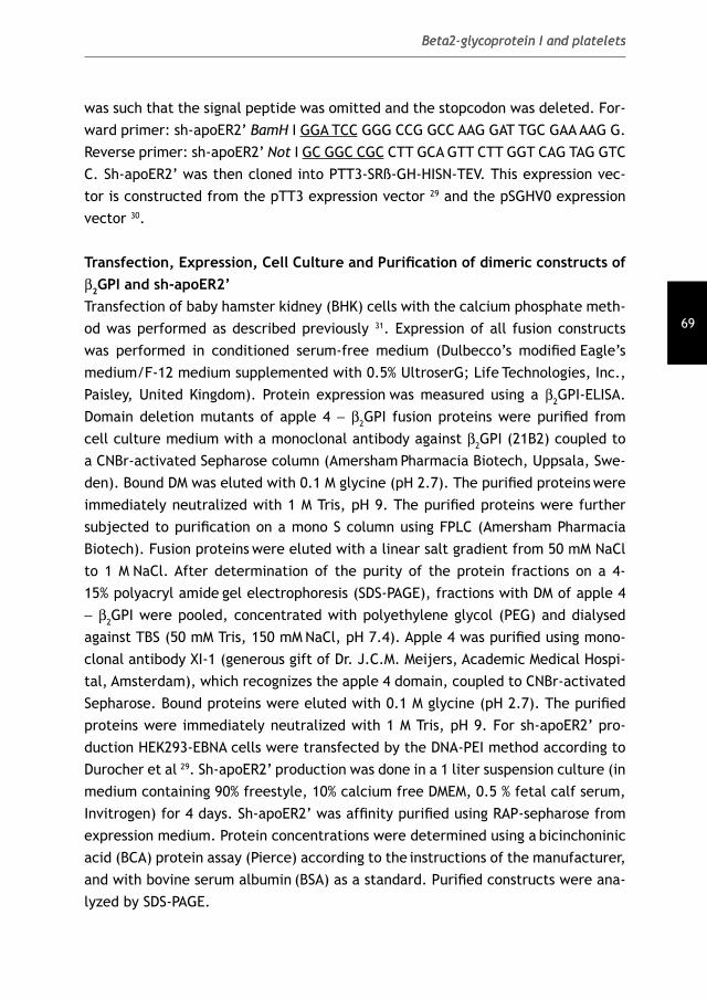
69
Beta2-glycoprotein I and platelets
was such that the signal peptide was omitted and the stopcodon was deleted. For-ward primer: sh-apoER2’ BamH I GGA TCC GGG CCG GCC AAG GAT TGC GAA AAG G. Reverse primer: sh-apoER2’ Not I GC GGC CGC CTT GCA GTT CTT GGT CAG TAG GTC C. Sh-apoER2’ was then cloned into PTT3-SRß-GH-HISN-TEV. This expression vec-tor is constructed from the pTT3 expression vector 29 and the pSGHV0 expressionvector 30.
Transfection, Expression, Cell Culture and Purifi cation of dimeric constructs of β2GPI and sh-apoER2’ Transfection of baby hamster kidney (BHK) cells with the calcium phosphate meth-od was performed as described previously 31. Expression of all fusion constructs
was performed in conditioned serum-free medium (Dulbecco’s modifi ed Eagle’s medium/F-12 medium supplemented with 0.5% UltroserG; Life Technologies, Inc., Paisley, United Kingdom). Protein expression was measured using a β2GPI-ELISA. Domain deletion mutants of apple 4 − β2GPI fusion proteins were purifi ed from cell culture medium with a monoclonal antibody against β2GPI (21B2) coupled to a CNBr-activated Sepharose column (Amersham Pharmacia Biotech, Uppsala, Swe-den). Bound DM was eluted with 0.1 M glycine (pH 2.7). The purifi ed proteins were immediately neutralized with 1 M Tris, pH 9. The purifi ed proteins were further subjected to purifi cation on a mono S column using FPLC (Amersham Pharmacia Biotech). Fusion proteins were eluted with a linear salt gradient from 50 mM NaCl to 1 M NaCl. After determination of the purity of the protein fractions on a 4-15% polyacryl amide gel electrophoresis (SDS-PAGE), fractions with DM of apple 4 − β2GPI were pooled, concentrated with polyethylene glycol (PEG) and dialysed against TBS (50 mM Tris, 150 mM NaCl, pH 7.4). Apple 4 was purifi ed using mono-clonal antibody XI-1 (generous gift of Dr. J.C.M. Meijers, Academic Medical Hospi-tal, Amsterdam), which recognizes the apple 4 domain, coupled to CNBr-activated Sepharose. Bou nd proteins were eluted with 0.1 M glycine (pH 2.7). The purifi ed proteins were immediately neutralized with 1 M Tris, pH 9. For sh-apoER2’ pro-duction HEK293-EBNA cells were transfected by the DNA-PEI method according to Durocher et al 29. Sh-apoER2’ production was done in a 1 liter suspension culture (in medium containing 90% freestyle, 10% calcium free DMEM, 0.5 % fetal calf serum, Invitrogen) for 4 days. Sh-apoER2’ was affi nity purifi ed using RAP-sepharose from expression medium. Protein concentrations were determined using a bicinchoninic acid (BCA) protein assay (Pierce) according to the instructions of the manufacturer, and with bovine serum albumin (BSA) as a standard. Purifi ed constructs were ana-lyzed by SDS-PAGE.
Beta2-glycoprotein I and platelets

70
Chapter 3
Purifi cation of plasma β2GPIPlasma β2GPI was isolated from fresh citrated human plasma as described previ-ously 32. In short, dialyzed human plasma was subsequently applied to the follow-ing columns; DEAE-Sephadex A50, protein G-Sepharose, S-Sepharose, and fi nally heparin-Sepharose (all Sepharoses were obtained from Amersham Pharmacia Bio-tech). Bound proteins were eluted with a linear salt gradient. Afterward, β2GPI was dialyzed against TBS. Purity of the protein was checked with SDS-PAGE analysis. Concentration of the protein was determined using the BCA protein assay.
Preparation of phospholipid vesicles Phospholipid vesicles containing 20% phosphatidylserine (PS) and 80% phosphati-dylcholine (PC) were prepared according to Brunner et al. 33, with some modifi -cations as described by Van Wijnen et al. 34. The phospholipid concentration was determined by phosphate analysis 35.
Binding of domain deletion mutants to phospholipid vesicles Binding of DM of apple 4 − β2GPI to PS/PC vesicles was tested in a solid phase bind-ing assay. High binding 96 wells ELISA plates (Costar, Corning Incorporated, 9102) were coated with 20% PS/PC 80% (25 μM in TBS; 50 μL/well) overnight at 4oC. Wells were blocked with TBS/0.5% gelatine (150 μL/well) for two hours at 37oC. Subse-quently, wells were incubated with different concentrations of DM (0.25-32 μg/mL) for 1.5 hours at 37oC, followed by incubation with Moab 2B2 (3 μg/mL; 50 μL/well; 1.5 hours at 37oC), a generous gift of Dr. J. Arnout, Leuven, Belgium. Apple 4 − β2GPI was used as a positive control and plasma β2GPI as a negative control. Af-terwards, the wells were incubated with peroxidase-conjugated rabbit anti-mouse antibody (RAMPO) (1:1000; 50 μL/well; 1.5 hours at 37oC), followed by staining procedure using orthophenylenediamine (OPD). Samples were diluted in TBS/0.5% gelatine. Non-specifi c binding was determined using non-coated wells. Results are expressed as mean ± SD (n = 3).
Determination of the effect of domain deletion mutants on clotting time Coagulation assays were performed in a KC-10 coagulometer (Amelung, Lemgo, Germany). To detect the effect of the DM on clotting time, the prothrombin time (PT) was performed as follows: 25 μl of normal pooled plasma and 25 μl of DM, apple 4 − β2GPI, plasma β2GPI (fi nal concentration 100 μg/ml) or buffer were incu-bated for 30 min at 4°C, followed by an incubation of 90 s at 37 °C. Clotting was initiated by the addition of 50 μl of Innovin (Dade Behring, Marburg, Germany).
Chapter 3

71
Beta2-glycoprotein I and platelets
Blood collection for perfusion experiments Freshly drawn venous blood was collected from healthy donors (with informed consent) into 1/10 of volume of 3.2% tri-sodium citrate (w/v). These donors denied taking aspirin or other platelet function inhibitors during the previous 10 days.
Perfusion Experiments Perfusions were performed in a single-pass perfusion chamber under nonpulsa-tile fl ow conditions using a modifi ed parallel plate perfusion with a slit width of 2 mm and a slit height of 0.1 mm. Experiments with collagen type III as a surface were performed with a perfusion time of 90 s, at a shear rate of 800 s-1, which represents the fl ow rate in small arteries. Plasma-derived β2GPI, apple 4, apple 2 − β2GPI, apple 4 − β2GPI, DM, or buffer were added to whole blood 5 min before the start of the perfusion at a concentration of 100 μg/ml and incubated at 37°C. The prewarmed blood was drawn through the perfusion chamber by an infusion pump (pump 22, model 2400-004; Harvard, Natrick, MA). Afterward, the coverslips were taken from the perfusion chamber and rinsed with Hepes-buffered saline (10 mM Hepes, 150 mM NaCl, pH 7.4), fi xed in 0.5% glutaraldehyde in PBS, dehydrated with methanol, and stained with May-Grünwald/Giemsa as described previously.
Platelet deposition was evaluated with a light microscope equipped with a JAI-CCD camera (Copenhagen, Denmark) coupled to a Matrox frame grabber (Matrox Elec-tronic Systems Ltd., Quebec, Canada) using Optimas 6.2 software (Optimas Inc., Seattle, WA) for image analysis. Evaluation for platelet adhesion was performed
on 20 fi elds, perpendicular to the fl ow direction. Platelet adhesion was expressed as the percentage of the surface covered with platelets relative to adhesion in the presence of buffer. Results are expressed as mean relative coverage (mean ± SD, n = 3). Statistical analysis was performed with a Student T-test.
Binding of apple 4 − β2GPI to immobilized soluble human apoER2’ Binding of apple 4 − β2GPI to soluble human (sh) apoER2’ was tested in a solid phase binding assay. All incubations were performed for one hour at room temperature. Hydrophobic 96 well ELISA plates were coated with increasing concentrations of sh-apoER2’ in PBS (0-10 μg/mL; 50μL/well). Wells were blocked with PBS/4% BSA (150 μL/well). After incubation with plasma-derived β2GPI, apple 4, apple 2 − β2GPI, apple 4 − β2GPI, apple 4 − β2GPI − Trp316Ser, delta V (3 μg/mL, 50μL/well) or plasma β2GPI (3 μg/mL) in the presence of a mouse monoclonal α-β2GPI antibody (19H9; 1 μg/mL) in PBS/1%BSA, bound protein was detected using rabbit polyclonal anti-β2GPI (1:500; 50μL/well). Wells were incubated with peroxidase-conjugated
Beta2-glycoprotein I and platelets

72
Chapter 3
swine anti-rabbit antibody (SWARPO) (1:500 50μL/well), followed by staining pro-cedure using OPD. A control protein with a C-terminus growth hormone-tag was used to measure non-specifi c binding of apple 4 − β2GPI. Results are expressed as mean ± SD (n = 3).
Association of apple 4 − ß2GPI with sh-apoER2’ in the presence of inhibiting peptides Binding of apple 4 − β2GPI to sh-apoER2’ was further investigated using peptides with the following sequences; VSRGGMRK (representing a cationic patch at aa po-sition 37-44 in domain I of β2GPI), CKNKEKKC (representing a cationic patch at aa position 282-287 in domain V of β2GPI) and EKCKNKCK (scrambled). Hydrophobic 96 well ELISA plates were coated with 5 μg/mL of sh-apoER2’ in PBS (50μL/well). Wells were blocked with PBS/4% BSA (150μL/well). After incubation with apple 4 − β2GPI (3 μg/mL, 50μL/well) with or without increasing concentrations of peptides (0 − 500 μg/mL), wells were subsequently incubated with a rabbit polyclonal anti-β2GPI antibody (1:500; 50μL/well) and SWARPO (1:500, 50μL/well). This was fol-lowed by a staining procedure using OPD. Binding of apple 4 − β2GPI in the absence of peptide was set at 100 %. Results are expressed as mean ± SD (n = 3).
Immunoprecipitations 500 μl aliquots of washed platelets (300.000/μL) resuspended in Hepes/Tyrode buffer were incubated for 5 min at 37°C with buffer or with plasma β2GPI, apple 4 − β2GPI, apple 4 − β2GPI − Trp316Ser or DM of apple 4 − β2GPI (fi nal concen-tration 100 μg/mL). Incubations were performed in the presence of 3 mM CaCl2. For competition experiments, proteins (fi nal concentration 100 μg/mL) were in-cubated with the inhibiting peptides (fi nal concentration 500 μg/mL) for 5 min at 37°C. As control, platelets were incubated with buffer and the inhibiting peptides. Afterwards, platelets were lysed on ice with 1% CHAPS (3-[(3-cholamidopropyl) dimethylammonio]-1-propanesulfonic acid), containing 50 mM MES (2-(N-Mor-pholino) ethanesulfonic acid) and 150 mM NaCl, pH 7.4. Proteins were precipi-tated with 1 μg/ml of a polyclonal anti-ApoER2’ antibody (sc-10112, Santa Cruz Biotechnology, Santa Cruz, CA) and protein G-Sepharose (Amersham Biociences). The immunoprecipitations were incubated for 18 h at 4 °C in a top-over-top rotor, washed three times with lysis buffer, resuspended in non-reducing Laemmli sample buffer (0.001% (w/v) bromphenol blue, 2% (w/v) SDS, 10% (v/v) glycerol in 62.5 mM
Tris, pH 6.8), and boiled for 5 min. The supernatants were subjected to 10% SDS-PAGE and electroblotted onto an Immobilon-P polyvinylidene difl uoride membrane.
Chapter 3

73
Beta2-glycoprotein I and platelets
Blots were blocked with TBS with 0.1% (v/v) Tween 20 (TBST) containing 2% (w/v) non-fat dry milk for 1 h at room temperature. Incubation with anti-β2GPI antibody 2B2 (3 μg/ml) was performed overnight in TBST supplemented with 1% non-fat dry milk. The membranes were washed three times and incubated with RAMPO (1:2500; Dako, Glostrup, Denmark) in the same buffer. Bands on blots were visu-alized with enhanced chemiluminescence. This experiment was performed with washed platelets form three different donors.
Results
Expression and Purifi cation of domain deletion mutants To study the effect of domain involvement of apple 4 − β2GPI on PL-binding, clot-ting time and platelet adhesion, dimeric constructs of β2GPI fused to the C ter-minus of the dimerization domain (apple 4) of factor XI were made. BHK cells were transfected with expression vectors containing DM of apple 4 − β2GPI. Protein expression was confi rmed by western blotting using an anti-β2GPI monoclonal an-tibody. Cell lines with the highest expression were selected using a β2GPI enzyme linked immunosorbent assay (ELISA). The proteins were purifi ed using a monoclo-nal α-β2GPI antibody (Moab 2B2) column followed by further purifi cation on a mono S column using FPLC. After purifi cation DM were applied on a 7.5 % SDS-PAGE under non-reducing (Figure 1, panel A) and reducing (Panel B) conditions and stained with Coommassie Brilliant Blue. In the presence of SDS full-length apple 4 − β2GPI (D), apple 4 − β2GPI − Trp316Ser (W), apple 4 − β2GPI − Δ1β2GPI (ΔI), apple 4 − Δ2β2GPI (ΔII) and apple 4 − Δ5β2GPI (ΔV) migrated as monomers with an apparent molecular mass of 50 kDa. Plasma β2GPI (M) migrated with a molecular mass of 45 kDa under non-reducing conditions. Under reducing conditions full-length apple 4 − β2GPI and apple 4 − β2GPI − Trp316Ser migrated with a molecular mass of ap-proximately 62 kDa. Apple 4 − Δ1β2GPI, apple 4 − Δ2β2GPI and apple 4 − Δ5β2GPI migrated slightly slower with a molecular mass of approximately 56 kDa. Plasma β2GPI migrated with a molecular mass of approximately 52 kDa.
Effect of apple 4 − β2GPI domain deletion mutants on clotting time To study the role of individual domains of apple 4 − β2GPI for competing with coa-gulation factors, we studied the effect of the DM on clotting time. For this purpose we performed the prothrombin time (PT). Concentrations of 200 μg/mL plasma derived β2GPI, apple 4 − β2GPI or DM, diluted in TBS, were mixed 1:1 with normal pooled plasma (NPP) and incubated for 30 minutes at 4oC (fi nal concentration 100
Beta2-glycoprotein I and platelets

74
Chapter 3
μg/mL). This was followed by measurement of the PT. The clotting time in the presence of buffer was set at 100 %. The observed effect for delta I and delta II was comparable with full length apple 4 − β2GPI; apple 4 − β2GPI showed a relative prolongation of the clotting time to 171.3 ± 3.7 %, delta I to 163.6 ± 7.9 % and delta II to 180.7 ± 23.8 %, respectively. Results are presented in Figure 2. The addition of plasma β2GPI, delta V or apple 4 − β2GPI − Trp316Ser to NPP did not infl uence the clotting time. Furthermore, the control proteins apple 4 and apple 2 − β2GPI did not infl uence the clotting time.
Figure 1. SDS-PAGE analysis of apple 4 − β2GPI constructs.Purifi ed plasma β2GPI (M), apple 4 − β2GPI (D), delta I (ΔI), delta II (Δ2), delta V (ΔV) and apple 4 − β2GPI − Trp316Ser (W) were analysed on a 10% SDS-PAGE under non-re-ducing- (A) and reducing (B) conditions. Gels were stained with Coommassie Brilliant Blue. The molecular masses of prestained markers are expressed in kilodalton (kDa).
T M a4 a2 D �I �II �V W
0
50
100
150
200
250
Clo
ttin
gti
me
(%)
**
Figure 2. Effect of apple − β2GPI constructs on the prothrombin time (PT).Plasma-derived β2GPI (M), apple 4 (a4), apple 2 − β2GPI (a2), apple 4 − β2GPI (D), delta I (ΔI), delta II (ΔII), delta V (ΔV) or apple 4 − β2GPI − Trp316Ser (W) were 1:1 diluted with nor-mal pooled plasma (fi nal protein concentration 100 μg/mL) followed by measurement of the PT. Clotting time with buffer (T) was set at 100%. Results represent mean clotting time ± SD in percentage (n = 3). Statistical analysis was performed using the Student T-test (p ≤0.001). Differences between apple 4 − β2GPI, delta I and delta II are not signifi cant.
Chapter 3
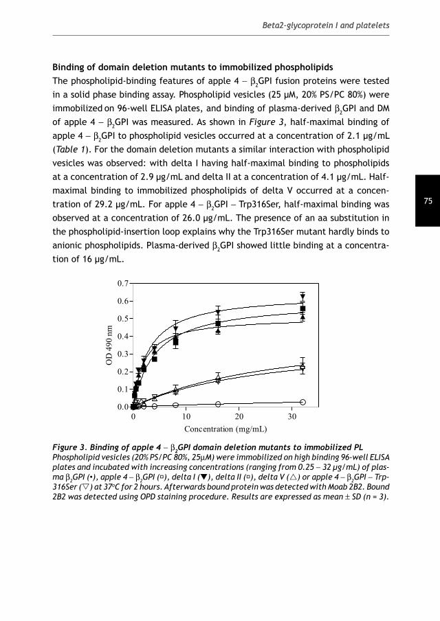
75
Beta2-glycoprotein I and platelets
Binding of domain deletion mutants to immobilized phospholipids The phospholipid-binding features of apple 4 − β2GPI fusion proteins were tested in a solid phase binding assay. Phospholipid vesicles (25 μM, 20% PS/PC 80%) were immobilized on 96-well ELISA plates, and binding of plasma-derived β2GPI and DM of apple 4 − β2GPI was measured. As shown in Figure 3, half-maximal binding of apple 4 − β2GPI to phospholipid vesicles occurred at a concentration of 2.1 μg/mL (Table 1). For the domain deletion mutants a similar interaction with phospholipid vesicles was observed: with delta I having half-maximal binding to phospholipids at a concentration of 2.9 μg/mL and delta II at a concentration of 4.1 μg/mL. Half-maximal binding to immobilized phospholipids of delta V occurred at a concen-tration of 29.2 μg/mL. For apple 4 − β2GPI − Trp316Ser, half-maximal binding was observed at a concentration of 26.0 μg/mL. The presence of an aa substitution in the phospholipid-insertion loop explains why the Trp316Ser mutant hardly binds to anionic phospholipids. Plasma-derived β2GPI showed little binding at a concentra-tion of 16 μg/mL.
0 10 20 300.0
0.1
0.2
0.3
0.4
0.5
0.6
0.7
Concentration (mg/mL)
OD
49
0n
m
Figure 3. Binding of apple 4 − β2GPI domain deletion mutants to immobilized PL Phospholipid vesicles (20% PS/PC 80%, 25μM) were immobilized on high binding 96-well ELISA plates and incubated with increasing concentrations (ranging from 0.25 − 32 μg/mL) of plas-ma β2GPI (•), apple 4 − β2GPI (), delta I (▼), delta II (), delta V (r) or apple 4 − β2GPI − Trp-316Ser (s) at 37oC for 2 hours. Afterwards bound protein was detected with Moab 2B2. Bound 2B2 was detected using OPD staining procedure. Results are expressed as mean ± SD (n = 3).
Beta2-glycoprotein I and platelets

76
Chapter 3
Apparent Kd Plasma β2GPI Apple 4-
β2GPI
Apple 4-
∆1β2GPI
Apple 4-
∆2β2GPI
Apple 4-
∆5β2GPI
Apple 4-β2GPI-
Trp316Ser
Kd(app) (μg/mL) 62.1 ± 24.4 2.1 ± 0.2 2.9 ± 0.4 4.1 ± 0.5 29.2 ± 2.4 26.0 ± 4.3
Kd(app) (nM) 1.4e3 ± 0.6e3 18.3 ± 1.8 29 ± 4.0 41 ± 5.0 314 ± 25.8 226.0 ± 37.4
Table 1. Apparent dissociation constants of domain deletion mutants for PS/PC vesiclesCurves of plasma β2GPI and apple 4 − β2GPI fusion proteins were fi tted according to a one-site binding model in GraphPad. Half-maximal binding is given as apparent Kd (Kd(app)) both in μg/mL as in nmol/L (nM). Results are expressed as mean ± SD (n = 3).
Effect of domain deletion mutants on platelet deposition to collagen type III under conditions of fl ow To determine which domain of apple 4 − β2GPI is involved in platelet sensitization, we performed perfusion experiments with citrated whole blood pre-incubated with plasma β2GPI, apple 4 − β2GPI or DM (fi nal concentration 100 μg/mL). To determine basal platelet adhesion to collagen type III, whole blood was pre-incubated with buffer. The basal platelet coverage after 90 s was 9.4 ± 2.1 %, which was set at 100 % (baseline). As shown in Figure 4A, no increase in platelet adhesion was found when plasma β2GPI, apple 4, apple 2 − β2GPI or delta V was added to whole blood (105.5 ± 11.4 %, 103.8 ± 15.6 %, 97.3 ± 16.1 % and 99.5 ± 5.5 %, respectively). As has also been shown by Lutters et al. 27, apple 4 − β2GPI − Trp316Ser did not induce increased platelet adhesion to collagen. In contrast, platelet adhesion increased signifi cantly when full length apple 4 −, delta I or delta II was added to whole blood (155.4 ± 11.0 %, 148.3 ± 8.6 % and 157.5 ± 7.9 %, respectively). Morphology of platelets and number of platelet aggregates were similar in conditions with full length apple 4 − β2GPI (Figure 4B, Panel a), delta I (not shown) and delta II (not shown). Plasma β2GPI, delta V (Panel b and d, respectively) and apple 4 − β2GPI − Trp316Ser (not shown) displayed comparable effects on morphology and number of platelet aggregates as incubation with buffer (Panel a).
Chapter 3
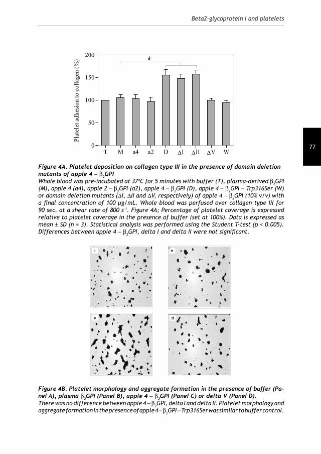
77
Beta2-glycoprotein I and platelets
T M a4 a2 D �I �II �V W
0
50
100
150
200
Pla
tele
tad
hes
ion
toco
llag
en(%
)
*
Figure 4A. Platelet deposition on collagen type III in the presence of domain deletion mutants of apple 4 − β2GPIWhole blood was pre-incubated at 37oC for 5 minutes with buffer (T), plasma-derived β2GPI (M), apple 4 (a4), apple 2 − β2GPI (a2), apple 4 − β2GPI (D), apple 4 − β2GPI − Trp316Ser (W) or domain deletion mutants (ΔI, ΔII and ΔV, respectively) of apple 4 − β2GPI (10% v/v) with a fi nal concentration of 100 μg/mL. Whole blood was perfused over collagen type III for 90 sec. at a shear rate of 800 s-1. Figure 4A; Percentage of platelet coverage is expressed relative to platelet coverage in the presence of buffer (set at 100%). Data is expressed as mean ± SD (n = 3). Statistical analysis was performed using the Student T-test (p < 0.005). Differences between apple 4 − β2GPI, delta I and delta II were not signifi cant.
c d
Figure 4B. Platelet morphology and aggregate formation in the presence of buffer (Pa-nel A), plasma β2GPI (Panel B), apple 4 − β2GPI (Panel C) or delta V (Panel D). There was no difference between apple 4 − β2GPI, delta I and delta II. Platelet morphology and aggregate formation in the presence of apple 4 − β2GPI − Trp316Ser was similar to buffer control.
Beta2-glycoprotein I and platelets

78
Chapter 3
Binding of apple 4 − β2GPI and the Trp316Ser mutant to immobilized soluble human apoER2’Binding of apple 4 − β2GPI and the Trp316Ser mutant to immobilised soluble human (sh)-apoER2’ was measured using an ELISA setup. As shown in Figure 5, half-maxi-mal binding of both apple 4 − β2GPI and apple 4 − β2GPI − Trp316Ser to immobilized sh-apoER2’ occurred at concentrations as low as 2.9 ± 0.7 and 6.0 ± 0.8 μg/mL, respectively (corresponds to 25 and 52 nM, respectively). Also, plasma β2GPI in the presence of a monoclonal α-β2GPI antibody (19H9) displayed binding to sh-apoER2’. Half-maximal binding occurred at a concentration of 2.4 ± 0.7 μg/mL (corresponds to 21 nM). No binding was found with plasma β2GPI, apple 4 or delta V. Apple 2 − β2GPI displayed only slight interaction with sh-apoER2’.
0 5 10 15 200.00
0.05
0.10
0.15
0.20
0.25
0.30
0.35
Concentration (mg/mL)
OD
49
0n
m
Figure 5. Binding of apple 4 − β2GPI and the Trp316Ser mutant to immobilized apoER2’Interaction between apple 4 − β2GPI and sh-apoER2’ was measured in a solid phase binding assay. Soluble human apoER2’ was coated on a hydrophobic 96 well ELISA pla-te in a concentration dependent manner (0-10 μg/mL). After blocking, plasma β2GPI (•), apple 4 (◊), apple 2 − β2GPI (+), apple 4 − β2GPI (), delta V (r), apple 4 − β2GPI − Trp316Ser (s) mutant and plasma β2GPI + 19H9 (•) were incubated (3 μg/mL). Af-terwards bound protein was detected with rabbit polyclonal anti-β2GPI. Bound an-tibody was detected using SWARPO. Results are expressed as mean ± SD (n = 3).
Association of domain deletion mutants and the Trp316Ser mutant of apple 4 − β2GPI with apoER2’ on platelets The only member of the LDL receptor family known to be present on platelets is apoER2’. To demonstrate that a cationic patch in domain V of β2GPI and not the hydrophobic PL-insertion loop in domain V (Ser311-Lys317) is responsible for inter-
Chapter 3

79
Beta2-glycoprotein I and platelets
action with apoER2’, immunoprecipitations were performed. Platelets were incu-bated with buffer, plasma-derived β2GPI, full length apple 4 − β2GPI, DM or apple 4 − β2GPI − Trp316Ser, lysed and subjected to immunoprecipitation with an anti-apoER2’ antibody. Afterwards, Western blots were incubated with a monoclonal anti-β2GPI antibody to detect interaction between β2GPI and apoER2’. Association with apoER2’ was observed with apple 4 − β2GPI (Figure 6). Hardly any association was observed when platelets were incubated with plasma β2GPI or delta V. To de-monstrate that this fi nding was not the result of the inability of delta V to bind to anionic phospholipids, immunoprecipitations were performed with apple 4 − β2GPI − Trp316Ser. The interaction of the Trp316Ser mutant with apoER2’ on the surface of platelets was similar to that of full length apple 4 − β2GPI.
M D �V�I �II WT
75 kDa
50 kDa
37 kDa
Figure 6. Immunoprecipitations with domain deletion mutants of apple 4 − β2GPI and apoER2’ on plateletsWashed platelets (300.000/μL), resuspended in Hepes/Tyrode buffer containing 3 mM CaCl2 were incubated with buffer, apple 4 − β2GPI (D), delta I (ΔI), delta II (ΔII), delta V (ΔV) or apple 4 − β2GPI − Trp316Ser (W) (fi nal concentration 100 μg/mL; 10% v/v) for 5 minutes at 37oC followed by lysis with CHAPS on ice. Lysed platelets were subjected to immunoprecipitation with an anti-apoER2’ antibody in the presence of protein G sep-harose. Afterwards Western blots were incubated with an anti-β2GPI antibody followed by visualization using chemiluminescence. Blot represents three different experiments.
Association of apple 4 − β2GPI with apoER2’ in the presence of CKNKEKKC, EKCKNKCK (scrambled) or VSRGGMRK peptides Association of ligands with members of the LDL receptor family is supported by electrostatic interactions. To investigate the possibility that association of ap-ple 4 − β2GPI with apoER2’ is supported by a cationic patch in domain V of apple 4 − β2GPI competition studies were performed with positively charged peptides. Incubation of washed platelets with apple 4 − β2GPI and CKNKEKKC (represents a cationic patch at aa position 282-287 in domain V of β2GPI) peptide resulted in decreased binding of apple 4 − β2GPI to apoER2’ (Figure 7A). This effect was not seen with the VSRGGMRK peptide (represents a cationic patch at aa position 37-
Beta2-glycoprotein I and platelets
79

80
Chapter 3
44 in domain I of β2GPI). The decreased binding of apple 4 − β2GPI to apoER2’ in the presence of CKNKEKKC was also seen for the Trp316Ser mutant. Incubation of washed platelets with apple 4 − β2GPI and EKCKNKCK (scrambled peptide) resul-ted in a minor reduction of the association between apple 4 − β2GPI and apoER2’ (Figure 7B).
+ CKNKEKKC + VSRGGMRK
T M D W
75 kDa
50 kDa
37 kDa
T M D W T M D W
+ CKNKEKKC + EKCKNKCK
T M D T M D T M D
75 kDa
50 kDa
37 kDa
Figure 7A Figure 7B
0 100 200 300 400 5000
25
50
75
100
Concentration peptide (mg/mL)
Rel
ativ
eb
ind
ing
(%)
Figure 7C
Figure 7. Immunoprecipitations with apple 4 − β2GPI and apoER2’ on platelets in the presence of CKNKEKKC, EKCKNKCK or VSRGGMRK peptidesCompetition experiments were performed to investigate electrostatic involvement between apple 4 − β2GPI and apoER2’. For this purpose washed platelets were incubated with buffer (T), plasma β2GPI (M), apple 4 − β2GPI (D), apple 4 − β2GPI − Trp316Ser (W) in the presence of cationic peptides; incubations in the presence of CKNKEKKC or the VSRGGMRK peptide (Panel A) or in the presence of the EKCKNKCK peptide (Panel B) (fi nal peptide concentration; 500 μg/mL) for 5 minutes at 37oC. Afterwards, immunoprecipitations were performed as described previously. Blots represent three different experiments. To show direct competition between apple 4 − β2GPI and the peptides CKNKEKKC () and EKCKNKCK (Δ), binding of apple 4 − β2GPI in the presence of the peptides was investigated in the solid phase binding assay (Panel C).
Chapter 3
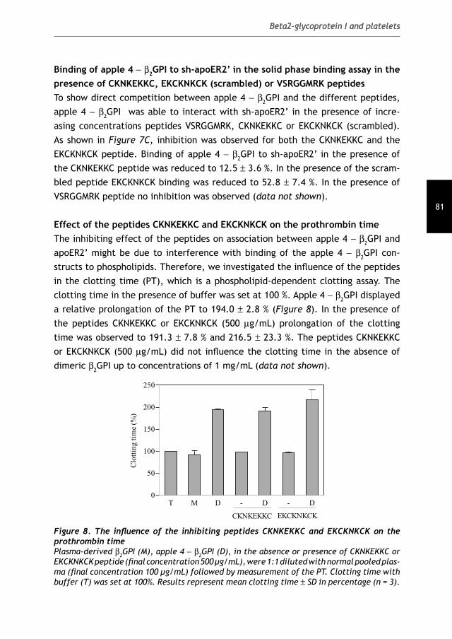
81
Beta2-glycoprotein I and platelets
Binding of apple 4 − β2GPI to sh-apoER2’ in the solid phase binding assay in the presence of CKNKEKKC, EKCKNKCK (scrambled) or VSRGGMRK peptides To show direct competition between apple 4 − β2GPI and the different peptides, apple 4 − β2GPI was able to interact with sh-apoER2’ in the presence of incre-asing concentrations peptides VSRGGMRK, CKNKEKKC or EKCKNKCK (scrambled). As shown in Figure 7C, inhibition was observed for both the CKNKEKKC and the EKCKNKCK peptide. Binding of apple 4 − β2GPI to sh-apoER2’ in the presence of the CKNKEKKC peptide was reduced to 12.5 ± 3.6 %. In the presence of the scram-bled peptide EKCKNKCK binding was reduced to 52.8 ± 7.4 %. In the presence of VSRGGMRK peptide no inhibition was observed (data not shown).
Effect of the peptides CKNKEKKC and EKCKNKCK on the prothrombin time The inhibiting effect of the peptides on association between apple 4 − β2GPI and apoER2’ might be due to interference with binding of the apple 4 − β2GPI con-structs to phospholipids. Therefore, we investigated the infl uence of the peptides in the clotting time (PT), which is a phospholipid-dependent clotting assay. The clotting time in the presence of buffer was set at 100 %. Apple 4 − β2GPI displayed a relative prolongation of the PT to 194.0 ± 2.8 % (Figure 8). In the presence of the peptides CKNKEKKC or EKCKNKCK (500 μg/mL) prolongation of the clotting time was observed to 191.3 ± 7.8 % and 216.5 ± 23.3 %. The peptides CKNKEKKC or EKCKNKCK (500 μg/mL) did not infl uence the clotting time in the absence of dimeric β2GPI up to concentrations of 1 mg/mL (data not shown).
T M D - D - D0
50
100
150
200
250
CKNKEKKC EKCKNKCK
Clo
ttin
gti
me
(%)
Figure 8. The infl uence of the inhibiting peptides CKNKEKKC and EKCKNKCK on the prothrombin timePlasma-derived β2GPI (M), apple 4 − β2GPI (D), in the absence or presence of CKNKEKKC or EKCKNKCK peptide (fi nal concentration 500 μg/mL), were 1:1 diluted with normal pooled plas-ma (fi nal concentration 100 μg/mL) followed by measurement of the PT. Clotting time with buffer (T) was set at 100%. Results represent mean clotting time ± SD in percentage (n = 3).
Beta2-glycoprotein I and platelets

82
Chapter 3
DiscussionIt is diffi cult to envision that mere binding of beta2-glycoprotein I (β2GPI) to an-ionic phospholipids (PL) on the cell surface can activate these cells. Therefore, a search for a cellular receptor for β2GPI on platelets was initiated. Lutters et al. have demonstrated that dimerization of β2GPI (either artifi cially by fusing β2GPI with the apple 4 domain of factor XI, or physiologically by binding aPL to β2GPI) results in increased affi nity of β2GPI for platelets (27) which results in an increased platelet deposition to collagen under conditions of fl ow. The increase in platelet adhesion could be blocked by the addition of receptor-associated protein (RAP), suggesting that a member of the LDL receptor family as the platelet receptor for dimeric β2GPI was involved. This receptor has later been identifi ed as apoER2’ (25). In the present study, the domain of β2GPI responsible for interaction with apoER2’ has been determined by using constructs of dimeric β2GPI lacking domain I, II or V and a construct with an aa substitution (Trp316Ser) in the hydrophobic loop in domain V. Substitution of Trp316 by a serine completely abolished binding of β2GPI to anionic phospholipids 14. In this study, we showed that a cationic patch in domain V of dimeric β2GPI is involved in interaction with apoER2’ on platelets and that, by using the Trp316Ser mutant, the binding site for apoER2’ does not coincide with the phospholipid-binding site within domain V. Deletion of domain I or domain II does not have an effect on the function of dime-ric β2GPI. Both domain deletion mutants (delta I and delta II) prolong the clotting time in the prothrombin-time (PT) comparable to full length dimeric β2GPI (Figure 2) and show comparable affi nity for anionic phospholipids (Figure 3). Furthermore, delta I and delta II cause an increased adhesion of platelets to collagen under conditions of fl ow comparable to full length dimeric β2GPI (Figure 4A). Deletion of domain V or the Trp316Ser substitution abolished the prolongation of the clotting time induced by dimeric β2GPI, which was to be expected, as domain V harbors the phospholipid-binding site. Indeed, both mutants show an approximate 15-fold decreased affi nity for anionic phospholipids. Furthermore, in the perfusion model both mutants were not able to increase platelet adhesion to collagen, stressing the necessity of domain V in the activation of platelets.Based on the observations in this paper, we propose the following model for the activation of platelets by β2GPI (Figure 9). β2GPI binds to platelets with a low af-fi nity. However, when bound to the platelet membrane a conformational change is induced in domain I of β2GPI 36 37 38 39, resulting in the exposure of a cryptic epitope in this domain. Binding of anti-β2GPI antibodies to this newly exposed epitope in domain I takes place. When one antibody interacts with two molecules of β2GPI,
Chapter 3
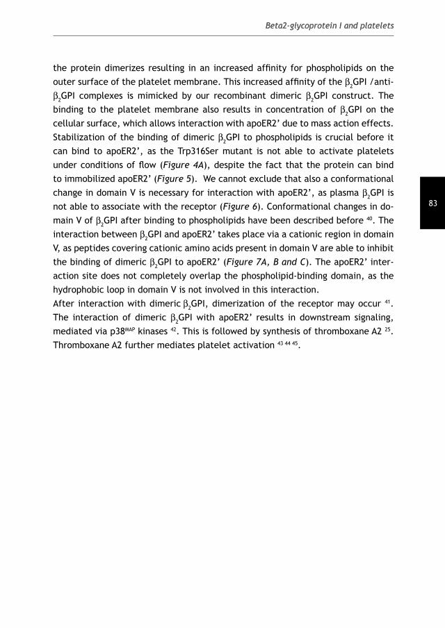
83
Beta2-glycoprotein I and platelets
the protein dimerizes resulting in an increased affi nity for phospholipids on the outer surface of the platelet membrane. This increased affi nity of the β2GPI /anti-β2GPI complexes is mimicked by our recombinant dimeric β2GPI construct. The binding to the platelet membrane also results in concentration of β2GPI on the cellular surface, which allows interaction with apoER2’ due to mass action effects. Stabilization of the binding of dimeric β2GPI to phospholipids is crucial before it can bind to apoER2’, as the Trp316Ser mutant is not able to activate platelets under conditions of fl ow (Figure 4A), despite the fact that the protein can bind to immobilized apoER2’ (Figure 5). We cannot exclude that also a conformational change in domain V is necessary for interaction with apoER2’, as plasma β2GPI is not able to associate with the receptor (Figure 6). Conformational changes in do-main V of β2GPI after binding to phospholipids have been described before 40. The interaction between β2GPI and apoER2’ takes place via a cationic region in domain V, as peptides covering cationic amino acids present in domain V are able to inhibit the binding of dimeric β2GPI to apoER2’ (Figure 7A, B and C). The apoER2’ inter-action site does not completely overlap the phospholipid-binding domain, as the hydrophobic loop in domain V is not involved in this interaction.After interaction with dimeric β2GPI, dimerization of the receptor may occur 41. The interaction of dimeric β2GPI with apoER2’ results in downstream signaling, mediated via p38MAP kinases 42. This is followed by synthesis of thromboxane A2 25. Thromboxane A2 further mediates platelet activation 43 44 45.
Beta2-glycoprotein I and platelets

84
Chapter 3
Figure 9. Proposed mechanism of platelet activation via dimerization of β2GPI by aPLBeta2-glycoprotein I circulates free in plasma with low affi nity for phospholipids. A confor-mational change in domain I of β2GPI is induced after low-affi nity binding to platelets. Di-merization of β2GPI via binding of aPL occurs followed by stabilization of the complex on the platelet surfaced which subsequently results in fi rm adhesion of the β2GPI/aPL complex to PL and association with apoER2’ on the surface of platelets. We cannot exclude the possibility that dimeric β2GPI dissociates from the cellular surface after interaction with apoER2’. The interaction results in down-stream signaling, TxA2 synthesis and furtherplatelet activation.
Acknowledgement: This study was supported by a grant from the Netherlands Organization for Health Research and Development (Zon-MW: 902-26-290).
Chapter 3

85
Beta2-glycoprotein I and platelets
References
1. Wilson, W. A., Gharavi, A. E., Koike, T., Lockshin, M. D., Branch, D. W., Piette, J.
C., Brey, R., Derksen, R. H. W. M., Harris, E. N., Hughes, G. R. V., Triplett, D. A., and
Khamashta, M. A. (1999) Arthritis Rheum. 42, 1309-1311
2. Cervera, R., Piette, J. C., Font, J., Khamashta, M. A., Shoenfeld, Y., Camps, M. T.,
Jacobsen, S., Lakos, G., Tincani, A., Kontopoulou-Griva, I., Galeazzi, M., Meroni, P.
L., Derksen, R. H., De Groot, P. G., Gromnica-Ihle, E., Baleva, M., Mosca, M.,
Bombardieri, S., Houssiau, F., Gris, J. C., Quere, I., Hachulla, E., Vasconcelos,
C., Roch, B., Fernandez-Nebro, A., Boffa, M. C., Hughes, G. R., and Ingelmo, M.
(2002) Arthritis Rheum. 46, 1019-1027
3. Derksen, R. H., Khamashta, M. A., and Branch, D. W. (2004) Arthritis Rheum. 50,
1028-1039
4. De Laat, B., Derksen, R. H., and De Groot, P. G. (2004) Clin. Immunol. 112, 161-
168
5. Galli, M., Comfurius, P., Maassen, C., Hemker, H. C., de Baets, M. H., van Breda-
Vriesman, P. J. C., Barbui, T., Zwaal, R. F. A., and Bevers, E. M. (1990) Lancet 335,
1544-1547
6. Matsuura, E., Igarashi, Y., Fujimoto, M., Ichikawa, K., and Koike, T. (1990) Lancet
336, 177-178
7. Caronti, B., Calderaro, C., Alessandri, C., Conti, F., Tinghino, R., Palladini, G., and
Valesini, G. (1999) Clin. Exp. Immunol. 115, 214-219
8. Chamley, L. W., Allen, J. L., and Johnson, P. M. (1997) Placenta 18, 403-410
9. Averna, M., Paravizzini, G., Marino, G., Lanteri, E., Cavera, G., Barbagallo, C. M.,
Petralia, S., Cavallaro, S., Magro, G., Grasso, S., Notarbartolo, A., and Travali, S.
(1997) Int. J. Clin. Lab Res. 27, 207-212
10. Hunt, J. E., Simpson, R. J., and Krilis, S. A. (1993) Proc. Natl. Acad. Sci. USA 90,
2141-2145
11. Bouma, B., De Groot, Ph. G., van den Elsen, J. M. H., Ravelli, R. B. G., Schouten,
A., Simmelink, M. J. A., Derksen, R. H. W. M., Kroon, J., and Gros, P. (1999) EMBO
J. 18, 5166-5174
12. Visvanathan, S., Geczy, C. L., Harmer, J. A., and McNeil, H. P. (2000) The journal of
immunology 165, 2258-2262
13. Hammel, M., Schwarzenbacher, R., Gries, A., Kostner, G. M., Laggner, P., and Prassl,
R. (2001) Biochemistry 40, 14173-14181
14. Horbach, D. A., Van Oort, E., Tempelman, M. J., Derksen, R. H. W. M., and De Groot,
Ph. G. (1998) Thromb. Haemost. 80, 791-797
15. Hong, D. P., Hagihara, Y., Kato, H., and Goto, Y. (2001) Biochemistry 40, 8092-8100
16. Hagihara, Y., Goto, Y., Kato, H., and Yoshimura, T. (1995) J. Biochem. (Tokyo) 118,
129-136
Beta2-glycoprotein I and platelets

86
Chapter 3
17. Kim, D. H., Iijima, H., Goto, K., Sakai, J., Ishii, H., Kim, H. J., Suzuki, H., Kondo, H.,
Saeki, S., and Yamamoto, T. (1996) The journal of biological chemistry 271, 8373-
8380
18. Hussain, M. M., Strickland, D. K., and Bakillah, A. (1999) Annu. Rev. Nutr. 19, 141-
172
19. Brandes, C., Kahr, L., Stockinger, W., Hiesberger, T., Schneider, W. J., and Nimpf, J.
(2001) J. Biol. Chem. 276, 22160-22169
20. Andersen, O. M., Benhayon, D., Curran, T., and Willnow, T. E. (2003) Biochemistry
42, 9355-9364
21. Chung, N. S. and Wasan, K. M. (2004) Adv. Drug Deliv. Rev. 56, 1315-1334
22. Korporaal, S. J., Relou, I. A., van, E. M., Strasser, V., Bezemer, M., Gorter, G., van
Berkel, T. J., Nimpf, J., Akkerman, J. W., and Lenting, P. J. (2004) J. Biol. Chem.
279, 52526-52534
23. Perez-Garcia, C. G., Tissir, F., Goffi net, A. M., and Meyer, G. (2004) Eur. J. Neurosci.
20, 2827-2832
24. Riddell, D. R., Vinogradov, D. V., Stannard, A. K., Chadwick, N., and Owen, J. S.
(1999) Journal of Lipid Research 40, 1925-1930
25. Lutters, B. C., Derksen, R. H., Tekelenburg, W. L., Lenting, P. J., Arnout, J., and De
Groot, P. G. (2003) J. Biol. Chem. 278, 33831-33838
26. Melman, L., Cao, Z. F., Rennke, S., Marzolo, M. P., Wardell, M. R., and Bu, G. (2001)
J. Biol. Chem. 276, 29338-29346
27. Lutters, B. C. H., Meijers, J. C. M., Derksen, R. H. W. M., Arnout, J., and De Groot,
P. G. (2001) The journal of biological chemistry 276, 3060-3067
28. den, D. E., Molin, D. G., Breikers, G., van, O. R., Akkerman, J. W., van Eys, G. J.,
and Heemskerk, J. W. (2001) Biochim. Biophys. Acta 1539, 243-255
29. Durocher, Y., Perret, S., and Kamen, A. (2002) Nucleic Acids Res. 30, E9
30. Leahy, D. J., Dann, C. E., III, Longo, P., Perman, B., and Ramyar, K. X. (2000) Protein
Expr. Purif. 20, 500-506
31. Meijers, J. C. M., Davie, E. W., and Chung, D. W. (1992) Blood 79, 1435-1440
32. Horbach, D. A., Van Oort, E., Donders, R. C. J. M., Derksen, R. H. W. M., and De
Groot, P. G. (1996) Thromb. Haemost. 76, 916-924
33. Brunner, J., Skrabal, P., and Hauser, H. (1976) Biochim. Biophys. Acta 455, 322-331
34. Van Wijnen, M., Stam, J. G., Van’t Veer, C., Meijers, J. C. M., Reitsma, P. H., Bertina,
R. M., and Bouma, B. N. (1996) Thromb. Haemost. 76, 397-403
35. Rouser, G., Fkeischer, S., and Yamamoto, A. (1970) Lipids 5, 494-496
36. Pengo, V., Biasiolo, A., and Fior, M. G. (1995) Thromb. Haemost. 73, 29-34
37. Matsuura, E., Igarashi, Y., Yasuda, T., Triplett, D. A., and Koike, T. (1994) J. Exp.
Med. 179, 457-462
38. Wang, S.-X., Sun, Y.-T., and Sui, S.-F. (2000) Biochem. J. 348, 103-106
Chapter 3

87
Beta2-glycoprotein I and platelets
39. Kuwana, M., Matsuura, E., Kobayashi, K., Okazaki, Y., Kaburaki, J., Ikeda, Y., and
Kawakami, Y. (2005) Blood 105, 1552-1557
40. Kuwana, M., Matsuura, E., Kobayashi, K., Okazaki, Y., Kaburaki, J., Ikeda, Y., and
Kawakami, Y. (2005) Blood 105, 1552-1557
41. Strasser, V., Fasching, D., Hauser, C., Mayer, H., Bock, H. H., Hiesberger, T., Herz, J.,
Weeber, E. J., Sweatt, J. D., Pramatarova, A., Howell, B., Schneider, W. J., and
Nimpf, J. (2004) Mol. Cell Biol. 24, 1378-1386
42. De Groot, P. G., van, L. M., Pennings, M., Urbanus, R., Bas de, L. H., Lenting, P. J.,
and Derksen, R. H. (2004) Thromb. Res. 114, 455-459
43. Forastiero, R. R., Martinuzzo, M., Carreras, L. O., and Maclouf, J. (1998) Thromb.
Haemost. 79, 42-45
44. Opara, R., Robbins, D. L., and Ziboh, V. A. (2003) J. Rheumatol. 30, 55-59
45. Arfors, L., Vesterqvist, O., Johnsson, H., and Green, K. (1990) Eur. J. Clin. Invest
20, 607-612
Beta2-glycoprotein I and platelets


Menno van Lummel, Maarten T.T. Pennings, Ronald H.W.M. Derksen, Rolf T. Urbanus,
Ronald A. Romijn, PeterJ. Lenting and Philip G. de Groot
Journal of Thrombosis and Haemostasis, 2006, In Press
4INTERACTION OF BETA2-GLYCOPROTEIN
I WITH MEMBERS OF THE LOW DENSITY
LIPOPROTEIN RECEPTOR FAMILY

91
Beta2-glycoprotein I and LDL-R family members
AbstractThe antiphospholipid syndrome (APS) is a non-infl ammatory autoimmune disease characterized by arterial and/or venous thrombosis and/or pregnancy morbidity in the presence of autoantibodies that recognize beta2-glycoprotein I (β2GPI) bound to phospholipids. We have previously demonstrated that dimerization of β2GPI by autoantibodies induces platelet activation, involving the platelet receptor apolipoprotein E receptor 2’ (apoER2’) a receptor belonging to the low-density lipoprotein receptor (LDL-R) family. Here, we show that dimeric β2GPI, but not monomeric β2GPI interacts with four other LDL-R family members: the LDL-R related protein (LRP), megalin, the LDL-R and the very-low density lipoprotein receptor (VLDL-R). Interaction between dimeric β2GPI and LDL-R, apoER2’ and VLDL-R was best described with a one-site binding model (half-maximal binding; ~ 20 nM for apoER2’ and VLDL-R and ~ 300 nM for LDL-R), whereas the interaction between dimeric β2GPI and LRP or megalin was best described with a two-site binding model, representing a high- (~ 3 nM) and a low-affi nity site (~ 0.2 μM). Binding to all receptors tested was unaffected by a tryptophane to serine (W316S) substitution in domain V of β2GPI, which is known to disrupt the phospholipid binding site of β2GPI. Also deletion of domain I or II left the interaction with the receptors unaffected. Deletion of domain V, however, signifi cantly decreased the affi nity for the receptors. In conclusion, our data show that dimeric β2GPI can interact with different LDL-R family members. This interaction is dependent on a binding site within domain V of β2GPI, which does not overlap with the phospholipid-binding site within domain V.
Keywords: antiphospholipid syndrome, beta2-glycoprotein I, antiphospholipid antibodies, low-density lipoprotein receptor family, protein-receptor interaction.
92
Chapter 4
IntroductionThe antiphospholipid syndrome (APS) is non-infl ammatory autoimmune disease. APS has been identifi ed as the association of arterial and/or venous thrombosis and/or pregnancy morbidity with the presence of antiphospholipid antibodies (aPL) in plasma of affected patients 1;2. Assays that depend on the presence of antibodies against beta2-glycoprotein I (β2GPI) display the highest correlation with thrombosis 3. The cause of haemostatic dysbalance induced by β2GPI in the presence of β2GPI antibodies in vivo still remains unraveled. Several possibilities have been proposed, of which cell activation by β2GPI/anti-β2GPI antibodies has received most attention. The involvement of cellular receptors that can bind these immune complexes is essential in all these hypotheses 4-7. β2-Glycoprotein I is an abundant plasma protein (approximately 200 μg/mL). At present, the physiological function of β2GPI is unclear, but the protein has been described to have both pro- and anti-coagulant properties in vitro 8-11. In previous studies we have shown that β2GPI/antibody complexes can activate platelets. This effect could be mimicked by a recombinant form of dimeric β2GPI. A splice variant of the apolipoprotein E receptor 2 (apoER2), that was recently identifi ed as apoER2’ on human platelets 12 has been demonstrated to be involved in the binding of dimeric β2GPI to human platelets 13. ApoER2 (also known as LDL receptor-related protein (LRP)-8) is among others expressed in brain, placenta and testis 14,15 and shares structural homology with other LDL receptor family members 16. In general, these multiligand receptors consist of a number of discrete domains and their extracellular component contains ligand-binding domains that consist of complement-type repeats. Differential clustering of these repeats within a domain may expose specifi city with respect to ligand recognition. These repeats are separated by one or more epidermal growth factor (EGF) precursor homology domains that contain YWTD motifs, responsible for ligand dissociation. The receptors are anchored in the membrane via an intracellular tail that harbors motifs for endocytosis and signaling. At present, twelve members of the LDL-receptor family have been identifi ed in mammals, including the LDL-receptor related protein (LRP), megalin, the low-density lipoprotein receptor (LDL-R), apoER2’ and the very-low density lipoprotein receptor (VLDL-R). These receptors are expressed on an array of cell types. The concept that they are multiligand receptors does not necessarily mean that they recognize identical ligands. In contrast, ligand binding may be tightly regulated between different members of the LDL-receptor family. In the present study we address a number of issues. Apart from apoER2’, other LDL-R homologues (LRP, megalin, LDL-R, VLDL-R) might recognize dimeric β2GPI

93
Beta2-glycoprotein I and LDL-R family members
with different affi nities. Furthermore, apoER2’-mediated platelet activation by β2GPI only occurred in the presence of anti-β2GPI antibodies. Since these antibodies possess the property to dimerize antigens, we investigated whether anti-β2GPI antibody induced dimerization of β2GPI also promotes the interaction with other members of the LDL-R family. It has been proposed that positive electrostatic surface potentials (mainly on domain I and V of β2GPI), not the primary sequences, in different ligands constitute receptor-binding domains. Therefore, we used domain deletion mutants of dimeric β2GPI to identify which domain of β2GPI is involved in the interaction with the receptors. Finally, to investigate whether the molecular interactions between phospholipid binding and binding to the receptors is similar, we tested binding of a non-phospholipid binding β2GPI mutant to LRP, megalin, apoER2’ and the VLDL-R.
Experimental procedures
Purifi cation of plasma β2GPI Plasma β2GPI was isolated from fresh citrated human plasma as described previously 17. In short, dialyzed human plasma was applied subsequently to the following columns: DEAE-Sephadex A50, protein G-Sepharose, S-Sepharose, and fi nally heparin-Sepharose (all Sepharoses were obtained from Amersham Pharmacia Biotech). Bound proteins were eluted with a linear salt gradient. Afterwards, β2GPI was dialyzed against TBS. Purity of the protein was checked on a 4-15% SDS-PAGE. Plasma β2GPI migrated with a molecular mass of 45 kDa under non-reducing conditions. Concentration of the protein was determined using the bicinchoninic acid (BCA) protein assay.
Construction of dimeric β2GPI Apple 4 — C321S — β2GPI (further referred to as dimeric β2GPI) and the non-dimeric apple 2 — β2GPI were constructed as described previously 18. Domain deletion mutants of dimeric β2GPI were constructed using full-length apple 4 — C321S — β2GPI cDNA as a template 19. In short, for deletion of domain I (delta I) one set of primers was used to construct domain II — V. For deletion of domain II (delta II) two sets of primers was used; one set to construct domain I and one set to construct domain III — V. The two created PCR products served as a template to obtain the full-length domain II deletion. For the domain V deletion mutant (delta V) one set of primers was used to construct domain I — IV. To construct chimaeric fusion proteins of the dimerization domain of factor XI (apple 4) and β2GPI, the

94
Chapter 4
PCR product was cloned into the vector apple 4-C321S-tissue-type plasminogen activator (tPA)-S478A. In the experiments we also used the non-phospholipid binding mutant, in which a tryptophane has been substituted to a serine (W316S). Construction of this mutant has been described previously 20. As a control, we also used dimeric apple 4 19. Sequence analysis was performed to confi rm correct amplifi cation of the β2GPI cDNA.
Construction of soluble VLDL-receptor Soluble human (sh) apoER2’ was constructed as described previously 19. Construction of sh-VLDL-receptor (VLDL-R) was performed as follows. Freshly isolated umbilical vein mRNA was prepared from endothelial cells to synthesize cDNA. This cDNA was the starting point for cloning sh-VLDL-R using Phusion DNA polymerase (Finnzymes). The signal peptide was excluded and the stopcodon was deleted. The following primers were used. Forward primer sh-VLDL-R; TA ATA GGA TCC GGG AGA AAA GCC AAA TGT GAA CCC. Reverse primer sh-VLDL-R; TA ATA GCG GCC GCA GTA GCA AGA TCC ATT TGA TAG CC. The VLDL-R was subsequently cloned into the vector PTT3-SRα-GH-HISN-TEV. This expression vector is constructed from the pTT3 expression vector 21 and the pSGHV0 expression vector 22. The sh-LDL-R was a generous gift of Dr. K. Mertens, Sanquin Bloodbank, Amsterdam, the Netherlands.
Transfection, Expression, Cell Culture and Purifi cation of Constructs Transfection of BHK cells with the calcium phosphate method was performed as described previously 13. Expression of all constructs was performed in conditioned serum-free medium (Dulbecco’s modifi ed Eagle’s medium/F-12 medium supplemented with 0.5% UltroserG; ITK Diagnostics, the Netherlands). Protein expression was measured using a β2GPI-ELISA [20]. Full-length and domain deletion mutants of dimeric β2GPI were purifi ed from cell culture medium using a monoclonal antibody (21B2; a generous gift of Dr. J. Arnout, Leuven, Belgium) column. The purifi ed proteins were further subjected to purifi cation on a mono S column using fast performance liquid chromatography (FPLC) (Amersham Pharmacia Biotech). Apple 4 was purifi ed using a monoclonal antibody (α-XI-1; a generous gift of Dr. J.C.M. Meijers, Academic Medical Center, Amsterdam). Transfection of HEK293E cells with sh-apoER2’ and sh-VLDL-R was performed using the PEI transfection method according to Durocher et al. Production of sh-apoER2 and sh-VLDL-R was done in a 1 liter suspension culture (in medium containing 90% freestyle, 10% calcium free DMEM, 0.1 % fetal calf serum, Invitrogen). Receptors were affi nity purifi ed using RAP-sepharose or Ni-NTA sepharose and purity of the
95
Beta2-glycoprotein I and LDL-R family members
protein fractions was determined on a 4-15% polyacryl amide gel electrophoresis (SDS-PAGE), subsequently followed by dialyzation against TBS (50 mM Tris, 150 mM NaCl, pH 7.4). Protein concentrations were determined using a bicinchoninic acid (BCA) protein assay (Pierce). Purifi ed constructs were analyzed on a 7.5% SDS-PAGE. LRP fragments cluster II and cluster IV were purifi ed as described previously 23. LRP was obtained from BioMac (Instruchemie, the Netherlands). Megalin was kindly provided by Dr. H. Pannekoek, Academic Medical Center, Amsterdam, the Netherlands.
Solid-phase immunosorbent assay for the interaction between β2GPI and apoER2’ and the VLDL-R Binding of β2GPI and the domain deletion mutants to apoER2’ and the VLDL-R was measured using a solid-phase immunosorbent assay. Briefl y, increasing concentrations (0 — 10 μg/mL in PBS) of receptors were immobilized on hydrophobic 96 wells plates (Costar). After blocking with PBS/4%BSA (150μL/well), proteins (3 μg/mL in PBS/1%BSA, 50μL/well) were allowed to interact with the receptors. After incubation, bound β2GPI was detected using subsequently a rabbit polyclonal anti-β2GPI antibody and a peroxidase-labeled swine anti-rabbit polyclonal antibody (SWARPO, 1:500 in PBS/1%BSA, 50μL/well), followed by a staining procedure using ortho-phenylenediamide (OPD).
Surface plasmon resonance analysis for the interaction between β2GPI and LRP, megalin and the LDL-R Surface plasmon resonance (SPR) binding assays were performed employing a Biacore 2000 system (Biacore AB, Uppsala, Sweden). LRP, cluster II or IV of LRP or megalin were immobilized on different CM5 sensor chips using the amine-coupling kit (Biacore AB, Uppsala, Sweden). The LDL-R was immobilized on a C1 sensor chip using the amine-coupling kit (Biacore AB, Uppsala, Sweden). Approximately 10 fmol/mm2 LRP, 26 fmol/mm2 cluster II, 16.5 fmol/mm2 cluster IV, 19.3 fmol/mm2 megalin and 29.4 fmol/mm2 LDL-R were immobilized. For each chip one channel was activated and blocked in the absence of protein and afterwards its signal (<5% of binding to coated channels) was used to correct the signal from the coated channels for a-specifi c binding. SPR analysis was performed in buffer containing TBS/0.005% Tween 20, 1 μM CaCl2 with a fl ow rate of 20 μl/min at 25ºC. Association and dissociation was followed for a period of 2 minutes. Regeneration of the surface was performed by application of 0.1 M sodium citrate containing 1 mM of EDTA and 1 M of NaCl, pH 5.0.

96
Chapter 4
Binding of a β2GPI/anti-β2GPI antibody complex to LDL-receptor family members Binding of plasma β2GPI in the presence of a monoclonal anti-β2GPI antibody (3B7) that recognizes domain I of β2GPI and prolongs the clotting time (lupus anticoagulant; LAC), was investigated for LRP, megalin, the LDL-R, apoER2’and the VLDL-R. For SPR analysis plasma β2GPI (100 nM) was incubated with 3B7 (nM) for 30 minutes at 37ºC. Afterwards, interaction of the immune complex with immobilized LRP, megalin and the LDL-R was investigated. Association and dissociation was followed for a period of two minutes. Analysis of binding of the immune complex to the immobilized receptors was measured in TBS/0.005% Tween 20, 1 μM CaCl2 with a fl ow rate of 20 μl/min at 25ºC. Because of the complexity of the interaction between antibody/β2GPI complexes and immobilized receptor, the kinetic parameters of this interaction cannot be calculated using the available software programs. Interaction between β2GPI/anti-β2GPI antibody complex and coated apoER2’ and the VLDL-R (5 μg/mL) was measured using the solid-phase immunosorbent assay. In this assay, plasma β2GPI (3 μg/mL) was incubated with 3B7 (1 μg/mL) in PBS/1%BSA. Bound β2GPI was visualized as described previously.
Inhibition studies in the presence of heparin or RAP Inhibition experiments were performed using SPR (LRP and megalin) analysis and with the solid-phase immunosorbent assay (apoER2’ and VLDL-R). For heparin inhibition studies, dimeric β2GPI was pre-incubated with increasing concentrations of heparin (0 — 2 mg/mL) in TBS/0.005% Tween 20 containing 1 μM CaCl2 for 30 minutes at 37oC prior to injection. For RAP inhibition studies, a pre-injection with increasing concentrations of RAP (0 — 200 nM) was performed. Association- and dissociation was followed for a period of 2 minutes. This was subsequently followed by injection of 100 nM dimeric β2GPI. For inhibition studies with apoER2’ and the VLDL-R, dimeric β2GPI (3 μg/mL, 50 μL/well) in the presence of increasing concentrations heparin (0 — 2 mg/mL) or RAP (0 — 200 nM) in PBS/1%BSA was incubated on coated apoER2’ or the VLDL-R (5 μg/mL). Bound protein was detected as described previously.
Analysis of SPR data Analysis of SPR data was performed using the GraphPad Prism program (GraphPad Prism version 4.0 for Windows, Graph Pad Software, San Diego, CA). Data obtained from SPR analysis was used for the calculation of the steady state constants as follows. Responses at equilibrium (Req) derived from sensorgrams were plotted

97
Beta2-glycoprotein I and LDL-R family members
against protein concentration. The resulting binding isotherms were subsequently fi tted to the following equations;
For a one-site binding model:
For a two-site (heterologous) binding model:
Scatchard plots were derived from the saturation curves to verify the use of a one-site- or a two-site (heterologous) ligand-binding model. Results are expressed as mean ± SD (n = 3).
Results
Binding of a β2GPI/anti-β2GPI antibody complex to LDL-receptor family membersTo investigate the effect of dimerization of β2GPI by an antibody on its interaction with members of the LDL-R homologues, we assessed the binding of plasma β2GPI in the absence or presence of an anti-β2GPI antibody (3B7) to LRP, megalin, the LDL-R, apoER2’ and the VLDL-R. Interaction of β2GPI with LRP, megalin and the LDL-R was investigated by SPR analysis and binding to apoER2’ and the VLDL-R was assessed in a qualitative manner using an immunosorbent assay. As shown in Figure 1, incubation of plasma β2GPI in the presence of 3B7 resulted in binding of the immune complex to all receptors tested, except for the LDL-R. Plasma β2GPI or 3B7 alone had less than 20% binding to the receptors. Thus, β2GPI binds to LRP, megalin, apoER2’ and the VLDL-R, however dimerization of β2GPI is necessary for optimal binding.
XK
XBY
d +×= max
XK
XB
XK
XBY
dd +×+
+×
=2
2max
1
1max
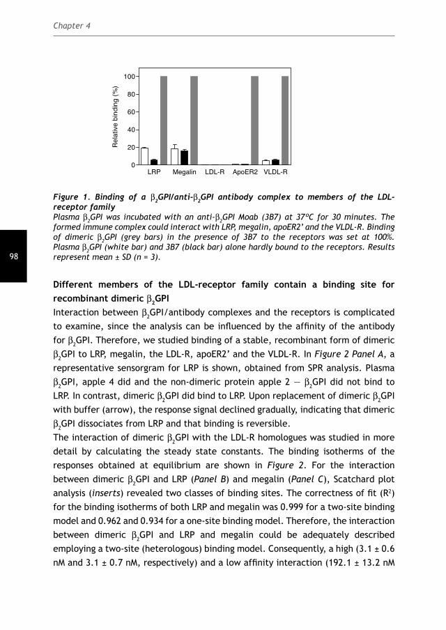
98
Chapter 4
LRP Megalin LDL-R ApoER2 VLDL-R0
20
40
60
80
100
Re
lativ
e b
ind
ing
(%
)
Figure 1. Binding of a β2GPI/anti-β2GPI antibody complex to members of the LDL-receptor family Plasma β2GPI was incubated with an anti-β2GPI Moab (3B7) at 37ºC for 30 minutes. The formed immune complex could interact with LRP, megalin, apoER2’ and the VLDL-R. Binding of dimeric β2GPI (grey bars) in the presence of 3B7 to the receptors was set at 100%. Plasma β2GPI (white bar) and 3B7 (black bar) alone hardly bound to the receptors. Results represent mean ± SD (n = 3).
Different members of the LDL-receptor family contain a binding site for recombinant dimeric β2GPI Interaction between β2GPI/antibody complexes and the receptors is complicated to examine, since the analysis can be infl uenced by the affi nity of the antibody for β2GPI. Therefore, we studied binding of a stable, recombinant form of dimeric β2GPI to LRP, megalin, the LDL-R, apoER2’ and the VLDL-R. In Figure 2 Panel A, a representative sensorgram for LRP is shown, obtained from SPR analysis. Plasma β2GPI, apple 4 did and the non-dimeric protein apple 2 — β2GPI did not bind to LRP. In contrast, dimeric β2GPI did bind to LRP. Upon replacement of dimeric β2GPI with buffer (arrow), the response signal declined gradually, indicating that dimeric β2GPI dissociates from LRP and that binding is reversible. The interaction of dimeric β2GPI with the LDL-R homologues was studied in more detail by calculating the steady state constants. The binding isotherms of the responses obtained at equilibrium are shown in Figure 2. For the interaction between dimeric β2GPI and LRP (Panel B) and megalin (Panel C), Scatchard plot analysis (inserts) revealed two classes of binding sites. The correctness of fi t (R2) for the binding isotherms of both LRP and megalin was 0.999 for a two-site binding model and 0.962 and 0.934 for a one-site binding model. Therefore, the interaction between dimeric β2GPI and LRP and megalin could be adequately described employing a two-site (heterologous) binding model. Consequently, a high (3.1 ± 0.6 nM and 3.1 ± 0.7 nM, respectively) and a low affi nity interaction (192.1 ± 13.2 nM
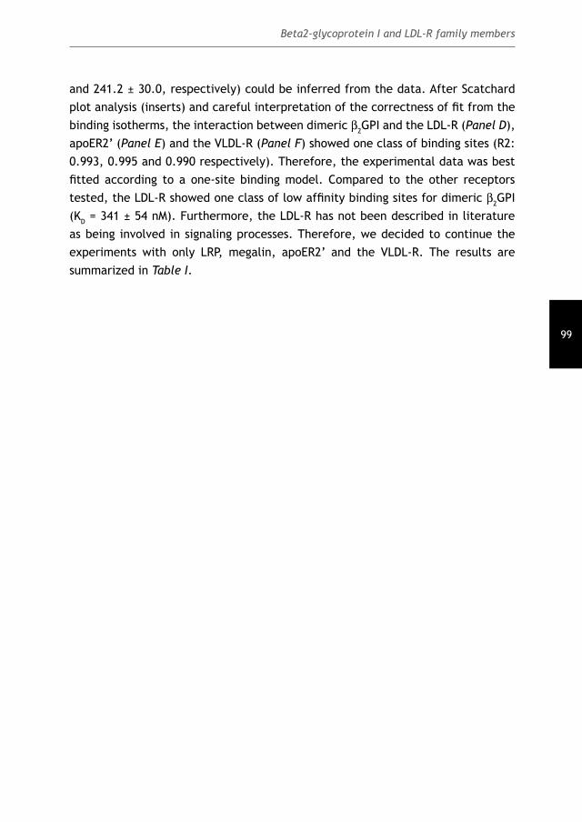
99
Beta2-glycoprotein I and LDL-R family members
and 241.2 ± 30.0, respectively) could be inferred from the data. After Scatchard plot analysis (inserts) and careful interpretation of the correctness of fi t from the binding isotherms, the interaction between dimeric β2GPI and the LDL-R (Panel D), apoER2’ (Panel E) and the VLDL-R (Panel F) showed one class of binding sites (R2: 0.993, 0.995 and 0.990 respectively). Therefore, the experimental data was best fi tted according to a one-site binding model. Compared to the other receptors tested, the LDL-R showed one class of low affi nity binding sites for dimeric β2GPI (KD = 341 ± 54 nM). Furthermore, the LDL-R has not been described in literature as being involved in signaling processes. Therefore, we decided to continue the experiments with only LRP, megalin, apoER2’ and the VLDL-R. The results are summarized in Table I.

100
Chapter 4
Figure 2. Interaction of plasma and dimeric β2GPI with LDL-R homologues A, a reproducible sensorgram showing interaction of dimeric β2GPI with LRP (solid line). Also the control proteins apple 2 — β2GPI (broken line), apple 4, and plasma β2GPI are displayed. B, LRP, C, megalin or D, LDL-R was immobilized on a CM5 or C1 sensor chip at a density of 10 fmol/mm2, 26 fmol/mm2, or 29.4 fmol/mm2 respectively. Seven different concentrations (5 — 500 nM) of dimeric β2GPI were passed over the immobilized receptors. E, apoER2’ or F, VLDL-R was coated with increasing concentrations (0 — 10 μg/mL) on a 96 wells plate and dimeric β2GPI (3 μg/mL) was able to interact with the receptors. Lines represent the data curves and their fi tted curves obtained with a one-site binding model or a two-site binding model. Scatchard plots (inserts) verify the use of the binding models. Data for LRP, megalin and the LDL-R is expressed as mol ligand bound to mol receptor. Steady state constants derived from the binding isotherms are listed in Table 1. Results represent mean ± SD (n = 3).
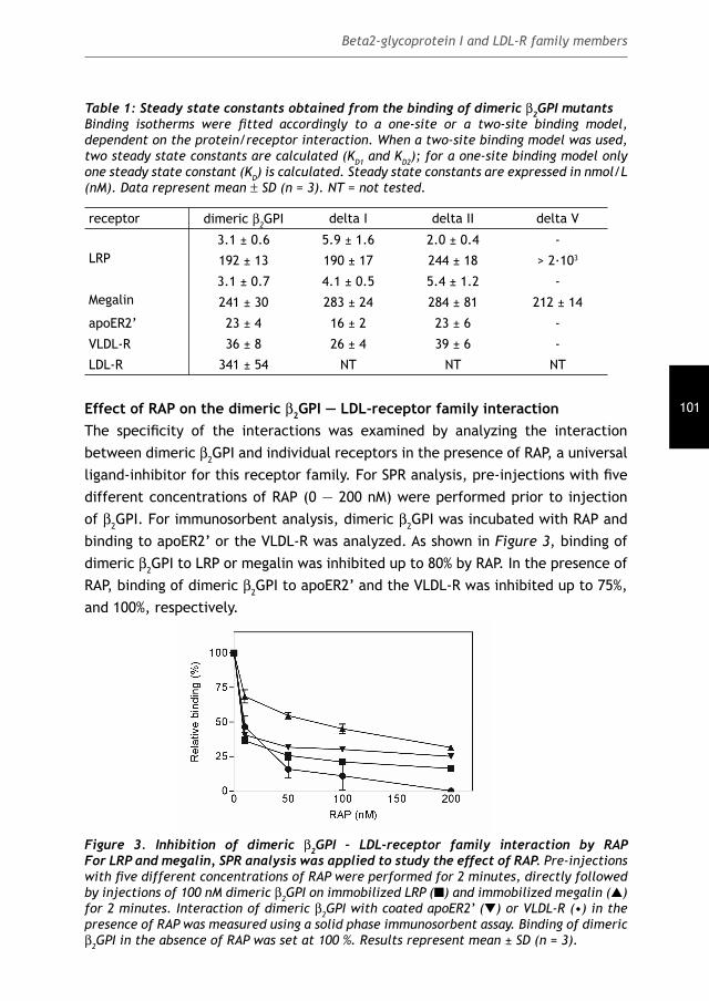
101
Beta2-glycoprotein I and LDL-R family members
Table 1: Steady state constants obtained from the binding of dimeric β2GPI mutants Binding isotherms were fi tted accordingly to a one-site or a two-site binding model, dependent on the protein/receptor interaction. When a two-site binding model was used, two steady state constants are calculated (KD1 and KD2); for a one-site binding model only one steady state constant (KD) is calculated. Steady state constants are expressed in nmol/L (nM). Data represent mean ± SD (n = 3). NT = not tested.
receptor dimeric β2GPI delta I delta II delta V
LRP3.1 ± 0.6 5.9 ± 1.6 2.0 ± 0.4 -
192 ± 13 190 ± 17 244 ± 18 > 2·103
Megalin3.1 ± 0.7 4.1 ± 0.5 5.4 ± 1.2 -
241 ± 30 283 ± 24 284 ± 81 212 ± 14
apoER2’ 23 ± 4 16 ± 2 23 ± 6 -
VLDL-R 36 ± 8 26 ± 4 39 ± 6 -
LDL-R 341 ± 54 NT NT NT
Effect of RAP on the dimeric β2GPI — LDL-receptor family interaction The specifi city of the interactions was examined by analyzing the interaction between dimeric β2GPI and individual receptors in the presence of RAP, a universal ligand-inhibitor for this receptor family. For SPR analysis, pre-injections with fi ve different concentrations of RAP (0 — 200 nM) were performed prior to injection of β2GPI. For immunosorbent analysis, dimeric β2GPI was incubated with RAP and binding to apoER2’ or the VLDL-R was analyzed. As shown in Figure 3, binding of dimeric β2GPI to LRP or megalin was inhibited up to 80% by RAP. In the presence of RAP, binding of dimeric β2GPI to apoER2’ and the VLDL-R was inhibited up to 75%, and 100%, respectively.
Figure 3. Inhibition of dimeric β2GPI – LDL-receptor family interaction by RAP For LRP and megalin, SPR analysis was applied to study the effect of RAP. Pre-injections with fi ve different concentrations of RAP were performed for 2 minutes, directly followed by injections of 100 nM dimeric β2GPI on immobilized LRP (■) and immobilized megalin (▲) for 2 minutes. Interaction of dimeric β2GPI with coated apoER2’ (▼) or VLDL-R (•) in the presence of RAP was measured using a solid phase immunosorbent assay. Binding of dimeric β2GPI in the absence of RAP was set at 100 %. Results represent mean ± SD (n = 3).

102
Chapter 4
Effect of heparin on the dimeric β2GPI — LDL-receptor family interaction β2-Glycoprotein I consists of two regions that are enriched in cationic residues. These residues are located on domain I and V, with the largest cationic patch on domain V (~2,000 Å2). Since LDL-R homologues are known to recognize such cationic residues, we investigated the ability of heparin (that also recognizes cationic residues) to inhibit the interaction between dimeric β2GPI and LRP, megalin, apoER2’ and the VLDL-R. As shown in Figure 4, heparin inhibited the interaction of dimeric β2GPI with LRP or megalin according to a bi-phasic inhibition model. An initial rapid inhibition phase up to approximately 50 — 75 % (residual binding of 46.2 ± 0.5 % and 23.2 ± 1.9 %, respectively) with 0.0625 mg/mL heparin and a slow inhibition phase up to >95% with 2.0 mg/mL heparin. In contrast, binding of dimeric β2GPI to apoER2’ or the VLDL-R was inhibited by heparin in accordance with a mono-phasic pattern. A gradual inhibition was observed up to >95%. Heparin alone did not bind to the receptors (not shown). Half-maximal inhibition was observed at 0.05 mg/mL for LRP and 0.03 mg/mL for megalin. In addition, for apoER2’ and the VLDL-R half-maximal inhibition was observed at 0.46 mg/mL and 0.17 mg/mL, respectively.
Figure 4. Effect of heparin on the interaction between dimeric β2GPI and receptors Dimeric β2GPI (100 nM) was pre-incubated with increasing concentrations of heparin (0 — 2 mg/mL) at 37ºC for 30 minutes. Afterwards the effect of heparin on binding of dimeric β2GPI to immobilized LRP (■) or megalin (▲) was investigated using SPR analysis. The effect of heparin on the interaction between dimeric β2GPI and apoER2’ (▼) or the VLDL-R (•) was investigated using a solid phase immunosorbent assay. Percentage of inhibition in the presence of heparin was calculated as a result of binding of proteins to the receptors without heparin. Results represent mean ± SD (n = 3).
Function of the different domains of β2GPI in LDL-receptor interaction To further characterize the interaction between dimeric β2GPI and LDL-receptor family members, we determined the binding characteristics of the domain deletion

103
Beta2-glycoprotein I and LDL-R family members
mutants for LRP, megalin, apoER2’ and the VLDL-R. SPR measurements and the immunosorbent assays were performed under the same conditions as for full-length dimeric β2GPI. Deletion of domain V of β2GPI appeared to be crucial for binding to LRP and megalin. A small increase in the response signal was observed with increasing concentrations of delta V, whereas a signifi cant increase in the response signal was observed when delta I and delta II were passed over LRP or megalin (data not shown). After replacement with buffer, dissociation of delta V started rapidly and protein was completely gone before the dissociation time (120 sec) was ended, whereas delta I and delta II dissociated gradually from LRP and megalin (similar to full-length dimeric β2GPI). As shown in Figure 5 Panel A, maximal binding to LRP was lower for delta I and higher for delta II compared with full-length dimeric β2GPI. The results are summarized in Table 1. A model describing the interaction between delta I and delta II and two independent binding sites (two-site binding model) was found to provide the best fi t of the experimental data for LRP (KD1 5.9 ± 1.6 nM, KD2 189.6 ± 16.4 nM and KD1 2.0 ± 0.4 nM, KD2 283.4 ± 80.7 nM, respectively). Delta V displayed a more than 600 times decreased affi nity (Kd > 2·103 nM) for LRP. Binding characteristics for megalin were similar to LRP; interaction of delta I and delta II with megalin (Panel B) displayed no major differences in maximal response compared with full-length dimeric β2GPI (Table 2). Except for delta V (KD 211.4 ± 13.7 nM), a two-site binding model could be applied for both delta I and delta II (KD1 4.1 ± 0.5 nM, KD2 283.1 ± 24.1 nM and KD1 5.4 ± 1.2 nM, KD2 283.4 ± 80.7 nM, respectively). For apoER2’ (Panel C) and the VLDL-R (Panel D) similar binding characteristics were observed; delta V displayed reduced binding to both receptors, whereas delta I and delta II displayed a dose-dependent interaction that was comparable with full-length dimeric β2GPI.
Interaction of dimeric β2GPI with recombinant LRP clusters LRP consists of four clusters enriched in complement-type repeats, of which clusters II and IV encompass the main ligand-binding domains of LRP. The observation that interaction between dimeric β2GPI and LRP was best characterized with a two-site binding model may suggest heterogeneity of LRP. To identify LRP regions involved in binding dimeric β2GPI, purifi ed recombinant cluster II and IV were used. When plasma β2GPI was passed over both clusters II and cluster IV, no signifi cant increase in the resonance signal was observed (data not shown). Binding of dimeric β2GPI displayed a reversible and dose dependent binding to both ligand-binding clusters. The experimental data for each cluster fi tted best using a two-site binding model. Therefore, as shown in Table 3, a high and a low affi nity binding site could be
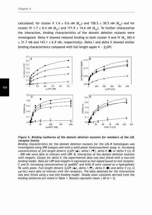
104
Chapter 4
calculated; for cluster II 1.6 ± 0.6 nM (KD1) and 158.5 ± 30.5 nM (KD2) and for cluster IV 1.7 ± 0.4 nM (KD1) and 171.9 ± 14.6 nM (KD2). To further characterize the interaction, binding characteristics of the domain deletion mutants were investigated. Delta V showed reduced binding to both cluster II and IV (KD 365.4 ± 31.7 nM and 143.1 ± 6.9 nM, respectively). Delta I and delta II showed similar binding characteristics compared with full-length apple 4 — β2GPI.
Figure 5. Binding isotherms of the domain deletion mutants for members of the LDL receptor familyBinding characteristics for the domain deletion mutants for the LDL-R homologues was investigated using SPR analysis and with a solid phase immunosorbent assay. A, increasing concentrations of full-length dimeric β2GPI (▲), delta I (▼), delta II (■) or delta V (Δ) (0 – 500 nM) were able to interact with LRP. B, interaction of the domain deletion mutants with megalin. Except for delta V, the experimental data was best fi tted with a two-site binding model. Data for LRP and megalin is expressed as mol ligand bound to mol receptor. C and D; increasing concentrations of apoER2’ and VLDL-R were coated on a hydrophobic 96 wells plate. Full-length dimeric β2GPI (▲), delta I (▼), delta II (■) and delta V (Δ) (3 μg/mL) were able to interact with the receptors. The data obtained for the interactions was best fi tted using a one-site binding model. Steady state constants derived from the binding isotherms are listed in Table 1. Results represent mean ± SD (n = 3).
105
Beta2-glycoprotein I and LDL-R family members
Table 2: Maximum binding (Req,max) for domain deletion mutants of β2GPIBinding isotherms were fi tted accordingly to a one-site or a two-site binding model, dependent on the type of interaction. When a two-site binding model was used, two binding constants (KD1 and KD2) are calculated; for a one-site binding model only one KD is calculated. Steady state constants are expressed in nmol/L (nM). Data represent mean ± SD (n = 3).
receptor dimeric β2GPI delta I delta II delta V
LRP (RU) 507 ± 30 283 ± 40 608 ± 13 59 ± 22
Megalin (RU) 375 ± 14 343 ± 34 310 ± 20 120 ± 5
Cluster II (RU) 253 ± 9 194 ± 7 209 ± 3 28 ± 3
Cluster IV (RU) 201 ± 8 164 ± 6 180 ± 3 52 ± 2
ApoER2’ (OD490 nm) 0.352 ± 0.06 0.294 ± 0.03 0.389 ± 0.05 0.026 ± 0.03
VLDL-R(OD490 nm) 0.437 ± 0.02 0.394 ± 0.01 0.404 ± 0.01 0.086 ± 0.02
Table 3: Steady state constants obtained from the binding of dimeric β2GPI mutants to cluster II and IV of LRPBinding isotherms were fi tted accordingly to a one-site or a two-site binding model, dependent on the type of interaction. When a two-site binding model was used, two binding constants (KD1 and KD2) are calculated; for a one-site binding model only one KD is calculated. Steady state constants are expressed in nmol/L (nM). Data represent mean ± SD (n = 3).
receptor fragment
dimeric β2GPI delta I delta II delta V
Cluster II of LRP
1.6 ± 0.6 2.4 ± 0.8 2.0 ± 0.3 -
159 ± 30 205 ± 38 157 ± 19 365 ± 32
Cluster IV of LRP
1.7 ± 0.4 1.4 ± 1.2 4.2 ± 0.7 -
172 ± 15 249 ± 53 186 ± 22 143 ± 7
Effect of a mutation in the phospholipid-binding site in domain V of dimeric β2GPI To address the possibility that binding of dimeric β2GPI to members of the LDL-receptor family has similar characteristics as binding to anionic phospholipids, we performed binding experiments with the non-phospholipid-binding mutant of dimeric β2GPI. As shown in Figure 6, the mutant displayed dose-dependent association with LRP (Panel A), megalin (Panel B), apoER2’ (Panel C) and the VLDL-R (Panel D). In contrast to full-length dimeric β2GPI, the maximal response of
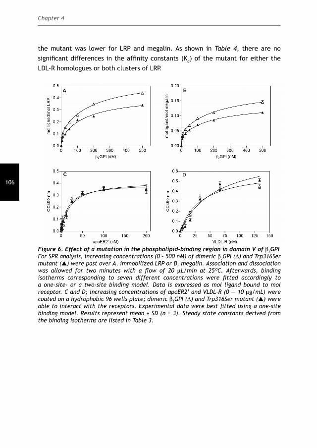
106
Chapter 4
the mutant was lower for LRP and megalin. As shown in Table 4, there are no signifi cant differences in the affi nity constants (Kd) of the mutant for either the LDL-R homologues or both clusters of LRP.
Figure 6. Effect of a mutation in the phospholipid-binding region in domain V of β2GPI For SPR analysis, increasing concentrations (0 – 500 nM) of dimeric β2GPI (Δ) and Trp316Ser mutant (▲) were past over A, immobilized LRP or B, megalin. Association and dissociation was allowed for two minutes with a fl ow of 20 μL/min at 25ºC. Afterwards, binding isotherms corresponding to seven different concentrations were fi tted accordingly to a one-site- or a two-site binding model. Data is expressed as mol ligand bound to mol receptor. C and D; increasing concentrations of apoER2’ and VLDL-R (0 — 10 μg/mL) were coated on a hydrophobic 96 wells plate; dimeric β2GPI (Δ) and Trp316Ser mutant (▲) were able to interact with the receptors. Experimental data were best fi tted using a one-site binding model. Results represent mean ± SD (n = 3). Steady state constants derived from the binding isotherms are listed in Table 3.

107
Beta2-glycoprotein I and LDL-R family members
Table 4: Steady state constants obtained from the binding of the Trp316Ser mutant of β2GPIMaximal binding to LRP and megalin is shown in response units (RU) and for apoER2’ and the VLDL-R the maximal response is expressed in OD280 nm. Data represent mean ± SD (n = 3).
receptor dimeric β2GPI Trp316Ser
LRP3.1 ± 0.6 3.7 ± 0.4
192 ± 13 162 ± 47
Megalin3.1 ± 0.7 2.6 ± 0.5
241 ± 30 198 ± 17
Cluster IIof LRP
1.6 ± 0.6 1.7 ± 0.19
158 ± 30 197 ± 10
Cluster IV of LRP
1.7 ± 0.4 1.2 ± 0.3
172 ± 15 175 ± 13
apoER2’ 22 ± 4 34 ± 9
VLDL-R 38 ± 8 60 ± 17
DiscussionIn the present study we have investigated interaction between beta2-glycopprotein I (β2GPI) and fi ve members of the low density lipoprotein receptor (LDL-R) family. Among these are 1. the low-density lipoprotein receptor related protein (LRP), a promiscuous and ubiquitously expressed receptor involved in several physiological processes, 2. the apolipoprotein E receptor 2’ (apoER2’) which participates in neuronal development but is also expressed on platelets, 3. the low-density lipoprotein receptor (LDL-R), 4. the very low-density lipoprotein receptor (VLDL-R) which is expressed for instance on endothelial cells (EC) and 5. megalin, a multifunctional receptor expressed in various resorptive epithelia (including the proximal renal tubule) pointing to a predominant role in endocytosis and transport. The current report addresses a number of specifi c questions. In previous experiments we have shown that platelets could be activated by a complex of b2GPI and an anti-b2GPI antibody, involving the receptor apoER2’ 13. The interaction between b2GPI and apoER2’ results in phosphorylation of apoER2’ followed by p38 MAPK phosphorylation and thromboxane synthesis 4.This raised the question if other LDL-R homologues also recognize a β2GPI/anti-β2GPI antibody complex. Indeed, we found that LRP, megalin and the VLDL-R recognize only a complex of β2GPI/anti-β2GPI but not β2GPI alone (Figure 1) and
108
Chapter 4
this interaction can be mimicked by our recombinant form of dimeric β2GPI (Figure 2). Two binding models were identifi ed for the interactions; interaction between dimeric β2GPI, apoER2’, the LDL-R and the VLDL-R could be described with a one-site binding model, whereas with LRP or megalin a two-site (heterologous) binding model was more suitable. In contrast to the other receptors tested, the LDL-R showed one class of low affi nity binding sites (340 nM) for dimeric β2GPI. The LDL-R is a classical endocytosis receptor, whereas other LDL-R family members have been shown to regulate intracellular signaling processes 24, suggesting the minor role of the LDL-R in β2GPI-mediated signaling. We could not observe a two-site binding model in the solid-phase binding assay, presumably because the multiple washing steps in this system may amplify association or dissociation defects. SPR analysis measures binding under equilibrium conditions and is therefore not affected by washing steps. In order to confi rm whether data obtained from SPR analysis or from solid-phase binding assays are comparable, we analyzed interaction between dimeric β2GPI and LRP using the solid-phase binding assays (as described for apoER2’ or the VLDL-R). The resulting saturation curve could be adequately described employing a one-site binding model, representing a high affi nity-binding site with half-maximal binding of 2.7 ± 1.0 nM (data not shown). This was in concordance with the high-affi nity site (3.1 ± 0.6 nM) derived from the SPR data. In this study we have further investigated the receptor/protein interaction by using several recombinant constructs of dimeric β2GPI. We demonstrated that domain V of β2GPI is the crucial domain for recognition by the LDL-R homologous tested (Figure 5) and that this recognition site is not identical to the phospholipid-binding site within domain V of β2GPI (Figure 6). It has been proposed that positive electrostatic surface potentials, not a primary sequence, in different ligands constitute receptor-recognition domains 25,26. Our observations from this study suggest that indeed positively charged residues are important (Figure 4). We cannot exclude that this observed inhibition is due to steric interference by heparin. However, in a previous study we have shown that interaction between dimeric β2GPI and apoER2’ on platelets can be inhibited by a peptide spanning amino acids 282-287 in domain V of β2GPI 19. Furthermore, it has been reported that Lys (284), Lys (286) and Lys (287) in domain V are essential for the interaction of β2GPI with heparin 27. This implicates that both electrostatic interactions and amino acid sequences are important for the interaction between dimeric β2GPI and LDL-R homologues. Interaction of dimeric β2GPI with LRP or megalin was best described with a two-site binding model, suggesting the presence of two interaction sites on the surface of

109
Beta2-glycoprotein I and LDL-R family members
the receptors or on β2GPI. For this purpose, we analyzed the interaction between β2GPI and cluster II and IV of LRP. We could still observe a two-site binding model with both clusters, suggesting that LRP recognizes two surface potentials on β2GPI. Two domains that expose cationic charges were of interest; domain I and domain V of β2GPI. However, delta I interacted similar with the receptors or both clusters of LRP compared to full-length dimeric β2GPI. Therefore, it is likely that domain V contains both regions involved in the receptor-interaction. Site-directed mutagenesis is required to establish the relative importance of cationic residues in domain V for the assembly of the β2GPI/LDL-R complex. Besides members of the LDL-R family, a number of other cellular receptors have been described for β2GPI. The contribution of LDL-R family members in β2GPI-mediated cell-activation in comparison with these other receptors is unknown. One of the candidate receptors, annexin A2 39, 40, does not have a transmembrane and intracellular domain, making it unlikely that annexin A2 is directly involved in signal transduction. It has been reported that anti-β2GPI antibodies display a signaling cascade in endothelial cells comparable to that induced by members of the toll-like receptor (TLR) family. TLR2 41 and TLR4 42 have been implicated in this event. Interesting, also glycoprotein Ib, another possible member of the TLR family, has been shown to bind β2GPI/anti-β2GPI antibody complexes 43. Our next challenge is to understand how cell signaling induced by anti-β2GPI antibodies is coordinated by all these different receptors.Besides β2GPI, other proteins have been implicated as possible target for aPL, in particular prothrombin. However, the majority of the performed studies indicate that anti-β2GPI antibodies correlate best with the observed clinical manifestations 28. In addition, addition of purifi ed anti-prothrombin antibodies to whole blood did not result in increased platelet deposition (unpublished observations). Therefore, we do not believe that interaction of prothrombin-anti-prothrombin antibody complexes with cells result in cellular activation.An important step in resolving the pathways that explain clinical symptoms in APS is the identifi cation of cellular receptors that can interact with the protein of interest. The classical clinical manifestations observed in APS are thrombosis and pregnancy morbidity 28-30. However, patients often suffer from other aPL associated clinical manifestations, such as heart valve abnormalities, thrombocytopenia, proteinuria, chorea, neuropathy and livedo reticularis 31-35, which are undoubtedly frequently observed in patients with aPL antibodies in their plasma. Here we have shown that β2GPI/antiβ2GPI-antibodies complexes can interact with different LDL-R homologues. It is clear now that these receptors have the potential to induce

110
Chapter 4
signaling processes in different cell types [36-38]. Different members of the LDL-R family are expressed on almost all cell types. It is interesting to speculate that β2GPI/anti-b2GPI antibody complexes can activate a large number of different cells via LDL-R homologues, resulting in responses specifi c for that cell type. We speculate that the heterogeneous clinical manifestations observed in APS are due to interaction between β2GPI/anti-β2GPI antibody complexes and LDL-R family members on different cell types.
Acknowledgement: This study was supported by a grant from the Netherlands Organization for Health Research and Development (Zon-MW: 902-26-290) and by a grant from the Netherlands Heart Foundation (2003B74).

111
Beta2-glycoprotein I and LDL-R family members
References
1. Wilson WA, Gharavi AE, Koike T et al. International consensus statement on
preliminary classifi cation criteria for defi nite antiphospholipid syndrome: report of
an international workshop. Arthritis Rheum 1999; 42: 1309-1311.
2. Roubey RA. Treatment of the antiphospholipid syndrome. Curr Opin Rheumatol 2002;
14: 238-242.
3. de Laat H, Derksen RH, Urbanus RT, De Groot PG. IgG antibodies that recognize
epitope Gly40-Arg43 in domain I of beta 2-glycoprotein I cause LAC, and their
presence correlates strongly with thrombosis. Blood 2005; 105: 1540-1545.
4. De Groot PG, van Lummel M, Pennings M et al. Beta2-glycoprotein I and LDL-receptor
family members. Thromb Res 2004; 114: 455-459.
5. Raschi E, Testoni C, Bosisio D et al. Role of the MyD88 transduction signaling pathway
in endothelial activation by antiphospholipid antibodies. Blood 2003; 101: 3495-
3500.
6. Moestrup SK, Schousboe I, Jacobsen C et al. beta2-glycoprotein-I (apolipoprotein H)
and beta2-glycoprotein-I-phospholipid complex harbor a recognition site for the
endocytic receptor megalin. J Clin Invest 1998; 102: 902-909.
7. Zhang J, McCrae KR. Annexin A2 mediates endothelial cell activation by
antiphospholipid/anti-beta2 glycoprotein I antibodies. Blood 2005; 105: 1964-1969.
8. Schousboe I. beta 2-Glycoprotein I: a plasma inhibitor of the contact activation of
the intrinsic blood coagulation pathway. Blood 1985; 66: 1086-1091.
9. Yasuda S, Atsumi T, Ieko M et al. Nicked beta2-glycoprotein I: a marker of cerebral
infarct and a novel role in the negative feedback pathway of extrinsic fi brinolysis.
Blood 2004; 103: 3766-3772.
10. Takeuchi R, Atsumi T, Ieko M et al. Suppressed intrinsic fi brinolytic activity by
monoclonal anti-beta-2 glycoprotein I autoantibodies: possible mechanism for
thrombosis in patients with antiphospholipid syndrome. Br J Haematol 2002; 119:
781-788.
11. Sheng Y, Reddel SW, Herzog H et al. Impaired thrombin generation in b2-glycoprotein
I null mice. J Biol Chem 2001; 276: 13817-13821.
12. Riddell DR, Vinogradov DV, Stannard AK, Chadwick N, Owen JS. Identifi cation and
characterization of LRP8 (apoER2) in human blood platelets. J Lipid Res 1999; 40:
1925-1930.
13. Lutters BC, Derksen RH, Tekelenburg WL et al. Dimers of beta 2-glycoprotein I increase
platelet deposition to collagen via interaction with phospholipids and the
apolipoprotein E receptor 2’. J Biol Chem 2003; 278: 33831-33838.
14. Kim DH, Iijima H, Goto K et al. Human apolipoprotein E receptor 2. A novel lipoprotein
receptor of the low density lipoprotein receptor family predominantly expressed in
the brain. J Biol Chem 1996; 271: 8373-8380.

112
Chapter 4
15. Herz J, Bock HH. Lipoprotein receptors in the nervous system. Annu Rev Biochem
2002; 71: 405-434.
16. Kim DH, Magoori K, Inoue TR et al. Exon/intron organization, chromosome
localization, alternative splicing, and transcription units of the human apolipoprotein
E receptor 2 gene. J Biol Chem 1997; 272: 8498-8504.
17. Horbach DA, Van Oort E, Donders RCJM, Derksen RHWM, De Groot PG. Lupus
anticoagulant is the strongest risk factor for both venous and arterial thrombosis in
patients with systemic lupus erythematosus - Comparison between different assays
for the detection of antiphospholipid antibodies. Thromb Haemost 1996; 76: 916-
924.
18. Lutters BCH, Meijers JCM, Derksen RHWM, Arnout J, De Groot PG. Dimers of b2-
glycoprotein I mimic the in vitro effects of b2-glycoprotein I-anti-b2-glycoprotein I
antibody complexes. J Biol Chem 2001; 276: 3060-3067.
19. van Lummel M, Pennings MT, Derksen RH et al. The binding site in {beta}2-glycoprotein
I for ApoER2’ on platelets is located in domain V. J Biol Chem 2005; 280: 36729-
36736.
20. Horbach DA, Van Oort E, Tempelman MJ, Derksen RHWM, De Groot PhG. The
prevalence of a non-phospholipid-binding form of b2-glycoprotein I in human plasma.
Consequences for the development of anti-b2-glycoprotein I antibodies. Thromb
Haemost 1998; 80: 791-797.
21. Durocher Y, Perret S, Kamen A. High-level and high-throughput recombinant protein
production by transient transfection of suspension-growing human 293-EBNA1 cells.
Nucleic Acids Res 2002; 30: E9.
22. Leahy DJ, Dann CE, III, Longo P, Perman B, Ramyar KX. A mammalian expression
vector for expression and purifi cation of secreted proteins for structural studies.
Protein Expr Purif 2000; 20: 500-506.
23. Neels JG, van Den Berg BM, Lookene A et al. The second and fourth cluster of class A
cysteine-rich repeats of the low density lipoprotein receptor-related protein share
ligand-binding properties. J Biol Chem 1999; 274: 31305-31311.
24. May P, Herz J, Bock HH. Molecular mechanisms of lipoprotein receptor signalling.
Cell Mol Life Sci 2005; 62: 2325-2338.
25. Melman L, Cao ZF, Rennke S et al. High affi nity binding of receptor-associated
protein to heparin and low density lipoprotein receptor-related protein requires
similar basic amino acid sequence motifs. J Biol Chem 2001; 276: 29338-29346.
26. Raffai R, Weisgraber KH, MacKenzie R et al. Binding of an antibody mimetic of
the human low density lipoprotein receptor to apolipoprotein E is governed through
electrostatic forces. Studies using site-directed mutagenesis and molecular modeling.
J Biol Chem 2000; 275: 7109-7116.
113
Beta2-glycoprotein I and LDL-R family members
27. Guerin J, Sheng Y, Reddel S et al. Heparin inhibits the binding of beta 2-glycoprotein
I to phospholipids and promotes the plasmin-mediated inactivation of this blood
protein. Elucidation of the consequences of the two biological events in patients
with the anti-phospholipid syndrome. J Biol Chem 2002; 277: 2644-2649.
28. De Groot PG, Derksen RH. The antiphospholipid syndrome: clinical characteristics,
laboratory features and pathogenesis. Curr Opin Infect Dis 2005; 18: 205-210.
29. Derksen RH, Khamashta MA, Branch DW. Management of the obstetric antiphospholipid
syndrome. Arthritis Rheum 2004; 50: 1028-1039.
30. Vasoo S, Sangle S, Zain M, D’Cruz D, Hughes G. Orthopaedic manifestations of the
antiphospholipid (Hughes) syndrome. Lupus 2005; 14: 339-345.
31. D’Cruz DP. Renal manifestations of the antiphospholipid syndrome. Lupus 2005;14:45-
48.
32. Antunes MC, D’Cruz D, Hughes GR. Neurological manifestations of anti-phospholipid
(Hughes) syndrome. Clin Exp Rheumatol 2004; 22: 771-775.
33. Suarez AL, Hughes GR, Khamashta MA. Neurological manifestations of the
antiphospholipid syndrome. Med Clin (Barc.) 2005; 124: 630-633.
34. Dayal NA, Isenberg DA. Endstage renal failure in primary antiphospholipid syndrome-
case report and review of literature. Rheumatology (Oxford) 2003; 42: 1128-1129.
35. Krause I, Blank M, Fraser A et al. The association of thrombocytopenia with systemic
manifestations in the antiphospholipid syndrome. Immunobiology 2005; 210: 749-
754.
36. Mayer H, Duit S, Hauser C, Schneider WJ, Nimpf J. Reconstitution of the Reelin
Signaling Pathway in Fibroblasts Demonstrates that Dab1 Phosphorylation Is
Independent of Receptor Localization in Lipid Rafts. Mol Cell Biol 2006; 26: 19-27.
37. Lillis AP, Mikhailenko I, Strickland DK. Beyond endocytosis: LRP function in cell
migration, proliferation and vascular permeability. J Thromb Haemost 2005; 3: 1884-
1893.
38. Zou Z, Chung B, Nguyen T et al. Linking receptor-mediated endocytosis and cell
signaling: evidence for regulated intramembrane proteolysis of megalin in proximal
tubule. J Biol Chem 2004; 279: 34302-34310.
39. Ma K, Simantov R, Zhang J-C, et al. High-affi nity binding of beta2-glycoprotein I to
human endothelial cells is mediated by annexin II. J Biol Chem 2000; 275; 15541-
15548.
40. Zhou H, Roubey RAS. Annexin A2 and the upregulation of monocyte tissue factor
activity by aPL. Lupus 2002; 11: 543.
41. Satta N, Dunoyer-Geindre S, Fish RJ, Reber G, Boehlen F, Kruithof EKO, de Moerloose
P. The activation of mouse fi broblasts by antiphospholipid antibodies requires TLR2.
J Thromb Haemost 2005; 3 (1): 1223.

114
Chapter 4
42. Meroni PL, Raschi E, Testoni C, Parisio A, Borghi MO. Innate immunity in the
antiphospholipid syndrom: role of toll-like receptors in endothelial cell activation by
antiphospholipid antibodies. Autoimmunity Reviews 2004; 3; 510-515.
43. Pennings MTT, Derksen RHWM, van Lummel M, Adelmeijer J, Lisman T, de
Groot PG. Platelet adhesion to dimeric beta2-glycoprotein I under conditions
of fl ow is mediated by glycoprotein Ib. J Thromb Haemost 2005; 3 (1): 256.

Menno van Lummel, Linda Hartkamp, Ronald H.W.M. Derksen,
Maarten T.T. Pennings, and Philip G. de Groot
In preparation
5MOLECULAR CHARACTERIZATION OF THE
APOLIPOPROTEIN E RECEPTOR 2’-BINDING SITE
AND PHOSPHOLIPID-BINDING SITE WITHIN BETA2-
GLYCOPROTEIN I

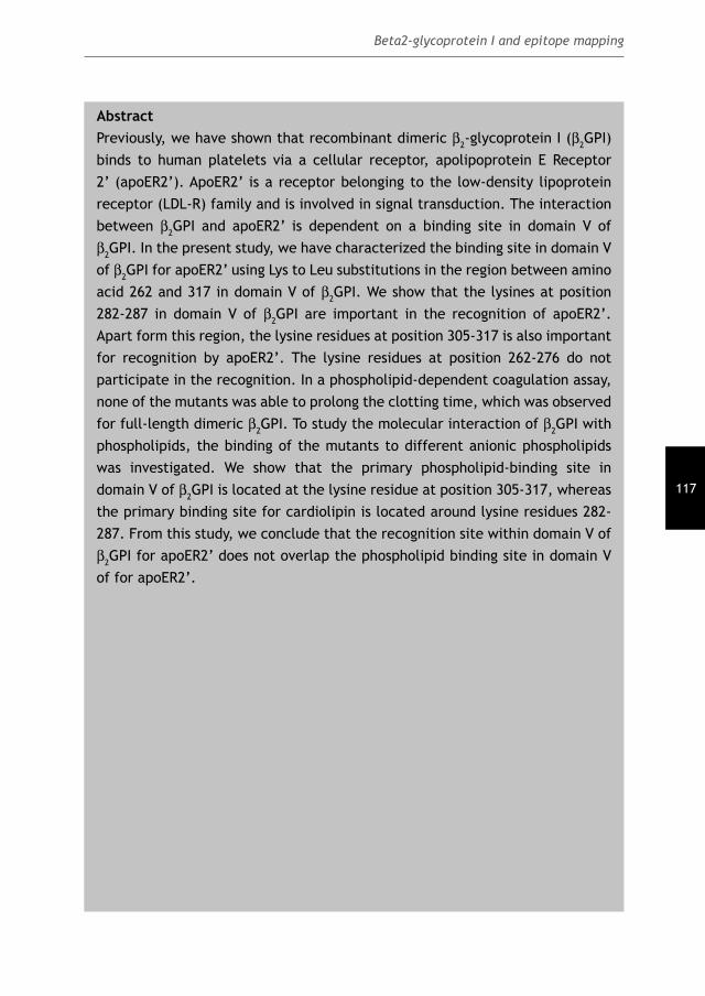
117
Beta2-glycoprotein I and epitope mapping
AbstractPreviously, we have shown that recombinant dimeric β2-glycoprotein I (β2GPI) binds to human platelets via a cellular receptor, apolipoprotein E Receptor 2’ (apoER2’). ApoER2’ is a receptor belonging to the low-density lipoprotein receptor (LDL-R) family and is involved in signal transduction. The interaction between β2GPI and apoER2’ is dependent on a binding site in domain V of β2GPI. In the present study, we have characterized the binding site in domain V of β2GPI for apoER2’ using Lys to Leu substitutions in the region between amino acid 262 and 317 in domain V of β2GPI. We show that the lysines at position 282-287 in domain V of β2GPI are important in the recognition of apoER2’. Apart form this region, the lysine residues at position 305-317 is also important for recognition by apoER2’. The lysine residues at position 262-276 do not participate in the recognition. In a phospholipid-dependent coagulation assay, none of the mutants was able to prolong the clotting time, which was observed for full-length dimeric β2GPI. To study the molecular interaction of β2GPI with phospholipids, the binding of the mutants to different anionic phospholipids was investigated. We show that the primary phospholipid-binding site in domain V of β2GPI is located at the lysine residue at position 305-317, whereas the primary binding site for cardiolipin is located around lysine residues 282-287. From this study, we conclude that the recognition site within domain V of β2GPI for apoER2’ does not overlap the phospholipid binding site in domain V of for apoER2’.
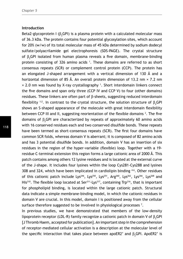
118
Chapter 5
IntroductionBeta2-glycoprotein I (β2GPI) is a plasma protein with a calculated molecular mass of 36.3 kDa. The protein contains four potential glycosylation sites, which account for 20% (w/w) of its total molecular mass of 45 kDa determined by sodium dodecyl sulfate/polyacrilamide gel electrophoresis (SDS-PAGE). The crystal structure of β2GPI isolated from human plasma reveals a fi ve domain, membrane-binding protein consisting of 326 amino acids 1. These domains are referred to as short consensus repeats (SCR) or complement control protein (CCP). The protein has an elongated J-shaped arrangement with a vertical dimension of 130 Å and a horizontal dimension of 85 Å. An overall protein dimension of 13.2 nm × 7.2 nm × 2.0 nm was found by X-ray crystallography 1. Short interdomain linkers connect the fi ve domains and span only three (CCP IV and CCP V) to four (other domains) residues. These linkers are often part of β-sheets, suggesting reduced interdomain fl exibility 1;2. In contrast to the crystal structure, the solution structure of β2GPI shows an S-shaped appearance of the molecule with great interdomain fl exibility between CCP III and II, suggesting reorientation of the fl exible domains 3. The fi ve domains of β2GPI are characterized by repeats of approximately 60 amino acids with 16 conserved residues each and two conserved disulfi de bonds. These repeats have been termed as short-consensus repeats (SCR). The fi rst four domains have common SCR folds, whereas domain V is aberrant; it is composed of 82 amino acids and has 3 potential disulfi de bonds. In addition, domain V has an insertion of six residues in the region of the hyper-variable (fl exible) loop. Together with a 19-residue C-terminal extension this region forms a large cationic area of 2000 Å. This patch contains among others 12 lysine residues and is located at the external curve of the J-shape. It includes four lysines within the loop Cys281-Cys288 and lysines 308 and 324, which have been implicated in cardiolipin binding 4-6. Other residues of this cationic patch include Lys246, Lys250, Lys251, Arg260, Lys262, Lys266, Lys268 and His310. The fl exible loop located at Ser311-Lys317, containing Trp316, that is important for phospholipid binding, is located within the large cationic patch. Structural data indicate a simple membrane-binding model, in which the cationic residues in domain V are crucial. In this model, domain I is positioned away from the cellular surface therefore suggested to be involved in physiological processes 7. In previous studies, we have demonstrated that members of the low-density lipoprotein receptor (LDL-R) family recognize a cationic patch in domain V of β2GPI [J Thromb Haem, accepted for publication]. An important step in the comprehension of receptor-mediated cellular activation is a description at the molecular level of the specifi c interaction that takes place between apoER2’ and β2GPI. ApoER2’ is

119
Beta2-glycoprotein I and epitope mapping
a member of the low-density lipoprotein receptor (LDL-R) family and is identifi ed on human platelets 8. ApoER2 is predominantly expressed in the central nervous system, but is also expressed in the testis and vascular cells. Multiple alternative splicing variants of apoER2 exist of which one, apoER2’, is expressed on platelets and megakaryocytes. ApoER2’ on human platelets functions as a signalling receptor, which has been shown for oxidized LDL 9 and artifi cially dimerized β2GPI 10. In the present study, the interaction between β2GPI and apolipoprotein E receptor 2’, as expressed on human platelets, has been studied on a molecular level by substituting lysine residues into leucine residues in domain V of β2GPI.
Experimental Procedures
A three-dimensional model illustrating the effect on the electrostatic potential after amino acid substitutions in a large cationic patch in domain V of β2GPIA detailed molecular study of β2GPI — LDL-R interaction was combined by a prediction of the electrostatic potential after substitution of lysine residues into leucine in the cationic patch between amino acids 262-317 in domain V of β2GPI. The template structure was that of the fi fth domain of β2GPI as determined by X-ray crystallography 1. The electrostatic potential on the surface β2GPI and the mutants were calculated with Swiss-PdbViewer version 3.7b2 (Expasy), an application used to generate protein models and obtaining amino acid substitutions. This programme uses the fi nite difference method to solve the linearized Poisson-Boltzmann equation. Each lysine was assigned a neutral charge (leucine) because β2GPI is active in plasma at pH of approximately 7.2. The molecular surface of domain V of β2GPI was determined with Discovery Studio Viewer Pro version 6.0 (Accelrys Inc.).
Cloning recombinant dimeric β2GPI The recombinant proteins apple 4 — C321S — β2GPI (further referred to as dimeric β2GPI), apple 4 — C321S — ∆5β2GPI (delta V), the non-phospholipid binding dimer apple 4 — C321S — β2GPI — W316S and the control proteins apple 4 (dimerization domain of factor XI) and apple 2 — β2GPI were cloned as described previously 10.
Site-directed mutagenesisPreviously described cloned β2GPI cDNA in eukaryotic expression vector (apple 4 — tissue — type plasminogen activator (tPA)-S478A) 10 was used to create the desired mutations using the QuikChange site-directed mutagenesis kit (Stratagene) for

120
Chapter 5
which only a forward primer for each mutation is required. The sequence of dimeric β2GPI was amplifi ed from the vector apple 4 — C321S — β2GPI tissue — tPA — S478A with the primers β2GPI — Xho I forward (CC CT CGA GGA CGG ACC TGT CCC AAG CCA) and β2GPI — Xba I reverse (CC TCT AGA AAA CAA GTG TGA CAT TTT ATG TGG). The PCR product was cloned into the pBluescript SK +/- cloning vector (Fermentas, Canada). This plasmid was used as a template to construct the pointmutations. Mutagenic primers were designed containing the desired mutation(s) in the middle of the primer. The following single lysine residues in domain V of dimeric β2GPI were substituted into a leucine (with the primer used for mutagenesis): Lys282Leu (5’ GAT AAA GTT TCT TTC TTC TGC CTG AAT AAG GAA AAG AAG TGT AGC 3’) Lys284Leu (GTT TCT TTC TTC TGC AAA AAT CTG GAA AAG AAG TGT AGC TAT ACA) Lys286Leu (5’ TTC TTC TGC AAA AAT AAG GAA CTG AAG TGT AGC TAT ACA GAG GAT 3’)Lys287Leu (5’ TTC TGC AAA AAT AAG GAA AAG CTG TGT AGC TAT ACA GAG GAT GCT 3’)In addition, in three different cationic patches in domain V of β2GPI, three to four lysines were substituted into a leucine in the following regions: Lys262Leu, Lys264Leu, Lys268Leu and Lys276Leu (group 1; further referred to as Lys262-276), Lys282Leu, Lys284Leu, Lys286Leu and Lys287Leu (group 2; further referred to as Lys282-287) and Lys305Leu, Lys308Leu and Lys317Leu (group 3; further referred to as Lys305-317). The primers for mutagenesis for these three groups were (mutated amino acids are underlined):Group 1 (Lys262-276): (5’ CTG ATT CAG GAA CTG TTT CTG AAT GGA ATG CTA 3’ for Lys262Leu, Lys264Leu, Lys268Leu and 5’ AAT GGA ATG CTA CAT GGT GAT CTG GTT TCT TTC TTC TGC AAA AAT 3’ for Lys262-276)Group 2 (Lys282-287): (5’ TTC TGC CTG AAT CTG GAA CTG CTG TGT AGC TAT ACA GAG GAT GCT 3’)Group 3 (Lys305-317): (5’ GTC CCC CTG TGC TTC CTG GAA CAC AGT TCT CTG 3’ for Lys305Leu, Lys308Leu and 5’ TCT CTG GCT TTT TGG CTG ACT GAT GCA TCC GAT 3’ for Lys305-317)The mutagenic primers were used to PCR amplify the entire plasmid using QuikChange Multi enzyme (Stratagene). The mutations were identifi ed by sequence analysis with a β2GPI forward primer (TAT CCT GCA AAA CCA ACA C). To construct fusion proteins of apple 4 and the β2GPI mutants, the PCR products were cloned with Xho I and Xba I into the original vector (apple 4 — C321S — β2GPI — tPA — S478A). In this way dimeric β2GPI with the pointmutations were constructed.

121
Beta2-glycoprotein I and epitope mapping
Transfection, Expression, Cell Culture and Purifi cation of dimeric β2GPI mutants Recombinant full-length and mutants of dimeric β2GPI were purifi ed from the culture supernatant of transfected baby hamster kidney cells by affi nity chromatography using monoclonal anti-β2GPI antibody 11. Bound proteins were eluted with 0.1 M glycine-HCl, pH 2.7. The eluted fractions were immediately neutralized with 1 M Tris, pH 9. The pooled fractions were further purifi ed on a mono S column using fast performance liquid chromatography (FPLC) (Amersham Pharmacia Biotech). Apple 4 was purifi ed using a monoclonal antibody (anti-XI-1). Purity of the protein fractions was determined on a 4-15% polyacryl-amide gel electrophoresis (SDS-PAGE). Concentrated eluants were dialyzed against 50 mM Tris, 150 mM NaCl, pH 7.4. Protein concentrations were determined using a bicinchoninic acid (BCA) protein assay (Pierce). Purifi ed constructs were analyzed on a 7.5% SDS-PAGE.
Gel Filtration StudiesThe dimeric β2GPI constructs proteins were applied to Superdex 200 gel fi ltration column using FPLC equipment. The column was equilibrated with TBS, pH 7.4, at a fl ow rate of 1.0 mL/min. The absorbance of the eluent was monitored at 280 nm. Molecular masses were determined by comparison to a standard curve of ferritin (440 kDa), catalase (232 kDa), aldolase (158 kDa), albumin (67 kDa), ovalbumin (43 kDa) and chymotrypsinogen A (25 kDa).
Purifi cation of plasma β2GPIPlasma β2GPI was purifi ed as described previously 10. In short, b2GPI was isolated from freshly, frozen citrated human plasma. Dialyzed plasma was subsequently applied to the following columns: DEAE-Sephadex A50 column, protein G-Sepharose, S-Sepharose and fi nally heparin-Sepharose chromatography (all Sepharoses were obtained from Amersham Pharmacia Biotech). Bound proteins were eluted with a linear salt gradient. Afterwards, b2GPI was dialyzed against buffer containing 50 mM Tris, 150 mM NaCl, pH 7.4. Purity of the protein was checked with SDS-PAGE analysis.
Preparation of phospholipid vesiclesPhospholipid vesicles containing 20% phosphatidylserine (PS), 40% phosphatidylcholine (PC) and 40% phosphatidylethanolamine (PE) were prepared according to Brunner et al. 12 with some modifi cations as described by Van Wijnen et al 13. The phospholipid concentration was determined by phosphate analysis 14.

122
Chapter 5
Binding of dimeric β2GPI mutants to phospholipid vesiclesBinding of dimeric β2GPI mutants to phosphatidylserine (PS) / phostidylcholine (PC) (20% PS/PC 80%, 5% PS/PC 95%) vesicles and cardiolipin was tested in a solid phase binding assay. High binding 96 wells ELISA plates (Costar, Corning Incorporated, 9102) were coated with PS/PC (25 μM in TBS; 50 μL/well) overnight at 4oC. Cardiolipin (50 μg/mL in ethanol; 50 μL/well) was coated under a stream of nitrogen at room temperature. Wells were blocked with TBS/0.5% gelatine (150 μL/well) for two hours at 37oC. Subsequently, wells were incubated with different concentrations of the dimeric mutants (0.25-32 μg/mL) for 1.5 hours at 37oC, followed by incubation with Moab 2B2 (3 μg/mL; 50 μL/well). Apple 4 — β2GPI was used as a positive control and plasma β2GPI as a negative control. Afterwards the wells were incubated with peroxidase-conjugated rabbit anti-mouse antibody (RAMPO) (1:1000; 50 μL/well; 1.5 hours at 37oC), followed by staining procedure using orthophenylenediamine (OPD). Samples were diluted in TBS/0.5% gelatine. Non-specifi c binding was determined using non-coated wells. Results are expressed as mean ± SD (n = 3).
Determination of the effect of the β2GPI mutants on the clotting timeAll coagulation assays were measured in a KC-10 coagulometer (Amelung, Lemgo, Germany). The prothrombin time (PT), a phospholipid-depending coagulation assay, was performed to detect effects of the lysine substitutions in domain V of β2GPI on clotting time. In short, 25 μL of normal pooled plasma and 25 μL of buffer, plasma β2GPI or recombinant full-length dimeric β2GPI or mutants were incubated for two minutes at 4ºC. After an incubation of 1.5 minute at 37ºC, clotting time was initiated by the addition of 50 μL of Innovin (Dade Behring, Marburg, Germany).
Surface plasmon resonance analysis for the interaction between dimeric β2GPI mutants and apolipoprotein E receptor 2’Surface plasmon resonance (SPR) binding assays were performed employing a Biacore 2000 system (Biacore AB, Uppsala, Sweden). ApoER2’ was immobilized on a CM1 sensor chips using the amine-coupling kit (Biacore AB, Uppsala, Sweden). Approximately 7.4 fmol/mm2 of apoER2’ was immobilized. One channel was immobilized with an irrelevant, non-binding protein and afterwards its signal was used to correct the signal from the apoER2’ coated channel for non-specifi c binding. SPR analysis was performed in buffer containing 25 mM HEPES, 125 mM NaCl, pH 7.4 with 3 mM CaCl2 Measurements were performed with a fl ow rate of 20 μl/min at 25ºC. Association and dissociation was followed for a period of

123
Beta2-glycoprotein I and epitope mapping
2 minutes. Regeneration of the surface was performed by application of 0.1 M sodium citrate containing 1 mM of ethylenediaminetetraacetic acid (EDTA) and 1 M of NaCl, pH 5.0.
Results
Three-dimensional model of β2GPI and its mutantsA three dimensional model of the fi fth domain of β2GPI has been generated with the X-ray structure as a template 1. The fi fth domain comprises residues 244 — 326 (Figure 1, panel A). The structure contains a large cationic area of 2000Å, which is illustrated in Figure 2, panel A as a large blue area. The positively charged patch on the surface of domain V decreases when lysine residues at position Lys262, Lys264, Lys268 and Lys276 (group 1; Lys262-276) are substituted by leucine residues (Figure 2, panel B), which is indicated by the smaller blue area in the lower part of domain V of β2GPI. Substituting residues at position Lys282, Lys284, Lys286 and Lys287 (group 2; Lys282-287) and Lys305, Lys308 and Lys317 (group 3; Lys305-317) obtain similar results (Figure 2, panel C and panel D, respectively).Most of the positively charged side chains (14 of 16) are located on the surface of two regions in domain V of β2GPI. The fi rst of the cationic regions is defi ned by amino acids Lys282-287 at the top of the molecule (Figure 1, panel C). In addition, mutation of these lysines is known to disrupt binding to phospholipids. The second region is defi ned by amino acids Lys305-317 and the third region by amino acids Lys262-
276. All three positively charged residues are predicted to interact with apoER2’.
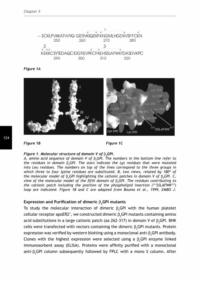
124
Chapter 5
-- SCKLPVKKATVVYQ GERVKIQEKFKNGMLHGDKVSFFCKN
250 260 270 280
** * * *1
290 300 310 320KEKKCSYTEDAQCIDGTIEVPKCFKEHSSLAFWKTDASDVKPC
** *** *32
Figure 1A
Figure 1B Figure 1C
Figure 1. Molecular structure of domain V of β2GPI. A, amino acid sequence of domain V of β2GPI. The numbers in the bottom line refer to the residues in domain β2GPI. The stars indicate the Lys residues that were mutated into Leu residues. The numbers on top of the lines correspond to the three groups in which three to four lysine residues are substituted. B, two views, related by 180º of the molecular model of β2GPI highlighting the cationic patches in domain V of β2GPI. C, view of the molecular model of the fi fth domain of β2GPI. The residues contributing to the cationic patch including the position of the phospholipid insertion (311SSLAFWK317) loop are indicated. Figure 1B and C are adapted from Bouma et al., 1999, EMBO J.
Expression and Purifi cation of dimeric β2GPI mutantsTo study the molecular interaction of dimeric β2GPI with the human platelet cellular receptor apoER2’, we constructed dimeric β2GPI mutants containing amino acid substitutions in a large cationic patch (aa 262-317) in domain V of β2GPI. BHK cells were transfected with vectors containing the dimeric β2GPI mutants. Protein expression was verifi ed by western blotting using a monoclonal anti-β2GPI antibody. Clones with the highest expression were selected using a β2GPI enzyme linked immunosorbent assay (ELISA). Proteins were affi nity purifi ed with a monoclonal anti-β2GPI column subsequently followed by FPLC with a mono S column. After

125
Beta2-glycoprotein I and epitope mapping
purifi cation and protein quantifi cation, the mutants were applied on a 7.5% SDS-PAGE under reducing and non-reducing conditions and stained by Coommassie Brilliant Blue. As shown in Figure 3, panel A, under non-reducing conditions all the mutants migrated with a molecular weight of approximately 50 kilo Dalton (kDa) comparable to that observed for full-length dimeric β2GPI. After reduction (Figure 3, Panel B), the mutants migrated as monomers with a molecular mass of approximately 57 kDa.
Figure 2. Electrostatic potential of the fi fth domain of β2GPI.Electrostatic potential at the phospholipid-binding region of native domain V and mutants. The fi gures were prepared by program Swiss-PdbViewer. A, native domain V at neutral pH. B, Lys262-
276 mutant at neutral pH. The molecular surface of the models are coloured by electrostatic potential (positive = bleu; negative = red). C and D, Lys282-287 and Lys305-317 at neutral pH.
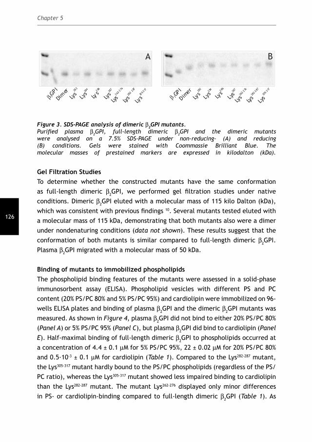
126
Chapter 5
A B
β 2GPI
Dim
erLy
s28
2
Lys284
Lys286
Lys28
7
Lys28
2-287
Lys262-
276
Lys30
5-317
β 2GPI
Dimer
Lys28
2
Lys284
Lys286
Lys28
7
Lys28
2-287
Lys26
2-276
Lys30
5-317
Figure 3. SDS-PAGE analysis of dimeric β2GPI mutants.Purifi ed plasma β2GPI, full-length dimeric β2GPI and the dimeric mutants were analysed on a 7.5% SDS-PAGE under non-reducing- (A) and reducing (B) conditions. Gels were stained with Coommassie Brilliant Blue. The molecular masses of prestained markers are expressed in kilodalton (kDa).
Gel Filtration StudiesTo determine whether the constructed mutants have the same conformation as full-length dimeric β2GPI, we performed gel fi ltration studies under native conditions. Dimeric β2GPI eluted with a molecular mass of 115 kilo Dalton (kDa), which was consistent with previous fi ndings 10. Several mutants tested eluted with a molecular mass of 115 kDa, demonstrating that both mutants also were a dimer under nondenaturing conditions (data not shown). These results suggest that the conformation of both mutants is similar compared to full-length dimeric β2GPI. Plasma β2GPI migrated with a molecular mass of 50 kDa.
Binding of mutants to immobilized phospholipidsThe phospholipid binding features of the mutants were assessed in a solid-phase immunosorbent assay (ELISA). Phospholipid vesicles with different PS and PC content (20% PS/PC 80% and 5% PS/PC 95%) and cardiolipin were immobilized on 96-wells ELISA plates and binding of plasma β2GPI and the dimeric β2GPI mutants was measured. As shown in Figure 4, plasma β2GPI did not bind to either 20% PS/PC 80% (Panel A) or 5% PS/PC 95% (Panel C), but plasma β2GPI did bind to cardiolipin (Panel E). Half-maximal binding of full-length dimeric β2GPI to phospholipids occurred at a concentration of 4.4 ± 0.1 μM for 5% PS/PC 95%, 22 ± 0.02 μM for 20% PS/PC 80% and 0.5·10-3 ± 0.1 μM for cardiolipin (Table 1). Compared to the Lys282-287 mutant, the Lys305-317 mutant hardly bound to the PS/PC phospholipids (regardless of the PS/PC ratio), whereas the Lys305-317 mutant showed less impaired binding to cardiolipin than the Lys282-287 mutant. The mutant Lys262-276 displayed only minor differences in PS- or cardiolipin-binding compared to full-length dimeric β2GPI (Table 1). As

127
Beta2-glycoprotein I and epitope mapping
can be seen from Figure 4, panel B and D, the three positively charged groups in domain V of β2GPI (Lys262-276, Lys282-287 and Lys305-317) contributed to PS-binding in the order of position 305-317 > 282-287 > 262-276 for 5% PS:PC 95% and 20% PS: PC 80%. For cardiolipin, the observations were in contrast with the fi ndings for PS containing surfaces. The contribution of cationic residues in domain V was in the order of position 282-287 > 305-317 > 262-276 (Figure 4, panel F). This suggests that binding of β2GPI to cardiolipin requires other regions in domain V of β2GPI than binding of β2GPI to PS/PC. These results demonstrate a physiological signifi cance of residues Lys305-317 in domain V of β2GPI for binding to anionic phospholipids.

128
Chapter 5
0 10 20 30
0.05
0.15
0.25
0.35
0.45
β2GPI (μg/mL)
OD
490
nm
A B
Dimer
GPI
2β W31
6S26
2-27
6
Lys
282-
287
Lys
305 -3
17
Lys
0
25
50
75
100
Res
idua
lbin
din
g(%
)
0 10 20 300.00
0.15
0.30
0.45
0.60
0.75
β2GPI (μg/mL)
OD
490
nm
C
Dimer GPI
2β W31
6S26
2- 276
Lys
282-2
87
Lys
305 -3
17
Lys
0
25
50
75
100
Re
sid
ua
lbin
din
g(%
)
D
0.00 0.25 0.50 0.75 1.000.00
0.25
0.50
0.75
1.00
1.25
β2GPI (μg/mL)
OD
49
0n
m
E
Dim
erG
PI2β W
316S
262-2
76
Lys
282-2
87
Lys
305 -3
17
Lys
0
25
50
75
100
Re
sid
ua
lbin
din
g(%
)
F
Figure 4. Binding of dimeric β2GPI mutants to immobilized phospholipids A and C, phospholipid vesicles (20% PS/PC 80% and 5% PS/PC 95%, 25μM) were immobilized on high binding 96-well ELISA plates. E, cardiolipin (50 μg/mL) was coated on 96-well ELISA plates under a stream of nitrogen. This was followed by incubation with increasing concentrations (ranging from 0 — 32 μg/mL for PS/PC and from 0 — 1 μg/mL) of plasma β2GPI (Δ), full-length dimeric β2GPI (■) and the dimeric mutants at 37ºC for 2 hours. The following constructs are shown: Lys262-276 (▲), Lys282-287 (▼), Lys305-317 (•) and the non-phospholipid binding dimer W316S (). Afterwards, bound protein was detected with Moab 2B2. Bound 2B2 was detected using OPD staining procedure. Results are expressed as mean ± SD (n = 3). B, D and E, percentage of binding to phospholipids is expressed relative to binding to phospholipids in the presence of full-length dimeric β2GPI (set at 100%).
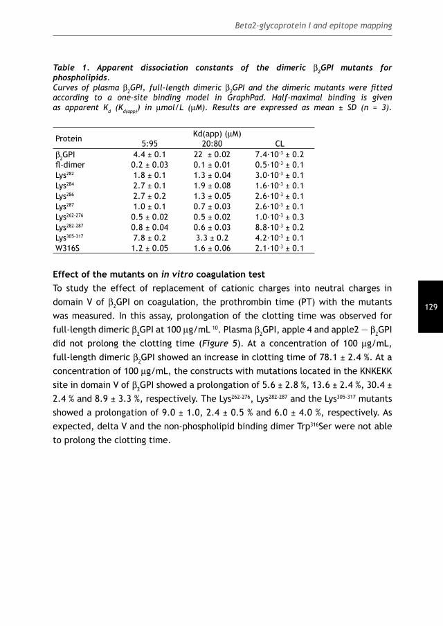
129
Beta2-glycoprotein I and epitope mapping
Table 1. Apparent dissociation constants of the dimeric β2GPI mutants for phospholipids.Curves of plasma β2GPI, full-length dimeric β2GPI and the dimeric mutants were fi tted according to a one-site binding model in GraphPad. Half-maximal binding is given as apparent Kd (Kd(app)) in μmol/L (μM). Results are expressed as mean ± SD (n = 3).
Protein Kd(app) (μM)5:95 20:80 CL
β2GPI 4.4 ± 0.1 22 ± 0.02 7.4·10-3 ± 0.2fl -dimer 0.2 ± 0.03 0.1 ± 0.01 0.5·10-3 ± 0.1Lys282 1.8 ± 0.1 1.3 ± 0.04 3.0·10-3 ± 0.1Lys284 2.7 ± 0.1 1.9 ± 0.08 1.6·10-3 ± 0.1Lys286 2.7 ± 0.2 1.3 ± 0.05 2.6·10-3 ± 0.1Lys287 1.0 ± 0.1 0.7 ± 0.03 2.6·10-3 ± 0.1Lys262-276 0.5 ± 0.02 0.5 ± 0.02 1.0·10-3 ± 0.3Lys282-287 0.8 ± 0.04 0.6 ± 0.03 8.8·10-3 ± 0.2Lys305-317 7.8 ± 0.2 3.3 ± 0.2 4.2·10-3 ± 0.1W316S 1.2 ± 0.05 1.6 ± 0.06 2.1·10-3 ± 0.1
Effect of the mutants on in vitro coagulation testTo study the effect of replacement of cationic charges into neutral charges in domain V of β2GPI on coagulation, the prothrombin time (PT) with the mutants was measured. In this assay, prolongation of the clotting time was observed for full-length dimeric β2GPI at 100 μg/mL 10. Plasma β2GPI, apple 4 and apple2 — β2GPI did not prolong the clotting time (Figure 5). At a concentration of 100 μg/mL, full-length dimeric β2GPI showed an increase in clotting time of 78.1 ± 2.4 %. At a concentration of 100 μg/mL, the constructs with mutations located in the KNKEKK site in domain V of β2GPI showed a prolongation of 5.6 ± 2.8 %, 13.6 ± 2.4 %, 30.4 ± 2.4 % and 8.9 ± 3.3 %, respectively. The Lys262-276, Lys282-287 and the Lys305-317 mutants showed a prolongation of 9.0 ± 1.0, 2.4 ± 0.5 % and 6.0 ± 4.0 %, respectively. As expected, delta V and the non-phospholipid binding dimer Trp316Ser were not able to prolong the clotting time.

130
Chapter 5
Buf
fer
Dimer
W31
6S 282
Lys
284
Lys
286
Lys
287
Lys 26
2-27
6
Lys
282-
287
Lys
305-
317
Lys
0
50
100
150
200
Clo
ttin
g tim
e (
%)
Figure 5. Effect of dimeric β2GPI mutants on the prothrombin time (PT).Buffer, full-length dimeric β2GPI and the dimeric mutants were 1:1 diluted with normal pooled plasma (fi nal protein concentration: 100 μg/mL) followed by measurement of the PT. Clotting time with buffer was set at 100%. Results represent mean clotting time ± SD in percentage (n = 3). Statistical analysis was performed using the Student T-test (p ≤ 0.001). Differences between the pointmutations are not signifi cant.
Interaction between dimeric β2GPI mutants and apoER2’Measurement of binding of the mutants Lys262-276, Lys282-287 and Lys305-317 to apoER2’ was performed in an ELISA. As shown in Figure 6, Panel A, both the Lys282-287 and the Lys305-317 mutant showed impaired binding to apoER2’ with residual binding of 15.5 ± 1.5 % and 53.5 ± 4.5 %, respectively. The Lys262-276 mutant showed less impaired binding to apoER2’ (residual binding 82 ± 10 %). The signifi cance of lysine residues within the KNKEKK site was further investigated using mutants with single Lys to Leu substitutions at position 282-287 in domain V of β2GPI. In Figure 6, panel B, a representative sensorgram is shown, obtained from SPR analysis. Full-length dimeric β2GPI (100 nM) associated with immobilized apoER2’. Upon replacement of full-length dimeric β2GPI with buffer, the response started to decline gradually indicating that the protein dissociated from apoER2’ and binding is reversible. Delta V (100 nM) hardly bound to apoER2’. As representative for the mutants, the sensorgram for Lys282-287 (100 nM) is illustrated. The interaction of the KNKEKK mutants with apoER2’ was studied in more detail by calculating the steady state constants. The binding isotherms of the responses obtained at equilibrium are shown in Figure 6, Panel C. Full-length dimeric β2GPI assembled effi ciently with immobilized apoER2’, whereas delta V displayed only minor association with the receptor. The dimeric β2GPI mutants tested showed a clearly impaired association with apoER2’. However, there is no signifi cant difference between the Lys282, Lys284, Lys286, Lys287 and the Lys282-287 mutants (residual binding 37.8 ± 3.1 %, 47.7 ± 5.2 % 47.7 ± 1.6 %, 48.4 ± 7.9 % and 38.7 ± 8.6 binding, respectively) (Panel D).

131
Beta2-glycoprotein I and epitope mapping
After Scatchard plot analysis and careful interpretation of the correctness of fi t from the binding isotherms derived from SPS analysis, the interaction between the dimeric mutants and apoER2’ showed one class of binding sites. Consequently, one affi nity constant could be derived from the data for each mutant. The derived affi nity constants are summarized in Table 2. The three positively charged groups (Lys262-276, Lys282-287 and Lys305-317) in domain V of β2GPI contributed to apoER2’- binding in the order of position 282-287 > 305-317 > 262-276. All together, these data clearly indicate that the interaction between β2GPI and apoER2’ is due to a combined recognition site in the region Lys282-287 and Lys305-317, but with the most important cationic patch at position Lys282-287 within domain V of β2GPI.
0 100 200 300 400 5000
25
50
75
100
β2GPI (nM)
Res
pons
e(R
U)
C
Dimer 28
2
Lys284
Lys
286
Lys28
7
Lys 282-2
87
Lys Delt
aV
0
25
50
75
100
Res
idua
lbin
din
g(%
)
D
A
Dimer
262-
276
Lys 28
2 -287
Lys 30
5 -317
Lys
0
25
50
75
100
Res
idua
lbin
ding
(%)
0 50 100 150 2000
10
20
30
40
50
60
Time (s)
Res
pons
e(R
U)
B
Figure 6. Binding of dimeric β2GPI mutants to immobilized apoER2’.SPR analysis and ELISA measured binding of full-length dimeric β2GPI and the dimeric mutants. A, binding of the dimeric mutants Lys262-276, Lys282-287 and Lys305-317 (3 μg/mL) to apoER2’ (5 μg/mL) measured by ELISA relative to binding of full-length dimeric β2GPI to apoER2’ (set at 100%). B, a reproducible sensorgram showing the association phase and dissociation phase of full-length dimeric β2GPI (—), Lys282-287 (---) and delta V (·····). C, saturation curves derived from SPR analysis showing the dose-dependent interaction between full-length dimeric β2GPI (■), Lys282 (▼), Lys284 (▲), Lys286 (•), Lys287 (+) and Lys282-287 (◊). D, apoER2’-binding of the mutants with pointmutations in the KNKEKK region, as measured by SPR analysis. Expression is relative to binding of full-length dimeric β2GPI to apoER2’ (set at 100%).

132
Chapter 5
Protein Kd (nM)fl -dimer 50 ± 14Lys282 450 ± 40Lys284 280 ± 20Lys286 320 ± 25Lys287 320 ± 25Lys262-276 NMLys282-287 800 ± 180Lys305-317 240 ± 50Delta V > 2⋅10-3
DiscussionPreviously, we have shown that apoER2’ recognizes a cationic region in domain V of β2GPI 15 and that the same domain is crucial for recognition by anionic phospholipids (J Throm Haem, 2006, accepted for publication). In the present study, we have addressed the functional role of basic residues in domain V of β2GPI for recognition by apoER2’ and anionic phospholipids. To investigate the specifi c lysine residues within domain V of β2GPI critical for binding to apoER2’ and to anionic phospholipids, site-directed mutagenesis was performed with the three-dimensional model of the fi fth domain of β2GPI as lead. Three potential cationic patches were predicted to be involved in the interaction with apoER2’ and subsequently the following dimeric mutants of β2GPI were generated in which a single or several lysines in a particular area in domain V were substituted into a leucine: Lys282, Lys284, Lys286, Lys287, Lys262-276 (a quatriple mutant), Lys282-287 (a quatriple mutant) and Lys305-317 (a triple mutant). The results of our studies are summarized in Table 3. Of cationic regions, Lys282-287 proved most important for the interaction with apoER2’ (Kd approximately 600 nM). A second cluster of lysine residues that is important for recognition by apoER2’ is located at Lys305-317, whereas Lys262-276 is hardly involved in the interaction with apoER2’.Phosphatidylcholine (PS) is one of the most important anionic phospholipids (comprises 8-15 mol% of the total phospholipid content of cellular membranes) and is thought to be involved in binding β2GPI to cellular surfaces. In quiescent cells, PS is located at the cytoplasmic side of the plasma membrane. After activation, PS is exposed at the outer surface of cells. Several studies have investigated binding of β2GPI to cardiolipin. Studies performed by other groups identifi ed residues Lys284, Lys286, and Lys287 in the fi fth domain of β2GPI important for binding to cardiolipin (CL) 6;16;17. So far, studies investigating the binding site within β2GPI for PS surfaces, which are physiologically more important than CL, have not been conducted.
Table 2. Steady state constants of the dimeric β2GPI mutants for apoER2’.Binding isotherms were fi tted accordingly to a one-site binding model. Steady state constants are expressed in nmol/L (nM). Data represent mean ± SD (n = 3). NM = not measured.

133
Beta2-glycoprotein I and epitope mapping
Based on the hypothesis that domain V carries the lipid-binding region within the sequence motif C281KNKEKKC288, we investigated the interaction between β2GPI and anionic phospholipids (PS and CL) more closely using the dimeric mutants. The PS-binding site within domain V of β2GPI is predominantly located at position Lys305-317, whereas for cardiolipin the residue Lys282-287 is more important. This observation was independent of the PS content tested in the binding assays. Our data are an addition on what is already known for binding of β2GPI to anionic phospholipids. Regarding the binding of β2GPI to cardiolipin, another group identifi ed a crucial role for a hydrophobic region at position 313-316 in domain V of β2GPI. This study also used recombinant β2GPI constructed by site-directed mutagenesis 18. However, the latter study only used recombinant β2GPI with mutations at position 306 and 316 and did not test other regions in domain V of β2GPI. The observed differences between PS-binding and CL-binding can be explained by the physical properties of both lipids. PS/PC forms unilamellar vesicles, whereas CL forms bilayers. The differences in lipid properties might explain the observation that various anionic phospholipids recognize different regions within domain V of β2GPI.A cluster of lysine residues at position C281KNKEKKC288 supports binding of β2GPI to endothelial cells 19. Our group has identifi ed several members of the LDL-R family on endothelial cells (unpublished data). We suggest that the cationic patch at position Lys282-287 might be involved in the interaction between β2GPI and LDL-R homologous on endothelial cells. However, the role of the cationic patch Lys282-287 for other members of the LDL-R family still needs to be elucidated. Different hypotheses for the interaction of β2GPI with membranes are discussed in the literature. One very interesting aspect outlines that the targets of β2GPI are cell surface receptor proteins rather than lipids 20. The underlying mechanisms for such a β2GPI-protein interaction might be quite different from the association with anionic phospholipids and certainly require further structural characterization. Some studies stress that binding of β2GPI to anionic phospholipids alters the conformation of the protein, thus exposing neo-epitopes 21;22. Based on several lines of evidence, β2GPI seems to be anchored in the lipid membrane via domain V. After a structural rearrangement of the protein, other regions in domain V might be exposed and can exert other biological functions, such as interacting with cellular receptors. An important feature of (anionic) phospholipid bilayers, present in cellular membranes, is that they are cooperative structures. They are held together by many reinforcing, covalent interactions. The β2GPI-binding properties of apoER2’ are dependent on interactions between different sites within domain V of β2GPI, with emphasis on Lys282-287, whereas Lys303-317 is crucial for β2GPI-binding to

134
Chapter 5
PS. These observations suggest that β2GPI-binding to PS cooperates β2GPI-binding to apoER2’.In conclusion, the interaction between i. β2GPI and ii. anionic phospholipids and apoER2’ involves different non-overlapping binding sites within domain V of β2GPI. Interestingly, all residues responsible for binding to apoER2’ and binding to anionic phospholipids are located at the aberrant non-SCR half of domain V of β2GPI.
Table 3. Binding characteristics of the dimeric β2GPI mutantsBinding characteristics of the dimeric mutants were compared with that of full-length dimeric β2GPI and assigned a plus mark according to the strength of the interaction.
Protein PT PS CL apoER2’fl -dimer + + + + + + + + + + +Lys282 - + + + +Lys284 - + + + + +Lys286 - + + + +Lys287 - + + + +Lys262-276 - + + + + + + + +Lys282-287 - + + + +Lys305-317 - - + + + +Delta V - - + + -
Acknowledgement: This study was supported by a grant from the Netherlands Organization for Health Research and Development (Zon-MW: 902-26-290).

135
Beta2-glycoprotein I and epitope mapping
References
1. Bouma B, De Groot PhG, van den Elsen JMH et al. Adhesion mechanism of human
β2-glycoprotein I to phospholipids based on its crystal structure. EMBO J.
1999;18:5166-5174.
2. Schwarzenbacher R, Zeth K, Diederichs K et al. Crystal structure of human b2-
glycoprotein I: implication for phospholipid binding and the antiphospholipid
syndrome. The EMBO journal 1999;18:6228-6239.
3. Hammel M, Kriechbaum M, Gries A et al. Solution structure of human and bovine
beta(2)-glycoprotein I revealed by small-angle X-ray scattering. J.Mol.Biol.
2002;321:85-97.
4. Hammel M, Schwarzenbacher R, Gries A et al. Mechanism of the interaction of
beta(2)-glycoprotein I with negatively charged phospholipid membranes. Biochemistry
2001;40:14173-14181.
5. Hong DP, Hagihara Y, Kato H, Goto Y. Flexible loop of beta 2-glycoprotein I domain V
specifi cally interacts with hydrophobic ligands. Biochemistry 2001;40:8092-8100.
6. Sheng YH, Sali A, Herzog H, Lahnstein J, Krilis SA. Site-directed mutagenesis of
recombinant human β2-glycoprotein I identifi es a cluster of lysine residues that
are critical for phospholipid binding and anti-cardiolipin antibody activity. J.Immunol.
1996;157:3744-3751.
7. Hagihara Y, Goto Y, Kato H, Yoshimura T. Role of the N- and C-terminal domains
of bovine β2- glycoprotein I in its interaction with cardiolipin. J.Biochem.(Tokyo)
1995;118:129-136.
8. Riddell DR, Vinogradov DV, Stannard AK, Chadwick N, Owen JS. Identifi cation and
characterization of LRP8 (apoER2) in human blood platelets. Journal of Lipid
Research 1999;40:1925-1930.
9. Korporaal SJ, Relou IA, van EM et al. Binding of low density lipoprotein to platelet
apolipoprotein E receptor 2’ results in phosphorylation of p38MAPK. J.Biol.Chem.
2004;279:52526-52534.
10. Lutters BC, Derksen RH, Tekelenburg WL et al. Dimers of beta 2-glycoprotein I increase
platelet deposition to collagen via interaction with phospholipids and the
apolipoprotein E receptor 2’. J.Biol.Chem. 2003;278:33831-33838.
11. Zaiou M, Arnold KS, Newhouse YM et al. Apolipoprotein E;-low density lipoprotein
receptor interaction. Infl uences of basic residue and amphipathic alpha-helix
organization in the ligand. J.Lipid Res. 2000;41:1087-1095.
12. Brunner J, Skrabal P, Hauser H. Single bilayer vesicles prepared without sonication.
Physio-chemical properties. Biochim.Biophys.Acta 1976;455:322-331.
13. Van Wijnen M, Stam JG, Van’t Veer C et al. The interaction of protein S with the
phospholipid surface is essential for the activated protein C-independent activity of
protein S. Thromb.Haemost. 1996;76:397-403.

136
Chapter 5
14. Rouser G, Fkeischer S, Yamamoto A. Two dimensional thin layer chromatography
separation of polar lipids and determination of phospholipids by phosphorus analysis
of spots. Lipids 1970;5:494-496.
15. van Lummel M, Pennings MT, Derksen RH et al. The binding site in {beta}2-glycoprotein
I for ApoER2’ on platelets is located in domain V. J.Biol.Chem. 2005;280:36729-
36736.
16. Hunt J, Krilis S. The fi fth domain of b2-glycoprotein I contains a phospholipid binding
site (Cys281-Cys288) and a region recognized by anticardiolipin antibodies.
J.Immunol. 1994;152:653-659.
17. Sheng Y, Krilis SA, Sali A. Site-directed mutagenesis of recombinant human β2-
glycoprotein I - Effect of phospholipid binding and anticardiolipin antibody activity.
Ann.NY Acad.Sci. 1997;815:331-333.
18. Mehdi H, Naqvi A, Kamboh MI. A hydrophobic sequence at position 313-316 (Leu-
Ala-Phe-Trp) in the fi fth domain of apolipoprotein H (β2-glycoprotein I) is crucial for
cardiolipin binding. European Journal of Biochemistry 2000;267:1770-1776.
19. Del Papa N, Sheng YH, Raschi E et al. Human β2-glycoprotein I binds to endothelial
cells through a cluster of lysine residues that are critical for anionic phospholipid
binding and offers epitopes for anti-β2-glycoprotein I antibodies. The journal of
immunology 1998;160:5572-5578.
20. Ma K, Simantov R, Zhang JC et al. High affi nity binding of beta 2-glycoprotein I to
human endothelial cells is mediated by annexin II. J Biol Chem. 2000;275:15541-
15548.
21. Matsuura E, Inagaki J, Kasahara H et al. Proteolytic cleavage of β2-glycoprotein
I: reduction of antigenicity and the structural relationship. Int.Immunol.
2000;12:1183-1192.
22. de Laat B, Derksen RH, van Lummel M, Pennings MT, De Groot PG. Pathogenic anti-
beta2-glycoprotein I antibodies recognize domain I of beta2-glycoprotein I only after
a conformational change. Blood. 2006;107:1916-1924.

Bas de Laat, Ronald H.W.M. Derksen, Menno van Lummel,
Maarten T.T. Pennings, and Philip G. de Groot
Blood 2006 Mar 1; 107 (5): 1916-24
6PATHOGENIC ANTI-BETA2-GLYCOPROTEIN
I ANTIBODIES RECOGNIZE DOMAIN I OF BETA2- GLYCOPROTEIN I ONLY AFTER A
CONFORMATIONAL CHANGE

139
Beta2-glycoprotein I and conformation
AbstractRecently, we published the existence of two populations of anti-β2-glycoprotein I (β2GPI) IgG antibodies. Type A antibodies recognize epitope G40-R43 in domain I of β2GPI and are strongly associated with thrombosis. Type B antibodies recognize other parts of β2GPI and are not associated with thrombosis. In this study, we demonstrate that type A antibodies only recognize plasma-purifi ed β2GPI when coated onto a negatively-charged surface, and not when coated onto a neutrally-charged surface. The affi nity of type B antibodies towards plasma-purifi ed β2GPI was independent of the charge of the surface to which β2GPI was coated. Type A antibodies did not recognize plasma-purifi ed β2GPI in solution, whereas they did recognize recombinant β2GPI both in solution and coated onto a neutrally-charged plate. When the carbohydrate chains were removed from plasma-purifi ed β2GPI, we found that type A antibodies did recognize the protein in solution. This supports the hypothesis that the difference in recognition of plasma-purifi ed and recombinant β2GPI is caused by the difference in glycosylation and that epitope G40-R43 of plasma-purifi ed β2GPI is covered by a carbohydrate chain. Type A anti-β2GPI antibodies can only recognize this epitope when this carbohydrate chain is displaced due to a conformational change. This fi nding has major implications both for the detection of pathogenic anti-β2GPI antibodies and the comprehension of the pathophysiology of the antiphospholipid syndrome.

140
Chapter 6
IntroductionThe antiphospholipid syndrome is a systemic autoimmune disease that is characterized serologically by the presence of antiphospholipid antibodies in plasma of patients, and clinically by vascular thrombosis and/or recurrent pregnancy morbidity 1,2,3. In 1990 it was shown that these so-called antiphospholipid antibodies do not recognize phospholipids directly, but a phospholipid-binding protein: β2-glycoprotein I (β2GPI) 4. Ever since, antiphospholipid antibodies with affi nity for a large number of other phospholipid-binding proteins have been described. Only antiphospholipid antibodies with affi nity for β2GPI are thought to be clinically relevant 5. Several assays are available to detect anti-β2GPI antibodies. The most common one is the anticardiolipin enzyme-linked immunosorbent assay (ELISA). In this assay, the antibodies recognize β2GPI bound to immobilized cardiolipin 6. A second assay is the lupus anticoagulant assay, which detects anti-β2GPI antibodies with capacity to prolong phospholipid-dependent coagulation assays (LAC) 7. A third method is an ELISA-based method in which antibodies recognize β2GPI coated directly onto ELISA plates 3.β2GPI (formerly known as apolipoprotein H) is a protein of 44 kDa with a plasma concentration of approximately 150 μg/ml. β2GPI is highly glycosylated as it contains 4 N-linked carbohydrate side-chains. Together these carbohydrate side-chains account for approximately 15% (w/w) of the total molecular mass of β2GPI. The protein β2GPI consists of 326 amino acids organized in 5 complement control protein domains 8,9. Each domain consists of 60 amino acids, except for domain V. Domain V consists of 82 amino acids, caused by a C-terminal extension of 19 amino acids and an insertion of 6 amino acids forming a hydrophobic loop. This specifi c structure of domain V is responsible for the binding properties of β2GPI to anionic phospholipids 8,9.Although the three-dimensional structure of β2GPI is known for over 5 years, the mechanism by which anti-β2GPI antibodies recognize β2GPI is unclear 9,10. Two main theories have been proposed to explain the binding of antiphospholipid antibodies to β2GPI. The fi rst and most accepted hypothesis is known as the “dimerization theory”; one antibody must bind two β2GPI molecules to obtain considerable avidity 11-14. To achieve this, a high density of β2GPI on an ELISA plate is essential. It has been reported that the coated amount of β2GPI must exceed a certain threshold before antiphospholipid antibodies are able to bind 15. The studies in favor of the “dimerization theory” seem rather convincing but several observations remain unexplained, and are in favor of a second hypothesis. This hypothesis is based on the recognition of a cryptic epitope by antiphospholipid antibodies. This epitope is

141
Beta2-glycoprotein I and conformation
only exposed after binding of β2GPI to a negatively charged surface 16-18. Supporting this latter hypothesis is the fact that the structure of β2GPI in solution differs from that of crystallized β2GPI 9,10,19. Moreover, no β2GPI-antibody complexes can be detected in the circulation of patients with the antiphospholipid syndrome (unpublished observations). Furthermore, it was shown by circular dichroism measurements that cardiolipin can induce a conformational change in β2GPI 16. We have recently published that the population of anti-β2GPI antibodies recognizing epitope G40-R43 (type A antibodies) cause LAC and strongly correlate with thrombosis (odds ratio 18.9, 95% confi dence interval: 53.2 - 6.8). The other group of anti-β2GPI antibodies (type B antibodies) recognized other parts of β2GPI and did not correlate with thrombosis (1.1, 95% confi dence interval: 2.8 – 0.4). In this study we have further investigated the epitope(s) on β2GPI recognized by type A and type B anti-β2GPI antibodies.
Experimental Procedures
Patients52 patient plasmas positive for anti-β2GPI IgG antibodies were included in this study; 36 patients with systemic lupus erythematosus (SLE), 11 patients with lupus like disease (LLD) and 5 patients with primary antiphospholipid syndrome. Patients with SLE meet at least 4 ACR (American College of Rheumatology) criteria for the classifi cation of SLE and patients with LLD 1 to 3 of these criteria 20. Patients with primary antiphospholipid syndrome have antiphospholipid antibodies (LAC and/or anticardiolipin antibodies) and a history of thrombosis in the absence of any sign of a systemic autoimmune disease. By chart review, the number of objectively verifi ed thromboembolic events was recorded for each patient 5. For the diagnosis of thrombosis of intracerebral vessels, computed tomographic scanning or magnetic resonance imaging was used. Myocardial infarction was diagnosed by typical electrocardiographic features and an elevated fraction creatine kinase myoglobin (CK-MB). Peripheral arterial thrombosis and thrombosis of the distal aorta was diagnosed by arteriography. Retinal thrombosis was documented by funduscopy and fl uorescence angiography. Deep vein thrombosis (DVT) was diagnosed by ultrasonography or venography, pulmonary embolism by radionuclide lung scanning. Portal vein thrombosis was diagnosed by angiography. A patient was diagnosed with superfi cial thrombophlebitis if the manifestation was diagnosed clinically. Blood was drawn from these patients at an arbitrary visit at the lupus clinic of the University Medical Center Utrecht, Utrecht, the Netherlands. The

142
Chapter 6
Institutional Review Board of the University Medical Center Utrecht approved this study, and informed consent was obtained from all patients.
Serological assays
Anti-β2GPI IgG antibody ELISA Antibodies against β2GPI were measured in an ELISA as described before (inter-assay variability 11.5%) 21. In short, plasma-purifi ed β2GPI (10 μg/ml diluted in a Tris based solution (TBS): 50 mM Tris in 100 mM NaCl) was incubated onto hydrophilic ELISA plates (Costar cat. nr. 9102, New York, USA). After one hour, the plates were incubated with 4% bovine serum albumin (BSA) in TBS for one hour. This was followed by incubation with patient plasma (1:50 diluted in blocking solution) for 1 hour. Then the plates were incubated with a goat-anti-human IgG alkaline-phosphatase-labeled antibody (diluted 1:1000, Biosource, Camarillo USA) followed by staining with para-nitrophenyl phosphatase (PnPP, 0.6 mg/ml diluted in diethynolamine (DEA) buffer, Sigma, St. Lious, MO, USA). Plasma samples were regarded positive when the absorbed value exceeded the cut-off value (mean +3SD of 40 healthy volunteers) and corrected for the absorption of standard positive plasma.
LAC To determine LAC activity, an APTT and a Dilute Russell Viper Venom Time (DRVVT) were performed 5. For the APTT (PTT-LA, inter-assay variability 2.0%, Diagnostica Stago, Gennevilliers, France), 50 μl patient plasma of patients was diluted 1:1 with normal pool plasma of 40 healthy volunteers and incubated with 50 μl APTT reagent. Coagulation was initiated by the addition of 50 μl CaCl2 (25 mM). As a control, samples were tested in an actin-FS based APTT (APTT-FS, Dade Behring, Marburg, Germany) which is a LAC insensitive assay. Patients were considered positive when the ratio PTT-LA/APTT-FS was greater than 1.20 (10). The DRVVT was performed according to the instructions of the manufacturers (inter-assay variability 3.2%, Gradipore Ltd, North Ryde, Australia) and considered positive when LAC screen/LAC confi rm > 1.20. A patient was considered LAC positive if one of the two LAC assays was positive.
Anticardiolipin antibody ELISAAnticardiolipin antibodies were measured in an ELISA as described before (inter-assay variability 6.9%) 22. Nine IgG/IgM calibrators were used to report

143
Beta2-glycoprotein I and conformation
anticardiolipin antibody levels as GPL or MPL units. Levels above 10 GPL or GPM units were considered positive.
Anti-domain I IgG ELISAWe recently described a method in which we were able to discriminate between anti-β2GPI IgG antibodies with reactivity towards epitope G40-R43 in domain I (type A antibodies) and anti-β2GPI IgG antibodies with other reactivity (type B antibodies) 23. In short, hydrophilic plates (Costar cat. nr. 9102, New York, USA) and hydrophobic plates (Costar cat. nr. 2595, New York, USA) were coated with domain I of β2GPI (5 μg/ml diluted in TBS) for 1 hour at 37ºC. After every incubation step, the plates were washed 3 times with 0.1% Tween/TBS. The plates were blocked with 4% BSA/0.1% Tween/TBS and subsequently incubated with patient plasma (diluted 1:100 in blocking solution) containing anti-β2GPI IgG antibodies. The bound IgG antibodies were detected by a goat-anti-human IgG alkaline-phosphatase-labeled antibody (diluted 1:1000, Biosource). This was followed by staining with PnPP (0.6 mg/ml in DEA solution). Anti-β2GPI IgG antibodies were regarded as type A antibodies when these showed decreased affi nity for domain I coated onto hydrophilic plates compared to that with domain I coated onto hydrophobic plates. Anti-β2GPI IgG antibodies were regarded as type B antibodies when they showed equal affi nity for domain I on both plates. A ratio of greater than 2 between the OD measured with the hydrophobic plate and the OD with the hydrophilic plate discriminates between type A and type B anti-β2GPI IgG antibodies. This ratio is an indication for the relative amount of anti-β2GPI IgG antibodies that recognize the positive charge (epitope G40-R43) on domain I (inter-assay variability 3.8%).
Purifi cation of β2GPI from plasma Human plasma was dialyzed against a solution containing 0.04 M Tris, 0.01 M succinate, 0.005% polybrene, 1 mM EDTA, 1mM benzamidin, 43 mM NaCl and 0.02% NaN3. Then, the plasma was added to DEAE-Sephadex column, and the fl ow-through was collected, pooled and added to a protein-G-Sepharose column. Then the effl uent pool was added to a mono-S Sepharose column. The bound β2GPI was eluted by a linear salt-gradient (138 mM NaCl – 550 mM NaCl), and checked for purity by SDS-PAGE 24. The purifi ed β2GPI was analyzed with gel fi ltration and showed a single peak at the expected molecular weight.
Recombinant β2GPI, and domain deleted mutants of β2GPIRecombinant full-length β2GPI (DI-V) and 8 domain deletion mutants of β2GPI
144
Chapter 6
(comprising domain I (DI), domains I and II (DI-II), domains I, II and III (DI-III), domains I, II, III, and IV (DI-IV), domains II, III, IV and V (DII-V), domains III, IV and V (III-V), domains IV and V (DIV-V) and domain V (DV)) were obtained by a generous gift from Dr. Iverson of La Jolla Pharmaceutical Company 25. The recombinant full-length β2GPI was analyzed with gel fi ltration and showed a single peak at the expected molecular weight. On SDS-PAGE electrophoresis, the molecular weight of recombinant β2GPI was slightly lower than the molecular weight of plasma-purifi ed β2GPI.
Purifi cation of immunoglobulin G (IgG) antibodies from plasma samples Blood samples were collected by venipuncture using plastic tubes containing 3.8% trisodium citrate (0.129 M) as the anticoagulant (9:1, v/v). To obtain platelet poor plasma the samples were centrifuged twice at 2000 g for 10 minutes, and subsequently stored at -50ºC until further use. The IgG fraction was purifi ed by the use of a protein G-Sepharose column. By adding the IgG fraction to a column coated with β2GPI, anti-β2GPI antibodies were purifi ed. The purity of the (affi nity-) purifi ed IgG fractions was checked by using SDS-PAGE.
Binding studies with β2GPI coated onto hydrophobic plates or hydrophilic platesHydrophilic plates (Costar cat. nr. 9102, New York, USA) and hydrophobic plates (Costar cat. nr. 2595, New York, USA) were incubated with 50 μl plasma-purifi ed β2GPI or recombinant β2GPI (10 μg/ml in TBS) for one hour at 37ºC. Subsequently, the plates were washed three times with washing solution (TBS/0.1% Tween) and blocked with 150 μl blocking solution (4% BSA)/TBS/0.1% Tween) for one hour at 37ºC. The plates were washed 3 times, and incubated with either patient plasma (diluted 1:50), patient IgG antibodies (type A and type B), monoclonal antibody 4F3, monoclonal antibody 2B2 or monoclonal antibody 27G7 in various concentrations (diluted in blocking buffer), for one hour at 37ºC 26,27. Monoclonal antibodies 2B2 and 27G7 were kindly provided by Dr. Tincani and Dr. J. Arnout, respectively. To detect the bound monoclonal antibodies, the plates were washed and incubated with 50 μl rabbit-anti-mouse peroxidase-labeled antibody (diluted 1:1000, Dako, Glostrup, Denmark), followed by staining with an ortho-phenylene diamine solution (OPD solution; 4 mg/ml OPD diluted in 0.1 M NaH2PO4.2H2O/0.1 M Na2HPO4.H2O). The coloring reaction was stopped with the addition of 1 M H2SO4, and absorbance was measured at 490 nm. To detect the bound patient IgG antibodies, the plates were incubated with an alkalinephosphatase-labeled goat-anti-human IgG antibody

145
Beta2-glycoprotein I and conformation
(diluted 1:1000, Biosource, Camarillo USA). PnPP was used as coloring substance. The reaction was stopped by the addition of 2.4 M NaOH, and absorbance was measured at 405 nm.
Binding of patient-derived IgG antibodies to recombinant and plasma-purifi ed β2GPI in solutionHydrophilic ELISA plates (Costar cat. nr. 9102) were coated with 10 μg/ml plasma-purifi ed β2GPI, for 1 hour at 37ºC. The plates were washed 3 times with washing solution (TBS/0.1% Tween) and blocked with 150 μl blocking solution (4% BSA/TBS/0.1% Tween) for one hour at 37ºC. Then, the patient-derived IgG type A antibodies (total IgG fraction) were incubated to the plate in the presence of various concentrations of plasma-purifi ed β2GPI and recombinant β2GPI. The patient-derived IgG antibodies bound to the plate were detected by using an alkaline-phosphatase-labeled goat-anti-human IgG antibody (diluted 1:1000, Biosource, Camarillo USA). PnPP (0.6 mg/ml diluted in DEA buffer) was used as coloring substance. The reaction was stopped by the addition of 2.4 M NaOH, and absorbance was measured at 405 nm.
Recognition of plasma-purifi ed β2GPI in solution in the presence of cardiolipin vesicles and phosphatidylserine/phosphatidylcholine (PS/PC) vesicles Affi nity-purifi ed patient IgG type A antibodies (20 μg/ml) of patient 1 and patient 2, were diluted in 150 mM Na2CO3.10H2O/350 mM NaHCO3 (pH 9.6) to reach a fi nal concentration of 20 μg/ml, and were coated onto hydrophobic ELISA plates (Costar cat. nr. 2595) for 12 hours at 4ºC. The plates were washed three times with TBS/0.1% Tween and subsequently blocked with 4% BSA/TBS/0.1% at 37ºC. After 1 hour the plates were washed 3 times, and incubated with plasma-purifi ed β2GPI (10 μg/ml), plasma-purifi ed β2GPI (10 μg/ml) in the presence of cardiolipin vesicles (20 μM), or plasma β2GPI (10 μg/ml) in the presence of PS/PC vesicles (20%/80%, 20 μM), all diluted in blocking solution. The cardiolipin and PS/PC vesicles were prepared as described before 7,28. Then the plates were washed and incubated with a polyclonal goat-anti-human anti-β2GPI antibody (10 μg/ml). After washing, the plates were incubated with a rabbit-anti-goat peroxidase-labeled antibody (diluted 1:1000, Biosource). Coloring was performed by using OPD. The coloring reaction was stopped by the addition of 1 M H2SO4, and absorbance was measured at 490 nm.

146
Chapter 6
The infl uence of aggregating plasma-purifi ed β2GPI on the recognition by type A antibodiesTo aggregate plasma-purifi ed β2GPI, the protein was incubated with 1% glutaraldehyde for 15 minutes at room temperature. Then, the solution was dialyzed against TBS and analyzed by using SDS-PAGE. The aggregated β2GPI, plasma-purifi ed β2GPI and recombinant β2GPI were coated onto a hydrophobic ELISA plate (Costar cat. nr. 2595), at a concentration of 10 ug/ml for one hour at 37ºC. Then, the plates were blocked with 4% BSA/TBS/0.1% Tween. After washing the plates, they were incubated with 2 type A plasma samples (1:100 diluted in blocking solution) for one hour at 37ºC. The bound IgG antibodies were detected with an alkaline-phosphatase-labeled goat-anti-human IgG antibody (diluted 1:1000, Biosource) and coloring was performed by using PnPP. The coloring reaction was stopped by the addition of 2.4 M NaOH, and absorbance was measured at 405 nm. For the inhibition ELISA we coated 10 ug/ml plasma-purifi ed β2GPI to a hydrophilic ELISA plate (Costar cat. nr. 2595). The plates were washed three times with TBS/0.1% Tween and subsequently blocked with 4% BSA/TBS/0.1% at 37ºC. After 1 hour the plates were washed 3 times, and incubated with 10 μg/ml anti-β2GPI antibodies of patient 1 (type A) in the presence of either 100 μg/ml plasma-purifi ed β2GPI, 100 μg/ml recombinant β2GPI or 100 μg/ml glutaraldehyde-treated β2GPI. The antibodies bound the coated β2GPI were detected with an alkaline-phosphatase-labeled goat-anti-human IgG antibody (diluted 1:1000, Biosource) and coloring was performed by using PnPP. The coloring reaction was stopped by the addition of 2.4 M NaOH, and absorbance was measured at 405 nm.
Removal of the carbohydrate side-chains of plasma-purifi ed β2GPIβ2GPI was deglycosylated by incubating 50 μl sample (β2GPI, 1.1 mg/ml diluted in TBS) with 5 units N-Glycosidase F (Roche, Mannheim, Germany) for 24 hours at 37ºC. The extent of deglycosylated β2GPI was analyzed by using SDS-PAGE. The deglycosylated β2GPI was analyzed with gel fi ltration and showed a single peak at the expected molecular weight.
Production of F(ab) fragments of monoclonal antibody 4F3First, monoclonal antibody 4F3 was dialyzed against 100 mM sodiumacetate (pH 6.2). Then cysteïn, EDTA, sodiumazide were added to the antibody solution to obtain a fi nal concentration of subsequently 50 mM, 1mM and 0.05%. This was followed by the addition of papaïn (10 μg papaïn per 1 mg antibody) (Sigma, P3125). After incubating the solution for 8 hours at 37ºC, iodoacetamide was added
147
Beta2-glycoprotein I and conformation
(fi nal concentration: 75 mM). By using a protein G coupled sepharose column the Fc parts of the antibodies were removed from the solution and the purity of the F(ab) fragments was analyzed by using SDS-PAGE.
Binding of patient IgG to plasma-purifi ed β2GPI, deglycosylated β2GPI and recombinant β2GPI in solutionAffi nity-purifi ed patient IgG type A antibodies (20 μg/ml) of patient 1, patient 2, monoclonal antibody 4F3 (3 μg/ml), F(ab) fragments of monoclonal antibody 4F3 (15 μg/ml) and monoclonal antibody 2B2 (3 μg/ml) were diluted in 150 mM Na2CO3.10H2O/350 mM NaHCO3 (pH 9.6) and coated onto hydrophobic ELISA plates (Costar cat. nr. 2595), for 12 hours at 4ºC. The plates were washed 3 times with TBS/0.1% Tween and subsequently blocked with 4% BSA/TBS/0.1% at 37ºC. After 1 hour the plates were washed 3 times, and incubated with different concentrations of either plasma-purifi ed β2GPI, deglycosylated plasma-purifi ed β2GPI or recombinant β2GPI, all diluted in blocking solution. Then the plates were washed and incubated with a polyclonal goat-anti-human anti-β2GPI antibody. After washing, the plates were incubated with a rabbit-anti-goat peroxidase-labeled antibody (diluted 1:1000). Coloring was performed by using OPD. The coloring reaction was stopped by the addition of 1 M H2SO4, and absorbance was measured at 490 nm.
Domain specifi city of patient IgG antibodies and monoclonal antibodies 4F3 and 2B2The reactivity of antibodies against β2GPI domain deletion mutants was tested by coating 96-well hydrophobic ELISA plates (Costar cat. nr. 2595) with deletion mutants in a concentration of 10 μg/ml, for 1 hour at 37ºC. The plates were washed 4 times with TBS/0.1% Tween and subsequently blocked with a 4% BSA/TBS solution. Patient IgG fractions (10 μg/ml) and monoclonal antibodies 4F3 and 2B2 (both 3 μg/ml) were diluted in blocking solution, and added to the wells (50μl/well, 1 hour at 37ºC). The plates were washed 4 times (0.1% Tween/TBS), and incubated with alkalic-phosphatase-labeled goat-anti-human IgG antibodies (diluted 1/1000, Biosource) to detect the bound patient IgG antibodies (1 hour at 37ºC). Staining was performed with PnPP (Sigma, St. Louis, MO, USA) at a concentration of 0.6 mg/ml diluted in DEA buffer. The coloring reaction was stopped with 2.4 M NaOH, and absorbance was measured at 405 nm. The bound monoclonal antibodies were detected by peroxidase-labeled rabbit-anti-mouse antibodies, and coloring was performed with an OPD solution. The coloring reaction was stopped by 1 M H2SO4, and absorbance was measured at 490 nm.
148
Chapter 6
Infl uence of increasing NaCl concentration on the recognition of β2GPI by anti-β2GPI IgG antibodiesHydrophilic and hydrophobic plates (Costar cat. nr. 9102; Costar cat. nr. 2595) were incubated with 10 μg/ml plasma-purifi ed β2GPI or recombinant β2GPI for 1 hour at 37ºC. After washing, the plates were blocked with 4% BSA/TBS/0.1%Tween at 37ºC for one hour. Subsequently the plates were washed and incubated with patient plasma diluted (1:100) in blocking solution containing either 150 mM NaCl or 5 M NaCl. After washing, the plates were incubated with a alkaline-phosphatase-labeled goat-anti-human IgG antibody (diluted 1:1000). Coloring was performed by using PnPP. The coloring reaction was stopped by the addition of 2.4 M NaOH, and absorbance was measured at 405 nm.
Results
Serological and clinical characterization of the patient populationThirty (58%) out of 52 plasma samples with anti-β2GPI IgG antibodies tested positive in the anti-domain I IgG ELISA (type A antibodies). Twenty-two (42%) plasma samples tested negative in the anti-domain I IgG ELISA (type B antibodies). All 52 (100%) plasma samples tested positive for anticardiolipin antibodies. Thirty-four (65%) plasma samples showed LAC activity. Of these, 28 (82%) were positive for type A and 6 (18%) for type B antibodies.Twenty-fi ve (83%) out of 30 patients with type A antibodies had a history of thrombosis. Of these, 16 (53%) patients had a history of venous thrombosis and 12 (40%) of arterial thrombosis. Seven (32%) patients with type B antibodies had a history of thrombosis. Of these, 5 (23%) had a history of venous thrombosis and 4 (18%) a history of arterial thrombosis.
The recognition of β2GPI when coated onto different types of platesTo investigate whether different coating conditions infl uence the recognition of β2GPI by type A and type B IgG antibodies, plasma-purifi ed β2GPI was coated onto hydrophilic and hydrophobic ELISA plates. Type A patient plasmas (n=30) recognized plasma-purifi ed β2GPI coated onto a hydrophilic plate much better than when coated onto a hydrophobic plate (median OD 0.262 vs. median OD 0.010, p=0.001). This is a reduction of 96% (OD 0.252). For type B plasmas (n=22) we did not observe a difference in recognition when plasma-purifi ed β2GPI coated onto either a hydrophilic plate or a hydrophobic plate (median OD 0.091 vs. median OD 0.076, no signifi cant difference). This is a reduction of 16% (OD 0.015). For both

149
Beta2-glycoprotein I and conformation
type A and type B plasma, a typical example is shown in fi gure 1A.For total IgG, we performed this type of experiment with 3 total IgG samples of each type of plasma. All type A patients we tested had suffered from venous thrombosis in the past, and 1 of the 3 type B patients had suffered from venous thrombosis. We observed the same pattern as with the plasma samples; Type A IgG antibodies showed a high affi nity for plasma-purifi ed β2GPI coated onto hydrophilic plates, but showed negligible affi nity for plasma-purifi ed β2GPI coated onto hydrophobic plates. Type B IgG antibodies showed equal affi nity for plasma-purifi ed β2GPI coated onto hydrophilic or hydrophobic plates. Both type A and type B IgG antibodies had similar affi nity for recombinant β2GPI coated onto hydrophilic or hydrophobic plates. For both type A and type B IgG a typical example is shown in fi gure 1B.Monoclonal antibody 4F3 showed the same recognition pattern as type A IgG antibodies (Figure 1C). For two other monoclonal antibodies, 2B2 and 27G7, the recognition of β2GPI was independent of the type of plate (Figure 1C). This indicates that both types of ELISA plates bound comparable amounts of plasma-purifi ed β2GPI and recombinant β2GPI.

150
Chapter 6
Figure 1A
Figure 1B
Figure 1C
Figure 1. Patient-derived antibodies (and monoclonal antibody 4F3) do not recognize plasma-purifi ed β2GPI coated onto a hydrophobic plate. Plasma-purifi ed β2GPI and recombinant β2GPI were coated onto hydrophilic and hydrophobic plates. Then, the plates were incubated with either patient plasma (A), patient IgG antibodies (B), monoclonal antibody 4F3, monoclonal antibody 2B2 or monoclonal antibody 27G7 (C). The bound mAbs were detected by a rabbit-anti-mouse peroxidase-labeled antibody, and coloring was performed by OPD. To detect the bound patient IgG antibodies, the plates were incubated with an alkaline-phosphatase-labeled goat-anti-human IgG antibody. PnPP was used as coloring substance. The obtained optical density was corrected for the optical density obtained with total IgG isolated from pooled plasma from 40 healthy volunteers ((A) hydrophilic plate, OD: 0.087; hydrophobic plate, OD: 0.130). Error bars represent mean ± S.E.M. of duplicate points. Ab: antibody, OD: optical density.

151
Beta2-glycoprotein I and conformation
The recognition of β2GPI in solutionTo further investigate the differences in recognition of plasma-purifi ed β2GPI and recombinant β2GPI by type A IgG antibodies, we performed several experiments with β2GPI in solution. We coated plasma-purifi ed β2GPI to a hydrophilic ELISA plate, and studied whether plasma-purifi ed β2GPI and recombinant β2GPI were able to inhibit the binding of patient type A IgG antibodies to the coated plasma-purifi ed β2GPI on the plate. Both type A patients tested in this experiment had suffered from venous thrombosis in the past. Figure 2 shows that the binding to the coated β2GPI could be completely inhibited when patient type A IgG antibodies were pre-incubated with recombinant β2GPI. In contrast, the binding of patient type A IgG antibodies to coated β2GPI could only be inhibited for approximately 25% by plasma-purifi ed β2GPI. To expand this observation, we coated affi nity-purifi ed patient type A IgG antibodies to a hydrophobic ELISA plate. Subsequently, the plate was incubated with plasma-purifi ed β2GPI, plasma-purifi ed β2GPI in the presence of cardiolipin vesicles, and with plasma-purifi ed β2GPI in the presence of PS/PC vesicles. We found that the patient type A IgG antibodies recognized plasma-purifi ed β2GPI only in the presence of cardiolipin vesicles (Figure 3).
Figure 2. Binding of type A IgG antibodies to β2GPI in solution. Hydrophilic ELISA plates were coated with 10 μg/ml plasma-purifi ed β2GPI, and patient type A IgG antibodies were incubated to the plate in the presence of various concentrations of plasma-purifi ed β2GPI and recombinant β2GPI. The patient type A IgG antibodies bound to the plate were detected by using an alkaline-phosphatase-labeled goat-anti-human IgG antibody. PnPP was used as coloring substance. The reaction was stopped by the addition of 2.4 M NaOH, and absorbance was measured at 405 nm. The obtained optical density was corrected for the optical density obtained with total IgG isolated from pooled plasma from 40 healthy volunteers (OD:0.168).

152
Chapter 6
Figure 3. Type A IgG antibodies recognize plasma-purifi ed β2GPI in solution only in the presence of cardiolipin. Affi nity-purifi ed type A IgG antibodies (20 μg/ml) of patient 1 and patient 2 were coated onto hydrophobic ELISA plates, and incubated with plasma-purifi ed β2GPI, plasma-purifi ed β2GPI in the presence of cardiolipin vesicles, or plasma β2GPI in the presence of PS/PC vesicles (20%/80%). Then, the plates were incubated with a polyclonal goat-anti-human anti-β2GPI antibody. Subsequently, the plates were incubated with a rabbit-anti-goat peroxidase-labeled antibody. Coloring was performed by using OPD. The obtained optical density was corrected for the optical density obtained with total IgG isolated from pooled plasma from 40 healthy volunteers (incubation with plasma β2GPI, OD:0.086; incubation with PS/PC, OD:0.087; incubation with CL, OD:0.088). CL: cardiolipin vesicles. Error bars represent mean ± S.E.M. of triplicate points.
The infl uence of aggregating of β2GPI on the recognition of β2GPI by type A antibodiesGalazka et al. described that modifying β2GPI with glutaraldehyde induces the exposure of a cryptic epitope that is recognized by anti-β2GPI antibodies 29. We investigated whether this modifi cation infl uences the recognition of plasma-purifi ed β2GPI by type A antibodies. We coated plasma-purifi ed β2GPI, recombinant β2GPI and plasmaβ2GPI modifi ed by glutaraldehyde onto a hydrophobic ELISA plate. Two randomly chosen plasma samples positive for type A antibodies (with a history of venous thrombosis) were incubated to the plate, and the bound IgG antibodies were detected. As with purifi ed IgG antibodies in fi gure 1B, we found that both plasma samples recognized recombinant β2GPI (Figure 4). Plasma-purifi ed β2GPI was hardly recognized by both samples, but treatment with glutaraldehyde induced binding of both samples to plasma-purifi ed β2GPI, to an extent that was comparable to recombinant β2GPI.

153
Beta2-glycoprotein I and conformation
We performed an inhibition experiment with the anti-β2GPI antibodies of patient 1. Plasma-purifi ed β2GPI was coated to a hydrophilic plate and antibodies of patient 1 were incubated to the plate in the presence of either plasma-purifi ed β2GPI, recombinant β2GPI or glutaraldehyde-treated plasma-purifi ed β2GPI. We observed a maximal decrease in signal of 15.2 % when the antibodies were incubated with plasma-purifi ed β2GPI. For glutaraldehyde-treated plasma-purifi ed β2GPI we observed a decrease of 66.7 % which was comparable to recombinant β2GPI (63.0 %).
Figure 4. Type A IgG antibodies recognize plasma-purifi ed β2GPI after treatment with glutardialdehyde. A hydrophobic ELISA plate was coated with plasma-purifi ed β2GPI (10 μg/ml), recombinant β2GPI (10 μg/ml), and plasma-purifi ed β2GPI treated with glutaraldehyde (10 μg/ml). After blocking the plates, plasma of 2 type A patients was added to the wells (1:100 dilution in blocking solution). The bound patient IgG antibodies were detected with an alkaline-phosphatase-labeled goat-anti-human IgG antibody. PnPP was used as coloring substance. The obtained optical density was corrected for the optical density obtained with total IgG isolated from pooled plasma from 40 healthy volunteers (OD: 0.126).
The importance of carbohydrate side-chains for the recognition of β2GPI in solutionThere is no difference in amino acid composition between plasma-purifi ed β2GPI and recombinant β2GPI (data not shown), therefore the difference in recognition by monoclonal antibody 4F3 and patient type A IgG antibodies might be due to a difference in glycosylation. By using N-Glycosidase F we removed the carbohydrate side-chains from plasma-purifi ed β2GPI (Figure 5A). Then, affi nity-purifi ed patient type A IgG antibodies (both with a history of venous thrombosis) were coated onto a hydrophobic ELISA plate, and incubated with recombinant β2GPI, plasma-

154
Chapter 6
purifi ed β2GPI, or deglycosylated plasma-purifi ed β2GPI. Figure 5B shows that the coated patient type A IgG antibodies did recognize recombinant β2GPI and plasma-purifi ed β2GPI without the carbohydrate chains, in contrast to plasma β2GPI (with carbohydrate chains). We obtained comparable results with monoclonal antibody 4F3 as with the patient type A IgG antibodies (Figure 5C). We found no difference in binding pattern between monoclonal antibody 4F3 and F(ab) fragments of this antibody (fi gure 5D). Monoclonal antibody 2B2 recognized all forms of β2GPI in solution (Figure 5E).
Figure 5A
Figure 5B

155
Beta2-glycoprotein I and conformation
Figure 5C Figure 5D
Figure 5E
Figure 5. Recognition of recombinant β2GPI and (deglycosylated) plasma-purifi ed β2GPI in solution. Type A IgG antibodies of patient 1 and patient 2 (B), monoclonal antibodies 4F3 (3 μg/ml) (C), F(ab) fragments of monoclonal antibody 4F3 (15 μg/ml) (D), and monoclonal antibody 2B2 (3 μg/ml) (D) were coated onto hydrophobic ELISA plates. Then, the plates were incubated with different concentrations of either plasma-purifi ed β2GPI, (A) deglycosylated plasma-purifi ed β2GPI or recombinant β2GPI. Subsequently, the plates were washed and the bound β2GPI was detected by a polyclonal goat-anti-human anti-β2GPI antibody. After washing, the plates were incubated with a rabbit-anti-goat peroxidase-labeled antibody. Coloring was performed with OPD. The obtained optical density was corrected for the optical density obtained with total IgG isolated from pooled plasma from 40 healthy volunteers ((B) OD:0.179; (D) OD:0.117).

156
Chapter 6
Domain-specifi city of the antibodiesTo investigate the specifi city of type A IgG antibodies, type B IgG antibodies, monoclonal antibody 4F3 and monoclonal antibody 2B2, we coated domain deletion mutants of β2GPI to a hydrophobic ELISA plate. All patient type A IgG antibodies (n=30) showed a high avidity for all domain deletion mutants that contained domain I (Figure 6A). Patient type B antibodies (n=22) recognized full-length recombinant β2GPI, domains I and V, and also domains I and V extended with additional domains (Figure 6B). Monoclonal antibody 4F3 showed a moderate affi nity for domain deletion mutant I-II, but showed no reactivity when domain I was not present. This indicates that monoclonal antibody 4F3 recognizes the intersection between domain I and domain II. Monoclonal antibody 2B2 predominantly recognized the domain deletion mutants that contained domain III.
Figure 6A Figure 6B
Figure 6. Reactivity of patient-derived IgG antibodies, and monoclonal antibodies 4F3 and 2B2 towards the different domain deletion mutants of β2GPI. Domain deletion mutants of β2GPI were coated onto an ELISA plate to test the domain specifi city of: (A) patient-derived type A IgG fractions and (B) patient-derived type B IgG fractions. The patient-derived IgG fractions and monoclonal antibody 4F3 and monoclonal antibody 2B2 were incubated to the plates. Subsequently, the plates were washed and incubated with alkaline-phosphatase labeled goat-anti-human IgG Abs to detect the bound patient IgG antibodies. Staining was performed by using PnPP. The coloring reaction was stopped by 2.4 M NaOH, and absorbance was measured at 405 nm. The obtained optical density was corrected for the optical density obtained with total IgG isolated from pooled plasma from 40 healthy volunteers ((A+B) OD:0.101).
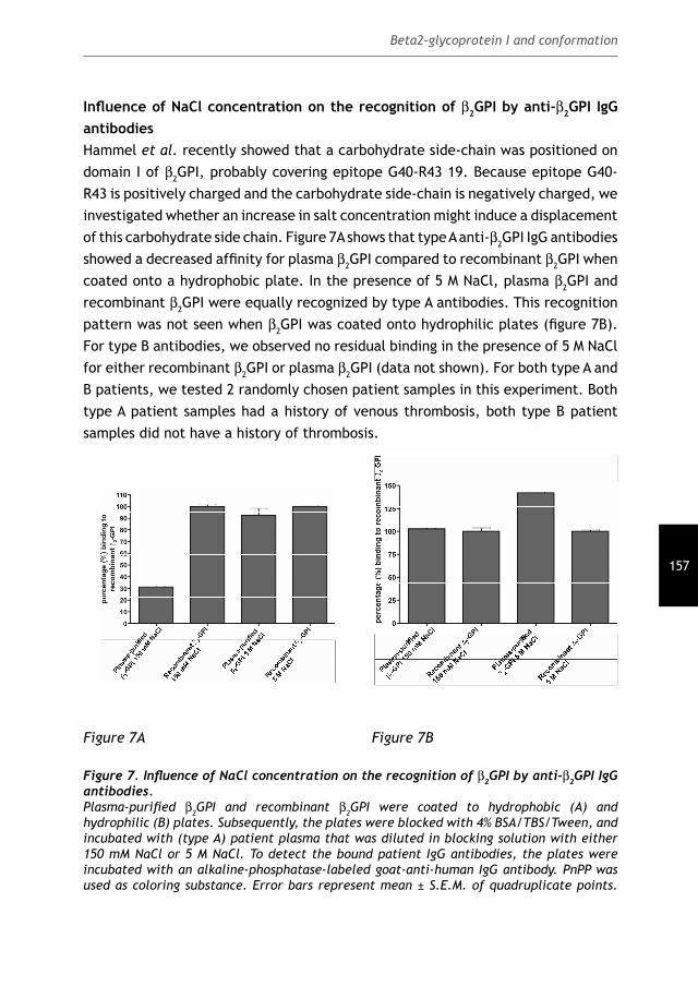
157
Beta2-glycoprotein I and conformation
Infl uence of NaCl concentration on the recognition of β2GPI by anti-β2GPI IgG antibodiesHammel et al. recently showed that a carbohydrate side-chain was positioned on domain I of β2GPI, probably covering epitope G40-R43 19. Because epitope G40-R43 is positively charged and the carbohydrate side-chain is negatively charged, we investigated whether an increase in salt concentration might induce a displacement of this carbohydrate side chain. Figure 7A shows that type A anti-β2GPI IgG antibodies showed a decreased affi nity for plasma β2GPI compared to recombinant β2GPI when coated onto a hydrophobic plate. In the presence of 5 M NaCl, plasma β2GPI and recombinant β2GPI were equally recognized by type A antibodies. This recognition pattern was not seen when β2GPI was coated onto hydrophilic plates (fi gure 7B). For type B antibodies, we observed no residual binding in the presence of 5 M NaCl for either recombinant β2GPI or plasma β2GPI (data not shown). For both type A and B patients, we tested 2 randomly chosen patient samples in this experiment. Both type A patient samples had a history of venous thrombosis, both type B patient samples did not have a history of thrombosis.
Figure 7A Figure 7B
Figure 7. Infl uence of NaCl concentration on the recognition of β2GPI by anti-β2GPI IgG antibodies. Plasma-purifi ed β2GPI and recombinant β2GPI were coated to hydrophobic (A) and hydrophilic (B) plates. Subsequently, the plates were blocked with 4% BSA/TBS/Tween, and incubated with (type A) patient plasma that was diluted in blocking solution with either 150 mM NaCl or 5 M NaCl. To detect the bound patient IgG antibodies, the plates were incubated with an alkaline-phosphatase-labeled goat-anti-human IgG antibody. PnPP was used as coloring substance. Error bars represent mean ± S.E.M. of quadruplicate points.

158
Chapter 6
DiscussionWhether antiphospholipid antibodies recognize a cryptic epitope on β2GPI has been the subject of a lengthy debate. Based on this study, we conclude that a conformational change in β2GPI is necessary for type A antibodies to recognize the protein. This conformational change is induced by binding of β2GPI to a negatively charged surface via a positive charged patch in domain V of β2GPI. We recently published that the population of anti-β2GPI antibodies that binds domain I of β2GPI at position 40-43 highly correlates with thrombosis 23. This area of domain I is probably covered by a carbohydrate chain in solution, as could be concluded from the studies by Hammel et al. 19. We hypothesized that this carbohydrate chain impedes antiphospholipid antibodies to bind epitope G40-R43 in solution. An anionic surface may induce a conformational change in β2GPI which results in the displacement of the carbohydrate side-chain. Antiphospholipid antibodies are then able to bind epitope G40-R43. Indeed, the removal of the carbohydrate chains with N-glycosidase F resulted in an increased affi nity for plasma-purifi ed β2GPI in solution by our monoclonal antibody 4F3 and patient type A IgG antibodies. Furthermore, by increasing the concentration of NaCl we were able to induce binding of anti-β2GPI IgG antibodies to plasma β2GPI on a hydrophobic plate. This indicates that the carbohydrate side chain no longer covers epitope G40-R43, and is now accessible for antibodies to bind. This hypothesis is further supported by the fact that proteins produced in insect cells have truncated carbohydrate side-chains instead of complex carbohydrate side-chains (as produced in mammalian cells). Due to these shorter carbohydrate side-chains, recombinant β2GPI exposes epitope G40-R43 constitutively. From our studies, it is clear that anti-β2GPI antibodies bind a cryptic epitope that is only exposed on β2GPI when bound to a negatively charged surface. This by no means excludes a role for dimerization of β2GPI. Antibody-induced dimerization of β2GPI seems important for functional activity because LAC activity is not seen when Fab(1) fragments of anti-β2GPI antibodies are used 30. Furthermore, we have also shown that dimerized β2GPI and not monomeric β2GPI induces LAC activity 11. We postulate that the recognition of β2GPI by anti-β2GPI antibodies occurs as follows (Figure 8): β2GPI has a relatively low affi nity for anionic phospholipids, and a certain concentration of β2GPI is necessary for binding. After binding of β2GPI to anionic phospholipids via domain V, the protein undergoes a conformational change. This results in the exposure of several cryptic epitopes, including epitope G40-R43 in domain I, enabling antibodies to bind this epitope 17,23,31. When the amount of β2GPI bound to the anionic phospholipids reaches a certain density,

159
Beta2-glycoprotein I and conformation
one antibody can bind two β2GPI molecules, thereby increasing the affi nity of the interaction with anionic phospholipids and stabilizing the complex.This model has several consequences for the assays we use to detect anti-β2GPI antibodies. It is obvious that for the LAC assay, both dimerization and exposure of the cryptic epitope are necessary. Clotting assays use PS as catalytic surface, but the affi nity of β2GPI for PS is low, thus stabilization of the interaction via dimerization is essential 3,13. In contrast to the LAC assay, in the anticardiolipin antibody ELISA cardiolipin is used as binding surface for β2GPI. The affi nity of β2GPI for cardiolipin is 30-40 times higher than the affi nity for PS 13. This relatively high affi nity enables β2GPI of becoming stabilized on the cardiolipin, and there is no need for dimerization via the antibodies to increase the affi nity of β2GPI for the surface. Only the exposure of a cryptic epitope is essential in this assay. In the ELISA in which β2GPI is directly coated onto the plate, the situation is more complex 32-34. Some brands of ELISA plates are unable to induce the conformational change (due to a difference in structure or charge), and are unsuitable for use in an anti-β2GPI ELISA 33. Other brands of ELISA plates do induce the conformational change, but their affi nity for β2GPI is rather low. These infl uences of different ELISA plates on our test results emphasize the need for standardization of the anti-β2GPI ELISA. In this respect it is interesting to mention that we observed differences in binding of β2GPI onto plates of different lot numbers of the same brand (unpublished data).Another consequence of our results concerns the use of different sources of β2GPI. We found that patient type A IgG antibodies (and monoclonal antibody 4F3) did show reactivity towards recombinant β2GPI coated onto hydrophobic plates, in contrast to plasma-purifi ed β2GPI coated onto hydrophobic plates. We also found that patient type A IgG antibodies (and monoclonal antibody 4F3) did recognize recombinant β2GPI in solution, but showed hardly any reactivity towards plasma-purifi ed β2GPI. This difference in binding characteristics between plasma-purifi ed β2GPI and recombinant β2GPI can be explained by the difference in glycosylation. Furthermore, bovine β2GPI has an additional glycosylation site at position 73 which can easily infl uence the binding of epitope G40-R43 by type A anti-β2GPI antibodies 19. In addition to this, it was already suggested that aggregation of β2GPI promotes the binding of certain anti-β2GPI antibodies 35. Galazka et al. found that a cryptic epitope was exposed on β2GPI when it was treated with glutaraldehyde 29. We found that these 2 features also hold true for type A antibodies. The importance of the carbohydrate chains and conformation of β2GPI has major consequences for the binding of antiphospholipid antibodies. One should check for aggregates of β2GPI in a (plasma) sample, and one should be careful in using β2GPI isolated from

160
Chapter 6
other sources than human plasma when studying the interaction between β2GPI and anti-β2GPI antibodies. In conclusion, we found that pathogenic antiphospholipid antibodies bind a cryptic epitope on domain I of β2GPI. This epitope (G40-R43) is accessible for antiphospholipid antibodies only after a conformational change.
Figure 8. Mechanism describing the binding of antiphospholipid antibodies to β2GPI. Our proposed mechanism is based on our fi ndings in this study together with both the crystal structure and the NMR structure of β2GPI in solution: (i) Antiphospholipid antibodies can not bind β2GPI in solution because epitope G40-R43 is covered by one of the carbohydrate chains. (ii) Binding to a phospholipid membrane induces a conformational change in β2GPI. (iii) As a result, the carbohydrate chain is not able to cover epitope G40-R43 anymore and is now able to bind antiphospholipid antibodies. aPL: antiphospholipid antibodies.
AcknowlegementsThis study was supported by a grant of the Netherlands Organization for Health Research and Development (ZonMw grant nr. 902-26-290).Authorship statement: Bas de Laat performed research, analyzed data and wrote the paper. Menno van Lummel and Maarten T.T. Pennings performed research and contributed vital reagents. Philip G. de Groot and Ronald H.W.M Derksen designed research and wrote the paper.

161
Beta2-glycoprotein I and conformation
References
1. Wilson WA, Gharavi AE, Koike T et al. International consensus statement on
preliminary classifi cation criteria for defi nite antiphospholipid syndrome: report of
an international workshop. Art Rheum. 1999;42:1309-11
2. Levine JS, Branch DW, Rauch J. The antiphospholipid syndrome. N Engl J Med.
2002;346:752-63
3. Bas De Laat H, Derksen RH, De Groot PG. Beta(2)-Glycoprotein I, the playmaker of
the antiphospholipid syndrome. Clin Immunol. 2004;112:161-68
4. McNeil HP, Simpson RJ, Chesterman CN, Krilis SA. Anti-phospholipid antibodies are
directed against a complex antigen that includes a lipid binding inhibitor of
coagulation: β2-glycoprotein I (Apolipoprotein H). Proc Natl Acad Sci USA.
1990;87:4120
5. de Laat HB, Derksen RHWM, Urbanus RT, Roest M, de Groot PG. β2-glycoprotein I
dependent lupus anticoagulant highly correlates with thrombosis in the
Antiphospholipid Syndrome. Blood. 2004;104:3598-602
6. Wong RC, Gillis D, Adelstein S, Baumgart K, Favaloro EJ, Hendle MJ, Homes P, Pollock
W, Smith S, Steele RH, Sturgess A, Wilson RJ. Consensus guidelines on anti-cardiolipin
antibody testing and reporting. Pathology. 2004;36:63-68
7. Simmelink MJ, Derksen RHWM, Arnout J, de Groot PG. A simple method to discriminate
between beta2-glycoprotein I and prothrombin-dependent lupus anticoagulants. J.
Thromb Haemost. 2003;1:740-47
8. Steinkasser A, Barlow PN, Willis AC, Kertesz Z, Campbell ID, Simm RB, Norman DG.
Activity, disulfi de mapping and structural modelling of the fi fth domain of human
β2-glycoprotein I. FEBS Letters. 1992;313:193-97
9. Bouma B, de Groot PhG, van den Elsen JMH, Ravelli RBG, Schouten A, Simmelink
MJA, Derksen RHWM, Kroon J, Gros P. Adhesion mechanism of human β2-glycoprotein
I to phospholipids based on its crystal structure. EMBO J. 1999;18:5166-74
10. Schwarzenbacher R, Zeth K, Diederichs K, Gries A, Kostner GM, Laggner P, Prassl R.
Crystal structure of human beta2-glycoprotein I: implications for phospholipid
binding and the antiphospholipid syndrome. EMBO J. 1999;15:6228-39
11. Lutters BC, Meijers JC, Derksen RH, Arnout J, de Groot PG. Dimers of beta 2-
glycoprotein I mimic the in vitro effects of beta 2-glycoprotein I-anti-beta 2-
glycoprotein I antibody complexes. J Biol Chem. 2001;276:3060-7
12. Sheng Y, Kandiah DA, Krilis SA. Anti-beta 2-glycoprotein I autoantibodies from
patients with the “antiphospholipid” syndrome bind to beta 2-glycoprotein I with
low affi nity: dimerization of beta 2-glycoprotein I induces a signifi cant increase in
anti-beta 2-glycoprotein I antibody affi nity. J Immunol. 1998;161:2038-43
162
Chapter 6
13. Willems GM, Janssen MP, Pelsers MAL, Comfurius P, Galli M, Zwaal RFA, Bevers EM.
Role of divalency in the high-affi nity binding of anticardiolipin antibody-β2-
glycoprotein I complexes to lipid membranes. Biochemistry. 1996;35:13833-42
14. Arnout J, Wittevrongel C, van Russelt M, Hoylaerts M, Vermijlen J. β2-glycoprotein I
dependent lupus anticoagulants form stable bivalent antibody β2-glycoprotein I
complexes on phospholipid surfaces. Thromb Haemost. 1998;79:79-86
15. Reddel SW, Wang YX, Krilis SA. Anti-beta2-glycoprotein I autoantibodies require an
antigen density threshold, consistent with divalent binding. Lupus. 2003;12:37-45
16. Wang SX, Sun YT, Sui SF. Membrane-induced conformational change in human
apolipoprotein H. Biochem J. 2000 May 15;348:103-6
17. Kuwana M, Matsuura E, Kobayashi K, Okazaki Y, Kaburaki J, Ikeda Y, Kawakami Y.
Binding of {beta}2-Glycoprotein I to anionic phospholipids facilitates processing and
presentation of a cryptic epitope that activates pathogenic autoreactive T cells.
Blood. 2005;105:1552-57
18. Merrill JT. What causes the antiphospholipid syndrome? Curr Rheumatol Rep.
2001;3:293-300
19. Hammel M, Kriechbaum M, Gries A, Kostner GM, Laggner P, Prassl R. Solution structure
of human and bovine beta(2)-glycoprotein I revealed by small-angle X-ray scattering.
J Mol Biol. 2002;321:85-97
20. Tan EM, Cohen AS, Fries JF, Masi AT, McShane DJ, Rothfi eld NF, et al. The 1982
revised criteria for the classifi cation of systemic lupus erythematosus. Arthritis
Rheum. 1982;25:1271-77
21. Horbach DA, van Oort E, Donders RCJM, Derksen RHWM, de Groot Ph.G. Lupus
anticoagulant is the strongest risk factor for both venous and arterial thrombosis in
patients with systemic lupus Erythematosus. Thromb Haemost. 1996;76:916-24
22. Derksen RH, Hasselaar P, Blokzijl L, Gmelig Meyling FH, de Groot PG. Coagulation
screen is more specifi c than the anticardiolipin antibody ELISA in defi ning a thrombotic
subset of lupus patients. Ann Rheum Dis. 1998;47:364-71
23. de Laat B, Derksen RH, Urbanus RT, de Groot PG. IgG antibodies that recognize
epitope Gly40-Arg43 in domain I of {beta}2-glycoprotein I cause LAC and their
presence correlates strongly with thrombosis. Blood. 2005;105:1540-45
24. Oosting JD, Derksen RHWM, Hackeng TM, van Vliet M, Preissner KT, Bouma BN, de
Groot PhG. In vitro studies of antiphospholipid antibodies and its cofactor, β2-
glycoprotein I, show negligible effects on endothelial cell mediated protein C
activation. hromb Haemost. 1991;66:666-71
25. McNeeley PA, Dlott JS, Furie RA, Jack RM, Ortel TL, Triplett DA, Victoria EJ, Linnik
MD. Beta2-glycoprotein I-dependent anticardiolipin antibodies preferentially bind
the amino terminal domain of beta2-glycoprotein I. Thromb Haemost. 2001;86:590-
95

163
Beta2-glycoprotein I and conformation
26. Lutters BC, Derksen RH, Tekelenburg WL, Lenting PJ, Arnout J, de Groot PG. Dimers
of beta 2-glycoprotein I increase platelet deposition to collagen via interaction with
phospholipids and the apolipoprotein E receptor 2’. J Biol Chem. 2003;278:33831-
38
27. Arnout J, Wittevrongel C, Vanrusselt M, Hoylaerts m, Vermylen J. Beta-2-glycoprotein
I dependent lupus anticoagulants form stable bivalent antibody beta-2-glycoprotein
I complexes on phospholipid surfaces. Thromb Haemost. 1998;79:79-86
28. Pengo V, Thiagarajan P, Shapiro SS, Heine MJ. Immunological specifi city and
mechanism of action of IgG lupus anticoagulants. Blood. 1987;70:69-76
29. Galazka M, Tang M, Debari VA, Kohles JD, Lee JK, Keil LB, Petersheid M. Modifi caton
of beta2glycoprotein I by Glutardialdehyde: Conformational Changes and Aggregation
Accompany Exposure of the Cryptic Autoepitope. Appl Biochem biotechnol.
1999;76:1-14
30. Roubey RA, Eisenberg RA, Harpr MF, Winfi eld JB. “Anticardiolipin” autoantibodies
recognize beta2-glycoprotein I in the absence of phospholipid. Importance of Ag
density and bivalent binding. J Immunol. 1995;154:954-60
31. Iverson GM, Reddel S, Victoria EJ et al. Use of single point mutations in domain
I of β2GPI to determine fi ne antigenic specifi city of antiphospholipid autoantibodies.
J Immunol. 2002;169:7097-103
32. Matsuura E, Garashi Y, Yasuda T, Triplett DA, Koike T. Anticardiolipin antibodies
recognize beta2-glycoprotein I structure altered by interacting with an oxygen
modifi ed solid phase surface. J Exp Med. 1994;179:457-62
33. Pengo V, Biasiolo A, Fior MG. Autoimmune antiphospholipid antibodies are directed
against a cryptic epitope expressed when beta2-glycoprotein I is bound to a suitable
surface. Thromb Haemost. 1995;73:29-34
34. Keil LB, Galazka M, el-Kadi HS, Erickson EN Jr, DeBari VA. Binding of beta 2
glycoprotein I to activated polystyrene and its recognition by human IgG
autoantibodies. Biotechnol Appl Biochem. 1995;22:305-13
35. Arvieux J, Regnault V, Hachulla E, Darnige L, Berthou F, Youinou P. Oxidation of
beta2-glycoprotein I (beta2GPI) by the hydroxyl radical alters phospholipid
binding and modulates recognition by anti-beta2GPI autoantibodies. Thromb
Haemost. 2001;86:1070-76


Introduction 167
β2GPI-induced platelet activation 167
Molecular regions within β2GPI that are recognized by apoER2’ on platelets 168
Members of the Low-Density Lipoprotein Receptor family and their role
the antiphospholipid syndrome` 169
Identifi cation of ligand binding sites within LRP 171
Mutational studies mapping the interaction between β2GPI and apoER2’ 172
Site-directed mutagenesis identifi es a cluster of lysine residues recognized
by apoER2’ 172
The fi fth domain of β2GPI contains the phospholipid-binding site 173
Implications for the antiphospholipid syndrome 175
Involvement of the receptors in the antiphospholipid syndrome 175
Anticoagulant therapy and antiphospholipid antibodies 175
Future prospects 177
Soluble receptors; a marker of the antiphospholipid syndrome? 177
Cellular activation in the antiphospholipid syndrome 177
Animal models 178
References 179
7General Discussion


167
General Discussion
IntroductionThe pathophysiology of the antiphospholipid syndrome (APS) is still not understood. It is diffi cult to envision that low affi nity anti-β2GPI antibodies that affect one single biological activity, not to mention several biologic pathways, cause the syndrome. A link must exist how anti-β2GPI antibodies induce prolongation of an in vitro clotting assay and the thrombotic manifestations observed in APS patients with circulating anti-β2GPI antibodies. There are two theories explaining this paradox. One is the “dimerization theory”. Due to binding of anti-β2GPI antibodies to β2GPI, the protein dimerizes resulting in increased affi nity for cellular surfaces. The other one is the “cryptic epitope” theory. Due to binding of β2GPI to anionic phospholipids, a cryptic epitope within β2GPI that is normally shielded off, is exposed thereby facilitating the binding of anti-β2GPI antibodies to β2GPI 1. The affi nity of the formed immune complex is high enough to compete with clotting factors on a cellular surface. We know how the antibodies induce the observed in vitro lupus anticoagulant (LAC). Several hypotheses have been put forward to explain the arterial and/or venous thrombosis in APS patients. We cannot exclude that arterial and venous thrombosis is the result of different actions of β2GPI/anti-β2GPI antibody complexes on cells. A number of interesting studies identifi ed mechanistic links between anti-β2GPI antibodies and thrombosis. Now, attention is mainly focussed on the interference of such immune complexes with antithrombotic pathways, such as fi brinolysis 2-
4 and the Protein C pathway 2. However, impaired Protein C pathway results in venous thrombosis 5;6, while in APS venous and arterial thrombosis are present. It is diffi cult to envision that simple binding to anionic phospholipids can activate cells. A second hit must occur to activate cells. An important observation is that β2GPI/anti-β2GPI antibodies complexes not only have an increased affi nity for anionic phospholipids but also for cellular surfaces. Binding to and activation of endothelial cells, platelets and monocytes by β2GPI/anti-β2GPI antibody complexes has been reported. There must be an additional mechanism how dimerization of β2GPI can explain the observed cellular activation.
β2GPI-induced platelet activationResearch into the pathology of APS was obscured by the idea that anionic phospholipids are crucial for binding β2GPI/antiβ2GPI antibody complexes. We now know that cellular activation by β2GPI/anti-β2GPI antibody complexes is the consequence of classic receptor-ligand interactions. These interactions represent a basis for understanding the pathology of APS. Previous studies of our group revealed that platelets are sensitized by β2GPI/anti-β2GPI antibody complexes or

168
Chapter 7
recombinant dimeric β2GPI under conditions of arterial fl ow using a whole blood perfusion model 7. The binding of dimeric β2GPI to platelets is mediated by the platelet receptor apolipoprotein E receptor 2’ (apoER2’). Platelet activation is among others dependent on the synthesis of thromboxane A2 (TxA2)
7-9. Release of TxA2 and binding to its receptor on the platelet membrane results in further platelet activation. Several investigators observed increased levels of thromboxane breakdown products in the urine of APS patients, indicating the existence of activated platelets in the circulation of APS patients. This is supported by the observation that activated platelets do circulate in APS patients 10;11. Apart form apoER2’, other receptors have been suggested for β2GPI, annexin A2 on human platelets and endothelial cells 12 and members of the toll-like receptor family (TLR) on endothelial cells, platelets and monocytes 13-15. Further studies should elucidate the role of different receptors.Several studies show that the addition of aPL to human platelets results in an increase in phosphorylation of p38 mitogen-activated protein kinase (p38MAPK) 9;16. P38MAPK is a member of a family of proline-directed Ser/Thr kinase that is dual-phosphorylated on residues Thr180 and Tyr182. Platelets contain members of the MAPKs, including ERK-1 (p44MAPK), ERK-2 (p42MAPK) and p38MAPK. Activated p38MAPK phosphorylates and activates cyclic phospholipase A2 (cPLA2), resulting in the release of arachidonic acid (AA), a precursor in the formation of TxA2. Increased phosphorylation of p38MAPK is also found when platelets are activated by dimeric β2GPI (unpublished data). The p38MAPK phosphorylation is transient and reaches its maximum at one minute. This indicates that binding of dimeric β2GPI to apoER2’ on the platelet membrane results in phosphorylation and activation of p38MAPK. Synthesis of TxA2 supports further platelet activation. Because of its known low rate of endocytosis and ligand degradation, apoER2’ is probably involved in signal transduction. Whether apoER2’ is involved in endocytosis by platelets remains to be determined.
Molecular regions within β2GPI that are recognized by apoER2’ on platelets It is proposed for members of the LDL-R family that positive electrostatic surface potentials, not the primary sequences, in different ligands constitute receptor-binding domains. For example, electrostatic interaction between basic residues in the lectin-like domain of LOX-1, a scavenger receptor sharing similar ligand-binding properties as the LDL-R family, and negatively charged oxidized LDL (oxLDL) is critical for the binding activity of LOX-1 17. Specifi c cationic residues within the RAP molecule 18 facilitate binding of receptor-associated protein (RAP)

169
General Discussion
to low density lipoprotein receptor-related protein (LRP). A specifi c domain within apoB100 mediates LDL binding to the LDL-R 19. This domain is termed the B-site and encodes a patch of cationic amino acids consisting of lysine and arginine residues. These residues are essential for binding of LDL to the LDL-R. Modifi cation of these residues results in reduced signaling in platelets 20. Instead of a short primary sequence, a patch rich in cationic residues thus compose a receptor-binding domain within ligands. Lysines and arginines on the outer surface of ligands comprise the cationic potential required for binding to LDL-R family members. X-ray diffraction analysis supports this proposal. The LDL-R recognizes specifi c basic amino acid residues within apolipoprotein E (apoE) 21. These basic amino acids are clustered into a surface patch on one large helix. For lipoprotein C lipase, amino acids 378-448 within the C-terminal binding domain are crucial for binding to LRP 22-24. Thus, a positive electrostatic potential exposed on the outer surface of different ligands may constitute a domain that is recognized by various members of the LDL-R family. β2GPI is composed of fi ve sushi or complement control protein (CCP) domains. The cationic charge in domain I and domain V is the result of multiple lysine and arginine residues. Domain V displays the largest cationic patch (2000 Å2) and might be essential for binding to apoER2’. Indeed, we show in Chapter 3 the importance of a cationic patch within domain V (amino acid 282-287) of β2GPI for the interaction with apoER2’. This is in accordance with other groups who found that lysine residues constitute a ligand recognition site for members of the LDL-R family (Table 1).
Table 1: Amino acid sequences in several ligand important for binding to LDL-R homologous
Ligand Receptor Amino acids within ligand
Residues important for binding References
ApoB100 LDL-R 3359-3369 RLTRKRGLKLA 19;775apoE LDL-R 134-150 RVRLASHLRKLRKRLLR 217β2-macroglobulin LRP 1366-1392 FIPLKPTVKMLERSNHVSRTEVSSNHV 784PAI-1 LRP 69-87 DKGMAPALRHLYKELMGPWN 793Lipoprotein lipase LRP 403-425 KIRVKAGETQKKVIFCSREKVSHL 802aprotinin LRP 38-47 CRAKRNNFKSA 811
Members of the Low-Density Lipoprotein Receptor family and their role in the antiphospholipid syndromeThe LDL-R gene family consists of structurally closely related transmembrane glycoproteins that participate in a wide range of biological activities, such as lipoprotein metabolism, protection against atherosclerosis, neurodevelopment
170
Chapter 7
and in the transport of nutrients, vitamins and drugs 25. At present, 12 members of the LDL-R family in mammals are identifi ed. The function of a receptor present on one cell type may not be restricted to a single function. Cellular distribution of LDL-R homologous is not restricted to specifi c cells or tissues. The LDL-R, for example, is distributed throughout the human body and is expressed in hepatocytes, macrophages and the central nervous system. Like the LDL-R, also the low-density lipoprotein receptor related protein (LRP) is expressed by a wide range of cell types and tissues, among others hepatocytes, macrophages and vascular smooth muscle 26. Megalin is expressed in the apical plasma membrane of secretory epithelia facing transcellular fl uid 27. Because of the various expression patterns of members of the LDL-R family, one can envision a role for these receptors in the pathophysiology of APS. The fi rst paper published on apoER2’ in platelets showed that this receptor might be involved in platelet inhibition 28. However, further research illustrated that human platelets are activated by LDL or dimeric β2GPI, which is mediated by apoER2’ 7;19;29. Megalin recognizes complexes of β2GPI and phospholipids, but also β2GPI alone 30. We show that several members of the LDL-R family recognize complexes of β2GPI/anti-β2GPI antibody complexes (Chapter 4). Apart from apoER2’, these receptors include the VLDL-R, megalin and LRP. Besides these receptors, dimeric β2GPI is also able to bind with low affi nity to the LDL-R. The LDL-R is known to be involved in ligand uptake and endocytosis 31;32, whereas a role in signal transduction is still unclear. Therefore, we believe that the LDL-R has little role in the pathophysiology of APS. RAP dose-dependently inhibited binding of dimeric β2GPI to members of the LDL-R receptor family, indicating a specifi c interaction. In accordance with data for apoER2’, domain V of β2GPI comprises the main recognition site for members of the LDL-R family. Heparin is able to inhibit the interaction of dimeric β2GPI with members of the LDL-R family (Chapter 4). Lys284, Lys286 and Lys287 in domain V of β2GPI are essential for the interaction of b2GPI with heparin 33, suggesting an involvement of this region in the interaction with LDL-receptor family members. Despite data indicating a crucial role for domain V of β2GPI, our group previously showed that a monoclonal anti-β2GPI antibody (4F3), that recognizes domain I of β2GPI, could inhibit platelet adhesion to a collagen surface 7. This suggests that domain I is involved in platelet activation. However, the data presented in Chapter 4 shows that domain I of β2GPI is not involved in the interaction with members of the LDL-R family. The observed inhibition with 4F3 can be due to steric interference; binding of 4F3 induces a conformational change within β2GPI that abrogates binding of β2GPI to apoER2’.

171
General Discussion
Members of the LDL-R family are widely distributed in cells and tissues and are associated with a number of metabolic processes and disorders such as embryonic development of the brain, Alzheimer disease (AD), viral and bacterial infections and lipid metabolism. For example, familial hypercholesterolemia (FH), which is one of the most common human inborn errors of metabolism, is caused by loss-of-function mutations in the LDL-R gene 34. LRP mediates critical biological pathways in AD such as the clearance of denatured proteins (beta-amyloid) 35;36. As mentioned, several cell types that are involved in the pathology of APS, among others platelets, monocytes and endothelial cells, express LDL-R family members. It is of interest to study whether dimeric β2GPI can activate these cells. This is currently under investigation. In conclusion, data presented here suggests that the antiphospholipid syndrome can be added to the expanding list of pathological conditions mediated by members of the LDL-R family.
Identifi cation of ligand binding sites within LRPThe identifi cation of ligand-binding sites within a member of the LDL-R family has revealed multiple sites for protein interaction throughout the whole of the receptor ligand binding domains. Information of the different ligand-binding sites is often provided from studies of how certain inhibitors with known binding sites displace the ligand of interest or how a potential ligand infl uences binding of well-established ligands. Two small cysteine-rich complement type (CR) domains within a receptor are suffi cient for high affi nity ligand binding. Some ligands bind to identical or partially overlapping sites, which results in competitive receptor binding, while other ligands have unique binding sites that are suggestive of multiple ligand epitopes. Also, in the extremely large receptors containing multiple CR domains (among others LRP and megalin) most, if not all, of the CR domains can mediate ligand binding. It is of interest to establish how many CR domains contribute to the direct binding of a ligand. Analysis of LRP minireceptors comprised of isolated cluster II and cluster IV (8 and 11 CR domains, respectively), as well as the VLDL-R and the LDL-R clusters (7 and 8 CR domains, respectively) demonstrate that a ligand can bind with high-affi nity to several of these CR domains. For example, RAP can bind with high-affi nity to cluster II and cluster IV of LRP 37-39, which displays only minor differences in ligand-binding properties 40. In Chapter 4 we demonstrate that i. RAP can inhibit dimeric β2GPI binding to LRP and ii. dimeric β2GPI can bind to both cluster II and cluster IV, which comprise the high-affi nity recognition regions within LRP. The domain deletion mutants, except for the domain V deletion mutant, bind with

172
Chapter 7
comparable affi nities (high- and low affi nity binding site) to these minireceptors. Therefore, LRP might recognizes two cationic patches in domain V of β2GPI, containing a low- and a high affi nity binding site. The high-affi nity binding site may be located at position Lys282-287 within domain V of β2GPI, as we show in Chapter 3 that this region is important for recognition by apoER2’. NMR and crystallization studies of LRP revealed that fl exible linkers connect the CR domains of LRP. The combination of several CR domains may comprise a suitable ligand-binding site. The observation that multiple CR domains (for example cluster II and IV) mediate ligand binding is in accordance with the fi ndings for LRP (Chapter 4). The cationic patch in domain V of β2GPI measures 2000 Å2. Given the fact that the overall dimensions of a CR domain is 20 × 20 × 27 Å 17, domain V of β2GPI can accommodate more than one CR domain. This is already true for binding of RAP and α2-macroglobulin (α2-M) to LRP; ligand-binding repeats from two clusters cooperate to generate a high affi nity-binding site for RAP (cluster II and cluster IV) and α2-M (cluster I and cluster II) 40;41. Receptor dimerization is an important process before signal transduction can occur. Bivalent antibodies directed to the extracellular part of LRP, but not Fab fragments of the same antibody, are able to induce calcium infl ux in cultured neurons, indicating the necessity of receptor dimerization 42. Interestingly, intact LRP interacted with dimeric β2GPI in a 1:2 stoichiometric (Chapter 4). These data provide biochemical evidence that dimeric β2GPI may require simultaneous association with two LRP molecules. This observation suggests that binding of dimeric β2GPI to LRP induces receptor multimerization on the cell surface, bringing signaling factors bound to the cytoplasmic tail of the individual receptors into close proximity. Receptor dimerization is already reported for apoER2; after binding of Reelin, apoER2 on neuronal cells is able to dimerize involving several CR domains. This is followed by subsequent signal transduction 43. The fact that soluble receptors are not able to associate with Reelin supports the receptor dimerization hypothesis.
Mutational studies mapping the interaction between β2GPI and apoER2’
Site-directed mutagenesis identifi es a cluster of lysine residues recognized by apoER2’Since a cationic peptide spanning amino acid residue Lys282-287 in domain V of β2GPI was able to inhibit binding of dimeric β2GPI to apoER2’ on human platelets (Chapter 3), it is of interest to explore domain V for potential recognition sites

173
General Discussion
for apoER2’. One approach to determine which residues are of importance for protein-protein interaction is site directed mutagenesis. Cationic residues within ligands are important for recognition by members of the LDL-R family, as has been established for several ligands (Table 1). These recognition sites mainly comprise lysine and arginine residues. Therefore, we predicted that lysine residues in domain V of β2GPI contain the receptor-binding site. A detailed molecular study for the interaction between β2GPI and apoER2’ is preceded by a prediction of the electrostatic surface potential of domain V after substation of lysine residues into neutral leucine residues (Chapter 5). Analysis of a three-dimensional molecular structure of domain V revealed that regions Lys262-276, Lys282-287 and Lys305-317 are located at the outer surface of the putative apoER2’-interacting surface area. This observation raises the question to what extent these regions contribute to the interaction with apoER2’. Employing site-directed mutagenesis in which lysine residues were replaced for leucine residues, we observed reduced binding of dimeric β2GPI to apoER2’ (Chapter 5). We clearly show that the Lys282-287 residue is crucial for interaction with apoER2’ and that a second recognition site resides at residue Lys305-317. We propose that instead of a short patch of a primary sequence in domain V of β2GPI, a cationic secondary or tertiary organization may comprise a receptor-binding domain. The cationic electrostatic potentials encompass stretches of lysine residues in domain V of β2GPI. These cationic residues may bind to several CR domains of apoER2’. The major binding site for RAP on apoER2 on neuronal cells is mapped to Asp35 on the fi rst CR domain of apoER2 and a minor role for the third CR domain 43. Only the third RAP domain (amino acid 216-323) binds to apoER2. It is of interest to study which CR domain of apoER2’ on human platelets is crucial for recognition of dimeric β2GPI. Therefore, binding studies with different complement-type repeats in CR domains of apoER2’ should be performed.
The fi fth domain of β2GPI contains the phospholipid-binding siteDomain V of β2GPI differs signifi cantly from the other four CCR domains. It folds into a central β-spiral and an extended C-terminal loop region. This loop contains several hydrophic residues and is implicated in binding to anionic phospholipids. Apart from phospholipids, domain V might be involved in binding to hydrophobic ligands 44 and dextran sulphates 45. Using synthetic peptides the major phospholipid-binding site is mapped to the basic patch of lysine residues (Cys281-288) that is located on the outer surface of the fi fth domain of β2GPI. Lys284, Lys286 and Lys287 are crucial for the electrostatic interaction with anionic phospholipids 46;47. However, the optimal

174
Chapter 7
integrity of the C-terminus and the nearby-located Leu313-316 residue is required for optimal interaction with anionic phospholipids 48;49. Based on the hypothesis that domain V comprises the phospholipid-binding site within the residue Cys281-288, we performed phospholipid-binding assays with several dimeric mutants of β2GPI (Chapter 5). Lys282-287 is a critical residue within β2GPI for binding to cardiolipin, as shown by site-directed mutagenesis 50. In that study, the investigators used β2GPI with mutations in the KNKEKK site and not in the region after the KNKEKK site. The same group confi rmed their results by performing competition experiments with peptides spanning regions within domain V of β2GPI 46. In Chapter 5, this fi nding is supported by data with the dimeric β2GPI mutants. However, we provide evidence that the residue Lys305-317 is more crucial for binding to phosphatidylserine (PS), a natural occurring phospholipid in cellular membranes than the Lys282-287 residue.Different hypotheses exist explaining the interaction between β2GPI and anionic phospholipids. An interesting observation outlines that the targets for β2GPI are cell surface receptors rather than phospholipids 51. However, evidence exist that β2GPI interacts directly with anionic phospholipids, which alters the conformation of the protein 52. In Chapter 6, we show that a conformational change within β2GPI is necessary for anti-β2GPI antibodies to recognize the protein. This conformational change is induced by the binding of β2GPI to an anionic surface via a cationic patch in domain V of β2GPI (Chapter 6). The fi fth domain of β2GPI is important for binding to apoER2’ on human platelets, but is also important for binding to phospholipids. Interestingly, Reelin binding to phospholipids might promote signal transduction via apoER2 by ensuring that Dab1, an intracellular adaptor protein, integrates into the signaling complex in a suitable orientation 53. This raises the question whether anionic phospholipids are required for binding and proper orientation of β2GPI before it can interact with apoER2’ and induce signal transduction in platelets. In the kinetic studies, several members of the LDL-R family recognized both dimeric β2GPI and a β2GPI/antiβ2GPI antibody complex. In the absence of phospholipids, LDL-R family members recognize both dimers in a purifi ed system (Chapter 4). We used a monoclonal antibody and not a patient derived IgG, as patients IgG do not recognize β2GPI in solution. Moreover, the non-phospholipid binding dimeric β2GPI did interact with the receptors (Chapter 3 and 4). This suggests that the recombinant dimeric β2GPI does not necessarily need phospholipids to bind to members of the LDL-R family. The following hypothesis is proposed. In patients with APS, monomeric (plasma) β2GPI circulates free in plasma at a concentration of approximately 200 μg/mL. Low-affi nity binding of β2GPI to anionic phospholipids mediated by a cationic patch at position Lys305-317 is

175
General Discussion
a prerequisite before anti-β2GPI antibodies can bind to and dimerize β2GPI. This leads to formation and stabilization of β2GPI/anti-β2GPI antibody complexes on the cellular surface, followed by binding of the complex to apoER2’ on platelets. This latter interaction involves region Lys282-287 in domain V of β2GPI. The hypothesis that plasma β2GPI needs to be dimerized by anti-β2GPI antibodies on an anionic phospholipid surface is supported by the fact that β2GPI/anti-β2GPI antibody complexes are not detected in the plasma of APS patients.
Implications for the antiphospholipid syndrome
Involvement of the receptors in the antiphospholipid syndromeThe understanding of the LDL-family has broadened since the discovery of the LDL-R as a cargo receptor. It is clear now that other members of the LDL-R family have additional functions in regulating signal transduction. A direct relation between endocytosis and signal transduction has been shown for megalin, whereas the role for the VLDL-R and apoER2’ seems to lie in signal transduction with a limited role for endocytosis. The crucial role of these receptors in the control of several physiological processes makes them attractive targets. Knowledge of the molecular basis for ligand recognition and of their many functions, members of the LDL-R family are attractive for the development of specifi c inhibitors. For example, if binding of β2GPI/antiβ2GPI–antibody complexes to LDL-R family members induces signal transduction, designing a compound selectively inhibiting this interaction would be attractive. This approach might be more effi cient or can cause fewer side effects.
Anticoagulant therapy and antiphospholipid antibodiesFrequent thrombotic manifestations in the antiphospholipid syndrome are deep venous thrombosis (DVT), pulmonary emboli (PE), ischaemic stroke and transient ischaemic attacks (TIA). Thrombosis is reported in almost any vessel in the body Initial treatment of deep venous thrombosis (DVT) is the administration of low molecular weight heparin (LMWH), unfractionated heparin (UFH) and vitamin K antagonists (VKA) 54. The starting dose for treatment of DVT is a bolus injection of 5.000 Units (U) unfractionated heparin (UFH), followed by a continuous infusion of 30.000 U for a period of 24 hours. Subsequent doses should be standardized according to weight. For administration with LMWH, a therapeutic range is 0.6 to
176
Chapter 7
1.0 IU/mL. The target range is less clear in patients treated with LMWH once a day, but a level between 1.0 IU/mL and 2.0 IU/mL seems reasonable.Initial treatment for VTE in patients with APS is the administration of low-molecular weight heparin (LMWH), overlapped with VKA or aspirin treatment 53;55;56. Based on the results from retrospective studies 54;57;58, for long-term treatment, high-intensity VKA is recommended for preventing recurrent VTE. In general, patients with ischaemic strokes receive antiplatelet drugs, notably low-dose aspirin. Low-dose aspirin may also be effective in patients with ischaemic strokes and aPL 59. In summary, a life-long prophylaxis to patients with thromboembolic events and prophylaxis with LMWH during risk periods is recommended. Several possibilities exist to explain the therapeutic effect of heparin toward decreasing the thrombotic risk in APS patients: 1. has an antithrombotic effect via antithrombin (AT) independent of aPL 2. modulates cell-mediated events to prevent production or alter the action of aPL or 3. facilitates the elimination of antiphospholipid antibodies 60-62. It is reported that unfractionated heparin and LMWH reduce the in vitro binding of aPL 63. As shown in Chapter 4, heparin can abrogate binding of dimeric β2GPI to members of the LDL-R family. Half-maximal binding of dimeric β2GPI to LRP or megalin is observed at 0.06 mg/mL of unfractionated heparin (corresponds to 11 IE/mL). We also observed inhibition with LMWH for LRP and megalin with half-maximal binding at approximately 0.05 mg/mL LMWH, which corresponds to 8 IE/mL (unpublished data). In conclusion, we show that heparin performs its therapeutic effect by binding to domain V of β2GPI. Thereby heparin interferes with binding of β2GPI to members of the LDL-family and possibly to other cellular receptors. Finally, β2GPI is not able to activate cells involved in APS (platelets, monocytes, endothelial cells).

177
General Discussion
Future prospects
Soluble receptors; a marker of the antiphospholipid syndrome?An interesting feature of the LDL-R family is the reports of soluble forms of these receptors. A soluble form of LRP has been identifi ed in human plasma that retains LRP-ligand binding ability 64 65 66. This soluble form is probably the result of proteolytic cleavage of the LRP form the cellular surface by a metalloproteinase. In addition, soluble megalin is present in urine that remains the ability to bind ligands 64. However, the physiological relevance of soluble megalin fi nding is unclear. For other receptors, cleavage from the cell membrane after ligand binding is reported. Several other cell surface proteins are shed from the cell membrane, including L-selectin 67, GpIb 68, interleukin receptors 69 and transferrin receptor 70. The hypothesis exist that these soluble receptors are the result of proteolytic cleavage after binding of ligands. There is no data, but we assume that members of the LDL-R family might be cleaved from the cell membrane (platelets, monocytes, endothelial cells) after interaction with β2GPI/antiβ2GPI antibody complexes. Soluble receptors in plasma of patients with APS can serve as a diagnostic marker for cellular activation and thus they might be a predictive tool for thrombosis. For several receptors, the diagnostic value has already been established. Measurement of soluble LOX-1, a scavenger receptor sharing similarities with the LDL/R family with regard to ligand binding, or soluble Fc gamma RIIIa (CD16) in vivo may provide a novel diagnostic tool for the evaluation and prediction of atherosclerosis and vascular disease 71;72. Interestingly, among other markers, soluble P-selectin in plasma, can be used in the assessment of platelet activation status 73. In summary, measurement of soluble members of the LDL-R family might serve as a diagnostic tool for APS.
Cellular activation in the antiphospholipid syndromeUntil know, we only have clear evidence that dimeric β2GPI can activate platelets via apoER2’. This is followed by phosphorylation of apoER2’, phosphorylation and activation of p38 MAPK and thromboxane synthesis. Recently, our group has identifi ed on endothelial cells several members of the LDL-R that recognize dimeric β2GPI, suggesting a role for the LDL-R family in dimeric β2GPI induced endothelial cell activation (unpublished data). Other groups have shown the involvement of other receptors (annexin A2, toll-like receptors) in cellular activation and signal transduction in the antiphospholipid syndrome 13;15. However, dimeric β2GPI-induced cellular activation is still under investigation.
178
Chapter 7
Animal modelsHow β2GPI dimers increase platelet deposition in vitro 74 and thrombus formation in vivo is subject for ongoing research. Passive administration of anti-β2GPI antibodies to pregnant BALB/c mice induced clinical fi ndings consistent with APS (increased fetal resorptions, reduced platelet counts, and prolonged activated partial thromboplastin time) 75. In another study, infusion of anti-β2GPI antibodies promoted thrombus formation in a photochemically induced thrombosis model 76. Cellular activation via the Fc portion of β2GPI/anti-β2GPI antibody complexes was not essential, because F(ab’)(2) fragments of the same antibodies still promoted thrombus formation. However, it is still unclear which receptors are involved in the induction of thrombosis in animal models.We have now clear evidence that members of the LDL-R family recognize dimeric β2GPI and this interaction results in cellular activation (Chapter 3). This involves a high-affi nity interaction that can compete with binding for other ligands of this receptor family. Whether the contribution of the LDL-R family is physiologically relevant for development of thrombosis needs to be established. To explore the role of the LDL-R family in the pathogenesis of APS, studies with LDL-R family
knockout animal models are essential.

179
General Discussion
References
1. Kuwana M, Matsuura E, Kobayashi K et al. Binding of beta 2-glycoprotein I to anionic
phospholipids facilitates processing and presentation of a cryptic epitope that
activates pathogenic autoreactive T cells. Blood 2005;105:1552-1557.
2. Yasuda S, Atsumi T, Ieko M, Koike T. Beta2-glycoprotein I, anti-beta2-glycoprotein I,
and fi brinolysis. Thromb.Res. 2004;114:461-465.
3. Ieko M, Ichikawa K, Atsumi T et al. Effects of beta2-glycoprotein I and monoclonal
anticardiolipin antibodies on extrinsic fi brinolysis. Semin.Thromb.Hemost.
2000;26:85-90.
4. Yasuda S, Atsumi T, Ieko M et al. Nicked beta2-glycoprotein I: a marker of cerebral
infarct and a novel role in the negative feedback pathway of extrinsic fi brinolysis.
Blood 2004;103:3766-3772.
5. Nojima J, Kuratsune H, Suehisa E et al. Acquired activated protein C resistance
associated with anti-protein S antibody as a strong risk factor for DVT in non-SLE
patients. Thromb.Haemost. 2002;88:716-722.
6. Rosen SB, Sturk A. Activated protein C resistance--a major risk factor for thrombosis.
Eur.J.Clin.Chem.Clin.Biochem. 1997;35:501-516.
7. Lutters BC, Derksen RH, Tekelenburg WL et al. Dimers of beta 2-glycoprotein I increase
platelet deposition to collagen via interaction with phospholipids and the
apolipoprotein E receptor 2’. J.Biol.Chem. 2003;278:33831-33838.
8. Vega-Ostertag M, Harris EN, Pierangeli SS. Intracellular events in platelet activation
induced by antiphospholipid antibodies in the presence of low doses of thrombin.
Arthritis Rheum. 2004;50:2911-2919.
9. Pierangeli SS, Vega-Ostertag M, Harris EN. Intracellular signaling triggered by
antiphospholipid antibodies in platelets and endothelial cells: a pathway to targeted
therapies. Thromb.Res. 2004;114:467-476.
10. Lopez-Pedrera C, Buendia P, Barbarroja N et al. Antiphospholipid-mediated
thrombosis: interplay between anticardiolipin antibodies and vascular cells. Clin.
Appl.Thromb.Hemost. 2006;12:41-45.
11. Fanelli A, Bergamini C, Rapi S et al. Flow cytometric detection of circulating activated
platelets in primary antiphospholipid syndrome. Correlation with thrombocytopenia
and anticardiolipin antibodies. Lupus 1997;6:261-267.
12. Zhang J, McCrae KR. Annexin A2 mediates endothelial cell activation by
antiphospholipid/anti-beta2 glycoprotein I antibodies. Blood 2005;105:1964-1969.
13. Meroni PL, Raschi E, Testoni C, Borghi MO. Endothelial cell activation by
antiphospholipid antibodies. Clin.Immunol. 2004;112:169-174.
14. Meroni PL, Raschi E, Testoni C, Parisio A, Borghi MO. Innate immunity in the
antiphospholipid syndrome: role of toll-like receptors in endothelial cell activation
by antiphospholipid antibodies. Autoimmun.Rev. 2004;3:510-515.

180
Chapter 7
15. Raschi E, Testoni C, Bosisio D et al. Role of the MyD88 transduction signaling pathway
in endothelial activation by antiphospholipid antibodies. Blood 2003;101:3495-
3500.
16. Gambino R, Ruiu G, Pagano G, Cassader M. Study of the glycosylation of apolipoprotein
H. Chem.Phys.Lipids. 1999;103:161-174.
17. Dolmer K, Huang W, Gettins PG. NMR solution structure of complement-like repeat
CR3 from the low density lipoprotein receptor-related protein. Evidence for specifi c
binding to the receptor binding domain of human alpha(2)-macroglobulin. J.Biol.
Chem. 2000;275:3264-3269.
18. Melman L, Cao ZF, Rennke S et al. High affi nity binding of receptor-associated
protein to heparin and low density lipoprotein receptor-related protein requires
similar basic amino acid sequence motifs. J.Biol.Chem. 2001;276:29338-29346.
19. Relou AM, Gorter G, van Rijn HJ, Akkerman JW. Platelet activation by the apoB/E
receptor-binding domain of LDL. Thromb.Haemost. 2002;87:880-887.
20. Hackeng CM, Relou IA, Pladet MW et al. Early platelet activation by low density
lipoprotein via p38MAP kinase. Thromb.Haemost. 1999;82:1749-1756.
21. Wilson C, Wardell MR, Weisgraber KH, Mahley RW, Agard DA. Three-dimensional
structure of the LDL receptor-binding domain of human apolipoprotein E. Science
1991;252:1817-1822.
22. Nykjaer A, Nielsen M, Lookene A et al. A carboxyl-terminal fragment of lipoprotein
lipase binds to the low density lipoprotein receptor-related protein and inhibits
lipase-mediated uptake of lipoprotein in cells. J.Biol.Chem. 1994;269:31747-
31755.
23. Williams SE, Inoue I, Tran H et al. The carboxyl-terminal domain of lipoprotein lipase
binds to the low density lipoprotein receptor-related protein/alpha 2-macroglobulin
receptor (LRP) and mediates binding of normal very low density lipoproteins to LRP.
J.Biol.Chem. 1994;269:8653-8658.
24. Nielsen MS, Nykjaer A, Warshawsky I, Schwartz AL, Gliemann J. Analysis of ligand
binding to the alpha 2-macroglobulin receptor/low density lipoprotein receptor-
related protein. Evidence that lipoprotein lipase and the carboxyl-terminal domain
of the receptor-associated protein bind to the same site. J.Biol.Chem. 1995;270:23713-
23719.
25. May P, Herz J, Bock HH. Molecular mechanisms of lipoprotein receptor signalling.
Cell Mol.Life Sci. 2005;62:2325-2338.
26. Hussain MM, Strickland DK, Bakillah A. The mammalian low-density lipoprotein
receptor family. Annu.Rev.Nutr. 1999;19:141-172.
27. Moestrup SK, Gliemann J, Pallesen G. Distribution of the alpha 2-macroglobulin
receptor/low density lipoprotein receptor-related protein in human tissues. Cell
Tissue Res. 1992;269:375-382.

181
General Discussion
28. Riddell DR, Vinogradov DV, Stannard AK, Chadwick N, Owen JS. Identifi cation and
characterization of LRP8 (apoER2) in human blood platelets. Journal of Lipid
Research 1999;40:1925-1930.
29. Korporaal SJ, Relou IA, van EM et al. Binding of low density lipoprotein to platelet
apolipoprotein E receptor 2’ results in phosphorylation of p38MAPK. J.Biol.Chem.
2004;279:52526-52534.
30. Moestrup SK, Schousboe I, Jacobsen C et al. beta2-glycoprotein-I (apolipoprotein H)
and beta2-glycoprotein-I-phospholipid complex harbor a recognition site for the
endocytic receptor megalin. J.Clin.Invest 1998;102:902-909.
31. Bovenschen N, Mertens K, Hu L, Havekes LM, van Vlijmen BJ. LDL receptor cooperates
with LDL receptor-related protein in regulating plasma levels of coagulation factor
VIII in vivo. Blood. 2005;106:906-912.
32. Defesche JC. Low-density lipoprotein receptor--its structure, function, and
mutations. Semin.Vasc.Med. 2004;4:5-11.
33. Guerin J, Sheng Y, Reddel S et al. Heparin inhibits the binding of beta 2-glycoprotein
I to phospholipids and promotes the plasmin-mediated inactivation of this blood
protein. Elucidation of the consequences of the two biological events in patients
with the anti-phospholipid syndrome. J.Biol.Chem. 2002;277:2644-2649.
34. Jeon H, Blacklow SC. Structure and physiologic function of the low-density lipoprotein
receptor. Annu.Rev.Biochem. 2005;74:535-562.
35. Herz J, Strickland DK. LRP: a multifunctional scavenger and signaling receptor.
J.Clin.Invest 2001;108:779-784.
36. Zerbinatti CV, Bu G. LRP and Alzheimer’s disease. Rev.Neurosci. 2005;16:123-135.
37. Andersen OM, Christensen LL, Christensen PA et al. Identifi cation of the minimal
functional unit in the low density lipoprotein receptor-related protein for binding
the receptor-associated protein (RAP). A conserved acidic residue in the complement-
type repeats is important for recognition of RAP. J.Biol.Chem. 2000;275:21017-
21024.
38. Vash B, Phung N, Zein S, DeCamp D. Three complement-type repeats of the low-
density lipoprotein receptor-related protein defi ne a common binding site for RAP,
PAI-1, and lactoferrin. Blood 1998;92:3277-3285.
39. Horn IR, van Den Berg BM, van der Meijden PZ, Pannekoek H, van Zonneveld AJ.
Molecular analysis of ligand binding to the second cluster of complement-type
repeats of the low density lipoprotein receptor-related protein. Evidence for an
allosteric component in receptor-associated protein-mediated inhibition of ligand
binding. J.Biol.Chem. 1997;272:13608-13613.
40. Neels JG, van Den Berg BM, Lookene A et al. The second and fourth cluster of class A
cysteine-rich repeats of the low density lipoprotein receptor-related protein share
ligand-binding properties. J.Biol.Chem. 1999;274:31305-31311.

182
Chapter 7
41. Mikhailenko I, Battey FD, Migliorini M et al. Recognition of alpha 2-macroglobulin
by the low density lipoprotein receptor-related protein requires the cooperation of
two ligand binding cluster regions. J.Biol.Chem. 2001;276:39484-39491.
42. Bacskai BJ, Xia MQ, Strickland DK, Rebeck GW, Hyman BT. The endocytic receptor
protein LRP also mediates neuronal calcium signaling via N-methyl-D-aspartate
receptors. Proc.Natl.Acad.Sci.U.S.A. 2000;97:11551-11556.
43. Andersen OM, Benhayon D, Curran T, Willnow TE. Differential binding of ligands to
the apolipoprotein E receptor 2. Biochemistry 2003;42:9355-9364.
44. Hong DP, Hagihara Y, Kato H, Goto Y. Flexible loop of beta 2-glycoprotein I domain V
specifi cally interacts with hydrophobic ligands. Biochemistry 2001;40:8092-8100.
45. Schousboe I, Rasmussen MS. The effect of beta 2-glycoprotein I on the dextran
sulfate and sulfatide activation of the contact system (Hageman factor system) in
the blood coagulation. Int.J.Biochem. 1988;20:787-792.
46. Hunt J, Krilis S. The fi fth domain of β2-glycoprotein I contains a phospholipid binding
site (Cys281-Cys288) and a region recognized by anticardiolipin antibodies.
J.Immunol. 1994;152:653-659.
47. Hammel M, Schwarzenbacher R, Gries A et al. Mechanism of the interaction of
beta(2)-glycoprotein I with negatively charged phospholipid membranes. Biochemistry
2001;40:14173-14181.
48. Mehdi H, Naqvi A, Kamboh MI. A hydrophobic sequence at position 313-316 (Leu-
Ala-Phe-Trp) in the fi fth domain of apolipoprotein H (β2-glycoprotein I) is crucial for
cardiolipin binding. European Journal of Biochemistry 2000;267:1770-1776.
49. Horbach DA, Van Oort E, Tempelman MJ, Derksen RHWM, De Groot PhG. The
prevalence of a non-phospholipid-binding form of β2-glycoprotein I in human
plasma. Consequences for the development of anti-β2-glycoprotein I antibodies.
Thromb.Haemost. 1998;80:791-797.
50. Sheng YH, Sali A, Herzog H, Lahnstein J, Krilis SA. Site-directed mutagenesis of
recombinant human β2-glycoprotein I identifi es a cluster of lysine residues that
are critical for phospholipid binding and anti-cardiolipin antibody activity. J.Immunol.
1996;157:3744-3751.
51. Ma K, Simantov R, Zhang J-C et al. High affi nity binding of β2-glycoprotein I to
human endothelial cells is mediated by annexin II. J.Biol.Chem. 2000;275:15541-
15548.
52. Matsuura E, Inagaki J, Kasahara H et al. Proteolytic cleavage of β2-glycoprotein
I: reduction of antigenicity and the structural relationship. Int.Immunol.
2000;12:1183-1192.
53. Meroni PL, Moia M, Derksen RH et al. Venous thromboembolism in the antiphospholipid
syndrome: management guidelines for secondary prophylaxis. Lupus 2003;12:504-
507.

183
General Discussion
54. Buller HR, Agnelli G, Hull RD et al. Antithrombotic therapy for venous thromboembolic
disease: the Seventh ACCP Conference on Antithrombotic and Thrombolytic Therapy.
Chest 2004;126:401S-428S.
55. Buller HR, Agnelli G, Hull RD et al. Antithrombotic therapy for venous thromboembolic
disease: the Seventh ACCP Conference on Antithrombotic and Thrombolytic Therapy.
Chest 2004;126:401S-428S.
56. Ortel TL. The antiphospholipid syndrome: what are we really measuring? How do we
measure it? And how do we treat it? J.Thromb.Thrombolysis. 2006;21:79-83.
57. Khamashta MA, Cuadrado MJ, Mujic F et al. The management of thrombosis in the
antiphospholipid-antibody syndrome. N.Engl.J.Med. 1995;332:993-997.
58. Levine JS, Rauch J, Branch DW. The antiphospholipid syndrome. New Eng.J.Med.
2002;346:752-763.
59. Derksen RH, De Groot PG, Kappelle LJ. Low dose aspirin after ischemic stroke
associated with antiphospholipid syndrome. Neurology 2003;61:111-114.
60. Franklin RD, Kutteh WH. Inhibition of antiphospholipid antibody activity with
unfractionated and low molecular weight heparin. Fertility and Sterility 2001
61. Shibata S, Harpel P, Bona C, Fillit H. Monoclonal antibodies to heparan sulfate inhibit
the formation of thrombin-antithrombin III complexes. Clin.Immunol.Immunopathol.
1993;67:264-272.
62. Tanne D, Triplett DA, Levine SR. Antiphospholipid-protein antibodies and ischemic
stroke: not just cardiolipin any more. Stroke 1998;29:1755-1758.
63. Franklin RD, Kutteh WH. Effects of unfractionated and low molecular weight heparin
on antiphospholipid antibody binding in vitro. Obstet.Gynecol. 2003;101:455-462.
64. Grimsley PG, Quinn KA, Owensby DA. Soluble low-density lipoprotein receptor-
related protein. Trends Cardiovasc.Med. 1998;8:363-368.
65. Quinn KA, Pye VJ, Dai YP, Chesterman CN, Owensby DA. Characterization of the
soluble form of the low density lipoprotein receptor-related protein (LRP). Exp.Cell
Res. 1999;251:433-441.
66. Quinn KA, Grimsley PG, Dai YP et al. Soluble low density lipoprotein receptor-related
protein (LRP) circulates in human plasma. J.Biol.Chem. 1997;272:23946-23951.
67. Schleiffenbaum B, Spertini O, Tedder TF. Soluble L-selectin is present in human
plasma at high levels and retains functional activity. J.Cell Biol. 1992;119:229-238.
68. Jandrot-Perrus M, Bouton MC, Lanza F, Guillin MC. Thrombin interaction with platelet
membrane glycoprotein Ib. Semin.Thromb.Hemost. 1996;22:151-156.
69. Mitsuyama K, Tomiyasu N, Suzuki A et al. A form of circulating interleukin-6 receptor
component soluble gp130 as a potential interleukin-6 inhibitor in infl ammatory
bowel disease. Clin.Exp.Immunol. 2006;143:125-131.
184
Chapter 7
70. Dassler K, Zydek M, Wandzik K, Kaup M, Fuchs H. Release of the soluble transferrin
receptor is directly regulated by binding of its ligand ferritransferrin. J.Biol.Chem.
2006;281:3297-3304.
71. Kume N, Kita T. Roles of lectin-like oxidized LDL receptor-1 and its soluble forms in
atherogenesis. Curr.Opin.Lipidol. 2001;12:419-423.
72. Masuda M, Takahashi H. [Soluble Fc gamma RIIIa derived from macrophages]. Rinsho
Byori 2003;51:1102-1105.
73. Kappelmayer J, Nagy B, Jr., Miszti-Blasius K, Hevessy Z, Setiadi H. The emerging
value of P-selectin as a disease marker. Clin.Chem.Lab Med. 2004;42:475-486.
74. Lutters BCH, Meijers JCM, Derksen RHWM, Arnout J, De Groot PG. Dimers of b2-
glycoprotein I mimic the in vitro effects of b2-glycoprotein I-anti-b2-glycoprotein I
antibody complexes. The journal of biological chemistry 2001;276:3060-3067.
75. George J, Blank M, Levy Y, Meroni P. differential effects of anti-beta2-glycoprotein I
antibodies on endothelial cells and on the manifestations of experimental
antiphospholipid synrdome. Circulation 1998;97:900-906.
76. Jankowski M, Vreys I, Wittevrongel C et al. Thrombogenicity of beta 2-glycoprotein
I-dependent antiphospholipid antibodies in a photochemically induced thrombosis
model in the hamster. Blood 2003;101:157-162.
77. Boren J, Lee I, Zhu W et al. Identifi cation of the low density lipoprotein receptor-
binding site in apolipoprotein B100 and the modulation of its binding activity by the
carboxyl terminus in familial defective apo-B100. J.Clin.Invest. 1998;101:1084-
1093.
78. Nielsen KL, Holtet TL, Etzerodt M et al. Identifi cation of residues in alpha-
macroglobulins important for binding to the alpha2-macroglobulin receptor/Low
density lipoprotein receptor-related protein. J.Biol.Chem. 1996;271:12909-12912.
79. Stefansson S, Muhammad S, Cheng XF et al. Plasminogen activator inhibitor-1
contains a cryptic high affi nity binding site for the low density lipoprotein receptor-
related protein. J.Biol.Chem. 1998;273:6358-6366.
80. Nielsen MS, Brejning J, Garcia R et al. Segments in the C-terminal folding domain of
lipoprotein lipase important for binding to the low density lipoprotein receptor-
related protein and to heparan sulfate proteoglycans. J.Biol.Chem. 1997;272:5821-
5827.
81. Moestrup SK, Cui S, Vorum H et al. Evidence that epithelial glycoprotein 330/megalin
mediates uptake of polybasic drugs. J.Clin.Invest 1995;96:1404-1413.

8Summary

187
Summary
Beta2-glycoprotein I (β2GPI), also known as apolipoprotein H (apoH), is a membrane-adhesion protein that circulates in plasma (100-300 μg/mL) in a free and a bound form to lipoproteins. The molecular weight of β2GPI is approximately 37 kDa. It consist of 326 amino acids residues with four carbohydrates attached to domain III and domain IV, contributing to ∼20% (w/w) of its total molecular weight of 45 kDa. The β2GPI gene is located on chromosome 17q23-24 and consists of 8 exons (∼1.2 kb) that are separated by large sequences (∼16.2 kb). β2GPI belongs to a super family of mainly complement proteins. This family of proteins is characterized by repeating units, called short consensus repeats (SCR), Sushi domains or complement control proteins and is involved in protein-protein interactions. β2GPI consists of fi ve of these SCR domains of which the fi rst four domains structurally resemble each other (∼60 amino acids and two conserved disulfi de bridges). The fi fth domain is aberrant and is comprised of 82 amino-acids and three internal disulfi de bonds. β2GPI has been implicated in blood coagulation and is a principal target for antiphospholipid antibodies present in plasma of patients with the antiphospholipid syndrome (APS). APS is a condition associated with venous/arterial thrombosis accompanied by pregnancy complications and the presence of anti-β2GPI antibodies in plasma of affected patients. Several studies have shown a signifi cant correlation between thrombotic complications and anti-β2GPI antibodies. The pathological mechanism by which β2GPI/anti-β2GPI antibody complexes exert their biological function remains unknown. Several reports clearly demonstrate that anti-β2GPI antibodies are implicated in the activation of platelets via the apolipoprotein receptor E’ (apoER2’) and activation of endothelial cells by binding to β2GPI complexed with annexin II or members of the Toll-like (TLR) receptor family. We have previously shown that dimerized β2GPI can bind to apoER2’ on human platelets. ApoER2’ belongs to a super-family of multi-ligand receptors, called the low-density lipoprotein receptor (LDL-R) family. This receptor family is expressed on a variety of cells and tissues, among others brain, heart, liver, lung, platelets, monocytes and smooth muscle cells (SMS). This diverse expressing pattern assumes their role in several pathological processes. In this thesis we have addressed the following questions regarding β2GPI and the LDL-R family. 1.) Which domain of β2GPI is recognized by the human platelet receptor apolipoprotein E receptor 2’ (apoER2’)? 2.) Do other members of the LDL-R family recognize β2GPI? 3.) Is their a specifi c sequence within β2GPI that is recognized by LDL-R homologous or is it merely a case of electrostatic potential? To answer these questions we have conducted molecular studies combined with cellular- and kinetic studies.

188
Chapter 8
The interaction between β2GPI and apoER2’ on human platelets is investigated using domain deletion mutants of recombinant dimeric β2GPI (Chapter 3). Based on the assumption that ligand-binding to LDL-R homologous is governed by electrostatic interactions we expected that domain V, having the most positively charged residues, is involved in the receptor interaction. In a fi rst screen, the effect of domain deletion mutants of recombinant dimeric β2GPI on binding to apoER2’ was assessed by solid phase immunosorbent assays. When we tested dimeric β2GPI lacking domain I or domain II, no effect on binding was measured. The calculated affi nity constants (steady state constants) were similar to full-length dimeric β2GPI. In contrast, dimeric β2GPI lacking domain V displayed impaired binding to apoER2’. These results indicated the importance of this domain in the interaction with apoER2’. These results were further confi rmed with isolated platelets expressing apoER2’ on their surface. Competition studies employing peptides spanning cationic regions in domain I and domain V of β2GPI, showed that a specifi c residue in domain V (Cys281-Cys288) is recognized by apoER2’. This residue does not overlap the phospholipid binding site in domain V of β2GPI (Ser311-Lys317). The results in this chapter argue a role for a specifi c residue within domain V of β2GPI that is crucial for the interaction with the platelet receptor apoER2’.
Considering the wide cell- and tissue expression of members of the LDL-R family, we further explored the interaction between dimeric β2GPI and several LDL-R homologous, including the LDL-R, the very low-density lipoprotein receptor (VLDL-R), megalin and LDL-R related protein (LRP). In Chapter 4, kinetic studies were employed to investigate whether other LDL-R family members can recognize dimeric β2GPI. These studies particularly addressed the measurement of steady state constants for β2GPI-receptor interactions. Surface plasmon studies (SPR) were performed with soluble forms of the receptors tested. These analyses revealed that dimeric β2GPI bound to all receptors tested. However, the steady state constant derived from the interaction between the LDL-R and dimeric β2GPI was remarkably low compared to the other receptors tested. To show that not only recombinant dimeric β2GPI bound to the receptors but also β2GPI/antiβ2GPI antibody complexes, we tested binding of β2GPI in the presence of a monoclonal anti-β2GPI antibody. This complex bound to all receptors, except for the LDL-R. These results were consistent with the results obtained with dimeric β2GPI. From literature data we know that the LDL-R is only involved in ligand-binding and not in ligand endocytosis and cannot be involved in signal transduction. Consistent with fi nding for apoER2’, the other receptors tested also recognized a cationic residue

189
Summary
within domain V of dimeric β2GPI, as 1) heparin, a mucopolysaccharide recognizing Lys282, Lys284 and Lys287 in domain V of β2GPI, could block binding of dimeric β2GPI to all receptors tested and 2) the domain V deletion mutant hardly bound to the receptors. Furthermore, this cationic residue did not overlap the phospholipid binding site within domain V; a non-phospholipid binding mutant of dimeric β2GPI showed no impaired binding to the receptors tested.
In Chapter 5, the interaction between dimeric β2GPI and apoER2’ is further investigated on a molecular level. Using dimeric β2GPI with several amino acid substations in domain V, we addressed the β2GPI-binding site for apoER2’. SPR analysis showed that the region Lys282-Lys287 within domain V is crucial for recognition by apoER2’, whereas the region Lys305-Lys317 plays a minor role in the interaction. Another mutant tested, the Lys262-Lys276 mutant hardly showed impaired binding to apoER2’. These results were consistent with the competition experiment using the peptides spanning region Cys281-Cys288 discussed in Chapter 3.Binding of β2GPI to a cellular surface via anionic phospholipids and subsequent dimerization by anti-β2GPI antibodies is a prerequisite before the dimerized protein can bind to apoER2’ on the platelet surface. In this chapter, we investigated the interaction between dimeric β2GPI and apoER2’ on a molecular level, but it is also important to elucidate the interaction of dimeric β2GPI with anionic phospholipids. Several groups have investigated binding of β2GPI to cardiolipin. They have concluded that the region Cys281-Cys288 in domain V is crucial for this interaction. However, binding of β2GPI to anionic phospholipids, such as phosphatidylserine (PS) present on (slightly) activated platelets and other cells, has not yet been performed. We know from X-ray data that the phospholipid-binding region is located at Ser311-Lys317. We conducted phospholipid-binding experiment with the pointmutations employing solid phase immunosorbent assays. We showed that the cardiolipin-binding region within domain V is not similar to the PS-binding region; replacement of the residues Lys282-Lys287 did not affect binding to apoER2’ in such an extent as the mutant Lys305-Lys317. Taken together, our fi ndings indicate that binding of β2GPI to a cellular surface occurs in a two-step process. 1.) Binding of dimeric β2GPI to anionic phospholipids (PS) accompanied by the residues Ser311-Lys317. 2.) Subsequent binding of dimeric β2GPI to apoER2’ supported by the cationic residue Cys281-Cys288 within domain V.
The interaction of β2GPI with anionic phospholipids is believed to be correlated with the biological function of the protein. Whether binding of β2GPI to anionic

190
Chapter 8
phospholipids induces a conformational change within β2GPI, has been a subject of many investigations. Several investigators have put forward data that support this theory. Hammel et al. have shown that a structural rearrangement of β2GPI after binding to anionic phospholipids, and not neutral phospholipids, can result in the exposure of a cryptic epitope located in the several domains. This conformational change may induce oligomerization of β2GPI on the lipid surface serving as the basis for binding of anti-β2GPI antibodies. In contrast, Krilis et al. have shown that binding of anti-β2GPI antibodies is dependent on the density of the antigen and not on cryptic (neo)-epitope formation. The study described in Chapter 6 shows that anti-β2GPI antibodies only bind a cryptic epitope in domain I that is exposed after β2GPI-binding to anionic phospholipids. These domain specifi c antibodies do not recognize β2GPI in solution. However, after removal of the carbohydrate side chains, attached to domain II and domain IV (deglycosylation), these antibodies do recognize β2GPI in solution. This indicated that the carbohydrate side chains no longer cover the cryptic epitope located in domain I. The cryptic epitope has been mapped to location Gly40-Arg43 in domain I of β2GPI. Therefore, it is supposed that the antibodies might actually bind to the new determinants of β2GPI that were specifi cally exposed during the interactions between the protein and anionic phospholipids.
In conclusion, the results in this thesis show that the interaction between β2GPI and LDL-R family members is based on the recognition of a cationic patch within domain V of β2GPI by LDL-R homologous. Molecular studies have identifi ed a specifi c region within domain V of β2GPI (Lys282, Lys284, Lys286 and Lys287) that is recognized by the platelet receptor apoER2’. Furthermore, the studies demonstrate that the apoER2’ recognition site does not overlap the phospholipid-binding site (the fl exible loop located at Ser311-Lys317) within domain V of β2GPI. These observations may learn us how β2GPI binds to cellular surfaces containing anionic phospholipids. First, low-affi nity binding of β2GPI to anionic phospholipids via the fl exible loop located at Ser311-Lys317 occurs, inducing a conformational change within β2GPI. These structural changes results in binding of anti-β2GPI antibodies to an exposed epitope in domain I of β2GPI followed by protein dimerization. Now, high-affi nity binding of β2GPI to LDL-R family members takes place, probably via the residue Lys282-Lys287 within domain V of β2GPI. However, several questions still not to be answered.So far, the studies described in this thesis show the relevance of β2GPI dimeriza-tion in cellular activation in in vitro and in biochemical experiments. Animal models, in which thrombosis can be studies, should be used to investigate the

191
Summary
physiological role of β2GPI dimerization. In addition, future studies must be performed to investigate the role of the LDL-R family in β2GPI/anti-β2GPI induced thrombosis. This can be accomplished using LDL-R knockout mice.


9Nederlandse Samenvatting


195
Nederlandse Samenvatting
Beta2-glycoproteïne I (β2GPI), ook wel bekend als apolipoproteine H (apoH), is een membraanadhesie eiwit dat in plasma circuleert (100-300 μg/mL) in een vrije- en een gebonden vorm. De molecuulmassa van β2GPI is ongeveer 37 kDa. Het eiwit is opgebouwd uit 326 aminozuren. Vier suikergroepen gebonden aan domein III en domein IV dragen voor ongeveer 20% bij aan de totale molecuulmassa van 45 kDa. Het gen van β2GPI ligt op chromosoom 17q23-24 en bestaat uit 8 exonen (∼1.2 kilobasen (kb)) die door grote sequenties (intronen; ∼16.2 kb) van elkaar worden gescheiden. β2GPI behoort tot een superfamilie van voornamelijk complement eiwitten. Deze familie van eiwitten wordt gekenmerkt door herhalende delen (units), genaamd short consensus repeats (SCR), Sushi domains of complement control proteins, welke betrokken zijn bij eiwit-eiwit interacties. β2GPI is opgebouwd uit vijf SCR domeinen waarvan de eerste vier domeinen gelijk zijn aan elkaar (∼60 aminozuren en twee geconserveerde disulfi de bruggen). Het vijfde domein is afwijkend en is opgebouwd uit 82 aminozuren en drie disulfi de bruggen.β2GPI schijnt betrokken te zijn bij bloedstolling en is een antigeen voor antifosfolipiden antistoffen die aanwezig zijn in plasma van patiënten met het antifosfolipiden syndroom (APS). APS wordt gekenmerkt door arteriële en/of veneuze trombose in combinatie met zwangerschap complicaties en de aanwezigheid van anti-β2GPI antistoffen in plasma van APS patiënten. Verschillende studies hebben een signifi cante correlatie tussen trombotische complicaties en anti-β2GPI antistoffen aangetoond. Het pathologische mechanisme via welke β2GPI/anti-β2GPI antistof complexen hun biologische functie uitoefenen, is onbekend. Meerdere studies tonen aan, dat anti-β2GPI antistoffen betrokken zijn bij de activatie van bloedplaatjes via de apolipoprotiëne E receptor 2’ (apoER2’) en activatie van endotheelcellen door binding van β2GPI gecomplexeerd met annexine II of leden van de Toll-like receptor (TLR) familie. Recentelijk heeft onze groep aangetoond dat gedimerizeerd β2GPI bindt aan apoER2’ op humane bloedplaatjes. ApoER2’ behoort tot een superfamilie van multi-ligand receptoren, genaamd de low-density lipoprotein receptor (LDL-R) familie. Deze familie van receptoren is geëxpresseerd op diverse cellen en weefsels, waaronder de hersenen, het hart, de lever, de longen, bloedplaatjes, monocyten en gladde spiercellen. Deze uitgebreide expressie suggereert dat de LDL-R familie in verschillende pathologische processen een rol kan spelen. In dit proefschrift zijn de volgende vragen met betrekking tot β2GPI en de LDL-R familie gesteld. 1.) Welk domein van β2GPI wordt herkend door apoER2’ op humane bloedplaatjes? 2.) Zijn andere leden van de LDL-R familie in staat β2GPI te herkennen? 3.) Wordt een specifi eke sequentie in β2GPI herkent door leden van de LDL-R familie of is er eerder

196
Chapter 9
sprake van een elektrostatische interactie? Om deze vragen te beantwoorden zijn moleculaire studies gecombineerd met cellulaire- en kinetische studies.
De interactie tussen β2GPI en apoER2’ op humane bloedplaatjes is onderzocht met behulp van domein deletie mutanten van recombinant dimeer β2GPI (Hoofdstuk 3). Gebaseerd op de veronderstelling dat binding van liganden aan leden van de LDL-R familie afhankelijk is van elektrostatische interacties, was domein V van β2GPI, welke de meeste positieve lading bevat, het meest interessant om te onderzoeken. In een eerste experimentele opzet werd binding van de geconstrueerde domein deletie mutanten aan geïmmobiliseerd apoER2’ gemeten in een enzym-linked immunosorbent assay (ELISA). Binding aan apoER2’ van dimeer β2GPI zonder domein I of domein II was vergelijkbaar met binding van wild-type dimeer β2GPI. De affi niteit constanten van de domein I en domein II deletie mutant waren niet afwijkend ten opzichte van wild-type dimeer β2GPI. In tegenstelling tot deze twee mutanten, vertoonde de domein V deletie mutant een aanzienlijk verminderde binding aan apoER2’. Deze resultaten tonen aan dat domein V belangrijk is voor de interactie met apoER2’. Deze resultaten werden geverifi eerd met geïsoleerde, humane bloedplaatjes welke apoER2’ expresseren. Competitie studies, gebruikmakende van peptiden die positief geladen gebieden in domein I en domein V van β2GPI omtreffen, toonden aan dat een specifi ek residu in domein V (Cys281-Cys288) wordt herkend door apoER2’. Dit residu overlapt niet het fosfolipiden-bindend gebied in domein V van β2GPI (Ser311-Lys317). De resultaten in dit hoofdstuk tonen aan dat een specifi ek residu in domein V van β2GPI van belang is voor de interactie met de bloedplaatjes receptor apoER2’.
Gezien de wijde expressie van leden van de LDL-R familie op cellen en weefsel, werd de interactie tussen dimeer β2GPI en andere leden van de LDL-R familie onderzocht, waaronder de LDL-R, de very low-density lipoprotein receptor (VLDL-R), megaline en de LDL-R related protein (LRP). In Hoofdstuk 4, worden kinetische studies toegepast om te onderzoeken ofdat bovengenoemde leden van de LDL-R familie dimeer β2GPI herkennen. Deze studies omvatten voornamelijk het berekenen van affi niteit constanten voor β2GPI-receptor interacties. Surface plasmon resonance (SPR) studies werden uitgevoerd met soluble vormen (zonder transmembraan deel) van de receptoren. Deze analyses toonden aan dat dimeer β2GPI aan alle geteste receptoren bond, maar de affi niteit constante voor de β2GPI/LDL-R interactie was lager in vergelijking met de andere receptoren. Niet alleen dimeer β2GPI bond aan de receptoren, maar ook β2GPI/anti-β2GPI complexen bonden aan alle receptoren,

197
Nederlandse Samenvatting
behalve aan de LDL-R. Deze resultaten zijn consistent met de behaalde resultaten gebruikmakende van dimeer β2GPI. Van data uit de literatuur is het bekend dat de LDL-R betrokken is bij binding van liganden en niet in de endocytose van gebonden liganden en niet betrokken is in signalering. Consistent met data voor apoER2’, herkenden de overige geteste receptoren ook een positief geladen residu in domein V van β2GPI, omdat 1) heparine, een mucopolysaccharide die Lys282, Lys284 en Lys287 in domein V van β2GPI, binding van dimeer β2GPI aan de receptoren kon remmen en 2) de domein V deletie mutant bond nauwelijks aan de receptoren. Deze positief geladen residu overlapt niet de fosfolipiden-bindend gebied in domein V van β2GPI, aangezien een dimeer mutant die niet in staat is aan fosfolipiden te binden, wel een normale interactie laat zien met de geteste receptoren.
In Hoofdstuk 5, is de interactie tussen β2GPI en apoER2’ op een moleculair niveau onderzocht. Door positief geladen lysine residuen in domein V van β2GPI te substitueren voor neutrale aminozuren, is het gebied dat apoER2’ herkent, verder ontrafeld. SPR analyse heeft aangetoond dat het gebied Lys282-Lys287 in domein V van β2GPI van cruciaal belang is voor binding aan apoER2’, terwijl het gebied Lys305-Lys317 van minder belang is voor de eiwit-receptor interactie. Een ander dimeer β2GPI mutant heeft aangetoond, dat de lysine residuen tussen Lys262-Lys276 geen signifi cante rol spelen in de interactie met apoER2’. Deze resultaten zijn consistent met de competitie studies zoals beschreven in Hoofdstuk 3. β2GPI binding aan negatief geladen fosfolipiden en opeenvolgend dimerizatie door anti-β2GPI antistoffen is van belang voor binding aan apoER2’ op bloedplaatjes. In dit hoofdstuk is de moleculaire interactie tussen β2GPI en apoER2’ bestudeerd, maar het is ook van belang om de interactie tussen β2GPI en negatief geladen fosfolipiden te onderzoeken. Uitgaande van diverse studies heeft men geconcludeerd dat het gebied Cys281-Cys288 in domein V van β2GPI belangrijk is voor de interactie met cardiolipine. Echter, binding van β2GPI aan negatief geladen fosfolipiden, waaronder fosfatidylserine (PS) aanwezig op (licht) geactiveerde bloedplaatjes en diverse andere cellen, is nog niet bestudeerd. X-ray data hebben aangetoond dat het fosfolipiden-bindend gebied in domein V van β2GPI gelokaliseerd is in het residu Ser311-Lys317. In dit hoofdstuk is de interactie tussen β2GPI en negatief geladen fosfolipiden onderzocht in een ELISA opzet, gebruikmakende van de dimeer β2GPI mutanten. Deze studies hebben aangetoond dat domein V van β2GPI niet met hetzelfde gebied bindt aan cardiolipine en PS; het residu Lys282-Lys287 is meer van belang voor binding van β2GPI aan cardiolipine, terwijl Lys305-Lys317 belangrijker is voor binding aan PS. Samenvattende, deze bevindingen tonen aan dat β2GPI binding

198
Chapter 9
aan membranen gebaseerd is op twee opeenvolgende processen. 1.) Binding van β2GPI aan negatief geladen fosfolipiden via Ser311-Lys317 gevolgd door dimerizatie. 2.) Binding aan apoER2’ via de positief geladen residu Cys281-Cys288 in domein V van β2GPI.
De veronderstelling bestaat dat de interactie tussen β2GPI en negatief geladen fosfolipiden gecorreleerd is aan de biologische functie van het eiwit. Ofdat binding van β2GPI aan deze fosfolipiden een conformationele verandering in β2GPI induceert, is al reeds een lange tijd een discussie. Diverse onderzoeken ondersteunen deze hypothese. Hammel et al. hebben aangetoond dat een structurele verandering van β2GPI na binding aan negatief (en niet positief) geladen fosfolipiden kan resulteren in het vrijkomen van een cryptische epitoop gelegen in meerdere domeinen van β2GPI. Deze conformationele verandering kan resulteren in oligomerizatie van β2GPI op het lipide oppervlak waardoor anti-β2GPI antistoffen aan β2GPI kunnen binden. In tegenstelling, Krilis et al. laten zien dat binding van anti-β2GPI antistoffen afhankelijk is van de dichtheid van het antigeen (β2GPI) en niet afhankelijk is van het vrijkomen van cryptische epitopen in β2GPI. De studie beschreven in Hoofdstuk 6 toont aan dat anti-β2GPI antistoffen alleen aan β2GPI kunnen binden, indien een cryptische epitoop (Gly40-Arg43) in domein I is vrijgekomen na binding van β2GPI aan negatief geladen fosfolipiden. Deze domein specifi eke antilichamen herkennen β2GPI niet in oplossing. Echter, na deglycosylatie, herkennen de anti-β2GPI antistoffen wel het antigeen in oplossing, omdat de cryptische epitoop niet meer wordt afgeschermd door de suikergroepen.
In conclusie, de resultaten in dit proefschrift hebben aangetoond dat de interactie tussen β2GPI en leden van de LDL-R familie gebaseerd is op de herkenning van een positief geladen residu in domein V van β2GPI. Moleculaire studies hebben een specifi ek gebied in domein V gekarakteriseerd (Lys282, Lys284, Lys286 and Lys287) welke door de LDL-R familie wordt herkend. Dit positief geladen residu overlapt niet het fosfolipiden-bindend gebied in domein V van β2GPI (het fl exibele gebied op positie Ser311-Lys317). Deze bevindingen geven ons een beter inzicht hoe β2GPI aan cellulaire oppervlakten bindt. Ten eerste, er vindt binding met een lage affi niteit plaats tussen Ser311-Lys317 in domein V van β2GPI en negatief geladen fosfolipiden, dat vervolgens een conformationele verandering in het molecuul teweegbrengt. Deze structurele verandering resulteert in dimerizatie van het eiwit door binding van anti-β2GPI antistoffen aan de epitoop Gly40-Arg43 in domein I van β2GPI. Nu kan β2GPI met een hoge affi niteit binden aan leden van de LDL-R familie via de positief

199
Nederlandse Samenvatting
geladen residu Cys281-Cys288 in domein V van β2GPI. Meerdere vragen blijven nog steeds onbeantwoord. De studies beschreven in dit proefschrift hebben de relevantie van β2GPI dimerizatie laten zien uitgaande van experimenten waarin is gekeken naar cellulaire activatie. Tevens is met behulp van biochemische en kinetische experimenten het belang van β2GPI dimerizatie aangetoond. Diermodellen, waarin β2GPI-geïnduceerde trombose kan worden onderzocht, moeten de fysiologische relevantie van dimerizatie van β2GPI aantonen. Toekomstige studies zullen moeten uitsluiten in hoeverre de LDL-R familie in vivo betrokken is in de interactie met β2GPI. Dit kan worden gerealiseerd door LDL-R knockout muizen te incorporeren in de diermodellen, waarin β2GPI-geïnduceerde trombose wordt onderzocht.


10Appendices

202
Appendices
Bibliography
List of publications
Pathogenic anti-β2-glycoprotein I antibodies recognize domain I of β2-glycoprotein I only after an anionic surface-induced conformational change.Bas de Laat, Ronald H.W.M. Derksen, Menno van Lummel, Maarten T.T. Pennings, Philip G. de Groot. Blood. 2006 Mar 1;107(5):1916-24
Beta2-glycoprotein I and LDL-receptor family members.P.G. de Groot, M. van Lummel, M. Pennings, R. Urbanus, B. de Laat, P.J. Lenting, R.H. Derksen. Thromb. Res. 2004;114:455-59
The binding site in beta2-glycoprotein I for ApoER2’ on platelets is located in domain Vvan Lummel M, Pennings MT, Derksen RH, Urbanus RT, Lutters BC, Kaldenhoven N, de Groot PG. J Biol Chem. 2005 Nov 4;280(44):36729-36
Interaction of beta2-glycoprotein I with members of the low density lipoprotein receptor family.van Lummel M, Pennings MT, Derksen RH, Urbanus RT, Lutters BC, Kaldenhoven N, de Groot PG. J. Thromb. Haemost., 2006, in press
Molecular characterization of the apolipoprotein E receptor 2’-binding site of beta2-glycoprotein I by site-directed mutagenesis Menno van Lummel, Linda M Hartkamp, Ronald H.W.M. Derksen, Maarten T.T. Pennings, Philip G. de Groot. In preparation
203
Appendices
Presentations
Beta2-glycoprotein I and members of the low density lipoprotein receptor familyMenno van Lummel, Ronald H.W.M. Derksen, Philip G. de Groot.Institute of Biomembranes, 2003, Utrecht, the Netherlands Oral presentation
Identifi cation of the domain of beta2-glycoprotein I involved in binding to apoER2’ on plateletsMenno van Lummel, Maarten T.T. Pennings, Ronald H.W.M. Derksen, Rolf T. Urbanus, Philip G. de Groot. 11th International Congress on Antiphospholipid Antibodies, 2004, Sicily, Italy Oral presentation
Anti-β2-glycoprotein I antibodies recognize an epitope on domain I only after a phospholipid induced conformational changeH. Bas de Laat, Menno van Lummel, Ronald H.W.M. Derksen, Philip G. de Groot. 11th International Congress on Antiphospholipid Antibodies, 2004, Sydney, Australia Poster presentation
The binding site in beta2-glycoprotein I for ApoER2’ on platelets is located in domain VMenno van Lummel, Maarten T.T. Pennings, Ronald H.W.M. Derksen, Rolf T. Urbanus, Niels Kaldenhoven, Philip G. de Groot. Congress of the International Society on Thrombosis and Haemostasis, 2005, Sydney, Australia Oral presentation Winner of the Young Investigator Award Travel grant from the Dutch Society for Thrombosis and Haemostasis (NVTH)
204
Appendices
Beta2-glycoprotein I and members of the low-density lipoprotein receptor familyMenno van Lummel, Maarten T.T. Pennings, Ronald H.W.M. Derksen, Rolf T. Urbanus, Philip G. de Groot Congress of the International Society on Thrombosis and Haemostasis, 2005, Sydney, Australia Poster presentation
Beta2-glycoprotein I and members of the low density lipoprotein receptor family Menno van Lummel, Maarten T.T. Pennings, Ronald H.W.M. Derksen, Rolf T. Urbanus, Philip G. de Groot Congress of the American Society of Hematology, 2005, Atlanta, USA Poster presentation Low density lipoprotein receptor family and the antiphospholipid syndromeMenno van Lummel, Maarten T.T. Pennings, Ronald H.W.M. Derksen, Rolf T. Urbanus, Philip G. de Groot Institute of Biomembranes, 2005, Utrecht, the Netherlands Oral presentation

205
Appendices
Curriculum vitae
Menno van Lummel werd geboren op 26 januari 1978 te Maassluis. De middelbare school werd in 1996 afgerond met het behalen van het diploma hoger algemeen voortgezet onderwijs aan de openbare scholengemeenschap ‘s Gravenlant te Schiedam. In datzelfde jaar werd begonnen met de studie klinische chemie aan de Hogeschool Rotterdam en omstreken te Delft. In 2000 heeft hij zijn bachelor diploma klinische chemie behaald met een afstudeerstage bij het Leids Universitair Medisch Centrum onder begeleiding van Dr. R. Nieuwland en Prof. dr. A. Sturk. In datzelfde jaar is hij gestart als specieel-analist op de afdeling Metabole Ziekten van het Wilhelmina Kinderziekenhuis te Utrecht. Vervolgens vergaarde hij werkervaring in de research bij Prof. dr. A.A. ten Have-Opbroek en Dr. W.J. de Vree op de afdeling Longziekten van het Leids Universitair Medisch Centrum te Leiden. Van mei 2002 tot juni 2006 heeft hij gewerkt aan het in dit proefschrift beschreven promotie-onderzoek onder supervisie van Prof. dr. Ph. G. De Groot en Dr. R.H.W.M. Derksen. Het promotie-onderzoek werd uitgevoerd op de afdeling Haematologie van het Universitair Medisch Centrum Utrecht. Tijdens deze periode ontving hij de ‘Young Investigator Award’ van het XXth Congress of the International Society on Thrombosis and Haemostasis in Sydney, Australië in 2005. Momenteel is hij werkzaam als postdoc bij de groep van Dr. W.J. van Blitterswijk op de afdeling Cellulaire Biochemie, Nederlands Kanker Instituut te Amsterdam.

206
Appendices
Dankwoord
Het einde van het eneIs altijd het begin van iets nieuws
Daarmee wil ik mijn dankwoord beginnen. Want na vier is het eindelijk af. Wat een geweldig gevoel! Zo’n boekje schrijven is haast onmogelijk als je geen mensen om je heen hebt die helpen bij de wetenschappelijke gedachte, de experimenten op het lab en die buiten je werk interesse tonen.
In het bijzonder wil ik mijn promoter Flip de Groot bedanken voor het gegeven vertrouwen en de vrijheid bij dit onderzoek. Veel succes in de toekomst met het antiphospholipiden syndroom, hopelijk komen er nog mooie resultaten uit. Mijn co-promotor Ron Derksen wil ik bedanken voor het altijd kritisch bekijken van mijn manuscripten. Dit heeft zeker het schrijven van papers beïnvloedt.
Mijn “klim en game vrienden” Bas en Irlando. Irlando, in Engeland had ik een iets scherpere tong bij de Indiër dan jij, maar ook voor mij was je een grote tegenstander. Binnen een uur anderhalve liter bier drinken is niet gemakkelijk. En trouwens, ooit klim ik je er nog eens uit!
Bas, het is fi jn om zo een keer per week de hogere sferen op te zoeken. Even je gedachten ergens anders op zetten. Ook zeer ontspannend was het altijd om even terug te keren naar de virtuele wereld, waarin je tenminste echt kunt racen, zonder bekeuringen te krijgen! Nog bedankt voor de ritjes naar België. Samen hebben jullie het promoveren een stuk ontspannender gemaakt. By the way, bedankt dat jullie mijn paranimfen willen zijn.
Maarten, bedankt voor de leuke samenwerking en wetenschappelijke discussies over het antiphospholipiden syndroom. Het is altijd fi jn om even na werk een biertje met je te drinken en wat te ouwehoeren. Onze tour door Melbourne zal ik niet snel vergeten. Veel succes met de laatste loodjes.
Rolf, mijn andere APS maat. Ik heb onze discussies over de experimenten tijdens de busreizen altijd erg boeiend gevonden. Goeie ideëen kwamen hier wel eens naar boven. Trouwens, de muziek die je meenam naar het lab maakte het doen van experimenten altijd een stuk meer enerverend. Samen met Maarten is het

207
Appendices
promoveren een stuk aangenamer geweest. Het bieren na vijven (of vieren) werkte hier zeker aan mee. Nog veel succes met je experimenten.
Linda, ‘mijn’ stagiaire, dank voor je enthousiasme waarmee je 10 maanden hebt meegewerkt aan het APS-project. Ik heb er in ieder geval veel van geleerd; hopelijk jij ook.
Susan, bedankt dat je me de p38MAPK assay hebt geleerd. Die pipetteerschema’s van jouw waren in het begin wat lastig te doorgronden. Maar ik geloof niet dat ik de enige was. Ik vond het tof om mijn ‘acteertalent’ te laten zien in je promotiestukje.
Tijdens het pipetteren ben ik altijd geholpen door een fl ink aantal personen van lab I, lab II en lab III. Astrid, jouw lach op het lab heeft altijd iets positiefs met zich meegebracht. Ik heb altijd tegen jouw kunnen zeggen wat ik wilde. Succes met je nieuwe baan in de clinical services. Mieke, altijd even dollen op de gang. Ik heb inderdaad een oude foto van je gevonden op het NKI. Nou, nou zeg. Sandra, jij hebt echt ballen. Je vind het geen probleem om klappen te geven. Sommigen voel ik nog. Veel succes trouwens met het zoeken naar een huis. Koen, nog altijd zo nu en dan aan het schelden? Ik wil je veel succes wensen met het schrijven van je vetgave papers. Marjolein, de raft specialist. Bedankt dat je me hebt geleerd om rafts te isoleren. Lucy, je was niet erg lang op lab II, maar ook jouw lach was erg welkom op het lab. Veel succes in Amsterdam. Bettina, het kan soms erg druk zijn op lab II, maar het is er altijd wel erg gezellig. Trouwens, niet meer zo schrikken, hè. Martin, bedankt dat je me het perfuseren hebt geleerd en al je verdere (regel) hulp. Cees, je muziek op de AiO kamer viel zo nu en dan in als een bom, maar was toch graag gehoord. Ronan, good luck with the fi nal steps of your thesis. I know that you still don’t know what to do after this, but I hope your stay here was cool. Chantal, Carina en Janine, bedankt dat ik jullie zo nu en dan kon lastigvallen. Jullie hadden vaak koekjes op het lab, waar ik helaas meestal te laat achterkwam.
Ook de speciele diagnostiek wil ik niet vergeten. Bedankt voor al jullie hulp bij het meten van samples en bij bloedafnames.
Ik vind dat ik toch nog wat stafl eden moet bedanken die me tevens gestimuleerd hebben. Jan Willem Akkerman, bedankt voor de lastige vragen die je tijdens de vrijdagochtend werkbesprekingen stelde. Peter Lenting, je hebt me veel geholpen

208
Appendices
bij het interpreteren van SPR data. Hier is een mooi artikel uit gekomen. Ton Lisman, jouw niet wetenschappelijke discussies waren vaak een goede afl eiding van het onderzoek. Je Fokke & Sukke plaatjes trouwens ook.
Joukje, de regelneef van het ‘T&H’ lab. Bedankt voor het verzenden van alle pakketjes en het papierwerk.
Verder wil ik nog de mensen op het lab van Jeff Arnout in Leuven bedanken voor het meewerken aan de dierexperimenten. Helaas is er geen te gebruiken data uitgekomen. Maar dit misschien voor in de toekomst.
De mensen op de afdeling kristallografi e van het departement Scheikunde, sectie Kristal- en Structuurchemie, waaronder Eric Huizinga. Het is jammer dat het kristalliseren van dimeer β2GPI tot nu toe niet is gelukt. De zoutkristallen zagen er wel goed uit. Maar misschien lukt het wel met gedeglycosyleerd β2GPI.
Mijn nieuwe collega’s van het NKI, ik wil jullie bedanken voor het meeleven in de laatste fase van het promoveren en voor het vertrouwen in mij als post-doc in jullie onderzoeksgroep.
Tot slot wil ik mijn vrienden en (schoon)familie bedanken voor de interesse die jullie in mijn AiO–project hadden, ook al was het voor jullie vaak onbegrijpelijk wat ik nu eigenlijk uitspookte. Bedankt voor jullie steun, gezelligheid en voor de nodige ontspanning.
Pap en Mam, jullie hebben mij altijd gestimuleerd om te studeren en te doen wat ik zelf het leukste vond, medische wetenschap.
Mijn lieve Ingrid, bedankt voor alle steun die je hebt gegeven en geduld die je hebt moeten opbrengen tijdens mijn promotie. Ik was dan ook niet altijd even goed gehumeurd, maar daar is nu een eind aan gekomen. Het zal nu een stuk minder druk worden. Lieffi es.
Menno