Embed Size (px)
Citation preview
Cellular/Molecular
The Role of PSD-95 and Cypin in Morphological Changes inDendrites Following Sublethal NMDA Exposure
Chia-Yi Tseng1,2 and Bonnie L. Firestein1
1Department of Cell Biology and Neuroscience, 2The Graduate Program in Neuroscience, Rutgers, The State University of New Jersey, Piscataway, NewJersey 08854-8082
Focal swelling or varicosity formation in dendrites and loss of dendritic spines are the earliest indications of glutamate-induced excito-toxicity. Although it is known that microtubule dynamics play a role in varicosity formation, very little is known about the proteins thatdirectly impact microtubules during focal swelling and dendritic spine loss. Our laboratory has recently reported that the postsynapticprotein PSD-95 and its cytosolic interactor (cypin) regulate the patterning of dendrites in hippocampal neurons. Cypin promotes micro-tubule assembly, and PSD-95 disrupts microtubule organization. Thus, we hypothesized that cypin and PSD-95 may play a role in alteringdendrite morphology and spine number in response to sublethal NMDA-induced excitotoxicity. Using an in vitro model of glutamate-induced toxicity in rat hippocampal cultures, we found that cypin overexpression or PSD-95 knockdown increases the percentage ofneurons with varicosities and the number of varicosities along dendrites, decreases the size of varicosities after sublethal NMDA expo-sure, and protects neurons from NMDA-induced death. In contrast, cypin knockdown or PSD-95 overexpression results in oppositeeffects. We further show that cypin regulates the density of spines/filopodia: cypin overexpression decreases the number of protrusionsper micrometer of dendrite while cypin knockdown results in an opposite effect. Cypin overexpression and PSD-95 knockdown attenuateNMDA-promoted decreases in protrusion density. Thus, we have identified a novel pathway by which the microtubule cytoskeleton isregulated during sublethal changes to dendrites.
IntroductionEarly results of glutamate-induced neuronal injury observed invivo and in vitro include focal swelling/varicosities in dendrites,spine loss, and cytoskeletal degradation (Hasbani et al., 1998,2001; Ikegaya et al., 2001; Monnerie et al., 2003; Greenwood et al.,2007). Disintegrated and fragmented microtubules are found inswollen dendrites, inside of varicosities (Yamamoto et al., 1986;Tomimoto and Yanagihara, 1992; Greenwood et al., 2007). It isthought that microtubule integrity relates to varicosity formationand dendrite recovery (Faddis et al., 1997). Indeed, Taxol, themicrotubule stabilizer, attenuates glutamate-induced varicosityformation (Emery and Lucas, 1995; Furukawa and Mattson,1995; Park et al., 1996). In addition, spine retraction and reemer-gence are associated with modification of cytoskeletal organiza-tion. Stabilization of actin filaments reduces dendritic spine lossafter ischemia (Gisselsson et al., 2005; Meller et al., 2008). How-ever, actin polymerization does not contribute to spine recovery(Hasbani et al., 2001). Thus, the precise molecular mechanisms
that regulate dendrite integrity and spine morphogenesis in isch-emia remain largely unknown.
Although dendritic varicosity formation is seen in a numberof conditions, including disease, traumatic brain injury, and isch-emic stroke, a number of recent studies have focused on thetiming and role of glutamate receptors in mediating thesechanges in the intact animal during ischemic stroke (Brown et al.,2007; Murphy et al., 2008). While these studies are of high im-portance, it is difficult to assess the roles of intracellular proteinsin mediating dendritic changes in vivo, since viral infection ortransfection to alter specific protein levels in neurons undergoingthese changes is technically challenging.
We have chosen to use an in vitro model, primary cultures ofhippocampal neurons exposed to sublethal concentrations ofNMDA, to study the roles of two proteins, postsynapticdensity-95 protein (PSD-95) and its cytosolic interactor (cypin),known to regulate cytoskeletal dynamics in dendrites. We previ-ously reported that PSD-95 decreases dendrite branching by dis-rupting microtubule organization (Charych et al., 2006; Sweet etal., 2011a,b). Moreover, PSD-95 is in a complex with F-actin, andPSD-95 overexpression enhances dendritic spine formation andstabilization (Okabe et al., 1999; El-Husseini et al., 2000; Horneand Dell’Acqua, 2007). Cypin increases dendrite branching bypromoting microtubule polymerization and negatively regulat-ing PSD-95 clustering (Firestein et al., 1999; Akum et al., 2004;Charych et al., 2006). Accordingly, cypin and PSD-95 may beinvolved in the machinery that maintains dendrite integrity andpromotes spine retraction and reemergence in response to sub-lethal glutamate exposure.
Received May 16, 2011; revised Aug. 26, 2011; accepted Sept. 5, 2011.Author contributions: C.-Y.T. and B.L.F. designed research; C.-Y.T. performed research; C.-Y.T. and B.L.F. analyzed
data; C.-Y.T. and B.L.F. wrote the paper.This work was supported in part by a Busch Biomedical Grant, National Science Foundation Grants IBN-0919747
and IBN-0548543, March of Dimes Foundation Grants 1-FY04-107 and 1-FY08-464, and a Grant-In-Aid from theAmerican Heart Association (to B.L.F). We thank Martin Schwander and members of the Firestein Laboratory fortheir comments on this manuscript.
Correspondence should be addressed to Dr. Bonnie L. Firestein, Department of Cell Biology and Neuroscience,Rutgers, The State University of New Jersey, Nelson Biological Laboratories, 604 Allison Road, Piscataway, NJ 08854-8082. E-mail: [email protected].
DOI:10.1523/JNEUROSCI.2442-11.2011Copyright © 2011 the authors 0270-6474/11/3115468-13$15.00/0
15468 • The Journal of Neuroscience, October 26, 2011 • 31(43):15468 –15480
To elucidate the roles of PSD-95 and cypin in mediatingchanges in dendrite morphology, we applied a sublethal dose ofNMDA to primary hippocampal neurons. We find that microtu-bules are disrupted in response to this treatment. Furthermore,overexpression of cypin and knockdown of PSD-95 result in de-creased varicosity size but increased percentage of neurons withvaricosities (beaded neurons) and number of varicosities per mi-crometer of dendrite after sublethal NMDA treatment. Further-more, neuroprotection from NMDA-induced excitotoxicity isseen under these conditions. We see opposite effects when cypinis overexpressed and PSD-95 is knocked down. Moreover, over-expression of cypin and knockdown of PSD-95 result in a reduc-tion in the loss of dendritic protrusions in response to NMDAtreatment. Our results suggest that PSD-95 and cypin are part ofa signaling pathway that regulates changes in the morphology ofdendrites and dendritic protrusions after sublethal NMDAexposure.
Materials and MethodsAll animal studies were approved by the Rutgers University Animal Careand Facilities Committee.
Antibodies. Rabbit polyclonal anti-ribosomal protein L9 (Rpl9) was pur-chased from Sigma. Mouse monoclonal anti-PSD-95 was purchased fromThermoFisher Scientific. Rabbit polyclonal anti-microtubule-associatedprotein 2 (MAP2) and mouse monoclonal anti-glyceraldehyde 3-phophatedehydrogenase (GAPDH) were purchased from Millipore. Chicken poly-
clonal anti-GFP was purchased from Rockland Immunochemicals. Mousemonoclonal anti-neuronal class III �-tubulin (Tuj1) was from Covance.Mouse monoclonal anti-lactate dehydrogenase antibody and rabbit poly-clonal anti-synapsin I were from Abcam. Rabbit polyclonal anti-PSD-95 andrabbit polyclonal anti-cypin were characterized previously (Firestein et al.,1999). Cyanine (Cy)2-, Cy3-, and Cy5-conjugated secondary antibodieswere from Jackson ImmunoResearch. HRP-conjugated anti-rabbit andanti-mouse antibodies were purchased from VWR International.
Neuronal culture and transfection. Neuronal cultures were preparedfrom hippocampi of rat embryos of both sexes at 18 d of gestation asdescribed previously (Firestein et al., 1999). Hippocampi were dissoci-ated, and cells were plated on glass coverslips (12 mm in diameter), 35mm glass bottom Petri dishes, or 35 mm Petri dishes coated with poly-D-lysine at a density of 600 – 800 cells/mm 2. Neurons were cultured inNeurobasal media supplemented with B27, penicillin, streptomycin, andGlutaMax (Invitrogen). For plasmid transfection, neurons were grownfor 14 d in culture (DIV 14) and transfected with the appropriate con-structs using Effectene (Qiagen) following the manufacturer’s instruc-tions, except for cell viability studies where neurons were transfectedusing the calcium phosphate method (Xia et al., 1996). The sequence forthe PSD-95 shRNA used for our studies is 5�-GCCTTCGACAGAGCCACGA-3� and 5�-GCCTTCGATCGTGCCACGA-3� for the rescue mu-tant. We previously published the sequence for the cypin shRNA andrescue plasmid (Chen and Firestein, 2007). All overexpression plasmidshave been previously published (Akum et al., 2004; Charych et al., 2006).Neurons were allowed to express the exogenous protein for 6 d and werethen subjected to NMDA treatment as described below.
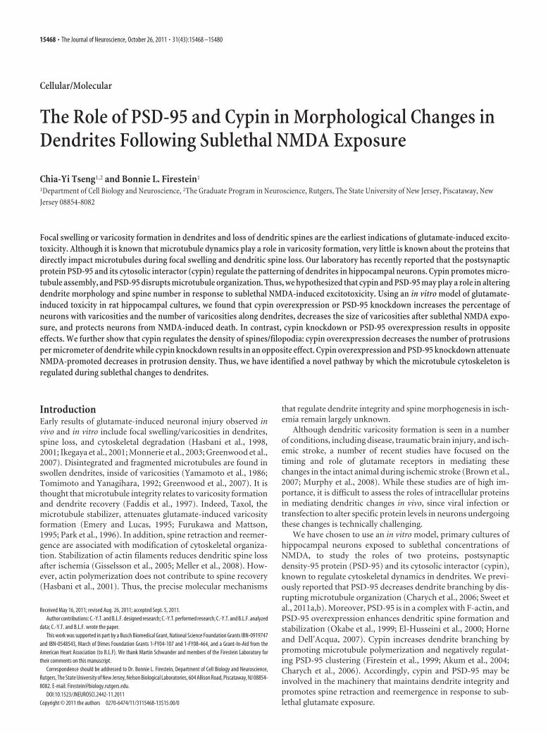
Figure 1. Changes in dendrite morphology following sublethal excitotoxic exposure. Primary hippocampal neurons were transfected with cDNAs encoding GFP on DIV 14. Live cell time-lapseimaging was performed at DIV 20, and frames were collected at 5 min intervals for 75 min. Neurons were observed for 5 min before 30 �M NMDA or vehicle treatment, 10 min during 30 �M NMDAapplication (asterisk) or vehicle treatment, and 60 min after returning to recovery medium. Representative live cell time-lapse imaging is shown. A, Dendrite and protrusion morphology remainsstable in the vehicle-treated condition. B, Varicosities form within minutes after application of 30 �M NMDA (plus symbol) and a number of them persist after recovery for 60 min. A number ofprotrusions retract during NMDA treatment. Some of the retracted protrusions reemerge when the neurons are returned to recovery medium (arrow) while some do not (arrowhead).Scale bars: 10 �m.
Tseng and Firestein • Cypin and PSD-95 in Dendritic Changes J. Neurosci., October 26, 2011 • 31(43):15468 –15480 • 15469

NMDA treatment, immunocytochemistry,and imaging. Treatment with a sublethal doseof NMDA (Sigma) was performed as previ-ously described (Hasbani et al., 1998) withsome modification. All steps were performed at37°C. NMDA was diluted in HEPES-bufferedbalanced salt solution (HBBSS; 200 mg/lCaCl2, 400 mg/l KCl, 77.3 mg/l MgCl2, 3 g/lNaCl, 2.2 g/l NaHCO3, 125 mg/l NaH2PO4
H2O, 4.5 g/l D-glucose, 2.6 g/l HEPES, pH 7.4).Briefly, on DIV 20, neurons expressing the ap-propriate constructs were exposed to 30 �M
NMDA for 10 min. In parallel, sister cultureswere treated only with HBBSS buffer to serve ascontrols. At the end of NMDA application, re-covery groups were returned to HBBSS buffercontaining 10 �M MK-801 (Sigma).
For fixed cell time-lapse imaging, exposuresor recoveries were terminated by fixation in 4%paraformaldehyde in PBS for 10 min at the ap-propriate time points. Neurons were visualizedby immunofluorescence under a 60� oil-immersion objective on an Olympus IX50 mi-croscope with a Cooke Sensicam CCD-cooledcamera, fluorescence, imaging system, and ImagePro software. The experimenter was blinded tothe condition when taking images and analyzingdendrite and spine morphology. For cell viabilityexperiments, fixed neurons were stained withrabbit anti-MAP2 (1:500) and chicken anti-GFP(1:500) followed by secondary antibodies conju-gated to Cy2 or Cy5. Nuclei were stained usingDAPI (Sigma) to determine cell survival. Stainedneurons were visualized by immunofluorescenceunder a 10� objective. Thirty different objectiveviews were randomly selected from three inde-pendent experiments. Only GFP-positive neu-rons with clear neuronal morphology wereexamined. The experimenter was blinded to the condition when taking im-ages and counting. Neurons with negative MAP2 immunostaining and con-densed DAPI staining were considered dead.
For live cell time-lapse imaging, culture medium was replaced withHBBSS buffer for 30 min before imaging, and neurons were placed ontoa heated stage. Images were acquired using the same equipment as de-scribed above at 5 min intervals for up to 75 min. One picture wascaptured before adding NMDA, followed by three pictures duringNMDA application, and finally, 13 pictures during the recovery stage. Tomodify microtubule organization, either 10 �g/ml Taxol (Sigma) or 10�g/ml nocodazole (Sigma) was applied 30 min before NMDA treatmentuntil the end of recording. Control sister cultures were exposed to vehicleconditions. For examination of presynaptic contacts for protrusions,neurons were fixed with 4% paraformaldehyde in PBS for 10 min imme-diately after the end of recovery. Neurons were stained with chickenanti-GFP (1:500) and rabbit anti-synapsin I (1:500) followed by second-ary antibodies conjugated to Cy2 and Cy5. Dendrites at least 10 �m awayfrom the soma were randomly chosen, and the percentage of protru-sions that appose immunostained clusters of the presynaptic markersynapsin I was determined. In addition to fixed cells, at least three livecells, imaged over time, were examined.
For endogenous PSD-95 cluster immunostaining, neurons expressing theappropriate constructs were fixed on DIV 20. Neurons were stained withmouse anti-PSD-95 (1:500), anti-synapsin I (1:500), and chicken anti-GFP(1:500) followed by secondary antibodies conjugated with Cy2, Cy3, andCy5. Dendrites at least 10 �m away from the soma were randomly chosen,and the percentage of PSD-95 clusters that appose synapsin I immunostain-ing versus total number of clusters was counted.
Statistical significance (p � 0.05) was determined using ANOVA fol-lowed by the appropriate post hoc test using GraphPad InStat or Graph-Pad Prism software.
Analysis of dendrite and protrusion morphology. All images were ana-lyzed using NIH ImageJ software. Quantitative analyses of dendriteand protrusion morphology are from fixed cell time-lapse imaging. Adendritic swelling is considered a varicosity when it is twice the width ofthe dendritic shaft. Neurons were considered beaded if they had threevaricosities along the same dendrite. Thirty neurons from three indepen-dent experiments were counted at each time point of each group. Datawere expressed as percentage beaded neurons (normalized to total neu-rons counted). In a beaded neuron, three dendrites at least 10 �m awayfrom the soma were randomly chosen, and the number of beaded struc-tures along the same 10 �m length of dendrite was counted. The datawere expressed as the number of varicosities per micrometer of dendrite.In addition, the area of each varicosity on the same dendrite was exam-ined, and the result was represented as varicosity size (�m 2).
Both filopodia and spines were counted as protrusions in order not torule out any alterations in spine morphology due to NMDA-inducedexcitotoxicity (Hasbani et al., 2001). Two dendrites per neuron wererandomly chosen, and the number of protrusions from a particularlength of dendrite (10 �m) was counted. The result was expressed asnumber of protrusions per �m of dendrite. Statistical differences (p �0.05) were determined using ANOVA followed by the appropriate posthoc test using GraphPad InStat or GraphPad Prism software.
Western blotting. Hippocampal cultures (DIV 20) were scraped into150 �l of TEE (20 mM TrisHCl pH 7.4, 1 mM EDTA, 1 mM EGTA) with1% Triton X-100 at the appropriate time points after NMDA application.Neurons were homogenized and lysed by passage through a 25.5 gaugeneedle 20 times. The homogenates were spun at 1000 � g for 10 min. Foranalysis of proteins released into the medium, HBBSS buffer (medium)was collected at the appropriate time points after NMDA application.Amicon Ultra-4 Centrifugal Filter Unit with Ultracel-10 membrane(Millipore) was used to concentrate the medium 20-fold. Bradford assays
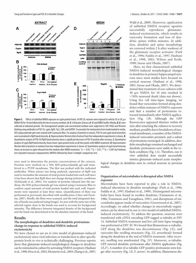
Figure 2. The organization of microtubules is disturbed after exposure to sublethal NMDA. Primary hippocampal neurons weretransfected with cDNAs encoding �-tubulin-GFP on DIV 14. On DIV 20, neurons were treated with vehicle or 30 �M NMDA. A–L,Frames were captured at 0 min (before NMDA treatment; A, D, G, J ), 15 min (end of NMDA treatment; B, E, H, K ), and 75 min (endof recovery after NMDA treatment; C, F, I, L) from live cell time-lapse imaging. Nocodazole (10 �g/ml; G–I ) or Taxol (10 �g/ml;J–L) was applied to the cultures 30 min before NMDA treatment until the end of recording. Arrowheads in E indicate regions ofswelling. Arrows in E and F or H and I indicate that protrusions that contain �-tubulin-GFP that are longer after recovery. Scale bar,10 �m.
15470 • J. Neurosci., October 26, 2011 • 31(43):15468 –15480 Tseng and Firestein • Cypin and PSD-95 in Dendritic Changes
were used to determine the protein concentrations of the extracts.Proteins were resolved on a 10% SDS-polyacrylamide gel and trans-ferred to a PVDF membrane. The blot was probed with the indicatedantibodies. When extract was being analyzed, expression of Rpl9 wasused to normalize the amount of total protein loaded into each well sinceit has been shown that Rpl9 does not change during ischemic conditions(Kobayashi et al., 2004). For analysis of proteins released into the me-dium, the SDS-polyacrylamide gel was stained using Coomassie Blue toconfirm equal amounts of total protein loaded into each well. Experi-ments were repeated at least three times. Quantification of blots wasperformed as previously described with modification (Chen and Firest-ein, 2007). Briefly, the blots were scanned, and the total integrated den-sity of bands was analyzed using ImageJ. An area with the same size of theselected region close to the bands was used to account for backgroundintensity. The difference between integrated density of the backgroundand the band was determined to be the absolute intensity of the band.
ResultsThe morphologies of dendrites and dendritic protrusionschange in response to sublethal NMDA-inducedexcitotoxicityWe have chosen to use an in vitro model of glutamate-inducedexcitotoxicity since viral infection or transfection to alter specificprotein levels in vivo is technically challenging. Previous reportsshow that glutamate-induced morphological changes in dendritescan be mimicked in culture by activating NMDA receptors (Hasbaniet al., 1998; Hou et al., 2002; Monnerie et al., 2003; Zhang et al., 2007;
Wahl et al., 2009). Moreover, applicationof sublethal NMDA receptor agonistssuccessfully reproduces glutamate-induced excitotoxicity, which results invaricosity formation and loss of den-dritic spines within minutes. In addi-tion, dendrite and spine morphologyare restored within 2 h after washout ofthe glutamate receptor activator (Parket al., 1996; Faddis et al., 1997; Hasbaniet al., 1998, 2001; Wilson and Keith,1998; Inoue and Okada, 2007).
Here, we first characterized sublethalNMDA-induced morphological changesin dendrites in primary hippocampal neu-rons since most studies have focused oncortical neurons (Hasbani et al., 1998,2001; Inoue and Okada, 2007). We deter-mined that treatment of our cultures with30 �M NMDA for 10 min resulted in�50% neuronal death (data not shown).Using live cell time-lapse imaging, wefound that varicosities formed along den-drites within minutes of NMDA exposureand that a number of protrusions re-tracted immediately after NMDA applica-tion (Fig. 1B). Although the GFPfluorescence appeared dimmer within 60min after returning the cultures to recoverymedium, possibly due to breakdown of neu-ronal membranes, a number of the NMDA-induced varicosities persisted, and a subsetof protrusions reemerged. In addition, den-drite morphology remained unchanged anddendritic protrusions were stable in the ve-hicle condition (Fig. 1A). Therefore, our invitro sublethal dose model successfullymimics glutamate-induced acute morpho-
logical changes in dendrites seen in cortical neurons in previousstudies.
Organization of microtubules is disrupted after NMDAexposureMicrotubules have been reported to play a role in NMDA-induced alterations in dendrite morphology (Park et al., 1996;Faddis et al., 1997; Hasbani et al., 1998). Disintegrated microtu-bules have been found in swollen dendrites (Yamamoto et al.,1986; Tomimoto and Yanagihara, 1992), and disruptions of mi-crotubules appear inside of varicosities (Greenwood et al., 2007).Accordingly, we asked whether changes in microtubule organi-zation can be observed in our in vitro model of sublethal NMDA-induced excitotoxicity. To address the question, neurons weretransfected with cDNA encoding GFP-tagged �-tubulin at DIV14. Sublethal NMDA treatment and live cell time-lapse imagingwas performed at DIV 20. We found that filamentous �-tubulin-GFP along the dendrites was discontinuous (Fig. 2 E), andvaricosity-like swelling structures (Fig. 2E, arrowhead) formedalong the dendrites at the end of NMDA treatment. After recov-ery, swelling was ameliorated (Fig. 2F). Moreover, �-tubulin-GFP entered dendritic protrusions after NMDA application (Fig.2E,F). A number of �-tubulin-GFP-positive protrusions were lon-ger after recovery (Fig. 2E, F, arrow). In addition, filamentous dis-
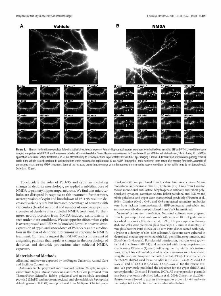
Figure 3. Effects of sublethal NMDA exposure on cypin protein levels. At DIV 20, neurons were exposed to vehicle (V) or 30 �M
NMDA (N) for 10 min followed by 60 min in recovery medium. A–E, Cell lysates (Extract; A–C) and HBBSS buffer (Media; D, E) werecollected at indicated periods. The homogenized samples and concentrated medium were subjected to SDS-PAGE and Westernblotting using antibodies to PSD-95, cypin, Rpl9, Tuj1, LDH, and GAPDH. To normalize for total protein from media loaded in media,SDS-polyacrylamide gels were stained with Coomassie Blue. For analysis of proteins in extract, PSD-95 and cypin band intensitieswere normalized to Rpl9 band intensity. A, Representative Western blot of extract from five independent experiments is shown. B,Quantitative analysis of PSD-95/Rpl9 band intensity shows a significant decrease in PSD-95 protein after recovery. C, Quantitativeanalysis of cypin/Rpl9 band intensity shows lower cypin protein levels at all time points with NMDA treatment. D, RepresentativeWestern blot of proteins in medium from four independent experiments is shown. E, Quantitative analysis of cypin band intensityshows an increase in cypin released into the medium after NMDA treatment. *p � 0.05, **p � 0.01, ***p � 0.001 by Friedmantest (repeated-measures nonparametric ANOVA) followed by Dunn’s multiple-comparisons test.
Tseng and Firestein • Cypin and PSD-95 in Dendritic Changes J. Neurosci., October 26, 2011 • 31(43):15468 –15480 • 15471
tribution of �-tubulin-GFP along thedendrite remained unchanged during vehi-cle treatment (Fig. 2A–C).
We then asked whether pharmacolog-ical manipulations of the cytoskeletoncould alter dendritic swelling after sub-lethal NMDA application in our in vitromodel. Taxol, a microtubule stabilizer,has been shown to reduce dendritic swell-ing in response to sublethal stimulation ofthe NMDA receptor (Emery and Lucas,1995; Furukawa and Mattson, 1995; Parket al., 1996). Taxol was applied to neurons30 min before NMDA treatment until theend of recording. We found that filamen-tous �-tubulin-GFP along dendrites re-mained intact and stable during and afterNMDA treatment (Fig. 2 J–L). In addi-tion, we treated neurons with nocodazole,a microtubule-depolymerizing agent. Weobserved that microtubules were dis-rupted before NMDA treatment (Fig.2G). As expected, a more severe disorga-nization of microtubules was observed af-ter NMDA receptor activation, whichpersisted throughout the recovery period(Fig. 2H, I). A number of �-tubulin-GFP-positive protrusions were longer after re-covery (Fig. 2H, I, arrow). Moreover,�-tubulin-GFP fluorescence was dimmerin nocodazole-treated neurons than incontrol neurons. Together, our data sug-gest that the disruption of microtubuleorganization plays a role in varicosity for-mation in neurons exposed to sublethalconcentrations of NMDA. Our data alsoshow that tubulin enters into dendritic pro-trusions after NMDA application, elucidat-ing the potential role of microtubules inNMDA-mediated changes in dendritemorphology.
PSD-95 and cypin protein decreaseafter exposure to a sublethal doseof NMDABecause PSD-95 and cypin play a role inthe modification of microtubule dynam-ics (Akum et al., 2004; Charych et al.,2006; Chen and Firestein, 2007; Sweet etal., 2011a,b), we next asked whether pro-tein levels of PSD-95 and cypin maychange in parallel with the alteration ofmicrotubule organization after sublethal NMDA application. Weperformed Western blot analysis from cellular extracts to deter-mine protein expression of PSD-95 and cypin with shock NMDAtreatment, after 10 min of NMDA treatment, and after 10 min ofNMDA treatment followed by 60 min recovery. Control neuronswere exposed to vehicle treatment. There is no significant differ-ence in the levels of Rpl9 protein, which should not change inresponse to treatment (Kobayashi et al., 2004), between con-trol and NMDA-treated groups (data not shown). We found thatPSD-95 protein significantly decreases after 10 min treatmentfollowed by 60 min recovery in the NMDA-treated group (Fig.
3A,B), and cypin protein is significantly lower in the NMDA-treated than control cultures at all time points (Fig. 3 A, C). Itwas surprising to us that cypin protein expression decreasedwithin such a short time after exposure to NMDA. We askedwhether cypin is released from the cells as a result of treatment. Toaddress this question, we collected and concentrated the mediumwith shock NMDA treatment, after 10 min of NMDA treatment,and after 10 min of NMDA treatment followed by 60 min recoveryand analyzed cypin protein levels by Western blot analysis. Controlneurons were exposed to vehicle. Interestingly, we found that themedium contained �25% of cypin levels found in cellular extract
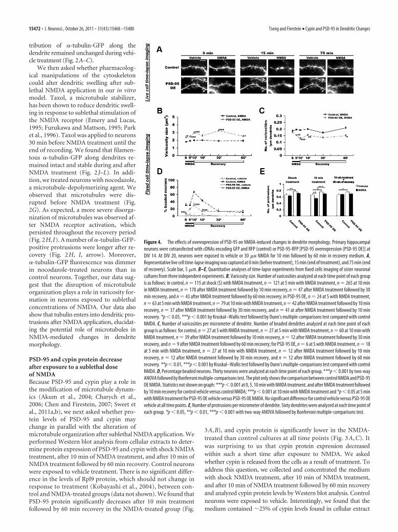
Figure 4. The effects of overexpression of PSD-95 on NMDA-induced changes in dendrite morphology. Primary hippocampalneurons were cotransfected with cDNAs encoding GFP and RFP (control) or PSD-95-RFP [PSD-95 overexpression (PSD-95 OE)] atDIV 14. At DIV 20, neurons were exposed to vehicle or 30 �M NMDA for 10 min followed by 60 min in recovery medium. A,Representative live cell time-lapse imaging was captured at 0 min (before treatment), 15 min (end of treatment), and 75 min (endof recovery). Scale bar, 5 �m. B–E, Quantitative analyses of time-lapse experiments from fixed cells imaging of sister neuronalcultures from three independent experiments. B, Varicosity size. Number of varicosities analyzed at each time point of each groupis as follows: in control, n � 115 at shock (S) with NMDA treatment, n � 121 at 5 min with NMDA treatment, n � 265 at 10 minin NMDA treatment, n � 178 after NMDA treatment followed by 10 min recovery, n � 47 after NMDA treatment followed by 30min recovery, and n � 43 after NMDA treatment followed by 60 min recovery; in PSD-95 OE, n � 24 at S with NMDA treatment,n � 63 at 5 min with NMDA treatment, n � 79 at 10 min with NMDA treatment, n � 42 after NMDA treatment followed by 10 minrecovery, n � 37 after NMDA treatment followed by 30 min recovery, and n � 41 at after NMDA treatment followed by 10 minrecovery. *p � 0.05, ***p � 0.001 by Kruskal–Wallis test followed by Dunn’s multiple-comparisons test compared with controlNMDA. C, Number of varicosities per micrometer of dendrite. Number of beaded dendrites analyzed at each time point of eachgroup is as follows: for control, n � 27 at S with NMDA treatment, n � 27 at 5 min with NMDA treatment, n � 60 at 10 min withNMDA treatment, n � 39 after NMDA treatment followed by 10 min recovery, n � 12 after NMDA treatment followed by 30 minrecovery, and n � 9 after NMDA treatment followed by 60 min recovery; for PSD-95 OE, n � 6 at S with NMDA treatment, n � 18at 5 min with NMDA treatment, n � 27 at 10 min with NMDA treatment, n � 12 after NMDA treatment followed by 10 minrecovery, n � 12 after NMDA treatment followed by 30 min recovery, and n � 12 after NMDA treatment followed by 60 minrecovery. **p � 0.01, ***p � 0.001 by Kruskal–Wallis test followed by Dunn’s multiple-comparisons test compared with controlNMDA. D, Percentage beaded neurons. Thirty neurons were analyzed at each time point of each group. ***p � 0.001 by two-wayANOVA followed by Bonferroni multiple-comparisons test. The plot only shows the comparison between control NMDA and PSD-95OE NMDA. Statistics not shown on graph: ***p � 0.001 at 0, 5, 10 min with NMDA treatment, and after NMDA treatment followedby 10 min recovery for control vehicle versus control NMDA; ***p � 0.001 at 10 min with NMDA treatment and *p � 0.05 at 5 minwith NMDA treatment for PSD-95 OE vehicle versus PSD-95 OE NMDA. No significant difference for control vehicle versus PSD-95 OEvehicle at all time points. E, Number of protrusions per micrometer of dendrite. Sixty dendrites were analyzed at each time point ofeach group. *p � 0.05, **p � 0.01, ***p � 0.001 with two-way ANOVA followed by Bonferroni multiple-comparisons test.
15472 • J. Neurosci., October 26, 2011 • 31(43):15468 –15480 Tseng and Firestein • Cypin and PSD-95 in Dendritic Changes

under vehicle conditions; however, we de-tected no LDH, Tuj1, PSD-95, or GAPDHin the medium under these conditions,which would reflect general cell death (Fig.3D,E). The amount of cypin found in themedium doubled at shorter exposures toNMDA (shock, 10 min) while neither LDHnor GAPDH was released (Fig. 3 D, E),implying that cypin is specifically re-leased into the medium as a result ofNMDA exposure. It is important to notethat all proteins examined increased inthe medium after 10 min NMDA treat-ment followed by 60 min recovery. Sincethere was no increase in cypin found inthe media in the vehicle group (Fig.3 D, E), the increases in release of all pro-teins most likely represent significantchanges in membrane integrity in responseto NMDA treatment. Thus, it appears thatcypin is released from neurons at all timepoints and PSD-95 is released at the 60 min/recovery time point, suggesting that releaseof cypin occurs in response to NMDAtreatment.
PSD-95 plays a role in NMDA-inducedvaricosity formationAs seen in Figures 2 and 3, the organizationof microtubules is disrupted and PSD-95protein levels are altered in response to sub-lethal NMDA exposure. Our previous find-ings show that overexpression of PSD-95decreases dendrite branching by disruptingmicrotubule dynamics (Charych et al.,2006; Sweet et al., 2011a,b). Therefore, wehypothesized that alterations in PSD-95protein expression would affect NMDA-induced varicosity formation. To test thehypothesis, PSD-95 protein was either in-creased (Fig. 4) or knocked down (Fig. 5) bytransfecting neurons with the appropriateconstructs at DIV 14. Sublethal NMDA ex-posure followed by live cell (Figs. 4A, 5A) orfixed cell (Figs. 4B–D, 5B–D) time-lapse im-aging was performed at DIV 20. Live celltime-lapse imaging was used to monitormorphological changes in the same den-drites while fixed cell time-lapse imagingwas used to avoid photobleaching and out-of-focus imaging. Quantitative analyses ofdendrite and protrusion morphology arefrom fixed cell time-lapse imaging. Wefound that sublethal NMDA treatment re-sulted in a significant decrease in thenumber of neurons with varicosities whenPSD-95 was overexpressed during NMDAtreatment and at the beginning of recov-ery (Fig. 4D). In beaded neurons, neuronsoverexpressing PSD-95 showed a decreasein the number of varicosities per micro-meter of dendrite (Fig. 4C) and an in-crease in the size of varicosities (Fig. 4B)
Figure 5. Effects of PSD-95 knockdown on NMDA-induced changes in dendrite morphology. Primary hippocampal neurons weretransfected at DIV 14 with control shRNA (control), or PSD-95 shRNA without [PSD-95 knockdown (PSD-95 shRNA)] or with [PSD-95knockdown plus rescue (PSD-95 shRNA�R)] cDNA encoding silent mutations in PSD-95 in the region targeted by our shRNA. At DIV 20,neurons were exposed to vehicle or 30�M NMDA for 10 min followed by 60 min in recovery medium. A, Representative live cell time-lapseimaging were captured at 0 min (before treatment), 15 min (end of treatment), and 75 min (end of recovery). Scale bar, 5 �m. B–E,Quantitative analyses of time-lapse experiment from fixed cell imaging of sister neuronal cultures from three independent experiments inB–D and from two independent experiments in E. B, Varicosity size. Number of varicosities analyzed at each time point of each group is asfollows: for control, n �108 at shock (S) with NMDA treatment, n �157 at 5 min with NMDA treatment, n �197 at 10 min with NMDAtreatment, n�206 after NMDA treatment followed by 10 min recovery, n�130 after NMDA treatment followed by 30 min recovery, andn�36 after NMDA treatment followed by 60 min recovery; for PSD-95 shRNA, n�172 at S with NMDA treatment, n�224 at 5 min withNMDA treatment, n � 324 at 10 min with NMDA treatment, n � 177 after NMDA treatment followed by 10 min recovery, n � 225 afterNMDA treatment followed by 30 min recovery, and n � 153 after NMDA treatment followed by 60 min recovery; for PSD-95 shRNA�R,n�52 at S with NMDA treatment, n�65 at 5 min with NMDA treatment, n�154 at 10 min with NMDA treatment, n�129 after NMDAtreatment followed by 10 min recovery, n � 94 after NMDA treatment followed by 30 min recovery, and n � 83 after NMDA treatmentfollowed by 60 min recovery. *p�0.05, **p�0.01, ***p�0.001 by Kruskal–Wallis test followed by Dunn’s multiple-comparisons test.The plot only shows the comparison of PSD-95 shRNA NMDA or PSD-95 shRNA�R NMDA to control NMDA. Statistics not shown on graph:***p � 0.001 at all time points for PSD-95 shRNA NMDA versus PSD-95 shRNA�R NMDA. C, Number of varicosities per micrometer ofdendrite. Number of beaded dendrites analyzed at each time point of each group is as follows: for control, n � 27 at S with NMDAtreatment, n�36 at 5 min with NMDA treatment, n�48 at 10 min with NMDA treatment, n�51 after NMDA treatment followed by 10min recovery, n�33 after NMDA treatment followed by 30 min recovery, and n�9 after NMDA treatment followed by 60 min recovery;forPSD-95shRNA,n�33atSwithNMDAtreatment,n�48at5minwithNMDAtreatment,n�69at10minwithNMDAtreatment,n�36afterNMDAtreatmentfollowedby10minrecovery,n�45afterNMDAtreatmentfollowedby30minrecovery,andn�30afterNMDAtreatment followed by 60 min recovery; for PSD-95 shRNA�R, n�15 at S with NMDA treatment, n�18 at 5 min with NMDA treatment,n � 39 at 10 min with NMDA treatment, n � 36 after NMDA treatment followed by 10 min recovery, n � 24 after NMDA treatmentfollowed by 30 min recovery, and n � 24 after NMDA treatment followed by 60 min recovery. *p � 0.05, **p � 0.01, ***p � 0.001 byKruskal–Wallis test followed by Dunn’s multiple-comparisons test. The plot only shows the comparison of PSD-95 shRNA NMDA or PSD-95shRNA�R NMDA with control NMDA. Statistics not shown on graph: not significant at 5 min, *p � 0.05 at 10 min with NMDA treatmentandafterNMDAtreatmentfollowedby10minrecovery,and***p�0.001at0minwithNMDAtreatment,afterNMDAtreatmentfollowedby 10 and 60 min recovery for PSD-95 shRNA NMDA versus PSD-95 shRNA�R NMDA. D, Percentage beaded neurons. Thirty neurons wereanalyzed at each time point of each group. *p�0.05, ***p�0.001 with two-way ANOVA followed by Bonferroni multiple-comparisonstest. The plot only shows the comparison of PSD-95 shRNA NMDA or PSD-95 shRNA�R NMDA with control NMDA. Statistics not shown ongraph: not significant after NMDA treatment followed by 10 and 60 min recovery, *p�0.05 at 0 min with NMDA treatment, and ***p�0.001 at 5, 10 min with NMDA treatment, and after NMDA treatment followed by 10 min recovery for PSD-95 shRNA NMDA versus PSD-95shRNA�RNMDA;***p�0.001forcontrolvehicleversuscontrolNMDAinall timepointsexceptafterNMDAtreatmentfollowedby60minrecovery (p � 0.05); ***p � 0.001 for PSD-95 shRNA vehicle versus PSD-95 shRNA NMDA in all time points. Not significant at 0 min withNMDA treatment, **p�0.01 at 5 min with NMDA treatment, and ***p�0.001 at 10 min with NMDA treatment, after NMDA treatmentfollowed by 10, 30, and 60 min recovery for PSD-95 shRNA�R vehicle versus PSD-95 shRNA�R NMDA; not significant for control vehicleversus PSD-95 shRNA vehicle, control vehicle with PSD-95 shRNA�R vehicle, and PSD-95 shRNA vehicle with PSD-95 shRNA�R vehicle inall time points. E, Number of protrusions per micrometer of dendrite. Forty dendrites were analyzed at each time point of each group. *p�0.05, **p � 0.01, ***p � 0.001 with two-way ANOVA followed by Bonferroni multiple-comparisons test.
Tseng and Firestein • Cypin and PSD-95 in Dendritic Changes J. Neurosci., October 26, 2011 • 31(43):15468 –15480 • 15473

when compared with control neurons.In addition, the size of varicosities re-mained elevated when neurons overex-pressing PSD-95 were returned torecovery medium for 60 min (Fig. 4 B).
In line with these data, knockdownof PSD-95 resulted in a significant in-crease in the number of neurons withvaricosities at the end of sublethalNMDA treatment and at 60 min afterreturning the cultures to recovery me-dium (Fig. 5D). An increase in the num-ber of varicosities per micrometer ofdendrite (Fig. 5C) and a decrease in thesize of varicosities (Fig. 5B) were ob-served in neurons with PSD-95 knockeddown. To demonstrate the specificity ofour PSD-95 shRNA, we coexpressed acDNA encoding silent mutations inPSD-95 in the region targeted by ourshRNA. As expected, the silent mutantPSD-95 rescued the effects of PSD-95knockdown (Fig. 5B–D). The effects ofPSD-95 rescue either returned varicos-ity characteristics to control levels or inbetween the levels of control and over-expression. This is expected as we can-not control for how much knockeddown PSD-95 is replaced by the silentmutant. Together, these results indicatethat PSD-95 plays a role in mediating sub-lethal NMDA-induced varicosity formationin dendrites.
Cypin plays a role in NMDA-inducedvaricosity formationPrevious data from our laboratory indi-cate that binding of cypin to PSD-95 re-sults in a decrease in PSD-95 clusteringand delocalization (Firestein et al., 1999;Charych et al., 2006). Moreover, cypin in-teracts with and acts upstream of PSD-95in dendritogenesis (Charych et al., 2006).Cypin increases dendrite branching bypromoting microtubule polymerizationand negatively regulating the synaptic lo-calization of PSD-95 (Akum et al., 2004;Charych et al., 2006; Chen and Firestein,2007). In addition, cypin protein levels arealtered in response to sublethal NMDAexposure (Fig. 3). Therefore, we asked whether cypin plays a rolein mediating changes in dendrite morphology after sublethalNMDA exposure. To answer this question, cypin protein waseither increased (Fig. 6) or knocked down (Fig. 7) by transfectingneurons with the appropriate constructs on DIV 14. SublethalNMDA exposure followed by live cell (Figs. 6A, 7A) or fixed cell(Figs. 6B–D, 7B–D) time-lapse imaging was performed on DIV20. We found that sublethal NMDA treatment resulted in anincrease in the number of beaded neurons in neurons overex-pressing cypin at the end of NMDA treatment and at the begin-ning of recovery (Fig. 6D). In beaded neurons, an increase in thenumber of varicosities per micrometer of dendrite (Fig. 6C) and
a decrease in the size of varicosities (Fig. 6B) were found in neu-rons overexpressing cypin.
Knockdown of cypin resulted in a significant decrease inthe number of beaded neurons during sublethal NMDA treat-ment and at the beginning of recovery (Fig. 7D). Decreasednumber of varicosities per micrometer of dendrite (Fig. 7C)and increased size of varicosities (Fig. 7B) were found in neu-rons with cypin knocked down. In addition, the size of vari-cosities in neurons with cypin knocked down was elevatedeven when cultures were returned to recovery medium for 60min. To demonstrate the specificity of our cypin shRNA, wecoexpressed a cDNA encoding a single point mutation in the
Figure 6. Effects of cypin overexpression on NMDA-induced changes in dendrite morphology. Primary hippocampal neuronswere transfected with cDNAs encoding GFP (control) or GFP-cypin (cypin OE) at DIV 14. At DIV 20, neurons were exposed to vehicleor 30 �M NMDA for 10 min followed by 60 min in recovery medium. A, Representative live cell time-lapse imaging were capturedat 0 min (before treatment), 15 min (end of treatment), and 75 min (end of recovery). Scale bar, 5 �m. B–E, Quantitative analysesof time-lapse experiments from fixed cell imaging of sister neuronal cultures from three independent experiments. B, Varicositysize. Number of varicosities analyzed at each time point of each group is as follows: for control, n � 220 at shock (S) with NMDAtreatment, n � 219 at 5 min with NMDA treatment, n � 230 at 10 min with NMDA treatment, n � 131 after NMDA treatmentfollowed by 10 min recovery, n � 144 after NMDA treatment followed by 30 min recovery, and n � 103 after NMDA treatmentfollowed by 60 min recovery; for cypin OE, n � 242 at S with NMDA treatment, n � 298 at 5 min with NMDA treatment, n � 370at 10 min with NMDA treatment, n � 276 after NMDA treatment followed by 10 min recovery, n � 220 after NMDA treatmentfollowed by 30 min recovery, and n � 210 after NMDA treatment followed by 60 min recovery. *p � 0.05; ***p � 0.001 byKruskal–Wallis test followed by Dunn’s multiple-comparisons test as compared with control NMDA. C, Number of varicosities permicrometer of dendrite. Number of beaded dendrites analyzed at each time point of each group is as follows: for control, n � 48at S with NMDA treatment, n � 57 at 5 min with NMDA treatment, n � 57 at 10 min with NMDA treatment, n � 39 after NMDAtreatment followed by 10 min recovery, n � 33 after NMDA treatment followed by 30 min recovery, and n � 27 after NMDAtreatment followed by 60 min recovery; for cypin OE, n � 51 at S with NMDA treatment, n � 63 at 5 min with NMDA treatment,n�75 at 10 min with NMDA treatment, n�57 after NMDA treatment followed by 10 min recovery, n�48 after NMDA treatmentfollowed by 30 min recovery, and n�42 after NMDA treatment followed by 60 min recovery. **p�0.01; ***p�0.001 by ANOVAwith Kruskal–Wallis test followed by Dunn’s multiple-comparisons test compared with control NMDA. D, Percentage beadedneurons. Thirty neurons were analyzed at each time point of each group. *p � 0.05 by two-way ANOVA followed by Bonferronimultiple-comparisons test. The plot only shows the comparison between control NMDA and cypin OE NMDA. Statistics not shownon graph: ***p � 0.001 for control vehicle versus control NMDA and cypin OE vehicle versus cypin OE NMDA at all time points; notsignificant for control vehicle versus cypin OE vehicle in all time points. E, Number of protrusions per micrometer of dendrite. Sixtydendrites were analyzed at each time point of each group. *p � 0.05; **p � 0.01 by two-way ANOVA followed by Bonferronimultiple-comparisons test.
15474 • J. Neurosci., October 26, 2011 • 31(43):15468 –15480 Tseng and Firestein • Cypin and PSD-95 in Dendritic Changes
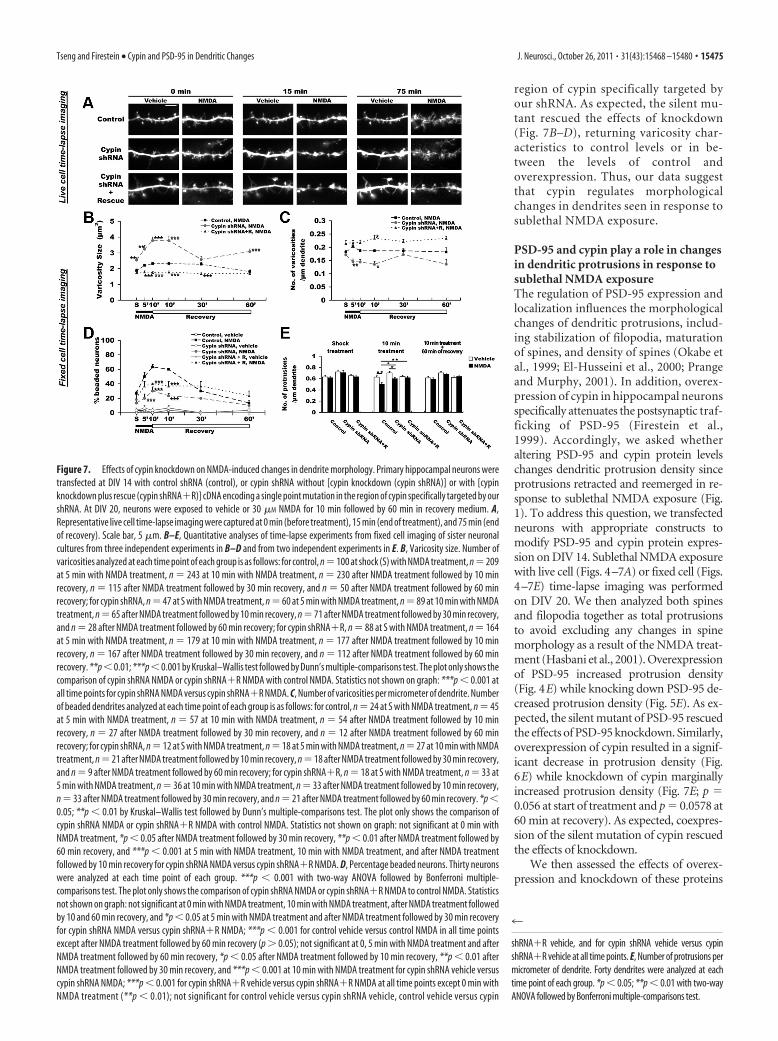
region of cypin specifically targeted byour shRNA. As expected, the silent mu-tant rescued the effects of knockdown(Fig. 7B–D), returning varicosity char-acteristics to control levels or in be-tween the levels of control andoverexpression. Thus, our data suggestthat cypin regulates morphologicalchanges in dendrites seen in response tosublethal NMDA exposure.
PSD-95 and cypin play a role in changesin dendritic protrusions in response tosublethal NMDA exposureThe regulation of PSD-95 expression andlocalization influences the morphologicalchanges of dendritic protrusions, includ-ing stabilization of filopodia, maturationof spines, and density of spines (Okabe etal., 1999; El-Husseini et al., 2000; Prangeand Murphy, 2001). In addition, overex-pression of cypin in hippocampal neuronsspecifically attenuates the postsynaptic traf-ficking of PSD-95 (Firestein et al.,1999). Accordingly, we asked whetheraltering PSD-95 and cypin protein levelschanges dendritic protrusion density sinceprotrusions retracted and reemerged in re-sponse to sublethal NMDA exposure (Fig.1). To address this question, we transfectedneurons with appropriate constructs tomodify PSD-95 and cypin protein expres-sion on DIV 14. Sublethal NMDA exposurewith live cell (Figs. 4–7A) or fixed cell (Figs.4–7E) time-lapse imaging was performedon DIV 20. We then analyzed both spinesand filopodia together as total protrusionsto avoid excluding any changes in spinemorphology as a result of the NMDA treat-ment (Hasbani et al., 2001). Overexpressionof PSD-95 increased protrusion density(Fig. 4E) while knocking down PSD-95 de-creased protrusion density (Fig. 5E). As ex-pected, the silent mutant of PSD-95 rescuedthe effects of PSD-95 knockdown. Similarly,overexpression of cypin resulted in a signif-icant decrease in protrusion density (Fig.6E) while knockdown of cypin marginallyincreased protrusion density (Fig. 7E; p �0.056 at start of treatment and p � 0.0578 at60 min at recovery). As expected, coexpres-sion of the silent mutation of cypin rescuedthe effects of knockdown.
We then assessed the effects of overex-pression and knockdown of these proteins
Figure 7. Effects of cypin knockdown on NMDA-induced changes in dendrite morphology. Primary hippocampal neurons weretransfected at DIV 14 with control shRNA (control), or cypin shRNA without [cypin knockdown (cypin shRNA)] or with [cypinknockdown plus rescue (cypin shRNA�R)] cDNA encoding a single point mutation in the region of cypin specifically targeted by ourshRNA. At DIV 20, neurons were exposed to vehicle or 30 �M NMDA for 10 min followed by 60 min in recovery medium. A,Representative live cell time-lapse imaging were captured at 0 min (before treatment), 15 min (end of treatment), and 75 min (endof recovery). Scale bar, 5 �m. B–E, Quantitative analyses of time-lapse experiments from fixed cell imaging of sister neuronalcultures from three independent experiments in B–D and from two independent experiments in E. B, Varicosity size. Number ofvaricosities analyzed at each time point of each group is as follows: for control, n�100 at shock (S) with NMDA treatment, n�209at 5 min with NMDA treatment, n � 243 at 10 min with NMDA treatment, n � 230 after NMDA treatment followed by 10 minrecovery, n � 115 after NMDA treatment followed by 30 min recovery, and n � 50 after NMDA treatment followed by 60 minrecovery; for cypin shRNA, n � 47 at S with NMDA treatment, n � 60 at 5 min with NMDA treatment, n � 89 at 10 min with NMDAtreatment, n�65 after NMDA treatment followed by 10 min recovery, n�71 after NMDA treatment followed by 30 min recovery,and n � 28 after NMDA treatment followed by 60 min recovery; for cypin shRNA�R, n � 88 at S with NMDA treatment, n � 164at 5 min with NMDA treatment, n � 179 at 10 min with NMDA treatment, n � 177 after NMDA treatment followed by 10 minrecovery, n � 167 after NMDA treatment followed by 30 min recovery, and n � 112 after NMDA treatment followed by 60 minrecovery. **p�0.01; ***p�0.001 by Kruskal–Wallis test followed by Dunn’s multiple-comparisons test. The plot only shows thecomparison of cypin shRNA NMDA or cypin shRNA�R NMDA with control NMDA. Statistics not shown on graph: ***p � 0.001 atall time points for cypin shRNA NMDA versus cypin shRNA�R NMDA. C, Number of varicosities per micrometer of dendrite. Numberof beaded dendrites analyzed at each time point of each group is as follows: for control, n � 24 at S with NMDA treatment, n � 45at 5 min with NMDA treatment, n � 57 at 10 min with NMDA treatment, n � 54 after NMDA treatment followed by 10 minrecovery, n � 27 after NMDA treatment followed by 30 min recovery, and n � 12 after NMDA treatment followed by 60 minrecovery; for cypin shRNA, n � 12 at S with NMDA treatment, n � 18 at 5 min with NMDA treatment, n � 27 at 10 min with NMDAtreatment, n�21 after NMDA treatment followed by 10 min recovery, n�18 after NMDA treatment followed by 30 min recovery,and n � 9 after NMDA treatment followed by 60 min recovery; for cypin shRNA�R, n � 18 at S with NMDA treatment, n � 33 at5 min with NMDA treatment, n � 36 at 10 min with NMDA treatment, n � 33 after NMDA treatment followed by 10 min recovery,n � 33 after NMDA treatment followed by 30 min recovery, and n � 21 after NMDA treatment followed by 60 min recovery. *p �0.05; **p � 0.01 by Kruskal–Wallis test followed by Dunn’s multiple-comparisons test. The plot only shows the comparison ofcypin shRNA NMDA or cypin shRNA�R NMDA with control NMDA. Statistics not shown on graph: not significant at 0 min withNMDA treatment, *p � 0.05 after NMDA treatment followed by 30 min recovery, **p � 0.01 after NMDA treatment followed by60 min recovery, and ***p � 0.001 at 5 min with NMDA treatment, 10 min with NMDA treatment, and after NMDA treatmentfollowed by 10 min recovery for cypin shRNA NMDA versus cypin shRNA�R NMDA. D, Percentage beaded neurons. Thirty neuronswere analyzed at each time point of each group. ***p � 0.001 with two-way ANOVA followed by Bonferroni multiple-comparisons test. The plot only shows the comparison of cypin shRNA NMDA or cypin shRNA�R NMDA to control NMDA. Statisticsnot shown on graph: not significant at 0 min with NMDA treatment, 10 min with NMDA treatment, after NMDA treatment followedby 10 and 60 min recovery, and *p � 0.05 at 5 min with NMDA treatment and after NMDA treatment followed by 30 min recoveryfor cypin shRNA NMDA versus cypin shRNA�R NMDA; ***p � 0.001 for control vehicle versus control NMDA in all time pointsexcept after NMDA treatment followed by 60 min recovery (p � 0.05); not significant at 0, 5 min with NMDA treatment and afterNMDA treatment followed by 60 min recovery, *p � 0.05 after NMDA treatment followed by 10 min recovery, **p � 0.01 afterNMDA treatment followed by 30 min recovery, and ***p � 0.001 at 10 min with NMDA treatment for cypin shRNA vehicle versuscypin shRNA NMDA; ***p � 0.001 for cypin shRNA�R vehicle versus cypin shRNA�R NMDA at all time points except 0 min withNMDA treatment (**p � 0.01); not significant for control vehicle versus cypin shRNA vehicle, control vehicle versus cypin
4
shRNA�R vehicle, and for cypin shRNA vehicle versus cypinshRNA�R vehicle at all time points. E, Number of protrusions permicrometer of dendrite. Forty dendrites were analyzed at eachtime point of each group. *p � 0.05; **p � 0.01 with two-wayANOVA followed by Bonferroni multiple-comparisons test.
Tseng and Firestein • Cypin and PSD-95 in Dendritic Changes J. Neurosci., October 26, 2011 • 31(43):15468 –15480 • 15475

on changes in protrusion density in response to sublethal NMDAexposure. Overexpression of PSD-95 blocked the decrease in protru-sion density seen in response to NMDA (Fig. 4E). Knockdown ofPSD-95 occluded changes in protrusion density mediated byNMDA treatment (Fig. 5E). As expected, coexpression of the silentmutant of PSD-95 rescued the effects of PSD-95 knockdown. Simi-larly, overexpression of cypin occluded NMDA-mediated changes inprotrusion density (Fig. 6E). In contrast, knockdown of cypin re-sulted in an attenuated decrease in protrusion density at the end ofNMDA exposure (Fig. 7E). As expected, coexpression of the silentmutant of cypin rescued the effects of PSD-95 knockdown. To-gether, our data suggest that PSD-95 and cypin play a role in medi-ating changes in protrusion retraction and reemergence in responseto sublethal NMDA exposure.
Presynaptic and postsynaptic elements remain in contactduring dendritic changes in response to NMDAThe Goldberg laboratory has previously shown that presynapticboutons remain associated with or near postsynaptic compo-nents during morphological changes in cortical neurons in re-sponse to NMDA treatment (Hasbani et al., 2001). Accordingly,we asked whether elimination of dendritic spines is associatedwith loss of synaptic contact using live cell time-lapse imaging inour in vitro model of sublethal NMDA-induced excitotoxicity,and whether altering cypin or PSD-95 protein expression influ-ences synaptic contact during morphological change. It should benoted that regardless of PSD-95 or cypin protein levels, PSD-95clusters are synaptic (Fig. 8G). We transfected neurons with theappropriate constructs to modify PSD-95 and cypin protein ex-
Figure 8. Synaptic connections remain during morphological changes, and cypin promotes neuroprotection. A–C, Primary hippocampal neurons were transfected with indicated constructs tooverexpress or knockdown cypin or PSD-95 at DIV 14. At DIV 20, neurons were exposed to vehicle or 30 �M NMDA for 10 min followed by 60 min in recovery medium. Representative live celltime-lapse imaging were captured at 0 min (before treatment), 15 min (end of treatment), and 75 min (end of recovery). At the end of recovery, neurons were fixed and stained with anti-GFP andanti-synapsin I (presynaptic marker). Arrows indicate protrusions that appose synapsin I-immunoreactive boutons and their corresponding position during live cell time-lapse imaging. Numbersbelow the images represent the percentage of protrusions that appose synapsin I clusters (%). At least three live cell time-lapse images were examined. There are no significant differences betweengroups in each panel as determined by two-way ANOVA followed by Bonferroni multiple-comparisons test. Scale bars: 5 �m. D–F, At DIV 20, transfected neurons were treated with vehicle or 30 �M
NMDA for 10 min followed by 24 h in recovery medium. Neurons were fixed and stained with anti-GFP, anti-MAP2, and DAPI. GFP immunostaining was performed to determine the transfected cells.MAP2 and DAPI staining was performed to determine cell survival. Thirty objective views were chosen at random from three independent experiments. Overexpressing cypin and knocking downPSD-95 protects neurons from NMDA-induced excitotoxicity while knocking down cypin and overexpressing PSD-95 have opposite effects. Nonsignificant, *p � 0.05, **p � 0.01, ***p � 0.001 byKruskal–Wallis test followed by Dunn’s multiple-comparisons test. G, At DIV 20, transfected neurons were fixed and stained with anti-GFP, anti-synapsin I, and anti-PSD-95. Numbers below theimages represent the percentage of PSD-95 clusters that appose synapsin I immunostaining versus total number of clusters (%). Outlines in bottom images represent dendrite as determined by GFPimage. Arrows indicate PSD-95 clusters that appose to synapsin I-immunoreactive boutons and their corresponding position in protrusions. At least three live cell time-lapse images were examined.The majority of PSD-95 clusters appose synapsin I immunostaining, and this does not change when cypin or PSD-95 protein levels are altered, as determined by Kruskal–Wallis test followed byDunn’s multiple-comparisons test. Scale bars: 5 �m.
15476 • J. Neurosci., October 26, 2011 • 31(43):15468 –15480 Tseng and Firestein • Cypin and PSD-95 in Dendritic Changes
pression on DIV 14. Sublethal NMDA exposure and live celltime-lapse imaging was performed on DIV 20. After 60 min inrecovery, neurons were fixed and immunostained with the pre-synaptic marker synapsin I followed by analysis of percentage ofprotrusions that appose synapsin I immunostaining. As shown inFigure 8A–C, the majority of protrusions remained associatedwith presynaptic elements regardless of whether cypin or PSD-95was overexpressed or knocked down. There was no significantdifference between synaptic contacts made in the control versusoverexpression and knockdown cultures.
Overexpression of cypin and knockdown of PSD-95 protectneurons from NMDA-induced cytotoxicityA number of studies have shown that increases in the number ofbeaded neurons or the formation of varicosities found in neuronsafter glutamate exposure indicates unhealthy or dying neurons(Inoue and Okada, 2007; Meller et al., 2008; Hou et al., 2009).Others have reported the opposite effects, that varicosity produc-tion is a self-protective response against excitotoxicity and thatprevention of focal swelling increases the level of cell death(Ikegaya et al., 2001). Moreover, knocking down PSD-95 proteinlevels modulates NMDA-induced excitotoxicity (Fan et al.,2009). Thus, we asked whether NMDA-induced neurotoxicity isaffected when PSD-95 or cypin protein levels are altered. Wetransfected neurons with the appropriate constructs on DIV 14.At DIV 20, neurons were treated with 30 �M NMDA and werereturned to recovery medium for 24 h followed by GFP andMAP2 immunostaining and DAPI staining. MAP2 and DAPIstaining was used to determine cell survival or death. Cell viabilitywas analyzed in GFP-positive neurons with clear neuronal mor-phology in each condition. As shown in Figure 8D–F, overex-pressing cypin or knocking down PSD-95 significantly protectsneurons from NMDA-induced excitotoxicity, while knockingdown cypin or overexpressing PSD-95 potentiates neuronaldeath. Furthermore, changes in cell viability are not due to aber-rant subcellular localization of PSD-95 that may interfere withthe NMDA receptor signaling, further influencing neuron sur-vival rates (Sattler et al., 1999; Aarts et al., 2002; Cui et al., 2007).As shown in Figure 8G, the majority of PSD-95 clusters are syn-aptic in all conditions. Together, our data suggest that the pro-motion of smaller varicosities by increased cypin or decreasedPSD-95 is neuroprotective.
DiscussionHigh levels of glutamate are found after traumatic brain injury(Nilsson et al., 1990; Hillered et al., 1992; Bullock et al., 1998),neurological disorders (Sloviter and Dempster, 1985; Olney et al.,1986; Rothstein, 1995), and stroke (Meldrum et al., 1985; Roth-man and Olney, 1986; Lee et al., 2000). These high levels result inoverstimulation of glutamate receptors, which leads to neurotox-icity. Focal swelling of dendrites and retraction of dendriticspines are early characteristics of glutamate-induced excitotoxic-ity (Hasbani et al., 1998, 2001; Inoue and Okada, 2007). Several invivo and in vitro models of glutamate-induced excitotoxicitymimic varicosity formation and the loss of dendritic spines (Parket al., 1996; Faddis et al., 1997; Hasbani et al., 1998, 2001; Zhanget al., 2005; Brown et al., 2007; Durukan and Tatlisumak, 2007;Inoue and Okada, 2007; Brown and Murphy, 2008; Brown et al.,2008; Li and Murphy, 2008; Murphy et al., 2008). For example, inthe Rose Bengal mouse cortical photothrombosis model, which
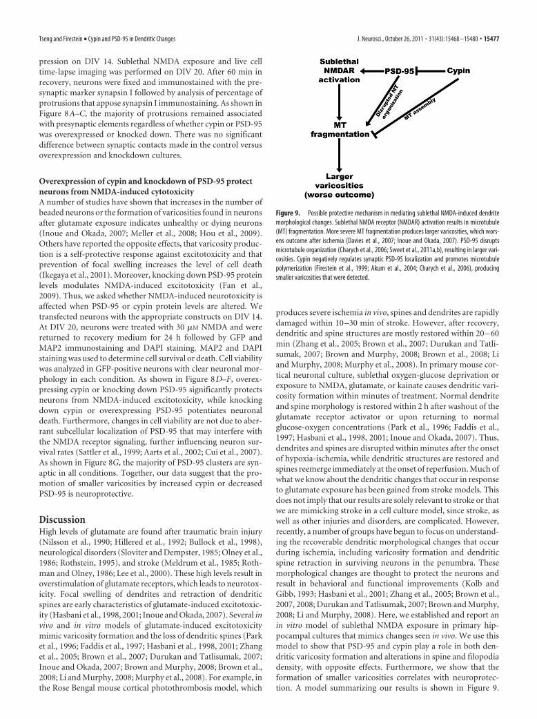
produces severe ischemia in vivo, spines and dendrites are rapidlydamaged within 10 –30 min of stroke. However, after recovery,dendritic and spine structures are mostly restored within 20 – 60min (Zhang et al., 2005; Brown et al., 2007; Durukan and Tatli-sumak, 2007; Brown and Murphy, 2008; Brown et al., 2008; Liand Murphy, 2008; Murphy et al., 2008). In primary mouse cor-tical neuronal culture, sublethal oxygen-glucose deprivation orexposure to NMDA, glutamate, or kainate causes dendritic vari-cosity formation within minutes of treatment. Normal dendriteand spine morphology is restored within 2 h after washout of theglutamate receptor activator or upon returning to normalglucose-oxygen concentrations (Park et al., 1996; Faddis et al.,1997; Hasbani et al., 1998, 2001; Inoue and Okada, 2007). Thus,dendrites and spines are disrupted within minutes after the onsetof hypoxia-ischemia, while dendritic structures are restored andspines reemerge immediately at the onset of reperfusion. Much ofwhat we know about the dendritic changes that occur in responseto glutamate exposure has been gained from stroke models. Thisdoes not imply that our results are solely relevant to stroke or thatwe are mimicking stroke in a cell culture model, since stroke, aswell as other injuries and disorders, are complicated. However,recently, a number of groups have begun to focus on understand-ing the recoverable dendritic morphological changes that occurduring ischemia, including varicosity formation and dendriticspine retraction in surviving neurons in the penumbra. Thesemorphological changes are thought to protect the neurons andresult in behavioral and functional improvements (Kolb andGibb, 1993; Hasbani et al., 2001; Zhang et al., 2005; Brown et al.,2007, 2008; Durukan and Tatlisumak, 2007; Brown and Murphy,2008; Li and Murphy, 2008). Here, we established and report anin vitro model of sublethal NMDA exposure in primary hip-pocampal cultures that mimics changes seen in vivo. We use thismodel to show that PSD-95 and cypin play a role in both den-dritic varicosity formation and alterations in spine and filopodiadensity, with opposite effects. Furthermore, we show that theformation of smaller varicosities correlates with neuroprotec-tion. A model summarizing our results is shown in Figure 9.
Figure 9. Possible protective mechanism in mediating sublethal NMDA-induced dendritemorphological changes. Sublethal NMDA receptor (NMDAR) activation results in microtubule(MT) fragmentation. More severe MT fragmentation produces larger varicosities, which wors-ens outcome after ischemia (Davies et al., 2007; Inoue and Okada, 2007). PSD-95 disruptsmicrotubule organization (Charych et al., 2006; Sweet et al., 2011a,b), resulting in larger vari-cosities. Cypin negatively regulates synaptic PSD-95 localization and promotes microtubulepolymerization (Firestein et al., 1999; Akum et al., 2004; Charych et al., 2006), producingsmaller varicosities that were detected.
Tseng and Firestein • Cypin and PSD-95 in Dendritic Changes J. Neurosci., October 26, 2011 • 31(43):15468 –15480 • 15477

Sublethal activation of the NMDA receptor results in microtu-bule fragmentation, which produces larger varicosities, worsen-ing the outcome after ischemia (Davies et al., 2007; Inoue andOkada, 2007). PSD-95 disrupts microtubule organization (Charychet al., 2006; Sweet et al., 2011a,b), therefore producing largevaricosities. Cypin negatively regulates synaptic PSD-95 local-ization and promotes microtubule polymerization (Firesteinet al., 1999; Akum et al., 2004; Charych et al., 2006), resultingin smaller varicosities.
Fragmented microtubules and loss of microtubule-associatedproteins, such as MAP2, have been found in dendrites after isch-emic injury (Yamamoto et al., 1986; Tomimoto and Yanagihara,1992; Emery and Lucas, 1995; Greenwood et al., 2007). We foundthat microtubule dynamics change with NMDA treatment (Fig.2) and that nocodazole worsened and Taxol blocked NMDA-induced microtubule breakdown. The finding using Taxol is con-sistent with previous findings (Jacobs and Stevens, 1986; Emeryand Lucas, 1995; Park et al., 1996). Until now, very little wasknown about the intracellular mediators of the changes in micro-tubules during stroke. PSD-95 disrupts microtubule organiza-tion (Charych et al., 2006; Sweet et al., 2011a,b), and thisdisruption translates into decreased number of beaded neuronswith increased varicosity size. Cypin promotes microtubule as-sembly (Akum et al., 2004; Chen and Firestein, 2007), translatinginto a larger number of beaded neurons with smaller varicosities.Thus, by understanding how PSD-95 and cypin affect varicosityformation, we can now identify how microtubules change duringthis process.
Cypin contains a domain that has high homology to collapsinresponse mediator protein-2 (CRMP-2), and this domain is re-sponsible for binding tubulin heterodimers for the promotion ofmicrotubule assembly (Firestein et al., 1999; Akum et al., 2004).Interestingly, overexpression of CRMP-2 increases axonal resis-tance to glutamate-induced varicosity formation (Hou et al.,2009). Like cypin, CRMP-2 promotes microtubule assembly (Fu-kata et al., 2002), and, together, this report and our data suggestthat promotion of microtubule assembly moderates dendriticswelling. What does varicosity formation mean for the neuron? Anumber of studies show that a reduction of beaded neurons ornumber of varicosities promotes functional recovery (Inoue andOkada, 2007; Meller et al., 2008; Hou et al., 2009). Others havereported the opposite and indicate that varicosity production is aself-protective response against excitotoxicity and that preven-tion of focal swelling increases the level of cell death (Ikegaya etal., 2001). We have found that cypin increases varicosity produc-tion (Fig. 6), which may be neuroprotective. Similar results, in-cluding the formation of smaller varicosities, were found whenPSD-95 was knocked down (Fig. 5). Smaller varicosity size hasbeen shown to indicate a metabolically healthier hippocampus inischemic stroke (Davies et al., 2007), and indeed, our results sup-port this idea (Figs. 4 – 8). Previous findings have shown thatswelling in distal dendrites occurs first, followed by swelling inproximal dendrites (Park et al., 1996; Oliva et al., 2002). Theincreased number of varicosities can be explained by the fact thatcypin overexpression, which results in decreased PSD-95 cluster-ing, results in increased number of distal dendrites (Firestein etal., 1999; Chen and Firestein, 2007). In line with these results,overexpression of PSD-95 and knockdown of cypin resulted in asmaller number of varicosities, which were larger in size (Figs. 4,7). Moreover, persistent beading, as is seen with overexpressionof PSD-95 and knockdown of cypin, implies an impaired regula-tion of dendritic volume after NMDA exposure (Inoue andOkada, 2007).
It is also interesting to note that cypin is released in response toNMDA exposure (Fig. 3). What is the significance of this finding?Release of cypin from the neuron during and after NMDA treat-ment may lead to cell death since intracellular cypin appears toconfer neuroprotection from this treatment. However, exoge-nously added cypin (guanine deaminase) protects spinal motorneurons from glutamate-induced toxicity (Hedlund et al., 2010).We added exogenous cypin to our cultures and observed no pro-tection from NMDA-induced toxicity (data not shown). Thisdifference in the effect of cypin may be due to the distinct neuro-nal types or the fact that glutamate, as used in the spinal cordstudy, affects multiple receptor subtypes, which may contributeto injury.
What is the role of changes in dendritic spine density in re-sponse to NMDA? The collapse of spines as an early response toreduce NMDA receptor activity may serve to render neurons lessvulnerable to excitotoxic stimulus (Halpain et al., 1998). In linewith this theory, a decrease in the number of dendritic spinespromoted by preconditioning ischemia protects against furtherspine loss resulting from NMDA-induced excitotoxicity (Melleret al., 2008). We have found that cypin decreases protrusionnumber on its own and occludes NMDA-mediated decreases inprotrusion number. What mechanism might regulate this? TheNMDA receptor interacts with small GTPases, and in specificRhoA, which are involved in regulation of spine number andmorphology (Nakayama et al., 2000; Calabrese et al., 2005; Goveket al., 2005). We have previously reported that the activation ofRhoA inhibits translation of cypin protein (Chen and Firestein,2007) and that overexpression of cypin results in decreasedPSD-95 clustering at the synapse (Firestein et al., 1999). Since it isbelieved that reducing the size of the postsynaptic density (i.e.,reducing PSD-95), the number of synaptic NMDA receptor clus-ters is decreased, protecting the neurons from ischemic damage(Gisselsson et al., 2010).
We have identified a novel pathway by which neurons altertheir dendritic morphology in response to sublethal concentra-tions of NMDA. We have shown that cypin and PSD-95, whichhave opposite effects on the microtubule cytoskeleton, also me-diate opposite effects on the formation of NMDA-induced vari-cosities and dendritic protrusions. Last, we have providedevidence that alterations in cypin or PSD-95 protein levels thatlead to smaller varicosities also confer neuroprotection fromNMDA-induced toxicity.
ReferencesAarts M, Liu Y, Liu L, Besshoh S, Arundine M, Gurd JW, Wang YT, Salter
MW, Tymianski M (2002) Treatment of ischemic brain damage by per-turbing NMDA receptor- PSD-95 protein interactions. Science298:846 – 850.
Akum BF, Chen M, Gunderson SI, Riefler GM, Scerri-Hansen MM, FiresteinBL (2004) Cypin regulates dendrite patterning in hippocampal neuronsby promoting microtubule assembly. Nat Neurosci 7:145–152.
Brown CE, Murphy TH (2008) Livin’ on the edge: imaging dendritic spineturnover in the peri-infarct zone during ischemic stroke and recovery.Neuroscientist 14:139 –146.
Brown CE, Li P, Boyd JD, Delaney KR, Murphy TH (2007) Extensive turn-over of dendritic spines and vascular remodeling in cortical tissues recov-ering from stroke. J Neurosci 27:4101– 4109.
Brown CE, Wong C, Murphy TH (2008) Rapid morphologic plasticity ofperi-infarct dendritic spines after focal ischemic stroke. Stroke39:1286 –1291.
Bullock R, Zauner A, Woodward JJ, Myseros J, Choi SC, Ward JD, MarmarouA, Young HF (1998) Factors affecting excitatory amino acid release fol-lowing severe human head injury. J Neurosurg 89:507–518.
15478 • J. Neurosci., October 26, 2011 • 31(43):15468 –15480 Tseng and Firestein • Cypin and PSD-95 in Dendritic Changes
Calabrese B, Halpain S (2005) Essential role for the PKC target MARCKS inmaintaining dendritic spine morphology. Neuron 48:77–90.
Charych EI, Akum BF, Goldberg JS, Jornsten RJ, Rongo C, Zheng JQ, Firest-ein BL (2006) Activity-independent regulation of dendrite patterningby postsynaptic density protein PSD-95. J Neurosci 26:10164 –10176.
Chen H, Firestein BL (2007) RhoA regulates dendrite branching in hip-pocampal neurons by decreasing cypin protein levels. J Neurosci27:8378 – 8386.
Cui H, Hayashi A, Sun HS, Belmares MP, Cobey C, Phan T, Schweizer J, SalterMW, Wang YT, Tasker RA, Garman D, Rabinowitz J, Lu PS, Tymianski M(2007) PDZ protein interactions underlying NMDA receptor-mediatedexcitotoxicity and neuroprotection by PSD-95 inhibitors. J Neurosci27:9901–9915.
Davies ML, Kirov SA, Andrew RD (2007) Whole isolated neocortical andhippocampal preparations and their use in imaging studies. J NeurosciMethods 166:203–216.
Durukan A, Tatlisumak T (2007) Acute ischemic stroke: overview of majorexperimental rodent models, pathophysiology, and therapy of focal cere-bral ischemia. Pharmacol Biochem Behav 87:179 –197.
El-Husseini AE, Schnell E, Chetkovich DM, Nicoll RA, Bredt DS (2000)PSD-95 involvement in maturation of excitatory synapses. Science290:1364 –1368.
Emery DG, Lucas JH (1995) Ultrastructural damage and neuritic beading incold-stressed spinal neurons with comparisons to NMDA and A23187toxicity. Brain Res 692:161–173.
Faddis BT, Hasbani MJ, Goldberg MP (1997) Calpain activation contrib-utes to dendritic remodeling after brief excitotoxic injury in vitro. J Neu-rosci 17:951–959.
Fan J, Cowan CM, Zhang LY, Hayden MR, Raymond LA (2009) Interactionof postsynaptic density protein-95 with NMDA receptors influences ex-citotoxicity in the yeast artificial chromosome mouse model of Hunting-ton’s disease. J Neurosci 29:10928 –10938.
Firestein BL, Brenman JE, Aoki C, Sanchez-Perez AM, El-Husseini AE, BredtDS (1999) Cypin: a cytosolic regulator of PSD-95 postsynaptic target-ing. Neuron 24:659 – 672.
Fukata Y, Itoh TJ, Kimura T, Menager C, Nishimura T, Shiromizu T, Wa-tanabe H, Inagaki N, Iwamatsu A, Hotani H, Kaibuchi K (2002)CRMP-2 binds to tubulin heterodimers to promote microtubule assem-bly. Nat Cell Biol 4:583–591.
Furukawa K, Mattson MP (1995) Taxol stabilizes [Ca 2�]i and protects hip-pocampal neurons against excitotoxicity. Brain Res 689:141–146.
Gisselsson LL, Matus A, Wieloch T (2005) Actin redistribution underlies thesparing effect of mild hypothermia on dendritic spine morphology afterin vitro ischemia. J Cereb Blood Flow Metab 25:1346 –1355.
Gisselsson L, Toresson H, Ruscher K, Wieloch T (2010) Rho kinase inhibi-tion protects CA1 cells in organotypic hippocampal slices during in vitroischemia. Brain Res 1316:92–100.
Govek EE, Newey SE, Van Aelst L (2005) The role of the Rho GTPases inneuronal development. Genes Dev 19:1– 49.
Greenwood SM, Mizielinska SM, Frenguelli BG, Harvey J, Connolly CN(2007) Mitochondrial dysfunction and dendritic beading during neuro-nal toxicity. J Biol Chem 282:26235–26244.
Halpain S, Hipolito A, Saffer L (1998) Regulation of F-actin stability in den-dritic spines by glutamate receptors and calcineurin. J Neurosci18:9835–9844.
Hasbani MJ, Hyrc KL, Faddis BT, Romano C, Goldberg MP (1998) Distinctroles for sodium, chloride, and calcium in excitotoxic dendritic injury andrecovery. Exp Neurol 154:241–258.
Hasbani MJ, Schlief ML, Fisher DA, Goldberg MP (2001) Dendritic spineslost during glutamate receptor activation reemerge at original sites ofsynaptic contact. J Neurosci 21:2393–2403.
Hedlund E, Karlsson M, Osborn T, Ludwig W, Isacson O (2010) Globalgene expression profiling of somatic motor neuron populations with dif-ferent vulnerability identify molecules and pathways of degeneration andprotection. Brain 133:2313–2330.
Hillered L, Persson L, Carlson H, Ungerstedt U, Ronne-Engstrom E, NilssonP (1992) Studies on excitatory amino acid receptor-linked brain disor-ders in rat and man using in vivo microdialysis. Clin Neuropharmacol 15[Suppl 1 Pt A]:695A– 696A.
Horne EA, Dell’Acqua ML (2007) Phospholipase C is required for changesin postsynaptic structure and function associated with NMDA receptor-dependent long-term depression. J Neurosci 27:3523–3534.
Hou ST, Jiang SX, Aylsworth A, Ferguson G, Slinn J, Hu H, Leung T, KapplerJ, Kaibuchi K (2009) CaMKII phosphorylates collapsin response medi-ator protein 2 and modulates axonal damage during glutamate excitotox-icity. J Neurochem 111:870 – 881.
Hou XY, Zhang GY, Yan JZ, Chen M, Liu Y (2002) Activation of NMDAreceptors and L-type voltage-gated calcium channels mediates enhancedformation of Fyn-PSD95-NR2A complex after transient brain ischemia.Brain Res 955:123–132.
Ikegaya Y, Kim JA, Baba M, Iwatsubo T, Nishiyama N, Matsuki N (2001)Rapid and reversible changes in dendrite morphology and synaptic effi-cacy following NMDA receptor activation: implication for a cellular de-fense against excitotoxicity. J Cell Sci 114:4083– 4093.
Inoue H, Okada Y (2007) Roles of volume-sensitive chloride channel inexcitotoxic neuronal injury. J Neurosci 27:1445–1455.
Jacobs JR, Stevens JK (1986) Experimental modification of PC12 neuriteshape with the microtubule-depolymerizing drug Nocodazole: a serialelectron microscopic study of neurite shape control. J Cell Biol103:907–915.
Kobayashi MS, Takahashi Y, Nagata T, Nishida Y, Murata A, Ishikawa K, AsaiS (2004) Screening for control genes in rat global cerebral ischemia us-ing high-density oligonucleotide array. J Neurosci Res 76:512–518.
Kolb B, Gibb R (1993) Possible anatomical basis of recovery of functionafter neonatal frontal lesions in rats. Behav Neurosci 107:799 – 811.
Lee JM, Grabb MC, Zipfel GJ, Choi DW (2000) Brain tissue responses toischemia. J Clin Invest 106:723–731.
Li P, Murphy TH (2008) Two-photon imaging during prolonged middlecerebral artery occlusion in mice reveals recovery of dendritic structureafter reperfusion. J Neurosci 28:11970 –11979.
Meldrum B, Evans M, Griffiths T, Simon R (1985) Ischaemic brain dam-age: the role of excitatory activity and of calcium entry. Br J Anaesth57:44 – 46.
Meller R, Thompson SJ, Lusardi TA, Ordonez AN, Ashley MD, Jessick V,Wang W, Torrey DJ, Henshall DC, Gafken PR, Saugstad JA, Xiong ZG,Simon RP (2008) Ubiquitin proteasome-mediated synaptic reorganiza-tion: a novel mechanism underlying rapid ischemic tolerance. J Neurosci28:50 –59.
Monnerie H, Shashidhara S, Le Roux PD (2003) Effect of excess extracellu-lar glutamate on dendrite growth from cerebral cortical neurons at 3 daysin vitro: Involvement of NMDA receptors. J Neurosci Res 74:688 –700.
Murphy TH, Li P, Betts K, Liu R (2008) Two-photon imaging of strokeonset in vivo reveals that NMDA-receptor independent ischemic depo-larization is the major cause of rapid reversible damage to dendrites andspines. J Neurosci 28:1756 –1772.
Nakayama AY, Harms MB, Luo L (2000) Small GTPases Rac and Rho in themaintenance of dendritic spines and branches in hippocampal pyramidalneurons. J Neurosci 20:5329 –5338.
Nilsson P, Hillered L, Ponten U, Ungerstedt U (1990) Changes in corticalextracellular levels of energy-related metabolites and amino acids follow-ing concussive brain injury in rats. J Cereb Blood Flow Metab 10:631– 637.
Okabe S, Kim HD, Miwa A, Kuriu T, Okado H (1999) Continual remodel-ing of postsynaptic density and its regulation by synaptic activity. NatNeurosci 2:804 – 811.
Oliva AA Jr, Lam TT, Swann JW (2002) Distally directed dendrotoxicityinduced by kainic Acid in hippocampal interneurons of green fluorescentprotein-expressing transgenic mice. J Neurosci 22:8052– 8062.
Olney JW, Collins RC, Sloviter RS (1986) Excitotoxic mechanisms of epi-leptic brain damage. Adv Neurol 44:857– 877.
Park JS, Bateman MC, Goldberg MP (1996) Rapid alterations in dendritemorphology during sublethal hypoxia or glutamate receptor activation.Neurobiol Dis 3:215–227.
Prange O, Murphy TH (2001) Modular transport of postsynaptic density-95clusters and association with stable spine precursors during early develop-ment of cortical neurons. J Neurosci 21:9325–9333.
Rothman SM, Olney JW (1986) Glutamate and the pathophysiology of hy-poxic–ischemic brain damage. Ann Neurol 19:105–111.
Rothstein JD (1995) Excitotoxic mechanisms in the pathogenesis of amyo-trophic lateral sclerosis. Adv Neurol 68:7–20; discussion 21–27.
Sattler R, Xiong Z, Lu WY, Hafner M, MacDonald JF, Tymianski M (1999)Specific coupling of NMDA receptor activation to nitric oxide neurotox-icity by PSD-95 protein. Science 284:1845–1848.
Tseng and Firestein • Cypin and PSD-95 in Dendritic Changes J. Neurosci., October 26, 2011 • 31(43):15468 –15480 • 15479

Sloviter RS, Dempster DW (1985) “Epileptic” brain damage is replicatedqualitatively in the rat hippocampus by central injection of glutamate oraspartate but not by GABA or acetylcholine. Brain Res Bull 15:39 – 60.
Sweet ES, Tseng CY, Firestein BL (2011a) To branch or not to branch: howPSD-95 regulates dendrites and spines. Bioarchitecture 1:69 –73.
Sweet ES, Previtera ML, Fernandez JR, Charych EI, Tseng CY, Kwon M,Starovoytov V, Zheng JQ, Firestein BL (2011b) PSD-95 Alters Microtu-bule Dynamics via an Association With EB3. J Neurosci 31:1038 –1047.
Tomimoto H, Yanagihara T (1992) Electron microscopic investigation ofthe cerebral cortex after cerebral ischemia and reperfusion in the gerbil.Brain Res 598:87–97.
Wahl AS, Buchthal B, Rode F, Bomholt SF, Freitag HE, Hardingham GE,
Ronn LC, Bading H (2009) Hypoxic/ischemic conditions induce expres-sion of the putative pro-death gene Clca1 via activation of extrasynapticN-methyl-D-aspartate receptors. Neuroscience 158:344 –352.
Wilson MT, Keith CH (1998) Glutamate modulation of dendrite out-growth: alterations in the distribution of dendritic microtubules. J Neu-rosci Res 52:599 – 611.
Xia Z, Dudek H, Miranti CK, Greenberg ME (1996) Calcium influx via theNMDA receptor induces immediate early gene transcription by a MAPkinase/ERK-dependent mechanism. J Neurosci 16:5425–5436.
Yamamoto K, Morimoto K, Yanagihara T (1986) Cerebral ischemia in thegerbil: transmission electron microscopic and immunoelectron micro-scopic investigation. Brain Res 384:1–10.
15480 • J. Neurosci., October 26, 2011 • 31(43):15468 –15480 Tseng and Firestein • Cypin and PSD-95 in Dendritic Changes