Embed Size (px)
Citation preview

at SciVerse ScienceDirect
Biochimie 94 (2012) 2242e2263
Contents lists available
Biochimie
journal homepage: www.elsevier .com/locate/b iochi
Review
The role of nutrition on epigenetic modifications and their implications on health
Josep C. Jiménez-Chillarón a,*, Rubén Díaz a,b, Débora Martínez a, Thais Pentinat a, Marta Ramón-Krauel b,Sílvia Ribó a, Torsten Plösch c,**
a Fundacio Sant Joan de Deu, Paediatric Hospital Sant Joan de Deu, SpainbHospital Sant Joan de Deu, Universitat de Barcelona, Endocrine Division, SpaincDepartment of Pediatrics, University Medical Center Groningen, University of Groningen, The Netherlands
a r t i c l e i n f o
Article history:Received 9 March 2012Accepted 11 June 2012Available online 5 July 2012
Keywords:Developmental origins of health and diseaseNutritional epigenomicsMetabolic syndromeDietary TransitionsCaloric restriction
* Corresponding author. Tel.: þ34 93 6009455; fax** Corresponding author.
E-mail addresses: [email protected] (J.C. Jiménez(T. Plösch).
URL: http://www.epigenetic-programming.nl
0300-9084/$ e see front matter � 2012 Elsevier Mashttp://dx.doi.org/10.1016/j.biochi.2012.06.012
a b s t r a c t
Nutrition plays a key role in many aspects of health and dietary imbalances are major determinants ofchronic diseases including cardiovascular disease, obesity, diabetes and cancer. Adequate nutrition isparticularly essential during critical periods in early life (both pre- and postnatal). In this regard, there isextensive epidemiologic and experimental data showing that early sub-optimal nutrition can have healthconsequences several decades later.
The hypothesis that epigenetic mechanisms may link such nutritional imbalances with altered diseaserisk has been gaining acceptance over recent years. Epigenetics can be defined as the study of heritablechanges in gene expression that do not involve alterations in the DNA sequence. Epigenetic marksinclude DNA methylation, histone modifications and a variety of non-coding RNAs. Strikingly, they areplastic and respond to environmental signals, including diet. Here we will review how dietary factorsmodulate the establishment and maintenance of epigenetic marks, thereby influencing gene expressionand, hence, disease risk and health.
� 2012 Elsevier Masson SAS. All rights reserved.
“You may be an undigested bit of beef, a blot of mustard, a crumbof cheese, a fragment of underdone potato. There’s more of gravythan of grave about you, whatever you are!”
A Christmas Carol, Charles Dickens
2012 marks the celebration of the Bicentennial of CharlesDickens. It is a good time to review his great narratives. They arefull of elaborated descriptions of children growing under somesort of nutritional deprivation (Oliver Twist, David Copperfield)or even famine (Tiny Tim). Here we will review our currentknowledge about the relationship between early malnutrition,later disease risk andhowepigeneticmechanismsmay link them.
1. Introduction: the rise of the field of NutritionalEpigenomics
Diet constitutes one of the major environmental factors thatexert a profound effect on many aspects of health and disease risk.
: þ34 93 6009771.
-Chillarón), [email protected]
son SAS. All rights reserved.
For example, in industrialized countries, excessive caloric intake isa major determinant of complex chronic diseases, such as obesity,type 2 diabetes, cardiovascular disease and even cancer. Accordingto the World Health Organization these diseases account for morethan half of the deaths worldwide and have a huge impact onnational economies (World Health Organization, 2003). Conversely,in poor countries, malnutrition and undernutrition, especiallyduring the perinatal period, increase not only neonatal mortalityand perinatal morbidities but also the risk of chronic diseasesduring adulthood [1e3]. This association between perinatal nutri-tion and late-onset disease has been conceptualized into theDevelopmental Origins of Health and Disease Hypothesis (DOHaD,Box 1 ([16e26,28,29,32])). Finally, a paradigmatic scenario isillustrated by chronic caloric restriction (CR). It has been shownthat moderate global caloric restriction is the most powerful way toincrease lifespan in various model organisms from different taxa,such as yeasts, worms (Caenorhabditis elegans), insects (Drosophilamelanogaster) or mammals (including mice, rats and monkeys) [4].
Herewewill review the evidence that supports a role for dietaryfactors, including micro-nutrients, macro-nutrients, and non-nutrient dietary components, in mediating disease risk throughepigenetic modifications. Special emphasis will be put on the roleof dietary factors during early perinatal development in the contextof DOHaD. We will focus primarily on the Metabolic Syndrome,
Box 1.
Developmental Origins of Health and Disease Hypothesis
(DOHaD)
By the early 1990s the epidemiologist David Barker first
came with his observation that the fetal environment has
life-long programming effects for the offspring. Barker and
his colleagues used birth weight as a surrogate marker for
poor intrauterine nutrition and could show correlations
between birth weight and the mortality risks for cardiovas-
cular disease, insulin resistance and hypertension [16e19].These seminal observations were followed by many epide-
miologic evidences demonstrating that prenatal and early
postnatal environmental challenges influence the risk of
developing various chronic diseases during adulthood,
including cardiovascular disease, diabetes, obesity, cancer
and even some behavioural disorders [20e22]. Among
environmental factors that program adult metabolic disor-
ders, poor intrauterine nutrition is the most extensively
studied [22e24]. Inadequate prenatal nutrition usually
results in intrauterine growth restriction and, ultimately, low
birth weight [3]. In developed countries, low birth weight
accounts for up to 7% from all lived births. These numbers
strongly aggravate in developing countries where average
low birth weight increases up to 15% and, in some Southern
Asian countries, it may even rise up to 27% (UNICEF Portal,
www.childinfo.org). This constitutes a major global health
problem, including developed countries, since the propor-
tion of people at risk for adult chronic diseases is achieving
alarming epidemic proportions [1,2].
These epidemiologic data has been further confirmed by
numerous animal models, including ours [25e28]. These
works clearly support causality between a) nutritional
challenges during early development and b) elevated risk
for adult metabolic syndrome [29]. Experimental and
human studies led to propose the Developmental Origins of
Adult Health and Disease hypothesis, (DOHaD) [30,31]. This
hypothesis proposes that environmental stimuli, like nutri-
tion, acting during fetal and/or neonatal development can
produce permanent changes in cell/tissue structure and
function, through permanently modifying expression of
target genes [30e32].
Several explanations have been put forward to explain the
correlation between events that, in human studies, were
separated by several decades. It has been proposed, and it
is currently widely accepted that epigenetic changes
induced by early nutrition influence later health and disease
(see box 2).
The Dutch Hunger Winter (1944e1945)
TheDutch famine of 1944 took place in theGerman-occupied
part of the Netherlands. FromSeptember, 1944 toMay, 1945,
theNazis began a blockade that cut off food supplies and fuel
shipments to the population of the western part of the
Netherlands, to punish the reluctance of the Dutch to aid the
Nazi war effort. Daily caloric supply during this time was
decreased to as few as 700 calories per day. People suffered
from chronic hunger and the diseases produced by malnu-
trition. Some 4.5 millions were affected and about 18,000
people died because of the famine. Most vulnerable accord-
ing to the death reports were elderly men and children.
The Dutch Hunger Winter provided science and clinical
medicinewithawell-characterizedpopulationsuitable for the
studyofDOHaD inhumans.Hence, thesocalledDutchCohort
is a population of pregnant mothers and fetuses that experi-
enced malnutrition during first, second, or third trimesters.
The main characteristics that made this population so
important are summarized as follows: first, the famine was
short in time (6 months), started and ended abruptly and
therefore it is clearly circumscribed in timeandplace.Second,
the population was ethnically homogeneous and without
remarkable prior differences in dietary patterns. Likewise,
food availability during rationing was largely unaffected by
social class. Third, the official food rations were known, so
that the number of calories available could be estimated by
place and time of birth. Finally, and most importantly, long-
term follow-up was possible, since the childhood and adult
medical histories of the fetuses that survived could be traced
through national population registers.
In sum, for all these reasons, theDutch Cohort constitutes an
“excellent” population for the study of developmental
programming of adult disease. In accord, a huge number of
critical reports have already been published andmanymore
will certainly be published in the future. Also, the first reports
describing an association between nutritional imbalances in
utero and altered epigenetic marks during adulthood have
been described in subjects from this cohort [102,103].
J.C. Jiménez-Chillarón et al. / Biochimie 94 (2012) 2242e2263 2243
characterized by insulin resistance, obesity, hypertension, hyper-triglyceridemia, hyperglycaemia and diabetes [5]. The potential roleof nutrition in EpigeneticseCancer is extensively reviewed else-where [6e9,135]. We will examine the role of nutrition on epige-netic modifications in mammals. Hence, the role of nutrition onother model organisms (plants, C. elegans, Drosophila, zebrafish)will not be discussed here.
Epigenetics can be pragmatically defined as the study of stableinheritance of gene expression that occurs withoutmodifications inthe DNA sequence [10]. Epigenetic mechanisms in mammalsinclude DNA methylation, histone modifications and, morerecently, a variety of non-coding RNAs (key epigenetic concepts aresummarized in Box 2) [11]. In the context of this review, it is rele-vant to state that epigenetic factors may be modulated by envi-ronmental cues, including nutrition, and thus provide a mechanismbywhich genomes integrate environmental signals into permanentchanges of gene expression that may ultimately lead to health anddisease risk [10e14]. This recognition has ignited the rapid growthof a novel field: Nutritional Epigenomics [15].
Box 2.
Epigenetics: Stable inheritance of gene expression that
occurs without modifications in the DNA sequence. Epige-
netic mechanisms include DNA methylation, histone
modifications and, recently, a variety of non-coding RNAs.
DNA methylation: It is a covalent modification that consists
on the addition of a methyl group at cytosines of the DNA
template. In mammals, DNA methylation occurs primarily
at CpG dinucleotides.
CpG islands are regions in the DNA with a disproportional
high abundance of dinucleotides CpG. Typically CpG
islands exist around promoter regions of the genes.
Very recently, other DNA modifications, like hydrox-
ymethylation, have been identified. The impact of these
modifications on programming of adult disease is currently
unknown.
Histones: Histones are alkaline proteins found in eukaryotic
cell nuclei that package the DNA into structural units called
nucleosomes. They are the main protein components of
chromatin, acting as spools around which DNA winds, and
play a role in gene regulation.

Histone modifications: Covalent modifications of histone
residues that can alter chromatin states and, thus, gene
regulation. Histone modifications include a series of
complex post-translational modifications including meth-
ylation (mono-, di-, and tri-methylation), acetylation,
SUMOylation, biotinylation, phosphorylation, ubiquitina-
tion and ADP-rybosilation.
Histone code: The histone code hypothesis suggests that
specific histone modifications (or combination of modifica-
tions) may confer unique biological functions to regions of
the genome where they associate. Given the fact that there
exist 4 different histones andmultiple types ofmodifications
across the residues of the proteins, the combination of
modifications is extremely high. This would result in
a complex, locus-specific regulation of gene transcription.
Non-codingRNAs (ncRNA):Theyare functionalRNAs thatare
not translated into proteins. Non-coding RNAs include
transfer RNA (tRNA) ribosomal RNA (rRNA) and small
nucleolarRNA (snoRNA). Recently, a series ofnewRNAswith
regulatory activity have been added to the list: siRNA (small
interfering RNA) miRNA (microRNA) and piRNA (piwi RNA).
Genome and Epigenome: The genome is the totality of the
genetic information of a cell/organism that is contained in
the DNA sequence.
The epigenome consists on all chemical modifications of
DNA and histones of a cell/organism that contribute to
regulate gene expression independently of DNA sequence.
One single genome may give rise to several epigenomes
depending on environmental conditions, tissue specificity,
developmental stages, etc. It is proposed that this relation
(1 Genome/ n Epigenomes) constitutes the basis for
fundamental biological issues such as pluripotency and cell
differentiation, phenotypic variation, etc.
Metastable epiallele: It is an epiallele (an allele that can
stably exist in more than one epigenetic state, resulting in
different phenotypes) at which the epigenetic state can
switch and establishment is a probabilistic event. Once
established, the state is mitotically inherited.
Sources: Molecular Biology of the Cell, Garland Science,
Taylor and Francis Group, 4th Edition.
Epigenetics, Cold Spring Harbor Laboratory Press, 1st
Edition.
Rakyan VK et al. Trends in Genetics. Volume 18, Issue 7,
348e351, 1 July 2002.
J.C. Jiménez-Chillarón et al. / Biochimie 94 (2012) 2242e22632244
This review is structured in order to address the following fourkey questions:
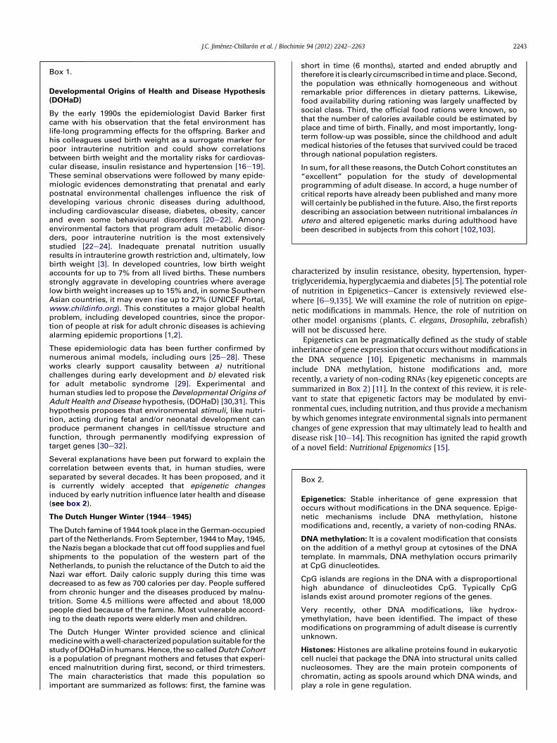
1. WHEN do dietary factors influence the epigenome, thusleading to long-term changes in gene expression? It isremarkable to note that current evidence linking diet toepigenetic modifications can be narrowed down to two specificscenarios: First, during “critical windows” of early develop-ment (specially during fetal development and/or early neonatalgrowth) and, second, in adult individuals, during “DietaryTransitions” (such as high fat feeding, caloric restriction, etc.)occurring over a relatively long period of time (Fig. 1). There-fore, before extensively reviewing most relevant exampleslinking nutrition and epigenetic modifications, we willsummarize the concepts of “critical windows” and “homeostasisvs. chronic dietary transitions” (Section 2).
2. WHAT are the evidences linking diet and epigenetic modifi-cations? Most relevant studies describing nutritional variation
and epigenetically-associated metabolic phenotypes will besummarized in Section 3 (Tables 1e3).
3. HOW do dietary factors influence the epigenome? In otherwords, what are the mechanisms that link dietary factors andepigenetic modifications?Molecular mechanisms are reviewedin Section 4 (Figs. 2e4).
4. WHY is nutrition regulating gene expression through epige-netic modifications, particularly during specific stages ofdevelopment or during the course of Dietary Transitions? Asyet, this is an open question that generates an intense debate.In this last section we will comment on the current thinkingrelating the biological meaning of nutrition during develop-ment and its impact on long-term regulation of geneexpression.
2. WHEN do dietary factors influence the epigenome?
Under what circumstances does nutrition induce epigeneticmodifications? Epidemiologic and experimental evidences linkingdiet to epigenetic modifications can be narrowed down to twoscenarios (Fig. 1): (1) First, during “critical windows” of develop-ment, including fetal and early neonatal growth. (2) Second, during“Dietary Transitions” occurring over a long period of time in adultindividuals. Typical examples of these Dietary Transitions arechronic overfeeding, high fat feeding or chronic caloric restriction.
2.1. Critical windows of development
Developing organisms are under dynamic changes, and organsystems undergo rapid development characterized by cell prolif-eration/differentiation. Epigenetic mechanisms during early stagesof development contribute to faithfully maintain undifferentiatedstem-cells on one hand, and organogenesis on the other one [33,34].Thus, early embryogenesis in mammals is the most critical periodfor the establishment of the epigenome. In particular, betweenfertilization and implantation, the embryo demethylates thegenomewidely [35e37]. Short after implantation, there is awave ofre-methylation that sets the epigenetic patterns for different celltypes. Therefore, these periods constitute critical spatiotemporalwindows of development during which the epigenetic marks areeither partially erased or re-set. Failure to complete these programsin time might be irreversible and lead to permanent dysregulationof gene expression [15,38]. Importantly, this is a period especiallyvulnerable to environmental cues, such as nutrition, that candisrupt the correct establishment of epigenetic marks that, onceestablished, remain highly stable. Arguably, this is the reason whynutritional challenges during early windows of development mighthave such long-term effects in the context of DOHaD.
A striking example of the critical-window-concept arises fromthe Dutch Famine (Box 1) [39e41]. At the end of the SecondWorld War, individuals from the Western Netherlands wereexposed to acute undernutrition for a defined period of 4 months.The disease risk of the offspring’s of women who were pregnantduring the Dutch Famine was different depending on if it wasduring the beginning, the middle or close to the end of gestationat the time of the famine. Individuals affected early in pregnancyhave cardiovascular complications, including a pro-atherogeniclipid profile, and reduced cognitive functions [41e44]. Mid-gestational maternal undernutrition was associated withimpaired kidney and lung function [41,45,46]. Lastly, individualssuffering starvation at the end of gestation had striking differ-ences with regards to glucose tolerance at adult age, although thisis a feature which is present in all groups at low levels [41,47].Whether these differences are mediated, in part, by epigenetic

F1 F2 F3
[107]
[111]
[63]
[89]
Life-course
[92]
[93]
[113]
[67]
[74]
[75]
[64]
[27]
[76]
[65]
[59]
[58]
[104]DN
A m
eth
yla
tio
n
[61]
[99]
[161]
[95]
[191]
[107]
[109]
[197]
[196]
[198]
[82]
[199]
[70]
fic
atio
ns
[70]
[200]
[58]
[201]
[68]
[80]
[84]
[86]
[61]
[60]
[81]
Histo
ne m
od
i
High fat dietNeonatal overfeedingLow protein dietMalnutrition (placental artery ligation)Methyl-supplemented dietMalnutrition (maternal caloric restriction)20-40% global caloric restriction
Birth
We
an
in
g
1s
t ye
ar
2n
d ye
ar
Fig. 1. Summary of studies, from Tables 1e3, showing length and time of dietary intervention over the life course of the mouse/rat, as model organism. Each horizontal colored linecorresponds to an individual study, and length-time of the intervention is projected against the black arrow representing the life-course (2 years average) of a laboratory rodent. Thestudies can be grouped into two distinctive clusters: First, interventions during early windows of development, including prenatal and early neonatal stages of development untilweaning. Second, interventions in adult individuals consisting on Dietary Transitions over a long period of time (from 9 weeks to over the lifespan of the individual).
J.C. Jiménez-Chillarón et al. / Biochimie 94 (2012) 2242e2263 2245
modifications remains unknown. But it is likely that (a) the time,(b) the intensity and (c) duration of an environmental factor mayinduce different epigenetic alterations in a tissue-dependentmanner. At this point we lack a systematic survey describingthe epigenomic modifications (and phenotypic effects) mediatedby different dietary factors during specific well-controlled periodsof development.
2.2. Dietary Transitions
Epigenetic variations are not only restricted to early windows ofdevelopment but also may occur throughout an individual life-course (Figs. 1 and 2). Such epigenetic variations accumulate overa long period of time and may ultimately influence phenotypicoutcomes (health and disease risk). This is clearly exemplified

Table 1Summary of relevant studies showing effects of dietary conditions on DNA methylation in humans and model organisms.
Dietary condition Species Period ofdietary input
Tissue(s) Methylation Epigeneticallyregulated gene(s)
Observed phenotype Reference
High fat diet Mouse Adult dietarytransition
Brain (variousregions)
[
[
[
Oprm1ThDat
Dopaminergic (Th and Dat) and theopioid systems (Oprm1), which participatein the central regulation of food intake andthe development of obesity, were altered.
[107,108]
Rat Adult dietarytransition
Islet cells Y Il13ra3 Progressive beta-cell dysfunction in isletcells from paternally high fat fed rats.
[111]
Mouse In utero Brain Y
Y
Y
DatMorPenk
Altered gene expression of dopamineand opioid-related genes may changebehavioral preference for palatable foodsand increase risk of obesity andobesity-related diseases.
[63]
Rat In utero Liver Y Cdkn1a Offspring from high fat fed damsdeveloped hepatic steatosis andcharacteristics of non-alcoholicliver disease.
[89]
Neonataloverfeeding/overgrowth
Rat Neonatal Hypothalamus [ YPomcexpr/leptinratioYPomc expr/Insulin receptorratio
Early overfeeding resulted in a metabolicsyndrome phenotype (obesity,hyperleptinemia, hyperinsulinemia,insulin resistance and diabetes).
[92]
Rat Neonatal Hypothalamus [ Same model than in [92]. Reducedinsulin receptor expression leads tohypothalamic insulin resistance andpredisposition to altered feedingbehavior characteristic of this model.
[93]
Human Neonatal Peripheral blood Y TACSTD2 Rapid postnatal growth is associatedwith increased childhood adiposity(9e15 years).
[189]
Low protein diet Mouse Adult dietarytransitionetransgenerationaleffect
Liver [ PPARa Increased hepatic cholesterol/lipidbiosynthesis, increasing risk of fattyliver and steatosis.
[113]
Rat In utero Liver Global DNAhypermethylation
None (globalanalysis)
Low maternal protein availabilityduring gestation results in glucoseintolerance and hypertension inthe adult.
[67]
Rat In utero Adrenal gland Y AT(1b) Maternal low-protein diet resultedin the development of hypertensionin the offspring.
[74,75]
Rat In utero Hypothalamus [ Pomc Maternal low-protein nutrition canaffect brain development and expressionof orexigenic/anorexigenic genes.
[64]
Mouse In utero Liver [ Lxra Protein restriction during pregnancyreduced Lxra-dependent hepaticcholesterol biosynthesis.
[27]
Mouse In utero Adipose tissue Y Lep Offspring from mothers fed alow-protein diet showed increasedfood intake and increased adiposity.
[76]
Rat In utero Islet cell [ Hnf4a Reduced expression of Hnf4acontributes to beta-cell dysfunctionand development of type 2 diabetes.
[65]
Pig In utero Liver Y Somaticcytochromec (CYCS),
Increased cytochrome c gene expression,may be involved in changedmitochondrial function
[206]
Rat In utero þneonatal
Liver Y
Y
PPARaGR
Altered expression of PPARa and theglucocorticoid receptor might contributeto altered carbohydrate/lipid homeostasisand hypertension, respectively.
[59,70,71]
Intrauterinemalnutrition;placental arteryligation
Rat In utero Liver Global DNAhypomethylationin fetal livers
None (globalanalysis)
Utero-placental insufficiency throughbilateral artery ligation caused insulinresistance and diabetes in the adult.
[58]
Rat In utero Islet cell [ Pdx1 Adult-onset type 2 diabetes. Diabeteswas associated with progressivesilencing of the transcription factor Pdx1.
[61]
Rat In utero Islet cell Genome-wideHELP assay:1400 locidifferentiallymethylated(both hyper-and hypo-methylated).Y
Validatedloci are:Fgfr1
Same model as in [61]. Type 2 diabetesdue, in part, to beta-cell dysfunction.Genome-wide DNA methylation analysisshowed that alterations occurred neargenes regulating processes such asvascularization, beta-cell proliferation,insulin secretion, and cell death.
[66]
J.C. Jiménez-Chillarón et al. / Biochimie 94 (2012) 2242e22632246
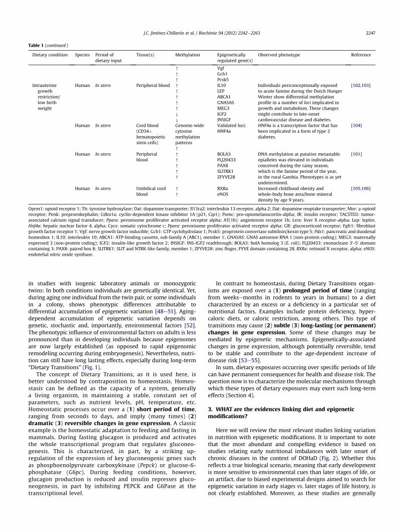
Table 1 (continued )
Dietary condition Species Period ofdietary input
Tissue(s) Methylation Epigeneticallyregulated gene(s)
Observed phenotype Reference
[
[
[
VgfGch1Pcsk5
Intrauterinegrowthrestriction/low birthweight
Human In utero Peripheral blood [
[
[
[
[
Y
Y
IL10LEPABCA1GNASASMEG3IGF2INSIGF
Individuals periconceptionally exposedto acute famine during the Dutch HungerWinter show differential methylationprofile in a number of loci implicated ingrowth and metabolism. These changesmight contribute to late-onsetcardiovascular disease and diabetes.
[102,103]
Human In utero Cord blood(CD34þhematopoieticstem cells)
Genome-widecytosinemethylationpatterns[
Validated loci:HNF4a
HNF4a is a transcription factor that hasbeen implicated in a form of type 2diabetes.
[104]
Human In utero Peripheralblood
[
[
[
[
[
BOLA3FLJ20433PAX8SLITRK1ZFYVE28
DNA methylation at putative metastableepialleles was elevated in individualsconceived during the rainy season,which is the famine period of the year,in the rural Gambia. Phenotypes is as yetundetermined.
[101]
Human In utero Umbilical cordblood
[
[
RXRaeNOS
Increased childhood obesity andwhole-body bone area/bone mineraldensity by age 9 years.
[105,106]
Oprm1: opioid receptor 1; Th: tyrosine hydroxylase; Dat: dopamine transporter; Il13ra2: interleukin 13 receptor, alpha 2; Dat: dopamine reuptake transporter; Mor: m-opioidreceptor; Penk: preproenkephalin; Cdkn1a: cyclin-dependent kinase inhibitor 1A (p21, Cip1); Pomc: pro-opiomelanocortin-alpha; IR: insulin receptor; TACSTD2: tumor-associated calcium signal transducer; Ppara: peroxisome proliferator activated receptor alpha; AT(1b): angiotensin receptor 1b; Lxra: liver X receptor-alpha; Lep: leptin;Hnf4a: hepatic nuclear factor 4, alpha; Cycs: somatic cytochrome c; Ppara: peroxisome proliferator activated receptor alpha; GR: glucocorticoid receptor; Fgfr1: fibroblastgrowth factor receptor 1; Vgf: nerve growth factor inducible; Gch1: GTP cyclohydrolase 1; Pcsk5: proprotein convertase subtilisin/kexin type 5; Pdx1: pancreatic and duodenalhomeobox 1; IL10: interleukin 10; ABCA1: ATP-binding cassette, sub-family A (ABC1), member 1; GNASAS: GNAS antisense RNA 1 (non-protein coding); MEG3: maternallyexpressed 3 (non-protein coding); IGF2: insulin-like growth factor 2; INSIGF: INS-IGF2 readthrough; BOLA3: bolA homolog 3 (E. coli); FLJ20433: exonuclease 30-50 domaincontaining 3; PAX8: paired box 8; SLITRK1: SLIT and NTRK-like family, member 1; ZFYVE28: zinc finger, FYVE domain containing 28. RXRa: retinoid X receptor, alpha; eNOS:endotelial nitric oxide synthase.
J.C. Jiménez-Chillarón et al. / Biochimie 94 (2012) 2242e2263 2247
in studies with isogenic laboratory animals or monozygotictwins: In both conditions individuals are genetically identical. Yet,during aging one individual from the twin pair, or some individualsin a colony, shows phenotypic differences attributable todifferential accumulation of epigenetic variation [48e51]. Aging-dependent accumulation of epigenetic variation depends ongenetic, stochastic and, importantly, environmental factors [52].The phenotypic influence of environmental factors on adults is lesspronounced than in developing individuals because epigenomesare now largely established (as opposed to rapid epigenomicremodeling occurring during embryogenesis). Nevertheless, nutri-tion can still have long lasting effects, especially during long-term“Dietary Transitions” (Fig. 1).
The concept of Dietary Transitions, as it is used here, isbetter understood by contraposition to homeostasis. Homeo-stasis can be defined as the capacity of a system, generallya living organism, in maintaining a stable, constant set ofparameters, such as nutrient levels, pH, temperature, etc.Homeostatic processes occur over a (1) short period of time,ranging from seconds to days, and imply (many times) (2)dramatic (3) reversible changes in gene expression. A classicexample is the homeostatic adaptation to feeding and fasting inmammals. During fasting glucagon is produced and activatesthe whole transcriptional program that regulates gluconeo-genesis. This is characterized, in part, by a striking up-regulation of the expression of key gluconeogenic genes suchas phosphoenolpyruvate carboxykinase (Pepck) or glucose-6-phosphatase (G6pc). During feeding conditions, however,glucagon production is reduced and insulin represses gluco-neogenesis, in part by inhibiting PEPCK and G6Pase at thetranscriptional level.
In contrast to homeostasis, during Dietary Transitions organ-isms are exposed over a (1) prolonged period of time (rangingfrom weeksemonths in rodents to years in humans) to a dietcharacterized by an excess or a deficiency in a particular set ofnutritional factors. Examples include protein deficiency, hyper-caloric diets, or caloric restriction, among others. This type oftransitions may cause (2) subtle (3) long-lasting (or permanent)changes in gene expression. Some of these changes may bemediated by epigenetic mechanisms. Epigenetically-associatedchanges in gene expression, although potentially reversible, tendto be stable and contribute to the age-dependent increase ofdisease risk [53e55].
In sum, dietary exposures occurring over specific periods of lifecan have permanent consequences for health and disease risk. Thequestion now is to characterize the molecular mechanisms throughwhich these types of dietary exposures may exert such long-termeffects (Section 4).
3. WHAT are the evidences linking diet and epigeneticmodifications?
Here we will review the most relevant studies linking variationin nutrition with epigenetic modifications. It is important to notethat the most abundant and compelling evidence is based onstudies relating early nutritional imbalances with later onset ofchronic diseases in the context of DOHaD (Fig. 2). Whether thisreflects a true biological scenario, meaning that early developmentis more sensitive to environmental cues than later stages of life, oran artifact, due to biased experimental designs aimed to search forepigenetic variation in early stages vs. later stages of life history, isnot clearly established. Moreover, as these studies are generally

Table 2Summary of relevant studies showing effects of dietary conditions on histone modifications in humans and model organisms.
Dietary condition Species Period ofdietary input
Tissue Histone modification(s) Epigeneticallyregulated gene(s)
Observed (or associated) phenotype(health and disease)
Reference
Highfat diet(HFD)
Japanesemacaques
In utero Liver [Acetylation (H3K14) Correlationsbetween hepaticH3 and geneexpression areabsent or subtle(P > 0.05).
Maternal high fat feeding increasedfetal liver triglyceride accumulation.Likewise, hepatic histology correlatedwith non-alcoholic liver disease.
[91]
Mouse Adultdietarytransition:fromweaning toage >18 weeks
Brain YAcetylation (H3K9)[Methylation (H3K9)
Oprm1 Chronic high fat diet resulted in alteredfood behavior (preference for sucrose diets)and obesity in the offspring.
[107]
Rat Adult dietarytransition: HFdiet containing45% Kcal fromfat for 13 weeks
Liver [Acetylation (H3, H4)YMethylation(H3K27 andH3K27Me3)[Methylation(H3K4Me2).
p16INK4a andp21Cip1
Obesity prone rats fed a high fat dietshowed activation of the cellularsenescence pathway (p16INK4a andp21Cip1), which was associatedwith hepatic steatosis.
[109]
Rat In utero Liver [Acetylation (H4)YMethylation (H3K9Me3and H3K27Me3)
Pck1 Foetal offspring of HF-fed dams hadsignificantly higher mRNA contentsof gluconeogenic genes, which cancontribute to late onset glucoseintolerance and diabetes.
[196]
Mouse Threeconsecutivegenerations(F0, F1, and F2)
Liver YMethylation (H3K9Me2) LXRa and ERO1-a The male offspring of the F2 generation(derived from both grand-maternal andmaternal obesity) were highly susceptibleto developing obesity and hepatic steatosis.
[110]
MaternalLowproteindiet (LP)
Pig Gestation andlactation
Skeletalmucle
[Acetylation (H3)[Methylation (H3K27Me3)YMethylation (H3K9Me)
Mstn Maternal low protein diet influencesmyostatin gene expression at weaningand finishing stages influencing musclemass, and potentially insulin sensitivity,in the offspring.
[197]
Rat Pregnancyand lactation
Liver YAcetylation (H3)[Methylation (H3K9Me3)
Cyp7a1 Body weight and liver growth wereimpaired in the male offspring.Likewise, circulating and hepaticcholesterol levels were increasedin the adult offspring.
[82]
Rat In utero Skeletalmuscle
[Acetylation (H3, H4) C/EBPb Low protein availability duringgestation altered amino acid andenergy homeostasis in skeletalmuscle and fat deposition duringmuscle development in the offspring.
[198]
Rat In utero Liver [Acetylation (H3, H4,and H3K9)YMethylation(H3K9Me3)
GR Increased hepatic expression of theglucocorticoid receptor in the offspringcontributed to glucose intolerance andincreased hepatic glucose production.
[70]
Rat In utero Liver [Acetylation (H4)[Methylation(H3K9Me3)
Asns; Atf3 Maternal low protein diet programmedthe amino acid response pathway in theliver of the offspring. These alterationsmight potentially lead to liver dysfunction,including defective glucose homeostasis.
[199]
In uteroundernutrition(utero-placentalinsufficiency, UPI)
Rat In utero Liver [Acetylation (H3) Global H3hyperacetylationin livers from P0and P21 rat offspring.
Uteroplacental insufficiency (UPI)leads to increased risk of insulinresistance, hypertriglyceridemia,hyperglycemia and overt diabetesin the adult rat offspring.
[58]
Rat In utero Liver [Acetylation (H3K9,H3K14 and H3K18)
PGC1a and CPT1a Same model as in [58]; changesin PGC1a and CPT1a may contributeto hepatic metabolic dysfunction.
[200]
Rat In utero Brain [Acetylation (H3K9Acand H3K14Ac)
Global histonemodifications(no specific lociare described)
UPI caused permanent changeschromatin structure of thehippocampus and the periventricularwhite matter of the offspring. Thesealterations might be associated to poorneurodevelopmental outcomes.
[68]
Rat In utero Liver [Acetylation (H3K9Acand H3K14Ac)
Dusp5 Same model as in [58]; Dusp5 is aphosphatase that dephosphorylatesErk1 and 2, which in turn increasesserine phosphorylation of IRS. IRSserine-phosphorylation contributesto hepatic insulin resistance.
[80]
Rat In utero Hippocampus GR Same model as [58,68]; intrauterinegrowth restricted rats showed
[84]
J.C. Jiménez-Chillarón et al. / Biochimie 94 (2012) 2242e22632248

Table 2 (continued )
Dietary condition Species Period ofdietary input
Tissue Histone modification(s) Epigeneticallyregulated gene(s)
Observed (or associated) phenotype(health and disease)
Reference
[Acetylation (H3K9)[Methylation(H3K4Me3)
increased expression ofhippocampal glucocorticoid receptor,which is an important regulator of thehypothalamic-pituitaryeadrenal axis.
Rat In utero Lung [,YMethylation in adevelopmental andgender-specific manner(H3K9Me3)
PPARg Intrauterine growth restriction alteredPPARg expression, causing altered lungalveolization and postnatal lung diseasein the male offspring.
[86]
Rat In utero Islet cells YAcetylation (H3 and H4)YMethylation (H3K4)[Methylation (H3K9)
Pdx1 Intrauterine growth restriction resultedin adult-onset type 2 diabetes. Adultdiabetes was associated with progressivesilencing of the transcription factor Pdx1,which is critical for beta-cell functionand development
[61,201]
In uteroundernutrition(50% caloricrestriction)
Rat In utero Skeletalmuscle
YAcetylation (H3K14)[Methylation (H3K9Me2)
Glut-4 50% caloric restriction during the lastweek of gestation represses skeletalmuscle Glut4 expression in the adultrat offspring.
[60]
Rat In utero Liver YMethylation (H3K4Me2)[Methylation (H3K4Me3)
Igf1 50% caloric restriction during gestationdecreased H3K4Me2 at the hepatic IGF1region of the newborn offspring.Intrauterine growth restricted rats thatexhibited postnatal catch-up growthhad decreased H3K4Me2 and increasedH3K4Me3 in the IGF1 locus.
[81]
Oprm1: m-opioid receptor; p16INK4a: cyclin-dependent kinase inhibitor; p21Cip1: cyclin-dependent kinase inhibitor 1A; Pck1: phosphoenolpyruvate carboxykinase;LXRa: liver X nuclear receptor alpha; ERO1-a: endoplasmic reticulum oxidation 1; Mstn: myostatin; Cyp7a1: colesterol 7 a-hydroxylase; C/EBPb: CCAAT/enhancer-bindingprotein beta; GR: glucocorticoid receptor; Asns: asparagine synthetase; Atf3: activating transcription factor 3; PGC1a: peroxisome proliferator activated receptor gamma,coactivator 1 alpha; CPT1a: carnitine palmitoyltransferase 1a; Dusp5: dual specificity phosphatase 5; Pparg: peroxisome proliferator-activated receptor gamma; Pdx1:pancreatic and duodenal homeobox 1; Glut4: Glucose transporter 4 insuline-responsive; Igf1: insulin-like growth factor 1.
J.C. Jiménez-Chillarón et al. / Biochimie 94 (2012) 2242e2263 2249
conducted from the clinical perspective, with pathologies asreadout, we currently do not know whether we miss the advan-tageous, evolutionary beneficial effects of epigenetic adaptationsbecause of this biased view.
Recent articles have reviewed some aspects covered in thissection [56,57]. Therefore, we have kept it short and summarizedmost experimental data in Tables 1e3.
3.1. Nutrition during early development: epigenetics and DOHaD
3.1.1. Animal modelsThe association between dietary changes during specific
windows of development and epigenomic modifications has beenreported in several animal models (Tables 1e3) [27,58e68]. Theyconstitute an excellent tool to understand how particular nutri-tional regimens or specific dietary factors may influence the epi-genome. The most widely studied nutritional challenges includeprotein deficiency, global caloric restriction, high fat feeding andexcessive neonatal food intake. A special chapter is constituted bythe Agouti mouse model which, although mechanistically likely tobe an exemption, serves as a visualization of the current ideas inthe field.
3.1.1.1. Protein malnutrition. Protein restriction is frequently usedas a model for maternal malnutrition. Often, diets of 18% casein(control) and 9% casein (restricted) are compared, but sometimesother percentages of protein are used or restricted diets arecompared to chow. This should be kept in mind when comparingdifferent studies. Feeding a low protein diet to pregnant ratsresulted in global DNA hypermethylation in livers from theoffspring [67]. This was among the first studies showing a linkbetween nutritional imbalances during intrauterine developmentand epigenetic modifications. More recent studies have alsoconfirmed that maternal low-protein feeding during gestation
may also result in locus-specific changes in DNA methylation(Fig. 2, Tables 1e3). More importantly, these changes remainstable until adulthood, thus providing a molecular basis forDOHaD. Reported genes (or loci) include the glucocorticoidreceptor (GR), peroxisome proliferator-activated receptor alpha(PPARa) and liver X receptor-alpha (Lxra) in liver [27,59,69e73];the hepatocyte nuclear factor-4-alpha (Hnf4a) in islet cells [65];the AT(1b) angiotensin receptor in adrenal gland [74,75]; theorexigenic/anorexigenic genes neuropeptide Y (Npy) and pro-opiomelanocortin C (Pomc) in hypothalamus [64]; and the lep-tin gene (Lep) in adipose tissue [76].
Importantly, in the previous examples, changes in DNA meth-ylation correlate with altered gene expression. Therefore, suchnutritionally-induced changes in DNA methylation may explain, atleast in part, metabolic dysfunction in the adult. Hence, alteredexpression of GR, PPARa and Lxra may explain altered lipidmetabolism and hepatic steatosis which in turn contributes tohepatic insulin resistance. Dysregulated expression of Hnf4a in isletcells may lead to beta-cell dysfunction and type 2 diabetes. Finally,Npy, Pomc and Lep regulate appetite in rodents. Therefore, aberrantexpression of these genes may alter feeding behavior and explainthe development of obesity and obesity-related diseases includinginsulin resistance and diabetes. In sum, there is now sufficientevidence to support that maternal protein malnutritionmay inducepermanent alterations in gene expression through epigeneticmodifications. These alterations can contribute, in part, to thedevelopment of obesity, insulin resistance and type 2 diabetes inthe adult.
3.1.1.2. Global caloric restriction: placental artery ligation. Globalcaloric restriction is another frequently used model for maternalmalnutrition. Caloric restriction in animal models has beenaccomplished by either placental artery ligation or by global caloricrestriction.

Table 3Summary of relevant studies showing effects of dietary conditions on micro-RNA expression in humans and model organisms.
Dietary factor Species,timing
Tissue(s) Observed phenotype (miRNAs modulated) Reference
Maternalhigh fatfeeding
Mouse(in utero)
Liver Maternal high fat feeding prior to conception,duringgestation and lactation changed the expressionof 23 miRNAs (from 579 miRNAs present ina microarray) in livers from the adult offspring.Strikingly, methyl-CpG binding protein 2 wasthe common predicted target for several of theidentified miRNAs (miR-709, -let7s, �122, �194and �26a).
[177]
High fatfeedingsupplementedwith linoleicacid
Mouse(adult)
Whiteadiposetissue(WAT)
Expression of miR-103, miR-107 (lipid metabolisms)and miR-103, miR-107 (altered in obesity) changedin response to the treatment with conjugated linolenicacid, currently used to induce fat loss.
[179]
Biotin Human(in vitro)
Primaryhumancells
Physiological concentrations of biotin increased miR-539abundance in a dose-dependent manner. miR-539 regulatesholocarboxylase synthetase, which catalyzes the covalentbinding of biotin to carboxylases and histones.
[202]
Polyphenolsfrom yauponholly leaves(quercetinand kaempferol3-rutinoside)
Human(in vitro);Mouse
Humancoloncells;
Flavonol-rich fractions extracted from yaupon holly leavesexert anti-inflammatory properties in both human andmouse cells:1. Quercetin and kaempferol 3-rutinoside up-regulatedmiR-146a in human colon cancer cells, which is a negativeregulator of the pro-inflammatory factor NF-kB.2. Quercetin treatment in mouse macrophagesdown-regulated the pro-inflammatory miR-155.
[180,203,204]
Ethanol Human;Mouse
Colonbiopsiesand caco-2cells;fetalbrain
Ethanol induced expression of miR-212, which causes gutleakiness, a key factor in human alcoholic liver disease;prenatal ethanol exposure changed expression of severalmiRNAs in fetal brain from mice (miiR-10a, 10b, 9, 145,30a, 152, 200a, 496, 296, 30e-5p, 362, 339, 29c, 154).miR-10 up-regulation mediated, in part, HoxaIdown-regulation. Co incubation with folate reverted theseeffects.
[178,205]
Vitamin E Rat(DietaryTransitions)
Liver Vitamin E-deficient diet (6 months)caused a down-regulation of miR-122a and miR-125b,which contribute to regulate lipid metabolism andcancer-inflammation, respectively.
[181]
Starvation Rat Liver Mild starvation (12 h) increased hepatic levels ofmiR-451, �122a, �29b. Insig1, which in turn inhibitsSrebp1 production, is a predicted target of miR-29.
[183]
J.C. Jiménez-Chillarón et al. / Biochimie 94 (2012) 2242e22632250
Bilateral placental artery ligation in rats has beenwidely used asa model of reduced nutrient and oxygen availability for the fetus[77,78]. This surgical procedure may both induce genome-wideDNA hypomethylation in fetal livers [58] and affect the histonecode at specific loci in the offspring (Tables 1 and 2) [58,60e62,68].For example, in utero undernutrition in rats reduces expression ofthe homeobox 1 transcription factor (Pdx1) in islet cells [61]; thedual specificity phosphatase 5 (Dusp5) [80] and cholesterol 7alpha-hydroxylase (Cyp7a1) [82] in liver; dual specific phosphatase 5(Dusp5) and the glucocorticoid receptor (GR) genes in hippocampus[83,84]; 11beta-hydroxysteroid dehydrogenase type 2 (Hsd11b2) inkidney [85]; and the peroxisome proliferator-activated receptorgamma (PPARg) in lungs [86].
Similar towhat we have described for the low protein diet, someof the previously described genes can contribute to differentaspects of the metabolic syndrome. For example, Pdx1 is a keytranscription factor that regulates beta-cell differentiation. Hence,altered Pdx1 expression may lead to beta-cell dysfunction anddiabetes. On the other hand, Dusp5 is a protein from MAPK-signaling pathway that can modulate insulin signaling. Thus,altered expression of Dusp5 may induce tissue-specific insulinresistance that can ultimately contribute to whole body insulinresistance and diabetes. In sum, all these data clearly establish thatin rodent models altered gestational nutrition may induce
chromatin remodeling at metabolically relevant loci, throughchanging histone marks.
To finish, we would like to notice a recent report from Nüskenand colleagues [79]. They have compared surgical uterine arteryligation with protein restriction in rats and found striking differ-ences in the resulting phenotype [79]. Therefore, these acute andsevere surgical interventions cannot be completely compared withany dietary regimen. It constitutes, though, a valuable model tounderstand developmental programming of the offspring inresponse to placental dysfunction/placental insufficiency whichcauses reduced nutrient and oxygen availability to the fetus.
3.1.1.3. Global caloric restriction: nutritional deprivation. In rats, 50%global caloric restriction during the last week of gestation resultedin reduced expression of the glucose transporter 4 (Glut4) inskeletal muscle from the offspring [60]. This alteration is mediatedby specific changes of histone modifications (H3K14 deacetylationand increased H3K9 di-methylation). Glut4 is a landmark proteinthat allows insulin-stimulated glucose uptake into peripheraltissues. Therefore, altered epigenetic regulation of glut4 maycontribute to the development of insulin resistance and diabetes inthis rat model. In a similar rat model, 50% caloric restrictiondecreased the abundance of H3K4Me2 at the IGF1 locus of liverfrom the newborn offspring. This epigenetic modification alters

DNA methylation
-Dopamine and Opioid-
related genes (brain;
mouse)
-Cdkn1a (liver; rat)
High fat diet
Low-protein diet
DNA methylation
-Angiotensin receptor
(adrenal gland; mouse)
-Npy, Cart, Pomc (brain; rat)
-Leptin (adipose; mouse)
-Lxra (liver; mouse)
-Hnf4a, PPARa,
Glucocorticoid receptor
(liver, rat)
DNA methylation
-IL10, LEP, ABCA1,
GNASAS,
MEG3, IGF2 (human)
-HNF4A (human)
-BOLA3, FLJ20433, PAX8,
SLITRK1, ZFYVE28 (human)
-RXRa, eNOS (human)
Histone modifications
-Pdx1 (islet cell; rat)
-Glut 4 (skeletal muscle; rat)
-Dusp5, IGF1 (liver; rat)
-Dusp5 (hoppocampus; rat)
-11-b-DH type (kidney; rat)
-PPARg (lung; rat)
Undernutrition
Birth Weaning
Fetal growth Neonatal growth
Adult
DNA methylation
-POMC
(hypothalamus; rat)
-Insulin receptor
(brain; rat)
-TACSTD2 (human)
Neonatal overfeeding
DNA methylation
-Neurotransmitter
System (brain; rat)
-Multiple genes in F2
(islet cells; rat)
High fat diet
DNA methylation
-H-ras (rat)
-RUNX (human)
-p16 (human)
-ATP10A, WT1, TNFa
(Adipose tissue; human)
Histone modifications
-p16INK4
(human)
-hTERT (human)
-SirT1 mediated changes
(FOXO, Pgc1a, HDAC1, etc;
Mouse, rat)
Caloric restriciton
Fig. 2. Summary of the loci that show altered expression in association with an epigenetic modification. Results are grouped by dietary intervention, type of epigenetic event andwindow of intervention. Data included in this figure is derived from Tables 1e3, including humans and model organisms.
J.C. Jiménez-Chillarón et al. / Biochimie 94 (2012) 2242e2263 2251
IGF1 expression and contributes to post-natal catch-up growth andsubsequent risk of diabetes in the adult [62,81].
Moderate caloric restriction (30%) to pregnant non-humanprimates (Baboon) decreased methylation in fetal kidney during
Die
FADNAD+
DNA methylation, his
histone ac
Metabolic P
Diet
DNA methylation, histone methylation,
histone acetylation
Metabolic Phenotypes
1 2
A B
Fig. 3. Intracellular signals that translate nutrition into epigenetically-mediated metabolic pbox, alters the epigenome. B, intracellular second-messengers synthesized in response to exthe production of the second-messengers depends, directly or indirectly, from the synthesiscell. ATP acts as a cofactor or it is necessary to fully activate the enzymes that catalize the(flavin adenine dinucleotide), a-KG (a-ketoglutarate), SAM (S-adenosyl methionine), ATP (adomain histone demethylase; 3: JumonjiC-containing domain histone demethylase; 4: Dmononucleotide adenylyltransferase; 7: riboflavin kinase and FAD synthase; 8: a-ketogluta
early stages of gestation, whereas it increased DNA methylation bythe end of gestation [87]. Likewise, DNA methylation was alsoincreased in the frontal cortex during late gestational stages [87]. Ina follow-up study, expression of the glucogenogenic enzyme
ATP
Diet
FAD α-KGNAD+
SAM
DNA methylation, histone methylation,
histone acetylation
Metabolic Phenotypes
t
α-KG SAM
tone methylation,
etylation
henotypes
3 4 5
67 8
C
9
henotypes. A, diet, through not completely known mechanisms depicted by the blacktracellular nutritional/energetic states and that are able to modulate the epigenome. C,of ATP (or the ATP/ADP ratio), which in turn is determined by the energetic state of thesynthesis of NAD, FAD, a-KG and SAM. NAD (nicotinamine adenine dinucleotide), FADdenosine triphosphate). 1: Class III histone deacetylase (sirtuins); 2: LSD1-containingNA methyl transferase; 5: histone methyl transferase; 6: nicotinamide/nicotinic acidrate dehydrogenase; 9: S-adenosyl methionine transferase.

DNMT
MTHFR
SHMT
MTR
MAT
Methionine SAM
SAHHcy
5-methylTHF
THF
5,10-
methylTHF
Histones
mHistone
HMT
DNA
mDNA
HDM-LSD1
HDM-Jumonji
Methionine
Cycle
Folate
Cycle
HDM-LSD1DNMT
MTHFR
SHMT
MTR
MAT
Methionine SAM
SAHHcy
5-methylTHF
THF
5,10-
methylTHF
Histones
HMT
DNA
HDM-JmjC
FAD
Diet
A-KG
Diet
Diet
FolateDietCholine
BetaineDiet
Diet
Dimethyl-
glycineB12
B6
Diet Polyphenols
mHistone
mDNA
HDM-LSD1DNMT
MTHFR
SHMT
MTR
MAT
Methionine SAM
SAHHcy
5-methylTHF
THF
5,10-
methylTHF
Histones
HMT
DNA
HDM-JmjC
FAD
Diet
A-KG
Diet
Diet
FolateDietCholine
BetaineDiet
Diet
Dimethyl-
glycineB12
B6
Diet Polyphenols
ATP
ATP
ATP
ATP
mHistone
mDNA
A
B
C
Fig. 4. The methionine cycle. A, connection between the methionine and folate cycles and their implication on DNA and histone methylation. B, interaction between the fola-teemethionine cycles and different dietary compounds that act as co-factors of the enzymes in the cycle. C, role of ATP as a common regulatory molecule in mediating the activity ofkey enzymes of the methionine cycle. Enzymes. SHMT: serine hydroxymethyl-transferase; MTHFR: methylentetrahydrofolate reductase; MTR: 5-methyltetragydrofolate-homo-cysteine methyl transferase; MAT: methionine adenosyl-transferase; DNMT: DNA methyl-transferase; HMT: histone methyl-transferase; HDM: histone demethylase. Metabolites.THF: tetrahydrofolate; SAM: S-adenosyl methionine; Hcy: homocysteine; SAH: S-adenosylhomocysteine; mDNA: methylated DNA; mHistone: methylated histone.
J.C. Jiménez-Chillarón et al. / Biochimie 94 (2012) 2242e22632252
phosphoenolpyruvate carboxykinase 1 (PCK1) was increased in thefetal liver [88]. Strikingly, up-regulation of this gene occurred inassociation with the hypomethylation of the PCK1 promoter. Thesedata support that moderate maternal nutrient reduction in non-
human primates causes organ-specific and gestational age-specific changes in DNA methylation. These changes may havelong-term effects on fetal organ development [87] and be causativefor metabolic dysfunction later in life [88].
J.C. Jiménez-Chillarón et al. / Biochimie 94 (2012) 2242e2263 2253
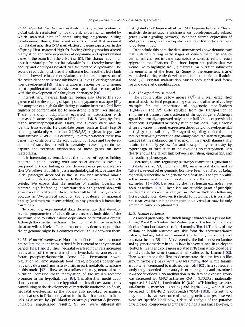
3.1.1.4. High fat diet. In utero malnutrition (by either protein orglobal caloric restriction) is not the only experimental model bywhich maternal diet influences offspring epigenome duringdevelopment. Hence, two recent reports showed that maternalhigh fat diet may alter DNAmethylation and gene expression in theoffspring. First, maternal high fat feeding during gestation alteredmethylation and gene expression of dopamine and opioid relatedgenes in the brain from the offspring [63]. This change may influ-ence behavioral preference for palatable foods, thereby increasingobesity and obesity-associated risk for metabolic syndrome. Thesecond report demonstrated that offspring frommothers fed a highfat diet showed reduced methylation, and increased expression, ofthe cyclin-dependent kinase inhibitor 1A (Cdkn1a) during neonatalliver development [89]. This alteration is responsible for changinghepatic proliferation and liver size, two aspects that are compatiblewith the development of a fatty liver phenotype [90]
Interestingly, maternal high fat feeding also altered the epi-genome of the developing offspring of the Japanese macaque [91].Consumption of a high fat diet during gestation increased fetal livertriglyceride content and led to non-alcoholic fatty liver disease.These phenotypic adaptations occurred in association withincreased histone acetylation at H3K14 and H3K18. Next, by chro-matin immunoprecipitation assays, the authors were able toidentify locus-specific H3 candidate genes, such as DnaJ (Hsp40)homolog, subfamily A, member 2 (DNAJA2) or glutamic pyruvatetransaminase 2(GPT2). It is currently unknown whether these twogenes may contribute to the accumulation of lipids and the devel-opment of fatty liver. It will be certainly interesting to furtherexplore the potential implication of these genes on livermetabolism.
It is interesting to remark that the number of reports linkingmaternal high fat feeding with late onset disease is lower ascompared to those linking caloric deprivation or protein restric-tion. We believe that this is just a methodological bias, because theinitial paradigm described in the DOHaD was maternal caloricdeprivation, starting already with Barker’s focus on low birthweight. We expect that the number of studies focusing onmaternal high fat feeding (or overnutrition, as a general idea) willgrow over the next years. These studies will be extremely relevantbecause in Westernized societies the prevalence of maternalobesity (and maternal overnutrition) during gestation is increasingalarmingly.
To conclude, experimental data demonstrate that develop-mental programming of adult disease occurs at both sides of thespectrum, due to either caloric deprivation or nutritional excess.Although the specific mechanisms leading to adult disease in bothsituationwill be likely different, the current evidences support thatthe epigenome might be a common molecular link between them.
3.1.1.5. Neonatal overfeeding. Nutritional effects on the epigenomeare not limited to the intrauterine life, but extend to early neonatalperiod (Figs. 1 and 2). Thus, neonatal overfeeding in rats increasedmethylation of the promoter of the hypothalamic anorexigenicfactor proopiomelanocortin, Pomc [92]. Permanent down-regulation of Pomc augments food intake, promotes obesity andmay provide a mechanism to explain, in part, metabolic syndromein this model [92]. Likewise, in a follow-up study, neonatal over-nutrition increased mean methylation of the insulin receptorpromoter in the hypothalamus [93]. This alteration might addi-tionally contribute to induce hypothalamic insulin resistance, thuscontributing to the development of metabolic syndrome. To finish,neonatal overfeeding in the mouse also provoked permanentmodifications in DNA methylation in the liver from adult individ-uals, as assessed by CpG island microarrays (Pentinat & Jimenez-Chillaron, unpublished results). 91 loci were differentially
methylated (49% hypermethylated, 51% hypomethylated). Clusteranalysis demonstrated enrichment on developmentally-relatedgenes (Wnt signaling pathway). Whether altered expression ofWnt proteins may mediate hepatic metabolic dysfunction remainsto be determined.
To conclude this part, the data summarized above demonstratethat nutrition during early stages of development can inducepermanent changes in gene expression of somatic cells throughepigenetic modifications. The three important points that wewould like to highlight are: (1) maternal malnutrition influencesthe epigenome of the fetus. (2) Some of the epigenetic marksestablished during early development remain stable until adult-hood. (3) Perinatal malnutrition causes both global and locus-specific epigenetic modifications.
3.1.2. The agouti mouse modelThe agouti viable yellow mouse (Avy) is a well established
animalmodel for fetal programming studies and often used as a keyexample for the importance of epigenetic modifications[13,94e97]. The Avy allele resulted from the transposition ofa murine retrotransposon upstream of the agouti gene. Althoughagouti is normally expressed only in hair follicles, its expression inother cells is regulated by methylation of this locus. Thus, isogenicoffspring varies in agouti expression depending on developmentalmethyl group availability. The agouti signaling molecule bothinduces yellow pigmentation and antagonizes the satiety signalingcascade (at the melanocortin 4 receptor in the hypothalamus). Thisresults in variably yellow fur and susceptibility to obesity byhyperphagia in correlation to the level of DNA methylation. Thisclearly shows the direct link between nutrition, epigenetics, andthe resulting phenotype.
Therefore, besides regulatory pathways involved in regulation ofmetabolism (like GR, Pomc and LXR, summarized above and inTable 1), several other genomic loci have been identified as beingespecially vulnerable to epigenetic modifications. The agouti viableyellow mouse and the axin fused mouse are the most prominentexamples [98e100], but recently the first human examples havebeen described [101]. These loci are suitable proof-of-principlecandidates for measuring changes in DNA methylation followingdietary challenges. However, it should be noted that it is currentlynot clear whether this phenomenon is universal or may be onlylimited to some exceptional loci.
3.1.3. Human evidencesAs noted previously, the Dutch hunger winter was a period late
duringWorldWar 2 when theWestern part of the Netherlands wasblocked from food transports for 4 months (Box 1). There is plentyof data on health outcome available from the abovementionedcohorts, linking fetal environment (particularly nutrition) andpostnatal health [39e41]. Very recently, the links between famineand epigenetic markers in adults have been examined. In an elegantstudy, Heijmans and colleagues isolated DNA fromwhite blood cellsof individuals being peri-conceptionally affected by famine [102].They were among the first to demonstrate that the insulin-likegrowth factor 2 (IGF2) locus was less methylated in the faminegroup when compared to matched controls [102]. In a subsequentstudy they extended their analysis to more genes and examinedsex-specific effects. DNA methylation in the famine-exposed groupwas increased for GNAS antisense RNA 1 (GNASAS), maternallyexpressed 3 (MEG3), interleukin 10 (IL10), ATP-binding cassette,sub-family A, member 1 (ABCA1) and leptin (LEP), while it wasdecreased for INS-IGF2 readthrough (INSIGF) [103]. Interestingly,they found that at least some of the epigenetic changes observedwere sex specific. Until now, a detailed analysis of the putativephysiological consequences of these findings is missing. However, it

J.C. Jiménez-Chillarón et al. / Biochimie 94 (2012) 2242e22632254
is tempting to speculate that methylation changes in promoters ofgenes such as LEP (involved in satiety regulation) and ABCA1(involved in cholesterol transport and HDL formation) may linkearly nutrition to adult metabolic disease. To finish, it is remarkablethat in both studies differences in DNA methylation were apparentmore than 60 years after birth. It remains to be determinedwhether this type of alterations are already present at birth andmaintained throughout life, or appeared secondarily in response toprogressive metabolic dysfunction. Here, careful physiologicalstudies have to follow in future.
Seminal studies from the Dutch cohort have been followed bya series of reports: A recent study by Waterland and colleaguesextended our knowledge of nutritional influences during gestationon the epigenome to seasonal changes in nutrition [101]. Theauthors examined DNA methylation in individuals from ruralGambia. There, nutrition during the rainy season is largely differentfrom nutrition during the dry season. The rainy season is charac-terized by reduced nutrient availability whereas the dry season ischaracterized by high nutrient availability. The authors reportedthat several putative metastable epialleles (Box 2) were differen-tially methylated (BOLA3, FLJ20433, PAX8, SLITRK1, ZFYVE28). Theseloci are stochastically methylated early during development and inmice reflect nutritional influences. Here, this phenomenon could bedemonstrated for the first time in humans. Importantly, the authorsalso examined the methylation of other loci which have beenpreviously been identified as targets of differential methylation(e.g., LINE1, GNASAS, IL10) and failed to demonstrate any nutritionalinfluences. This may indicate that the duration and severity of themalnutrition has a pronounced effect on the establishment ofepigenetic effects.
The key question is what the relevance of these changes inmetastable epialleles for human disease is. On one hand, it is notknown whether they can influence adult metabolism in any way.Theymight be useful, though, as biomarkers of early nutrition. Theycan be a good tool to determine whether an individual has devel-oped under nutritional stress or not. This information might beextremely useful in order to enroll positive individuals into specificprograms aimed to prevent late onset metabolic dysfunction.Nevertheless, the validity of these markers needs further evalua-tion including the presence in other independent human cohorts.
To finish, a set of very recent studies have determined patternsof DNA methylation in cells from cord blood [104e106]. Forexample, Einstein and colleagues analyzed global patterns of DNAmethylation in hematopoietic stem cells (CD34þ) from cord bloodin intrauterine growth restricted and control babies by microarrayanalysis [104]. Bioinformatic analysis yielded that a small subset of56 loci showed significant differences in methylation betweengroups. These genes were involved in processes critical for stem cellfunction (cell cycle, cellular maintenance). Strikingly, the diabetes-related gene hepatocyte nuclear factor 4, alpha (HNF4A) appearedamong these differentially methylated loci. It remains unclearthough whether these changes will remain stable into adulthoodand therefore contribute to diabetes risk (or chronic disease risk ingeneral) later in life. In this regard, the authors suggest thatepigenetic modifications in multipotent progenitor cells (such asthe CD34þ cells analyzed in this study) might influence chronicdiseases later in life as the cell population expands over time andinduce functional changes during tissue differentiation and matu-ration. While very attractive, this hypothesis deserves furtherinvestigation. In any case, these types of studies are extremelyimportant because of the potential use of DNA methylation at birthas an early marker of future disease risk [104e106].
In another recent set of studies, DNA methylation of severalcandidates was assessed in cord blood from two independentpopulations of children with normal birth weights [105,106].
Strikingly, the authors show that the methylation of retinoid Xreceptor alpha (RXRa) and endothelial nitric oxide synthase (eNOS)at birth correlated with adiposity by age 9 years [105]. In addition,in a follow-up study, DNA methylation of the promoter region ofeNOS also correlatedwith bonemineral density at age 9 years [106].Thus, these studies constitute the first proof of principle to showthat DNA methylation at birth might be a powerful molecularmarker (of early nutrition) for later risk of disease (adiposity, bonedensity). Additional data from other cohorts will validate thisconcept and additional follow-up studies to define whether thesechanges in methylation persist well into adulthood.
3.2. Adult nutrition during “Dietary Transitions”
As previously mentioned, epigenetic variations are not onlyrestricted to early windows of development and may also occurthroughout an individual life-course. However, the amount of datalinking adult dietary interventions with epigenetic modifications ismuch more limited than that for dietary interventions during earlydevelopment (Figs. 1 and 2), and it is yet unknown whether this isa bias or truly shows differential biological responses to differentdevelopmental stages. Regardless, as we will discuss here, dietaryfactors may influence the epigenome in adult individuals(Tables 1e3). Taking into account the available data, nutrition mayinduce epigenetic modifications in adults when it fulfills at leastthese two conditions: First, dietary interventions take place overa long period of time and, second, there is a transition from theprevious to a novel type of diet. This is clearly exemplified innumerous animal models: from chow diet-to-high fat diet, fromchow diet containing normal protein content-to-chow diet con-taining low protein content, from ad lib feeding-to-caloric restric-tion (CR), etc.
3.2.1. Chronic high fat feedingChronic high fat diet in mice (from weaning until 20 weeks of
age) altered patterns of DNA methylation within the promoterregions of the genes encoding tyroxine hydroxylase, the dopaminetransporter and the m-opioid receptor in the brain [107,108]. Thesegenes are part of the neurotransmitter systems that participate inthe regulation of food intake. Thus, these epigenetically-inducedalterations can contribute to the development of obesity andobesity-related diseases occurring later in life. In another ratmodel,high fat feeding in obese prone rats for 13 weeks resulted inincreased transcription of p16INK4a and p21Cip1 in the liver [109].These changes, which might contribute to liver disease, occur inresponse to modifications in the histones residing in the regulatoryand coding regions of both genes.
Very recently, an interesting study explored the effect ofcontinuous high fat feeding for three generations on the develop-ment of fatty liver in the mouse offspring [110]. At 4e6 weeks ofage, C57BL/6 females (F0) were fed with a diet containing 60% Kcalof fat. This high-fat feeding was continued for two more genera-tions, F1 and F2. After this nutritional intervention, the authorsreport that obesity occurred earlier and became more severe in F2male offspring that in F1 and F0 mice. Likewise, F2 offspring alsodeveloped the highest degree of hepatic steatosis. Hepatic steatosisin F2 mice was accompanied by a transgenerational trend to up-regulate lipogenic genes, including fatty acid synthase (Fasn),stearoylecoenzyme A desaturase 1 (Scd1), sterol regulatoryelement binding protein-1 (Srebp), liver X nuclear receptor alpha(Lxra), liver X nuclear receptor beta (Lxrb) or the endoplasmicreticulum oxidation 1 (Ero-1a). Strikingly, Lxra and Ero1-a expres-sion are explained, in part, by reduced relative protein levels ofH3K9Me2 and H3K27Me3 binding to their promoter regions. Thus,the authors conclude that the effects described in F2 male offspring

J.C. Jiménez-Chillarón et al. / Biochimie 94 (2012) 2242e2263 2255
are “presumably consequence of transgenerational accumulation ofepigenetic modifications leading to accumulation of lipogenesis inthe liver” [110]. In sum, a sustained dietary change for threegenerations leads to progressive accumulation of epigeneticmodifications that may modulate metabolic phenotypes. To note,the effects described in F2 male mice are actually a combination oflong dietary interventions, plus the nutritional impact receivedduring development. It will be important to design appropriateexperiments to dissect the relative contribution of developmentalvs. adult nutrition on the development of fatty liver.
Interestingly, effects of high fat feeding may induce trans-generational (epigenetic) consequences: chronic high fat diet(during 10 weeks, from age 4 weeks) in male SpragueeDawley ratsprogrammed beta-cell dysfunction in their female offspring, whichhas not been exposed to high fat diet during its development [111].Beta-cell dysfunction was characterized by altered expression ofgenes involved in Calcium-, MAPK- and Wnt-signaling pathways.This alteration may be attributed, in part, to changes in DNAmethylation. This is exemplified by the interleukin 13 receptoralpha-2 gene (Il13ra2), which shows the highest fold change inexpression in concordance with hypomethylation of its regulatoryregion. These authors argue that this is an example of non-genetic,intergenerational transmission of metabolic dysfunction throughthe paternal lineage. Since males only contribute to their offspringthrough the information contained in the sperm, it is pointed outthat nutritional variations may influence the epigenome not only insomatic cells but also in cells from the germ line. Next, thesemodifications should remain after the reprogramming of the epi-genome during the processes of meiosis and first post-zygoticdivisions and inherited into the next generation offspring. Whileextremely plausible, direct evidence that this is actually happeningin germ cells from this model is not experimentally provided [112]and alternative explanations might occur: For example, it might bepossible that reported epigenetic alterations occurring in the ratoffspring are not inherited from the father, but develop secondarilyto the pre-diabetic phenotype that develops in response to thebeta-cell dysfunction. Undoubtedly, an accurate analysis of theepigenome of germ cells and sperm will be necessary to ascertainthat nutritional imbalances, such as high fat diet, may induceheritable epigenetic modifications in mammals.
3.2.2. Low protein dietTransgenerational effects have also been shown in C57/Bl6 male
mice fed a low protein diet fromweaning to age 9e12 weeks [113].Offspring of males fed a low protein diet showed elevated hepaticexpression of genes involved in cholesterol and lipid metabolism.Likewise, paternal low protein diet induced numerous changes ofDNA methylation, as assessed by microarray analysis, in livers fromthe offspring. Among positive loci, an enhancer of the lipid regu-latory protein PPARa was identified [113]. The authors conclude, asin the previous study, that paternal nutrition may programme theepigenome of the germ line that, in turn, might be inherited andinfluence offspring disease risk, such as lipidecholesterol metab-olism. Again, a direct molecular link has not been shown yet, sincethe sperm epigenome from low protein fed male mice appearednormal [114]. Thus, the identification of the environmentally-induced epigenetic marks that are transmitted to the offspringwill be a matter of intense research over the next years.
3.2.3. Diets containing methyl-supplementsA recently published work explored the contribution of a sus-
tained dietary change on the epigenome of isogenic mice over thecourse of six generations [115]. The authors fed founder mice withmethyl-supplements from 2 weeks prior of mating and maintainedthis diet over 6 generations. They report that such sustained diet
increased DNAmethylation variation in liver from the isogenic C57/BL6 mice. This study concludes that epigenetic modifications (DNAmethylation) are stochastic in nature, and occur in both controlsand nutritionally-treated mice. But, methyl-supplemented miceshow a greater variability on positive differentially methylated loci.Again, as previously described by Li et al. [110], the accumulatedvariation in DNA methylation observed in mice offspring from thesixth generation results from combining inherited- andnutritionally-induced-epigenetic variation.
3.2.4. Caloric restriction (CR)The effects of chronic caloric restriction have deserved special
attention to the scientific community since it is, by far, the mostpowerful mechanism to extend lifespan in many animal modelssuch as yeast, C. elegans, Drosophila and mammals (mice, rat, andmonkeys) [116e118]. It is important to note that CR not onlyincreases maximal lifespan but also delays onset of chronic age-related diseases, including cardiovascular disease, type 2 diabetes,degenerative diseases and cancer in both nonhuman primates andhumans [118e122]. Thus, as stated in the title of this review, CRconstitutes an example where dietary interventions influencehealth, as opposed to disease risk. A number of recent reviews havecovered the potential role of nutrition involved in aging andlongevity through epigenetic mechanisms [123e128]. In thissection we will just summarize the main aspects.
CR may exert its beneficial effects on aging-related degenerativediseases through multiple mechanisms, including (1) reduction ofoxidative stress and (2) modulation of metabolic pathways throughthe endocrine system (insulin/IGF1 signaling) [4,129]. Morerecently, chromatin remodeling has been included as an additionalkey mechanism in mediating lifespan extension through CR[52,124]. In this regard, early evidences have shown that aging isassociated with global DNA hypomethylation, in conjunction withhypermethylation of specific promoter regions, such as cyclin-dependent kinase inhibitor 2A (p16), Harvey rat sarcoma virusoncogene (H-Ras), runt-related transcription factor (RUNX), orretinoic acid receptor responder (tazarotene induced) 1 (TIG1)[130e135]. Likewise, global DNA hypomethylation has beenobserved in many different age-related diseases, including cancer,atherosclerosis or neurodegenerative diseases [136,137]. GlobalDNA hypomethylation and multiple changes in the histone coderesult in loss of chromatin integrity [138]. There is now emergingdata to support that CR mediates its beneficial effects by modu-lating chromatin function and increasing genomic stability throughreversing DNA methylation and increasing global histone deace-tylases activity [124]. Thus, it has been shown that CR may reverseaberrant DNA methylation in specific loci, such as H-ras in rats, orp16 and RUNX3 in human samples, but not global hypomethylationassociated to the process of aging [139]. Likewise, CR may alsoreverse aberrant locus-specific DNA methylation in age-relateddisorders such as obesity. Accordingly, short-term CR on obesepeople may change DNA methylation is specific loci includingATPase, class V, type 10a (ATP10a), Wilms tumor 1 (WT1) or tumornecrosis factor a (TNFa) [140e143]. It has been proposed that thesechanges might be useful as indicators of diet-induced weight lossresponders vs. non-responders. To finish, CR influences expressionof specific genes associated to age-related diseases (p16INK4a;cancer) and senescence (Human Telomerase Reverse Transcriptase,hTERT) through modulating the enrichment binding of HDAC1 totheir promoter regions [144,145].
3.2.5. CR and sirtuinsRecent experimental data suggests that CR mediates its effects
through the activation of the members of the Class III of histonedeacetylases (HDAC), also known as the sirtuin family. Sirtuins are

J.C. Jiménez-Chillarón et al. / Biochimie 94 (2012) 2242e22632256
NADþ dependent HDAC (see section 4) that have been linked toregulation of CR-mediated lifespan [55]. Among mammalian sir-tuins, sirtuin 1 (SirT1) is best characterized and has been one of thekey players translating CR into biological responses in mammals[146,147]. SirT1 is activated in response to CR and increases lifespanin most model organisms [148]. The role of other sirtuins inmammals in CR-mediated increased lifespan is not clearly estab-lished [55]. SirT1 acts as a metabolic sensor and its activation inresponse to CR mediates a series of metabolic adaptationscompatible with aging retardation: (1) Increased stress resistanceby regulating the tumor protein p53 and the forkhead box O gene(FOXO); (2) inhibition of lipogenesis and regulation of mitochon-drial function and glucose homeostasis [53]. Importantly, thebeneficial effects of SirT1 are mediated through directly deacety-lating target proteins, such as stress-dependent transcriptionfactors (FOXO, NF-kB, p53), transcription factors involved in regu-lation of metabolism, including liver X receptor (LXR), glucocorti-coid receptor (GR), peroxisome proliferator activated receptorgamma coactivator 1 alpha (PGC1a) and liver kinase B1 (LKB1), orcell growth-proliferation Target of rapamycin (TOR) [149e153]. Onthe other hand, SirT1 regulates multiple functions through coor-dination of heterochromatin formation deacetylation of H4K16Acand H3K9Ac residues [124].
To finish, given the promising role of sirtuins (or at least SirT1 inmammals) in mediating lifespan, the search for activators of sir-tuins has been a very active field. In this regard, a component ofgrape and red wine, resveratrol, has been shown to be a potentactivator of SirT1 in vitro and in vivo [55,142]. Although it is notcompletely clear whether the effects of resveratrol in vivo are SirT1-dependent or independent, its identification opens the possibilityof searching for molecules in the diet that might mimic, in part, thebeneficial effects of CR. Accordingly, two chemical activators ofSirT1 (SRT1720 and SRT2183) have protective effects against age-related effects on metabolic dysfunction [154].
4. HOW do nutrients modify the epigenome? MECHANISMS
So far, along this review we have described that nutrition mayinduce epigenetic modifications in mammals. But, how do dietarycomponents bring about epigenetic modifications? This question isvisually depicted by the black box in the Fig. 3A. Over the last fewyears, the molecular mechanisms that translate nutritional varia-tion into epigenetic modifications have started to emerge. We willdescribe the main findings below.
4.1. Nutrition factors and DNA methylation
There are now moulting evidences supporting that nutrientsmay modify the pattern of DNA methylation, either at the globalscale or at locus-specific sites (Table 1). It has been proposed thatnutrition influences patterns of DNA methylation in three possibleways (Fig. 4A,B): First, by providing directly the substrates neces-sary for proper DNA methylation. Second, by providing the co-factors that modulate the enzymatic activity of DNA methyl-transferases (DNMTs) which catalyze the incorporation of methyl-groups into DNA. Three, by altering the activity of the enzymesthat regulate the methionine cycle (also known as one-carboncycle) which in turn provide the bioavailability of methyl-groups.Obviously, all 3 mechanisms are not mutually incompatible andmay operate together in time. Evidence that supports these threemechanisms is reviewed in more detail below.
4.1.1. Methyl-donors from dietS-Adenosyl-methionine (SAM) is the universal methyl-donor for
methyltransferases, including both DNA methyltransferases and
protein methyltransferases [155] (Fig. 4A). SAM is synthesized inthe methionine cycle from several precursors present in the diet,including methionine, folate, choline, betaine and vitamins B2, B6and B12 (Fig. 4B) (reviewed in [12,56]). All of them enter at differentsites in the methionine pathway and contribute to the net synthesisof SAM. Therefore, it has been proposed that reduced availability ofmethyl donors will result in low SAM synthesis and global DNAhypomethylation. Conversely, increased availability of methyldonors will result in the opposite effect.
Accordingly, it has been shown that diets deficient in methyldonors (no folate, no choline and very low methionine) result inglobal DNA hypomethylation in rodents [156,157,190e192]. Like-wise, low protein diets may result in reduced availability of themethionine precursor homocysteine and lead to DNA hypo-methylation [158]. Conversely, maternal diet supplemented withmethyl donors increases DNA methylation in specific loci[99,100,159,194]. Whether high methyl-donor intake also results inglobal DNA hypermethylation remains as yet undetermined.
Although the previous data support the idea that changes inDNA methylation are mediated, in part, through the provision ofmethyl-donors from diet, recent studies have pointed out to a morecomplex scenario: First, global methylation profiling, by means ofspecific microarrays, has shown that, in mice, low protein or 50%global malnutrition during gestation leads to both hyper-methylation and hypomethylation at specific loci in the offspring[27] (Martinez and JimenezeChillaron, unpublished data). Also,human studies have shown that exposure to maternal folic acidsupplementation before or during pregnancy decreased methyla-tion levels at the differential methylation region of H19, which isa negative regulator of IGF2 [195]. Likewise, in utero undernutritionin humans resulted in both hypo- and hyper-methylation ofdifferent specific loci [101e103]. Although it is not reportedwhether the amount of methionine (andmethyl-donors) is reducedin these specific studies, it is commonly accepted that maternalundernutrition correlates with reduced methyl-donor availability.Thus, an accurate measurement of these precursors will beextremely helpful to understand the role of methyl-donors on theestablishment of methyl-DNA. In sum, these and other forthcomingarticles point out that there is not a simple correlation betweenmethyl donor concentration and DNA methylation. Hence, othermechanisms might contribute, together with the availability ofmethyl donors, to set patterns of DNA methylation in cells.
4.1.2. DNMT activityDNA methyltransferases require SAM as a cofactor for their full
activation (Fig. 4B). As we have outlined in the previous section,methyl donors from the diet may contribute to modulate DNMTactivity by changing the intracellular concentration of SAM. Inaddition, dietary polyphenols, such as epigallocatechin 3-gallate(EGCG), found in green tea, or genistein, present in soybean, areable to inhibit DNMT, at least in vitro [160]. Genistein may alsoinfluence DNA methylation in vivo, at least in mice [95,161,193].Importantly, in one study the authors confirm that genistein doesnot seem to exert its effects on DNA methylation through the one-carbon cycle because both SAM and S-adenosyl-homocysteineconcentrations remained unaltered [161].
The clinical interest that arises from these types of studies is thatit is potentially feasible to modulate patterns of DNA methylationby increasing the availability of polyphenols through dietarysupplementation. It is questioned, though, whether consumption ofthese polyphenols from beverages and diets may have any effect onDNAmethylation in humans, because they are present at a very lowconcentrations in a normal diet [162]. As yet, experimental data islacking to show that this type of supplementation will influenceDNA methylation with no side undesired toxic effects. Therefore,

J.C. Jiménez-Chillarón et al. / Biochimie 94 (2012) 2242e2263 2257
more studies are needed in order to fully establish its viability asa dietary supplement with therapeutic effects.
4.1.3. Activity of enzymes from the methionine cycleVitamins B6 and B12 are cofactors involved in the regulation of
the catalytic activity of enzymes from the folate cycle, thus deter-mining SAM bioavailability (Fig. 4A,B). Specifically, vitamin B6regulates the activity of serine hydroxymethyl-transferase (SHMT)favoring the conversion of folic acid into 5,10-methylene THF.Vitamin B12 is a cofactor of the 5-methyltetrahydrofolate-homo-cysteine methyltransferse (MTR) that catalyzes the conversion ofhomocysteine (Hcy) into methionine, the direct precursor of SAM.Therefore, bioavailability of these cofactors may influence DNAmethylation by modifying the activity of the one-carbon cycle andthe production of SAM [12].
Thus, it is conceivable that supplementing diets with thesevitamins will contribute to the maintenance or establishment ofDNAmethyl marks. An indirect proof of principle is provided by theeffects induced by excessive ethanol consumption: High ethanolconsumption inhibits the availability of vitamins B6 and B12, thusinterfering with the production of SAM and appropriate DNAmethylation, through the folate/methionine cycles [163].
4.2. Nutrition factors and histone modifications
Histones may undergo a series of post-translational modifica-tions, including methylation, acetylation, SUMOylation, bio-tinylation, phosphorylation, ubiquitination, or ADP ribosylation,which alter their activity and, therefore, chromatin states(reviewed elsewhere in this special issue of Biochimie). There isevidence supporting that nutritional factors may influence somehistone modifications.
4.2.1. Nutrition and histone methylationWe have described multiple examples where nutrition changes
patterns of histone methylation (Table 2). Similar to their role inDNA methylation described in the previous Section 4.1, dietarymethyl donors may contribute to change patterns of histonemethylation through the provision of SAM, produced through theone-carbon cycle (Fig. 4A,B).
Histone methylation is a function of the opposing activities ofhistone methyltransferases (HMTs) and histone demethylases(HDMs). SAM is a cofactor necessary to fully activate HMTs (Fig. 4B).Therefore, dietary methyl donors may modulate levels of histonemethylation through the regulation of HMT activity. On the otherhand, the activity of histone demethylases may be modulated bymetabolic cofactors produced during themetabolismof high-energynutrients (carbohydrates, proteins or fat). There are two types ofHDMs: The LSD1-containing domain demethylases and theJumomjiC (JmjC) domain containing demethylases [162]. Each typeof HDM requires a different coenzyme: the LSD1-containing domainHDM uses flavin adenine dinucelotide (FAD) as a cofactor, whereasthe JmjC-containing domain HDM requires a-ketoglutarate (a-KG)[164,165]. Therefore, as we will discuss later, it is proposed thatextracellular nutrient availability will influence histone methylationthrough metabolism of energy-containing molecules and produc-tion of these coenzymes [162]. Nevertheless, a formal demonstrationof this hypothesis is as yet lacking and it is unknown whetherextracellular nutrient availabilitywill truly change pattern of histonemethylation through this proposed mechanism.
4.2.2. Nutrition and histone acetylationHistone acetylation depends on the opposing activities of
histone deacetylases (HDAC) and histone acetyl-transferases (HAT).Many studies show that several nutrients are able to modify the
activity of Histone Deacetylases (HDAC) (Table 2). There exist threeclasses of histone deacetylases (I, II, III). Classes I and II HDAC areinhibited by short-chain carboxylic acids and polyphenols, whereasClass III HDACs, also known as sirtuins, require nicotinamideadenine dinucelotide (NADþ) as a cofactor.
4.2.2.1. HDAC I and II. It is long-known that butyrate, a short-chaincarboxylic acid (C4) produced by bacterial carbohydrate fermen-tation in the intestinal lumen, is a potent inhibitor of Classes I andII HDAC, thus leading to histone hyperacetylation in vitro andin vivo [166e168]. A series of studies have linked the production ofintestinal butyrate with transcriptional regulation mediated bychanges in histone acetylation and colon cancer risk. Whether thisalso applies for metabolic dysfunction is still controversial andneeds further evaluation. It has been proposed that diet compo-sition will result in different concentration of butyrate that willlead to a gradient of histone acetylation. This mechanism maytheoretically link nutrition, the bacterial flora and epigeneticregulation.
To note, butyrate is not the only fatty acid in mediating changesin histone acetylation: Indeed, acetate (C2), propionate (C3),valerate (C5) and caproate (C6) may also induce hyperacetylation ofhistones, but to a lesser extent than butyrate (C4) [167]. In additionto carboxylic acids, other dietary compounds, including iso-thiocyanates and allyl sulfides present in cruciferous plants andgarlic respectively, may modulate histone acetylation, throughmodulation of HDAC and/or HAT activities [169,170].
4.2.2.2. HDAC III (sirtuins). Special attention has recently beenreceived by the Class III of histone deacetylases, also known assirtuins because they can mediate, in part, the beneficial effects ofcaloric restriction on lifespan [123,171]. The role of nutritionalregulation on Class III HDAC has been recently reviewed [55]. Sir-tuins use NADþ as cofactor to deacetylate target proteins [172],which is synthesized from amino acids. Thus, hypercaloric dietsgive rise to a low NADþ/NADH ratio and, consequently, low sirtuinactivity. Conversely, caloric restriction results in a high NADþ/NADHratio, thus increasing sirtuin 1 activity. Therefore, it has beenproposed that sirtuins can mediate nutritional-dependent chro-matin states, through its capacity to sense cellular energy state,based on the NAD/NADH ratio [124].
To finish, natural dietary polyphenols may influence histoneacetylation through modulating the activity of HDAC or HAT. Thus,it has been shown that SirT1 activitymay bemodulated by a naturalpolyphenol, resveratrol, that is particularly abundant in red grapes(and red wine) [173]. Likewise, dietary polyphenols from green teamay act as histone acetyl transferases inhibitors (HAT) [174e176].Given this relationship it is tempting to suggest that dietarycompoundsmay influence, at least in part, gene expression throughmodulation of HDAC-HATactivity and resulting in histone hyper- orhypo-acetylation.
4.2.3. Nutrition and other histone modificationsAt this point it is not known whether dietary factors may
influence other histone marks. It is plausible through, that thismight be the case given the fact that nutrients have a wide range ofimplications in the cell. Nevertheless, the impact of specific dietarycomponents on histone modifications other than methylation oracetylation, thus influencing gene expression and phenotype,remains to be fully characterized.
4.3. Nutrition factors and non-coding RNAs
Recently, non-coding RNAs have extended the list of molecularmechanisms with epigenetic regulatory potential [11]. One of the

J.C. Jiménez-Chillarón et al. / Biochimie 94 (2012) 2242e22632258
most widely studied non-coding RNA is the microRNA (miRNA). Asreviewed in this special issue of Biochimie, miRNAs are a largefamily of small non-coding RNAs (20e22 nucleotides long). Theycan regulate expression of up to 30% of the human genome,primarily through post-transcriptional targeting of mRNA. Recentevidences support that a wide range of nutrients, including fatfeeding, protein, alcohol, vitamin E, hormones and a number ofpolyphenols may alter expression of specific miRNAs[177e183,202e205] (Table 3).
Specifically, maternal high fat feeding during gestation andlactation changed the expression of 23 miRNAs in liver from theoffspring [177]. Likewise, maternal exposure to ethanol alsochanged the expression of several miRNAs in the fetal brain fromthe offspring [178]. At this point, it remains undetermined whetherthese altered patterns of miRNA expression contribute to increaseadult disease risk. Given its wide regulatory capacity it is highlyplausible that some of the altered miRNAs may contribute to thedevelopment of unhealthy phenotypes later in life.
Likewise, adult Dietary Transitions also contribute to alter eitherglobal or specific expression of miRNAs. Thus, supplementinglinoleic acid for 4e9 weeks to high fat fed mice changed theexpression of lipid/obesity specific miRNAs in white adipose tissue(miR-103, �107) [179]. On the other hand, polyphenols fromyaupon holly leaves, such as quercetin, down-regulated the pro-inflammatory miR-155 in mouse macrophages [180]. In addition,vitamin E deficiency in rats (6months) caused a down-regulation ofmiR-122a and miR-125b, which contribute to regulate lipidmetabolism and inflammation, respectively [181].
At the molecular level, it is not well characterized the waynutrition modulates miRNA abundance. But it is proposed that itcan be achieved through transcriptional regulation, via RNA-Pol II,in a similar fashion than mRNAs [179].
4.4. Nutrition: physiological and pharmacological regulation of theepigenome
In this section we will discuss how dietary nutritional factors onone hand and non-nutrient dietary compounds on the other one
Nutrients
Macronutrients Micronut
SAMFAD a-KGNAD+
ATP
Methyl donors
Methionine
Folate
Choline
Betaine
Vitamins B2, B6, B12
Histone
acetylation
(Class III)
DNA and histone
methylation
Carbohydrates, fat, protein
Fig. 5. Epigenetic modifications mediated by dietary compounds: mi
might have clinical relevance. Nutritional factors may play a role asphysiological regulators of the epigenome whereas non-nutrientsmight be relevant as pharmacological modulators of the epi-genome (Fig. 5).
4.4.1. Dietary nutritional factors: physiological regulation of theepigenome
Nutrients can be subdivided arbitrarily into two categories:macronutrients and micronutrients. Macronutrients includecarbohydrates, protein and fat. Macronutrients are metabolized inthe cell, giving rise to a number of intracellular signals, includingSAM, FAD, a-ketoglutarate or NADþ, that, in turn, influence theestablishment of epigenetic marks (DNA methylation, histonemethylation and histone acetylation) (Fig. 3B). Therefore, thesecofactors can be considered as intracellular signals that conveyextracellular nutritional status into epigenetically-derived meta-bolic responses (phenotypic variation).
It has been recently proposed that ATP might be a potentialsignal that integrates the energy contained in high-energy macro-nutrients [162] onto the biosynthesis of these specific coenzymesSAM, FAD and a-KG (Figs. 3C, 4C and 5). Specifically, ATP is requiredfor the activity of the enzyme S-adenosyl methionine transferase(MAT), which in turn converts methionine into SAM. Likewise, FAD,required for the LSD1-containing domain histone demethylases,depends on ATP for it synthesis. a-ketoglutarate, the coenzyme forthe JmjC class of HDMs, is produced in the TCA cycle from gluta-mate, through the catalytic action of a-ketoglutarate dehydroge-nase (a-KGDH). Strikingly, ATP regulates levels of a-ketoglutaratethrough inhibition of a-KGDH activity. To finish, intracellular NADþ
level also fluctuate in response to extracellular macronutrientavailability. During periods of feast ATP/ADP ratios are high andthere is net conversion of NAD to NADH (and low sirtuin activation).In contrast, during fasting, or periods of caloric restriction, intra-cellular concentrations of NAD are high and consequently increasedsirtuin activity [55]. In sum, ATP might be a common link betweennutrition (i.e. the energetic state of the cell) and the generation ofthe multiple second-messengers that induce physiological appro-priate epigenetic adaptations to intracellular energetic conditions.
Diet
Non-nutrients
rients Polyphenols
Short-chain FFAs
Acetate (C2)
Propionate (C3)
Butyrate (C4)
Valeroic (C5)
Caproate (C6)
Genistein
Curcumin
Zebularine
Resveratrol
Isothiocyanates
Allyl sulfides
Etc.
Histone
acetylation
(Classes I and II)
DNA and histone
methylation, acetylation
ncRNAs
?
cronutrients, macronutrients and non-nutritional dietary factors.

J.C. Jiménez-Chillarón et al. / Biochimie 94 (2012) 2242e2263 2259
In the context of this review, we have included the groupmethyldonors in the class of micronutrients because they are present ata very low concentration in a regular diet. Methyl donors aremetabolized through the folate and methionine cycles in order toproduce SAM. To note, SAM production through the one-carboncycle requires ATP. So, although methyl donors may influenceSAM content in a substrate concentration-dependent manner, ATP(possibly produced from macronutrient metabolism) is alsorequired in order to provide SAM for DNA and protein methylation.This suggests that micronutrients (or at least methyl donors) atphysiological levels do not largely influence the epigenome ifadditional signals from diet (ATP) are missing. Whether super-physiological pharmacological doses may induce epigeneticmodifications independently from ATP (or any other dietary-derived signal) clearly needs further investigation.
4.4.2. Dietary non-nutritional factors: “pharmacological”regulation of the epigenome
Non-nutrient (i.e. non-metabolized) dietary factors may alsoinduce changes in the epigenome (Fig. 5). The main differencewith nutrients is that they do not require its own metabolism/oxidation to generate additional signals and messengers that canindeed modify epigenetic marks. Therefore, its influence on theepigenome will largely depend on its bioavailability characterizedby its extracellular concentration, its transport into cells andstability.
Despite moulting proof to support that these dietary factors,primarily polyphenols, may induce epigenetic modifications, thequestion is whether they are physiologically relevant in humans ona normal diet. Generally, these non-nutrient dietary factors arepresent at very low concentrations and, although may potentiallyinfluence the epigenome, their relevance might be limited. Incontrast, diets deficient in one or more polyphenols or diets andbeverages containing them at pharmacologic levels might have animpact on the epigenome. Nevertheless, more studies are currentlyneeded in order to fully establish their relevance on health anddisease.
To conclude, diet can remodel chromatin as a function of (1)intracellular energy status and (2) bioavailability of non-nutrientdietary coenzymes.
This distinction leads to the following implications: first,nutrients (both micro- and macro-nutrients) are the main meta-bolic substrates that influence the epigenome. Therefore, nutri-tional imbalances, specially occurring during sensitive periods ofgrowth or during chronic Dietary Transitions, may permanentlychange patterns of gene expression through modifying the epi-genome. Second, non-nutrient dietary factors, when present inphysiological concentrations, should not largely influence theestablishment of epigenetic marks. In contrast, diets deficient inone or more polyphenols or at pharmacologic levels might have animpact. This point is of great relevance because it opens a windowfor the development of pharmacological or nutritional interven-tions aimed to modify metabolic phenotypes by influencing, atleast in part, the epigenome.
5. Final considerations: WHY does nutrition regulate geneexpression through epigenetic mechanisms?
The aforementioned studies convincingly show that nutritionduring critical periods of development induces epigenetic changesin a variety of organs and thus permanently influences the physi-ology of the individual. Similarly, long-term dietary interventions(Dietary Transitions) can induce epigenetic changes at least inanimal models. Therefore, it is not under debate that nutrition is
influencing the epigenome, but why. In this section, we note thequestions that we believe need to be addressed urgently.
In this regard, it has been proposed that the nutritionally-derived epigenetic changes induced during the perinatal develop-ment in mammals may be a preparation for the environment-to-be-expected. This is now discussed as the Predictive AdaptiveResponse Hypothesis [30,31,184]. It states that developing indi-viduals sense their environment as a prediction of the environ-mental conditions that a foetus/neonate will eventually encounterduring adulthood. Thus, such developing individual will adoptphysiological and anatomical modifications that will be advanta-geous in a predicted environmental condition. The epigenome isthe substrate where the environment may induce such long-termpermanent changes. Thus, the fetal/neonatal epigenome is modi-fied as a reaction to maternal environment to prepare the offspringfor future environmental clues after birth and therefore increase itsevolutionary fitness. Although this hypothesis is compelling for thesurvival of small, short lived mammals, its significance in humansneeds further debate.
Accordingly, we note that, until now, it is not clear whetherepigenetic modifications upon maternal malnutrition are a patho-logical side effect of the shortage in nutrients, or a coordinatedresponse to environmental challenges. It has beenproposed that theformer is true,mainly basedon the fact thatmaternal undernutritionin animal studies led to hypomethylation of several gene promoters[59]. However, untargeted genome-wide studies have shown thatthe number of hypermethylated genes under these conditions iscomparable to that of hypomethylated genes [27,87,88]. Moreover,pioneering studies on protein restriction have even shown globalhypermethylation [67]. Similarly, also the Dutch Famine studiesrevealed both hypo- and hypermethylated loci in individuals peri-natally undernourished [103]. This together, points to a directedresponse rather than a simple shortage of methyl donors.
5.1. Final considerations
So far, the DOHaD ideas are merely of academic value, but theymight have a strong practical impact in the near future. Many of theepigenetic modifications induced by nutrition can be assessed inperipheral blood, as exemplified by the Dutch Famine data[102,103]. Therefore, theymay be useful as additional biomarkers toretrospectively determine nutrient deficiency and prospectively todefine individuals at risk for metabolic diseases. This is linked toa potential application of nutrients as epigenetic modifiers: oppo-site to the fetal situation, the newborn is easily reachable bynutritional manipulations. This means that early (baby) nutritionmay in future be used to epigenetically program an individualwhich is less susceptible to chronic disease at adult age. This couldeven be combined with the aforementioned use of epigeneticbiomarkers. However, this use clearly needs not only furtherscientific but also ethical consideration which goes far beyond thefocus of this review.
More and more studies now focus on the influence of the gutmicrobiota for human health, especially with regards to thedevelopment of obesity. It has been demonstrated inmousemodelsthat germ-free mice consume more food but accumulate less bodyfat than conventional, non-germ free mice [185]. Colonization ofgerm-free mice by conventional murine bacterial flora induceshepatic lipogenesis in the host. Moreover, germ-free mice areprotected from high-fat diet induced obesity. Lastly, it has beendemonstrated that the colonic flora of obese mice (ob/ob) variesfrom that of lean control mice; when transferred to germ-freemice,the recipients of the ob/ob-derived flora showed a significantlyhigher fat gain [186]. Interestingly, the bacterial flora produceslarge quantities of specific metabolites. It has been proposed that

J.C. Jiménez-Chillarón et al. / Biochimie 94 (2012) 2242e22632260
these metabolites directly influence gene expression in the hostand, hence, influence metabolic rates [187,188]. It is important tonote that these substances include several epigenetic modifiers(folate, butyrate) which in large quantities reach the intestinalepithelium, epithelial stem cells and, via the portal system, theliver. The epigenetic properties of folate and butyrate have exten-sively been discussed above. In our opinion future research needs toinvestigate to what extend these bacterial metabolites influencethe epigenome of the host.
In summary, it has been known for a long time that thecomposition of the diet, and adequate amount, are a pre-requisitefor a healthy life. Charles Dickens, with whom we started thisreview, already knew about healthy infant nutrition. Data from thelast two decades now prove that this needs to be extended to eventhe fetal period. Thus the old saying you are what you eat is toosimple and we should start considering that in addition you arewhat your parents ate.
Acknowledgments
We apologize to researchers whose work could not be cited dueto space limitation. We thank the helpful comments from threeanonymous reviewers that have contributed to improve the qualityof the original manuscript.
References
[1] R. Horton, Maternal and child undernutrition: an urgent opportunity, Lancet371 (2008) 179.
[2] C.G. Victora, L. Adair, C. Fall, et al., Maternal and child undernutrition:consequences for adult health and human capital, Lancet 371 (2008)340e357.
[3] P. Saenger, P. Czernichow, I. Hughes, E.O. Reiter, Small for gestational age:short stature and beyond, Endocr. Rev. 28 (2007) 219e251.
[4] S.J. Russell, C.R. Kahn, Endocrine regulation of ageing, Nat. Rev. Mol. Cell Biol.8 (2007) 681e691.
[5] G.M. Reaven, The insulin resistance syndrome: definition and dietaryapproaches to treatment, Annu. Rev. Nutr. 25 (2005) 391e406.
[6] M.F. Fraga, R. Agrelo, M. Esteller, Cross-talk between aging and cancer: theepigenetic language, Ann. N. Y. Acad. Sci. 1100 (2007) 60e74.
[7] S. Huang, Histone methyltransferases, diet nutrients and tumour suppres-sors, Nat. Rev. Cancer 2 (2002) 469e476.
[8] J.A. Milner, Nutrition and cancer: essential elements for a roadmap, CancerLett. 269 (2008) 189e198.
[9] J.A. Milner, Diet and cancer: facts and controversies, Nutr. Cancer 56 (2006)216e224.
[10] R. Jaenisch, A. Bird, Epigenetic regulation of gene expression: how thegenome integrates intrinsic and environmental signals, Nat. Genet. 33(Suppl) (2003) 245e254.
[11] B.E. Bernstein, A. Meissner, E.S. Lander, The mammalian epigenome, Cell 128(2007) 669e681.
[12] R. Feil, M.F. Fraga, Epigenetics and the environment: emerging patterns andimplications, Nat. Rev. Genet. 13 (2012) 97e109.
[13] R.L. Jirtle, M.K. Skinner, Environmental epigenomics and disease suscepti-bility, Nat. Rev. Genet. 8 (2007) 253e262.
[14] P.D. Gluckman, Epigenetics and metabolism in 2011: epigenetics, the life-course and metabolic disease, Nat. Rev. Endocrinol. 8 (2011) 74e76.
[15] C. Gallou-Kabani, C. Junien, Nutritional epigenomics of metabolic syndrome:new perspective against the epidemic, Diabetes 54 (2005) 1899e1906.
[16] D.J. Barker, C. Osmond, Infant mortality, childhood nutrition, and ischaemicheart disease in England and Wales, Lancet 1 (1986) 1077e1081.
[17] D.J. Barker, A.R. Bull, C. Osmond, S.J. Simmonds, Fetal and placental size andrisk of hypertension in adult life, BMJ 301 (1990) 259e262.
[18] C.N. Hales, D.J. Barker, P.M. Clark, et al., Fetal and infant growth and impairedglucose tolerance at age 64, BMJ 303 (1991) 1019e1022.
[19] C.H. Fall, D.J. Barker, C. Osmond, P.D. Winter, P.M. Clark, C.N. Hales, Relationof infant feeding to adult serum cholesterol concentration and death fromischaemic heart disease, BMJ 304 (1992) 801e805.
[20] D.J. Barker, The developmental origins of chronic adult disease, Acta Paediatr.Suppl. 93 (2004) 26e33.
[21] P.D. Gluckman, M.A. Hanson, A.S. Beedle, Early life events and their conse-quences for later disease: a life history and evolutionary perspective, Am. J.Hum. Biol. 19 (2007) 1e19.
[22] P.D. Gluckman, M.A. Hanson, C. Cooper, K.L. Thornburg, Effect of in utero andearly-life conditions on adult health and disease, N. Engl. J. Med. 359 (2008)61e73.
[23] C.N. Hales, D.J. Barker, Type 2 (non-insulin-dependent) diabetes mellitus: thethrifty phenotype hypothesis, Diabetologia 35 (1992) 595e601.
[24] C.N. Hales, D.J. Barker, The thrifty phenotype hypothesis, Br. Med. Bull. 60(2001) 5e20.
[25] J.C. Jimenez-Chillaron, Beta-cell Secretory Dysfunction in the Pathogenesis ofLow Birth Weight-Associated Diabetes: A Murine Model (2005).
[26] J.C. Jimenez-Chillaron, M. Hernandez-Valencia, A. Lightner, et al., Reductionsin caloric intake and early postnatal growth prevent glucose intolerance andobesity associated with low birthweight, Diabetologia 49 (2006) 1974e1984.
[27] E.M. van Straten, V.W. Bloks, N.C. Huijkman, et al., The liver X-receptor genepromoter is hypermethylated in a mouse model of prenatal protein restric-tion, Am. J. Physiol. Regul. Integr. Comp. Physiol. 298 (2010) R275eR282.
[28] T. Pentinat, M. Ramon-Krauel, J. Cebria, R. Diaz, J.C. Jimenez-Chillaron,Transgenerational inheritance of glucose intolerance in a mouse model ofneonatal overnutrition, Endocrinology 151 (2010) 5617e5623.
[29] I.C. McMillen, J.S. Robinson, Developmental origins of the metabolicsyndrome: prediction, plasticity, and programming, Physiol. Rev. 85 (2005)571e633.
[30] P.D. Gluckman, M.A. Hanson, Living with the past: evolution, development,and patterns of disease, Science 305 (2004) 1733e1736.
[31] P.D. Gluckman, M.A. Hanson, The developmental origins of the metabolicsyndrome, Trends Endocrinol. Metab. 15 (2004) 183e187.
[32] P. Monaghan, Early growth conditions, phenotypic development and envi-ronmental change, Philos. Trans. R. Soc. Lond. B. Biol. Sci. 363 (2008)1635e1645.
[33] G.N. Corry, B. Tanasijevic, E.R. Barry, W. Krueger, T.P. Rasmussen, Epigeneticregulatory mechanisms during preimplantation development, Birth DefectsRes. C. Embryo. Today 87 (2009) 297e313.
[34] W. Reik, Stability and flexibility of epigenetic gene regulation in mammaliandevelopment, Nature 447 (2007) 425e432.
[35] F. Santos, B. Hendrich, W. Reik, W. Dean, Dynamic reprogramming of DNAmethylation in the early mouse embryo, Dev. Biol. 241 (2002) 172e182.
[36] H.D. Morgan, F. Santos, K. Green, W. Dean, W. Reik, Epigenetic reprogram-ming in mammals, Hum. Mol. Genet. 14 (Spec. No. 1) (2005) R47eR58.
[37] D.J. Lees-Murdock, C.P. Walsh, DNA methylation reprogramming in the germline, Epigenetics 3 (2008) 5e13.
[38] C. Faulk, D.C. Dolinoy, Timing is everything: the when and how of environ-mentally induced changes in the epigenome of animals, Epigenetics 6 (2011)791e797.
[39] L.H. Lumey, A.C. Ravelli, L.G. Wiessing, J.G. Koppe, P.E. Treffers, Z.A. Stein, TheDutch famine birth cohort study: design, validation of exposure, and selectedcharacteristics of subjects after 43 years follow-up, Paediatr. Perinat. Epi-demiol. 7 (1993) 354e367.
[40] L.H. Lumey, A.D. Stein, A.C. Ravelli, Timing of prenatal starvation in womenand birth weight in their first and second born offspring: the DutchFamine Birth Cohort study, Eur. J. Obstet. Gynecol. Reprod. Biol. 61 (1995)23e30.
[41] T. Roseboom, S. de Rooij, R. Painter, The Dutch famine and its long-termconsequences for adult health, Early Hum. Dev. 82 (2006) 485e491.
[42] T.J. Roseboom, J.H. van der Meulen, C. Osmond, D.J. Barker, A.C. Ravelli,O.P. Bleker, Plasma lipid profiles in adults after prenatal exposure to theDutch famine, Am. J. Clin. Nutr. 72 (2000) 1101e1106.
[43] T.J. Roseboom, J.H. van der Meulen, C. Osmond, et al., Coronary heart diseaseafter prenatal exposure to the Dutch famine, 1944e45, Heart 84 (2000)595e598.
[44] S.R. de Rooij, H. Wouters, J.E. Yonker, R.C. Painter, T.J. Roseboom, Prenatalundernutrition and cognitive function in late adulthood, Proc. Natl. Acad. Sci.U. S. A. 107 (2010) 16881e16886.
[45] C.E. Lopuhaa, T.J. Roseboom, C. Osmond, et al., Atopy, lung function, andobstructive airways disease after prenatal exposure to famine, Thorax 55(2000) 555e561.
[46] R.C. Painter, T.J. Roseboom, G.A. van Montfrans, et al., Microalbuminuria inadults after prenatal exposure to the Dutch famine, J. Am. Soc. Nephrol. 16(2005) 189e194.
[47] A.C. Ravelli, J.H. van der Meulen, R.P. Michels, et al., Glucose tolerance inadults after prenatal exposure to famine, Lancet 351 (1998) 173e177.
[48] M.F. Fraga, E. Ballestar, M.F. Paz, et al., Epigenetic differences arise during thelifetime of monozygotic twins, Proc. Natl. Acad. Sci. U. S. A. 102 (2005)10604e10609.
[49] P. Poulsen, M. Esteller, A. Vaag, M.F. Fraga, The epigenetic basis of twindiscordance in age-related diseases, Pediatr. Res. 61 (2007) 38Re42R.
[50] G. Vogt, M. Huber, M. Thiemann, G. van den Boogaart, O.J. Schmitz,C.D. Schubart, Production of different phenotypes from the same genotype inthe same environment by developmental variation, J. Exp. Biol. 211 (2008)510e523.
[51] Z.A. Kaminsky, T. Tang, S.C. Wang, et al., DNA methylation profiles inmonozygotic and dizygotic twins, Nat. Genet. 41 (2009) 240e245.
[52] S. Rodriguez-Rodero, J.L. Fernandez-Morera, A.F. Fernandez, E. Menendez-Torre, M.F. Fraga, Epigenetic regulation of aging, Discov. Med. 10 (2010)225e233.
[53] Y. Li, M. Daniel, T.O. Tollefsbol, Epigenetic regulation of caloric restriction inaging, BMC Med. 9 (2011) 98.
[54] J.E. Cropley, T.H. Dang, D.I. Martin, C.M. Suter, The penetrance of an epige-netic trait in mice is progressively yet reversibly increased by selection andenvironment, Proc. Biol. Sci. (2012).
J.C. Jiménez-Chillarón et al. / Biochimie 94 (2012) 2242e2263 2261
[55] A. Chalkiadaki, L. Guarente, Sirtuins mediate mammalian metabolicresponses to nutrient availability, Nat. Rev. Endocrinol. (2012).
[56] J.A. McKay, J.C. Mathers, Diet induced epigenetic changes and their impli-cations for health, Acta Physiol. (Oxf) 202 (2011) 103e118.
[57] J. Wang, Z. Wu, D. Li, et al., Nutrition, epigenetics, and metabolic syndrome,Antioxid. Redox Signal. (2012).
[58] N.K. MacLennan, S.J. James, S. Melnyk, et al., Uteroplacental insufficiencyalters DNA methylation, one-carbon metabolism, and histone acetylation inIUGR rats, Physiol. Genomics 18 (2004) 43e50.
[59] K.A. Lillycrop, E.S. Phillips, A.A. Jackson, M.A. Hanson, G.C. Burdge, Dietaryprotein restriction of pregnant rats induces and folic acid supplementationprevents epigenetic modification of hepatic gene expression in the offspring,J. Nutr. 135 (2005) 1382e1386.
[60] N. Raychaudhuri, S. Raychaudhuri, M. Thamotharan, S.U. Devaskar, Histonecode modifications repress glucose transporter 4 expression in the intra-uterine growth-restricted offspring, J. Biol. Chem. 283 (2008) 13611e13626.
[61] J.H. Park, D.A. Stoffers, R.D. Nicholls, R.A. Simmons, Development of type2 diabetes following intrauterine growth retardation in rats is associatedwith progressive epigenetic silencing of Pdx1, J. Clin. Invest. 118 (2008)2316e2324.
[62] Q. Fu, X. Yu, C.W. Callaway, R.H. Lane, R.A. McKnight, Epigenetics: intra-uterine growth retardation (IUGR) modifies the histone code along the rathepatic IGF-1 gene, FASEB J. 23 (2009) 2438e2449.
[63] Z. Vucetic, J. Kimmel, K. Totoki, E. Hollenbeck, T.M. Reyes, Maternal high-fatdiet alters methylation and gene expression of dopamine and opioid-relatedgenes, Endocrinology 151 (2010) 4756e4764.
[64] B. Coupe, V. Amarger, I. Grit, A. Benani, P. Parnet, Nutritional programmingaffects hypothalamic organization and early response to leptin, Endocri-nology 151 (2010) 702e713.
[65] I. Sandovici, N.H. Smith, M.D. Nitert, et al., Maternal diet and aging alter theepigenetic control of a promoter-enhancer interaction at the Hnf4a gene inrat pancreatic islets, Proc. Natl. Acad. Sci. U. S. A. 108 (2011) 5449e5454.
[66] R.F. Thompson, M.J. Fazzari, H. Niu, N. Barzilai, R.A. Simmons, J.M. Greally,Experimental intrauterine growth restriction induces alterations in DNAmethylation and gene expression in pancreatic islets of rats, J. Biol. Chem.285 (2010) 15111e15118.
[67] W.D. Rees, S.M. Hay, D.S. Brown, C. Antipatis, R.M. Palmer, Maternal proteindeficiency causes hypermethylation of DNA in the livers of rat fetuses,J. Nutr. 130 (2000) 1821e1826.
[68] X. Ke, Q. Lei, S.J. James, et al., Uteroplacental insufficiency affects epigeneticdeterminants of chromatin structure in brains of neonatal and juvenile IUGRrats, Physiol. Genomics 25 (2006) 16e28.
[69] G.C. Burdge, J. Slater-Jefferies, C. Torrens, E.S. Phillips, M.A. Hanson,K.A. Lillycrop, Dietary protein restriction of pregnant rats in the F0generation induces altered methylation of hepatic gene promoters in theadult male offspring in the F1 and F2 generations, Br. J. Nutr. 97 (2007)435e439.
[70] K.A. Lillycrop, J.L. Slater-Jefferies, M.A. Hanson, K.M. Godfrey, A.A. Jackson,G.C. Burdge, Induction of altered epigenetic regulation of the hepaticglucocorticoid receptor in the offspring of rats fed a protein-restricted dietduring pregnancy suggests that reduced DNA methyltransferase-1 expres-sion is involved in impaired DNA methylation and changes in histonemodifications, Br. J. Nutr. 97 (2007) 1064e1073.
[71] K.A. Lillycrop, E.S. Phillips, C. Torrens, M.A. Hanson, A.A. Jackson, G.C. Burdge,Feeding pregnant rats a protein-restricted diet persistently alters themethylation of specific cytosines in the hepatic PPAR alpha promoter of theoffspring, Br. J. Nutr. 100 (2008) 278e282.
[72] G.C. Burdge, K.A. Lillycrop, E.S. Phillips, J.L. Slater-Jefferies, A.A. Jackson,M.A. Hanson, Folic acid supplementation during the juvenile-pubertal periodin rats modifies the phenotype and epigenotype induced by prenatalnutrition, J. Nutr. 139 (2009) 1054e1060.
[73] E.M. van Straten, Sex-Dependent Programming of Glucose and Fatty AcidMetabolism in Mouse Offspring by Maternal Protein Restriction (2012).
[74] I. Bogdarina, S. Welham, P.J. King, S.P. Burns, A.J. Clark, Epigenetic modifi-cation of the renin-angiotensin system in the fetal programming of hyper-tension, Circ. Res. 100 (2007) 520e526.
[75] I. Bogdarina, A. Haase, S. Langley-Evans, A.J. Clark, Glucocorticoid effects onthe programming of AT1b angiotensin receptor gene methylation andexpression in the rat, PLoS One 5 (2010) e9237.
[76] C. Jousse, L. Parry, S. Lambert-Langlais, et al., Perinatal undernutrition affectsthe methylation and expression of the leptin gene in adults: implication forthe understanding of metabolic syndrome, FASEB J. 25 (2011) 3271e3278.
[77] F.L. Lueder, E.S. Ogata, Uterine artery ligation in the maternal rat alters fetaltissue glucose utilization, Pediatr. Res. 28 (1990) 464e468.
[78] R.H. Lane, A.S. Flozak, E.S. Ogata, G.I. Bell, R.A. Simmons, Altered hepatic geneexpression of enzymes involved in energy metabolism in the growth-retarded fetal rat, Pediatr. Res. 39 (1996) 390e394.
[79] K.D. Nusken, H. Schneider, C. Plank, et al., Fetal programming of geneexpression in growth-restricted rats depends on the cause of low birthweight, Endocrinology 152 (2011) 1327e1335.
[80] Q. Fu, R.A. McKnight, X. Yu, C.W. Callaway, R.H. Lane, Growth retardationalters the epigenetic characteristics of hepatic dual specificity phosphatase 5,FASEB J. 20 (2006) 2127e2129.
[81] D.N. Tosh, Q. Fu, C.W. Callaway, et al., Epigenetics of programmed obesity:alteration in IUGR rat hepatic IGF1 mRNA expression and histone structure
in rapid vs. delayed postnatal catch-up growth, Am. J. Physiol. Gastrointest.Liver Physiol. 299 (2010) G1023eG1029.
[82] G. Sohi, K. Marchand, A. Revesz, E. Arany, D.B. Hardy, Maternal proteinrestriction elevates cholesterol in adult rat offspring due to repressivechanges in histone modifications at the cholesterol 7alpha-hydroxylasepromoter, Mol. Endocrinol. 25 (2011) 785e798.
[83] X. Ke, R.A. McKnight, D. Caprau, et al., Intrauterine growth restriction affectshippocampal dual specificity phosphatase 5 gene expression and epigeneticcharacteristics, Physiol. Genomics 43 (2011) 1160e1169.
[84] X. Ke, M.E. Schober, R.A. McKnight, et al., Intrauterine growth retardationaffects expression and epigenetic characteristics of the rat hippocampalglucocorticoid receptor gene, Physiol. Genomics 42 (2010) 177e189.
[85] M. Baserga, R. Kaur, M.A. Hale, et al., Fetal growth restriction alters tran-scription factor binding and epigenetic mechanisms of renal 11beta-hydroxysteroid dehydrogenase type 2 in a sex-specific manner, Am. J.Physiol. Regul. Integr. Comp. Physiol. 299 (2010) R334eR342.
[86] L.A. Joss-Moore, K.H. Albertine, R.H. Lane, Epigenetics and the developmentalorigins of lung disease, Mol. Genet. Metab. 104 (2011) 61e66.
[87] A. Unterberger, M. Szyf, P.W. Nathanielsz, L.A. Cox, Organ and gestational ageeffects of maternal nutrient restriction on global methylation in fetalbaboons, J. Med. Primatol. 38 (2009) 219e227.
[88] M.J. Nijland, K. Mitsuya, C. Li, et al., Epigenetic modification of fetal baboonhepatic phosphoenolpyruvate carboxykinase following exposure to moder-ately reduced nutrient availability, J. Physiol. 588 (2010) 1349e1359.
[89] K.J. Dudley, D.M. Sloboda, K.L. Connor, J. Beltrand, M.H. Vickers, Offspring ofmothers fed a high fat diet display hepatic cell cycle inhibition and associ-ated changes in gene expression and DNA methylation, PLoS One 6 (2011)e21662.
[90] K.D. Bruce, F.R. Cagampang, M. Argenton, et al., Maternal high-fat feedingprimes steatohepatitis in adult mice offspring, involving mitochondrialdysfunction and altered lipogenesis gene expression, Hepatology 50 (2009)1796e1808.
[91] K.M. Aagaard-Tillery, K. Grove, J. Bishop, et al., Developmental origins ofdisease and determinants of chromatin structure: maternal diet modifies theprimate fetal epigenome, J. Mol. Endocrinol. 41 (2008) 91e102.
[92] A. Plagemann, T. Harder, M. Brunn, et al., Hypothalamic proopiomelanocortinpromoter methylation becomes altered by early overfeeding: an epigeneticmodel of obesity and the metabolic syndrome, J. Physiol. 587 (2009)4963e4976.
[93] A. Plagemann, K. Roepke, T. Harder, et al., Epigenetic malprogramming of theinsulin receptor promoter due to developmental overfeeding, J. Perinat. Med.38 (2010) 393e400.
[94] V.K. Rakyan, M.E. Blewitt, R. Druker, J.I. Preis, E. Whitelaw, Metastable epi-alleles in mammals, Trends Genet. 18 (2002) 348e351.
[95] D.C. Dolinoy, D. Huang, R.L. Jirtle, Maternal nutrient supplementationcounteracts bisphenol A-induced DNA hypomethylation in early develop-ment, Proc. Natl. Acad. Sci. U. S. A. 104 (2007) 13056e13061.
[96] D.C. Dolinoy, The agouti mouse model: an epigenetic biosensor for nutri-tional and environmental alterations on the fetal epigenome, Nutr. Rev. 66(Suppl. 1) (2008) S7e11.
[97] D.C. Dolinoy, J.R. Weidman, R.L. Jirtle, Epigenetic gene regulation: linkingearly developmental environment to adult disease, Reprod. Toxicol. 23(2007) 297e307.
[98] H.D. Morgan, H.G. Sutherland, D.I. Martin, E. Whitelaw, Epigenetic inheri-tance at the agouti locus in the mouse, Nat. Genet. 23 (1999) 314e318.
[99] R.A. Waterland, R.L. Jirtle, Transposable elements: targets for early nutritionaleffects on epigenetic gene regulation, Mol. Cell. Biol. 23 (2003) 5293e5300.
[100] R.A. Waterland, D.C. Dolinoy, J.R. Lin, C.A. Smith, X. Shi, K.G. Tahiliani,Maternal methyl supplements increase offspring DNA methylation at Axinfused, Genesis 44 (2006) 401e406.
[101] R.A. Waterland, R. Kellermayer, E. Laritsky, et al., Season of conception inrural gambia affects DNA methylation at putative human metastable epi-alleles, PLoS Genet. 6 (2010) e1001252.
[102] B.T. Heijmans, E.W. Tobi, A.D. Stein, et al., Persistent epigenetic differencesassociated with prenatal exposure to famine in humans, Proc. Natl. Acad. Sci.U. S. A. 105 (2008) 17046e17049.
[103] E.W. Tobi, L.H. Lumey, R.P. Talens, et al., DNA methylation differences afterexposure to prenatal famine are common and timing- and sex-specific, Hum.Mol. Genet. 18 (2009) 4046e4053.
[104] F. Einstein, R.F. Thompson, T.D. Bhagat, et al., Cytosine methylation dysregu-lation inneonates following intrauterine growth restriction, PLoSOne5 (2010)e8887.
[105] K.M. Godfrey, A. Sheppard, P.D. Gluckman, et al., Epigenetic gene promotermethylation at birth is associated with child’s later adiposity, Diabetes 60(2011) 1528e1534.
[106] N.C. Harvey, K.A. Lillycrop, E. Garratt, et al., Evaluation of methylation statusof the eNOS promoter at birth in relation to childhood bone mineral content,Calcif. Tissue Int. 90 (2012) 120e127.
[107] Z. Vucetic, J. Kimmel, T.M. Reyes, Chronic high-fat diet drives postnatal epige-netic regulation of m-opioid receptor in the brain, Neuropsychopharmacology36 (2011) 1199e1206.
[108] Z. Vucetic, J.L. Carlin, K. Totoki, T.M. Reyes, Epigenetic dysregulation of thedopamine system in diet-induced obesity, J. Neurochem. (2012).
[109] X. Zhang, D. Zhou, R.S. Strakovsky, Y. Zhang, Y.X. Pan, Hepatic cellularsenescence pathway genes are induced through histone modifications in

J.C. Jiménez-Chillarón et al. / Biochimie 94 (2012) 2242e22632262
a diet-induced obese rat model, Am. J. Physiol. Gastrointest. Liver Physiol.(2011).
[110] J. Li, J. Huang, J.S. Li, H. Chen, K. Huang, L. Zheng, Accumulation of endo-plasmic reticulum stress and lipogenesis in the liver through generationaleffects of high fat diets, J. Hepatol. (2011).
[111] S.F. Ng, R.C. Lin, D.R. Laybutt, R. Barres, J.A. Owens, M.J. Morris, Chronic high-fat diet in fathers programs beta-cell dysfunction in female rat offspring,Nature 467 (2010) 963e966.
[112] M.K. Skinner, Metabolic disorders: fathers’ nutritional legacy, Nature 467(2010) 922e923.
[113] B.R. Carone, L. Fauquier, N. Habib, et al., Paternally induced transgenerationalenvironmental reprogramming of metabolic gene expression in mammals,Cell 143 (2010) 1084e1096.
[114] A.C. Ferguson-Smith, M.E. Patti, You are what your dad ate, Cell. Metab. 13(2011) 115e117.
[115] C.C. Li, J.E. Cropley, M.J. Cowley, T. Preiss, D.I. Martin, C.M. Suter, A sustaineddietary change increases epigenetic variation in isogenic mice, PLoS Genet.7 (2011) e1001380.
[116] C.M.McCay,M.F. Crowell, L.A.Maynard, Theeffect of retardedgrowthupon thelength of life span and upon the ultimate body size, 1935, Nutrition 5 (1989)155e171. discussion 172.
[117] T.M. Cooper, R.J. Mockett, B.H. Sohal, R.S. Sohal, W.C. Orr, Effect of caloricrestriction on life span of the housefly, Musca domestica, FASEB J. 18 (2004)1591e1593.
[118] R.J. Colman, R.M. Anderson, S.C. Johnson, et al., Caloric restriction delaysdisease onset and mortality in rhesus monkeys, Science 325 (2009)201e204.
[119] L. Fontana, S. Klein, J.O. Holloszy, Long-term low-protein, low-calorie dietand endurance exercise modulate metabolic factors associated with cancerrisk, Am. J. Clin. Nutr. 84 (2006) 1456e1462.
[120] L. Fontana, D.T. Villareal, E.P. Weiss, et al., Calorie restriction or exercise:effects on coronary heart disease risk factors. A randomized, controlled trial,Am. J. Physiol. Endocrinol. Metab. 293 (2007) E197eE202.
[121] J.O. Holloszy, L. Fontana, Caloric restriction in humans, Exp. Gerontol. 42(2007) 709e712.
[122] L. Fontana, S. Klein, J.O. Holloszy, Effects of long-term calorie restriction andendurance exercise on glucose tolerance, insulin action, and adipokineproduction, Age (Dordr) 32 (2010) 97e108.
[123] L. Guarente, F. Picard, Calorie restriction e the SIR2 connection, Cell 120(2005) 473e482.
[124] A. Vaquero, D. Reinberg, Calorie restriction and the exercise of chromatin,Genes Dev. 23 (2009) 1849e1869.
[125] L.A. Wakeling, L.J. Ions, D. Ford, Could Sirt1-mediated epigenetic effectscontribute to the longevity response to dietary restriction and be mimickedby other dietary interventions? Age (Dordr) 31 (2009) 327e341.
[126] D. Ford, L.J. Ions, F. Alatawi, L.A. Wakeling, The potential role of epigeneticresponses to diet in ageing, Proc. Nutr. Soc. 70 (2011) 374e384.
[127] M.D. Niculescu, D.S. Lupu, Nutritional influence on epigenetics and effects onlongevity, Curr. Opin. Clin. Nutr. Metab. Care 14 (2011) 35e40.
[128] L.K. Park, S. Friso, S.W. Choi, Nutritional influences on epigenetics and age-related disease, Proc. Nutr. Soc. 71 (2012) 75e83.
[129] L.M. Redman, E. Ravussin, Endocrine alterations in response to calorierestriction in humans, Mol. Cell. Endocrinol. 299 (2009) 129e136.
[130] V.L. Wilson, R.A. Smith, S. Ma, R.G. Cutler, Genomic 5-methyldeoxycytidinedecreases with age, J. Biol. Chem. 262 (1987) 9948e9951.
[131] J.P. Issa, Y.L. Ottaviano, P. Celano, S.R. Hamilton, N.E. Davidson, S.B. Baylin,Methylation of the oestrogen receptor CpG island links ageing and neoplasiain human colon, Nat. Genet. 7 (1994) 536e540.
[132] J.P. Issa, P.M. Vertino, C.D. Boehm, I.F. Newsham, S.B. Baylin, Switch frommonoallelic to biallelic human IGF2 promoter methylation during aging andcarcinogenesis, Proc. Natl. Acad. Sci. U. S. A. 93 (1996) 11757e11762.
[133] T. Waki, G. Tamura, M. Sato, T. Motoyama, Age-related methylation of tumorsuppressor and tumor-related genes: an analysis of autopsy samples,Oncogene 22 (2003) 4128e4133.
[134] T. Waki, G. Tamura, M. Sato, M. Terashima, S. Nishizuka, T. Motoyama,Promoter methylation status of DAP-kinase and RUNX3 genes in neoplasticand non-neoplastic gastric epithelia, Cancer Sci. 94 (2003) 360e364.
[135] M. Esteller, Epigenetics in cancer, N. Engl. J. Med. 358 (2008) 1148e1159.[136] A. Agrawal, J. Tay, G.E. Yang, S. Agrawal, S. Gupta, Age-associated epigenetic
modifications in human DNA increase its immunogenicity, Aging (AlbanyNY) 2 (2010) 93e100.
[137] R.J. Wierda, S.B. Geutskens, J.W. Jukema, P.H. Quax, P.J. van den Elsen,Epigenetics in atherosclerosis and inflammation, J. Cell. Mol. Med. 14 (2010)1225e1240.
[138] A.R. Heydari, A. Unnikrishnan, L.V. Lucente, A. Richardson, Caloric restrictionand genomic stability, Nucleic Acids Res. 35 (2007) 7485e7496.
[139] Y. Li, L. Liu, T.O. Tollefsbol, Glucose restriction can extend normal cell lifespanand impair precancerous cell growth through epigenetic control of hTERTand p16 expression, FASEB J. 24 (2010) 1442e1453.
[140] J. Campion, F.I. Milagro, J.A. Martinez, Individuality and epigenetics inobesity, Obes. Rev. 10 (2009) 383e392.
[141] L. Bouchard, R. Rabasa-Lhoret, M. Faraj, et al., Differential epigenomic andtranscriptomic responses in subcutaneous adipose tissue between low andhigh responders to caloric restriction, Am. J. Clin. Nutr. 91 (2010)309e320.
[142] F.I. Milagro, J. Campion, P. Cordero, et al., A dual epigenomic approach for thesearch of obesity biomarkers: DNA methylation in relation to diet-inducedweight loss, FASEB J. 25 (2011) 1378e1389.
[143] J. Campion, F.I. Milagro, E. Goyenechea, J.A. Martinez, TNF-alpha promotermethylation as a predictive biomarker for weight-loss response, Obes. (SilverSpring) 17 (2009) 1293e1297.
[144] M. Meyerson, C.M. Counter, E.N. Eaton, et al., hEST2, the putative humantelomerase catalytic subunit gene, is up-regulated in tumor cells and duringimmortalization, Cell 90 (1997) 785e795.
[145] T. Kanaya, S. Kyo, M. Takakura, H. Ito, M. Namiki, M. Inoue, hTERT is a criticaldeterminant of telomerase activity in renal-cell carcinoma, Int. J. Cancer 78(1998) 539e543.
[146] S.J. Lin, P.A. Defossez, L. Guarente, Requirement of NAD and SIR2 for life-spanextension by calorie restriction in Saccharomyces cerevisiae, Science 289(2000) 2126e2128.
[147] I.B. Leibiger, P.O. Berggren, Sirt1: a metabolic master switch that modulateslifespan, Nat. Med. 12 (2006) 34e36. discussion 36.
[148] D.K. Ingram, M. Zhu, J. Mamczarz, et al., Calorie restriction mimetics: anemerging research field, Aging Cell. 5 (2006) 97e108.
[149] J.T. Rodgers, C. Lerin, W. Haas, S.P. Gygi, B.M. Spiegelman, P. Puigserver,Nutrient control of glucose homeostasis through a complex of PGC-1alphaand SIRT1, Nature 434 (2005) 113e118.
[150] H. Li, C.K. Tsang, M. Watkins, P.G. Bertram, X.F. Zheng, Nutrient regulatesTor1 nuclear localization and association with rDNA promoter, Nature 442(2006) 1058e1061.
[151] R. Amat, G. Solanes, M. Giralt, F. Villarroya, SIRT1 is involved inglucocorticoid-mediated control of uncoupling protein-3 gene transcription,J. Biol. Chem. 282 (2007) 34066e34076.
[152] X. Li, S. Zhang, G. Blander, J.G. Tse, M. Krieger, L. Guarente, SIRT1 deacetylatesand positively regulates the nuclear receptor LXR, Mol. Cell. 28 (2007)91e106.
[153] R. Amat, A. Planavila, S.L. Chen, R. Iglesias, M. Giralt, F. Villarroya, SIRT1controls the transcription of the peroxisome proliferator-activated receptor-gamma co-activator-1alpha (PGC-1alpha) gene in skeletal muscle throughthe PGC-1alpha autoregulatory loop and interaction with MyoD, J. Biol.Chem. 284 (2009) 21872e21880.
[154] J.N. Feige, M. Lagouge, C. Canto, et al., Specific SIRT1 activation mimics lowenergy levels and protects against diet-induced metabolic disorders byenhancing fat oxidation, Cell. Metab. 8 (2008) 347e358.
[155] W.A. Loenen, S-Adenosylmethionine: jack of all trades and master ofeverything? Biochem. Soc. Trans. 34 (2006) 330e333.
[156] I.P. Pogribny, A.R. Karpf, S.R. James, S. Melnyk, T. Han, V.P. Tryndyak,Epigenetic alterations in the brains of Fisher 344 rats induced by long-termadministration of folate/methyl-deficient diet, Brain Res. 1237 (2008)25e34.
[157] I.P. Pogribny, V.P. Tryndyak, T.V. Bagnyukova, et al., Hepatic epigeneticphenotype predetermines individual susceptibility to hepatic steatosis inmice fed a lipogenic methyl-deficient diet, J. Hepatol. 51 (2009) 176e186.
[158] R. Deminice, G.V. Portari, J.S. Marchini, H. Vannucchi, A.A. Jordao, Effects ofa low-protein diet on plasma amino acid and homocysteine levels andoxidative status in rats, Ann. Nutr. Metab. 54 (2009) 202e207.
[159] R.A. Waterland, M. Travisano, K.G. Tahiliani, M.T. Rached, S. Mirza, Methyldonor supplementation prevents transgenerational amplification of obesity,Int. J. Obes. (Lond) 32 (2008) 1373e1379.
[160] M. Fang, D. Chen, C.S. Yang, Dietary polyphenols may affect DNA methyla-tion, J. Nutr. 137 (2007) 223Se228S.
[161] D.C. Dolinoy, J.R. Weidman, R.A. Waterland, R.L. Jirtle, Maternal genisteinalters coat color and protects Avy mouse offspring from obesity by modifyingthe fetal epigenome, Environ. Health Perspect. 114 (2006) 567e572.
[162] R. Teperino, K. Schoonjans, J. Auwerx, Histone methyl transferases anddemethylases; can they link metabolism and transcription? Cell. Metab. 12(2010) 321e327.
[163] M.S. Ballard, M. Sun, J. Ko, Vitamin A, folate, and choline as a possiblepreventive intervention to fetal alcohol syndrome, Med. Hypotheses (2012).
[164] R. Anand, R. Marmorstein, Structure and mechanism of lysine-specificdemethylase enzymes, J. Biol. Chem. 282 (2007) 35425e35429.
[165] Y. Tsukada, J. Fang, H. Erdjument-Bromage, et al., Histone demethylation bya family of JmjC domain-containing proteins, Nature 439 (2006) 811e816.
[166] L. Sealy, R. Chalkley, The effect of sodium butyrate on histone modification,Cell 14 (1978) 115e121.
[167] J.R. Davie, Inhibition of histone deacetylase activity by butyrate, J. Nutr. 133(2003) 2485Se2493S.
[168] D.C. Drummond, C.O. Noble, D.B. Kirpotin, Z. Guo, G.K. Scott, C.C. Benz,Clinical development of histone deacetylase inhibitors as anticancer agents,Annu. Rev. Pharmacol. Toxicol. 45 (2005) 495e528.
[169] B. Delage, R.H. Dashwood, Dietary manipulation of histone structure andfunction, Annu. Rev. Nutr. 28 (2008) 347e366.
[170] H. Nian, B. Delage, E. Ho, R.H. Dashwood, Modulation of histone deacetylaseactivity by dietary isothiocyanates and allyl sulfides: studies with sulfor-aphane and garlic organosulfur compounds, Environ. Mol. Mutagen. 50(2009) 213e221.
[171] M.J. Holness, P.W. Caton, M.C. Sugden, Acute and long-term nutrient-ledmodifications of gene expression: potential role of SIRT1 as a central co-ordinator of short and longer-term programming of tissue function, Nutri-tion 26 (2010) 491e501.

J.C. Jiménez-Chillarón et al. / Biochimie 94 (2012) 2242e2263 2263
[172] S. Imai, C.M. Armstrong, M. Kaeberlein, L. Guarente, Transcriptional silencingand longevity protein Sir2 is an NAD-dependent histone deacetylase, Nature403 (2000) 795e800.
[173] J.A. Baur, D.A. Sinclair, Therapeutic potential of resveratrol: the in vivoevidence, Nat. Rev. Drug Discov. 5 (2006) 493e506.
[174] S.K. Kang, S.H. Cha, H.G. Jeon, Curcumin-induced histone hypoacetylationenhances caspase-3-dependent glioma cell death and neurogenesis of neuralprogenitor cells, Stem Cells Dev. 15 (2006) 165e174.
[175] J. Kang, J. Chen, Y. Shi, J. Jia, Y. Zhang, Curcumin-induced histone hypo-acetylation: the role of reactive oxygen species, Biochem. Pharmacol. 69(2005) 1205e1213.
[176] K.C. Choi, M.G. Jung, Y.H. Lee, et al., Epigallocatechin-3-gallate, a histoneacetyltransferase inhibitor, inhibits EBV-induced B lymphocyte trans-formation via suppression of RelA acetylation, Cancer Res. 69 (2009)583e592.
[177] J. Zhang, F. Zhang, X. Didelot, et al., Maternal high fat diet during pregnancyand lactation alters hepatic expression of insulin like growth factor-2 andkey microRNAs in the adult offspring, BMC Genomics 10 (2009) 478.
[178] L.L. Wang, Z. Zhang, Q. Li, et al., Ethanol exposure induces differentialmicroRNA and target gene expression and teratogenic effects which can besuppressed by folic acid supplementation, Hum. Reprod. 24 (2009) 562e579.
[179] P. Parra, F. Serra, A. Palou, Expression of adipose microRNAs is sensitive todietary conjugated linoleic acid treatment in mice, PLoS One 5 (2010)e13005.
[180] C. Boesch-Saadatmandi, A. Loboda, A.E. Wagner, et al., Effect of quercetin andits metabolites isorhamnetin and quercetin-3-glucuronide on inflammatorygene expression: role of miR-155, J. Nutr. Biochem. 22 (2011) 293e299.
[181] S. Gaedicke, X. Zhang, C. Schmelzer, et al., Vitamin E dependent microRNAregulation in rat liver, FEBS Lett. 582 (2008) 3542e3546.
[182] A. Granjon, M.P. Gustin, J. Rieusset, et al., The microRNA signature inresponse to insulin reveals its implication in the transcriptional action ofinsulin in human skeletal muscle and the role of a sterol regulatory element-binding protein-1c/myocyte enhancer factor 2C pathway, Diabetes 58 (2009)2555e2564.
[183] L. Cheung, C. Gustavsson, G. Norstedt, P. Tollet-Egnell, Sex-different andgrowth hormone-regulated expression of microRNA in rat liver, BMC Mol.Biol. 10 (2009) 13.
[184] P.D. Gluckman, M.A. Hanson, H.G. Spencer, Predictive adaptive responsesand human evolution, Trends Ecol. Evol. 20 (2005) 527e533.
[185] F. Backhed, J.K. Manchester, C.F. Semenkovich, J.I. Gordon, Mechanismsunderlying the resistance to diet-induced obesity in germ-free mice, Proc.Natl. Acad. Sci. U. S. A. 104 (2007) 979e984.
[186] P.J. Turnbaugh, R.E. Ley, M.A. Mahowald, V. Magrini, E.R. Mardis, J.I. Gordon,An obesity-associated gut microbiome with increased capacity for energyharvest, Nature 444 (2006) 1027e1031.
[187] F. Backhed, H. Ding, T. Wang, et al., The gut microbiota as an environmentalfactor that regulates fat storage, Proc. Natl. Acad. Sci. U. S. A. 101 (2004)15718e15723.
[188] J.K. DiBaise, H. Zhang, M.D. Crowell, R. Krajmalnik-Brown, G.A. Decker,B.E. Rittmann, Gut microbiota and its possible relationship with obesity,Mayo Clin. Proc. 83 (2008) 460e469.
[189] A. Groom, C. Potter, D.C. Swan, et al., Postnatal growth and DNA methylationare associated with differential gene expression of the TACSTD2 gene andchildhood fat mass, Diabetes 61 (2012) 391e400.
[190] M.D. Niculescu, C.N. Craciunescu, S.H. Zeisel, Dietary choline deficiency altersglobal and gene-specific DNA methylation in the developing hippocampus ofmouse fetal brains, FASEB J. 20 (2006) 43e49.
[191] M.G. Mehedint, C.N. Craciunescu, S.H. Zeisel, Maternal dietary choline defi-ciency alters angiogenesis in fetal mouse hippocampus, Proc. Natl. Acad. Sci.U. S. A. 107 (2010) 12834e12839.
[192] C.N. Craciunescu, A.R. Johnson, S.H. Zeisel, Dietary choline reverses some, butnot all, effects of folate deficiency on neurogenesis and apoptosis in fetalmouse brain, J. Nutr. 140 (2010) 1162e1166.
[193] K. Vanhees, S. Coort, E.J. Ruijters, R.W. Godschalk, F.J. van Schooten,S. Barjesteh van Waalwijk van Doorn-Khosrovani, Epigenetics: prenatalexposure to genistein leaves a permanent signature on the hematopoieticlineage, FASEB J. 25 (2011) 797e807.
[194] Y. Ba, H. Yu, F. Liu, et al., Relationship of folate, vitamin B12 and methylationof insulin-like growth factor-II in maternal and cord blood, Eur. J. Clin. Nutr.65 (2011) 480e485.
[195] C. Hoyo, A.P. Murtha, J.M. Schildkraut, et al., Methylation variation at IGF2differentially methylated regions and maternal folic acid use before andduring pregnancy, Epigenetics 6 (2011) 928e936.
[196] R.S. Strakovsky, X. Zhang, D. Zhou, Y.X. Pan, Gestational high fat diet programshepatic phosphoenolpyruvate carboxykinase gene expression and histonemodification in neonatal offspring rats, J. Physiol. 589 (2011) 2707e2717.
[197] X. Liu, J. Wang, R. Li, et al., Maternal dietary protein affects transcriptionalregulation of myostatin gene distinctively at weaning and finishing stages inskeletal muscle of Meishan pigs, Epigenetics 6 (2011) 899e907.
[198] S. Zheng, M. Rollet, Y.X. Pan, Maternal protein restriction during pregnancyinduces CCAAT/enhancer-binding protein (C/EBPbeta) expression throughthe regulation of histone modification at its promoter region in femaleoffspring rat skeletal muscle, Epigenetics 6 (2011) 161e170.
[199] D. Zhou, Y.X. Pan, Gestational low protein diet selectively induces the aminoacid response pathway target genes in the liver of offspring rats throughtranscription factor binding and histone modifications, Biochim. Biophys.Acta 1809 (2011) 549e556.
[200] Q. Fu, R.A. McKnight, X. Yu, L. Wang, C.W. Callaway, R.H. Lane, Uteroplacentalinsufficiency induces site-specific changes in histone H3 covalent modifi-cations and affects DNA-histone H3 positioning in day 0 IUGR rat liver,Physiol. Genomics 20 (2004) 108e116.
[201] S.E. Pinney, L.J. Jaeckle Santos, Y. Han, D.A. Stoffers, R.A. Simmons, Exendin-4increases histone acetylase activity and reverses epigenetic modificationsthat silence Pdx1 in the intrauterine growth retarded rat, Diabetologia 54(2011) 2606e2614.
[202] B. Bao, R. Rodriguez-Melendez, S.S. Wijeratne, J. Zempleni, Biotin regulatesthe expression of holocarboxylase synthetase in the miR-539 pathway inHEK-293 cells, J. Nutr. 140 (2010) 1546e1551.
[203] G.D. Noratto, G. Angel-Morales, S.T. Talcott, S.U. Mertens-Talcott, Poly-phenolics from acai (Euterpe oleracea Mart.) and red muscadine grape (Vitisrotundifolia) protect human umbilical vascular endothelial cells (HUVEC)from glucose- and lipopolysaccharide (LPS)-induced inflammation andtarget microRNA-126, J. Agric. Food Chem. 59 (2011) 7999e8012.
[204] G.D. Noratto, Y. Kim, S.T. Talcott, S.U. Mertens-Talcott, Flavonol-rich fractionsof yaupon holly leaves (Ilex vomitoria, Aquifoliaceae) induce microRNA-146aand have anti-inflammatory and chemopreventive effects in intestinalmyofibroblast CCD-18Co cells, Fitoterapia 82 (2011) 557e569.
[205] Y. Tang, A. Banan, C.B. Forsyth, et al., Effect of alcohol on miR-212 expressionin intestinal epithelial cells and its potential role in alcoholic liver disease,Alcohol. Clin. Exp. Res. 32 (2008) 355e364.
[206] S. Altmann, E. Murani, M. Schwerin, C.C. Metges, K. Wimmers, S. Ponsuksili,Somatic cytochrome c (CYCS) gene expression and promoter-specific DNAmethylation in a porcine model of prenatal exposure to maternal dietaryprotein excess and restriction, Br. J. Nutr. 107 (2012) 791e799.