Embed Size (px)
Citation preview

1
THE ROLE OF EZH2 IN GENOMIC STABILITY AND TUMORIGENESIS
IN BREAST CANCER
MATTHEW DUPRIE 4-19-2010
This thesis has been read and approved by _______________ Date: __/__/____

2
ABSTRACT:
Enhancer of Zeste Homolog 2 (EZH2) is a protein which over the years has come to be
recognized as a bona fide oncogene. Originally discovered to be overexpressed in prostate
cancer, my research has focused on the role of EZH2 in breast cancer. Preliminary data showed
that increased EZH2 expression is associated with higher tumor malignancy and increased
invasiveness in the estrogen receptor (ER) negative sub-type of breast cancer. My research
initially probed possibilities for EZH2 overexpression in this type of breast cancer. Associated
with my work, our lab published data showing that EZH2 overexpression leads to increased
proliferation in breast cancer cells, and that this effect is mediated through the tumor suppressor
BRCA1. Our studies then moved to further explore the relation between EZH2 and BRCA1
proteins. We show that increased EZH2 expression causes breast cells to sequester BRCA1 in
the cytosol, and that this localization is mediated through the phophatidylinositol 3-kinase/AKT
(PI3/AKT) pathway. My research specifically showed that increased EZH2 expression leads to
an increase in aneuploidy in CAL51 breast cancer cells and in MCF10A benign breast cells, and
that in the presence of AKT pathway inhibitors aneuploidy rates return to wild-type levels. Our
work in elucidating the mechanisms of EZH2-driven oncogenesis is intimately related to many
of the current debates surrounding cancerous transformation as a more general phenomenon. In
this thesis, I will highlight my work investigating the relation between EZH2 and genomic
stability in the context of our lab‟s work in investigating the role of EZH2 in breast cancer cells.
I shall end with a discussion of the implications of cancer stem cell theory and aneuploidy-driven
tumorigenesis theory as possible models of how EZH2 may lead to cancer, as well as the
therapeutic implications.

3
INTRODUCTION:
Breast cancer is the most common cancer among women and is a leading cause of cancer
death in the United States and worldwide (1-2). The term “breast cancer” refers to a malignant
tumor that has developed from cells in the breast. Breast cancer may occur in both men and
women, although male breast cancer is rare (3). Usually breast cancer either begins in the epithelial
cells of the lobules and ducts, which are the milk-producing glands and the passages that drain milk from
the lobules to the nipple, respectively. Less commonly breast cancer can begin in the stromal tissues,
which include the fatty and fibrous connective tissues of the breast. Over time, cancer cells can
invade nearby healthy breast tissue and make their way into the underarm lymph nodes, small
organs that filter out foreign substances in the body (4). If cancer cells get into the lymph nodes,
they then have a pathway into other parts of the body. The cancer cells may attach to other
tissues and grow to form new tumors that may damage those tissues. The spread of cancer is
called metastasis. For breast cancer, the most common sites for metastatic tumors are the bones,
liver, lungs, and brain (5).
Based on rates from 2004-2006, 1 in 8 women will be diagnosed with cancer of the breast
during their lifetime. It is estimated that 192,370 women will be diagnosed with and 40,170
women will die of cancer of the breast in 2009 (6). Despite advances in breast cancer treatment,
once metastases develop there is no cure (7). This provides great incentive for the discovery of
biomarkers of breast cancer risk and prognosis that have potential clinical utility.
With the advent and popularization of the DNA microarray in the late 1990‟s and early
2000‟s, this process of searching for prognostic biomarkers in cancer was greatly accelerated due
to the ability to compare expression levels of a large number of genes between normal and
cancerous cells. In 2002, Varambally et al (as part of the Chinnaiyan research group at the

4
University of Michigan‟s Comprehensive Cancer Center) used microarray analysis to compare
gene expression between localized and metastatic prostate cancer. At the top of the “list” of
genes upregulated in prostatic cancer was the protein EZH2 (Enhancer of Zeste, Homolog 2)
(Figure 1), a polycomb group protein (PcG) (8). Polycomb group proteins, along with the
trithorax (trxG) group proteins, were known to be implicated in maintaining homeotic gene
expression (genes determining cellular identity) (9-10). Knowing that a lack of cell
differentiation is a hallmark of cancer, it was hypothesized that EZH2 may contribute to the
lethal progression of prostate cancer through dysregulating the transcriptional memory
machinery. It was found that a decrease in EZH2 expression led to a significant (≈80%) growth
inhibition in these cells as compared to the control cells. The work from this paper provided the
first line of evidence that the protein EZH2 mediates cell proliferation and transcriptional
repression in prostate cancer cells, as well as that EZH2 mis-expression is a key alteration in
malignancies of epithelial origin (10).
Collaborating with the authors of the previous study, in 2003 Kleer et al. took a similar
approach towards the investigation of EZH2 expression in relation to breast cancer phenotype
(11). Comparing past gene expression sets between neoplastic and normal breast tissue, as well
as investigating EZH2 expression in new breast tissue samples, showed that EZH2 protein levels
are elevated in the more advanced phases of breast cancer as compared to earlier benign-
epithelium. Also a strong association was found between EZH2 levels and patient outcome, with
higher EZH2 levels being associated with lower overall survival and a high probability of disease
specific death (the ten year survival rates for patients with high and low EZH2 expressing tumors
was 24.76% and 58.92%, respectively). Overexpressing EZH2 in normal breast cells was found
to promote anchorage independent growth and cell invasion, both mechanisms associated with

5
Figure 1
aggressive cancers. This study thoroughly established EZH2 as an independent predictor
of survival of women with breast cancer and a functionally important protein in tumorigenesis
(11). Subsequent investigations have confirmed these findings on the prognostic value of EZH2
in breast cancer (12-14). Taken together, these data strongly suggest that EZH2 has a pivotal
role in the tumorigenic process in multiple organs and tissues.
These two papers have laid the foundation for our laboratory‟s ongoing research. In the
years since, research has grown exponentially on EZH2, leading to further illustration of its
cellular functions, as well as demonstrating its role in tumorigenesis in an assortment of other
human malignancies including transitional cell carcinoma of the bladder (15-16), aggressive and
Figure 1: Major proteins of concern. (A) Part of EZH2 (red), bound to binding partner EED (green) as part of its functional complex in the cell. EZH2 contains a SET domain responsible for its methyltranserase activity, as well as two closely spaced SANT domains which are often found in chromatin remodeling enzymes. (73)
(B) BRCA1 (magenta) in complex with BARD1 (teal), forming the heterodimer observed in its main cellular function. BRCA1 forms its heterdimer through its RING domain. It also contains a BRCT domain which binds phosphorylated proteins, and is present in many DNA-damage response proteins. (74)
A
B
6
invasive urothelial carcinomas (17), endometrial cancer (18), Wilms tumor (19), and
heptocellular carcinoma (20).
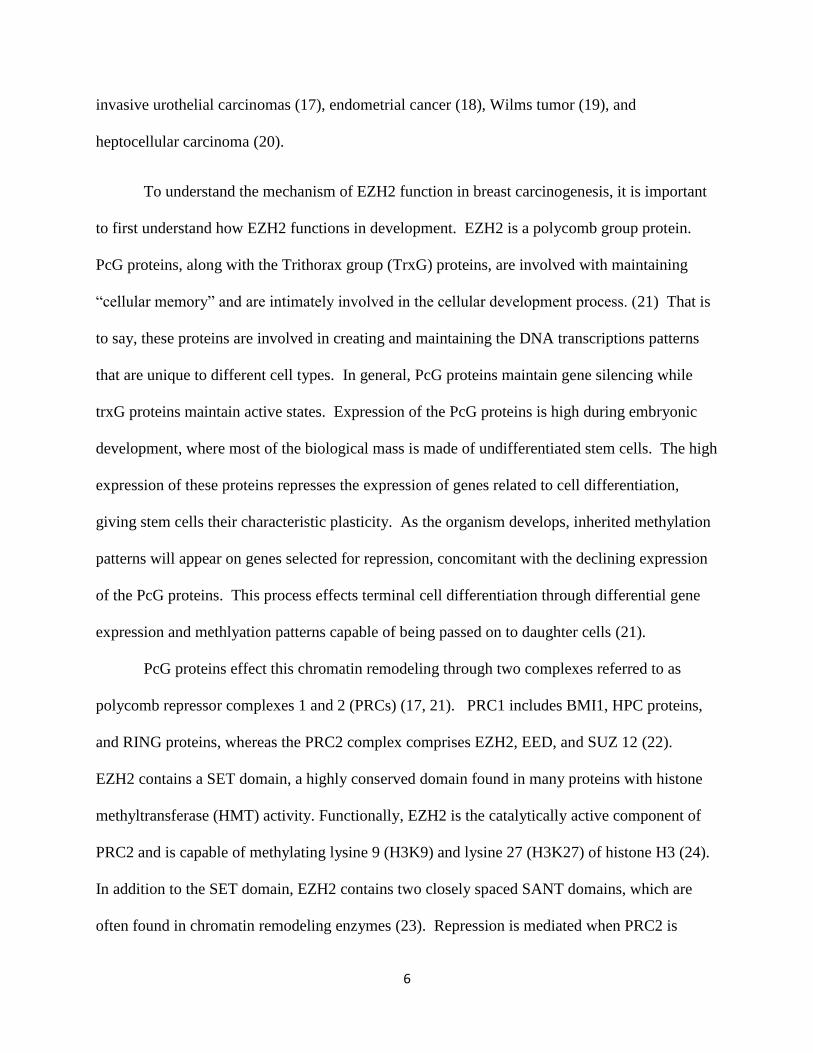
To understand the mechanism of EZH2 function in breast carcinogenesis, it is important
to first understand how EZH2 functions in development. EZH2 is a polycomb group protein.
PcG proteins, along with the Trithorax group (TrxG) proteins, are involved with maintaining
“cellular memory” and are intimately involved in the cellular development process. (21) That is
to say, these proteins are involved in creating and maintaining the DNA transcriptions patterns
that are unique to different cell types. In general, PcG proteins maintain gene silencing while
trxG proteins maintain active states. Expression of the PcG proteins is high during embryonic
development, where most of the biological mass is made of undifferentiated stem cells. The high
expression of these proteins represses the expression of genes related to cell differentiation,
giving stem cells their characteristic plasticity. As the organism develops, inherited methylation
patterns will appear on genes selected for repression, concomitant with the declining expression
of the PcG proteins. This process effects terminal cell differentiation through differential gene
expression and methlyation patterns capable of being passed on to daughter cells (21).
PcG proteins effect this chromatin remodeling through two complexes referred to as
polycomb repressor complexes 1 and 2 (PRCs) (17, 21). PRC1 includes BMI1, HPC proteins,
and RING proteins, whereas the PRC2 complex comprises EZH2, EED, and SUZ 12 (22).
EZH2 contains a SET domain, a highly conserved domain found in many proteins with histone
methyltransferase (HMT) activity. Functionally, EZH2 is the catalytically active component of
PRC2 and is capable of methylating lysine 9 (H3K9) and lysine 27 (H3K27) of histone H3 (24).
In addition to the SET domain, EZH2 contains two closely spaced SANT domains, which are
often found in chromatin remodeling enzymes (23). Repression is mediated when PRC2 is
7
recruited to chromatin, where the methyltransferase EZH2 catalyzes H3 trimethylation of lysine
27 (triMeK27-H3) (24). This histone mark then provides a platform to recruit PRC1 (24) which
aids in PcG-mediated repression either by chromatin compaction or by interfering with the
transcription machinery (21,26,27). Working together, PRC1 and PRC2 are responsible for
creating and maintaining gene expression patterns specific to different types of differentiated
cells, and their proper function is required to correctly pass these methylation patterns on to
daughter cells. Without EZH2 activity, PRC1 cannot be recruited to chromatin, and PcG-
mediated repression is not established (24,28).
Non-canonical roles for EZH2 have also been discovered. Shi et al. have reported that
EZH2 was able to activate gene transcription through mechanisms that do not involve histone
methylation (29). It has also been reported that EZH2 can function by forming transcriptional
complexes independent of its methyltransferase activity (30). In addition, cytosolic EZH2
complexes have been found to control cellular signaling via actin polymerization in various cell
types (31). With such duplicity in functional roles, it has been difficult to elucidate precise
mechanisms through which EZH2 influences breast cancer progression.
Breast and ovarian cancer type 1 susceptibility protein (BRCA1) is a tumor suppressor
involved in DNA damage repair, activation of cell cycle checkpoints and maintenance of
chromosome stability (32). The BRCA1 gene mutation was one of the first deregulations
correlated with breast cancer occurrence, and accounts for most familial cases of breast and
ovarian cancer (33). Heterozygous germ-line mutations in the BRCA1 gene predispose women
to breast and ovarian cancer with a lifetime risk of breast cancer up to 80% (34). Although
somatic mutations of BRCA1 are not common, expression of its messenger RNA and protein are
reduced in approximately 40% of sporadic breast carcinomas (35-37). The vast majority of breast
8
tumors in these patients display a basal-like phenotype, and BRCA1 dysfunction by
downregulation, mutation or other mechanisms has been suggested to have an etiological role in
the development of this aggressive breast cancer subtype (38-39).
It has been found that BRCA1 plays a major role in maintaining genome integrity
through its ability to form various distinct protein complexes, each of which is dedicated to a
specific cellular function in the DNA damage response pathway. To date at least three distinct
BRCA1-containing protein complexes have been emerged, identified as BRCA1A, BRCA1B,
and BRCA1C. BRCA1A functions to target BRCA1 to double strand breaks in DNA for repair,
and also to transiently inhibit entry into mitosis in the presence of DNA damage to avoid
aberrant chromosome segregation (40). BRCA1B is serves to inhibit DNA synthesis if DNA
damage is present, thereby avoiding the stalling or collapsing of replication forks during the S-
phase (41). BRCA1C functions in damage signaling and homologous recombination repair of
double strand DNA breaks. BRCA1 has emerged as a master regulator of genome integrity
through these diverse protein associations involved in somewhat redundant yet important cellular
processes ensuring genomic stability and promoting cell survival.
Our lab‟s research aim is to evaluate the oncogenic function of EZH2 in the breast and
elucidate the relevant mechanisms of this process. This knowledge may be applied to the clinical
field as inhibition of EZH2 may halt or prevent cancer progression.
Estrogen receptor (ER) negative breast cancer cell lines exhibit high EZH2 protein
expression. Our initial studies involved knocking down EZH2 expression in CAL51 and MDA-
MB-231 breast cancer cells using lentivirus-mediated short-hairpin RNA (shRNA) and
comparing phenotypes between these cells and control cancer cells. The results of our
experiments showed that EZH2 knockdown decreases proliferation of ER-negative breast cancer
9
cells and causes a delay in the G2-M transition of the cell cycle. Our data revealed that EZH2
modulates BRCA1 protein levels, as well as its phosphorylation at serine 1423, an important
modification in the regulation of the G2-M transition. We also show that BRCA1 is required for
the proliferative and G2-M effects of EZH2. We then highlighted the relevance of our in vitro
and animal findings by demonstrating that ER-negative invasive carcinomas have high levels of
EZH2 and concomitant low levels of BRCA1 protein. These initial studies provided the first
published functional link between EZH2 and BRCA1 proteins in breast cancer cells, and further
proposed that EZH2 knockdown in BRCA1 deficient cancer cells could possibly restore BRCA1
levels and function, as well as providing novel insight as to ER-negative breast cancer tumor
treatment.
Our research next aimed to investigate this newly found relationship between EZH2 and
BRCA1, as well as its implications in EZH2-mediated tumorigenesis. To do this, we created an
inducible expression system in MCF10A normal breast cells, which would overexpress EZH2
protein in the presence of Doxycylcline in the cell media. To complement these studies, we also
used the CAL51 cancer cell shRNA system previously described.
In investigating the link between these two proteins, we discovered that EZH2 regulates
the intracellular localization of BRCA1 protein in benign breast cells and in breast cancer cells
during cell cycle progression. Increased EZH2 expression leads to increased nuclear export of
BRCA1 in both MCF10A and CAL51 cell lines, and this is sufficient to trigger aberrant mitosis
with extra centrosomes and aneuploidy. EZH2 inhibition in tetraploid CAL51 breast cancer cells
induces BRCA1 nuclear accumulation and rescues defects in ploidy and mitosis.
Mechanistically, this data shows that EZH2-induced BRCA1 nuclear export, mitotic defects, and
aneuploidy require activation of the PI3/Akt signaling pathway.

10
In all, my research in the Kleer lab has followed the long of this story, beginning in 2007
through the present day. Although beginning with more modest projects, my data now
represents significant contributions to our lab‟s research aim, to illustrate the role of EZH2 in
breast cancer and breast cancer transformation. The experiments I have performed are part of
two research manuscripts, one published, and the other one in preparation. In this thesis I will
highlight the data I personally gathered (while giving credit to collaborating faculty for
corroborating data) in the context of our lab‟s larger story, and through this I will incorporate the
future directions and current debate at the cutting edge of breast cancer research.

11
RESULTS:
EZH2 expression is increased and deregulated in breast cancer cells in the absence of EZH2
gene mutation
A panel of breast cancer cell lines was investigated for EZH2 expression. These cells
include the nontumorigenic line MCF10A and breast cancer cells MDA-MB-231, SUM149,
CAL51, and MCF7. Western Blot analysis shows that breast cancer cells have higher EZH2
expression than the benign breast cells. (Figure 2)
Figure 2
Figure 2: EZH2 is upregulated and deregulated in breast cancer cells. (a) Immunoblot for EZH2 in a panel of breast
cells shows that EZH2 protein is elevated in breast cancer cells when compared with the nontumorigenic MCF10A
cell line. (b) EZH2 is deregulated in breast cancer cell lines compared with MCF10A cells. Data from M.E.G.
EZH2 expression has been observed to be cell cycle regulated in diploid fibroblasts, and
so our lab sought to determine whether EZH2 expression exhibited a similar pattern of regulated
expression in both normal and cancerous breast cells (42). To this end, benign and cancerous
breast cells were subjected to double thymidine block and subsequently restimulated by their
medium to enter the cycle. Figure 2 shows that EZH2 protein expression is cell growth regulated

12
and accumulates at the G1/S transition in benign MCF10A cells, similar to the expression seen in
fibroblasts (42). This regulation of EZH2 expression is lost in cancer, as seen in the four breast
cancer lines studied which exhibit unregulated, high expression of EZH2.
To determine whether this deregulation of EZH2 expression in breast cancer cells was
due to an activating mutation in the EZH2 locus, I sequenced the locus for MCF10A and CAL51
cell lines by direct sequencing. The EZH2 locus contains 2,695 base pairs, so sequencing
involved designing numerous forward and reverse primers to cover this length. After obtaining
the data, the experimentally determined sequences were then compared to that of the normal
sequence found in the NCBI database, checking for any possible mutations between the cell lines.
The results of this sequencing showed that there were no mutations in the CAL51 EZH2 locus
(Figure), ruling out the possibility of an activating mutation in the EZH2 reading frame being the
cause of its deregulated expression in breast cancer.
Increased EZH2 levels increase cyclin transcription levels and proliferation rates in breast
cancer cells
To study the effects of EZH2 expression on cell proliferation, we used shRNA
interference to stably knockdown EZH2 in breast cancer cells. EZH2 knockdown in MDA-MB-
231 and CAL51 cells significantly decreased proliferation (Figure 4). In searching to determine
how EZH2 increases proliferation in cells, we hypothesized that it may be due to upregulating
the expression of key cell cycle proteins, such as members of the cyclin family. Cyclins are
proteins which are expressed at varying levels during the cell cycle, and their association with
Cyclin dependent kinases (Cdks) is what drives cells through mitosis. To investigate if EZH2
13
expression regulated mRNA levels of key cyclins, I performed Real time PCR (RT-PCR) in
CAL51 shVector and shEZH2 cell lines. My results show that EZH2 increases the mRNA
expression of these key cyclins, therefore consistent with the observed increased proliferative
phenotype of EZH2 overexpressing cells (Figure 4).
This data was supported by other techniques performed in our lab. We found that EZH2
knockdown significantly increased the number of cells at the G2-M transition, while also
decreasing the phosphorylation of many key proteins necessary for entry into mitosis. Using
flow cytometry, we found that EZH2 knockdown caused over a 50% reduction in the percentage
of breast cancer cells undergoing mitosis. Collectively, these data show that EZH2 is important
in the regulation of the G2-M transition and the number of cells undergoing mitosis, which have
major influence in tumor growth. Our lab complemented these studies with in vivo experiments,
where MDA-MB-231 breast cancer cells (both shEZH2 and shVector) were injected into
immunocompromised mice and tumor growth was examined. We found that EZH2 knockdown
mice had decreased tumor growth rate, decreased tumor size, and better survival rates when
compared to control mice. It was also observed that mitotic activity also significantly decreased
in shEZH2 tumors as compared to control.

14
Figure 4
Figure 4: EZH2 inhibition decreases proliferation of breast cancer cells (a) Increased EZH2 expression is associated
with increased cyclin mRNA levels, providing link to its observed effect on breast cell proliferation. Quantitative
real-time PCR (qRT-PCR) measured the mRNA levels of cyclins A, B, and D in both CAL51 vector cells and EZH2
knockdown cells. Cyclin A expression is reduced by half, cylin B is reduced by a fifth, and cyclin D expression is
reduced by a third in CAL51 shEZH2 as compared to shVector cells. This data provides a possible functional insight
into how EZH2 overexpression in breast cancer cells increases proliferation EZH2 inhibition decreases proliferation
of breast cancer cells. (b) Time course of proliferation determined using the Wst-1 assay. shEZH2 in CAL51 and
MDA-MB-231 cells significantly decreases their doubling time when compared with vector control-transduced cells
(Student's t-test, P<0.001 for both cell lines). Proliferation data from M.E.G.
0
0.2
0.4
0.6
0.8
1
1.2
1.4
1.6
Cyclin A Cyclin B Cyclin D
EZH2 and cyclin expression
shVector
shEZH2
a
b

15
EZH2’s influence on proliferation is mediated through BRCA1
The rest of our first paper gave first light to the relation between EZH2 and BRCA1
proteins. Although I did not contribute to this section of our paper, I will summarize our
findings to illustrate the scope and significance of our findings. Studies have demonstrated that
EZH2 overexpression in human breast carcinomas is associated with the ER-negative basal-like
phenotype, characterized by low BRCA1 protein expression (11,14). From this, it was
hypothesized that EZH2 may promote breast tumorigenesis by regulating BRCA1 in this subtype
of breast cancer. Studies in ER-negative breast cancer cells showed that EZH2 downregulation
increased the protein levels of total BRCA1 and BRCA1 phosphorylated at serine 1423 in the
nuclei. Consistantly, overexpression of EZH2 in nontumorigenic MCF10A cells resulted in a 75%
decrease in nuclear BRCA1 protein and a 67% decrease in nuclear pBRCA1 s1423.
To investigate whether the effects of EZH2 expression on cell proliferation and transition
from the G2 phase to mitosis requires BRCA1 protein, the expression of EZH2 and BRCA1 were
both knocked down using shRNA in CAL51 breast cancer cells. We observed that BRCA1
knockdown in shEZH2 CAL51 cells was sufficient to completely rescue the reduction in
proliferation and the G2-M cell cycle arrest caused by EZH2 downregulation. Inhibition of
BRCA1 alone in CAL51 cells had no effect on proliferative activity. We also found that BRCA1
knockdown abolished the effect of EZH2 inhibition on important mitotic proteins. Taken
together, these data provide strong evidence supporting the hypothesis that the decrease in cell
proliferation and prolongation of G2 caused by EZH2 knockdown is mediated through BRCA1.
To investigate the relevance of these previous in vitro and in vivo studies, a large
sampling of invasive breast carcinoma tissue was probed for EZH2 and pBRCA1 expression and
16
possible correlation. It was found that in 76% of ER-negative tumors (associated with high
EZH2 expression) were negative for pBRCA1 s1423, further strengthening this link between
EZH2 and BRCA1.
The functional connection between EZH2 and BRCA1 proteins was our next area of
investigation.
Inducible EZH2-expressing MCF10A System Engineered
To further our investigation into the effect of EZH2 on cell proliferation and
tumorigenesis, as well as the relationship between EZH2 and BRCA1, we wanted to create a
cell-line system in which we could overexpress EZH2 in benign breast cell. I was put in charge
of the project to engineer this system which would overexpress EZH2 in the presence of
Doxycycline in the cell media. Greater detail as to how this system works and how it was
engineered to express our protein is found in the methods section. MCF10A cells naturally
express very low concentrations of EZH2, and Western blot analysis confirmed that EZH2
concentrations significantly increased in the cell when exposed to Dox.
EZH2 regulates the nuclear-cytoplasmic shuttling of BRCA1 in benign and in breast cancer cells.
EZH2 upregulation in the MCF10A cells resulted in nuclear export of BRCA1 protein
and increased BRCA1 cytoplasmic concentrations, as evinced by both Western blot and
immunofluorescence analysis (Figure 5). Similar results were found in CAL51 shVector and
shEZH2 cells, which showed that CAL51 breast cancer cells exhibit predominantly cytoplasmic

17
and perinuclear BRCA1 protein whereas CAL51 shEZH2 cells had accumulation of BRCA1 in
the nucleus. This data reveals that EZH2 influences the intracellular localization of BRCA1
protein in non-tumorigenic breast cells and in breast cancer cells.
Figure 5
MCF10A Cells (+/-) Dox
CAL51 Cells
BRCA1 DAPI Merge
Do
x
N
o D
ox
BRCA1 DAPI Merge
sh
EZH
2
sh
Vec
tor
Figure 5: EZH2 regulates
nuclear/cytoplasmic shuttling of
BRCA1. DAPI is a fluorescent stain
which strongly binds DNA, identifying
the nucleus. The top picture shows
fluorescence data gathered from the Tet-
On EZH2 overexpressing MCF10A cell
line. BRCA1 and DAPI are
independently detected in the first two
columns, and the images are merged in
the third column. The MCF10A data
shows that there is increased cytosolic
BRCA1 concentration with increasing
EZH2 concentration. The bottom
pictures show similar data for CAL51
breast cancer cells. In vector CAL51
cells (with high EZH2 expression), there
is observed a high cytosolic BRCA1
concentration. Knocking down EZH2
expression using shRNA shows
increased nuclear BRCA1
concentrations. Together this shows
that EZH2 plays a significant role in
BRCA1 subcellular localization.
Pictures from M.E.G.

18
Overexpression of EZH2 protein induces centrosome amplification and abnormal mitosis
Most malignant tumor cells have been found to have an abnormal number of
chromosomes, or aneuploidy, and frequently contain multiple centrosomes, which are the
microtubule-organizing centers required for proper chromosome segregation (43). Seeing as
EZH2 expression is elevated in many ER-negative invasive breast carcinomas, which display
many of these malignant characteristics, we hypothesized that EZH2 overexpression may induce
alterations in mitosis and centrosome number. Immunofluorescence analysis of mitotic nuclei
with α-tubulin and p-H3 (SER10) antibodies revealed that doxycycline-induced EZH2
upregulation in MCF10A cells led to mitotic defects characterized by the presence of multiple
mitotic spindles and extra centrosomes (Figure 6A). This contrasted with the absence of mitotic
defects in control MCF10A cells.
Aurora A is a key protein involved in centrosome maturation, separation, and spindle
formation. We hypothesized that the defective mitotic spindles observed upon EZH2
overexpression in MCF10A cells resulted from centrosome abnormality. We found that Aurora
A properly localizes to the centrosome during metaphase, as evidenced by the two distinct foci
that colocalized to the spindle poles and MCF10A control cells had two Aurora A foci. However,
although Aurora A was properly localized, EZH2 overexpression caused a greater than 6 fold
increase in the percentage of mitotic cells with more than two Aurora A foci, indicating
increased Aurora A expression and suggesting centrosome amplification. These cells contained
either a single giant nucleus or multiple multilobed nuclei. EZH2-induced extra centrosomes
were associated with multipolar mitosis (more than three and up to six spindle poles) as
compared with control MCF10A cells (example pictures given in Figure 6).

19
We next investigated the effect of EZH2 downregulation on mitotic spindle formation
and Aurora A containing centrosomes in CAL51 breast cancer cells (Figure 6B). CAL51 breast
cancer cells are diploid but contain a tetraploid population with centrosome amplification,
multiple mitotic spindles, and aberrant mitoses. Thus, they constitute a good model to test the
effect of EZH2 knockdown on the number of centrosomes, mitotic spindle formation, and
mitotic defects. 13.6% of CAL51 cells transduced with scrambled shRNAs have aberrant
mitoses characterized by increased number of centrosomes. In contrast, CAL51 shEZH2 cells
exhibit a 5-fold reduction in the number of cells with aberrant mitoses and supernumerary
centrosomes. Collectively, these data show that EZH2 may play a role in mitosis by regulating
the number of centrosomes in benign and breast cancer cells.
Further data our lab collected also showed that EZH2 expression is correlated with the
protein kinases Aurora A and B expression, suggesting further that EZH2 may mediate its
influence on mitosis through the regulation of these proteins.

20
Figure 6
A. MCF10A
B. CAL51
DAPI α-tubulin Aurora A Merge
DAPI α-tubulin Aurora A Merge
DAPI α-tubulin p-H3 (SER10) Merge
Do
x
N
o D
ox
Do
x
N
o D
ox
Figure 6: EZH2 overexpression
increases frequency of mitotic
aberrations. Confocal fluorescence
imaging was used to visualize the
localization of mitotic machinery in high
and low concentrations of EZH2 in
MCF10A and CAL51 cells. DAPI stains
the nucleus, α-tubulin is involved in the
mitotic spindle connecting centrosomes
and chromosomes, p-H3 is a histone
indicative of chromosomal alignment, and
Aurora-A is involved and localizes on the
centrosome structures. (A) shows the
experiments for the MCF10 Tet-On
System. With no dox added, the normal
bipolar mitotic alignment can be seen
(two spindle poles with actin extending
from poles and connecting to the
chromosomes, which are a linearly
aligned in middle). The merge in the top
section shows this proper alignment.
With dox added, mitotic aberrations are
more frequently observed. In this
example, actin can be seen to come from
many directions, and the chromosomes
are much more disorganized. Also seen is
an increase in Aurora A foci observed,
indicative of centrosome amplification,
known to cause aberrant mitosis. (B)
shows complementary data in the CAL51
system. In control cells the mitotic
spindle is more frequently disorganized,
although knocking down EZH2
expression decreases mitotic aberrations
in these cells. Pictures from M.E.G.
shEZ
H2
Co
ntr
ol
shEZ
H2
Co
ntr
ol
DAPI α-tubulin p-H3 (SER10) Merge

21
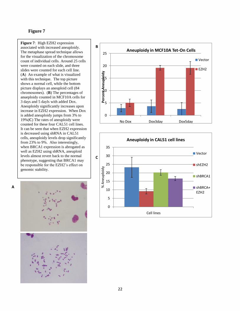
EZH2 overexpression causes genomic instability
Because errors in mitosis can lead to genomic instability and the development of cancer,
we next investigated whether EZH2 exerts a role in the maintenance of genomic integrity. To
investigate aneuploidy in our target cell lines I used a metaphase spread technique, a method
which fixes cells on a microscope slide and with the proper staining renders the chromosomes of
each individual cell visible and quantifiable (Figure 7A). EZH2 expression was found to be
correlated with increased prevalence of aneuploidy (Figure 7B). Tet-On MCF10A cells were
exposed to Dox for 1, 3, and 5 days before performing the metaphase spread technique.
Significant increases in aneuploidy were not observed until the third day of EZH2
overexpression. Giemsa stained metaphase spreads revealed that while 95% of untreated
MCF10A controls are diploid, 19.2% of cells became aneuploid after 72 hours of exposure to
Dox. We did not observe structural chromosomal abnormalities. Since CAL51 cells are diploid
with a tetraploid population, we reasoned that they constitute a good model to test if EZH2 can
rescue ploidy abnormalities. Giemsa stained metaphase spreads showed that EZH2 knockdown
decreased the percentage of tetraploid CAL51 cells from 23.2% to 9.2%, indicating that EZH2
downregulation can revert tetraploidy (Figure 7C). Taken together, this data reveals that besides
its ability to regulate the number of centrosomes and mitotic spindle formation, EZH2 plays a
role in the maintenance of genomic stability.
22
Figure 7
0
5
10
15
20
25
No Dox Dox3day Dox5day
Pe
rce
nt
An
eu
plo
idy
Aneuploidy in MCF10A Tet-On Cells
Vector
EZH2
0
5
10
15
20
25
30
35
Cell lines
% A
neu
plo
idy
Aneuploidy in CAL51 cell lines
Vector
shEZH2
shBRCA1
shBRCA+EZH2
Figure 7: High EZH2 expression
associated with increased aneuploidy.
The metaphase spread technique allows
for the visualization of the chromosome
count of individual cells. Around 25 cells
were counted on each slide, and three
slides were counted for each cell line.
(A) An example of what is visualized
with this technique. The top picture
shows a normal cell, while the bottom
picture displays an aneuploid cell (84
chromosomes). (B) The percentages of
anueploidy counted in MCF10A cells for
3 days and 5 dayls with added Dox.
Aneuploidy significantly increases upon
increase in EZH2 expression. When Dox
is added aneuploidy jumps from 3% to
19%(C) The rates of aneuploidy were
counted for these four CAL51 cell lines.
It can be seen that when EZH2 expression
is decreased using shRNA in CAL51
cells, aneuploidy levels drop significantly
from 23% to 9%. Also interestingly,
when BRCA1 expression is abrogated as
well as EZH2 using shRNA, aneuploid
levels almost revert back to the normal
phenotype, suggesting that BRCA1 may
be responsible for the EZH2‟s effect on
genomic stability.
A
B
C

23
EZH2-induced BRCA1 nuclear export, mitotic defects, and aneuploidy require activation of
PI3K/Akt
Although the mechanism that regulate the intracellular localization of BRCA1 protein
are not fully elucidated, recent studies suggest that the membrane serine/threonine protein kinase
Bα (AKT1) may play a role. In 2008 Plo et al. reported that AKT1 activation by
phosphorylation of serine 473 results in cytoplasmic localization of BRCA1 (44). This led us to
hypothesize that EZH2 may regulate the intracellular distribution of BRCA1 in normal breast
cells and in breast cancer cells through the PI3/AKT1 pathway.
Our laboratory shows that overexpression of EZH2 in MCF10A cells increases AKT and
AKT phosphorylation at serine 473, which is required to promote its maximal activation. The
levels of total and phosphorylated at SER473 AKT1 were also upregulated when EZH2 was
overexpressed. These results were similarly observed in CAL51 breast cancer cells. CAL51
EZH2 knockdown cells exhibited a drastic decrease in both AKT and AKT1 phosphorylation at
serine 473.
The significance of the PI3K/AKT pathway for BRCA1 intracellular localization in
EZH2 overexpressing MCF10A cells was next evaluated in a series of time course experiments.
BRCA1 protein was detected by immunofluorescence and by Western blot in the nuclear and
cytoplasmic-enriched fractions in the presence or absence of the PI3K inhibitor LY294002 or
Dox (Figure 8A). In the absence of the PI3K inhibitor, induction of EZH2 overexpression with
Dox led to high levels of pAkt SER473 and pAkt1 SER 473. Following LY294002 treatment,
pAKT and pAKT1expression dropped to almost undetectable levels. While Dox-induced EZH2
upregulation decreased BRCA1 and pBRCA1 proteins in the nuclei of MCF10A cells, inhibition

24
of PI3/AKT with LY294002 rescued BRCA1 and pBRCA1 nuclear expression. Similar results
were observed using Wortmannin to inhibit PI3K/AKT pathway. Immunofluoresence analysis
supported the immunoblot results as LY294002 or Wortmannin increased the expression of
BRCA1 in the nuclei reverting EZH2-induced nuclear export of BRCA1 (Figure 8A).
To investigate if EZH2 requires activation of the PI3/AKT pathway to trigger mitotic
defects and aneuploidy, I analyzed chromosome numbers and mitotic aberrations utilizing
metaphase spreads and immunofluorescence in the absence and presence of doxycylcline and the
PI3K/AKT inhibitors, LY294002 or Wortmannin (Figure 8B). As shown previously,
doxycycline-induced EZH2 overexpression causes aneuploidy in MCF10A cells after 72 hours.
LY294002 or Wortmannin treatment rescues EZH2-induced aneuploidy (figure). It was also
found that LY294002 or Wortmannin treatment also reverted the percentage of cells with extra
centrosomes back to near wild-type levels. Altogether, these results demonstrate that EZH2
upregulation increases AKT and AKT1 phosphorylation at serine 473 and that activation of the
PI3K/AKT is essential in the EZH2-induced BRCA1 nuclear export, aneuploidy, and mitotic
defects.

25
No Dox Dox LY294002 Wortmannin
Figure 8
0
5
10
15
20
25
% A
ne
up
loid
y
MCF10A Aneuploidy % with AKT inhibitor
Vector No Dox
EZH2 No Dox
EZH2 +Dox 3 Days
EZH2 +Dox 3 days +LYEZH2 +Dox 3 days +Wort
Figure 8: EZH2 mediates BRCA1 localization and effects on mitosis through PI3K/AKT pathway. (A)
Fluroescence data of MCF10A Tet-On cells. With low EZH2 (no dox) BRCA1 is found predominantly in the
nucleas. With the addition of dox, EZH2 protein levels increase and BRCA1becomes predominantly found in
cytosol. Adding the AKT inhibitors LY and Wortmannin abrogates the cytosolic shuttling of BRCA1.
Pictures from M.E.G. (B) Aneuploidy counting from MCF10A cell line again, this time in presence of AKT
inhibitors. Adding Akt inhibitor decreases aneuploidy to near wild-type levels in these cell lines. This graph
shows that the addition of the PI3/Akt pathway inhibitors LY and Wortmannin decrease the prevalence of
aneuploidy in MCF10A cells.
A
B
BR
CA
1
DA
PI
26
DISCUSSION:
Our research significantly contributes to our modern understanding of the function of
EZH2 in breast cancer. We have first shown that EZH2 overexpression in ER-negative breast
cells increases cell proliferation and tumor growth both in vitro and in vivo. Next we have
shown that EZH2 plays a role in regulating the sub-cellular localization of BRCA1 through the
PI3K/AKT pathway. Finally, we have shown that EZH2 overexpression increases the frequency
of mitotic aberrations and aneuploidy in breast cells. This brings us to the pertinent question of
if and how these newfound functions of EZH2 contribute to breast cancer initiation or
predisposition. I will discuss the implications and interpretation of our data in the context of
current cancer models later in the discussion. The bottom line of our research is that in providing
a greater illustration of how EZH2 functions in ER-negative breast cancers, we are providing
essential information for the guidance of novel treatments and cures that could target EZH2 and
mitigate its tumorigenic effects.
EZH2 has been found to be significantly overexpressed in certain subtypes of breast
cancer (basal-type tumors, characterized by estrogen receptor (ER), progesterone receptor (PR)
and Her-2/neu negative status as well as low levels of BRCA1 protein) (14), and is associated
with larger tumor development, increased tumor proliferation rates, increased invasiveness, and
worse prognosis in patients (11). While EZH2 overexpression leads to this litany of negative
tumor phenotypes, knowledge of this fact has allowed EZH2 to become an independent marker
of prognosis, recurrence and metastasis in women with breast cancer (11). My first research
project delved into investigating potential mechanisms of EZH2 overexpression in breast cancer.
My data showed that the deregulated expression is not due to genetic mutations, and other groups
have come to show other possible mechanisms of EZH2 deregulation independent of mutation.
27
Varambally et al. has shown that microRNA-101 regulates EZH2 expression levels in prostate
cancer cells by binding to the 3‟ untranslated region of its mRNA (45). Bracken et al. 2003 have
shown that the EZH2 promoter is a target of E2F transcription factors (42), and that EZH2
expression is therefore regulated through the pRb-E2F pathway. Other groups have traced back
EZH2 regulation to p53 control (46).
EZH2‟s effect on proliferation has also been well documented in the past, and my data
has helped shed more light on the mechanisms through which EZH2 influences proliferation in
breast cancer cells. I found that increased EZH2 expression is associated with an increase in
cyclin levels, while others in our lab have found that increased activation levels of other essential
cell-cycle proteins are also associated with higher EZH2 expression. Another major novel
finding presented in our first paper was that EZH2 downregulation in aggressive ER-negative
breast cancer cells greatly decreases their proliferative capacity and rate of progression through
the cell cycle, causing a prolonged doubling time of ER-negative breast cancer cells and caused
an arrest at the G2/M transition of the cell cycle. These studies were then complimented with
previously unexplored in vivo experiments; targeted downregulation of EZH2 in aggressive ER-
negative breast cancer cells resulted in significant reduction of mammary tumor size as well as
improved survival.
One of the most important conclusions of our first paper was to show that EZH2 mediates
its proliferative effects through BRCA1. BRCA1 is known to play a significant role in breast
cancer, and in addition to its function in hereditary breast cancer, BRCA1 expression is reduced
in up to 40% of sporadic breast carcinomas (Wilson, Yoshikawa, Turner). Although BRCA1
promoter methylation is responsible for 10-15% of sporadic breast carcinomas, it does not
explain BRCA1 deficiency in the remainder of the tumors, leaving the door open to other

28
possible alternatives of BRCA1 modulation. We demonstrate that EZH2 knockdown in ER-
negative breast cancer cell lines causes upregulation of BRCA1 protein levels with a
concomitant increase in pBRCA1 s1423, the total amount of the latter being crucial for G2/M
arrest (47-48). Consistantly, ectopic overexpression of EZH2 in benign breast cells decreases
nuclear BRCA1 and pBRCA1 s1423 protein levels. Our data demonstrate that the observed
effects of EZH2 downregulation on breast cancer proliferation and G2/M transition require
BRCA1, as BRCA1 inhibition was sufficient to completely rescue the decrease in cell
proliferation and the delay in G2 caused by EZH2 downregulation.
Our experiments show that EZH2 knockdown is sufficient to increase BRCA1 levels in
breast cancer cells. We propose that EZH2 knockdown in breast cancer cells reduces their
growth by enhancing the cell-cycle regulatory effects of BRCA1, slowing the transition from G2
to M phases, and allowing more time for DNA repair to occur. This hypothesis is supported by
our experiments showing that breast cancer cells with EZH2 knockdown have increased levels of
pBRCA1 s1423 protein, which is essential for G2/M arrest and is activated during the DNA
damage response (48). EZH2 knockdown had no significant effect on BRCA1 messenger RNA.
We have no evidence by co-immunoprecipitation of a direct interaction between EZH2 and
BRCA1 proteins.
The relevance of the association between EZH2 and BRCA1 proteins to human breast
cancer is highlighted by the finding that 76% of ER-negative invasive carcinomas overexpress
EZH2 and are negative for pBRCA1 s1423. Our work demonstrated a previously undescribed
function of EZH2 during ER-negative breast cancer progression by showing that EZH2
knockdown decreases tumor proliferation and growth in vivo and in vitro, and influences the
transition from G2 phase to mitosis. Our first paper revealed the first link between EZH2 and

29
BRCA1 proteins and showed that EZH2 knockdown depends on BRCA1 upregulation to
decrease breast cancer proliferation and progression through G2. We could hypothesize that
restoration of BRCA1 function by EZH2 knockdown may effectively decrease tumor
progression enabled by BRCA1 deficiency, informing future strategies tailored to restore
BRCA1 levels and function, and for possibly aiding in the treatement of ER-negative breast
cancer tumors.
Continuing, our research ventured further into investigating the functional connection
between EZH2 and BRCA1 proteins. Engineering the inducible expression system in MCF10A
benign breast cell line was essential to our continued investigation into this relation, providing a
stably overexpressing system with which to complement our studies in the CAL51 breast cancer
cell line. It was from investigations in both these cells lines that most of our data was obtained.
Knowing that EZH2 and BRCA1 protein expressions were inversely correlated in basal-
like ER-negative breast cancer, and with our previous research showing that EZH2
overexpression leads to decreases in nuclear BRCA1, our research provided new insight into the
development of this phenotype. Using confocal fluorescence microscopy and western blot
analysis, our lab discovered that EZH2 regulates the distribution of BRCA1 protein between the
cytoplasm and the nucleus in benign breast cells and in ER negative breast cancer cells. By
measuring the nuclear and cytoplasmic expression of BRCA1 protein at different timepoints after
release from cell cycle synchronization, we concluded that EZH2 overexpression in MCF10A
induces nuclear export of BRCA1 protein. Consistent with this observation, BRCA1 protein was
translocated from the cytoplasm to the nucleus upon EZH2 downregulation in the high-EZH2
expressing CAL51 breast cancer cells.

30
To further the links between EZH2, BRCA1, and genomic stability, our studies go on to
demonstrate that transient EZH2 overexpression in MCF10A cells causes aberrant mitosis with
extra centrosomes, contrasting with control MCF10A cells showing organized mitotic spindles.
Further strengthening the function of EZH2 in mitosis, we observed that EZH2 overexpression
increased the levels of important centrosome protein kinases Aurora A and Aurora B as well as
dysregulating their expression during cell cycle. The effect of EZH2 on mitosis was also
analyzed in CAL51 breast cancer cells. While CAL51 cells transduced with scrambled shRNAs
exhibited aberrant mitosis with supernumerary centrosomes and mitotic spindles, EZH2
knockdown with the proper shRNA mitigated these abnormalities. EZH2 knockdown also led to
downregulation of Aurora A and Aurora B kinases.
These described effects of EZH2 on centrosome amplification and mitosis underscore a
very important connection between EZH2 and tumorigenesis. Regulation of mitosis is a major
cell cycle control mechanism that guards against production of aneuploid daughter cells and
tumorigenesis. We hypothesized that the mitotic defects induced by EZH2 overexpression may
lead to aneuploidization of MCF10A cells. These cells constitute a good model to study ploidy
alterations since they are diploid. Conditional EZH2 upregulation caused aneuploidy in
MCF10A cells as early as 72 hours after addition of doxycycline. EZH2 knockdown in CAL51
cells diminished the number of tetraploid cells compared to scrambled shRNA controls.
Importantly, we observed that knocking down both EZH2 and BRCA1 expression in CAL51
cells lead to the reemergence of wild-type aneulploidy levels, giving further credence to the
conclusion that it is through regulating BRCA1 that EZH2 exerts its effect on genomic stability
and tumorigenesis.

31
While the mechanism responsible for the nuclear-cytoplasmic shuttling of BRCA1
protein is not fully elucidated, recent literature suggests that the membrane serine/threonine
protein kinase B (AKT) plays a role. Our data provides compelling evidence that the effect of
EZH2 on BRCA1 intracellular localization requires the activation of PI3K/Akt.
Pharmocological inhibition of the PI3K/Akt pathway with LY294002 or Wortmannin reverted
EZH2-induced cytoplasmic retention of BRCA1 and promoted its nuclear translocation.
The PI3K/Akt pathway has been reported to play a role in the maintenance of genomic
stablility. We observed that the effect of EZH2 overexpression on mitotic defects and
aneuploidy requires activation of PI3K/Akt. Inhibition of this pathway using LY294002 or
Wortmannin was sufficient to rescue the increased number of centrosomes, abnormal mitosis,
and aneuploidy caused by EZH2 overexpression. Our data not only reveal a novel function of
EZH2 in maintenance of genomic stability, but strongly suggest that the promotion of aberrant
mitosis and aneuploidy may be important for the oncogenic role of EZH2 in breast cancer.
Our results demonstrate a previously undescribed function of EZH2 in breast
tumorigenesis. We show that EZH2 overexpression influences BRCA1 nuclear-cytosolic
shuttling and is sufficient to promote aberrant mitosis and aneuploidy of benign breast cells. In
breast cancer cells, EZH2 knockdown induces nuclear localization of BRCA1, decreases mitotic
aberrations and reverts tetraploidy. Our results enable us to pinpoint one mechanism by which
EZH2 controls BRCA1 intracellular localization and genomic stablility, implicating the
PI3K/Akt-1 pathway. We show that Akt activation is indispensable for driving EZH2-mediated
BRCA1 nuclear export, mitotic defects and aneuploidy of mammary epithelial cells. In view of
our results and based on the profound effects of EZH2 knockdown to breast cancer cells, we
propose that modulation of EZH2 expression levels may be a valid strategy to prevent or halt

32
neoplastic progression in the breast. Our data also provides an answer to observation of why it is
that although BRCA1 gene mutation is very infrequent in sporadic breast cancer, its expression
is still found to be dysregulated in many cases. Whereas mutations in tumor suppressor genes
such as BRCA1 or p53 can clearly lead to cancer, there do exist examples of tumor suppressor
inactivation independent of their genetic mutations. Such an example is seen with p53, where
wild-type p53 protein is found mislocated in the cytoplasm in breast cancer cells, while mutant
p53 remains in the nucleus (49). Our research suggests that this observed BRCA1 dysregulation
in sporadic cancers may be a result of such a mechanism mediated by EZH2 and AKT1.
My research, along with that of my colleagues, cuts to the heart of the question
surrounding the oncogenic mechanism underlying EZH2 overexpression. Over the course of my
time in the lab, we have highlighted two novel proteins associated with EZH2, BRCA1 and
AKT1. In looking at the large picture again, our data now allows for the more comprehensive
analysis of the role of EZH2 in breast cancer tumorigenesis.
The question of the role of EZH2 in breast cancer, and my main research aim in the Kleer
lab, sits at the crux of two large debates currently taking place in breast cancer research: that of
the role of aneuploidy in tumorigenesis, and the cancer stem cell theory. Pursuing future
directions in our research has led down both of these paths, and these theories play a large role in
the further interpretation of our data as well as the development of further clinical utility.
Our data has shown that EZH2 overexpression can cause anueploidy in breast cells, but
can this aneuploidy leads to tumor formation? This hypothesis has been debated since it was
first proposed over one-hundred years ago (50), and research since this time has only revealed
the intrinsic complexity inherent in tumorigenesis and anuploidy formation. Aneuploidy is the
33
most common characteristic of human solid tumors, yet it is still unclear whether it is aneuploidy
is driving tumorigenesis or if it is merely a product of other malignant mutations in the cell (51).
By investigating in more detail the observed anuploidies in MCF10A and CAL51 cells, we have
constructed a more detailed image of how EZH2 overexpression may lead to aneuploidy and
tumorigenesis.
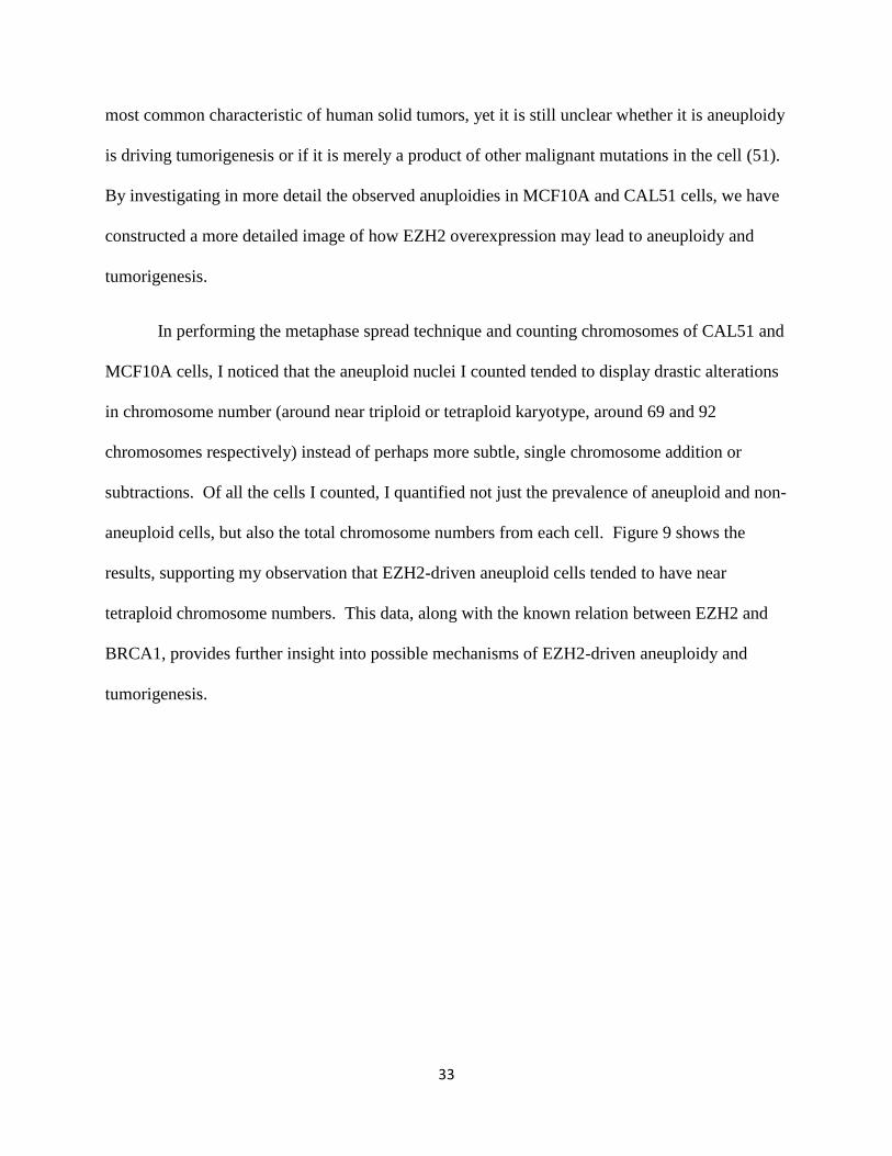
In performing the metaphase spread technique and counting chromosomes of CAL51 and
MCF10A cells, I noticed that the aneuploid nuclei I counted tended to display drastic alterations
in chromosome number (around near triploid or tetraploid karyotype, around 69 and 92
chromosomes respectively) instead of perhaps more subtle, single chromosome addition or
subtractions. Of all the cells I counted, I quantified not just the prevalence of aneuploid and non-
aneuploid cells, but also the total chromosome numbers from each cell. Figure 9 shows the
results, supporting my observation that EZH2-driven aneuploid cells tended to have near
tetraploid chromosome numbers. This data, along with the known relation between EZH2 and
BRCA1, provides further insight into possible mechanisms of EZH2-driven aneuploidy and
tumorigenesis.
34
Figure 9
Tetraploidy in cells usually arise from aberrations in mitosis (52). During mitosis,
chromosomes attach via their kinetochores (proteinaceous structure on the DNA) to spindle
microtubules from the centrosomes, with this formation favoring even chromosome segregation
between the two daughter cells. Cells have natural checkpoints which delay anaphase until all
kinetochores are properly attached. In the presence of a persistant error however, mitotic cells
can escape the checkpoint arrest and exit mitosis without undergoing anaphase or cytokinesis,
thereby producing a tetraploid cell with a single nucleus and two centrosomes (53). Abnormal
0
20
40
60
80
100
120
40-50 51-60 61-80 81-100 > 100
Tota
l Nu
mb
er
of
Ce
lls c
ou
nte
d
Karyotype Range
Chromosome Range in MCF10A Tet-On Cells
No Dox
EZH2 3days
EZH2 5days
Figure 9: Near-tetraploid phenotype is the most commonly observed aneuploidy in the EZH2
overexpressing MCF10A system.

35
spindle positioning and movements are known to interfere with cytokinesis and lead to the
accumulation of tetraploid cells (54). Recalling our earlier work, we have shown that EZH2
overexpression leads to centrosome amplification and mulitipolar spindle formation, so it is
likely though these aberrations in mitosis that EZH2 favorably yields more tetraploid cells.
Important in the discussion of aneuploidy and tumorigenesis is also the characterization
of chromosomal instability (CIN), where cells display an increase in the rate of gain or loss of
whole chromosomes during cell division, which is not synonymous with aneuploidy (55).
Tetraploidy has been observed to instigate chromosomal instability (CIN) in yeast and
mammalian cells (56-57). Supernumerary centrosomes are proposed to be the major source of
CIN (58-59). Extra centrosomes can lead to the formation of multiple spindle poles during
mitosis, resulting in the unequal distribution of chromosomes and the production of aneuploid
daughter cells. Multipolar mitoses have been shown to result in high CIN owing to
unsynchronized sister-chromatid separation, a high frequency of non-disjunction and the
occurrence of diplochromosomes (60). Our data again suggest that EZH2 may induce CIN
through its role in centrosome amplification.
Of greater importance now is asking how these characteristics may lead to tumor
formation. It can be reasoned that the appearance of CIN in a diploid cell would quickly lead to
cell death due to aberrant mitosis, however CIN in tetraploid cells would have a higher chance of
survival due to greater chromosomal redundancy, hinting at a selection method to pair CIN and
aneuploidy (52). Pairing CIN and aneuploidy together would lead to the production of progeny
that may be grossly aneuploid, though viable, and able to acquire further mutations. Chaotic
multipolar mitosis may break chromosomes directly, abnormal mitosis and prolonged mitotic
arrest can lead to the accumulation of DNA breaks, the double amount of DNA leads to twice the
36
amount of spontaneous DNA damage, coupled with the fact that DNA repair is probably much
less efficient; all of these events may lead to increased genetic mutation and therefore increased
likeliness for malignant transformation.
Tetraploidy has been identified in early stages of cancers, in which it precedes the
development of CIN and aneuploidy (62-64). Overexpression of Aurora A in the mammary
gland of mice leads to an increase in the generation of tetraploidy, CIN, and the formation of
mammary tumors, providing a further detailed picture of EZH2‟s possible effects in
tumorigenesis (65-66). Although aneuploidy has been shown to lead to tumor formation in some
studies, others have shown that aneupolidy can also play a neutral and even a tumor-suppressing
role in cancer (67). What hinders the hypothesis of aneuploidy-driven tumorigenesis is that to
date, there has been no found direct correlation between the level of aneuploidy and the
incidence of spontaneous tumor development. A comprehensive review on the role of
aneuploidy across a spectrum of cancers and models has shown that aneuploidy can increase the
risk of neoplastic transformation in some tissue lines, although a predisposed background or
some other activating mutation is usually required for tumorigenesis. The effect of aneuploidy
might not be driven by a particular combination of chromosomes per se, but rather by the
specific interaction of the karyotype with the various genetic contexts and microenviroments
found in different tissues. In relation to our research, a clear goal for the future could be to
investigate further whether aneuploidy promotes tumorigenesis specifically in the genetic context
of ER-negative breast cancers.
Another interesting model of tumorigenesis to consider in light of our data is the cancer
stem cell theory of breast cancer. This model posits that cancers develop from a subset of
malignant cells that possess stem cell characteristics (called cancer stem cells) and has been

37
proposed to account for the development of a variety of malignancies, including breast cancer.
These cancer stem cells originate from either mutated stem cells or from the dedifferentiation of
a lineage committed cell that has acquired stem cell characteristics through mutation. This
theory holds special relevance to our work seeing as how striking parallels can be found between
our studied subset of ER-negative breast cancer cells and stem cells. As brought up in the
introduction, EZH2 is an epigenetic repressor found in high concentrations in embryonic stem
cells to keep them proliferating and in an undifferentiated state. This is a very similar situation
as observed in our studied breast cancer cells, where EZH2 overexpression in breast cells is
associated with both increased proliferation and cell dedifferentiation, both hallmarks of more
malignant forms of breast cancer.
This cancer stem cell model of oncogenesis, first proposed for haematopoietic stem cells
in 2001 (68) and then for breast cancer in 2003 (69) (by the Wicha group at the University of
Michigan) details, implies a different mechanism of tumor progression and would therefore
recommend a different type of treatement in patients. Whereas the anueploidy-driven model
suggests that tumors become more homogenous masses of aneuploid cells, and so treatement
would need to be directly targeted against the entire tumor, the cancer stem cell model implies
that only a small subset of cells within a tumor possess the ability to self-renew and possibly
metastasize. This cancer model would then recommend that specifically these stem cells must be
targeted for treatement, and the rest of the tumor would soon die off (70). Implicit in designing
treatement then is identifying these cancer stem cells from the other tumor cells. In breast cancer,
cancer stem cells have been found to typically have the CD44+/CD24- phenotype (the CD
proteins are cell surface proteins), while the presence of ALDH-1 has also been used to
distinguish breast cancer stem cells. Our lab investigated into the possible relevance to our
38
cancer model by performing a cDNA microarray to test differential gene expression between
SUM149 cancer cells, both ALDH + and ALDHA -, as well as having EZH2 expression knocked
down using shRNA (Figure 10). The genes assayed in the microarray specifically pertained to
stem cell development pathway. Of the many observed difference, of note was the differential
expression of the Notch signaling proteins, which varied as a function of EZH2 expression
between the ALDH+ and ALDH- populations. Notch signaling plays a role in self renewal in
both normal and malignant stem cells, and finding differential expression could support the
theory that it is a small subset of breast cancer stem cells that are responsible for tumor growth
and invasion (71). This stem cell model would be an interesting model to look into, however its
restrictions of its applicability have been already heavily decried by critics. While this model of
tumorigenesis may explain the observed heterogeneity in tumors, there still has been a marked
lack of correlation between stem cell marking and cancer severity (72). This model is still
currently contested, however the great amount of research put into this investigation has left its
indelible mark on the future of cancer research.

39
Figure 10
A B C D E F G H
0.000.10
10.001000.00
100000.00
1 2 3 4 5 6 7 8 910 11 12
Row
Fold
Dif
fere
nce
(Te
st/c
on
tro
l)
Column
Microarray Stem Cell Gene Expression
1.E-07
1.E-06
1.E-05
1.E-04
1.E-03
1.E-02
1.E-01
1.E+00
1.E+01
1.E+02
1.E+03
1.E+04
1.E
-07
1.E
-06
1.E
-05
1.E
-04
1.E
-03
1.E
-02
1.E
-01
1.E
+00
1.E
+01
1.E
+02
1.E
+03
1.E
+04
Figure 10: Microarray
comparing SUM149
shEZH2 ALDH+ cells
with SUM149 parental
ALDH- cells. These
figures are given as
illustration as to the
power of microarray
analysis for widespread
gene analysis. The top
figure shows the
differential gene
expression as observed
in the differing well
locations on the
microarray plate, while
the bottom scatter plot
provides another view
with overexpressed
genes above the purple
lines and repressed
genes below the purple
line.
40
Both of these models of EZH2 driven tumorigenesis provide interesting yet varying ideas
as to how best treat these types of breast cancers. In reviewing these hypotheses, I have brought
us to the forefront not just of our research of EZH2 in breast cancer, but also of the current state
of cancer research. While in the end, the data I have collected while part of the Kleer lab has
helped us form a more comprehensive view of the role of EZH2 in breast cancer tumorigenesis,
there still remains many more questions and untreaded directions with which to continue our
research. We have discovered novel associations between EZH2 and BRCA1 expression in
breast cancer through AKT. We have provided potential in targeting any one of these relations
to increase breast cancer survival in patients. As quoted from Max Wicha himself, we are within
five years of being able to sequence patients‟ entire genome to search for the activating
mutations and genetic relations that we are elucidating today. The future of breast cancer
research is evolving through the beautiful amalgamation of advancing technology and intense
effort. Older models of breast cancer tumorigenesis are being questioned, and treatment
advances along with every new development. Though many questions remain, our work
continues.

41
MATERIALS AND METHODS:
Cell Lines
Breast cancer cell lines and immortalized human mammary epithelial cell lines (MCF10A) were
obtained from the American Type Culture Collection (ATCC, Manassas, VA, USA) and grown
under recommended conditions.
DNA Direct Sequencing
Direct Sequencing performed through the DNA sequencing core at the University of Michigan.
Primers were designed to cover entire EZH2 locus and ordered through Applied Biosystems.
Data received from the core was analyzed with FinchTV (Geospiza). The nucleotide sequences
were then compared using the NCBI BLAST (Basic Local Alignment Search Tool)
(http://blast.ncbi.nlm.nih.gov/Blast.cgi) program. Samples were compared to the NCBI
reference sequence for the Homo sapiens enhancer of zeste homolog 2 (Drosophila) (EZH2),
transcript variant 1, mRNA, locus NM_004456. If a mutation was found in the forward strand,
then the reverse stand was then consulted. A mutation would be considered valid if it was found
in both the forward and reverse strands.
Western immunoblots
Immunoblot analysis was performed as described in Kleer et al., 2003 using 100 mg of nuclear
enriched fractions extracted with NE-Per kit (Pierce, Rockford, IL, USA), or whole cell extract
as indicated in the legends. The following antibodies were used: mouse anti-b-actin (1:10 000),

42
mouse anti-a-tubulin (1:1000), goat anti-rabbit:horseradish peroxidase (HRP) secondary
antibody (1:10 000) and goat antimouse HRP (also 1:10 000). These antibodies were purchased
fromSigm a (St Louis, MO, USA). In addition, we used anti-EZH2 (1:1000; BD Biosciences,
San Jose, CA, USA); anti-BRCA1 (D-9) (1:200; Santa Cruz Biotechnology, Santa Cruz, CA,
USA); anti-phospho-BRCA1 (Ser1423) (1:1000; Abcam, Cambridge, MA, USA); anti-cdc25C
(5H9); anti-cdc2; anti-phosphor-cdc2-tyr15, used at 1:1000 dilution, fromCell Signaling
Technology, Danvers, MA, USA; anti-Cyclin B1 (1:1000 dilution, Calbiochem; EMD Chemicals,
La Jolla, CA, USA) and anti-phospho-Cyclin B1 (Ser126) (1:1000 dilution; Novus Biologicals,
Littleton, CO, USA). Control immunoblots using preimmune immunoglobulin G (IgG)
confirmed the specificity of the antibodies. Semiquantitative protein expression levels were
determined by densitometry using IMAGE J 1.38x software.
Cell synchronization
Cells were synchronized with double thymidine block as described previously (Fan et al., 2000).
Briefly, cells were incubated in medium containing 2mM thymidine for 12 h, released into their
normal medium for 8–10 h and then incubated for 12 h in medium containing 2mM thymidine
(see Supplementary methods for details).
Knockdown of shEZH2 and shBRCA1 in breast cancer cells
To generate stable short-hairpin interfering RNA-EZH2 and RNA-BRCA1 in MDA-MB-231 and
CAL51 breast cancer cells, cells were transduced with lentivirus and selected for antibiotic
resistance in ATCC-recommended media with puromycin (100 mg/ml, Sigma), at 37 1C under

43
10% CO2. Lentivirus was purchased fromthe Vector Core, University of Michigan. Background
vector control was Lenti- PuroEMPTY-VSVG. For targeting EZH2 (NM_152998
NCBI) and BRCA1 (NM_009764 NCBI), the shRNA oligos ID used were as follows:
V2LHS_17507 targeting EZH2 and V2LHS_254648 targeting BRCA1, corresponding to these
catalog numbers RHS4430-99139126 and RHS4430-99157192, respectively, fromOpen
Biosystems, Huntsville, AL, USA.
Cell proliferation
Cells were plated at the same density and cultured for 24 h in a 96-well microplate. WST-1
reagent was added and absorbance at 450nmwas measured after 3 h of incubation, following the
manufacturer‟s instructions (Roche Molecular Systems, Pleasanton, CA, USA).
Real-time quantitative PCR
Total RNA was isolated following the manufacturer‟s instructions with the RNeasy kit
(QIAGEN Inc., Valencia, CA, USA). cDNA samples from breast cancer cells were amplified in
triplicate from the same starting material of total RNA following the manufacturer‟s instructions
(High-Capacity cDNA Reverse Transcription Kit; Applied Biosystems, Foster City, CA, USA).
Samples were amplified using TaqMan MGB FAM dye-labeled probes from Applied
Biosystems in an ABI7900HT model real-time PCR machine. The following probes were used:
Hs99999903_m1 (ACTIN), Hs00173233_m1 (BRCA1) and Hs00544830_m1
(EZH2).
Engineering Tet-On System in MCF10A Cell Line

44
This project was one of my more significant undertakings in the lab, so I will go into a
little more detail about how the system works and how I created our system. Our lab purchased
the Lenti-XTM
Tet-On®
Advanced Inducible Expression System from Clontech. This involved a
system which would overexpress EZH2 when Doxycycline (Dox) was added to the cell culture
media. The system is derived from E. coli‟s tetracycline operon. In E. coli, the Tet repressor
protein (TetR) negatively regulates the genes of the tetracycline-resistance operon. TetR blocks
transcription of these genes by binding to the tet operator sequence (tetO) in the absence of
tetracycline (Tc). In the presence of Tc, TetR dissociates from tetO and transcription of
resistance mediating genes begins. This interaction between TetR and tetO are the basis for this
inducible system
The system comes with two elements, the regulator vector (pLVX-Tet-On Advanced) and
the response vector (pLVX-Tight-Puro). The pLVX-Tet-On Advanced vector constitutively
expresses the tetracycline-controlled transactivator, rtTA-Advanced. This protein consists of a
mutant TetR (rTetR) that posseses a “reverse” tetO binding phenotype in that this protein will
bind to tetO sequences in the presence of Dox, rather than its absence. The pLVX-Tight-Puro
response vector contains Ptight, the inducible promoter that controls the expression of our gene of
interest EZH2. Ptight consists of a modified Tet-Responsive Element (TREmod), made up of seven
direct repeats of an altered tetO sequence, joined to a minimal CMV promoter (PminCMVΔ). Upon
induction in the presence of Dox, rtTA-Advanced binds to the PTight promoter on the response
vector, activating transcription of the downstream gene (see Fig). These expression vectors
where introduced into the cell using viral vectors.
I will outline my strategy to splice the EZH2 locus from our stored plasmid into the PTight
promoter. The pLVX tight-puro vector was opened with a double digestion with the restriction

45
enzymes BAMHI and EcoRI. The EZH2 locus was cut from its vector using the same enzymes,
typical of this type of procedure, since this will allow for a simple mix and anneal step.
Complications arise however since EZH2 contains an internal EcoRI cut site, which separates an
almost 300bp segment from the rest of the gene. To get around this, I used PCR to magnify and
isolate the missing 300bp segment, while also engineering complementary cut site so this small
segment would fit in with the main vector fragment. After inserting the large part of the EZH2
locus into the pLVX tight-puro vector, I then double digested the small fragment with EcoRI and
BAMHI and then inserted and ligated the small segment into the larger vector construct using
viral T4 DNA ligase. Calf Intestinal (CIP) Alkaline Phosphatase was used to dephosphorylate
the 5‟ phosphate groups from the fragments to prevent self annealing.
The vector was then sequenced to confirm that our intended vector was obtained.
Western Blot analysis confirmed that EZH2 expression is at normal wild type levels in the
absence of Dox and that EZH2 expression increases dramatically upon the addition of Dox into
the media.
Metaphase Spead
Cells were grown in culture and colecemid added 24 hours before harvesting at around 70%
confluency. Colecemid limits microtubule formation and therefore freezes cells in metaphase. If
doxycycline was to be added (as in to collect data from the MCF10A inducible expression cell
line), it would be added to cell culture at around 50% confluency, in order to attain the desired
70-80% confluency. This level of confluency is need since colcemid will freeze cells currently
going through mitosis, however if the cells are already at 100% confluency then the mitotic

46
index will be very low and very few metaphases will be visible on the slide. Cells were collected
by trypsinization and resuspended in 0.075 mol/L KCl on ice for 30 min. Cells were fixed in a
3:1 mixture of methanol and glacial acetic acid with mild vortexing, dropped onto glass slides,
and stained with 544 Ag/mL Giemsa solution. To determine ploidy, the number of chromosomes
was counted in at least 25 metaphases for each cell line.

47
ACKNOWLEDGEMENT:
I would first like to thank Maria E. Gonzalez, with whom I‟ve spent most of my time
learning and working with in the lab and already owe a huge debt of gratitude for, and whose
data she kindly lent to me in order to be able to fully tell our intertwining research stories. It has
been a pleasure working with her, and I know I‟ve come a long way with her guidance since the
first day I walked into the lab as the inchoate student-researcher. I own much of my progression
in the lab to her help, as well as a greater understanding of what it means to pursue research as a
career yet still enjoying the other parts of life. I would next like to thank Dr. Celina Kleer, for
giving me the exceptional opportunity to work in her lab and engage in such an interesting
research project. I draw much inspiration from her ability to forge our research group out of her
own discoveries, and I‟m grateful everyday that I get to work and learn under her expert
authority on breast cancer. I would like to acknowledge the rest of our lab, Kathy, Xin, Heather,
Wei, and Anu, for making the workplace such a stimulating and always enjoyable research
environment. I thank my parents and family for raising me well and always giving me
encouragement to pursue my ambition, I‟ll always be proud of who I am and where I‟m from.
And thank you to my friends, given one condition, we will always drive our own cars.

48
REFERENCES:
1. American Cancer Society, Cancer Facts & Figures 2009
2. Bray F, McCarron P, Parkin DM. The changing global patterns of female breast cancer
incidence and mortality. Breast Cancer Res. 2004;6:229–239.
3. http://www.cancer.gov/cancertopics/types/breast
4. http://www.breastcancer.org/symptoms/understand_bc/what_is_bc.jsp
5. http://www.medicinenet.com/breast_cancer/page2.htm#tocd
6. Horner MJ, Ries LAG, Krapcho M, Neyman N, Aminou R, Howlader N, Altekruse SF,
Feuer EJ, Huang L, Mariotto A, Miller BA, Lewis DR, Eisner MP, Stinchcomb DG,
Edwards BK (eds). SEER Cancer Statistics Review, 1975-2006, National Cancer
Institute. Bethesda, MD, http://seer.cancer.gov/csr/1975_2006/, based on November
2008 SEER data submission, posted to the SEER web site, 2009.
7. Ellis M, Hayes D, Lippman M. (2000). Treatment of metastatic disease. In: Harris J,
Lippman ME, MorrowM (eds). Diseases of the Breast. Lippincott-Raven: Philadelpha,
pp 749–798.
8. Varambally S, Dhanasekaran SM, Zhou M, Barrette TR, Kumar-Sinha C, Sanda MG et
al. (2002). The polycomb group protein EZH2 is involved in progression of prostate
cancer. Nature 419: 624–629.
9. Mahmoudi, T. & Verrijzer, C. P. Chromatin silencing and activation by Polycomb and
trithorax group proteins. Oncogene 20, 3055–3066 (2001).
10. LaJeunesse, D. & Shearn, A. E(z): a polycomb group gene or a trithorax group gene?
Development 122, 2189–2197 (1996).
11. Kleer CG, Cao Q, Varambally S, Shen R, Ota I, Tomlins SA et al. (2003). EZH2 is a
marker of aggressive breast cancer and promotes neoplastic transformation of breast
epithelial cells. Proc Natl Acad Sci USA 100: 11606–11611.
12. Collett K, Eide GE, Arnes J, Stefansson IM, Eide J, Braaten A, Aas T, Otte AP, Akslen
LA (2006) Expression of enhancer of zeste homologue 2 is significantly associated with
increased tumor cell proliferation and is a marker of aggressive breast cancer. Clin
Cancer Res 12:1168–1174
13. Raaphorst FM, Meijer C, Fieret E, Blokzijl T, Mommers E, Buerger H, Packeisen J,
Sewalt RA, Otte AP, van Diest PJ (2003) Poorly differentiated breast carcinoma is
associated with increased expression of the human polycomb group EZH2 gene.
Neoplasia 5:481–488
14. Bachmann IM, Halvorsen OJ, Collett K, Stefansson IM, Straume O, Haukaas SA,
Salvesen HB, Otte AP, Akslen LA (2006) EZH2 expression is associated with high
proliferation rate and aggressive tumor subgroups in cutaneous melanoma and cancers of
the endometrium, prostate, and breast. J Clin Oncol 24:268–273
15. Raman JD, Mongan NP, Tickoo SK, Boorjian SA, Scherr DS, Gudas LJ (2005)
Increased expression of the Polycomb group gene, EZH2, in transitional cell carcinoma
of the bladder. Clin Cancer Res 11:8570–8576
16. Arisan S, Buyuktuncer ED, Palavan-Unsal N, Caskurlu T, Cakir OO, Ergenekon E (2005)
Increased expression of EZH2, a Polycomb group protein, in bladder carcinoma. Urol Int
75:252–257
17. Weikert S, Christoph F, Kollermann J, Muller M, Schrader M, Miller K, Krause H (2005)
Expression levels of the EZH2 Polycomb transcriptional repressor correlate with
aggressiveness and invasive potential of bladder carcinomas. Int J Mol Med 16:349–353

49
18. Bachmann IM, Halvorsen OJ, Collett K, Stefansson IM, Straume O, Haukaas SA,
Salvesen HB, Otte AP, Akslen LA (2006) EZH2 expression is associated with high
proliferation rate and aggressive tumor subgroups in cutaneous melanoma and cancers of
the
19. Zirn B, Hartmann O, Samans B, Krause M, Wittmann S, Mertens F, Graf N, Eilers M,
Gessler M (2006) Expression profiling of Wilms tumors reveals new candidate genes for
different clinical parameters. Int J Cancer 118:1954–1962
20. Sudo T, Utsunomiya T, Mimori K, Nagahara H, Ogawa K, Inoue H, Wakiyama S, Fujita
H, Shirouzu K, Mori M (2005) Clinicopathological significance of EZH2 mRNA
expression in patients with hepatocellular carcinoma. Br J Cancer 92:1754–1758
21. Ringrose, L., and Paro, R. (2004). Epigenetic regulation of cellular memory by the
Polycomb and Trithorax group proteins. Annu. Rev. Genet 38, 413–443.[
22. Levine SS, King IF, Kingston RE (2004) Division of labor in Polycomb group repression.
Trends Biochem Sci 29:478–485
23. Yu J, Li Y, Ishizuka T, Guenther MG, Lazar MA (2003) A SANT motif in the SMRT
corepressor interprets the histone code and promotes histone deacetylation. Embo J
22:3403–3410
24. Cao R, Wang L, Wang H, Xia L, Erdjument-Bromage H, Tempst P, Jones RS, Zhang Y
(2002) Role of histone H3 lysine 27 methylation in Polycomb-group silencing. Science
298:1039–1043
25. Min J, Zhang Y, Xu RM. Structural basis for specific binding of Polycomb
chromodomain to histone H3 methylated at Lys 27. Genes Dev. 2003;17:1823–1828.
26. Francis, N.J., Kingston, R.E., and Woodcock, C.L. (2004). Chromatin compaction by a
polycomb group protein complex. Science 306, 1574–1577.
27. Sarma, K., Margueron, R., Ivanov, A., Pirrotta, V., and Reinberg, D. (2008). Ezh2
requires PHF1 to efficiently catalyze H3 lysine 27 trimethylation in vivo. Mol. Cell. Biol.
28, 2718–2731.
28. Rastelli, L., Chan, C.S., and Pirrotta, V. (1993). Related chromosome binding sites for
zeste, suppressors of zeste and Polycomb group proteins in Drosophila and their
dependence on Enhancer of zeste function. EMBO J. 12, 1513–1522.
29. Shi B, Liang J, Yang X, Wang Y, Zhao Y, Wu H et al. (2007). Integration of estrogen
and Wnt signaling circuits by the polycomb group protein EZH2 in breast cancer cells.
Mol Cell Biol 27: 5105–5119.
30. Tonini T, Bagella L, D‟Andrilli G, Claudio PP, Giordano A. (2004). Ezh2 reduces the
ability of HDAC1-dependent pRb2/p130 transcriptional repression of cyclin A.
Oncogene 23: 4930–4937.
31. Su IH, Dobenecker MW, Dickinson E, Oser M, Basavaraj A, Marqueron R et al. (2005).
Polycomb group protein ezh2 controls actin polymerization and cell signaling. Cell 121:
425–436.
32. Venkitaraman AR. (2002). Cancer susceptibility and the functions of BRCA1 and
BRCA2. Cell 108: 171–182.
33. Miki, Y. et al. A strong candidate for the breast and ovarian cancer susceptibility gene
BRCA1. Science 266, 66–71 (1994).
34. Narod SA, Foulkes WD. (2004). BRCA1 and BRCA2: 1994 and beyond. Nat Rev
Cancer 4: 665–676.

50
35. Wilson CA, Ramos L, Villasenor MR, Anders KH, Press MF, Clarke K et al. (1999).
Localization of human BRCA1 and its loss in high-grade, non-inherited breast
carcinomas. Nat Genet 21: 236–240.
36. Yoshikawa K, Ogawa T, Baer R, Hemmi H, Honda K, Yamauchi A et al. (2000).
Abnormal expression of BRCA1 and BRCA1-interactive DNA repair proteins in breast
carcinomas. Int J Cancer 88: 28–36.
37. Turner N, Tutt A, Ashworth A. (2004). Hallmarks of „BRCAness‟ in sporadic cancers.
Nat Rev Cancer 4: 814–819.
38. FoulkesWD, Stefansson IM, Chappuis PO, Begin LR, Goffin JR, Wong N et al. (2003).
Germline BRCA1 mutations and a basal epithelial phenotype in breast cancer. J Natl
Cancer Inst 95: 1482–1485.
39. Lakhani SR, Reis-Filho JS, Fulford L, Penault-Llorca F, van der Vijver M, Parry S et al.
(2005). Prediction of BRCA1 status in patients with breast cancer using estrogen
receptor and basal phenotype. Clin Cancer Res 11: 5175–5180.
40. Wang, B. et al. Abraxas and RAP80 form a BRCA1 protein complex required for the
DNA damage response. Science 316, 1194–1198 (2007).
41. Xu, B., Kim, S. & Kastan, M. B. Involvement of BRCA1 in S-phase and G2-phase
checkpoints after ionizing irradiation. Mol. Cell. Biol. 21, 3445–3450 (2001).
42. Bracken AP, Pasini D, Capra M, Prosperini E, Colli E, Helin K. 2003. EZH2 is
downstream of the pRB–E2F pathway, essential for proliferation and amplified in cancer.
EMBO J 22: 5323–5335.
43. Storchova, Z., and C. Kuffer. 2008. The consequences of tetraploidy and aneuploidy. J.
Cell Sci. 121:3859–3866.
44. Plo I, Laulier C, Gauthier L, Lebrun F, Calvo F, Lopez BS. AKT1 inhibits homologous
recombination by inducing cytoplasmic retention of BRCA1 and RAD51. Cancer Res.
2008 Nov 15;68(22):9404-12.
45. Varambally S, Cao Q, Mani R, Shankar S, Wang X, Ateeq B, Laxman B, Cao X, Jing X,
Ramnarayanan K, Brenner J, Yu J, Kim J, Han B, Tan P, Sinha C, Lonigro R,
Palanisamy N, Maher C, Chinnaiyan A. Genomic Loss of microRNA-101 Leads to
Overexpression of Histone Methyltransferase EZH2 in Cancer. Science. 2008;322:1695–
1699.
46. Tang, X., Milyavsky, M., Shats, I., Erez, N., Goldfinger, N., Rotter, V. Activated p53
suppresses the histone methyltransferase EZH2 gene. Oncogene 2004, 23: 5759-5769.
47. Cortez D, Wang Y, Qin J, Elledge SJ. (1999). Requirement of ATMdependent
phosphorylation of brca1 in the DNA damage response to double-strand breaks. Science
286: 1162–1166.
48. Xu B, KimS, Kastan MB. (2001). Involvement of Brca1 in S-phase and G(2)-phase
checkpoints after ionizing irradiation. Mol Cell Biol 21: 3445–3450.
49. Moll, U. M., Riou, G., and Levine, A. J. (1992) Two distinct mechanisms alter p53 in
breast cancer: mutation and nuclear exclusion. Proc. Natl. Acad. Sci. U. S. A. 89, 7262–
7266
50. Boveri, T. Über mehrpolige mitosen als mittel zur analyse des zellkerns. Verh. Phys.
Med. Ges. Würzburg 35, 67–90 (1902) (in German).
51. Kops GJ, Weaver BA, Cleveland DW. On the road to cancer: aneuploidy and the mitotic
checkpoint. Nat Rev Cancer 2005; 5: 773-785

51
52. Storchova Z, Pellman D. From polyploidy to aneuploidy, genome instability and cancer.
Nat Rev Mol Cell Biol. 2004 Jan;5(1):45-54. Review.
53. Azeddine, E., Cunha, M. and Kirsch-Volders, M. (1998). Spindle poisons can induce
polyploidy by mitotic slippage and micronucleate mononucleates in the cytokinesis-
block assay. Mutagenesis 13, 193-198.
54. Reverte, C. G., Benware, A., Jones, C. W. and LaFlamme, S. E. (2006). Perturbing
integrin function inhibits microtubule growth from centrosomes, spindle assembly, and
cytokinesis. J. Cell Biol. 174, 491-497
55. Lengauer, C., Kinzler, K. W. & Vogelstein, B. Genetic instability in colorectal cancers.
Nature 386, 623–627 (1997).
56. Fujiwara, T., Bandi, M., Nitta, M., Ivanova, E. V., Bronson, R. T. and Pellman, D.
(2005). Cytokinesis failure generating tetraploids promotes tumorigenesis in p53-null
cells. Nature 437, 1043-1047
57. Storchova, Z., Breneman, A., Cande, J., Dunn, J., Burbank, K., O'Toole, E. and Pellman,
D. (2006). Genome-wide genetic analysis of polyploidy in yeast. Nature 443, 541-547.
58. Boveri, T. (2008). Concerning the Origin of Malignant Tumours by Theodor Boveri.
Translated and annotated by Henry Harris. J. Cell Sci. 121, S1-S84.
59. Nigg, E. A. (2002). Centrosome aberrations: cause or consequence of cancer progression?
Nat. Rev. Cancer 2, 815-825.
60. Gisselsson, D., Hakanson, U., Stoller, P., Marti, D., Jin, Y., Rosengren, A. H., Stewenius,
Y., Kahl, F. and Panagopoulos, I. (2008). When the genome plays dice: circumvention of
the spindle assembly checkpoint and near-random chromosome segregation in multipolar
cancer cell mitoses. PLoS ONE 3, e1871
61. Storchova ref
62. Galipeau, P. C. et al. 17p (p53) allelic losses, 4N (G2/tetraploid) populations, and
progression to aneuploidy in Barrett‟s esophagus. Proc. Natl Acad. Sci. USA 93, 7081–
7084 (1996).
63. Olaharski, A. J. et al. Tetraploidy and chromosomal instability are early events during
cervical carcinogenesis. Carcinogenesis 27, 337–343 (2006).
64. Ornitz, D. M., Hammer, R. E., Messing, A., Palmiter, R. D. & Brinster, R. L. Pancreatic
neoplasia induced by SV40 T-antigen expression in acinar cells of transgenic mice.
Science 238, 188–193 (1987).
65. Zhang, D. et al. Cre-loxP-controlled periodic Aurora-A overexpression induces mitotic
abnormalities and hyperplasia in mammary glands of mouse models.Oncogene 23, 8720–
8730 (2004).
66. Wang, X. et al. Overexpression of Aurora kinase A in mouse mammary epithelium
induces genetic instability preceding mammary tumor formation. Oncogene 25, 7148–
7158 (2006).
67. Holland AJ, Cleveland DW. Boveri revisited: chromosomal instability, aneuploidy, and
tumorigenesis. Nat Rev Mol Cell Biol. 2009 Jul;10(7):478-87. Review.
68. Reya T, Morrison SJ, Clarke MF, Weissman IL. Nature. 2001.Stem cells, cancer, and
cancer stem cells. Nov 1;414(6859):105-11. Review.

52
69. Al-Hajj M, Wicha MS, Benito-Hernandez A, Morrison SJ, Clarke MF. Prospective
identification of tumorigenic breast cancer cells. Proceedings of the National Academy
of Sciences of the United States of America. 2003;100(7):3983–3988.
70. Kakarala M, Wicha MS. Implications of the cancer stem-cell hypothesis for breast cancer
prevention and treatement. J Clin Oncol. 2008 Jun 10;26(17):2813-20. Review.
71. Dontu G, Jackson KW, McNicholas E, Kawamura MJ, Abdallah WM, Wicha MS: Role
of Notch signaling in cell-fate determination of human mammary stem/progenitor cells.
72. Lawson JC, Blatch GL, Edkins AL. Cancer stem cells in breast cancer and metastasis.
Breast Cancer Res Treat. 2009 Nov;118(2):241-54. Review.
73. Zeidler M, Kleer CG. The Polycomb group protein Enhancer of Zeste 2: its links to DNA
repair and breast cancer. J. Mol. Histol. (2006) 37:219–223
74. Huen MSY, Sy SMH, Chen J. BRCA1 and its toolbox for the maintenance of genome
integrity. Nat Rev Mol Cell Biol. 2010 Feb;11(2):138-48. Epub 2009 Dec 23. BRCA1
Review

![GENOMIC ANALYSIS OF METALLOTHIONEIN EXPRESSION IN … · 2018-01-09 · i genomic analysis of metallothionein expression in breast carcinogenesis lai yiyang [b.sc.(hons), nus] a thesis](https://img.dokumen.tips/doc/110x75/5e7c19873901b6286307dbcb/genomic-analysis-of-metallothionein-expression-in-2018-01-09-i-genomic-analysis.jpg)


![Breast cancer diagnosis based on genomic data and extreme … · 2020-01-15 · used for the prediction of breast cancer recurrence [18]. Researchers analyzed breast cancer data using](https://img.dokumen.tips/doc/110x75/5f93fc2f603b0841bd6a8cb6/breast-cancer-diagnosis-based-on-genomic-data-and-extreme-2020-01-15-used-for.jpg)