Embed Size (px)
Citation preview

Twenty Retinal Pathways Convey Visual Information from the Eye to the Brain
Classical studies of the functional architecture of the
retina have found that the image projected onto the
retina and captured by the photoreceptors is processed
locally by multiple parallel circuits ( Masland, 2001 ;
W ä ssle, 2004 ). This parallel processing results in several
different dynamic activity patterns at the retinal output
( Roska & Werblin, 2001 ) that are simultaneously trans-
mitted to the brain by ganglion cells, the output neurons
of the retina ( figure 13.1 ).
There is a growing consensus that the ganglion cell
population comprises ~20 different types. Each type
forms a subpopulation that covers the entire retina,
usually in a regularly spaced arrangement called a
“ mosaic. ” Thus, the unit of cellular infrastructure that
underlies parallel processing in the retina is a mosaic
of ganglion cells with similar morphology and response
properties, together with an associated mosaic of local
circuits ( figure 13.2 ). The retina embodies 20 such
mosaics that independently extract different features
from the visual world, although the underlying circuits
share many of the interneurons. It is as if our eye com-
prised multiple different TV crews pointing their
cameras at the same event but each broadcasting to
their audience (the relevant brain region) a subjectively
cut and processed version of the captured image flow
( figure 13.3 ). Some workers are shared among all of the
different crews, others specialize for jobs with few crews,
and some are only participating in one crew.
Each ganglion cell in a given mosaic has a local circuit
with different circuit elements. These elements can be
ranked according to how many synapses separate them
from the sensory receptors. Rods and cones are first-
order neurons; bipolar and horizontal cells are second-
order; amacrine and ganglion cells are third-order. The
ganglion cells, as the sole output element of the retina,
are positioned clearly at the top of the hierarchy in
these circuits. Bipolar, amacrine, and horizontal cells
each come in a variety of different morphological and
physiological variants. Again, neurons with the same
shapes and response properties are arranged in a
mosaic ( W ä ssle, 2004 ). Thus, the retina is built from
multiple mosaics of cells, which we will call “ cell types ”
for short.
Because each retinal patch contains 20 different gan-
glion cell circuits with more than 60 different circuit
elements, it is not surprising that there are strict orga-
nizational rules for the spatial arrangement of the
various circuit components. The first rule is that the cell
bodies of different circuits are packed in three different
cell body layers. Connections between circuit elements
occur between these three layers in two “ plexiform ”
layers of synapses. The second rule is that the dendrites
of the various ganglion cell types and the axon termi-
nals of the different bipolar cell types are stacked verti-
cally above each other in the inner plexiform layer,
forming ~10 narrow strata ( Siegert et al., 2009 ) ( figure
13.4 ). If the axon terminals of a bipolar cell type and
the dendrites of a given ganglion cell type are in differ-
ent strata, there can be no direct communication
between the two. Some bipolar cells have axon termi-
nals in more than one stratum and therefore give input
to these strata. There are more ganglion cell types than
bipolar cell types, and, therefore, the axon terminals of
a bipolar type typically excite more than one ganglion
cell type. Consistent with this, each stratum of the IPL
tends to contain dendrites of more than one ganglion
cell type.
Comparing the stratification of the three main cell
classes of the inner retina, the bipolar, ganglion, and
amacrine cells, we find striking differences. The pro-
cesses of ganglion and bipolar cells are generally con-
fined to one or two IPL strata. By contrast certain
amacrine cells, such as the AII amacrines, are arranged
vertically. With only a narrow horizontal extent, they
receive input or provide output in several strata. Other
amacrines, such as the starburst cells and polyaxonal
cells, are thinly stratified with a wide horizontal extent
within the stratum ( Masland, 2012 ). It appears that
the “ tall and narrow ” amacrines and their “ flat and
broad ” classmates have entirely different computational
roles.
13 The Retina Dissects the Visual Scene
into Distinct Features
Botond Roska and Markus Meister
Roska, B, & Meister, M (2014) The Retina Dissects the Visual Scene into Distinct Features. In: The New Visual Neurosciences (Werner, JS, Chalupa, LM, eds), pp 163–182. Cambridge, MA: MIT Press.

164 Botond Roska and Markus Meister
Figure 13.1 The retina creates 20 neural representations of the “ movie ” that enters the eye.
1
2
3
4
5
6
7
8
9
10
11
12
13
14
15
16
17
18
19
20
Retinal input
Retinal output
In evolution, the 20 retinal circuits could have been
organized into 20 separate pairs of eyes with overlap-
ping fields of view. Although this would have been
simpler with regard to the wiring and positioning of the
circuit elements, some cell types, such as photorecep-
tors, are needed for all circuits. Photoreceptors are
indeed numerous — they account for > 80% of all retinal
neurons ( Jeon, Strettoi, & Masland, 1998 ) — and it is
economical to share bulk common resources across cir-
cuits. The layered structure presented above, which
allows an efficient use of common resources, gives rise
to a hierarchical organization of cell types. Cells at the
bottom of the hierarchy, such as photoreceptors,
provide input to many ganglion cell types, whereas a
specialized amacrine cell higher in the hierarchy influ-
ences few ganglion cell types. Shared resources and cell
type hierarchy have important consequences for under-
standing both retinal processing and visual disorders.
Common operations needed for all circuits, such as the
gain control required for light adaptation ( Fain, 2011 ),
are more likely to be carried out at the front by common
elements. Similarly, one expects that cells whose dys-
function gives rise to noticeable visual defects are low
in the hierarchy. On the other hand circuit elements
responsible for specialized ganglion cell computations
are higher in the hierarchy.
The structure of the retina appears to be tailor-
made to extract many different features from the
visual scene. Under daylight conditions the image is
captured by cone photoreceptors. The first processing
stage, an interaction with the inhibitory horizontal
cells, contributes a step of lateral inhibition that
affects all downstream circuits ( Kamermans & Fahren-
fort, 2004 ; Wu, 1992 ). In dim light visual transduction
is accomplished by the rods, and their signals are sub-
sequently fed into the cone system by several elabo-
rate pathways ( Bloomfield & Dacheux, 2001 ). From
then on the rod-derived signals are largely processed
as though they came from cones. For the purposes of
this chapter, we therefore focus on circuits down-
stream of the cone bipolar cells.
Each cone is connected to ~10 types of bipolar cell
( W ä ssle et al., 2009 ). Some of these bipolar types are
distinguished by their neurotransmitter receptors with
different kinetics ( DeVries, 2000 ), and in turn they ter-
minate at different levels of the inner plexiform layer.
Therefore, the signals in different strata of the inner
retina already parse the visual input according to differ-
ent temporal features. Because there are more ganglion
cell types than strata, the activity carried from the outer
to the inner retina by each bipolar cell type is further
diversified. Different features can emerge within a
stratum because ganglion cell types have different
spatial extents and different receptors, and their cir-
cuits may include different amacrine cell types ( Taylor
& Smith, 2011 ). Notably, features carried by bipolar
cells can also be recombined locally by the action of
vertical amacrine cells.
The elaboration of visual features as discussed above
is a columnar operation: restricted in space and orga-
nized across or within strata. Retinal processing also
occurs laterally across space as a result of the lateral

The Retina Dissects the Visual Scene into Distinct Features 165
On autoOff autoOn:Off cross
0.0 0.2 0.4 0.6
Distance (mm)
Den
sity
(m
m–2
)
0.8 1.0
12
10
8
6
4
2
0
A
B
C
1 m
m
Figure 13.2 Ganglion cells of one type cover the retina with a regular mosaic. (A) Cell bodies and dendrites of ON alpha
ganglion cells in a wholemount view of the cat retina. Note that the dendrites cover space uniformly, and the cell bodies are
placed at regular distances ( W ä ssle, 2004 ). (B) Cell body locations of ON alpha (open circles) and OFF alpha (closed circles)
ganglion cells in a patch of cat retina ( W ä ssle, Peichl, & Boycott, 1981 ). (C) Each of the two cell types forms a regular mosaic
independent of the other. Spatial autocorrelation of the ON (solid blue line) and OFF (red) cell locations, showing the prob-
ability per unit area of finding a cell at a given distance from another cell of the same type. Note the prominent hole for dis-
tances < 0.2 mm. Cross-correlation (green) shows the probability of finding an OFF cell at a given distance from an ON cell.
Dotted lines are the average densities of ON (blue) and OFF (red) cells in this patch.
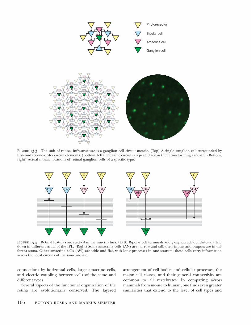
166 Botond Roska and Markus Meister
Ganglion cell
Amacrine cell
Bipolar cell
Photoreceptor
Figure 13.3 The unit of retinal infrastructure is a ganglion cell circuit mosaic. (Top) A single ganglion cell surrounded by
first- and second-order circuit elements. (Bottom, left) The same circuit is repeated across the retina forming a mosaic. (Bottom,
right) Actual mosaic locations of retinal ganglion cells of a specific type.
G1
B1
G2
B2
G3
B3
G4
B4
C C C C
B1
G4
B4 B4
C C C
AN AW
Figure 13.4 Retinal features are stacked in the inner retina. (Left) Bipolar cell terminals and ganglion cell dendrites are laid
down in different strata of the IPL. (Right) Some amacrine cells (AN) are narrow and tall; their inputs and outputs are in dif-
ferent strata. Other amacrine cells (AW) are wide and flat, with long processes in one stratum; these cells carry information
across the local circuits of the same mosaic.
connections by horizontal cells, large amacrine cells,
and electric coupling between cells of the same and
different types.
Several aspects of the functional organization of the
retina are evolutionarily conserved. The layered
arrangement of cell bodies and cellular processes, the
major cell classes, and their general connectivity are
common to all vertebrates. In comparing across
mammals from mouse to human, one finds even greater
similarities that extend to the level of cell types and

The Retina Dissects the Visual Scene into Distinct Features 167
their lamination ( figure 13.5 ). Several antibody markers
label the same strata across these species, and a number
of cell types are conserved. For example, both the
mouse ( Puller & Haverkamp, 2011 ) and the macaque
( Dacey & Packer, 2003 ) have a bipolar cell specialized
for signals from blue cones. In table 13.1 we compile a
catalog of retinal ganglion cell types across the major
species in which the topic has been studied. This illus-
trates a number of “ canonical ” cell types found in many
species (Berson, 2008). For other cell types the corre-
spondence is more difficult to identify, although this
may improve as we learn more about their visual
responses.
There are also distinct differences among mammals.
For example, in the mouse retina the spacing of cells
in a given mosaic is almost uniform across the retina;
at the other extreme, in the primate retina the cell
density rises sharply toward a small patch of retina in
the center called the fovea. The fovea has, therefore,
high spatial resolution and is used for encoding details
in the visual scene. Different mammals have different
degrees of nonuniformity in the spatial density of
ganglion cell mosaics, resulting in specialized retinal
regions such as the area centralis in cats or the visual
streak in rabbits.
A second difference is in the circuits processing color.
Most mammals have two cone types, one expressing a
short-wavelength pigment and the other medium
wavelength. Some primates also have cones with a
long-wavelength pigment. The circuitry connected to
short-wavelength cones has common circuit motifs
across mammals, such as the specialized blue cone cell,
but the differential handling of color information for
medium and long wavelengths is unique to a group of
primates. Some mammals such as mice and rats express
more than one pigment in many of their cones, and the
ratio of these pigments varies in a dorsoventral gradi-
ent. Because of this gradient the part of the eye that
looks at the blue sky is more sensitive at short wave-
lengths, and the part that looks at the ground is more
sensitive at longer wavelengths.
The anatomical evidence that the retina contains
20 ganglion cell mosaics along with their associated
circuits has emerged gradually over the last 50 years.
TH
ChAT
PKCa
MouseHuman
Figure 13.5 Comparing the retinas of humans and mice. Vertical sections of human (left) and mouse (right) retinas. Staining
with three antibodies against tyrosine hydroxylase (TH), choline acetyl transferase (ChAT), and protein kinase C alpha (PKCa)
identifies strata with similar positions in the two species.

168 Botond Roska and Markus Meister
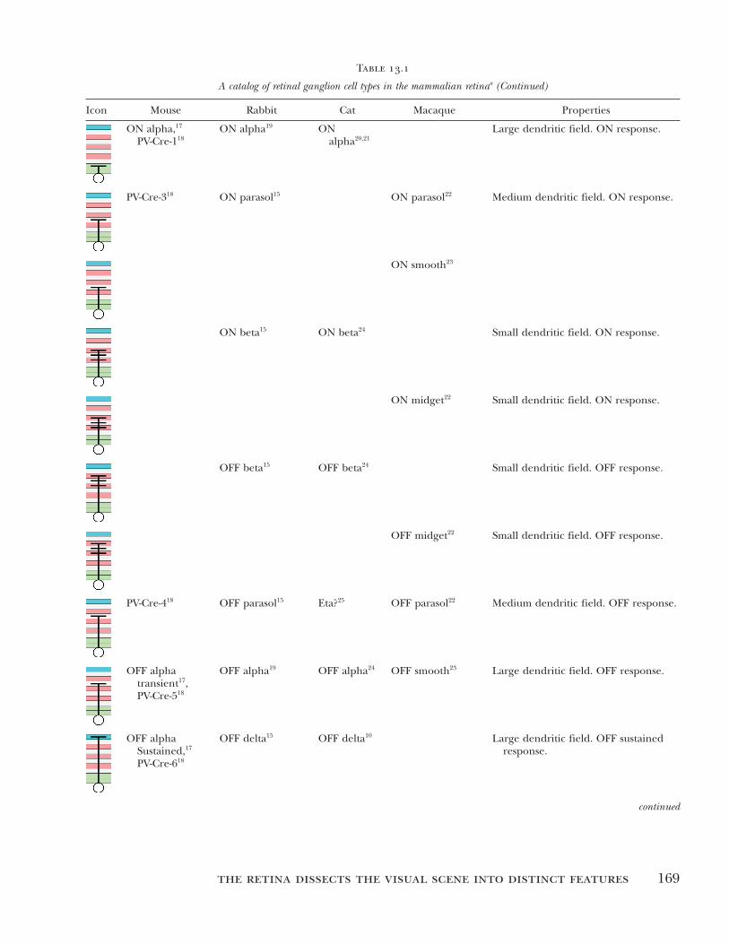
Table 13.1
A catalog of retinal ganglion cell types in the mammalian retina a
Icon Mouse Rabbit Cat Macaque Properties
M1 1,2 Outer
melanopsin 3
Large sparse dendrites. ON sluggish
synaptic response.
M2 1,2 Inner
melanopsin 3
Large complex dendrites. ON sluggish
synaptic response.
ON DS
temporal 4
ON DS temporal 5 ON DS. Preferred direction temporal.
ON DS ventral, 4
Spig-1 EGFP 6,7
ON DS ventral 5 ON DS. Preferred direction ventral.
ON, DS
dorsal 4,6,7
ON DS dorsal 5 ON DS. Preferred direction dorsal.
ON – OFF DS
temporal 8
ON – OFF DS
temporal 9
Theta? 10 Recursive
bistratified? 11
ON-OFF DS. Preferred direction
temporal.
ON-OFF DS
dorsal 8
ON-OFF DS
dorsal 9
Theta? 10 Recursive
bistratified? 11
ON-OFF DS. Preferred direction
dorsal.
Drd4-EGFP, 12
W9 8
ON – OFF, DS
nasal 9
Theta? 10 Recursive
bistratified? 11
ON-OFF DS. Preferred direction nasal.
BD-CreER, 8
Hb9-EGFP 13
ON – OFF, DS
ventral 9
Theta? 10 Recursive
bistratified? 11
ON-OFF DS. Preferred direction
ventral. Asymmetric dendrites in
mouse.
JAM-B 14 OFF coupled 15 ,
G3 16
OFF DS. Preferred direction ventral.
Highly asymmetric dendrites point
ventral.
The Retina Dissects the Visual Scene into Distinct Features 169
Icon Mouse Rabbit Cat Macaque Properties
ON alpha, 17
PV-Cre-1 18
ON alpha 19 ON
alpha 20,21
Large dendritic field. ON response.
PV-Cre-3 18 ON parasol 15 ON parasol 22 Medium dendritic field. ON response.
ON smooth 23
ON beta 15 ON beta 24 Small dendritic field. ON response.
ON midget 22 Small dendritic field. ON response.
OFF beta 15 OFF beta 24 Small dendritic field. OFF response.
OFF midget 22 Small dendritic field. OFF response.
PV-Cre-4 18 OFF parasol 15 Eta? 25 OFF parasol 22 Medium dendritic field. OFF response.
OFF alpha
transient 17 ,
PV-Cre-5 18
OFF alpha 19 OFF alpha 24 OFF smooth 23 Large dendritic field. OFF response.
OFF alpha
Sustained, 17
PV-Cre-6 18
OFF delta 15 OFF delta 10 Large dendritic field. OFF sustained
response.
Table 13.1
A catalog of retinal ganglion cell types in the mammalian retinaa (Continued)
continued

170 Botond Roska and Markus Meister
Icon Mouse Rabbit Cat Macaque Properties
ON-bistratified? 15 Small-bistratified 11 ON excitation, OFF inhibition,
blue-yellow opponent in macaque.
Large-bistratified 11 Blue-yellow opponent.
W3 26 Local edge
Detector 5,27
Zeta 28 Broad thorny 11 ON – OFF, strong surround, fast
ON – OFF inhibition.
Epsilon? 29 Recursive
monostratified 11
ON narrow
thorny 11
OFF narrow
thorny 11
Uniformity
Detector 30
Uniformity
Detector 31
Transiently suppressed by visual stimuli.
ON-OFF response. Dendrites just
outside the ChAT bands.
a Each graphic icon illustrates stratification of the dendritic tree in the IPL, divided into 10 laminae ( Siegert et al., 2009 ). For
each type we list the defining morphological and physiological features and identify its plausible correspondences in four
species, as supported by the cited literature. For further detail on cross-species comparisons, see Berson (2008). Note that many
of these ganglion cell types have only sparse and partial entries, emphasizing the need for future work to round out the catalog
of retinal output signals.
References : 1 Hattar et al. (2006). 2 Schmidt et al. (2011b). 3 Dacey et al. (2005). 4 Sun et al. (2006). 5 Barlow, Hill, & Levick (1964),
but see Kanjhan & Sivyer (2010) and Hoshi et al. (2011) for finer divisions. 6 Yonehara et al. (2009). 7 Yonehara et al. (2008).
8 Kay et al. (2011). 9 Oyster & Barlow (1967). 10 Isayama, Berson, & Pu (2000). 11 Dacey (2004). 12 Huberman et al. (2009).
13 Trenholm et al. (2011) erroneously identified the preferred direction as temporal. 14 Kim et al. (2008). 15 Roska, Molnar, &
Werblin (2006). 16 Hoshi et al. (2011). 17 Pang, Gao, & Wu (2003). 18 M ü nch et al. (2009). 19 Zhang et al. (2005). 20 Cleland, Levick,
& W ä ssle (1975). 21 W ä ssle, Peichl, & Boycott (1981). 22 Dacey & Packer (2003). 23 Crook et al. (2008). 24 W ä ssle, Boycott, & Illing
(1981). 25 Berson, Isayama, & Pu (1999). 26 Kim et al. (2010). 27 van Wyk, Taylor, & Vaney (2006). 28 Berson, Pu, & Famiglietti
(1998). 29 Pu, Berson, & Pan (1994). 30 Sivyer & Vaney (2010). 31 Cleland & Levick (1974).
Table 13.1
A catalog of retinal ganglion cell types in the mammalian retinaa (Continued)
However, with exception of a few ganglion cell types,
the functional distinctions among all these visual path-
ways have been more difficult to understand. Recent
technical advances have greatly accelerated this research
program, in particular the ability to genetically mark
and manipulate cell types ( Azeredo da Silveira & Roska,
2011 ; Huberman et al., 2009 ; Kay et al., 2011 ; Kim et
al., 2008 ; Yonehara et al., 2008 ). The fundamental new
insight is that the gene expression patterns of distinct
cell types are quite different. With advanced molecular,

The Retina Dissects the Visual Scene into Distinct Features 171
genetic, and viral tools one can hijack the cellular
machinery that controls these expression patterns to
manipulate neurons selectively. Now it is possible to
target specific cell types for physiological recording, to
modify them, to observe the effects on network func-
tion, and — importantly — to communicate the scientific
results without ambiguity about the identity of retinal
neurons under study.
In the following two sections we discuss what these
various ganglion mosaics extract from the visual scene
and how the associated circuitry performs the necessary
computations.
Pixel Sensors versus Feature Detectors
What is the role of the retina for the overall function
of the visual system? In the conventional view — still
dominant in textbooks and held by many vision
researchers today — the retina ’ s primary task is to get
the visual image transmitted to the brain, where the
cortex and other heavy-duty circuits can get on with the
challenges of processing the information. For that
purpose, the retina must first format the image signals
a bit to deal with the vicissitudes of the physical environ-
ment. Because the illumination conditions can change
so dramatically, the retina applies a gain control through
the cellular processes of light adaptation. And because
natural images tend to be highly redundant in their
pixel patterns, the retina performs some image com-
pression through the circuits that implement lateral
inhibition. In this view the defining characteristics of
retinal ganglion cell function are the center-surround
receptive field and gain control.
In an alternate view the retina shapes the visual rep-
resentation much more dramatically. Rather than
simply recoding the image for more efficient transmis-
sion through the optic nerve, the retina extracts from
the scene only a few specific features and transmits
those very selectively to the brain through several spe-
cific image channels. In this picture much of the raw
information in the visual scene is discarded. The gan-
glion cells transmit signals that result from very nonlin-
ear computations, for example, the speed of image
motion in a specific direction, that relate only distantly
to the raw data of image intensity.
Both of these rival conceptions of retinal processing
date back to the earliest days of retinal neurophysiology
( Barlow, 1953 ; Kuffler, 1953 ; Lettvin et al., 1959 ). Today,
with a complete catalog of ganglion cell types within
reach, one can envision an end to this debate. As usual,
the resolution will likely be a compromise. It appears
that a few ganglion cell types match the notion of “ pixel
sensors, ” whereas many others are better described as
“ feature detectors. ” Here we give these two concepts
explicit meaning and assess how they apply to the dif-
ferent retinal pathways.
Pixel Sensor
In its idealized form a pixel sensor ganglion cell would
simply measure the light intensity at a particular point
on the retina and convey that value directly to the brain.
A technological example of this is a single pixel sensor
of a digital camera. In practice, of course, ganglion cells
do not observe light at a single point but over a recep-
tive field. Furthermore, they cannot signal instanta-
neously but integrate the light over the retina ’ s response
time. Finally, they cannot put out a continuous signal
but only spikes. With these realistic constraints we can
define a pixel sensor retinal ganglion cell as one that
performs these image operations: compute a weighted
average of the light intensity over the receptive field
and the integration time and then use the result to
modulate the firing rate accordingly ( Meister & Berry,
1999 ). Such neural responses are generally character-
ized as “ linear ” because they derive from a linear sum-
mation of light intensity across time and space in the
receptive field (see appendix 13.1).
Remarkably, there are in fact retinal ganglion cells
that approach this ideal. For example, the midget P
cells in the primate fovea (including our own) receive
excitation mostly from a single bipolar cell, which in
turn gets input from a single cone ( Kolb & Marshak,
2003 ). Their response is truly dominated by a single
pixel in the photoreceptor array, and thus, there is no
opportunity for sophisticated nonlinear image compu-
tations. Actual response measurements from ganglion
cells in the fovea are rare ( McMahon et al., 2000 ), but
midget cells at greater eccentricity seem to integrate
light in a mostly linear manner ( Benardete & Kaplan,
1997a , 1997 b ).
Similarly, the X cells of the cat retina respond quite
linearly to light ( Enroth-Cugell et al., 1983 ). They have
a substantial maintained firing rate when the stimulus
remains constant. When the light increases they fire
more; when it decreases they fire less (or vice versa,
depending on polarity of the ganglion cell). A brighten-
ing in one part of the receptive field can be counterbal-
anced by a dimming in another part to completely cancel
the response, which illustrates that the circuit sums light
over space ( Enroth-Cugell & Robson, 1966 ). If the visual
stimulus varies in time like a sine-wave function, the
firing rate is modulated like a sine wave of the same fre-
quency; this is a common indicator of linear processing.
Both macaque P cells and cat X cells are the smallest
ganglion cells in the respective retinas. A tempting

172 Botond Roska and Markus Meister
suggestion is that in any given species the ganglion cells
with the finest receptive fields are pixel sensors that
convey a high-resolution version of the scene to the
brain. However, the mouse violates this simple notion:
The smallest ganglion cells in the mouse retina (called
local edge detectors or “ W3 ” cells) do not participate at
all in the signaling of routine visual scenes and respond
only very sparsely to specific events ( Zhang et al., 2012 ).
Feature Detector
A prototype of feature detection, again using a man-
made example, would be the face-detection circuit used
in current point-and-shoot cameras. Recognizing a face
in the visual scene requires an interesting combination
of selectivity and invariance: selectivity so the detector
remains silent in the many image regions that do not
contain a face; invariance so it responds to many differ-
ent faces under different views and illuminations. Obvi-
ously, this kind of performance requires computations
that are very different from mere linear filtering of the
image.
Again, there exist retinal ganglion cell types whose
performance approaches this ideal. One example
would be the “ bug perceivers ” described early on in the
frog retina ( Lettvin et al., 1959 ): These cells fire if a
small fly moves against a patterned background but not
if the same fly and the background move together.
Among the new ganglion cell types that were identified
or better explored in recent years, most have the char-
acteristics of feature detectors: highly nonlinear behav-
ior, selectivity for a certain visual feature, and invariance
to many other aspects of the scene. Often one can
understand the feature selectivity from ecological and
ethological considerations: the particular images pro-
duced by the natural environment, the needs of the
visual system, and the observer ’ s own behavior during
active vision. In the following section we illustrate some
of these cases.
Many Retinal Ganglion Cells Are Feature Detectors
For each of the sample ganglion cell types we begin by
discussing what it computes, namely what aspects of the
visual scene define its selectivity and its invariance. In
many cases we also understand how this stimulus selec-
tivity emerges from the interaction of retinal neurons,
and we present these explanations in the form of a
circuit diagram that summarizes the relevant connectiv-
ity and signal flow. These circuits are not intended to
be complete and exhaustive, so some cautionary com-
ments are in order.
First, for simplicity all the circuits begin with bipolar
cells. The outer retina circuits of photoreceptors and
horizontal cells perform some low-level formatting of
the visual signal, including light adaptation and lateral
inhibition. As a result of this processing, bipolar cells
have simple center-surround receptive fields. They
produce essentially linear light responses under the
same conditions where ganglion cells act as nonlinear
feature detectors ( Baccus et al., 2008 ). Therefore, not
much computation has occurred by the bipolar cell
level. Interesting selectivity emerges largely in the inner
retina through the interaction of bipolars, amacrines,
and ganglion cells. Second, the circuit diagram is
intended as schematic, not accurate in detail. For
example, the diagram does not spell out the correct
number of elements: a single component marked “ A ”
may stand for an entire population of amacrine cells of
that type. Third, the diagram is not exhaustive: It spells
out the minimal circuit that has been confirmed and is
essential to producing the function in question, but the
full circuit likely includes other components.
Y Cells
These ganglion cells drew attention in early studies of
cat retina because they clearly violate the notion of
linear summation ( Enroth-Cugell & Robson, 1966 ). If
the receptive field center is divided into a dark half and
a light half, and then the two regions are switched, the
X cells described above will remain silent because the
total light on the receptive field remains unchanged. By
contrast, the Y cells fire a strong burst on each of these
transitions. Y cells come in both polarities: An ON Y cell
is excited transiently by an ON transition in any small
region of the receptive field, even if it coincides with a
dimming elsewhere. These neurons are exquisitely sen-
sitive to a moving pattern because any such motion will
produce a brightening somewhere in the receptive field
( figure 13.6A ). The same applies to OFF Y cells, which
are excited by local OFF transitions. Thus, the Y cell
shows a form of invariance: It responds well to a fine
stimulus independent of where it occurs within the
receptive field or of the direction of motion. However,
the Y cells are not usually described as selective. They
do have an antagonistic surround that also includes
some nonlinear pooling over space similar to that in
the center ( Crook et al., 2008; Enroth-Cugell &
Freeman, 1987 ), but it has not been associated with
isolating any specific visual feature.
The unique response properties of the Y cell suggest
that its circuit pools excitatory inputs from small subre-
gions in the receptive field whose signals are individu-
ally rectified ( Enroth-Cugell & Freeman, 1987 ) ( figure

The Retina Dissects the Visual Scene into Distinct Features 173
Figure 13.6 Visual features extracted by retinal ganglion cells. For each of the four types of ganglion cells highlighted in the
text, this illustrates visual stimuli that excite the neuron (preferred) or suppress it (null). Arrows indicate movement. Dotted
line marks the receptive field center. The examples are taken from conditions that occur during natural vision: (A) excitation
by pattern motion on the receptive field center (Y cell and OMS cell); (B) suppression by a simultaneous pattern motion in
the surround (OMS cell); (C) excitation by expanding motion but not translating motion (looming detector); and (D) move-
ment in one direction but not in the opposite direction (DS cell).
AY cell
BOMS cell
Peferred Null
CLooming detector
DDS cell
13.7A ).There is good evidence now that the subfields
correspond to individual bipolar cells: These interneu-
rons match the size of the subfields ( Crook et al., 2008;
Demb et al., 2001 ), and their synaptic output can
indeed show strong rectification ( Baccus et al., 2008;
Demb et al., 2001 ). At a rectifying bipolar cell synapse,
only the depolarizations of the bipolar cell are transmit-
ted to the ganglion cell as excitation, whereas hyperpo-
larizations have no postsynaptic effect. This rectification
arises when the basal transmitter release rate at the
bipolar cell synapse is low because the resting potential
lies below the activation voltage of synaptic calcium
channels ( Matsui, Hosoi, & Tachibana, 1998 ; Palmer,
2010 ). The Y-cell circuit ( figure 13.7A ) explains
qualitatively how the neuron responds to moving tex-
tures regardless of the direction or the spatial pattern.
Small features of the texture activate different bipolar
cells as they move around. Bipolar cells often have
biphasic impulse responses ( Awatramani & Slaughter,
2000 ; Baccus et al., 2008 ; DeVries, 2000 ), which make
them sensitive to rapid changes but not to static pat-
terns. The rectification at the bipolar cell synapse then
allows accumulation of these transient signals from the
activated bipolars while it prevents cancellation from
other bipolars that experience opposite stimulus
changes. A time-varying velocity of the image pattern
leads to a time-varying firing rate, and the simple Y-cell
circuit model ( figure 13.7A ) can indeed predict this

174 Botond Roska and Markus Meister
Figure 13.7 Retinal circuits leading to different feature detector ganglion cells. (A) The Y-type ganglion cell. This ganglion
cell collects excitation from many bipolar cells. The bipolar cell synapses are rectifying: At baseline the release rate of transmit-
ter is low, so depolarization increases transmitter release, but hyperpolarization has little or no effect. In subsequent panels,
this rectifying quality is assumed for all bipolar cell synapses. (B) The object-motion-sensitive cell. Note that the ganglion cell
pools over both ON and OFF bipolars, but this process is gated by the action of a wide-field amacrine cell. (C) The looming
detector. Again there is pooling over ON and OFF channels, but with opposite sign because of an interposed narrow amacrine
cell. (D) The direction-selective ganglion cell. The asymmetric interaction that defines the null direction occurs between the
dendrite of a starburst amacrine cell and local bipolar cells. An additional threshold nonlinearity arises from spike generation
within the dendritic tree of the ganglion cell.
output quantitatively ( Baccus et al., 2008 ; Enroth-Cugell
& Freeman, 1987 ; Victor & Shapley, 1979 ).
The defining Y-cell characteristic of nonlinear sum-
mation over space has now been encountered in many
types of ganglion cell, but these differ strongly in other
response features that confer certain selectivities, as
seen in the following examples.
Object-Motion-Sensitive Cells
Ganglion cells of the “ bug perceiver ” type ( Lettvin et
al., 1959 ) have now been identified in several species,
and they likely represent one of the canonical types.
They have been called “ OMS ” cells in the salamander
( Ö lveczky, Baccus, & Meister, 2003 ), “ W3 ” cells in the
mouse retina ( Zhang et al., 2012 ), and “ local edge
detector ” cells in the rabbit retina ( Levick, 1967 ). They
produce transient responses to both ON and OFF
events in the receptive field center; thus, they process
the stimulus in a very nonlinear fashion, beyond that
of the Y cells. They are highly sensitive to moving pat-
terns within the receptive field center, for the most
part independent of the precise content of the pattern.
But if the receptive field surround experiences pattern

The Retina Dissects the Visual Scene into Distinct Features 175
motion, and the motion is synchronized with that of
the center, the ganglion cell remains silent ( figure
13.6B ).
One can speculate that such ganglion cells would be
very useful for detecting a moving object within a visual
scene. The ethological challenge here is that the eye of
the observer is almost always in motion, be it from small
fixational jitter or from the observer ’ s own locomotion
through the environment ( Kowler, 1990 ; Martinez-
Conde, Macknik, & Hubel, 2004 ). Thus, the default
condition on the retina is one of incessant image flow,
and identifying a moving object requires more than
simply flagging locations where the image moves.
Instead, the computation performed by the OMS cells
identifies image regions that move with a trajectory dif-
ferent from the surroundings. These are likely small
objects that move relative to the larger background,
such as bugs among leaves.
We now understand how this computation is per-
formed. The OMS ganglion cell pools excitation from
many bipolar cells with rectifying synapses ( figure
13.7B ). Moreover, unlike the typical Y cell, it receives
excitation from both ON- and OFF-type bipolars
( Levick, 1967 ; Zhang et al., 2012 ). Thus, movement of
a small object anywhere within the receptive field will
depolarize some bipolars and hyperpolarize others, and
in each case the ganglion cell receives a short pulse of
excitation. This will happen regardless of the exact
shape or pattern of the object or its direction of motion.
We see that the nonlinear summation over space and
over bipolars of opposite polarity already introduces a
great deal of invariance.
What about selectivity? The same ganglion cell
receives strong inhibition from amacrine cells in the
receptive field surround, up to large distances from the
center. The amacrine cell synapses act both directly on
the ganglion cell and at the bipolar cell terminal, where
they suppress transmission presynaptically ( Baccus
et al., 2008 ; Zhang et al., 2012 ). The amacrine cells are
themselves driven by the same kind of nonlinear pooling
mechanism that excites the ganglion cell ( Baccus et al.,
2008 ; Russell & Werblin, 2010 ; van Wyk, Taylor, & Vaney,
2006 ; Zhang et al., 2012 ). So if the visual pattern in the
surround moves at the same time as the pattern in the
center, excitation and inhibition cancel each other in
the ganglion cell, and it remains silent. But if a spurt of
motion in the center occurs independently of that in
the surround, the ganglion cell fires ( Ö lveczky, Baccus,
& Meister, 2003 ). This selectivity can be exquisite; for
example, the mouse W3 ganglion cell remains com-
pletely silent during natural stimuli that result from the
animal ’ s own locomotion because they contain a great
deal of global optic flow. These neurons are induced to
fire only in special conditions where a small target
moves against a static background ( Zhang et al., 2012 ).
Looming Detectors
These ganglion cells, called PV5 in the mouse retina,
fire strongly when a dark spot expands within the recep-
tive field, as would occur when an object approaches
the observer ( figure 13.6C ). Again, the receptive field
center is sensitive to both ON and OFF events in the
stimulus but now with opposite sign ( M ü nch et al.,
2009 ). A local dimming produces a transient excitation,
whereas a local brightening produces transient inhibi-
tion. When a dark spot moves through the receptive
field laterally, the leading edge produces excitation,
and the trailing edge inhibition; the two effects cancel,
and the ganglion cell remains silent. However, if a dark
spot expands within the receptive field, there is no ON
edge to contribute inhibition, and the ganglion cell
fires strongly. A symmetric looming detector for bright
objects has not been found.
The circuit that achieves the approach-specific
responses is based on the pooling of excitation from
the OFF pathway and inhibition from the ON pathway
( figure 13.7C ). The PV5 ganglion cell is excited by OFF
bipolar cells and inhibited by AII amacrine cells ( M ü nch
et al., 2009 ). The AII cell is a local interneuron that in
turn is excited by ON bipolar cells. Again, these synap-
tic inputs to the ganglion cell are rectified. As an edge
travels across the receptive field, it stimulates in turn
each of the small bipolar-size subunits, triggering a tran-
sient pulse of excitation or inhibition. When a dark
object expands over the receptive field, the excitatory
pulses are unopposed by any inhibition, and the gan-
glion cell fires throughout the period of expansion. If
the object moves laterally, on the other hand, excitation
from its leading edge is balanced by inhibition from the
trailing edge, and the ganglion cell remains silent.
Inhibition thus serves to suppress responses to the
nonpreferred motion signal, similar to the strategy of
the OMS cell circuit. In contrast to the OMS cells,
however, it is essential that the inhibition act postsynap-
tically rather than presynaptically at bipolar terminals
because signals from different parts of the object must
be combined. Again, rectification at the bipolar synapse
constitutes an essential element in the circuit, but here
we encounter an additional twist. This ganglion cell
combines rectified excitation and inhibition from path-
ways of opposite polarities. It has been suggested that
such “ crossover inhibition ” serves to make the overall
response of ganglion cells more linear ( Werblin, 2011 ),
with the ON pathway implementing responses to bright-
ening and the OFF pathway those to dimming. This is

176 Botond Roska and Markus Meister
not the case for PV5 cells. A neuron that pools the light
stimulus linearly over its receptive field can at best make
a dimming sensor but will not be selective for looming
objects. By contrast, the looming detector is excited by
an expanding dark edge, even if other parts of the
receptive field experience a gradual brightening. This
can be understood if the inhibitory pathway has a high
threshold such that a gradual brightening is ignored
but the sudden brightening at a traveling ON edge gets
transmitted ( M ü nch et al., 2009 ).
Note that the AII amacrine cell in this circuit also
serves an entirely different function during scotopic
vision, namely to feed rod signals into the cone bipolar
cells ( Bloomfield & Dacheux, 2001 ; Demb & Singer,
2012 ). This is an interesting example of a single cell
type that serves quite different roles, even signaling in
opposite directions ( Manookin et al., 2008 ).
Direction-Selective Cells
Again these ganglion cells are very sensitive to move-
ment within the receptive field. However, they respond
preferentially to motion in one direction and remain
silent to motion in the opposite direction ( Vaney, Sivyer,
& Taylor, 2012 ) ( figure 13.6D ). In some cases this direc-
tion selectivity applies even for tiny spots moving as little
as 1/10 of the RF diameter; thus, the computation is
performed on a very local scale, and the overall result
is pooled over the receptive field ( Barlow & Levick,
1965 ). Such a direction-selective (DS) cell is invariant
to the precise pattern or shape that moves within its
receptive field but selective for the direction in which
it moves.
Three classes of DS ganglion cells have been identi-
fied, distinguished by the polarity of the response in the
receptive field center. ON – OFF DS cells are excited
transiently by both ON and OFF steps of light ( Barlow
& Levick, 1965 ; Weng, Sun, & He, 2005 ), ON DS cells
by ON steps only ( Oyster, 1968 ; Sun et al., 2006 ), and
OFF DS cells by OFF steps ( Kim et al., 2008 ). These
three classes encompass multiple distinct cell types. The
four types of ON – OFF DS cells in the mammalian retina
have different preferred directions of motion in the
receptive field center, aligned with the cardinal direc-
tions on the eye: dorsal, ventral, nasal, and temporal
( Elstrott et al., 2008 ; Kay et al., 2011; Oyster, 1968 ).
Motion in the surround exerts a powerful suppression
( Barlow & Levick, 1965 ; Wyatt & Daw, 1975 ). As for
OMS cells, this suppression is particularly strong when
surround motion matches the center motion in speed
and direction ( Chiao & Masland, 2003 ; Ö lveczky,
Baccus, & Meister, 2003 ). Thus, the ON – OFF DS gan-
glion cells appear tuned to the local motion of objects
within the scene. Their axons project to both the thala-
mus and the superior colliculus ( Huberman et al.,
2009 ; Kay et al., 2011 ; Stewart, Chow, & Masland, 1971 ;
Vaney, Sivyer, & Taylor, 2012 ) and thus make this infor-
mation available to the two major streams for higher
visual processing. By contrast, the ON DS cells include
three types, with preferred directions on the retina
roughly dorsal, ventral, and temporal ( Oyster, 1968 ;
Yonehara et al., 2009 ). They are not suppressed by sur-
round motion and respond very well to moving patterns
that extend over the whole retina. Thus, they can serve
to encode the overall optic flow in the scene, as pro-
duced by slip of the image on the retina when the
animal or the eye moves relative to the scene. Interest-
ingly these neurons do not project to the major visual
pathways but exclusively into the accessory optic system
( Buhl & Peichl, 1986 ; Oyster et al., 1980 ; Yonehara et
al., 2008 , 2009 ) whose role is to sense self-motion for
the regulation of eye movements ( Simpson, 1984 ;
Giolli, Blanks, & Lui, 2006 ). Finally, a single type of
OFF DS cell has been described that prefers motion in
the ventral direction ( Kim et al., 2008 ). Again, these
neurons project to both superior colliculus and thala-
mus, but their role in downstream processing remains
unclear. This list represents the consensus types of DS
ganglion cells (DSGC), but there are recent indications
that the population may yet split into finer types whose
distinctions and downstream projections remain to be
established ( Hoshi et al., 2011 ; Kanjhan & Sivyer, 2010 ;
Rivlin-Etzion et al., 2011 ).
The retinal circuitry underlying the ON – OFF DSGC
has been studied intensely, and we now have a great
wealth of physiological, anatomical, and computational
results available. As may be expected, there is some
discordance in this large set of reports. As a result it has
become difficult to integrate all the observations into a
coherent model of neuronal circuitry. We present here
one subcircuit that almost certainly contributes to the
observed direction selectivity, although it leaves some
aspects unexplained ( figure 13.7D ). In this we largely
follow a recent review ( Vaney, Sivyer, & Taylor, 2012 ),
which is recommended for an overview of this retinal
subcircuit.
The discoverers of retinal direction selectivity pro-
posed a simple model of how it might be achieved
through the interaction of excitatory and inhibitory
synaptic inputs to the ganglion cell ( Barlow & Levick,
1965 ). This model has four required ingredients: spatial
asymmetry — inhibition should be laterally offset from
excitation; temporal asymmetry — inhibition should be
delayed relative to excitation; nonlinear pooling — the
ganglion cell responds only if its pooled synaptic input
exceeds a threshold; and small subunits — this pooling

The Retina Dissects the Visual Scene into Distinct Features 177
should occur independently within many small subunits
of the receptive field, to explain the selectivity for even
small motions. There now exists compelling evidence
that assigns these various functions to specific cellular
elements in the inner retina ( Vaney, Sivyer, & Taylor,
2012 ).
In this circuit ( figure 13.7D ), the independent sub-
units correspond to individual electrotonically distinct
dendritic compartments of the DSGC. Each such com-
partment pools excitation and inhibition and generates
a spike if the net depolarization exceeds a threshold.
These dendritic spikes travel to the soma reliably and
cause a spike in the axon. Each compartment receives
excitation from bipolar cells and inhibition from star-
burst amacrine cells (SACs). Whereas the receptive
field of a bipolar cell directly overlies its terminal, the
receptive field of the starburst cell is displaced laterally
toward the null side (the side from which null stimuli
arrive). This spatial asymmetry results from a peculiar
rule of connectivity between the two cell types. First,
although the SAC receives bipolar cell inputs all along
the dendrite, its inhibitory terminals are at the den-
dritic tips. Second, the DSGC connects preferentially to
those SAC dendrites that course in the null direction,
by a factor of 10 to 1 ( Briggman, Holmstaedter, & Denk,
2011 ). As a result the receptive field of the contributing
starburst dendrite is displaced toward the null side of
the DSGC ( figure 13.7D ). Another cellular mechanism
contributes to asymmetry: The depolarization at the
SAC dendritic tip is itself direction-selective, favoring
outward motion over inward motion ( Euler, Detwiler,
& Denk, 2002 ). Thus, the DSGC receives stronger inhi-
bition for null than for preferred motion. Finally, the
temporal delay and extended duration of inhibition
result from the additional synapse in the SAC pathway
as well as the prolonged time course of GABA release.
Given this circuit, one can understand the direction-
selective processing of moving stimuli ( figure 13.7D ). A
small spot moving in the null direction first excites the
SAC and then the bipolar cell. Because the SAC input
is delayed and more sustained, inhibition and excita-
tion arrive at the GC dendrite at the same time, the
resulting signal remains below threshold, and no spikes
are produced. The same sequence recurs in each of the
other compartments. With motion in the preferred
direction, excitation from the bipolar cell is triggered
before the inhibition can quench it, and this launches
a dendritic spike followed by somatic firing. The same
circuit is found in both the inner and the outer star-
burst stratum of the IPL, allowing the DSGC to process
ON and OFF edges independently.
As mentioned above, this should be considered a
minimal circuit. It leaves a number of observations
unexplained, for example, that excitatory inputs to the
DSGC are already direction selective ( Borg-Graham,
2001 ; Taylor & Vaney, 2002 ). This might occur if SACs
also inhibit the bipolar cell terminals ( figure 13.7D ).
Starburst amacrines also release acetylcholine, which
excites the DSGC; the function of these synapses is
unclear. The minimal circuit also does not account for
the suppressive effects of motion in the surround, which
may involve input from another type of amacrine cell.
The circuits for the other DS ganglion cells are less
well understood. Some ON DS cells seem to interact
with starburst amacrines ( Yonehara et al., 2011 ) much
as the ON – OFF DS cells do. However, the newly
described ON DS types that ramify outside the starburst
stratum suggest there must be other mechanisms to
achieve the same effects ( Hoshi et al., 2011 ). The same
holds for the OFF DS cells. This cell type has a strongly
asymmetic dendritic tree that points in the preferred
direction of motion ( Kim et al., 2008 ). In that case the
key asymmetry may well be provided by the morphology
of the ganglion cell itself, although the details of its
function remain to be explored.
Diverse Circuits Using Common Components
Although the various feature detectors discussed above
seem to select very different visual features, their under-
lying circuits share much in common. In fact, all these
circuits make use of the same kinds of simple elements:
small-field bipolar cells of two polarities, rectification at
a bipolar cell synapse, spatial pooling, narrow-field ama-
crines for sign inversion, wide-field amacrines for lateral
inhibition. The only differences lie in the sequence and
combination of the elements. As in the man-made field
of electronics, varying the arrangement and combina-
tion of simple elements results in dramatically different
functions. Still the above account falls short of filling
out the catalog of all 20 morphological ganglion cells,
which suggests that other retinal feature detectors and
their associated circuit computations remain to be iden-
tified ( table 13.1 ).
Open Questions
Visual Features and Ecology
The above examples motivate a deeper consideration
of feature selectivity. Are these neurons truly selective
for just one type of stimulus, and if not, can one justify
associating them with a specific feature? The answer to
the first question is clearly negative: For example, every
known ganglion cell will respond to a small spot flash-
ing in the receptive field center. This includes the

178 Botond Roska and Markus Meister
object-motion cells and the looming detectors. However,
under conditions of natural vision, flashing spots simply
do not happen very often. Except right after an eye
blink, objects rarely appear on the retina out of thin air;
instead, they move into a neuron ’ s receptive field from
a neighboring region, or they move within the receptive
field. Within the rather constrained set of stimuli that
occur commonly in natural vision, the looming detec-
tor is selective primarily for objects that expand, and
the OMS cell for those that move differently from their
background.
An interesting theme is that most of the feature
detectors identified to date seem to process some form
of image motion: wide-field, local, or differential. This
has a simple ethological interpretation: Moving objects
in the visual scene tend to be interesting points, either
as threats or opportunities. Similarly the global image
flow on the retina is a useful indicator of self-motion
through the environment. It is perhaps not surprising
that specific circuits have evolved to extract and sepa-
rate these important cues from the image rapidly and
efficiently. However, these qualitative arguments will
need to be tested more seriously. One approach is to
study retinal signaling under conditions that truly
reflect vision in the natural environment, including the
ever-present observer and eye motion. Such stimuli can
be gathered now thanks to ultralight video cameras that
can travel on the head of a rodent moving freely in the
natural environment ( Zhang et al., 2012 ). It will be
important to test for each ganglion cell type how selec-
tive it is under these conditions and whether the trigger
features are indeed those identified using the more
conventional synthetic stimuli.
Downstream Processing
Where in the brain are all these different parallel rep-
resentations sent? A simple suggestion, consistent with
the concept of a retina with many independent image
processors, would be that each ganglion cell type pro-
vides input to a different retinorecipient region. But
that is not the case. There are three patterns of gan-
glion cell projections ( figure 13.8 ). A few ganglion cell
types project to a single target region, such as the three
types of ON DS cells ( Vaney, Sivyer, & Taylor, 2012 ). A
few others project to multiple regions such as some of
the melanopsin-expressing ganglion cells ( Schmidt et
al., 2011a ). However, most ganglion cell types project
to two main visual centers, the lateral geniculate nucleus
(LGN) and the superior colliculus (SC). In fact, the
axons of most individual ganglion cells branch and
innervate both target areas.
There are two important points to note. First, many
visual features are copied to both the LGN and the SC,
and it is, therefore, intriguing to ask whether these
copies will ever be compared or, alternatively, will live
independent lives to drive behavior or perception.
Second, in most species studied, including primates,
many of the specialized visual features are sent to the
LGN. On the other hand, most cortical researchers are
convinced that only a few pathways — perhaps three —
arrive at the primary cortex from the LGN. One
Figure 13.8 Three types of retinal projections. (Left) Ganglion cell mosaic projecting to a single nucleus. (Center) Ganglion
cell mosaic projecting to two nuclei. (Right) Ganglion cell mosaic projecting to multiple nuclei.
G G G
G G G
G G G
G G G
G G G
G G G
G G G
G G G
G G G
Retina
Projections

The Retina Dissects the Visual Scene into Distinct Features 179
possibility is that multiple retinal features combine
immediately within the LGN into few visual channels.
Alternatively we may still have an incomplete under-
standing of the pathways that drive the visual cortex.
Fortunately a new set of tools is coming available that
includes the means to trace the downstream pathways
from genetically identified cells, to activate or silence
specific types of ganglion cells, and to record activity
from hundreds of neurons in the cortex. Therefore, it
is likely that this controversy will be resolved soon. More
broadly, it seems possible now to map out the relation
between the distinct ganglion cell pathways and specific
aspects of the animal ’ s visual behavior.
So far the silencing of types of ganglion cells has led
to controversial conclusions. Targeted elimination of a
few or single types of melanopsin-containing ganglion
cells caused well-defined behavioral deficits in mice
( Chen, Badea, & Hattar, 2011; Guler et al., 2008 ; Hatori
et al., 2008 ). Similar marked deficits in mouse behavior
were found when the starburst amacrine cells were
eliminated ( Yoshida et al., 2001 ), and likely the direc-
tional selectivity of at least seven types of directional
selective ganglion cells was abolished. In contrast, the
acute silencing of all types of ON ganglion cells led to
minor changes in primate visual behavior ( Schiller,
Sandell, & Maunsell, 1986 ). A mutation that is pre-
dicted to result in a similar silencing of all ON cells in
humans does not lead to any major visual defects at
light intensities where cones are active ( Dryja et al.,
2005 ; Zeitz et al., 2005 ). Many ganglion cell types come
in pairs, including an ON and an OFF version. It appears
that for a significant part of our visual perception and
function one version of a type is enough.
Clinical Tests of Feature Processing
The conservation across species of retinal structure and
function also provides new opportunities to diagnose
retinal diseases. When we visit the ophthalmologist, our
vision is tested on a chart from which we read small and
large letters. This test mostly diagnoses the optics of the
eye, based on the performance of 2 of the 20 types of
ganglion cells, the ON and OFF midget cells. Although
the retina incudes a massive infrastructure to analyze
and dissect different categories of motion, there is,
remarkably, not a single quantitative or even qualitative
test used regularly by ophthalmologists that would eval-
uate how well we can perceive motion. If, for example,
a mutation had produced a defect in the development
of amacrine cell networks, the patient may be unable
to see motion or, conversely, might see motion all the
time. Such a patient would likely end up in the office
of a psychiatrist, even though the defect originates in
the sensory periphery. By understanding one by one the
computations that different ganglion cells perform and
by understanding the behavioral phenotypes that result
from silencing identified ganglion cell mosaics, it may
be possible to discover abnormalities in human visual
perception that arise within the retina.
Appendix 13.1
Linear Visual Responses
Mathematically, a linear light response is derived by
convolving the stimulus with a filter function:
r t r s x t F x t t dt dx( ) = + ′( ) − ′( ) ′∫∫0 , ,
In this expression, s x t,( ) is the stimulus intensity as a
function of space and time, and r0 is the firing rate in
absence of any stimulus. The weighting function F x t,( ) specifies the weight applied to the intensity at location
x and time t in the past and is commonly called the
spatiotemporal receptive field of the neuron.
Clearly the range of a neuron ’ s firing rate is restricted,
namely to zero firing at the bottom, and some maximal
rate determined by cellular biophysics at the top. There-
fore, this linear relationship cannot persist when the
light intensity varies over too large a range. In fact one
generally finds distortions in the response to strong
stimuli. A more general version of a pixel sensor allows
for such distortions in the relation between stimulus
and response:
r t N s x t F x t t dt dx( ) = ′( ) − ′( ) ′( )∫∫ , ,
where the function N ( ) is the distortion function that
relates the linear-weighted stimulus to the firing rate,
generally with a sigmoid shape. A response function of
this kind is often called an LN model: a linear filter
followed by a nonlinearity ( Chichilnisky, 2001 ). Note
that the nonlinearity N ( ) does not fundamentally alter
what the ganglion cell reports about the visual scene,
only how it is reported. The visual meaning of the
message is fully defined by the spatiotemporal receptive
field F x t,( ) . In summary then, we can consider a retinal
ganglion cell a pixel sensor if its stimulus – response
function follows the LN model under most visual
stimuli. The spatial and temporal extent of the image
pixel that this ganglion cell reports is embodied by the
spatiotemporal receptive field F x t,( ) .
References
Awatramani , G. B. , & Slaughter , M. M. ( 2000 ). Origin of tran-
sient and sustained responses in ganglion cells of the retina.
Journal of Neuroscience , 20 , 7087 – 7095 .
Azeredo da Silveira , R. , & Roska , B. ( 2011 ). Cell types, circuits,
computation. Current Opinion in Neurobiology , 21 , 664 – 671 .
180 Botond Roska and Markus Meister
Baccus , S. A. , Ö lveczky , B. P. , Manu , M. , & Meister , M. ( 2008 ).
A retinal circuit that computes object motion. Journal of Neuroscience , 28 , 6807 – 6817 .
Barlow , H. B. ( 1953 ). Summation and inhibition in the frog ’ s
retina. Journal of Physiology , 119 , 69 – 88 .
Barlow , H. B. , Hill , R. M. , & Levick , W. R. ( 1964 ). Retinal
ganglion cells responding selectively to direction and speed
of image motion in the rabbit. Journal of Physiology , 173 , 377 –
407 .
Barlow , H. B. , & Levick , W. R. ( 1965 ). The mechanism of
directionally selective units in rabbit ’ s retina. Journal of Physiology , 178 , 477 – 504 .
Benardete , E. A. , & Kaplan , E. ( 1997a ). The receptive field of
the primate P retinal ganglion cell, II: Nonlinear dynamics.
Visual Neuroscience , 14 , 187 – 205 .
Benardete , E. A. , & Kaplan , E. ( 1997b ). The receptive field of
the primate P retinal ganglion cell, I: Linear dynamics.
Visual Neuroscience , 14 , 169 – 185 .
Berson , D. M. , Isayama , T. , & Pu , M. ( 1999 ). The eta ganglion
cell type of cat retina. Journal of Comparative Neurology , 408 ,
204 – 219 .
Berson , D. M. , Pu , M. , & Famiglietti , E. V. ( 1998 ). The zeta
cell: A new ganglion cell type in cat retina. Journal of Com-parative Neurology , 399 , 269 – 288 .
Bloomfield , S. A. , & Dacheux , R. F. ( 2001 ). Rod vision: Path-
ways and processing in the mammalian retina. Progress in Retinal and Eye Research , 20 , 351 – 384 .
Borg-Graham , L. J. ( 2001 ). The computation of directional
selectivity in the retina occurs presynaptic to the ganglion
cell. Nature Neuroscience , 4 , 176 – 183 .
Briggman , K. L. , Helmstaedter , M. , & Denk , W. ( 2011 ). Wiring
specificity in the direction-selectivity circuit of the retina.
Nature , 471 , 183 – 188 .
Buhl , E. H. , & Peichl , L. ( 1986 ). Morphology of rabbit retinal
ganglion cells projecting to the medial terminal nucleus of
the accessory optic system. Journal of Comparative Neurology , 253 , 163 – 174 .
Chen , S. K. , Badea , T. C. , & Hattar , S. ( 2011 ). Photoentrain-
ment and pupillary light reflex are mediated by distinct
populations of ipRGCs. Nature , 476 , 92 – 95 .
Chiao , C. C. , & Masland , R. H. ( 2003 ). Contextual tuning of
direction-selective retinal ganglion cells. Nature Neuroscience , 6 , 1251 – 1252 .
Chichilnisky , E. J. ( 2001 ). A simple white noise analysis of
neuronal light responses. Network , 12 , 199 – 213 .
Cleland , B. G. , Levick , W. R. , & W ä ssle , H. ( 1975 ). Physiolog-
ical identification of a morphological class of cat
retinal ganglion cells. Journal of Physiology , 248 , 151 –
171 .
Crook , J. D. , Peterson , B. B. , Packer , O. S. , Robinson , F. R. ,
Troy , J. B. , & Dacey , D. M. ( 2008 ). Y-cell receptive field
and collicular projection of parasol ganglion cells in
macaque monkey retina. Journal of Neuroscience , 28 , 11277 –
11291 .
Dacey , D. M. ( 2004 ). Origins of perception: Retinal ganglion
cell diversity and the creation of parallel visual pathways . In
M. S. Gazzaniga (Ed.), The cognitive neurosciences (pp. 281 –
301 ). Cambridge, MA : MIT Press .
Dacey , D. M. , Liao , H. W. , Peterson , B. B. , Robinson , F. R. ,
Smith , V. C. , Pokorny , J. , et al. ( 2005 ). Melanopsin-express-
ing ganglion cells in primate retina signal colour and irradi-
ance and project to the LGN. Nature , 433 , 749 – 754 . doi:
10.1038/nature03387 .
Dacey , D. M. , & Packer , O. S. ( 2003 ). Colour coding in the
primate retina: Diverse cell types and cone-specific cir-
cuitry. Current Opinion in Neurobiology , 13 , 421 – 427 .
Demb , J. B. , & Singer , J. H. ( 2012 ). Intrinsic properties and
functional circuitry of the AII amacrine cell. Visual Neurosci-ence , 29 , 51 – 60 .
Demb , J. B. , Zaghloul , K. , Haarsma , L. , & Sterling , P. ( 2001 ).
Bipolar cells contribute to nonlinear spatial summation in
the brisk-transient (Y) ganglion cell in mammalian retina.
Journal of Neuroscience , 21 , 7447 – 7454 .
DeVries , S. H. ( 2000 ). Bipolar cells use kainate and AMPA
receptors to filter visual information into separate chan-
nels. Neuron , 28 , 847 – 856 .
Dryja , T. P. , McGee , T. L. , Berson , E. L. , Fishman , G. A. ,
Sandberg , M. A. , Alexander , K. R. , et al. ( 2005 ). Night
blindness and abnormal cone electroretinogram ON
responses in patients with mutations in the GRM6 gene
encoding mGluR6. Proceedings of the National Academy of Sci-ences of the United States of America , 102 , 4884 – 4889 . doi:
10.1073/pnas.0501233102 .
Elstrott , J. , Anishchenko , A. , Greschner , M. , Sher , A. , Litke ,
A. M. , Chichilnisky , E. J. , et al. ( 2008 ). Direction selectivity
in the retina is established independent of visual experi-
ence and cholinergic retinal waves. Neuron , 58 , 499 – 506 .
Enroth-Cugell , C. , & Freeman , A. W. ( 1987 ). The receptive-
field spatial structure of cat retinal Y cells. Journal of Physiol-ogy , 384 , 49 – 79 .
Enroth-Cugell , C. , & Robson , J. G. ( 1966 ). The contrast sen-
sitivity of retinal ganglion cells of the cat. Journal of Physiol-ogy , 187 , 517 – 552 .
Enroth-Cugell , C. , Robson , J. G. , Schweitzer-Tong , D. E. , &
Watson , A. B. ( 1983 ). Spatio-temporal interactions in cat
retinal ganglion cells showing linear spatial summation.
Journal of Physiology , 341 , 279 – 307 .
Euler , T. , Detwiler , P. B. , & Denk , W. ( 2002 ). Directionally
selective calcium signals in dendrites of starburst amacrine
cells. Nature , 418 , 845 – 852 .
Fain , G. L. ( 2011 ). Adaptation of mammalian photoreceptors
to background light: Putative role for direct modulation of
phosphodiesterase. Molecular Neurobiology , 44 , 374 – 382 .
Giolli , R. A. , Blanks , R. H. , & Lui , F. ( 2006 ). The accessory
optic system: Basic organization with an update on con-
nectivity, neurochemistry, and function. Progress in Brain Research , 151 , 407 – 440 .
Guler , A. D. , Ecker , J. L. , Lall , G. S. , Haq , S. , Altimus , C. M. ,
Liao , H. W. , et al. ( 2008 ). Melanopsin cells are the principal
conduits for rod-cone input to non-image-forming vision.
Nature , 453 , 102 – 105 .
Hatori , M. , Le , H. , Vollmers , C. , Keding , S. R. , Tanaka , N. ,
Buch , T. , et al. ( 2008 ). Inducible ablation of melanopsin-
expressing retinal ganglion cells reveals their central role
in non-image forming visual responses. PLoS One , 3 , e2451 .
doi: 10.1371/journal.pone.0002451 .
Hattar , S. , Kumar , M. , Park , A. , Tong , P. , Tung , J. , Yau , K. W. ,
et al. ( 2006 ). Central projections of melanopsin-expressing
retinal ganglion cells in the mouse. Journal of Comparative Neurology , 497 , 326 – 349 . doi: 10.1002/cne.20970 .
Hoshi , H. , Tian , L. M. , Massey , S. C. , & Mills , S. L. ( 2011 ).
Two distinct types of ON directionally selective ganglion
cells in the rabbit retina. Journal of Comparative Neurology , 519 , 2509 – 2521 .
Huberman , A. D. , Wei , W. , Elstrott , J. , Stafford , B. K. , Feller ,
M. B. , & Barres , B. A. ( 2009 ). Genetic identification of an

The Retina Dissects the Visual Scene into Distinct Features 181
On-Off direction-selective retinal ganglion cell subtype
reveals a layer-specific subcortical map of posterior motion.
Neuron , 62 , 327 – 334 . doi: 10.1016/j.neuron.2009.04.014 .
Isayama , T. , Berson , D. M. , & Pu , M. ( 2000 ). Theta ganglion
cell type of cat retina. Journal of Comparative Neurology , 417 ,
32 – 48 .
Jeon , C. J. , Strettoi , E. , & Masland , R. H. ( 1998 ). The major
cell populations of the mouse retina. Journal of Neuroscience , 18 , 8936 – 8946 .
Kamermans , M. , & Fahrenfort , I. ( 2004 ). Ephaptic interac-
tions within a chemical synapse: Hemichannel-mediated
ephaptic inhibition in the retina . Current Opinions in Neuro-biology , 14 , 531 – 541 . doi: 10.1016/j.conb.2004.08.016 .
Kanjhan , R. , & Sivyer , B. ( 2010 ). Two types of ON direction-
selective ganglion cells in rabbit retina. Neuroscience Letters , 483 , 105 – 109 . doi: 10.1016/j.neulet.2010.07.071 .
Kay , J. N. , De la Huerta , I. , Kim , I. J. , Zhang , Y. , Yamagata , M. ,
Chu , M. W. , et al. ( 2011 ). Retinal ganglion cells with distinct
directional preferences differ in molecular identity, struc-
ture, and central projections. Journal of Neuroscience , 31 ,
7753 – 7762 . doi: 10.1523/JNEUROSCI.0907-11.2011 .
Kim , I. J. , Zhang , Y. , Yamagata , M. , Meister , M. , & Sanes , J. R.
( 2008 ). Molecular identification of a retinal cell type that
responds to upward motion. Nature , 452 , 478 – 482 .
Kolb , H. , & Marshak , D. ( 2003 ). The midget pathways
of the primate retina. Documenta Ophthalmologica , 106 , 67 –
81 .
Kowler , E. ( 1990 ). Eye movements and their role in visual and cognitive processes . New York : Elsevier Science .
Kuffler , S. W. ( 1953 ). Discharge patterns and functional orga-
nization of mammalian retina. Journal of Neurophysiology , 16 ,
37 – 68 .
Lettvin , J. Y. , Maturana , H. R. , McCulloch , W. S. , & Pitts ,
W. H. ( 1959 ). What the frog ’ s eye tells the frog ’ s brain.
Proceedings of the Institute of Radio Engineers , 47 , 1940 – 1951 .
doi: 10.1109/JRPROC.1959.287207 .
Levick , W. R. ( 1967 ). Receptive fields and trigger features of
ganglion cells in the visual streak of the rabbits retina.
Journal of Physiology , 188 , 285 – 307 .
Manookin , M. B. , Beaudoin , D. L. , Ernst , Z. R. , Flagel , L. J. ,
& Demb , J. B. ( 2008 ). Disinhibition combines with excita-
tion to extend the operating range of the OFF visual
pathway in daylight. Journal of Neuroscience , 28 , 4136 –
4150 .
Martinez-Conde , S. , Macknik , S. L. , & Hubel , D. H. ( 2004 ).
The role of fixational eye movements in visual perception.
Nature Reviews. Neuroscience , 5 , 229 – 240 .
Masland , R. H. ( 2001 ). The fundamental plan of the retina.
Nature Neuroscience , 4 , 877 – 886 .
Masland , R. H. ( 2012 ). The tasks of amacrine cells. Visual Neuroscience , 29 , 3 – 9 .
Matsui , K. , Hosoi , N. , & Tachibana , M. ( 1998 ). Excitatory
synaptic transmission in the inner retina: Paired recordings
of bipolar cells and neurons of the ganglion cell layer.
Journal of Neuroscience , 18 , 4500 – 4510 .
McMahon , M. J. , Lankheet , M. J. , Lennie , P. , & Williams , D.
R. ( 2000 ). Fine structure of parvocellular receptive fields in
the primate fovea revealed by laser interferometry. Journal of Neuroscience , 20 , 2043 – 2053 .
Meister , M. , & Berry , M. J. ( 1999 ). The neural code of the
retina. Neuron , 22 , 435 – 450 .
M ü nch , T. A. , Azeredo da Silveira , R. , Siegert , S. , Viney , T. J. ,
Awatramani , G. B. , & Roska , B. ( 2009 ). Approach sensitivity
in the retina processed by a multifunctional neural circuit.
Nature Neuroscience , 12 , 1308 – 1316 . doi: 10.1038/nn.2389 .
Ö lveczky , B. P. , Baccus , S. A. , & Meister , M. ( 2003 ). Segrega-
tion of object and background motion in the retina. Nature , 423 , 401 – 408 .
Oyster , C. W. ( 1968 ). The analysis of image motion by the
rabbit retina. Journal of Physiology , 199 , 613 – 635 .
Oyster , C. W. , & Barlow , H. B. ( 1967 ). Direction-selective units
in rabbit retina: Distribution of preferred directions. Science , 155 , 841 – 842 .
Oyster , C. W. , Simpson , J. I. , Takahashi , E. S. , & Soodak , R. E.
( 1980 ). Retinal ganglion cells projecting to the rabbit acces-
sory optic system. Journal of Comparative Neurology , 190 , 49 – 61 .
Palmer , M. J. ( 2010 ). Characterisation of bipolar cell synaptic
transmission in goldfish retina using paired recordings.
Journal of Physiology , 588 , 1489 – 1498 .
Pang , J. J. , Gao , F. , & Wu , S. M. ( 2003 ). Light-evoked excit-
atory and inhibitory synaptic inputs to ON and OFF alpha
ganglion cells in the mouse retina. Journal of Neuroscience , 23 , 6063 – 6073 .
Pu , M. , Berson , D. M. , & Pan , T. ( 1994 ). Structure and func-
tion of retinal ganglion cells innervating the cat ’ s genicu-
late wing: An in vitro study. Journal of Neuroscience , 14 ,
4338 – 4358 .
Puller , C. , & Haverkamp , S. ( 2011 ). Bipolar cell pathways for
color vision in non-primate dichromats. Visual Neuroscience , 28 , 51 – 60 .
Rivlin-Etzion , M. , Zhou , K. , Wei , W. , Elstrott , J. , Nguyen , P. L. ,
Barres , B. A. , et al. ( 2011 ). Transgenic mice reveal unex-
pected diversity of on-off direction-selective retinal gan-
glion cell subtypes and brain structures involved in motion
processing. Journal of Neuroscience , 31 , 8760 – 8769 .
Roska , B. , Molnar , A. , & Werblin , F. S. ( 2006 ). Parallel process-
ing in retinal ganglion cells: How integration of space-time
patterns of excitation and inhibition form the spiking
output. Journal of Neurophysiology , 95 , 3810 – 3822 .
Roska , B. , & Werblin , F. ( 2001 ). Vertical interactions across
ten parallel, stacked representations in the mammalian
retina. Nature , 410 , 583 – 587 .
Russell , T. L. , & Werblin , F. S. ( 2010 ). Retinal synaptic path-
ways underlying the response of the rabbit local edge detec-
tor. Journal of Neurophysiology , 103 , 2757 – 2769 .
Schiller , P. H. , Sandell , J. H. , & Maunsell , J. H. ( 1986 ). Func-
tions of the ON and OFF channels of the visual system.
Nature , 322 , 824 – 825 .
Schmidt , T. M. , Chen , S. K. , & Hattar , S. ( 2011a ). Intrinsically
photosensitive retinal ganglion cells: Many subtypes, diverse
functions. Trends in Neurosciences , 34 , 572 – 580 . doi: 10.1016/j.
tins.2011.07.001 .
Schmidt , T. M. , Do , M. T. H. , Dacey , D. , Lucas , R. , Hattar , S. ,
& Matynia , A. ( 2011b ). Melanopsin-positive intrinsically
photosensitive retinal ganglion cells: From form to func-
tion. Journal of Neuroscience , 31 , 16094 – 16101 . doi: 10.1523/
JNEUROSCI.4132-11.2011 .
Siegert , S. , Scherf , B. G. , Del Punta , K. , Didkovsky , N. , Heintz ,
N. , & Roska , B. ( 2009 ). Genetic address book for retinal
cell types. Nature Neuroscience , 12 , 1197 – 1204 .
Simpson , J. I. ( 1984 ). The accessory optic system. Annual Review of Neuroscience , 7 , 13 – 41 . doi: 10.1146/annurev.
neuro.7.1.13 .
Stewart , D. L. , Chow , K. L. , & Masland , R. H. ( 1971 ). Recep-
tive-field characteristics of lateral geniculate neurons in the
rabbit. Journal of Neurophysiology , 34 , 139 – 147 .

182 Botond Roska and Markus Meister
Sun , W. , Deng , Q. , Levick , W. R. , & He , S. ( 2006 ). ON direc-
tion-selective ganglion cells in the mouse retina. Journal of Physiology , 576 , 197 – 202 .
Taylor , W. R. , & Smith , R. G. ( 2011 ). Trigger features and
excitation in the retina. Current Opinion in Neurobiology , 21 ,
672 – 678 .
Taylor , W. R. , & Vaney , D. I. ( 2002 ). Diverse synaptic mecha-
nisms generate direction selectivity in the rabbit retina.
Journal of Neuroscience , 22 , 7712 – 7720 .
Trenholm , S. , Johnson , K. , Li , X. , Smith , R. G. , & Awatramani ,
G. B. ( 2011 ). Parallel mechanisms encode direction in the
retina. Neuron , 71 , 683 – 694 .
Vaney , D. I. , Sivyer , B. , & Taylor , W. R. ( 2012 ). Direction selec-
tivity in the retina: Symmetry and asymmetry in structure
and function. Nature Reviews. Neuroscience , 13 , 194 – 208 .
van Wyk , M. , Taylor , W. R. , & Vaney , D. I. ( 2006 ). Local edge
detectors: A substrate for fine spatial vision at low temporal
frequencies in rabbit retina. Journal of Neuroscience , 26 ,
13250 – 13263 .
Victor , J. D. , & Shapley , R. M. ( 1979 ). The nonlinear pathway
of Y ganglion cells in the cat retina. Journal of General Physi-ology , 74 , 671 – 689 .
W ä ssle , H. ( 2004 ). Parallel processing in the mammalian
retina. Nature Reviews. Neuroscience , 5 , 747 – 757 .
W ä ssle , H. , Boycott , B. B. , & Illing , R. B. ( 1981 ). Morphology
and mosaic of on- and off-beta cells in the cat retina and
some functional considerations. Proceedings of the Royal Society of London. Series B, Biological Sciences , 212 , 177 – 195 .
W ä ssle , H. , Peichl , L. , & Boycott , B. B. ( 1981 ). Morphology
and topography of on- and off-alpha cells in the cat retina.
Proceedings of the Royal Society of London. Series B, Biological Sciences , 212 , 157 – 175 .
W ä ssle , H. , Puller , C. , Muller , F. , & Haverkamp , S. ( 2009 ).
Cone contacts, mosaics, and territories of bipolar cells in
the mouse retina. Journal of Neuroscience , 29 , 106 – 117 .
Weng , S. , Sun , W. , & He , S. ( 2005 ). Identification of ON-OFF
direction-selective ganglion cells in the mouse retina.
Journal of Physiology , 562 , 915 – 923 .
Werblin , F. S. ( 2011 ). The retinal hypercircuit: A repeating
synaptic interactive motif underlying visual function. Journal of Physiology , 589 , 3691 – 3702 .
Wu , S. M. ( 1992 ). Feedback connections and operation of the
outer plexiform layer of the retina . Current Opinions in Neu-robiology , 2 , 462 – 468 . doi: 10.1016/0959-4388(92)90181-J .
Wyatt , H. J. , & Daw , N. W. ( 1975 ). Directionally sensitive
ganglion cells in the rabbit retina: Specificity for stimulus
direction, size, and speed. Journal of Neurophysiology , 38 ,
613 – 626 .
Yonehara , K. , Balint , K. , Noda , M. , Nagel , G. , Bamberg , E. , &
Roska , B. ( 2011 ). Spatially asymmetric reorganization of
inhibition establishes a motion-sensitive circuit. Nature , 469 ,
407 – 410 .
Yonehara , K. , Ishikane , H. , Sakuta , H. , Shintani , T. , Naka-
mura-Yonehara , K. , Kamiji , N. L. , et al. ( 2009 ). Identifica-
tion of retinal ganglion cells and their projections involved
in central transmission of information about upward and
downward image motion. PLoS One , 4 , e4320 . doi: 10.1371/
journal.pone.0004320 .
Yonehara , K. , Shintani , T. , Suzuki , R. , Sakuta , H. , Takeuchi ,
Y. , Nakamura-Yonehara , K. , et al . ( 2008 ). Expression of
SPIG1 reveals development of a retinal ganglion cell
subtype projecting to the medial terminal nucleus in the
mouse. PLoS One, 3 , e1533. doi: 10.1371/journal.
pone.0001533 .
Yoshida , K. , Watanabe , D. , Ishikane , H. , Tachibana , M. , Pastan ,
I. , & Nakanishi , S. ( 2001 ). A key role of starburst amacrine
cells in originating retinal directional selectivity and opto-
kinetic eye movement. Neuron , 30 , 771 – 780 .
Zeitz , C. , van Genderen , M. , Neidhardt , J. , Luhmann , U. F. ,
Hoeben , F. , Forster , U. , et al. ( 2005 ). Mutations in GRM6
cause autosomal recessive congenital stationary night blind-
ness with a distinctive scotopic 15-Hz flicker electroretino-
gram. Investigative Ophthalmology & Visual Science , 46 ,
4328 – 4335 . doi: 10.1167/iovs.05-0526 .
Zhang , Y. , Kim , I.-J. , Sanes , J. R. , & Meister , M. ( 2012 ). The
high-resolution ganglion cells of the mouse retina are
feature detectors. Proceedings of the National Academy of Sci-ence s of the United States of America , 109 , E2391-8.
Zhang , J. , Li , W. , Hoshi , H. , Mills , S. L. , & Massey , S. C. ( 2005 ).
Stratification of alpha ganglion cells and ON/OFF direc-
tionally selective ganglion cells in the rabbit retina. Visual Neuroscience , 22 , 535 – 549 .