Embed Size (px)
Citation preview
873Copyright © 2021 The Korean Neurosurgical Society
Laboratory InvestigationJ Korean Neurosurg Soc 64 (6) : 873-881, 2021https://doi.org/10.3340/jkns.2021.0078 pISSN 2005-3711 eISSN 1598-7876
The Restorative Effect of Gallic Acid on the Experimental Sciatic Nerve Damage Model
Gokhan Gurkan,1 Mumin Alper Erdogan,2 Gurkan Yigitturk,3 Oytun Erbas4
Department of Neurosurgery,1 Katip Celebi University Atatürk Training and Research Hospital, Izmir, Turkey Department of Physiology,2 Faculty of Medicine, Katip Celebi University, Izmir, Turkey Department of Histology,3 Faculty of Medicine, Sitki Kocman University, Mugla, Turkey Department of Physiology,4 Istanbul Bilim University Faculty of Medicine, Istanbul, Turkey
Objective : Peripheral nerve injuries occur mostly as a result of mechanical trauma. Due to the microvascular deterioration in peripheral nerve damage, it becomes challenging to remove free oxygen radicals. Gallic acid is a powerful antioxidant with anti-inf-lammatory effects and a free radical scavenger. The purpose of the study is to show that gallic acid contributes to the restorative effect in mechanical nerve damage, considering its antioxidant and anti-inflammatory effects.Methods : Thirty male Sprague Dawley albino mature rats were included in the study. Ten of them constituted the control group, 10 out of 20 rats for which sciatic nerve damage was caused, constituted the saline group, and 10 formed the gallic acid group. Post-treatment motor functions, histological, immunohistochemical, and biochemical parameters of the rats were evaluated.Results : Compared to the surgery+saline group, lower compound muscle action potential (CMAP) latency, higher CMAP amplitu-de, and higher inclined plane test values were found in the surgery+gallic acid group. Similarly, a higher nerve growth factor (NGF) percentage, a higher number of axons, and a lower percentage of fibrosis scores were observed in the surgery+gallic acid group. Fi-nally, lower tissue malondialdehyde (MDA) and higher heat shock protein-70 (HSP-70) values were determined in the surgery+gallic acid group.Conclusion : Gallic acid positively affects peripheral nerve injury healing due to its anti-inflammatory and antioxidant effects. It has been thought that gallic acid can be used as a supportive treatment in peripheral nerve damage.
Key Words : Gallic acid · Peripheral nerve injuries · Experimental animal models · Sciatic nerve.
• Received : April 2, 2021 • Revised : May 11, 2021 • Accepted : May 21, 2021• Address for reprints : Gokhan Gurkan
Department of Neurosurgery, Katip Celebi University Atatürk Training and Research Hospital, Basin Sitesi Mah. Hasan Tahsin Street No: 143, Karabaglar, Izmir 35150, TurkeyTel : +90 506 420 75 90, Fax : +90 232 243 15 30, E-mail : [email protected], ORCID : https://orcid.org/0000-0003-1839-1014
This is an Open Access article distributed under the terms of the Creative Commons Attribution Non-Commercial License (http://creativecommons.org/licenses/by-nc/4.0) which permits unrestricted non-commercial use, distribution, and reproduction in any medium, provided the original work is properly cited.
INTRODUCTION
Peripheral nerve damage is a critical reason for morbidity in
trauma patients due to the long-term disability it causes7). Al-
though peripheral nerve injuries develop for many reasons,
they mostly occur due to mechanical trauma9). The treatment
method adopted to provide post-traumatic nerve integrity is
end-to-end repair, especially in short-distance gap injuries.
After end-to-end repair, nerve regeneration results are still
unsatisfactory regardless of the surgical technique17). The
complex structure of peripheral nerve cells causes dependence
on many factors besides mechanical factors that play a role in
J Korean Neurosurg Soc 64 | November 2021
874 https://doi.org/10.3340/jkns.2021.0078
nerve healing, such as surgical techniques, in axonal regenera-
tion13).
Gallic acid is an organic acid known as 3,4,5-trihydroxy ben-
zoic acid. This phenolic acid derivative, which has antitumoral
and anti-inflammatory effects, is a powerful antioxidant and
free radical scavenger. Gallic acid, containing multiple hydroxyl
groups, inhibits lipid peroxidase, and it breaks free radical cha-
ins by providing multiple protons18). Gallic acid has been shown
in experiments to have anti-hyperlipidemic, cardioprotective,
and anti-diabetic effects25,27). Additionally, gallic acid can exert
neuroprotective effects as a result of its mono- and poly-targe-
ted behavior31,33). Gallic acid also acts as an anti-oxidant in bio-
logical organisms23). Furthermore, an acute and subacute toxi-
city analysis of gallic acid (5000 mg/kg; oral) demonstrated that
gallic acid has no significant side effects or organ damage24). As
a result of its enhanced safety margins, gallic acid could be ap-
proved for use in chronic neurodegenerative diseases. Nevert-
heless, academic evidence supporting the use of gallic acid in
sciatic nerve damage is insufficient. It is crucial to develop new
treatment strategies to improve and significantly accelerate re-
generation in mechanical nerve damage. The study aims to
show that gallic acid contributes to the restorative effect in me-
chanical nerve damage, considering its antioxidant and an-
ti-inflammatory effects, by immunohistochemical, electrophy-
siological, and motor function tests.
MATERIALs AND METHODs
The experimental procedures employed in the present study
were approved by the Animal Ethics Committee (2011-176/b).
AnimalsThirty male Sprague Dawley albino mature rats weighing
200–220 g were used in this study. Animals were fed ad libi-
tum and housed in pairs in steel cages having a temperature-
controlled environment (22±2°C) with 12-hour light/dark cy-
cles.
The rats used in the experiment were obtained from the ex-
perimental animal laboratory. All experiments were per-
formed according to the Guide for the Care and Use of Labo-
ratory Animals, as confirmed by the National Institutes of
Health (USA).
Chemicals and drugsGallic acid (purity ≥98%; Sigma Chemical Co., St Louis,
MO, USA), Ketamine and Xylazine (Alfasan International B.V.
Holland, Holland, The Netherlands), heat shock protein-70
(HSP-70) enzyme-linked immunosorbent assay (ELISA) kit
(USCN; Life Science Inc., Wuhan, China), and NGF antibody
(Santacruz Biotechnology, Santa Cruz, CA, USA) were used.
All other chemicals for various analyses were purchased either
from Sigma Chemical Co.
Experimental protocolThirty male Sprague Dawley rats were included in the study.
Twenty rats were considered as experimental groups, and surgi-
cal sciatic nerve dissection and repair operation was performed.
Ten rats formed a control group (n=10), and no surgical opera-
tion or drug treatment was applied. The experimental group
(20 rats) was divided into two groups. Surgery+saline group
(n=10) rats were assigned to a placebo group and were given 1
mL/kg/day 0.9% NaCl saline intraperitoneally (i.p.) following
the surgery. Surgery+gallic acid group (n=10) rats were given 20
mg/kg/day gallic acid i.p., following the surgery. All medica-
tions were administered for 12 weeks. The motor function test
was performed at the end of 12 weeks. Electromyography
(EMG) recordings were done after the motor function test. Fi-
nally, blood samples were taken by tail vein puncture for bio-
chemical analysis, and then the animals were euthanized, and
sciatic nerve samples were taken for immunohistochemistry
analysis.
Surgical procedureUnder the induction of general anesthesia of 75 mg/kg ket-
amine (Alfamine; Alfasan International B.V. Holland) and 10
g/kg xylazine (Alfazyne; Alfasan International B.V. Holland)
administered by intraperitoneal injection, rats were fixed to the
operating table in the prone position. Both sciatic nerves were
exposed from 1 cm distal of the sciatic notch to 1 cm distal to
trifurcation of the nerve using an aseptic technique. 3–3.5 cm
long nerve segments above the trifurcation were dissected care-
fully to isolate the sciatic nerve from surrounding soft tissue.
The nerves were then transected by the micro scissors at a level
of 1.5 cm above the trifurcation (i.e., starting point of the tibial
nerve, common peroneal nerve, and caudal sural cutaneous
nerve). Nerves were repaired with three epineural sutures (Ethi-
lon® 9-0; Ethicon, Somerville, NJ, USA) by the same surgeon.
Gallic Acid on Sciatic Nerve Damage | Gurkan G, et al.
875J Korean Neurosurg Soc 64 (6) : 873-881
The wound was closed with a Vicryl® 3-0 (Ethicon), and the
rats were allowed to recover. After the recovery from anesthesia,
rats were put back to their cages and allowed freely to get food
and water.
Assessment of motor function The inclined-plate test evaluated the rats’ motor performan-
ces according to the method described by Tator and Fehlin-
gs30). Briefly, the rat was placed oblique to the long axis of an
inclined plate. The initial angle of the inclined plate was 10 de-
grees. The incline angle slowly increased, and the plate’s maxi-
mum angle on which the rat preserved its position for 5 sec-
onds without falling was recorded as a motor score. The
inclined plate angle was measured three times in each rat to
find an average value.
Electrophysiological recordingsRats were anesthetized by a combination of ketamine hy-
drochloride at a dose of 80 mg/kg (Alfamine; Alfasan Interna-
tional B.V. Holland) and 10 mg/kg of xylazine hydrochloride
(Alfazyne; Alfasan International B.V. Holland). Electrophysio-
logical recordings (EMG studies) were performed in all groups
at the end of the study. EMG was obtained three times from
both sciatic nerves stimulated supramaximal (intensity 10 V,
duration 0.05 ms, frequency 1 Hz, in the range of 0.5‒5000
Hz, 40 kHz/s with a sampling rate) by a bipolar subcutaneous
needle stimulation electrode (BIOPAC Systems, Inc., Santa
Barbara, CA, USA) from the sciatic notch. Compound muscle
action potential (CMAP) was recorded from 2‒3 interosseous
muscles through unipolar platinum electrodes. Data were
evaluated using Biopac Student Lab Pro version 3.6.7 software
(BIOPAC Systems, Inc.) with distal latency and amplitude of
CMAP as the parameters. During the EMG recordings, the
rectal temperatures of the rats were monitored by a rectal
probe (HP Viridia 24-C; Hewlett-Packard Company, Palo
Alto, CA, USA), and the temperature of each rat was kept at
approximately 36‒37°C by a heating pad. All experiments
were performed between 10:00 a.m. and 02:00 p.m.
Biochemical analysis of sciatic nerve tissueAfter decapitation, sciatic nerves were rapidly removed and
stored at -20°C until biochemical analysis. For tissue analysis,
whole nerve tissues were homogenized with a glass homoge-
nizer in 5 volumes of phosphate-buffered saline (pH, 7.4) and
centrifuged at 5000 g for 15 minutes. The supernatant was
then collected, and total protein concentration in the nerve
homogenates was determined according to Bradford’s method
using bovine serum albumin as the standard6).
The sciatic levels of HSP-70 in the tissue supernatants were
measured using commercially available rat ELISA kits. All
samples from each animal were measured in duplicate accor-
ding to the manufacturer’s guidelines.
Measurement of nerve lipid peroxidationLipid peroxidation was determined in tissue samples by
measuring malondialdehyde (MDA) levels as thiobarbituric
acid reactive substances. Briefly, trichloroacetic acid and tri-
chloroacetic acid and thiobarbituric acid reactive substance
reagent were added to the tissue samples, then mixed and in-
cubated at 100°C for 60 minutes. After cooling on ice, the
samples were centrifuged at 3000 rpm for 20 minutes, and the
absorbance of the supernatant was read at 535 nm. MDA lev-
els were calculated from the standard calibration curve using
tetra ethoxy propane and expressed as nmol/µg protein.
Histology and quantitative immunohistochemis-try
Rats were perfused intracardially with 4% formaldehyde for
histology and quantitative immunohistochemistry. Briefly, sci-
atic nerves were embedded in paraffin, sectioned at 5 µm thick-
ness via microtome (Leica RM 2145; Leica Instruments GmbH,
Nussloch, Germany), and stained with hematoxylin-eosin
(H&E). Axons were detected through H&E staining. The sciatic
epineurium nerve’s thickness was measured, and the stained
tissue sections were then examined with an Olympus C-5050
digital camera (Olympus, Tokyo, Japan) mounted on an Olym-
pus BX51 microscope (Olympus). In each group, the Image-Pro
Express 4.5 (Media Cybernetics, Inc., Rockville, MD, USA)
program was used to measure the total axon number, the
thicknesses of the perineural layers in the middle regions of the
grafts, and the level of fibrosis covering these layers in the histo-
logical specimens and then analyzed statistically.
For immunohistochemical examination, sections were in-
cubated with H2O2 (10%) for 30 minutes. to eliminate endog-
enous peroxidase activity and then blocked with normal goat
serum (Invitrogen, Carlsbad, CA, USA) for 1 hour at room
temperature. Subsequently, sections were incubated with pri-
mary antibodies (1/100; Santacruz Biotechnology) against
J Korean Neurosurg Soc 64 | November 2021
876 https://doi.org/10.3340/jkns.2021.0078
nerve growth factor (NGF) for 24 hours at 4°C. Antibody de-
tection was carried out by the Histostain-Plus Bulk kit (Invit-
rogen) against rabbit immunoglobulin G, and 3,3’ diamino-
benzidine was used to visualize the final product. All sections
were washed in phosphate-buffered saline, examined under
an Olympus BX51 microscope (Olympus), and photographed
by Olympus C-5050 digital camera (Olympus). All groups and
six sections from each animal were used for quantitative im-
munohistochemistry. Two blinded observers counted the total
immune-positive Schwann cells and the number of axons un-
der a light microscope at ×10 and ×20 magnification. Data
were expressed as the mean±standard error of the mean
(SEM).
Statistical analysis SPSS version 20.0 (IBM Corp., Armonk, NY, USA) was used
to perform statistical analysis. All data were evaluated by one‐
way analysis of variance. Post‐hoc Tukey’s honestly significant
difference test was used for post hoc multiple comparisons.
Also, the groups of nonparametric variables were compared
using the Mann-Whitney U test. In addition, the Shapiro-
Wilk test was used for parametric-nonparametric differentia-
tion. Results are presented as mean+SEM. p<0.05 was accept-
ed as statistically significant.
REsULTs
Inclined plane test/motor function resultsAt the end of the study, motor functions were evaluated by
using the inclined plane test. A statistically significant decrease
of inclined plane degrees was observed in the surgery+saline
group (41.70±7.20) compared to the control group (88.10±6.50)
(p<0.001). The rats in the surgery+gallic acid group were able to
climb significantly higher degrees (65.80±8.40) in the inclined
plane compared to the surgery+saline group (41.70±7.20)
(p<0.001) (Table 1).
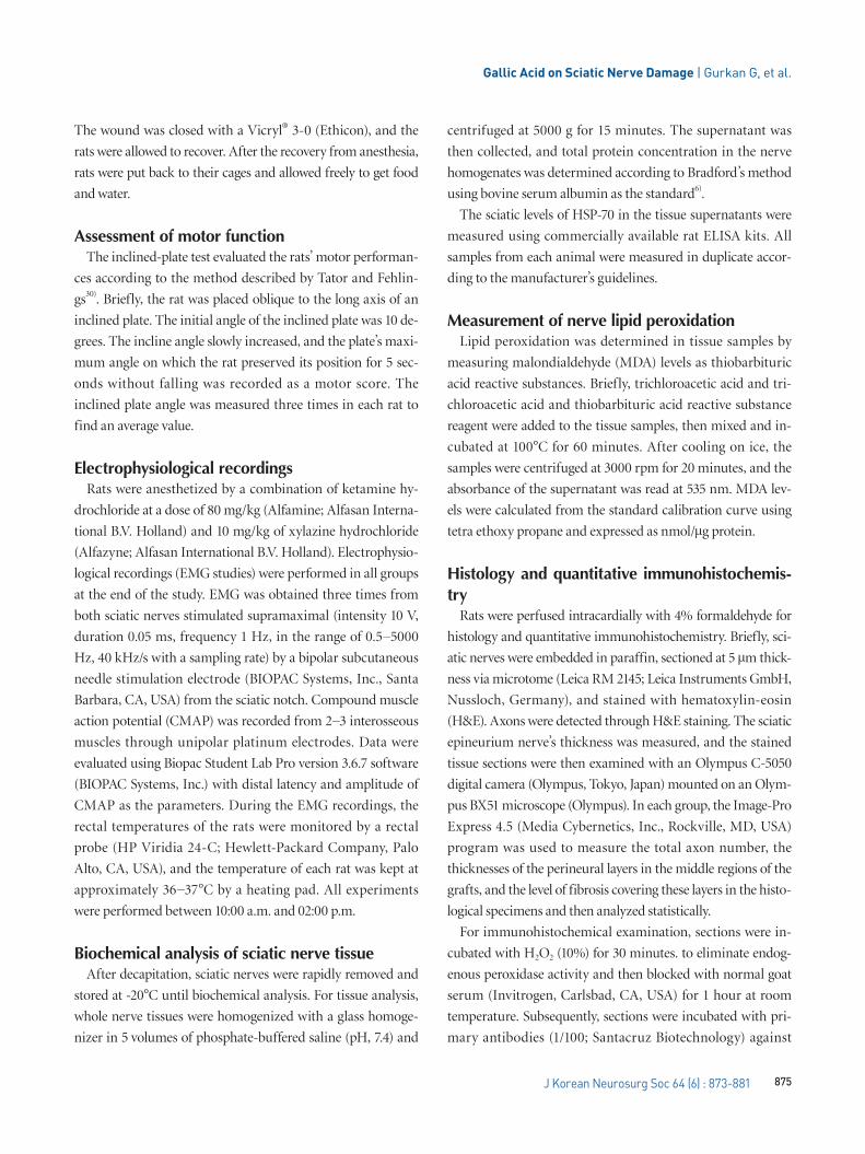
Evaluation of electrophysiological recordingsThe amplitudes of CMAP decreased significantly in the
surgery+saline group (2.50±0.31) compared to the control
group (11.53±2.04) (p<0.001). When compared to the control
group (2.31±0.19), the latency of CMAP was significantly ex-
tended in the surgery+saline group (3.69±0.44) (p<0.05).
Even then, the amplitudes of CMAP were significantly
greater in the surgery+gallic acid group (6.72±1.13) than in the
surgery+saline group (2.50±0.31) (p<0.001). CMAP latency
was significantly reduced in the surgery+gallic acid group
(2.90±0.19) over the surgery+saline group (3.69±0.44)
(p<0.001) (Fig. 1 and Table 1).
Fig. 1. Electromyography of (A) control group, (B) surgery+saline group, and (C) surgery+gallic acid group.
1 msec
1 mV
A B C
Table 1. Intergroup comparison of motor functions and EMG values
Control group Surgery+saline group Surgery+gallic acid group
EMG CMAP latency (MS) 2.31±0.19 3.69±0.44* 2.90±0.19‡
EMG CMAP amplitude (mV) 11.53±2.04 2.50±0.31† 6.72±1.13§
Inclined plane score (°) 88.10±6.50 41.70±7.20† 65.80±8.40§
Values are presented as mean±standard error of the mean. *p<0.05, †p<0.001; surgery+saline group compared with control group. ‡p<0.001, §p<0.001; surgery+gallic acid group compared with surgery+saline group. EMG : electromyography, CMAP : compound muscle action potential
Gallic Acid on Sciatic Nerve Damage | Gurkan G, et al.
877J Korean Neurosurg Soc 64 (6) : 873-881
Histology and quantitative immunohistochemis-try
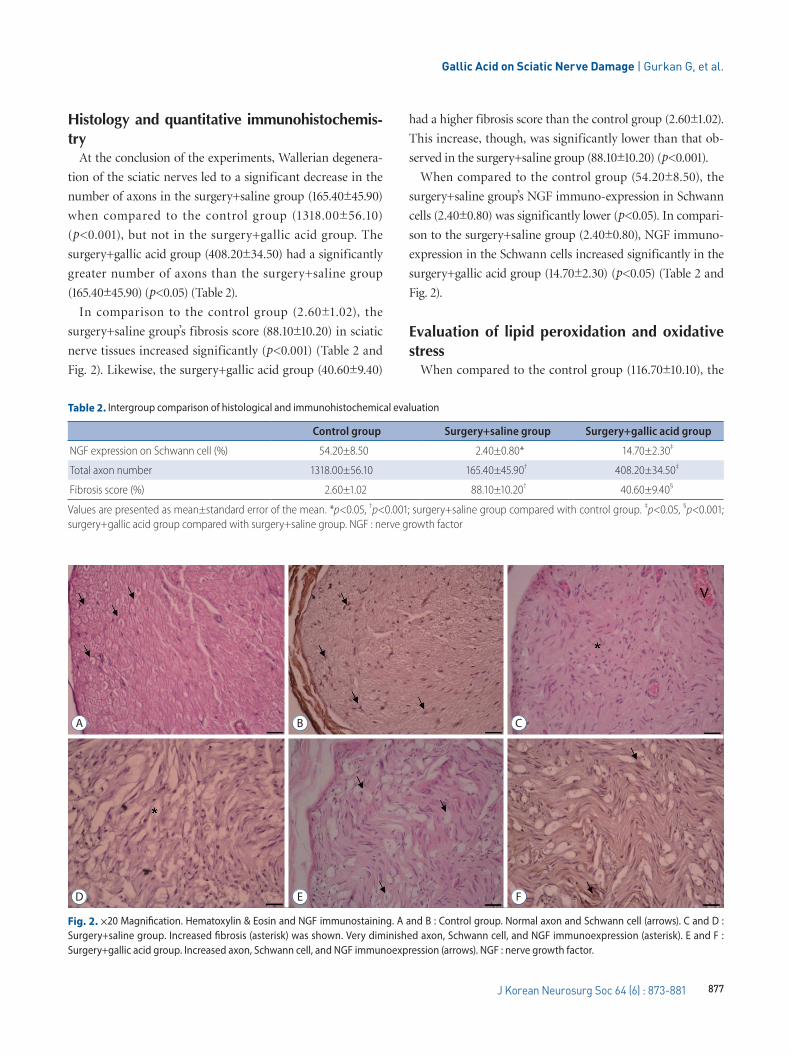
At the conclusion of the experiments, Wallerian degenera-
tion of the sciatic nerves led to a significant decrease in the
number of axons in the surgery+saline group (165.40±45.90)
when compared to the control group (1318.00±56.10)
(p<0.001), but not in the surgery+gallic acid group. The
surgery+gallic acid group (408.20±34.50) had a significantly
greater number of axons than the surgery+saline group
(165.40±45.90) (p<0.05) (Table 2).
In comparison to the control group (2.60±1.02), the
surgery+saline group’s fibrosis score (88.10±10.20) in sciatic
nerve tissues increased significantly (p<0.001) (Table 2 and
Fig. 2). Likewise, the surgery+gallic acid group (40.60±9.40)
had a higher fibrosis score than the control group (2.60±1.02).
This increase, though, was significantly lower than that ob-
served in the surgery+saline group (88.10±10.20) (p<0.001).
When compared to the control group (54.20±8.50), the
surgery+saline group’s NGF immuno-expression in Schwann
cells (2.40±0.80) was significantly lower (p<0.05). In compari-
son to the surgery+saline group (2.40±0.80), NGF immuno-
expression in the Schwann cells increased significantly in the
surgery+gallic acid group (14.70±2.30) (p<0.05) (Table 2 and
Fig. 2).
Evaluation of lipid peroxidation and oxidative stress
When compared to the control group (116.70±10.10), the
Table 2. Intergroup comparison of histological and immunohistochemical evaluation
Control group Surgery+saline group Surgery+gallic acid group
NGF expression on Schwann cell (%) 54.20±8.50 2.40±0.80* 14.70±2.30‡
Total axon number 1318.00±56.10 165.40±45.90† 408.20±34.50‡
Fibrosis score (%) 2.60±1.02 88.10±10.20† 40.60±9.40§
Values are presented as mean±standard error of the mean. *p<0.05, †p<0.001; surgery+saline group compared with control group. ‡p<0.05, §p<0.001; surgery+gallic acid group compared with surgery+saline group. NGF : nerve growth factor
Fig. 2. ×20 Magnification. Hematoxylin & Eosin and NGF immunostaining. A and B : Control group. Normal axon and Schwann cell (arrows). C and D : Surgery+saline group. Increased fibrosis (asterisk) was shown. Very diminished axon, Schwann cell, and NGF immunoexpression (asterisk). E and F : Surgery+gallic acid group. Increased axon, Schwann cell, and NGF immunoexpression (arrows). NGF : nerve growth factor.
A
D
B
E
C
F
J Korean Neurosurg Soc 64 | November 2021
878 https://doi.org/10.3340/jkns.2021.0078
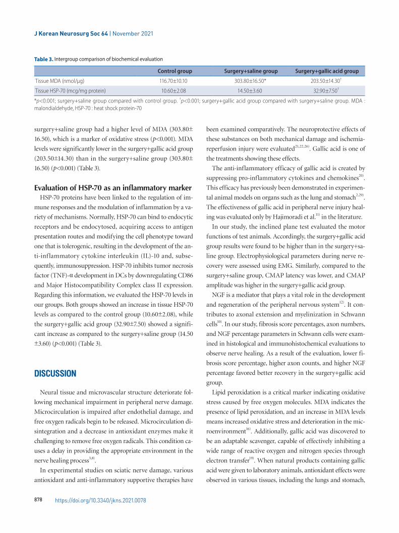
surgery+saline group had a higher level of MDA (303.80±
16.50), which is a marker of oxidative stress (p<0.001). MDA
levels were significantly lower in the surgery+gallic acid group
(203.50±14.30) than in the surgery+saline group (303.80±
16.50) (p<0.001) (Table 3).
Evaluation of HSP-70 as an inflammatory markerHSP-70 proteins have been linked to the regulation of im-
mune responses and the modulation of inflammation by a va-
riety of mechanisms. Normally, HSP-70 can bind to endocytic
receptors and be endocytosed, acquiring access to antigen
presentation routes and modifying the cell phenotype toward
one that is tolerogenic, resulting in the development of the an-
ti-inf lammatory cytokine interleukin (IL)-10 and, subse-
quently, immunosuppression. HSP-70 inhibits tumor necrosis
factor (TNF)-α development in DCs by downregulating CD86
and Major Histocompatibility Complex class II expression.
Regarding this information, we evaluated the HSP-70 levels in
our groups. Both groups showed an increase in tissue HSP-70
levels as compared to the control group (10.60±2.08), while
the surgery+gallic acid group (32.90±7.50) showed a signifi-
cant increase as compared to the surgery+saline group (14.50
±3.60) (p<0.001) (Table 3).
DIsCUssION
Neural tissue and microvascular structure deteriorate fol-
lowing mechanical impairment in peripheral nerve damage.
Microcirculation is impaired after endothelial damage, and
free oxygen radicals begin to be released. Microcirculation di-
sintegration and a decrease in antioxidant enzymes make it
challenging to remove free oxygen radicals. This condition ca-
uses a delay in providing the appropriate environment in the
nerve healing process3,8).
In experimental studies on sciatic nerve damage, various
antioxidant and anti-inflammatory supportive therapies have
been examined comparatively. The neuroprotective effects of
these substances on both mechanical damage and ischemia-
reperfusion injury were evaluated21,22,26). Gallic acid is one of
the treatments showing these effects.
The anti-inflammatory efficacy of gallic acid is created by
suppressing pro-inflammatory cytokines and chemokines20).
This efficacy has previously been demonstrated in experimen-
tal animal models on organs such as the lung and stomach2,29).
The effectiveness of gallic acid in peripheral nerve injury heal-
ing was evaluated only by Hajimoradi et al.11) in the literature.
In our study, the inclined plane test evaluated the motor
functions of test animals. Accordingly, the surgery+gallic acid
group results were found to be higher than in the surgery+sa-
line group. Electrophysiological parameters during nerve re-
covery were assessed using EMG. Similarly, compared to the
surgery+saline group, CMAP latency was lower, and CMAP
amplitude was higher in the surgery+gallic acid group.
NGF is a mediator that plays a vital role in the development
and regeneration of the peripheral nervous system12). It con-
tributes to axonal extension and myelinization in Schwann
cells10). In our study, fibrosis score percentages, axon numbers,
and NGF percentage parameters in Schwann cells were exam-
ined in histological and immunohistochemical evaluations to
observe nerve healing. As a result of the evaluation, lower fi-
brosis score percentage, higher axon counts, and higher NGF
percentage favored better recovery in the surgery+gallic acid
group.
Lipid peroxidation is a critical marker indicating oxidative
stress caused by free oxygen molecules. MDA indicates the
presence of lipid peroxidation, and an increase in MDA levels
means increased oxidative stress and deterioration in the mic-
roenvironment16). Additionally, gallic acid was discovered to
be an adaptable scavenger, capable of effectively inhibiting a
wide range of reactive oxygen and nitrogen species through
electron transfer19). When natural products containing gallic
acid were given to laboratory animals, antioxidant effects were
observed in various tissues, including the lungs and stomach,
Table 3. Intergroup comparison of biochemical evaluation
Control group Surgery+saline group Surgery+gallic acid group
Tissue MDA (nmol/μg) 116.70±10.10 303.80±16.50* 203.50±14.30†
Tissue HSP-70 (mcg/mg protein) 10.60±2.08 14.50±3.60 32.90±7.50†
*p<0.001; surgery+saline group compared with control group. †p<0.001; surgery+gallic acid group compared with surgery+saline group. MDA : malondialdehyde, HSP-70 : heat shock protein-70
Gallic Acid on Sciatic Nerve Damage | Gurkan G, et al.
879J Korean Neurosurg Soc 64 (6) : 873-881
according to the literature1,28). Gallic acid has also been shown
to have anti-inflammatory properties by inhibiting the deve-
lopment of pro-inf lammatory cytokines and chemokines4).
According to another study, gallic acid significantly healed
MDA and superoxide dismutase levels but had little impact on
the catalase amounts in diabetic rat testes. With the same an-
ti-inflammatory pathways, it drastically reduced TNF-α, nit-
ric oxide synthase-2, and vascular endothelial growth factor
levels34). The treatment of gallic acid has ameliorative effects
against TNF-α in paclitaxel-induced neuropathic pain in mice
model15). HSP-70 is in the heat shock protein family and has
cell protective efficacy14). Studies revealed that HSP-70 pre-
vents cell death in damaged neurons are available in the litera-
ture32). HSP treatment has been shown to inhibit or arrest in-
f lammatory damage in laboratory disease models, and in
early clinical trials in patients with chronic inflammatory dis-
orders, HSP proteins and peptides have been shown to stimu-
late the development of anti-inf lammatory cytokines, sug-
gesting that HSP possesses immunoregulatory capacity. Thus,
the involvement of immune responses to HSP in inflamma-
tory diseases can be interpreted as the immune system at-
tempting to fix the inflammatory state5). As we mentioned be-
fore, HSP-70 proteins have been linked to the regulation of
immune responses and the modulation of inflammation by a
variety of mechanisms. Normally, HSP-70 can bind to endo-
cytic receptors and be endocytosed, acquiring access to anti-
gen presentation routes and modifying the cell phenotype
toward one that is tolerogenic, resulting in the development of
the anti-inflammatory cytokine IL-10 and, subsequently, im-
munosuppression. HSP-70 inhibits TNF-α development in
DCs by downregulating CD86 and MHC class II expression5).
For all these reasons, tissue MDA and HSP-70 levels were
compared in our study. Accordingly, statistically significant
lower MDA levels and higher HSP-70 levels were found in the
surgery+gallic acid group compared to the surgery+saline
group. All these results showed that gallic acid had a more be-
neficial effect on recovery than placebo.
There is a limited number of studies in the literature inves-
tigating the effect of gallic acid on sciatic nerve damage with
our study parameters. Kaur and Muthuraman15) showed that
gallic acid could ameliorate paclitaxel-induced neuropathic
pain in mice model. However, this was a neuropathic pain
model, and in our study, we focused on the ameliorative effe-
cts of gallic acid on motor functions in the sciatic nerve dama-
ge model. Therefore, it was challenged to compare our study
findings with the literature. This situation is also one of the li-
mitations of our study. Another limitation is that since our
study is an experimental animal study, the number of animals
we used could be high due to ethical concerns. Further studies
involving more subjects and comparing gallic acid with other
accepted treatments are needed.
CONCLUsION
In conclusion, we demonstrated that gallic acid positively
affects peripheral nerve injury healing due to its anti-oxidant
and anti-inflammatory effects. A significant enhancement in
healing was observed in the surgery+gallic acid group com-
pared to the placebo group. Regarding our findings, it has
been thought that gallic acid may be used as a supportive
treatment for peripheral nerve damages. More comparative
studies are also needed.
CONFLICTs OF INTEREsT
No potential conflict of interest relevant to this article was
reported.
INFORMED CONsENT
This type of study does not require informed consent.
AUTHOR CONTRIBUTIONs
Conceptualization : GG, MAE, OE
Data curation : GG, MAE, GY
Formal analysis : GG, MAE, OE
Funding acquisition : OE
Methodology : GG, MAE, OE
Project administration : OE
Visualization : GY, OE
Writing - original draft : GG
Writing - review & editing : GG, MAE
J Korean Neurosurg Soc 64 | November 2021
880 https://doi.org/10.3340/jkns.2021.0078
ORCID
Gokhan Gurkan https://orcid.org/0000-0003-1839-1014
Mumin Alper Erdogan https://orcid.org/0000-0003-0048-444X
Gurkan Yigitturk https://orcid.org/0000-0002-5315-253X
Oytun Erbas https://orcid.org/0000-0002-2515-2946
References
1. Arora P, Ansari SH, Anjum V, Mathur R, Ahmad S : Investigation of anti-
asthmatic potential of Kanakasava in ovalbumin-induced bronchial
asthma and airway inflammation in rats. J Ethnopharmacol 197 : 242-249, 2017
2. Arora P, Ansari SH, Najmi AK, Anjum V, Ahmad S : Investigation of anti-
asthmatic potential of dried fruits of Vitis vinifera L. in animal model of
bronchial asthma. Allergy Asthma Clin Immunol 17 : 12, 2016
3. Bagdatoglu C, Saray A, Surucu HS, Ozturk H, Tamer L : Effect of trapidil
in ischemia/reperfusion injury of peripheral nerves. Neurosurgery 51 : 212-219; discussion 219-220, 2002
4. Bolzani V, Soares C, Pezzuto J, Luqman S, Morais M, Kondratyuk T, et
al. : Suppression of TNF-α induced NFκB activity by gallic acid and its
semi-synthetic alkyl-gallates: possible role in cancer chemoprevention.
Nature Precedings, 2012 [Epub ahead of print]
5. Borges TJ, Wieten L, van Herwijnen MJ, Broere F, Van Der Zee R,
Bonorino C, et al. : The anti-inflammatory mechanisms of Hsp70. Front Immunol 3 : 95, 2012
6. Bradford MM : A rapid and sensitive method for the quantitation of mi-
crogram quantities of protein utilizing the principle of protein-dye bind-
ing. Anal Biochem 72 : 248-254, 1976
7. Chen MB, Zhang F, Lineaweaver WC : Luminal fillers in nerve conduits
for peripheral nerve repair. Ann Plast Surg 57 : 462-471, 2006
8. Fairbairn NG, Meppelink AM, Ng-Glazier J, Randolph MA, Winograd JM :
Augmenting peripheral nerve regeneration using stem cells: a review of
current opinion. World J Stem Cells 7 : 11-26, 2015
9. Faroni A, Mobasseri SA, Kingham PJ, Reid AJ : Peripheral nerve regen-
eration: experimental strategies and future perspectives. Adv Drug Deliv Rev 82-83 : 160-167, 2015
10. Gao C, Ma S, Ji Y, Wang JE, Li J : Siatic nerve regeneration in rats stimu-
lated by fibrin glue containing nerve growth factor: an experimental
study. Injury 39 : 1414-1420, 2008
11. Hajimoradi M, Fazilati M, Gharib-Naseri MK, Sarkaki A : Gallic acid and
exercise training improve motor function, nerve conduction velocity but
not pain sense reflex after experimental sciatic nerve crush in male rats.
Avicenna J Phytomed 5 : 288-297, 2015
12. Henderson CE : Role of neurotrophic factors in neuronal development.
Curr Opin Neurobiol 6 : 64-70, 1996
13. Huang W, Begum R, Barber T, Ibba V, Tee NC, Hussain M, et al. : Regen-
erative potential of silk conduits in repair of peripheral nerve injury in
adult rats. Biomaterials 33 : 59-71, 2012
14. Kalmar B, Burnstock G, Vrbová G, Urbanics R, Csermely P, Greensmith
L : Upregulation of heat shock proteins rescues motoneurones from
axotomy-induced cell death in neonatal rats. Exp Neurol 176 : 87-97,
2002
15. Kaur S, Muthuraman A : Ameliorative effect of gallic acid in paclitaxel-
induced neuropathic pain in mice. Toxicol Rep 6 : 505-513, 2019
16. Koracevic D, Koracevic G, Djordjevic V, Andrejevic S, Cosic V : Method
for the measurement of antioxidant activity in human fluids. J Clin Pathol 54 : 356-361, 2001
17. Lundborg G : Enhancing posttraumatic nerve regeneration. J Peripher Nerv Syst 7 : 139-140, 2002
18. Ma J, Luo XD, Protiva P, Yang H, Ma C, Basile MJ, et al. : Bioactive novel
polyphenols from the fruit of Manilkara zapota (Sapodilla). J Nat Prod
66 : 983-986, 2003
19. Marino T, Galano A, Russo N : Radical scavenging ability of gallic acid
toward OH and OOH radicals. Reaction mechanism and rate constants
from the density functional theory. J Phys Chem B 118 : 10380-10389,
2014
20. Morais MC, Luqman S, Kondratyuk TP, Petronio MS, Regasini LO, Silva
DH, et al. : Suppression of TNF-α induced NFκB activity by gallic acid
and its semi-synthetic esters: possible role in cancer chemoprevention.
Nat Prod Res 24 : 1758-1765, 2010
21. Ogut E, Yildirim FB, Sarikcioglu L, Aydin MA, Demir N : Neuroprotective
effects of ozone therapy after sciatic nerve cut injury. Kurume Med J 65 : 137-144, 2020
22. Ozyigit F, Kucuk A, Akcer S, Tosun M, Kocak FE, Kocak C, et al. : Dif-
ferent dose-dependent effects of ebselen in sciatic nerve ischemia-
reperfusion injury in rats. Bosn J Basic Med Sci 15 : 36-43, 2015
23. Park HH, Ko SC, Oh GW, Jang YM, Kim YM, Park WS, et al. : Character-
ization and biological activity of PVA hydrogel containing chitooligosac-
charides conjugated with gallic acid. Carbohydr Polym 198 : 197-
205, 2018
24. Rajalakshmi K, Devaraj H, Niranjali Devaraj S : Assessment of the no-
observed-adverse-effect level (NOAEL) of gallic acid in mice. Food Chem Toxicol 39 : 919-922, 2001
25. Sanlier N, Atik I, Atik A : A minireview of effects of white tea consump-
tion on diseases. Trends Food Sci Technol 82 : 82-88, 2018
26. Shokouhi G, Tubbs RS, Shoja MM, Hadidchi S, Ghorbanihaghjo A,
Roshangar L, et al. : Neuroprotective effects of high-dose vs low-dose
melatonin after blunt sciatic nerve injury. Childs Nerv Syst 24 : 111-
117, 2008
27. Singh J, Saha L, Singh N, Kumari P, Bhatia A, Chakrabarti A : Study of
nuclear factor-2 erythroid related factor-2 activator, berberine, in pacli-
taxel induced peripheral neuropathy pain model in rats. J Pharm Phar-macol 71 : 797-805, 2019
28. Tamboli FA, More HN : Evaluation of antiulcer and antioxidant activity of
Barleria gibsoniDalz. leaves. Pharmacognosy Res 8 : 226-230, 2016
29. Tamboli FA, More HN : Anthelmintic activity of leaves extract of Barleria
gibsoni Dalz. against Pheretima posthuma. JPP 5 : 250-252, 2016
30. Tator CH, Fehlings MG : Review of the secondary injury theory of acute
Gallic Acid on Sciatic Nerve Damage | Gurkan G, et al.
881J Korean Neurosurg Soc 64 (6) : 873-881
spinal cord trauma with emphasis on vascular mechanisms. J Neuro-surg 75 : 15-26, 1991
31. Teixeira J, Oliveira C, Cagide F, Amorim R, Garrido J, Borges F, et al. :
Discovery of a new mitochondria permeability transition pore (mPTP)
inhibitor based on gallic acid. J Enzyme Inhib Med Chem 33 : 567-
576, 2018
32. Tidwell JL, Houenou LJ, Tytell M : Administration of Hsp70 in vivo inhib-
its motor and sensory neuron degeneration. Cell Stress Chaperones
9 : 88-98, 2004
33. Verma S, Singh A, Mishra A : Gallic acid: molecular rival of cancer. Envi-ron Toxicol Pharmacol 35 : 473-485, 2013
34. Yigitturk G, Acara AC, Erbas O, Oltulu F, Yavasoglu NUK, Uysal A, et al. :
The antioxidant role of agomelatine and gallic acid on oxidative stress
in STZ induced type I diabetic rat testes. Biomed Pharmacother 87 : 240-246, 2017


![Quantitative HPLC Analysis of Ascorbic Acid and Gallic Acid in … · 2017-12-04 · promotion such as alteration of protein kinase C (PKC) activity [8]. The fruits of Phyllanthus](https://img.dokumen.tips/doc/110x75/5e92f9636edd9a71666ed2d5/quantitative-hplc-analysis-of-ascorbic-acid-and-gallic-acid-in-2017-12-04-promotion.jpg)