Embed Size (px)
Citation preview
The Rockefeller University Press, 0021-9525/98/12/1831/14 $2.00The Journal of Cell Biology, Volume 143, Number 7, December 28, 1998 1831–1844http://www.jcb.org 1831
Homotypic Fusion of Immature Secretory Granules during Maturation in a Cell-free Assay
Sylvie Urbé, Lesley J. Page, and Sharon A. Tooze
Secretory Pathways Laboratory, Imperial Cancer Research Fund, London WC2A 3PX, United Kingdom
Abstract.
The biogenesis of secretory granules embod-ies several morphological and biochemical changes. In particular, in neuroendocrine cells maturation of secre-tory granules is characterized by an increase in size which has been proposed to reflect homotypic fusion of immature secretory granules (ISGs). Here we describe an assay that provides the first biochemical evidence for such a fusion event and allows us to analyze its regu-lation. The assay reconstitutes homotypic fusion be-tween one population of ISGs containing a [
35
S]sulfate-labeled substrate, secretogranin II (SgII), and a second population containing the prohormone convertase PC2. Both substrate and enzyme are targeted exclusively to ISGs. Fusion is measured by quantification of a cleav-
age product of SgII produced by PC2. With this assay we show that fusion only occurs between ISGs and not between ISGs and MSGs, is temperature dependent, and requires ATP and GTP and cytosolic proteins. NSF (
N
-ethylmaleimide–sensitive fusion protein) is amongst the cytosolic proteins required, whereas we could not detect a requirement for p97. The ability to reconstitute ISG fusion in a cell-free assay is an important advance towards the identification of molecules involved in the maturation of secretory granules and will increase our understanding of this process.
Key words: fusion • immature secretory granule • prohormone convertase 2 • secretogranin • NSF
S
ecretory
granules are specialized exocytic vesiclesresponsible for the controlled release of bioactivemolecules, such as hormones. During the lifetime of
the secretory granule, from formation to consumption, aseries of regulatory events control its composition andsize. Control of these events is crucial for the fidelity ofregulated secretion. Secretory granules originate from theTGN with a selected subset of lumenal and membraneproteins: this protein selection during formation is pre-sumably controlled by proteins both within the lumen ofthe TGN and in the cytoplasm (Tooze, 1998). Cargo selec-tion may be via receptor–ligand type interactions. For ex-ample, carboxypeptidase E recognizes a motif present inseveral prohormones and has been implicated as a generalcargo receptor (Cool et al., 1997) although this is currentlythe subject of intense debate (Irminger et al., 1997; Thieleet al., 1997). To date, no cytoplasmic coat proteins havebeen implicated in the selection of cargo receptors orsecretory granule membrane proteins. Patches of clathrinhave been detected on the TGN membrane in the region
of the budding immature secretory granule (ISG)
1
al-though there is no evidence that clathrin is involved in for-mation of secretory granules (for review see Urbé et al.,1997
b
). Formation of ISGs is regulated by heterotrimericG proteins (Barr et al., 1991) and proteins required forlipid metabolism (Chen et al., 1997; Ohashi et al., 1995) al-though how these are involved in protein sorting duringISG formation is not known.
Additional proteins and machinery will undoubtedly beidentified and found to be involved in secretory granulebiogenesis at the TGN. However, it is already well estab-lished that the selection mechanisms operative in the TGNalone are not sufficient to produce a mature secretorygranule (MSG). Isolation of ISGs (Tooze et al., 1991) con-firmed the previous morphological data (Salpeter and Far-quhar, 1981) showing that ISGs are smaller than MSGs.Subsequently, proteins were found in ISGs that are notpresent in MSGs, such as furin (Dittié et al., 1997), lysoso-
S. Urbé’s present address is Physiological Laboratory, University of Liver-pool, Crown Street, Liverpool L34BU, UK.
Address correspondence to S.A. Tooze, Secretory Pathways Labora-tory, Imperial Cancer Research Fund, 44 Lincoln’s Inn Fields, LondonWC2A 3PX, UK. Tel.: (44) 171 269 3122. Fax: (44) 171 269 3417. E-mail:[email protected]
1.
Abbreviations used in this paper:
CCV, clathrin-coated vesicle; ISG, im-mature secretory granules; MP, membrane pellet; MPR, mannose-6-phos-phate receptor; MSG, mature secretory granule; NEM,
N
-ethylmaleimide;NSF;
N
-ethylmalemide–sensitive fusion protein; PC2, prohormone con-vertase 2; PNS, postnuclear supernatant; SgII, secretogranin II; SNAP,soluble NSF attachment protein; SNARE, SNAP receptor; STI, soybeantrypsin inhibitor; t-SNARE, target SNARE; v-SNARE, vesicle SNARE.
on December 19, 2018jcb.rupress.org Downloaded from http://doi.org/10.1083/jcb.143.7.1831Published Online: 28 December, 1998 | Supp Info:
The Journal of Cell Biology, Volume 143, 1998 1832
mal enzymes (Kuliawat et al., 1997) and the mannose-6-phos-phate receptor (Klumperman, 1998). In addition, the lu-menal pH of the ISG is
z
6.3 whereas the lumenal pH ofthe MSG is
z
pH 5.5 (Orci et al., 1994; Urbé et al., 1997
a
).The differences in size and content between ISGs and
MSGs have led to the proposal that the nascent ISG,which buds from the TGN is an intermediate stage insecretory granule biogenesis (Tooze et al., 1991). Matura-tion of the intermediate ISG to an MSG would requireboth fusion and budding events. Fusion events betweenISGs would result in an increase in the size of ISGs to thatof MSGs, whereas budding of vesicles from the ISG wouldallow removal of nonessential proteins and excess mem-brane. The coordinated action of budding and fusionwould exert a tight control on both the composition andquantal size of the released secretory granule contentupon stimulation. This hypothesis is supported by mor-phological data demonstrating that MSGs in endocrinecells have a defined size depending on the cell type. Forexample, within the anterior pituitary gland the mam-motrophs have MSGs 350
m
m in diameter, whereas the go-nadotrophs have MSGs 200
m
m in diameter (Smith andFarquhar, 1966). The size differences may be related tothe biophysical properties of the major content proteinand the structure of the dense core, although the mecha-nisms that underlie this phenomenon are not known (Bur-gess and Kelly, 1987).
We are interested in understanding the mechanisms thatcontrol secretory granule composition and size, and deter-mining if these might involve fusion and budding from theISG. In one approach we have developed a cell-free assaythat reconstitutes fusion between ISGs. The assay is basedon the interaction of an endopeptidase with its substrate,both of which are specifically sorted into secretory gran-ules. We have designed the assay so that the substrate,which is labeled by radioactive sulfate on tyrosine resi-dues, is present in one ISG population, whereas the en-zyme is present in another population of ISGs. Upon fu-sion of the two ISG populations the enzyme will cleave thelabeled substrate into discrete fragments. Fusion is mea-sured by immunoprecipitation with a specific antibodywhich only recognizes one of these fragments and not theintact substrate.
With this assay we have investigated the requirementsfor cytosolic components involved in ISG fusion. In partic-ular, we have asked if either one of the two well-character-ized
N
-ethylmaleimide (NEM)–sensitive proteins, NEMsensitive fusion protein (NSF), or p97 are involved in ISGfusion. NSF, first identified by Rothman and colleagues(Block et al., 1988) is involved in vesicular transport alongthe secretory pathway, fusion events in the endocytic path-way, and regulated exocytosis (for review see Whiteheartand Kubalek, 1995; Woodman, 1997). p97, or its yeast ho-mologue cdc48p, was shown to be necessary for homotypicfusion events involved in organelle maintenance (Acharyaet al., 1995
a
; Latterich et al., 1995; Rabouille et al., 1995
a
).Both are ATPases which bind to membranes via attach-ment proteins, soluble NSF attachment proteins (SNAPs)(Whiteheart et al., 1993) in the case of NSF (Clary et al.,1990; Weidman et al., 1989) and p47 for p97 (Kondo et al.,1997; Rabouille, 1998). NSF and SNAPs interact with de-tergent-solubilized soluble NSF attachment protein recep-
tors (SNAREs) to form a 20 S particle (Sollner et al.,1993). Over 20 SNAREs have been identified (for reviewsee Advani et al., 1998; Bock and Scheller, 1997) which areclassified into two families, v-SNAREs found on the vesi-cle and t-SNAREs found on the target membrane. Thestructural similarity of the SNAREs, combined with theirrestricted localization is the basis of the current model forvesicular transport through the secretory pathway (Roth-man, 1994; for review see Götte and von Mollard, 1998).Recently, interaction of a v-SNARE with a t-SNARE hasbeen shown to be the minimal requirement for fusion of li-posomes (Weber et al., 1998).
The interaction of p97 or cdc48p with membranes alsorequires a SNARE protein, the t-SNARE syntaxin 5(Rabouille, 1998), and Ufe1p (Patel, 1998), respectively. Ithas recently been proposed that whereas heterotypicfusion requires the pairing of cognate v- and t-SNAREs,homotypic fusion can also proceed via the pairing oft-SNAREs alone (Nichols et al., 1997) and may be in somecases mediated by cdc48p or p97 (Patel, 1998; Rabouille,1998, respectively).
Using the in vitro system we describe here we reportthat fusion of ISGs can be reconstituted in a cell-free as-say, and provide the first biochemical evidence that ISG–ISG fusion can occur. This fusion requires ATP, GTP, andelevated temperature plus cytosolic components. We havefound that NSF, but not p97, is required for homotypicISG fusion. Further understanding of the role of ISG–ISGfusion during maturation, and how the generation ofMSGs may be controlled by ISG fusion will be greatly fa-cilitated with this assay.
Materials and Methods
Materials and Reagents
Carrier-free [
35
S]sulfate as a disodium salt was supplied by AmershamPharmacia Biotech (Piscataway, NJ). Fine chemicals were supplied bySigma Chemical Co. (St. Louis, MO) or Boehringer Mannheim (Mann-heim, Germany). PC12 cells (clone 251) were originally obtained fromH. Thoenen (Max Planck Institute, Martinsried, Germany) (Heumann etal., 1983). PC12/PC2 cell lines stably expressing the endoprotease PC2have been previously characterized (Dittié and Tooze, 1995). PC12 andPC12/PC2 cells were grown in DME supplemented with 10% horse serumand 5% fetal calf serum at 10% CO
2
.
Antibodies and Recombinant Proteins
Horseradish peroxidase (HRP)-conjugated anti-rabbit and anti-mouseIgGs were supplied by Amersham (Little Chalfont, UK). Antibodiesraised against SgII (175 and 100) have been previously described (Dittiéet al., 1996). Anti-NSF antibody was provided by M. Tagaya (Tokyo Uni-versity, Tokyo, Japan), anti–
a
-SNAP by T. Söllner (Sloan-Kettering, NewYork, NY), purified p97 and his-tagged p47, anti-p97, and p47 by H.Kondo (Imperial Cancer Research Fund [ICRF], London, UK). His-tagged NSF and His-tagged
a
-SNAP were a kind gift of G. Schiavo(ICRF).
Antibodies specific for the free COOH terminus of p18 were raisedagainst the peptide (KEENSRENPF) from rat SgII (Gerdes et al., 1988).Polyclonal antisera were obtained from rabbits (ab69), whereas the mono-clonal antibody was developed from hybridomas using a standard proce-dure (6B1). Both the polyclonal and monoclonal antibodies are specificfor the free COOH terminus of p18 as assessed by standard Western blot-ting, immunoprecipitation, and immunofluorescence techniques using ly-sates obtained from either PC12 cells or PC12/PC2 cells. Experimentshave been performed with either or both polyclonal antibody (ab69) andmonoclonal antibody (6B1).

Urbé et al.
Fusion of ISGs in a Cell-free Assay
1833
Preparation of Postnuclear Supernatant, ISGs,and MSGs
A postnuclear supernatant (PNS) was prepared in HB-PMSF (250 mM su-crose, 10 mM Hepes-KOH, pH 7.2, 1 mM Mg[OAc]
2
, 1 mM EDTA, pH7.2, with 0.5 mM PMSF) as described (Tooze and Huttner, 1990) and con-tained
z
10 mg/ml of protein as determined by Bio-Rad protein microas-say (using bovine IgG as a standard; Hercules, CA). PC12 cells were la-beled with [
35
S]sulfate and chased as previously described (Tooze andHuttner, 1990) for the times specified in the figure legends.
ISGs and MSGs were prepared as described in Dittié et al. (1996). Inbrief, 1.3 ml of PC12 or PC12/PC2 PNS was loaded on linear sucrose gra-dients of 0.3–1.2 M sucrose in 10 mM Hepes-KOH, pH 7.2, and subjectedto velocity controlled centrifugation. Fractions from the top of the velocitygradient containing ISGs and MSGs were pooled and subjected to equilib-rium sucrose gradient centrifugation. Fractions of 1 ml were collectedfrom the top and fractions seven to nine were pooled for the ISGs whereasfractions 10–12 were pooled for the MSGs. Aliquots were stored in liquidnitrogen and used within 6 mo. All gradient preparations were routinelychecked using [
35
S]sulfate labeling to ensure that the preparations wereconsistent and correct sorting of the soluble proteins was achieved (Toozeand Huttner, 1990).
Preparation of PC12 Cytosol
PC12 cells (
z
10
7
–10
8
cells) were homogenized in HB-PMSF and a PNSwas prepared as described above. The PNS was then subjected to centrifu-gation for 30 min at 30,000
g
av
, followed by a second centrifugation of thesupernatant at 100,000
g
av
for 90 min at 4
8
C. Cytosol was stored in liquidnitrogen and used within 3 mo. The protein concentration of the PC12 cy-tosol was determined using the Bio-Rad microassay and was typically be-tween 8 and 13 mg/ml.
Preparation of Rat Liver Cytosol
Rat livers either from Wistar or Sprague-Dawley male or female rats wereused. The livers were washed, minced, and then homogenized either in FB(20 mM Hepes-KOH, pH 7.4, 50 mM KOAc, 3 mM MgCl
2
, 1 mM DTT),containing 250 mM sucrose or protease inhibitors (0.5 mM PMSF, 10.5
m
Mleupeptin) and then centrifuged as described for the PC12 cell cytosolabove. Alternatively, cytosol was prepared as described (Rabouille et al.,1995
b
) in 0.5 M sucrose, 0.1 M KPO
4
, pH 6.7, 5 mM MgCl
2
and proteaseinhibitors. After centrifugation both cytosol preparations were precipi-tated with 40% ammonium sulfate, resuspended in FB containing 250 mMsucrose and protease inhibitors, and either dialyzed overnight against FBcontaining 250 mM sucrose or desalted into FB containing 250 mM su-crose with a PD10 column (Bio-Rad). Aliquots were stored in liquid nitro-gen and used within 3 mo. The protein concentration of these cytosols wastypically between 20 and 40 mg/ml.
Cell-free Assays for ISG–ISG Fusion and Processing
Reconstitution of ISG Fusion with a PNS.
PC12 cells were pulse-labeledwith [
35
S]sulfate or pulse-labeled and chased, before being washed, har-vested, and homogenized to prepare a PNS as described above. PC2-ISGswere thawed rapidly at 37
8
C, diluted at 4
8
C with 2 vol of 10 mM Hepes-KOH, pH 7.2, pelleted by centrifugation at 100,000
g
av
for 1 h. The pelletswere resuspended in HB-PMSF at 50-fold the concentration of the origi-nal ISG pool. If not indicated otherwise, standard conditions were as fol-lows: 100
m
l of the PC12 PNS per condition were supplemented with 10
3
FB, ATP-regenerating system (see below), and 10
m
l PC2-ISGs in a finalvolume of 120–160
m
l.After incubation at 4
8
or 37
8
C the samples were transferred to ice anddiluted with low pH buffer (50 mM MES, pH 5.5, in 0.3 M sucrose) to a fi-nal volume of 800
m
l. The samples were preincubated for 30 min on ice toequilibrate the pH in the ISGs with the buffer and then transferred to37
8
C to allow processing of SgII by PC2 to take place. After this process-ing incubation, the samples were placed on ice and 200
m
l of 5
3
lysisbuffer (500 mM Tris-HCl, pH 7.5, 750 mM NaCl, 1.5% Triton X-100, 25 mMEDTA for ab69, or 500 mM Tris-HCl, pH 7.5, 750 mM NaCl, 5% TritonX-100, 0.5% SDS, 5% Na deoxycholate for 6B3) was added. The lysedsamples were precleared by a 5-min spin in an Eppendorf centrifuge(Eindhoven, The Netherlands) and subjected to immunoprecipitation asdescribed below.
ATP-regenerating system (Davey et al., 1985) stock solutions con-tained 100 mM ATP, pH 7, 800 mM creatine phosphate, and 3,200 U/ml
creatine phosphokinase that were mixed before use in equal volumes togive a 30
3
stock. ATP depletion was performed with 3.3 mM
d
-glucoseand 25 U/ml hexokinase. AMP-PNP (final concentration 100
m
M), GTP(final concentration 300
m
M), and GTP
g
S (final concentration 100
m
M)were made up as a 100
3
stock solutions in H
2
O.
Immunoprecipitation and Quantitation of [
35
S]sulfate-labeled p18.
Forstandard immunoprecipitations from cell-free fusion assay incubationspolyclonal ab69 and protein A–Sepharose (Amersham Pharmacia Bio-tech) or monoclonal 6B1 and protein G–Sepharose (Sigma Chemical Co.)were used. The immunoprecipitates were washed with lysis buffer, elutedfrom the beads by boiling in Laemmli sample buffer, and then analyzed bySDS-PAGE using a modified Laemmli system (Lee and Huttner, 1983),followed by fluorography with 1 M sodium salicylate for 30 min and expo-sure to a preflashed Kodak XAR5 film (Eastman-Kodak, Rochester,NY). The fluorographs were usually exposed to film for 3–5 d, except forthose obtained with material labeled as in Fig. 3
a
where exposures longerthan 2 wk were required. Quantitation of [
35
S]sulfate-labeled p18 was bydensitometry using the NIH Image package version 68k/1.58 (Bethesda,MD). If not indicated otherwise, the PC2-independent signal was sub-tracted from the signal obtained in the presence of PC2-ISGs.
Treatment of PC2-ISGs with Trypsin Before and After Fusion.
PC2-ISGswere incubated with trypsin (20 ng trypsin/
m
g ISG protein or 200 ngtrypsin/
m
g ISG protein) either with or without 25
m
g of soybean trypsin in-hibitor (STI) for 15 min at room temperature, placed on ice, and thenPMSF and STI were added. The ISGs were then pelleted for 1 h, 5 min at100,000
g
av
. Postfusion and postprocessing treatment with trypsin was per-formed with a final concentration of 0.1 mg/ml trypsin (200 ng trypsin/
m
gISG protein) in the absence or presence of 0.3% Triton X-100 for 15 minat 37
8
C. After digestion STI was added and the samples were solubilizedin Laemmli sample buffer.
Cell-free Assay Using a Membrane Pellet.
To obtain a membrane pellet,the PNS was layered on top of a 1 ml sucrose cushion (0.5 M sucrose in 10mM Hepes-KOH, pH 7.2). The membranes were pelleted by centrifuga-tion for 1 h, 5 min at 100,000
g
av
at 4
8
C. The supernatant was removed andthe membranes were resuspended in HB or cytosol to their original vol-ume. An ATP-regenerating system, 10
3
FB, and PC2-ISGs were added asdescribed for the standard cell-free assay using a PNS. GTP was includedwhen rat liver cytosol was used.
Cell-free Assay Using Sucrose Gradient Fractions.
For the type of exper-iment shown in Fig. 8, the PNS was first subjected to velocity controlledsucrose centrifugation to separate the TGN and post-TGN vesicles. Thefractions from the top of the velocity gradient (fractions 2–4) were pooled,diluted with 10 mM Hepes-KOH, pH 7.2, and subjected to centrifugationas above. The membranes were then resuspended in cytosol or HB in avolume corresponding to the volume of the PNS loaded and used as de-scribed.
NEM Treatment of Cytosol and Membranes.
NEM treatment of PNS orcytosol was performed using freshly made 50 mM NEM stock solution indH
2
O added to a final concentration of 3 mM. The samples were mixedimmediately, incubated for 10 min on ice, and then quenched by additionof 3.6 mM glutathione for 15 min on ice. The membranes were then pel-leted through a sucrose cushion as described above while cytosol was usedwithout further treatment. Control samples were treated with glutathioneonly.
Results
Experimental System to Reconstitute Fusion of ISGs
To study the molecular requirements for fusion of ISGswe have developed an assay that allows reconstitution offusion in a cell-free extract. The assay is based on contentmixing after fusion of ISGs which allows an enzyme–substrate interaction to occur. Prohormone convertasescleave prohormones, and other regulated secretory pro-teins containing a dibasic amino acid site, in a pH andCa
2
1
-dependent fashion (Steiner, 1991). PC2, a prohor-mone convertase that preferentially cleaves dibasic aminoacid sites composed of lysine and arginine at a pH opti-mum of 5.5 and at micromolar Ca
2
1
concentrations (Ben-nett et al., 1992) was used in this assay. As the substratefor PC2, we have used secretogranin II (SgII), an abun-
The Journal of Cell Biology, Volume 143, 1998 1834
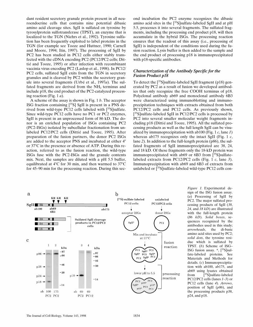
dant resident secretory granule protein present in all neu-roendocrine cells that contains nine potential dibasicamino acid cleavage sites. SgII is sulfated on tyrosine bytyrosylprotein sulfotransferase (TPST), an enzyme that islocalized to the TGN (Niehrs et al., 1992). Tyrosine sulfa-tion has been frequently exploited to label proteins in theTGN (for example see Tooze and Huttner, 1990; Carnelland Moore, 1994; Itin, 1997). The processing of SgII byPC2 has been studied in PC12 cells either stably trans-fected with the cDNA encoding PC2 (PC12/PC2 cells; Dit-tié and Tooze, 1995) or after infection with recombinantvaccinia virus encoding PC2 (Laslop et al., 1998). In PC12/PC2 cells, sulfated SgII exits from the TGN in secretorygranules and is cleaved by PC2 within the secretory gran-ule into several fragments (Urbé et al., 1997
a
). The sul-fated fragments are derived from the NH
2
terminus andinclude p18, the end product of the PC2-catalyzed process-ing reaction (Fig. 1
a
).A scheme of the assay is shown in Fig. 1
b
. The acceptorISG fraction containing [
35
S] SgII is present in a PNS de-rived from wild-type PC12 cells labeled with [
35
S]sulfate.Since wild-type PC12 cells have no PC1 or PC2 enzymes,SgII is present in an unprocessed form of 86 kD. The do-nor is an enriched population of ISGs containing PC2(PC2-ISGs) isolated by subcellular fractionation from un-labeled PC12/PC2 cells (Dittié and Tooze, 1995). Afterpreparation of the fusion partners, the donor PC2 ISGsare added to the acceptor PNS and incubated at either 4
8
or 37
8
C in the presence or absence of ATP. During this re-action, referred to as the fusion reaction, the wild-typeISGs fuse with the PC2-ISGs and the granule contentsmix. Next, the samples are diluted with a pH 5.5 buffer,equilibrated at 4
8
C for 30 min, and then warmed to 37
8
Cfor 45–90 min for the processing reaction. During this sec-
ond incubation the PC2 enzyme recognizes the dibasicamino acid sites in the [
35
S]sulfate-labeled SgII and at pH5.5, processes it into several fragments. The sulfated frag-ments, including the processing end product p18, will thenaccumulate in the hybrid ISGs. The processing reactionensures that the readout of this assay (i.e., processing ofSgII) is independent of the conditions used during the fu-sion reaction. Lysis buffer is then added to the sample andthe end product of processing p18 is immunoprecipitatedwith p18-specific antibodies.
Characterization of the Antibody Specific for the Fusion Product p18
To detect the [
35
S]sulfate-labeled SgII fragment (p18) gen-erated by PC2 as a result of fusion we developed antibod-ies that only recognize the free COOH terminus of p18.Polyclonal antibody ab69 and monoclonal antibody 6B3were characterized using immunoblotting and immuno-precipitation techniques with extracts obtained from bothPC12/PC2 cells and PC12 cells. As previously shown,[
35
S]sulfate-labeled SgII in PC12/PC2 cells is processed byPC2 into several smaller molecular weight fragments in-cluding p18 (Dittié and Tooze, 1995). All the sulfated pro-cessing products as well as the full length SgII can be visu-alized by immunoprecipitation with ab100 (Fig. 1
c
, lane
1
)whereas ab175 recognizes only the intact SgII (Fig. 1
c
,lane
2
). In addition to the full-length protein (p86), the sul-fated fragments of SgII immunoprecipitated are 38, 24,and 18 kD. Of these fragments only the 18-kD protein wasimmunoprecipitated with ab69 or 6B3 from [
35
S]sulfate-labeled extracts from PC12/PC2 cells (Fig. 1
c
, lane
3
).Immunoprecipitation with ab69 and 6B3 of extracts fromunlabeled or [
35
S]sulfate-labeled wild-type PC12 cells con-
Figure 1. Experimental de-sign of the ISG fusion assay.(a) Processing of SgII byPC2. The major sulfated pro-cessing products of SgII (38,24, and 18 kD) are illustratedwith the full-length protein(86 kD). Solid boxes, se-quences recognized by theantibodies used in this study;arrowheads, the di-basicamino acid sites used by PC2;solid dots, the tyrosine resi-due which is sulfated byTPST. (b) Scheme of ISG–ISG fusion assay. *, [35S]sul-fate-labeled proteins. SeeMaterials and Methods fordetails. (c) Immunoprecipita-tion with ab100, ab175, andab69 using lysates obtainedfrom [35S]sulfate-labeledPC12/PC2 cells (lanes 1–3) orPC12 cells (lane 4). Arrows,position of SgII (p86), andthe processing products p38,p24, and p18.

Urbé et al. Fusion of ISGs in a Cell-free Assay 1835
firmed that there was no detectable p18 (Fig. 1 c, lane 4) asexpected since there is no PC2 in PC12 cells.
Optimization of the Processing Reaction
As detection of fusion between ISGs depends on the cata-lytic activity of the PC2 enzyme it is important that theconditions within the fused ISGs support optimal PC2 ac-tivity to ensure that the processing reaction is not rate lim-iting and will proceed to completion. In particular, the lu-menal pH within the ISGs after fusion must be acidic toachieve optimal enzymatic activity. The ISG preparationsfrom both the PC12 cells and the PC12/PC2 cells have a lu-menal pH equivalent to that of the buffer used duringtheir isolation, i.e., pH 7.2. We have shown that whenthese isolated ISGs are incubated at 48C in a low pHbuffer they will equilibrate to the pH of the buffer. Usingthis equilibration step, performed at 48C for 30 min, wecan manipulate the lumenal pH of the ISG to promote ef-ficient processing of SgII by PC2 in isolated ISGs (Urbé et al.,1997a).
We have previously demonstrated that p18 can be pro-duced from [35S]sulfate-labeled SgII in ISGs isolated fromPC12/PC2 cells after equilibration at pH 5.5 followed byincubation at 378C (Urbé et al., 1997a). To obtain evidencethat the conditions required for fusion do not interferewith the processing of SgII in vitro by PC2 we performed amock fusion assay with a PNS obtained from PC12/PC2cells pulse labeled with [35S]sulfate for 10 min and chasedfor 15 min. After this pulse labeling condition the [35S]sul-fate-labeled SgII in this PNS is unprocessed (Urbé et al.,1997a). If this PNS, which contains [35S]sulfate-labeledSgII and PC2 in the same ISG, is kept at 48C, equilibratedto pH 5.5, and then incubated at 378C, SgII processing canbe detected by immunoprecipitation of p18 (Fig. 2 a, lane1). Incubation at 378C before the equilibration to pH 5.5 inthe presence of ATP (Fig. 2 a, lane 2) under conditionswhich mimic the fusion step in our assay does not interferewith the generation of p18. In a parallel processing reac-tion preceded by a mock fusion incubation using a PNSfrom PC12 cells labeled and isolated by an identical proce-dure, no p18 is detected (Fig. 2 a, lanes 3 and 4) as ex-pected in the absence of PC2.
Reconstitution of ISG–ISG Fusion In Vitro
Fusion of ISGs, as detected by the appearance of p18, canbe observed when the donor PC2-ISGs are added to an ac-ceptor PNS derived from [35S]sulfate-labeled PC12 cellsand the reaction mixture is incubated at 378C for fusionand then processing (Fig. 2 b, lane 2). A low amount ofp18 can be detected if the fusion reaction is kept at 48C(Fig. 2 b, lane 1). Addition of the PC2-ISGs after the fu-sion reaction does not result in a significant signal overbackground demonstrating that the processing reactiondoes not support fusion (Fig. 2 b, lane 3). In the absence ofthe donor PC2-ISGs a low amount of p18 is detected atthis exposure, and we define this as the PC2-independentbackground. We attribute this background p18 signal(seen in Fig. 2 b, lane 4) to a minor protease activitypresent in PC12-ISGs. To maximize the p18 signal a 30-min labeling protocol of the PC12 cells was thereafteradopted and routinely used. All of the controls performed
Figure 2. Generation of p18 in a cell-free fusion assay. (a) Pro-cessing of SgII in PC12/PC2 ISGs is not affected by previous in-cubation for fusion. A PNS was prepared from PC12/PC2 cells(lanes 1 and 2) or PC12 cells (lanes 3 and 4) labeled with a 10-minpulse followed by a 15-min chase. Each PNS was then incubatedeither at 48 or 378C under conditions which allow fusion in thepresence of ATP, then for processing at 378C for 45 min. (b) Re-constitution of ISG fusion. A PNS was prepared from PC12 cellslabeled as above and incubated in the presence of 7 ml of PC2-ISGs and ATP at 48 (lane 1) or 378C (lane 2) for 45 min. Alterna-tively, the PC2-ISGs were added after the fusion incubation (lane3) or omitted entirely (lane 4). After equilibration for 60 min onice, processing reactions were incubated at 378C for 45 min.[35S]sulfate-labeled p18 was immunoprecipitated and subjected toSDS-PAGE and autoradiography. Note the autoradiographshown in b was exposed for 2 mo. *, a high molecular weight bandwhich is brought down during the immunoprecipitation step bybinding nonspecifically to both protein A– and G–Sepharose.
The Journal of Cell Biology, Volume 143, 1998 1836
in Fig. 2 were done with the extended labeling period andgave the same result (data not shown). The high molecularweight material present in all conditions in Fig. 2 is an uni-dentified [35S]sulfate-labeled molecule which binds non-specifically to the protein A– and protein G–Sepharose in-dependently of added antibody.
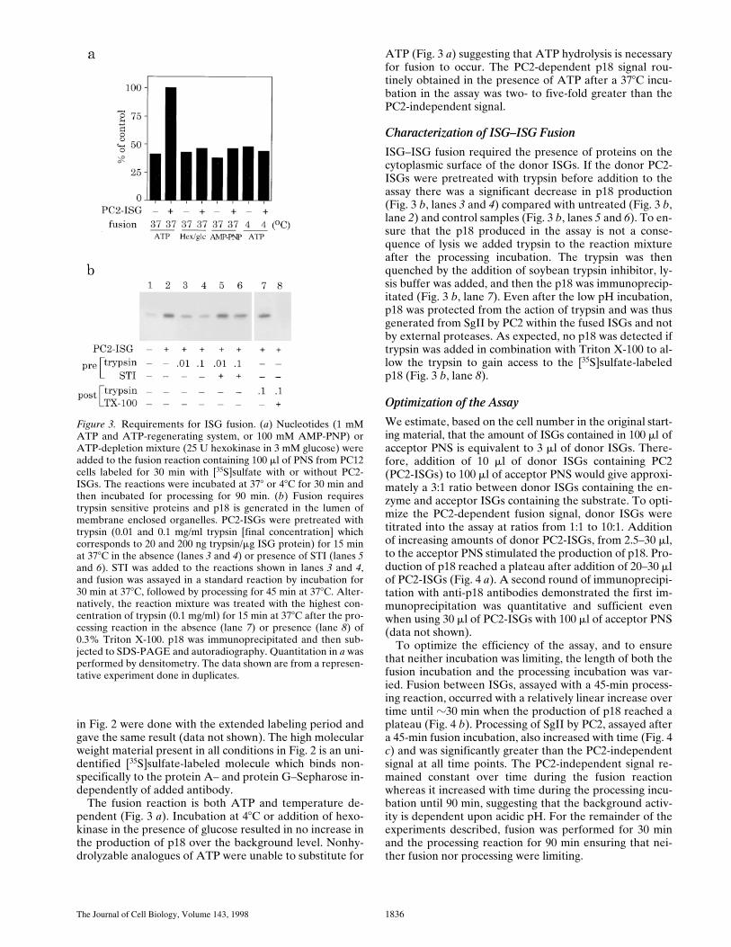
The fusion reaction is both ATP and temperature de-pendent (Fig. 3 a). Incubation at 48C or addition of hexo-kinase in the presence of glucose resulted in no increase inthe production of p18 over the background level. Nonhy-drolyzable analogues of ATP were unable to substitute for
ATP (Fig. 3 a) suggesting that ATP hydrolysis is necessaryfor fusion to occur. The PC2-dependent p18 signal rou-tinely obtained in the presence of ATP after a 378C incu-bation in the assay was two- to five-fold greater than thePC2-independent signal.
Characterization of ISG–ISG Fusion
ISG–ISG fusion required the presence of proteins on thecytoplasmic surface of the donor ISGs. If the donor PC2-ISGs were pretreated with trypsin before addition to theassay there was a significant decrease in p18 production(Fig. 3 b, lanes 3 and 4) compared with untreated (Fig. 3 b,lane 2) and control samples (Fig. 3 b, lanes 5 and 6). To en-sure that the p18 produced in the assay is not a conse-quence of lysis we added trypsin to the reaction mixtureafter the processing incubation. The trypsin was thenquenched by the addition of soybean trypsin inhibitor, ly-sis buffer was added, and then the p18 was immunoprecip-itated (Fig. 3 b, lane 7). Even after the low pH incubation,p18 was protected from the action of trypsin and was thusgenerated from SgII by PC2 within the fused ISGs and notby external proteases. As expected, no p18 was detected iftrypsin was added in combination with Triton X-100 to al-low the trypsin to gain access to the [35S]sulfate-labeledp18 (Fig. 3 b, lane 8).
Optimization of the Assay
We estimate, based on the cell number in the original start-ing material, that the amount of ISGs contained in 100 ml ofacceptor PNS is equivalent to 3 ml of donor ISGs. There-fore, addition of 10 ml of donor ISGs containing PC2(PC2-ISGs) to 100 ml of acceptor PNS would give approxi-mately a 3:1 ratio between donor ISGs containing the en-zyme and acceptor ISGs containing the substrate. To opti-mize the PC2-dependent fusion signal, donor ISGs weretitrated into the assay at ratios from 1:1 to 10:1. Additionof increasing amounts of donor PC2-ISGs, from 2.5–30 ml,to the acceptor PNS stimulated the production of p18. Pro-duction of p18 reached a plateau after addition of 20–30 mlof PC2-ISGs (Fig. 4 a). A second round of immunoprecipi-tation with anti-p18 antibodies demonstrated the first im-munoprecipitation was quantitative and sufficient evenwhen using 30 ml of PC2-ISGs with 100 ml of acceptor PNS(data not shown).
To optimize the efficiency of the assay, and to ensurethat neither incubation was limiting, the length of both thefusion incubation and the processing incubation was var-ied. Fusion between ISGs, assayed with a 45-min process-ing reaction, occurred with a relatively linear increase overtime until z30 min when the production of p18 reached aplateau (Fig. 4 b). Processing of SgII by PC2, assayed aftera 45-min fusion incubation, also increased with time (Fig. 4c) and was significantly greater than the PC2-independentsignal at all time points. The PC2-independent signal re-mained constant over time during the fusion reactionwhereas it increased with time during the processing incu-bation until 90 min, suggesting that the background activ-ity is dependent upon acidic pH. For the remainder of theexperiments described, fusion was performed for 30 minand the processing reaction for 90 min ensuring that nei-ther fusion nor processing were limiting.
Figure 3. Requirements for ISG fusion. (a) Nucleotides (1 mMATP and ATP-regenerating system, or 100 mM AMP-PNP) orATP-depletion mixture (25 U hexokinase in 3 mM glucose) wereadded to the fusion reaction containing 100 ml of PNS from PC12cells labeled for 30 min with [35S]sulfate with or without PC2-ISGs. The reactions were incubated at 378 or 48C for 30 min andthen incubated for processing for 90 min. (b) Fusion requirestrypsin sensitive proteins and p18 is generated in the lumen ofmembrane enclosed organelles. PC2-ISGs were pretreated withtrypsin (0.01 and 0.1 mg/ml trypsin [final concentration] whichcorresponds to 20 and 200 ng trypsin/mg ISG protein) for 15 minat 378C in the absence (lanes 3 and 4) or presence of STI (lanes 5and 6). STI was added to the reactions shown in lanes 3 and 4,and fusion was assayed in a standard reaction by incubation for30 min at 378C, followed by processing for 45 min at 378C. Alter-natively, the reaction mixture was treated with the highest con-centration of trypsin (0.1 mg/ml) for 15 min at 378C after the pro-cessing reaction in the absence (lane 7) or presence (lane 8) of0.3% Triton X-100. p18 was immunoprecipitated and then sub-jected to SDS-PAGE and autoradiography. Quantitation in a wasperformed by densitometry. The data shown are from a represen-tative experiment done in duplicates.

Urbé et al. Fusion of ISGs in a Cell-free Assay 1837
Characterization of Fusion Product
Incubation of ISGs containing [35S]sulfate-labeled SgIIwith PC2-ISGs results in the appearance of [35S]sulfate-labeled p18. To ensure that the vesicles which containedthe p18 were derived from the fusion of two populations ofISGs, we studied the sedimentation behavior of the fusedISGs by subcellular fractionation on sequential velocityand equilibrium sucrose gradients. A fusion incubationwas performed as above and chilled to 48C while an equalportion of the [35S]sulfate-labeled PC12-PNS was kept onice (starting material). Both the fusion reaction and thestarting material were subjected to velocity controlled cen-trifugation. ISG containing fractions (fractions 2–4) werepooled and either loaded on equilibrium gradients or aportion (one-fifth) of each pool was incubated for process-ing. The latter was analyzed after processing for the con-tent of [35S]sulfate-labeled SgII or p18 (Fig. 5, a and b). Inthe pool obtained after fusion (postfusion) [35S]sulfate-labeled p18 (Fig. 5 b) can be detected but not in the poolkept at 48C (prefusion). As expected, a decrease in the to-tal [35S]sulfate-labeled SgII is detectable after fusion (Fig.5 a), reflecting the processing of SgII to fragments in-cluding p18 (Fig. 1). To verify that the vesicles containingp18 were ISGs, the remaining four-fifths of the velocitygradient pools were subjected to equilibrium gradient cen-trifugation. After equilibrium gradient centrifugation andfractionation, the fractions were analyzed for [35S]sulfate-labeled SgII (Fig. 5 c) or [35S]sulfate-labeled p18 (Fig. 5 d).
The p18 containing vesicles produced by fusion (Fig. 5 d)sedimented to the same position on the equilibrium gradi-ent as the ISGs containing [35S]sulfate-labeled SgII in thestarting material (Fig. 5 c). This confirms that the fusion oftwo ISGs results in a “hybrid” ISG with similar propertiesas the starting ISG. Interestingly, the hybrid ISG (Fig. 5 d)displayed a slight increase in density, suggesting they werelarger than the original population of ISGs. This experi-ment was also used to calculate the percentage of SgII pro-cessed after fusion by comparing the amount of p86 in thestarting material with p86 after fusion and processing (Fig.5 a, Table I, and see below).
Comparison of SgII Processing in Fused ISGswith PC2-ISGs
Direct comparison of the extent of processing in the hy-brid ISGs with the PC2-ISGs where the enzyme and sub-strate are copackaged when ISGs are formed in the cellalso allowed us to estimate the fusion efficiency under thestandard conditions of the assay (Table I). A PNS fromPC12/PC2 cells labeled with [35S]sulfate was isolated andincubated in a fusion assay under standard conditions ex-cept that no unlabeled PC2-ISGs were added. The extentof processing obtained was then compared with that ob-served for the hybrid ISGs formed in a typical fusion as-say. The relative processing index in hybrid ISGs wasfound to be z25% compared with 69% for PC2-ISGs.
Figure 4. Optimization of the fusion assay conditions. (a) Increasing amounts of PC2-ISGs(from 0 to 30 ml) were added to 100 ml of PNS from PC12 cells labeled for 30 min with[35S]sulfate and then assayed for fusion by incubation for 30 min at 378C followed by theprocessing reaction for 90 min at 378C. Top and bottom panel are from separate represen-tative experiments. (b) Time course of fusion. 20 ml of PC2-ISGs were added to the assaymixture and incubated for fusion. At the time points shown the samples were transferredto ice and diluted with pH 5.5 buffer and subjected to the processing at 378C for 45 min. (c)Time course of processing. Fusion assays were performed with either 0 or 20 ml of PC2-ISGs for 45 min at 378C. The reactions were then subjected to processing reactions at 378Cfor times ranging from 0 to 150 min. p18 was immunoprecipitated and quantified as de-scribed in Fig. 3 and the PC2-independent background was subtracted. Data shown arefrom representative experiments done in duplicate.

The Journal of Cell Biology, Volume 143, 1998 1838
Thus, the efficiency of fusion between ISGs in the assaywas estimated to be z37%.
Comparison of the time course of SgII processing in thehybrid ISG (Fig. 4 c) and in ISGs isolated from PC12/PC2cells in a previous study (Urbé et al., 1997a) reveals thatSgII processing in hybrid ISGs occurs with different kinet-ics. In the PC2-ISGs 50% of the maximum processing sig-nal is reached after 15 min of incubation (Fig. 5 in Urbé etal., 1997a) whereas the processing signal in the hybridISGs is less than 10% of the maximum signal at that time.In addition, the maximum p18 signal in the PC2-ISGs isreached at 60 min compared with 90 min for the hybridISGs. These differences may be due to the fact that in thehybrid ISGs the PC2 enzyme will process the PC12-ISGderived [35S]sulfate-labeled SgII as well as the unlabeledSgII contained in the PC2-ISGs. If one assumes that onePC2-ISG fuses with one PC12-ISG containing [35S]sulfate-labeled SgII to generate a hybrid ISG, at most only half ofthe total SgII processed at any one time will be [35S]sul-fate-labeled, thus processing of [35S]sulfate-labeled SgIIwill take about twice as long in the hybrid ISG.
Fusion Only Occurs between ISGs and ISGs
The ISG is defined as an early stage intermediate that canundergo changes such as acidification, membrane remod-eling, and homotypic fusion (Tooze, 1991) whereas theMSG is a late stage stable organelle which has a constantsize and content. One prediction of these definitions is thatfusion should be specific to the early stage and will not oc-cur at the later mature granule stage. Two approacheswere used to investigate the stage specificity of fusion: firstwe asked whether MSGs were able to substitute for ISGsin the fusion assay and second whether ISGs or MSGscould compete for generation of labeled p18.
MSGs, isolated from PC12/PC2 cells, were substitutedfor the PC2-ISGs in a standard assay (Fig. 6 a). MSGsfrom PC12/PC2 cells contain large amounts of the maturePC2 enzyme (Dittié and Tooze, 1995; data not shown) andthus the processing of the [35S]sulfate-labeled SgII shouldbe readily detected if fusion occurs. Substitution of thePC2-ISGs with either one or two times the amount ofPC2-MSGs did not result in the appearance of appreciableamounts of p18 over the PC2-independent background.
The specificity of fusion was next tested by competitionfor fusion using ISGs isolated from PC12 cells (PC12-IGSs) that contain cold substrate but no enzyme. Additionof increasing amounts of cold PC12-IGSs to the completereaction mixture (containing 100 ml of [35S]sulfate-labeledPNS from PC12 cells, 10 ml PC2-ISGs, ATP, and an ATP-regenerating system) resulted in a decrease in the amountof p18 produced (Fig. 6 b, open squares). When the ratio ofPC12-ISGs to PC2-ISGs added to the assay was z3:1, a50% reduction in the p18 signal was detected. Impor-tantly, the addition of an equivalent amount of MSGs, pre-pared from PC12 cells (PC12-MSGs), to the assay (i.e., 3MSGs:1 ISG) resulted in no competition (Fig. 6 b, closeddiamond) confirming the observation that MSGs do notfuse with ISGs.
Competition leading to a decrease of the total p18 signalcould result from either competition for fusion or enzyme.If the ISGs have a limited capacity to fuse they will be-
Figure 5. Characterization of the membranes containing p18 af-ter fusion. A PNS was prepared from PC12 cells labeled with[35S]sulfate for 30 min. One-half of this PNS was kept on ice whilethe remainder was incubated under standard conditions for fu-sion with PC2-ISGs. Both the starting material and the reactionmixture were loaded onto velocity gradients and subjected tofractionation. Fractions 2–4 from the velocity gradients werepooled, and one-fifths were analyzed for p18 using the standardincubation for processing while the remaining four-fifths weresubjected to equilibrium gradient centrifugation and fraction-ation. (a) Samples of the velocity gradient pools analyzed for p86,or (b) subjected to processing and then immunoprecipitation withanti-p18 antibodies from either the starting material (pre fusion)or after fusion (post fusion). (c) Samples of equilibrium gradientfractions 5–11 from the gradient loaded with the starting materialwere analyzed for p86, or (d) equilibrium gradient fractions 5–11from the gradient loaded with the fusion reaction were subjectedto processing followed by immunoprecipitation to detect p18 asabove. Analysis of p86 was performed by SDS-PAGE followedby fluorography.
Table I. Determination of the Fusion Efficiency
PNS PNS VG
PC2-ISGsfusion NR*
1 processingip 175
hybrid ISGs1 fusion
1 processingip 175
hybrid ISGs1 fusion
1 processingtotal p86
p86 in starting material (st) 76.0 62.73 34.2p86 after incubations (fin) 23.6 46.76 20.8Percent processing efficiency 69.0% 25.5% 39.2%(p86st 2 p86fin) 3 100
(p86st)Percent fusion efficiency 100% 37% —
*NR, not required.A PNS was prepared from PC12 or PC12/PC2 cells labelled for 30 min with [35S]sul-phate. Fusion assays were performed as described using a PNS or enriched fractions(VG). The amount of p86 before (starting material 5 st) and after the fusion reaction(final 5 fin) was determined by immunoprecipitation with Ab 175 (ip 175) or by directanalysis of the total p86 from enriched fractions containing ISGs (Fig. 5) and is ex-pressed as arbitrary units. Processing efficiency is calculated from the values shownwhich are from representative experiments. Fusion efficiency in hybrid ISGs was ex-pressed as a percentage of SGII processing in intact PC2-ISGs using the values ob-tained with the PNS samples.

Urbé et al. Fusion of ISGs in a Cell-free Assay 1839
come refractory to fusion after a defined number of fusionevents. Thus, addition of excess ISGs will result in compe-tition and a decrease in the p18 produced. However, it isequally possible that competition is the result of unlabeledsubstrate competing with labeled substrate for enzyme inan unrestricted fusion reaction where PC2 is limiting. Fur-ther experiments need to be carried out to confirm themode of competition, however, whether it is a result ofcompetition for fusion or competition for substrate, bothdepend on an initial fusion event between ISGs.
Cytosol Dependence of ISG–ISG Fusion
To determine if ISG–ISG fusion required cytosolic com-ponents we modified the assay by isolating the membranesand membrane bound organelles from the acceptor PNSderived from the [35S]sulfate-labeled PC12 cells. The iso-lated membranes were resuspended in buffer or cytosolprepared from PC12 cells, and tested for activity in the fu-sion assay. Comparison of the results obtained with thePNS assay showed that the efficiency of the fusion reac-tion with the membrane pellet was similar and requiredcytosol (Fig. 7 a). To obtain a more abundant source of cy-tosol we explored the feasibility of substituting cytosolfrom tissues, such as rat liver, for the PC12 cell cytosol.Rat liver cytosol was demonstrated to be active in the fu-sion assay and the maximum fusion signal was obtainedwith z7 mg/ml cytosol which was comparable to PC12 cellcytosol.
We next investigated the requirements for the nucle-otides ATP and GTP in the cytosol-dependent assay. Ratliver cytosol was prepared and added to the assay with ei-ther ATP, GTP, or GTPgS. Both ATP and GTP togetherwere required for maximum efficiency, and interestinglyGTPgS could substitute for GTP but not ATP (Fig. 7 b).
Although the requirement for ATP may be attributed to arequirement for NSF (see below) we do not yet under-stand the GTP requirement. It may be that a GTP-bindingprotein is required in its GTP bound form for fusion tooccur.
Fusion of ISGs Can Occur with an Enriched Preparation of Acceptor ISGs
To ensure that the fusion we measure was homotypic, thatis occurring between identical populations of ISGs, we at-tempted to reconstitute fusion using an acceptor ISG frac-tion of comparable purity to the donor PC2-ISGs in thepresence of cytosol. Several attempts to do so failed (datanot shown) and we speculate that centrifugation throughtwo consecutive sucrose gradients removes essential com-ponents that are required on at least one ISG population.To overcome this problem we used a partially purifiedpool enriched in post-Golgi vesicles, in particular ISGs,which does not contain detectable levels of TGN mem-branes. Isolation of acceptor ISGs from PC12 cells labeledwith [35S]sulfate for 30 min was performed using the su-crose velocity gradients normally used to prepare donorPC2-ISGs. The pool of fractions 2–4 which was then usedin a standard assay supplemented with rat liver cytosolwith or without PC2 ISGs. Rat liver cytosol was added be-cause the velocity gradient fractions are largely depletedof cytosolic proteins which remain in the load. Fusion wasdetected in the reaction mixtures containing the velocitygradient fractions and PC2-ISGs and cytosol (Fig. 8). Al-though the total signal was reduced compared with thatobtained with a membrane pellet derived from a PNS of[35S]sulfate-labeled PC12 cells, when normalized to back-ground the PC2-dependent fusion signal obtained with theenriched donor pool (1.9-fold over background) was com-parable to that obtained with a membrane pellet (1.7-foldover background, Fig. 8).
Role of NEM-sensitive Proteins in ISG Fusion
Previously identified proteins involved in membrane fu-sion reactions include the ATPases NSF and p97 both ofwhich can be inactivated by NEM treatment. Analysis ofthe distribution of these NEM-sensitive proteins in themembrane fractions and rat liver cytosol used in the assayis shown in Fig. 9 a. The majority of the NSF contained inthe PNS (z3 mg NSF per mg of total protein) remains as-sociated with the membranes after preparation of themembrane pellet (MP). Most of the a-SNAP present inthe PNS was recovered on the membrane pellet, but verylittle was detected on PC2-ISGs. The distribution of p97on the PNS and membrane pellet is similar compared withNSF. However, there was very little p47 detected on themembrane pellet or the PC2-ISG fraction.
To explore the possibility that one, or both of thesecomplexes, are required for fusion we treated either thePC12 cell membrane pellet, containing [35S]sulfate-labeledSgII, rat liver cytosol, or both with NEM (Fig. 9 b, left).Treatment of both membranes and cytosol inhibited fu-sion more than 90%. In the presence of NEM treated ratliver cytosol the efficiency of fusion of untreated mem-branes was only z30% of the control value. The fusion ef-ficiency of NEM-treated membranes could be recovered
Figure 6. MSGs cannot fuse with ISGs. (a) PC2-ISGs, or one ortwo times the volume of MSGs prepared from PC12/PC2 cellswere added to the fusion reactions containing 100 ml of PNS fromPC12 cells labeled with 30 min [35S]sulfate. (b) Increasingamounts of ISGs (open squares) or 30 ml of MSGs (solid dia-mond) prepared from PC12 cells, were added to fusion reactionscontaining 10 ml of PC2-ISGs and 100 ml of PNS from labeledPC12 cells. Fusion reactions, carried out for 30 min at 378C werefollowed by processing reactions for 90 min at 378C. p18 was im-munoprecipitated from the reactions and quantitated as above.Results shown are after subtraction of the PC2-independentbackground and are representative experiments done in dupli-cate.

The Journal of Cell Biology, Volume 143, 1998 1840
to z40% of the original fusion efficiency by the additionof untreated rat liver cytosol to the NEM treated mem-branes.
To determine if we could attribute the decrease in fu-sion efficiency after NEM treatment to inactivation ofNSF or p97 we added recombinant NSF and a-SNAP, orpurified p97 and recombinant p47 to the fusion assay af-ter various NEM treatments. Using NEM-treated mem-branes, addition of NSF to NEM treated rat liver cytosolcould restore fusion nearly to the level observed after ad-dition of untreated rat liver cytosol (Fig. 9 c). Addition ofa-SNAP did not increase the rescue by NSF, in fact wasslightly inhibitory (data not shown). Interestingly, supple-menting untreated cytosol with similar amounts of NSFstimulated the recovery of NEM-treated membranes to al-most 90% of the control value (Fig. 9 b, right). Addition ofa complex of purified p97 and his-tagged p47 to NEM-treated membranes and NEM-treated cytosol did not re-store fusion (Fig. 9 c). Neither did the addition of p97/47to untreated cytosol restore the ability of NEM-treatedmembranes to undergo fusion (Fig. 8 b, right). Experi-ments were also performed using purified p97 alone, or acrude p97–p47 fraction (purified by 40% ammonium sul-fate precipitation of rat liver cytosol, then sucrose gradientcentrifugation using the method of Kondo et al. [1997]),both of which were unable to rescue NEM-treated mem-branes and cytosol.
DiscussionFusion of ISGs has been reconstituted in a cell-free assaybased on the proteolytic processing of a substrate prohor-mone (SgII) by a prohormone convertase (PC2). An im-
portant feature of the assay is the fact that proteolytic pro-cessing of substrates by PC2 only occurs in the regulatedsecretory pathway after the substrates and enzymes arepackaged into secretory granules (Urbé et al., 1997a). Us-ing this assay we have determined that ISG fusion is de-pendent upon cytosolic and membrane associated pro-teins, ATP, GTP, and physiological temperature. Fusion isspecific for ISGs in that MSGs cannot fuse with ISGs, andcompetition is observed after addition of excess ISGs, butnot MSGs, derived from unlabeled PC12 cells.
In the absence of PC2-ISGs we can detect a low amountof endopeptidase activity which processes the labeled SgIIin the PC12-ISGs. This background signal is produced dur-ing the processing reaction by an endogenous proteasepresent in PC12 cell ISGs. This endopeptidase activity isnot detected in intact PC12 cells after prolonged [35S]sul-fate-labeling during which time the MSGs accumulate un-processed [35S]sulfate-labeled SgII. Furthermore, subcel-lular fractionation data suggest that the activity is found inISGs but not MSGs (data not shown). We speculate thatthis unknown endopeptidase activity may have a pH opti-mum closer to pH 5.5 than 6.3, and therefore is normallyinactive in ISGs. Furthermore, this enzyme might be re-moved from ISGs during maturation and thus never reachthe more acidic milieu of the MSG where it would be ac-tive. Additional information about the nature and functionof this enzyme requires further study.
The assay described reconstitutes ISG–ISG fusion, andwas developed with the aim of understanding the role offusion in MSG biogenesis. Homotypic fusion, defined as thefusion of two like compartments, is thought to be involvedin the steady-state maintenance of organelles, unlike het-erotypic fusion which results in the transfer of molecules
Figure 7. Cytosol-dependent reconstitution of ISG fusion. (a) A membrane pellet (MP) was prepared from 100 ml of PNS obtainedfrom PC12 cells labeled for 30 min with [35S]sulfate. An equal volume of PNS was used as control. PC2-ISGs (10 ml), and PC12 cell cyto-sol (final concentration 7 mg/ml) was added to the reaction mixtures as indicated. Fusion and processing was carried out under standardconditions. p18 was immunoprecipitated and quantitated as described above. (b) A standard fusion reaction was performed using a MPprepared from the PNS of PC12 cells labeled as above, in the presence or absence of 10 ml of PC2-ISGs, rat brain cytosol (final amounts7 mg/ml), and nucleotides as indicated. p18 was immunoprecipitated and quantitated as described above. Data shown are representativeexperiments performed in duplicate.

Urbé et al. Fusion of ISGs in a Cell-free Assay 1841
and lipids from one compartment to another. Examples ofhomotypic fusion include the reassembly of the ER (Lat-terich et al., 1995; Turner et al., 1997), the Golgi complex(Acharya et al., 1995b; Rabouille et al., 1995b), endosome-endosome fusion (Gruenberg and Howell, 1986; Diaz etal., 1988; Woodman and Warren, 1988), lysosome–lyso-some fusion (Ward et al., 1997) and vacuole–vacuole fu-sion (Conradt et al., 1992).
In the case of assays which rely on internalized markersto define the endocytotic compartments (for example seeGruenberg and Howell, 1986; Diaz et al., 1988), fusion isassumed to be homotypic if the compartments undergoingfusion contain markers internalized for the same length oftime. In the ISG–ISG fusion assay described here fusioncan potentially occur amongst heterologous populations ofISGs defined by the [35S]sulfate-labeling protocol. ISGsform de novo from the TGN then mature into MSGs. Mat-uration of ISGs takes place over a relatively long period oftime (t1/2 5 z45 min), compared with the time it takes anISG to bud (budding t1/2 5 z5 min; Tooze and Huttner,1990). The possibility therefore exists that the labeledISGs in the assay are at different stages of maturation, andconsequently might display variable fusionogenicity. Al-
Figure 8. Fusion occurs with enriched acceptor ISGs. A mem-brane pellet (MP) or velocity gradient (VG) fraction (pool offractions 2–4) were isolated from a PNS obtained from PC12 cellslabeled with [35S]sulfate for 30 min. A standard reaction was per-formed with or without 10 ml of PC2-ISGs and with or without ratliver cytosol. After a 30-min incubation for fusion, processing wasperformed for 90 min at 378C. p18 was immunoprecipitated andquantitated as above. Data shown is of representative experimentperformed in triplicate (error bars are 6SD of the mean) ex-pressed as percent of the control. For comparison, the PC2-dependent signal is shown relative to the PC2-independent signalas an inset to emphasize that ISG–ISG fusion is comparable inthe MP and VG fractions.
Figure 9. NSF can rescue the inhibition of ISG fusion after NEMtreatment. (a) Immunoblot of a PNS, membrane pellet (MP) ob-tained from the PNS, PC2-ISGs, and rat liver cytosol with anti-bodies directed against p97, p47, NSF, and a-SNAP. The blot wasprepared from material loaded in amounts which are propor-tional to that in the assay, i.e., 110 mg of a PNS, 38.5 mg of MP, 4.1 mgof ISGs, 49 mg of rat liver cytosol. Bound antibodies were de-tected using 125I-protein A. (b) NEM treatment was performedon the membrane pellet obtained from a PNS of PC12 cells la-beled with [35S]sulfate for 30 min or rat liver cytosol or both. Afusion assay was performed with the treated or untreated compo-nents using 10 ml of PC2-ISGs. NSF (19 mg/ml; 230 nM) or p97/p47 (28 mg/ml p97; 290 nM p97) was added to NEM-treatedmembranes and cytosol before incubation for fusion. (c) NSF canrescue NEM inhibition of fusion. NEM-treated membrane pelletwas incubated with either rat liver cytosol or NEM-treated ratliver cytosol in the presence or absence of NSF, p94/p47 in a stan-dard fusion assay with 10 ml of PC2-ISGs. Values shown are aftersubtraction of the PC2-independent background and are repre-sentative of three independent experiments for NSF and two in-dependent experiments for p97/47 performed in duplicates ortriplicates (error bars are 6SD of the mean).

The Journal of Cell Biology, Volume 143, 1998 1842
though there might be subpopulations of ISGs betweenwhich we can not discriminate, we define this fusion as ho-motypic in contrast with heterotypic fusion of ISGs withthe plasma membrane.
Key components required for either or both heterotypicand homotypic membrane fusion are the related ATPases,NSF and p97 (for review see Mellman, 1995; Whiteheartand Kubalek, 1995; Woodman, 1997). To begin to under-stand the exact nature of ISG–ISG fusion we asked if ei-ther or both of these molecules were required. We foundthat NSF, but not p97, is required for fusion of ISGs. NEMtreatment of cytosol and membranes resulted in inhibitionof fusion, which could be rescued by the addition of NSF.We found that a-SNAP was not required for NSF activity,and the addition of a-SNAP alone had no effect. In lightof the results from the reconstitution of homotypic fusionbetween Golgi membranes (Acharya et al., 1995a; Rabou-ille et al., 1995a) one might have anticipated a role for p97in ISG fusion. We found that addition of p97, or a complexof p97 and p47, to NEM-treated membranes and cytosolwas not sufficient to rescue fusion.
The distinction between the function of NSF and p97 inheterotypic fusion versus homotypic fusion is somewhatblurred by the ability of NSF to rescue NEM-treated Golgimembranes (Acharya et al., 1995a; Rabouille et al., 1995a)in the presence of a-SNAP, g-SNAP, and p115, and the de-monstration that both p47 and a-SNAP bind the t-SNAREsyntaxin 5 (Rabouille, 1998). In addition, homotypic fu-sion between yeast vacuoles, mammalian endosomes, andlysosomes has been shown to require NSF (Diaz et al.,1989; Rodriguez, et al., 1994; Robinson, et al., 1997) andfusion between yeast vacuoles and mammalian endosomeshas been shown not to require cdc48p (Mayer et al., 1996)or p97 (Robinson et al., 1997), respectively. Thus, until weobtain more data on the exact role of either NSF or p97 inmembrane fusion, a requirement for one or the other doesnot seem to be a primary factor in classifying fusion eventsas either homotypic or heterotypic.
What is the purpose of ISG–ISG fusion? We proposethat fusion of ISGs with each other is an integral part ofthe maturation process and may be a prerequisite for post-TGN sorting of proteins from the maturing ISG. Fusion ofISGs may be necessary to create enough excess membraneto form a clathrin-coated vesicle (CCV) from the ISG.Formation of one CCVs from an ISG (80-nm-diam) wouldresult in the removal of nearly one-half the surface area ofthe ISG. If three to five ISGs fuse to form a MSG (120-nm-diam) (Tooze et al., 1991) enough excess membranewould become available to allow two to seven CCVs toform. In addition, fusion of ISGs might be a prerequisitefor formation of CCVs by increasing for example the num-ber of AP-1–binding sites present in the ISG. Furin, aTGN-resident endopeptidase and both mannose-6-phos-phate receptors (MPRs) associate with AP-1 and are re-moved in CCVs which bud from the ISGs (Dittié et al.,1996, 1997; Klumperman, 1998; Kuliawat, et al., 1997).Other molecules which are not found in MSGs such as cer-tain isoforms of peptidylglycine-a-amidating-mono-oxygen-ase (Milgram et al., 1994), as well as soluble moleculessuch as C-peptide (Kuliawat and Arvan, 1994) may also bepresent in these CCVs although how they are selected isnot clear.
If ISG fusion is part of the maturation process it followsthat after maturation is completed, homotypic fusionwould no longer be required. In agreement with this wehave shown here that MSGs can no longer fuse with ISGs.It is possible that the ISG–ISG fusion machinery maybegradually inactivated during maturation. We have previ-ously shown that ISGs have a less acidic luminal pH thanMSGs (pH 6.3 versus pH 5.5) and proposed that the de-crease in pH during maturation may be a direct conse-quence of the membrane remodelling events during matu-ration (Urbé et al., 1997a). Increasing lumenal acidity mayinfluence the properties of both the membrane and pro-teins within the membrane and might thus modify the fu-sion competence of the secretory granule. An alternativebut not exclusive explanation maybe that factors involvedin homotypic fusion of ISGs, for instance a t-SNARE, areremoved during maturation by budding of CCVs or consti-tutive-like secretory vesicles such that the MSG is left withonly the molecule(s), for instance a v-SNARE, required tomediate heterotypic fusion with the plasma membraneduring regulated exocytosis. Control of the final size of theMSG would then be dictated by the number of ISG fusionevents needed to generate enough vesicles to remove allthe SNAREs involved in ISG fusion.
There are to date two possible candidate t-SNAREsthat may be involved in ISG fusion, syntaxin 3 and 6. Syn-taxin 3 has been localized to zymogen granules (Gaisanoet al., 1996) whereas syntaxin 6 has been localized to ISGsin PC12 cells (Klumperman, 1998). Syntaxin 6 has beenshown to be present in coated buds emanating from theISGs as well as in CCVs, endosomes, (Klumperman, 1998),and in TGN membranes (Bock, et al., 1997; Klumperman,1998). It has been proposed that syntaxin 6 is involvedin targeting the CCVs to an endosomal compartment(Klumperman, 1998). It is equally plausible that syntaxin 6is involved in ISG fusion and only subsequently removedby CCVs together with furin and MPR and targeted to theendosome and/or returned to the TGN.
Selection and removal of putative sorting receptors,membrane proteins, or missorted cargo before regulatedexocytosis is the most important goal of maturation and iscritical for the homeostasis of the organism. Althoughsorting and fusion events during secretory granule biogen-esis are likely to be linked, more experiments are requiredto understand when and why ISGs lose their ability to fusewith each other and how this is controlled to generate aMSG. The assay described here represents an ideal experi-mental system to investigate secretory granule biogenesis.
We thank all the past and present members of the Tooze and Warren labsfor helpful discussions. We are grateful to G. Warren, G. Schiavo, and J.Tooze (all from ICRF, London, UK) for critically reading the manuscriptas well as for discussions, advice, and encouragement. We thank H.Kondo for generously supplying purified p97, p97–p47 complex, and his-tagged p47 as well as for his time and advice.
Received for publication 6 July 1998 and in revised form 9 October 1998.
References
Acharya, U., R. Jacobs, J.M. Peters, N. Watson, M.G. Farquhar, and V. Mal-hotra. 1995a. The formation of Golgi stacks from vesiculated Golgi mem-branes requires two distinct fusion events. Cell. 82:895–904.
Acharya, U., J.M. McCaffery, R. Jacobs, and V. Malhotra. 1995b. Reconstitu-tion of vesiculated Golgi membranes into stacks of cisternae: requirement of

Urbé et al. Fusion of ISGs in a Cell-free Assay 1843
NSF in stack formation. J. Cell Biol. 129:577–589.Advani, R.J., H.-R. Bae, J.B. Bock, D.S. Chao, Y.-C. Doung, R. Prekeris, J.-S.
Yoo, and R.H. Scheller. 1998. Seven novel mammalian SNARE proteins lo-calize to distinct membrane compartments. J. Biol. Chem. 273:10317–10324.
Barr, F.A., A. Leyte, S. Mollner, T. Pfeuffer, S.A. Tooze, and W.B. Huttner.1991. Trimeric G-proteins of the trans-Golgi network are involved in the for-mation of constitutive secretory vesicles and immature secretory granules.FEBS (Fed. Eur. Biochem. Soc.) Lett. 294:239–243.
Bennett, D.L., E.M. Bailyes, E. Nielsen, P.C. Guest, N.G. Rutherford, S.D.Arden, and J.C. Hutton. 1992. Identification of the type 2 proinsulin process-ing endopeptidase as PC2, a member of the eukaryote subtilisin family. J.Biol. Chem. 267:15229–15236.
Block, M.R., B.S. Glick, C.A. Wilcox, F.T. Wieland, and J.E. Rothman. 1988.Purification of an N-ethylmaleimide-sensitive protein catalyzing vesiculartransport. Proc. Natl. Acad. Sci. USA. 85:7852–7856.
Bock, J.B., and R.H. Scheller. 1997. Protein transport A fusion of new ideas.Nature. 36:133–135.
Bock, J.B., J. Klumperman, S. Davanger, and R.H. Scheller. 1997. Syntaxin 6functions in trans Golgi network vesicle trafficking. Mol. Biol. Cell. 8:1261–1271.
Burgess, T.L., and R.B. Kelly. 1987. Constitutive and regulated secretion ofproteins. Annu. Rev. Cell Biol. 3:243–293.
Carnell, L., and H.P. Moore. 1994. Transport via the regulated secretory path-way in semi-intact PC12 cells: role of intra-cisternal calcium and pH in thetransport and sorting of secretogranin II. J. Cell Biol. 127:693–705.
Chen, Y.G., A. Siddhanta, C.D. Austin, S.M. Hammond, T.C. Sung, M.A. Froh-man, A.J. Morris, and D. Shields. 1997. Phospholipase D stimulates releaseof nascent secretory vesicles from the trans-Golgi network. J. Cell Biol. 138:495–504.
Clary, D., I.C. Griff, and J.E. Rothman. 1990. SNAPs, a family of NSF attach-ment proteins involved in intracellular membrane fusion in animals andyeast. Cell. 61:723–733.
Conradt, B., J. Shaw, T. Vida, S. Emr, and W. Wickner. 1992. In vitro reactionsof vacuole inheritance in Saccharomyces cerevisiae. J. Cell Biol. 119:1469–1479.
Cool, D.R., E. Normant, F.-s. Shen, H.-C. Chen, L. Pannell, Y. Zhang, and Y.P.Loh. 1997. Carboxypeptidase E is a regulated secretory pathway sorting re-ceptor: genetic obliteration leads to endocrine disorders in Cpefat mice. Cell.88:73–83.
Davey, J., S.M. Hurtley, and G. Warren. 1985. Reconstitution of an endocyticfusion event in a cell-free system. Cell. 43:643–652.
Diaz, R., L. Mayorga, and P. Stahl. 1988. In vitro fusion of endosomes followingreceptor-mediated endocytosis. J. Biol. Chem. 263:6093–6100.
Diaz, R., L. Mayorga, P.J. Weidman, J.E. Rothman, and P.D. Stahl. 1989. Vesi-cle fusion following receptor-mediated endocytosis requires a protein activein Golgi transport. Nature. 339:398–400.
Dittié, A., and S. Tooze. 1995. Characterization of the endopeptidase PC2 ac-tivity towards SgII in stably transfected PC12 cells. Biochem. J. 310:777–787.
Dittié, A.S., N. Hajibagheri, and S.A. Tooze. 1996. The AP-1 adaptor complexbinds to immature secretory granules from PC12 cells, and is regulated byADP- ribosylation factor. J. Cell Biol. 132:523–536.
Dittié, A.S., L. Thomas, G. Thomas, and S.A. Tooze. 1997. Interaction of furinin immature secretory granules from neuroendocrine cells with the AP-1adaptor complex is modulated by casein kinase II phosphorylation. EMBO(Eur. Mol. Biol. Organ.) J. 16:4859–4870.
Gaisano, H.Y., M. Ghai, P.N. Malkus, L. Sheu, A. Bouquillion, M.K. Bennett,and W.S. Trimble. 1996. Distinct cellular locations of the syntaxin family ofproteins in rat pancreatic acinar cells. Mol. Biol. Cell. 7:2019–2027.
Gerdes, H.-H., E. Phillips, and W.B. Huttner. 1988. The primary structure of ratsecretogranin II deduces from a cDNA sequence. Nucl. Acids Res. 16:11811.
Götte, M., and G.F. von Mollard. 1998. A new beat for the SNARE drum.Trends Cell Biol. 8:215–218.
Gruenberg, J.E., and K.E. Howell. 1986. Reconstitution of vesicle fusions oc-curring in endocytosis with a cell-free system. EMBO (Eur. Mol. Biol. Or-gan.) J. 5:3091–3101.
Heumann, R., V. Kachel, and H. Thoenen. 1983. Relationship between NGF-mediated volume increase and “priming effect” in fast and slow reactingclones of PC12 pheochromocytoma cells. Exp. Cell Res. 145:179–190.
Irminger, J.-C., B. Verchere, K. Meyer, and P.A. Halban. 1997. Proinsulin tar-geting to the regulated pathway is not impaired in carboxypeptidase E-defi-cient Cpefat/Cpefat mice. J. Biol. Chem. 272:27532–27534.
Itin, C., C. Rancanos, Y. Nakajima, and S.R. Pfeffer. 1997. A novel assay re-veals a role for soluble N-ethylmaleimide-sensitive fusion attachment pro-tein in mannose 6-phosphate receptor transport from endosomes to thetrans-Golgi network. J. Biol. Chem. 272:27737–27744.
Klumperman, J., R. Kuliawat, J.M. Griffith, H.J. Geuze, and P. Arvan. 1998.Mannose 6-phosphate receptors are sorted from immature secretory gran-ules via adaptor protein AP-1, clathrin, and syntaxin 6-positive vesicles. J.Cell Biol. 141:359–371.
Kondo, H., C. Rabouille, R. Newman, T.P. Levine, D. Pappin, P. Freemont,and G. Warren. 1997. p47 is a cofactor for p97-mediated membrane fusion.Nature. 388:75–78.
Kuliawat, R., and P. Arvan. 1994. Distinct molecular mechanisms for proteinsorting within immature secretory granules of pancreatic b-cells. J. Cell Biol.126:77–86.
Kuliawat, R., J. Klumperman, T. Ludwig, and P. Arvan. 1997. Differential sort-ing of lysosomal enzymes out of the regulated secretory pathway in pancre-atic b-cells. J. Cell Biol. 137:595–608.
Laslop, A., C. Weiss, D. Savaria, C. Eiter, S.A. Tooze, N.G. Seidah, and H.Winkler. 1998. Proteolytic processing of chromogranin B and secretograninII by prohormone convertases. J. Neurochem. 70:374–383.
Latterich, M., K.U. Frohlich, and R. Schekman. 1995. Membrane fusion and thecell cycle: Cdc48p participates in the fusion of ER membranes. Cell. 82:885–893.
Lee, R.W.H., and W.B. Huttner. 1983. Tyrosine-O-sulfated proteins of PC12pheochromocytoma cells and their sulfation by a tyrosylprotein sulfotrans-ferase. J. Biol. Chem. 258:11326–11334.
Mayer, A., W. Wickner, and A. Haas. 1996. Sec18p (NSF)-driven release ofSec17p (a-SNAP) can precede docking and fusion of yeast vacuoles. Cell. 85:83–94.
Mellman, I. 1995. Enigma variations: protein mediators of membrane fusion.Cell. 82:869–872.
Milgram, S.L., B.A. Eipper, and D.E. Mains. 1994. Differential trafficking ofsoluble and integral membrane secretory granule-associated proteins. J. CellBiol. 124:33–41.
Nichols, J.B., C. Ungermann, H.R.B. Pelham, W.T. Wickner, and A. Haas.1997. Homotypic vacuolar fusion mediated by t-and v-SNAREs. Nature. 387:199–202.
Niehrs, C., J. Stinchcombe, and W.B. Huttner. 1992. Two membrane-boundforms of tyrosylprotein sulfotransferase as revealed by phase partitioning inTriton X-114. Eur. J. Cell Biol. 58:35–43.
Ohashi, M., K. Jan de Vries, R. Frank, G. Snoek, V. Bankaitis, K. Wirtz, andW.B. Huttner. 1995. A role for phosphatidylinositol transfer protein insecretory vesicle formation. Nature. 377:544–547.
Orci, L., P. Halban, A. Perrelet, M. Amherdt, M. Ravazzola, and R.G.W.Anderson. 1994. pH-independent and -dependent cleavage of proinsulin inthe same secretory vesicle. J. Cell Biol. 126:1149–1156.
Patel, S.K., F.E. Indig, N. Olivieri, N.D. Levine, and M. Latterich. 1998. Or-ganelle membrane fusion: a novel function for the syntaxin homolog Ufe1pin ER membrane fusion. Cell. 92:611–620.
Rabouille, C., T.P. Levine, J.M. Peters, and G. Warren. 1995a. An NSF-likeATPase, p97, and NSF mediate cisternal regrowth from mitotic Golgi frag-ments. Cell. 82:905–914.
Rabouille, C., T. Misteli, R. Watson, and G. Warren. 1995b. Reassembly ofGolgi stacks from mitotic Golgi fragments in a cell-free system. J. Cell Biol.129:605–618.
Rabouille, C., H. Kondo, R. Newman, N. Hui, P. Freemont, and G. Warren.1998. Syntaxin 5 is a common component of the NSF- and p97-mediated re-assembly pathways of Golgi cisternae from mitotic Golgi fragments in vitro.Cell. 92:603–610.
Robinson, L.J., F. Aniento, and J. Gruenberg. 1997. NSF is required for trans-port from early to late endosomes. J. Cell Sci. 110:2079–2087.
Rodriguez, L., C.J. Stirling, and P.G. Woodman. 1994. Multiple N-ethylmale-imide-sensitive components are required for endosomal vesicle fusion. Mol.Biol. Cell. 5:773–783.
Rothman, J.E. 1994. Mechanisms of intracellular protein transport. Nature. 372:55–63.
Salpeter, M.M., and M.G. Farquhar. 1981. High resolution analysis of the secre-tory pathway in mammotrophs of the rat anterior pituitary. J. Cell Biol. 91:240–246.
Smith, R.E., and M.G. Farquhar. 1966. Lysosome function in the regulation ofthe secretory process in cells of the anterior pituitary gland. J. Cell Biol. 31:319–347.
Sollner, T., M.K. Bennet, S.W. Whiteheart, R.H. Scheller, and J.E. Rothman.1993. A protein assembly-disassembly pathway in vitro that may correspondto sequential steps of synaptic vesicle docking, activation, and fusion. Cell.75:409–418.
Steiner, D.F. 1991. Prohormone convertases revealed at last. Curr. Biol. 1:375–377.Thiele, C., H.H. Gerdes, and W.B. Huttner. 1997. Protein secretion: puzzling
receptors. Curr. Biol. 7:496–500.Tooze, S.A. 1991. Biogenesis of secretory granules Implications arising from
the immature secretory granule in the regulated pathway of secretion. FEBS(Fed. Eur. Biochem. Soc.) Lett. 285:220–224.
Tooze, S.A. 1998. Biogenesis of secretory granules in the trans-Golgi networkof neuroendocrine and endocrine cells. Biochim. Biophys Acta. 1404:231–244.
Tooze, S.A., and W.B. Huttner. 1990. Cell-free protein sorting to the regulatedand constitutive secretory pathways. Cell. 60:837–847.
Tooze, S.A., T. Flatmark, J. Tooze, and W.B. Huttner. 1991. Characterizationof the immature secretory granule, an intermediate in granule biogenesis. J.Cell Biol. 115:1491–1503.
Turner, M.D., H. Plutner, and W.E. Balch. 1997. A Rab GTPase is required forhomotypic assembly of the endoplasmic reticulum. J. Biol. Chem. 272:13479–13483.
Urbé, S., S. Dittié, and S.A. Tooze. 1997a. pH-dependent processing of secre-togranin II by the endopeptidase PC2 in isolated immature secretory gran-ules. Biochem. J. 321:65–74.
Urbé, S., S.A. Tooze, and F.A. Barr. 1997b. Formation of secretory vesicles inthe biosynthetic pathway. Biochim Biophys Acta. 1358:6–22.
Ward, D.M., J.D. Leslie, and J. Kaplan. 1997. Homotypic lysosome fusion in

The Journal of Cell Biology, Volume 143, 1998 1844
macrophages: analysis using an in vitro assay. J. Cell Biol. 139:665–673.Weber, T., B.V. Zemelman, J.A. McNew, B. Westerman, M. Gmachi, F. Par-
lati, T.H. Söllner, and J.E. Rothman. 1998. SNAREpins: minimal machineryfor membrane fusion. Cell. 92:759–772.
Weidman, P.J., P. Melancon, M.R. Block, and J.E. Rothman. 1989. Binding ofan N-ethylmaleimide–sensitive fusion protein to golgi membranes requiresboth a soluble protein(s) and an integral membrane protein. J. Cell Biol. 108:1589–1596.
Whiteheart, S.W., I.C. Griff, M. Brunner, D.O. Clary, T. Mayer, S.A. Buhrov,
and J.E. Rothman. 1993. SNAP family of NSF attachment proteins includesa brain- specific isoform. Nature. 362:353–355.
Whiteheart, S.W., and E.W. Kubalek. 1995. SNAPs and NSF: general membersof the fusion apparatus. Trends Cell Biol. 5:64–68.
Woodman, P. 1997. The roles of NSF, SNAPs and SNAREs during membranefusion. Biochim Biophys Acta. 1357:155–172.
Woodman, P.G., and G. Warren. 1988. Fusion between vesicles from the path-way of receptor-mediated endocytosis in a cell-free system. Eur. J. Biochem.173:101–108.