Embed Size (px)
Citation preview
HAL Id: tel-00116844https://tel.archives-ouvertes.fr/tel-00116844
Submitted on 28 Nov 2006
HAL is a multi-disciplinary open accessarchive for the deposit and dissemination of sci-entific research documents, whether they are pub-lished or not. The documents may come fromteaching and research institutions in France orabroad, or from public or private research centers.
L’archive ouverte pluridisciplinaire HAL, estdestinée au dépôt et à la diffusion de documentsscientifiques de niveau recherche, publiés ou non,émanant des établissements d’enseignement et derecherche français ou étrangers, des laboratoirespublics ou privés.
The regulation of type III secretion system ofPseudomonas aeruginosa
Dakang Shen
To cite this version:Dakang Shen. The regulation of type III secretion system of Pseudomonas aeruginosa. Life Sciences[q-bio]. Université Joseph-Fourier - Grenoble I, 2006. English. <tel-00116844>

UNIVERSITE JOSEPH FOURIER-GRENOBLE 1
SCIENCE & GEOGRAPHIE
THESE
Pour obtenir le garde de
DOCTEUR DE L’UNIVERSITE JOSEPH FOURIER-GRENOBLE 1
Discipline : Microbiologie, Biologie moléculaire
Présenté et soutenue publiquement par
DaKang SHEN le 20 décembre 2006
TITRE DE THESE
Régulation du Système de Sécrétion de Type III de Pseudomonas aeruginosa
COMPOSITION DU JURY
Professeur Benoît POLACK Président Professeur Benoît GUERY Rapporteur Docteur Thierry JOUENNE Rapporteur Professeur Alain DUFOUR Examinateur Docteur Bertrand TOUSSAINT Directeur de thèse
Thèse préparée au sein du Groupe de Recherche et d’Etude du Processus Inflammatoire
GREPI EA 2938, CHU Grenoble, Université Joseph Fourier Grenoble 1
REMERCIEMENTS
Je tiens tout d’abord à remercier le Docteur Bertrand TOUSSAINT , directeur de ma thèse, pour son encadrement, sa patience et pour l’ensemble du savoir pratique et théorique qu’il m’a transmis pendant ces trois années. Je remercie sincèrement Madame le professeur Françoise MOREL pour m’avoir accueilli au sein de son laboratoire GREPI et son soutien tout de long de ma thèse. Je remercie sincèrement Monsieur le professeur Pierre AMBROISE-THOMAS , Madame le professeur Renée GRILLOT et Monsieur le professeur Chun-Liang XUE , pour leurs aides et leurs soutiens. Je remercie sincèrement Monsieur le professeur Benoît POLACK pour sa gentillesse, sa générosité et le partage de ses connaissances. Je veux aussi le remercier d’avoir accepté la présidence de mon jury de thèse. Je remercie à Jean-François BRUGERE, Claudine PINEL , Hervé PELLOUX et Hélène FRICKER-HIDALGO pour leurs aides amicales. J’adresse mes remerciements aux professeurs Benoît GUERY, Thierry JOUENNE, ainsi qu’au Professeur Alain DUFOUR pour m’avoir fait l’honneur de juger ce travail. J’adresse un grand merci à tous les membres du GREPI, en particulier à Olivier EPAULARD , Lauriane QUENEE, Didier FILOPON et Madiha DEROUAZI pour leurs aides et leurs conseils amicaux. Je remercie la Région Rhône-Alpes, l’Association de Recherche en Pathologie Ostéoarticulaire et l’Agence Universitaire de la Francophonie qui m’ont financé pendant ces trois années. Enfin et surtout, je voudrais adresser plus qu’un grand merci à mes parents, à ma femme, ma fille, à mes beaux-parents qui ont toujours été là pour moi et sans qui tout cela n’aurait pas été possible.
A mes parents, Xue-Zhen et Yong-Zhang
A ma femme, Xia, et à ma fille, Xin-Yu
ABBREVIATIONS
bp base pair
Da dalton
DMSO dimethyl sulfoxide
DNA deoxyribonucleic acid
EB ethidium bromide
EDTA ethylenediaminetetraacetate
EGTA ethylene glycol bis (β-aminoethyl ether) N,N,N’,N’-tetraacetic acid
EMSA electrophoretic mobility shift assay
HEPES N-(2-hydroxyethyl)piperazine-N’-(2-ethanesulfonic acid)
IPTG isopropyl-β-D-thiogalactopyranoside
kb kilobase
kDa kilodalton
Max maximum
mHBS modified HEPES-buffered saline
MOI multiplicity of infection
MW molecular weight
OD optical density
PBS phosphate-buffered saline
RLU relative luciferase units
RNA ribonucleic acid
SDS sodium dodecyl sulfate
SDS-PAGE SDS-PolyAcrylamide Gel Electrophoresis
TAE Tris acetate EDTA buffer
TBE Tris borate EDTA buffer
TE Tris EDTA buffer
TEMED N, N, N’, N’-tetramethylethylenediamine
Tris Tris(hydroxymethyl)aminomethane
UV ultraviolet
X-Gal 5-Bromo-4-Chloro-3-Indolyl-b-D-galactopyranoside

CONTENTS
Introduction (en anglais) 1
1. Pseudomonas aeruginosa and P. aeruginosa infections 2
1.1 P. aeruginosa and cystic fibrosis 2
1.2 P. aeruginosa and burn wound infections 4
1.3 P. aeruginosa and ulcerative keratitis 4
1.4 P. aeruginosa and nosocomial pneumonia 5
2. Virulence factors 5
2.1 Lipopolysaccharide 5
2.2 Exotoxin A 6
2.3 Alginate 7
2.4 Phospholipases 8
2.5 Rhamnolipids 8
2.6 Phenazines 9
2.7 Proteases 10
2.8 Pili 11
2.9 Flagella 11
3. Secretion pathways 12
3.1 Sec-dependent secretion pathways 13
3.1.1 Type II secretion system 13
3.1.2 Autotransporter 15
3.1.3 Chaperone/usher secretion system 15
3.2 Sec-independent secretion pathways 16
3.2.1 Type I secretion system 16
3.2.2 Type IV secretion system 17
3.2.3 Type III secretion system (TTSS) 17
4. Type III secretion system of P. aeruginosa 19
4.1 Genetic organization and subcellular function 19
4.1.1 pscNOPQRSTU 19
4.1.2 popNpcr1234DR 21
4.1.3 pcrGVHpopBD 22
4.1.4 exsCEBA 23
4.1.5 exsDpscBCDEFGHIJKL 23
4.2 Type III secretion apparatus (TTSA) 24
4.3 Type III secretion translocon 26

4.4 Type III secretion Effectors 26
4.4.1 ExoS and ExoT 27
4.4.2 ExoY 28
4.4.3 ExoU 29
4.5 Type III secretion chaperones 29
4.5.1 Class I chaperones 30
4.5.2 Class II chaperones 30
4.5.3 Class III chaperones 31
4.6 Type III secretion signals 32
4.6.1 The 5’ mRNA signal 32
4.6.2 The N-terminal amino acid signal 32
4.6.3 Chaperone signal 33
4.6.4 Model for type three secretion signals 33
4.7 Posttranslational and/or cotranslational secretion 34
5. Regulation of TTSSs 34
5.1 Environment stimuli 34
5.1.1 Temperature 35
5.1.2 Divalent cations 35
5.1.3 Cell contact 36
5.1.4 Serum 37
5.1.5 Other factors 37
5.2 Secreted regulators 38
5.3 AraC-like transcriptional activators 39
5.4 Regulatory chaperones 40
5.5 Two-components regulatory system 41
5.6 TTSA components 42
5.7 Quorum sensing 42
6. Regulation of TTSS of P. aeruginosa 43
6.1 ExsA-dependent regulation 44
6.1.1 Regulatory operon exsCEBA 44
6.1.2 ExsD 45
6.1.3 PtrA 45
6.2 ExsA-independent regulation 46
6.2.1 Two-components regulatory system 46
6.2.2 Metabolic factors 47

6.2.3 Quorum sensing 48
6.2.4 Others 49
Introduction (résumée en français) 51
1. P. aeruginosa et la mucoviscidose 52
2. Les facteurs de virulence de P. aeruginosa 52
3. Les différents systèmes de sécrétion bactériens 54
3.1 La sécrétion sec-dépendante 54
3.2 La sécrétion sec-indépendante 55
4. Le système de sécrétion de type III de P. aeruginosa 56
4.1 L’organisation génétique 56
4.2 Effecteurs 56
4.3 Chaperonnes 57
4.4 Signaux de sécrétion 58
5. La régulation de l’expression du SSTT chez P. aeruginosa 58
5.1 La régulation ExsA-dépendante 58
5.2 La régulation ExsA-indépendante 59
6. Objectifs du travail 60
Materials and Methods 61
1. Bacterial media 62
2. Bacterial strains and plasmids 62
2.1 Strain construction 64
2.1.1 Strain containing chromosomal transcriptional gene reporter 64
2.1.2 Mutants 65
2.1.3 Complementation construct 68
2.2 Strain maintenance, storage, recovery, culture and quantitation 68
2.3 Bacterial transformation 69
2.3.1 Chemical transformation of E.coli DH 5α by heat shock method 69
2.3.2 Transformation of P. aeruginosa by electroporation 70
2.4 Plasmid manipulation 71
2.4.1 Miniprep 71
2.4.2 Restriction enzyme digestion 71
2.4.3 DNA ligation 71
2.4.4 Transformation 72
2.4.5 DNA sequencing 72
2.4.6 Cloning in plasmid vectors 72
2.4.6.1 TOPO and PCR-Script cloning 72
2.4.6.2 Expressing cloning 73
2.4.6.3 Genomic DNA cloning 73
2.4.6.4 Other cloning 74
3. Preparation of genomic DNA from P. aeruginosa 74
4. Quantitation of nucleic acids 75
5. Polymerase chain reaction 76
6. Agarose Gel Electrophoresis 78
7. Recovery of DNA from agarose gel 80
8. SDS-PAGE of proteins 80
8.1 SDS-PAGE 81
8.2 Staining SDS-polyacrylamide gels by Coomassie Brilliant Blue 83
8.3 Air drying SDS-polyacrylamide gels 83
9. Preparation of labeled DNA probes 84
9.1 Labeling probes with 35S 84
9.2 Labeling probes with Biotin 85
9.3 Labeling probes with Digoxigenin 86
10. Southern hybridization 86
11. Western blotting 89
12. Heparin-SepharoseTM Affinity Chromatography 90
13. Mass spectrometry analysis 92
14. Quantitation of protein 92
15. Electrophoretic mobility shift assay 93
16. Expression and purification of recombinant protein 95
16.1 Expression of recombinant protein 95
16.2 Purification of recombinant protein 96
17. Gene reporting analysis: TTSS transcriptional activation 98
17.1 Transcription in response to calcium depletion 98
17.2 Transcription in response to human serum 99
17.3 Transcription in response to cell contact 99
18. Functionality of TTSS 99
19. Resistance to phagocyte killing 100
20. Search of TTSS inhibitor(s) from stationary phase culture supernanant 101

20.1 Supernatant preparation and its activity on the transcription of exsCEBA 101
20.2 Construction and screening transposon insertion mutant library 101
20.3 Mutant characterization 102
21. High Performance Liquid Chromatography 103
Results 104
Chapter 1: PsrA is a positive transcriptional regulator of type III secretion system
in P. aeruginosa 105
Résumé en français 106
En anglais 106
1. Background 106
2. Objective and strategy 108
3. Results 109
3.1 Chromosomal transcriptional gene reporter pClux and pSlux 109
3.2 PhpA doesn’t play a role in the regulation of TTSS expression 110
3.2.1 pC binding activity of rPhpA 110
3.2.2 Phenotype of mutant ∆phpA 112
3. 3 PsrA is an activator of TTSS in P. aeruginosa 113
3.3.1 PsrA was identified from extracts possessing pC binding activity 113
3.3.2 Recombinant protein PsrA binds to the pC region 114
3.3.3 Mutant ∆psrA and ∆psrA containing gene reporter pClux or pSlux 116
3.3.4 PsrA is required for the full transcriptional activation of exsA and exoS 117
3.3.5 Secretion of the type III effectors is decreased in the ∆psrA strain 121
3.3.6 Strain ∆psrA is less resistant to the phagocyte-like PLB-985 cells 122
4. Discussion 123
Chapter 2: Study of the inhibition of TTSS genes expression during the stationary phase 127
Résumé en français 128
En anglais 129
1. Background 129
2. Objective and strategy 130
3. Results 131
3.1 TTSS expression is cell density-dependent 131

3.2 PARST inhibits exsCEBA expression during the stationary phase 132
3.3 Construction and screening transposon insertion mutant library 133
3.4 Isolation and characterization of mutants not producing PARST 135
3.5 Tryptophan is implicated in the production of PARST 138
3.6 Tryptophan itself is not responsible for the TTSS inhibitory activity 139
3.7 Kynurenine and PQS are not implicated in the regulation of TTSS expression 140
3.8 IAA and NAA has a regulatory role in the TTSS expression 141
3.8.1 IAA and NAA inhibit TTSS expression 141
3.8.2 IAA inhibits also TTSS secretion 144
3.8.3 VirA is not involved in the IAA-dependent inhibition of TTSS expression 145
3.9 Tentative of determination of the nature of PARST 147
3.10 PARST might be a molecule of m/z 224 149
4. Discussion 149
Conclusion et perspectives 152
En français 153
En anglais 156
References 159

Introduction
1
INTRODUCTION (En anglais)

Introduction
2
1. Pseudomonas aeruginosa and P. aeruginosa infections
P. aeruginosa is a common environmental gram-negative bacillus, which acts as an
opportunistic pathogen under several circumstances (Bodey et al., 1983). The ubiquitous
occurrence of P. aeruginosa in the environment (Green et al., 1974) is due to several factors,
including its abilities to colonize multiple environmental niches and to utilize many
environmental compounds as energy sources (Williams and Worsey, 1976).
P. aeruginosa was likely first reported in human infections in 1862 by Luke, who observed
rod-shaped particles in blue-green pus of some infections. Similar coloration had been
previously observed by Sedillot on surgical dressings, and is now known to be caused by the
pigment pyocyanin produced by P. aeruginosa. The microorganism was first isolated from
infections in 1882 by Gessard, who called it Bacillus pyocyaneus. Given the widespread
occurrence of P. aeruginosa in the environment, it is noteworthy that human disease
attributable to it is quite rare in otherwise healthy individuals (Lyczak et al., 2000).
Nearly all-clinical cases of P. aeruginosa infection can be associated with the compromise of
host defense. While many cases of P. aeruginosa infection can be attributed to general
immunosuppression such as in AIDS patients (Franzetti et al., 1992; Kielhofner et al., 1992)
and in neutropenic patients undergoing chemotherapy (Bendig et al., 1987), such scenarios
predispose the host to a variety of bacterial and fungal infections, and are not specific to the
pathogenesis of P. aeruginosa. In this respect, four of the more informative human diseases
caused by P. aeruginosa are: 1) chronic lung infection in cystic fibrosis patients; 2) burn
wound infections; 3) acute ulcerative keratitis in users of extended-wear soft contact lenses;
and 4) nosocomial pneumonia.
1.1 P. aeruginosa and cystic fibrosis
Cystic fibrois (CF), fatal autosomal recessive disorder, is most prevalent in the Caucasian
population with one of every 3,300 live births. CF is one of the most common fatal genetic
disorders in the United States, affecting about 30,000 individuals, and a comparable number
of people in Europe also have CF (Bye, et al., 1994).
Although immense progress has been made in the elucidation of the molecular and cellular
pathophysiology of CF since the cloning of the CF gene (Rommens, et al., 1989; Riordan et

Introduction
3
al., 1989; Kerem et al., 1989), the median life-span of CF patients at the turn of the century
was approximately 40 years of age (Elborn et al., 1991) and the great majority of CF patients
die from lung infection (Orenstein et al., 2002; Boucher, 2004). The most common bacterium
to infect the CF lung is P. aeruginosa. Lungs of most children with CF become colonized
(inhabited long-term) by P. aeruginosa before their 10th birthday (www.pseudomonas.com).
CF transmembrane conductance regulator (CFTR) (Rommens, et al., 1989; Riordan et al.,
1989; Kerem et al., 1989), gene involved in CF, locates on human chromosome 7. CFTR,
normally produced in a number of tissues throughout the body, including the pancreas, sweat
glands, vas deferens and intestines, regulates the movement of salt and water in and out of
these cells. Mutation of the CFTR gene alters the CFTR protein in people with CF. As a
result, one hallmark of CF is the presence of a thick mucus secretion, which clogs the
bronchial tubes in the lungs and plugs the exit passages from pancreas and intestines, leading
to loss of function of these organs. When CF was first identified as a disease, most affected
individuals died within the first few years of life, owing to insufficient absorption of nutrients
secondary to pancreatic dysfunction. As treatment for CF patients improved over the years,
particularly after the introduction of improved nutritional regiments, patients began living
much longer. As a consequence of this increased survival, chronic bacterial lung infection
emerged as the primary cause of mortality in CF patients. Although Staphylococcus aureus is
often cited as an early pathogen for CF patients, no credible study documents direct lung
pathology resulting from the presence of S. aureus in throat cultures of CF patients. In
contrast, it is abundantly clear that P. aeruginosa is the most prevalent and problematic
pulmonary pathogen in CF patients (Lyczak et al., 2000). Once established in the lung of CF
patients, P. aeruginosa can never be completely eliminated by antibiotics due to the formation
of biofilm (Govan and Fyfe, 1978).
In addition to its role in salt transport, CFTR may influence P. aeruginosa lung infection
directly through its role as an epithelial cell receptor for this microorganism (Pier et al.,
1996). Because bacterial internalization by airway epithelium constitutes a host defense
mechanism, served to minimize the bacterial load in the airway in the normal lung. It was
proposed that in the CF lung, epithelium fails to function properly due to the mutation of

Introduction
4
CFTR, resulting in abnormally high bacterial carriage, which promotes the establishment of
chronic bacterial infection.
To date, 1465 candidate mutations in the CF gene have been identified and deposited in bank
of ‘CF mutation database’ (http://www.genet.sickkids.on.ca/cftr/).
1.2 P. aeruginosa and burn wound infections
Bacterial infection following severe thermal injury can be most simplistically attributed to
extensive breaches in the skin barrier. The fact that P. aeruginosa occurs so commonly in the
environment makes it the most common cause of burn wound infections (Mayhall, 2003).
Exacerbating this situation, hospitals often harbor multidrug-resistant P. aeruginosa that can
serve as the source of infection (Hsueh et al., 1998). P. aeruginosa has been found to
contaminate the floors, bed rails, and sinks of hospitals, and has also been cultured from the
hands of nurses (Chitkara and Feierabend, 1981). Besides transmission through fomites
(contaminant) and vectors, bacterial flora can be carried into a hospital by the patient and can
be an important source of infection for the same individual after injury (Phillips et al., 1989).
The successful colonization of P. aeruginosa at a burn site depends upon not only the
concerted impairment of several host immune mechanisms, but also a variety of bacterial
virulence factors (Lyczak et al., 2000), which will be discussed later.
1.3 P. aeruginosa and ulcerative keratitis
Ulcerative keratitis is a rapidly progressing inflammatory response to bacterial infection of the
cornea, and has been called the most destructive bacterial disease of the human cornea
(Laibson et al., 1972). Historically, this infection was usually associated with injury or trauma
to the cornea. However, in 1984, cases of ulcerative keratitis were reported among individuals
who had suffered no acute injury, but who were users of extended-wear soft contact lenses
(Weissman et al., 1984; Galentine et al., 1984).
In the cornea, the basal epithelial cells expressing the most CFTR and possibly internalizing
P. aeruginosa are buried beneath of several layers of the superficial epithelial cells, which
express undetectable quantities of CFTR protein (Zaidi et al., 1999). Corneal injury due to the
use of contact lenses permits access of the bacteria to the basal epithelial cells. In such case,

Introduction
5
rather than serving to remove P. aeruginosa, as occurs in the airway, internalization of P.
aeruginosa by corneal epithelium creates a reservoir of intracellular bacteria which are
capable of replicating within the corneal epithelial cells (Fleiszig et al., 1995).
1.4 P. aeruginosa and nosocomial pneumonia
Nosocomial pneumonia (NP), a frequent complication in critically ill patients requiring
mechanical ventilation, is responsible for a significant in-hospital morbidity and mortality
(Kollef, 2004). P aeruginosa was the most common Gram-negative bacterial pathogen
isolated from the respiratory tract in infected patients with both early-onset NP and late-onset
NP, while hospital mortality is significantly greater for patients with early-onset and late-
onset NP compared to patients without NP (Ibrahim et al., 2000).
2. Virulence factors
It is now recognized that the relatively large genome of P. aeruginosa (6.3 Mb) carries an
extensive repertoire of genes compatible with the various niches that this organism can
occupy, and possesses many potential virulence factors that contribute to its pathogenesis
(Stover et al., 2000).
2.1 Lipopolysaccharide
Lipopolysaccharide (LPS) molecules are located in the cell wall and thus play an important
structural role while mediating interaction with the neighboring environment. The tripartite
nature of LPS includes from inner to outer i) a hydrophobic lipid A region, ii) a central core
oligosaccharide region and iii) a repeating polysaccharide portion referred to as O antigen or
O polysaccharide.
The lipid A region of LPS, composed of a phosphorylated diglucosamine moiety substituted
with fatty acids, is thought to be responsible for most of the biological activities of LPS (also
referred to as endotoxin) through the induction of the immunomodulating molecules via its
interaction with the toll-like receptor 4 signaling complex. (Lynn and Golenbock, 1992; Kopp
and Medzhitov, 2003).
Introduction
6
The core oligosaccharide region of LPS can be subdivided into an inner and an outer core.
The outer core region is believed to be the ligand mediating the association with CF
transmembrane regulator protein (CFTR), a receptor for the binding of P. aeruginosa to host
epithelial surfaces, since strains producing rough LPS (lacking O antigen) and semirough LPS
(core-lipid A plus one O-antigen repeat) with a complete core region were ingested more
readily than were those expressing wild-type smooth LPS (attachment of the O antigen to
core-lipid A) (Pier, et al., 1996).
O antigen or O polysaccharide is the most heterogeneous portion of LPS and it confers serum
resistance to the organism. P. aeruginosa is capable of concomitantly synthesizing two types
of LPS referred to as A band and B band. The A-band LPS contains a conserved O
polysaccharide region composed of D-rhamnose (homopolymer), while the B-band O-antigen
(heteropolymer) structure varies among the 20 O serotypes. It is worth of pointing out that A
band is the LPS molecule selectively maintained on the P. aeruginosa cell surface during
chronic CF lung infections and the presence of anti-A-band antibodies within CF patients
correlates with both increased duration of P. aeruginosa infections and lower pulmonary
function (Rocchetta et al., 1999).
Taken together, LPS do play a key role in pathogenesis. In acute infections, a smooth LPS
protects the organism from complement-mediated killing and, during chronic lung infections,
an altered rough LPS helps the organism evade host defense mechanisms (Goldberg and Pier,
1996).
The regulatory mechanisms governing LPS synthesis and changes in LPS production in P.
aeruginosa have not been determined.
2.2 Exotoxin A
Exotoxin A (ETA), a 66-kDa protein encoded by toxA, is an ADP-ribosylating enzyme
(Iglewski and Kabat, 1975; Gray et al., 1984). It is generally accepted that ETA is
internalized by the cell surface receptor CD91, the α2-macroglobulin receptor/low density
lipoprotein receptor-related protein (Kounnas et al., 1992) and asserts its cellular toxicity by
blocking protein synthesis through ADP ribosylation of translation elongation factor 2
(Perentesis et al., 1992). ETA is considered to be the most toxic factor secreted by P.
aeruginosa.
Maximum production of ETA is usually detected when P. aeruginosa is grown in iron-
deficient medium (Lory, 1986). The complicated process of ETA production by P.
Introduction
7
aeruginosa also involves several positive regulatory genes, including regA, ptxR and pvdS
(Hamood and Iglewski, 1990; Hamood et al., 1996; Hunt et al., 2002).
2.3 Alginate
Alginate, a copolymer of mannuronic and guluronic acids (Evans and Linker, 1973), is an
exopolysaccharide that makes it very difficult to eradicate P. aeruginosa from the lungs of CF
patients due to its ability to shield the bacteria from antibiotics and host defenses (Bayer et al.,
1991; Cabral et al., 1987; Learn et al., 1987; Hatch and Schiller, 1998; Nivens et al., 2001;
Pier et al., 2001; Lyczak et al., 2002). The over-production of alginate in CF patients converts
the nonmucoid bacteria to mucoid form, which is associated with a decline of pulmonary
function and survival rate (Pedersen et al., 1992). Alginate can also act as an adhesin (Doig et
al., 1987), which facilitates bacterial colonization of the respiratory tract. Only the two
bacterial genera Pseudomonas (Linker and Jones, 1966) and Azotobacter (Pindar and Bucke,
1975) are known to produce alginates.
Most of the genes involved in alginate biosynthesis are clustered in an operon, which
comprises 12 genes (algD, alg8, alg44, algK, algE, algG, algX, algL, algI, algJ, algF, and
algA) under tight control of the alginate promoter, upstream of algD (Chitnis and Ohman,
1993; Schurr et al., 1993; Shankar et al., 1995).
algC is the only gene involved in alginate synthesis that is not located in the cluster, but it is
also involved in lipopolysaccharide synthesis and expressed from its own promoter (Goldberg
et al., 1993; Zielinski et al., 1991).
The alginate switch, including known algU, mucA, mucB, mucC, and mucD, is responsible for
the conversion of P. aeruginosa to the alginate-producing forms found predominantly in
lungs of CF patients (Deretic et al., 1994; Martin et al., 1993; Mathee et al., 1997; Shankar et
al., 1995). Regulatory genes algR, algP, algQ and algB, are important for transcriptional
activation of the alginate biosynthetic genes (Gacesa, 1998; Shankar et al., 1995).
Historically, alginate has been considered the major exopolysaccharide of the P. aeruginosa
biofilm matrix, however, recent chemical and genetic studies have demonstrated that alginate
is not involved in the initiation of biofilm formation in P. aeruginosa strains PAO1 and PA14
(Wozniak et al., 2003; Stapper et al., 2004). In fact, exopolysaccharide encoded by psl

Introduction
8
(polysaccharide synthesis locus) and pel (pellicle) genes is implicated in the biofilm formation
(Matsukawa and Greenberg, 2004; Jackson et al., 2004; Friedman and Kolter, 2004; Vasseur
et al., 2005).
2.4 Phospholipases
Phospholipases are a heterogeneous group of enzymes that are able to hydrolyse one or more
ester linkages in glycerophospholipids and include phospholipase A (PLA), B (PLB), C
(PLC) and D (PLD). Phospholipase activity can destabilize host membranes, lyse cells and
release lipid second messengers (Salyers and Witt, 1994). P. aeruginosa produces several
extracellular phospholipases, including i) three PLCs: a haemolytic PLC (PlcH), a non-
haemolytic PLC (PlcN) (Ostroff et al., 1990) and PLcB (Barker et al., 2004); All presently
known PLCs of P. aeruginosa hydrolyse phosphatidylcholine (PC), which is abundant in lung
surfactant (Krieg et al., 1988), therefore, PLCs probably has a role in virulence; Anti-PLC
antibody is readily detectable in CF patients, and the isolation of strains capable of secreting
PLCs is usually associated with a poor clinical status (Granstrom et al., 1984); Only PlcB is
also active on phosphatidylethanolamine (PE) and required for phospholipid chemotaxis
(Barker et al., 2004); ii) a PLD (PldA), which plays a role for persistence in a chronic
pulmonary infection model (Wilderman et al., 2001a); iii) a cytosolic PLA (cPlA2), which
plays an important role in the induction of host cell death (Kirschnek and Gulbins, 2006).
Expression of the PlcH is induced under phosphate starvation conditions or in the presence of
the osmoprotectants choline and glycine betaine (Sage and Vasil, 1997).
2.5 Rhamnolipids
Rhamnolipids are glycolipidic biosurfactants, which reduce water surface tension and
emulsify oil. Rhamnolipids are comprised of mono- and dirhamnose groups linked to 3-
hydroxy fatty acids that vary in length, the most common being L-rhamnosyl-3-
hydroxydecanoyl-3-hydroxydecanoate (monorhamnolipid) and L-rhamnosyl-L-rhamnosyl-3-
hydroxydecanoyl-3-hydroxydecanoate (dirhamnolipid) (Lang and Wullbrandt, 1995; Maier
and Soberon-Chavez, 2000; Deziel et al., 2003). For the synthesis of monorhamnolipid, the
enzyme rhamnosyltransferase 1 (Rt 1) catalyses the rhamnose transfer to β-hydroxydecanoyl-
Introduction
9
β-hydroxydecanoate, while rhamnosyltransferase 2 (Rt 2) synthesizes dirhamnolipid from
TDP-L-rhamnose and monorhamnolipid (Maier and Soberon-Chavez, 2000). Genes for
biosynthesis, regulation and induction of the Rt 1 enzyme are organized in tandem in the
rhlABRI gene cluster (Ochsner et al., 1994a; 1994b; Ochsner and Reiser, 1995). The gene
rhlC encoding the Rt 2 enzyme has been described (Rahim et al., 2001), and is homologous to
rhamnosyltransferases involved in lipopolysaccharide biosynthesis. RhlG, a β-ketoacyl
reductase encoded by rhlG, which contains RhlR binding sites in the promoter region but
does not require RhlR for activation, is also involved in biosynthesis of rhamnolipids
(Campos-Garcia et al., 1998).
Rhamnolipids are found in the sputa of CF patients and can inactivate tracheal cilia of
mammalian cells, indicating that they are virulence factors (Hastie et al., 1986; Kownatzki et
al., 1987). Rhamnolipid also play a role in the development of normal biofilm architecture
(Davey et al., 2003; Lequette and Greenberg, 2005) and might be the actual surfactant
involved in swarming motility (Kohler et al., 2000; Caiazza et al., 2005).
Rhamnolipid production coincides with stationary-phase growth (Hauser and Karnovsky,
1957) and is under RhlR-RhlI quorum-sensing control (Ochsner et al., 1994b, 1995; Campos-
Garcia et al., 1998; Rahim et al., 2001).
2.6 Phenazines
Phenazine compounds produced by fluorescent Pseudomonas species are biologically active
metabolites that function in microbial competitiveness (Mazzola et al., 1992) and virulence in
human and animal hosts (Mahajan-Miklos et al., 1999). P. aeruginosa produce a variety of
redox-active phenazine compounds, including pyocyanin, phenazine-1-carboxylic acid
(PCA), 1-hydroxyphenazine (1-OH-PHZ), and phenazine-1-carboxamide (PCN)
(Budzikiewicz et al., 1993).
Genes involved in phenazine biosynthesis in P. aeruginosa include two homologous core loci
(phzA1B1C1D1E1F1G1 and phzA2B2C2D2E2F2G2) responsible for synthesis of PCA and
three additional genes (phzM, phzS, and phzH) encoding unique enzymes involved in the
conversion of PCA to pyocyanin and PCN (Mavrodi et al., 2001).

Introduction
10
From 90 to 95% of P. aeruginosa isolates produce pyocyanin (Mavrodi et al., 2001), and the
presence of high concentrations of pyocyanin in the sputum of CF patients has suggested that
this compound plays a role in pulmonary tissue damage observed with chronic lung infections
(Wilson et al., 1988). This idea is supported by several studies, which demonstrated that
pyocyanin contributes in a variety of ways to the pathophysiological effects observed in
airways infected by P. aeruginosa. (Warren et al., 1990; Hussain et al., 1997; Vukomanovic
et al., 1997; Denning et al., 1998).
The unusually broad range of biological activity associated with phenazines is thought to be
due to their ability to undergo redox cycling in the presence of various reducing agents and
molecular oxygen, which leads to the accumulation of toxic superoxide (O2-) and hydrogen
peroxide (H2O2) and eventually to oxidative cell injury or death (Hassan and Fridovich, 1980;
Britigan et al., 1997).
2.7 Proteases
The major role of proteases in P. aeruginosa virulence in various infection models is thought
to involve tissue penetration (Cowell et al., 2003).
Proteases synthesized by this organism include 2 elastases, LasB encoded by the lasB gene
(Bever and Iglewski, 1988) and LasA encoded by the lasA gene (Olson and Ohman, 1992),
alkaline protease (AprA) encoded by the aprA gene (Okuda et al., 1990), and protease IV
encoded by prpL gene (Wilderman et al., 2001b; Traidej et al., 2003).
LasA enhances microbial virulence via modulating host defenses by shedding of syndecan-1,
the predominant cell-surface heparan sulphate proteoglycans, which are ubiquitous and
abundant receptors/co-receptors of extracellular ligands, including many microbes (Park et
al., 2001).
LasB decreases the host innate defense by cleaving the N-terminal domain of proteinase-
activated receptor 2 and disarms its subsequent activation, which is implicated in the
pulmonary innate defense (Dulon et al., 2005).
Protease IV contributes to the pathogenicity of Pseudomonas keratitis (Engel et al., 1997,
1998a; O'Callaghan et al., 1996). Purified protease IV (50-200 ng) induced corneal epithelial
damage within 3 hours after injection into the corneal stroma and increased the virulence of
Introduction
11
protease IV-deficient bacteria (Engel et al., 1998b). It appears that a combination of AprA,
LasB, LasA and protease IV work in concert to promote tissue invasion (Suter et al., 1994;
Matsumoto et al., 2004).
Proteases production is regulated by quorum-sensing with the first discovery of LasR,
regulator of lasB in the early 1990s (Gambello and Iglewski et al., 1991; Toder et al., 1991;
Gambello et al., 1993).
2.8 Pili
Pili, also known as fimbriae, are thin, hairlike appendages on the surface of many
microorganisms, especially gram-negative and gram-positive bacteria. After aspiration or
inhalation, the initial step in the establishment of a P. aeruginosa infection is the adherence to
susceptible host cells via type IV pili (Woods et al., 1980). In addition to their role as
colonization factors, the type IV pili of many species also play a role in twitching motility
(Mattick et al., 2002), biofilm formation (O'Toole and Kolter, 1998), natural transformation
(Dubnau et al., 1999), and bacteriophage infection (Bradley, 1974).
Pilus synthesis and assembly require at least 40 genes, which are located in several unlinked
regions on the chromosome (Hobbs et al., 1993; Mattick et al., 2002). The expression of pili
is mainly controlled by a two-component transcriptional regulatory system, PilS and PilR
(Hobbs et al., 1993) and the alternative sigma factor RpoN (Ishimoto and Lory, 1989). The
nature of the environmental signal that triggers the expression of pili is not known. Proteins
involved in type IV pili biogenesis share homology with the Xcp secreton, a type II secretion
machinery involved in the release of toxins and enzymes in P. aeruginosa (Russel, 1998).
2.9 Flagella
Flagella, comprised of the membrane-spanning hook-basal body and an external filament, are
complex organelles capable of propelling bacteria through liquids (swimming) and through
highly viscous environments or along surfaces (swarming) (McCarter, 2006).
In acute infections, flagella are essential for P. aeruginosa pathogenesis. This has been
documented in a murine thermal injury model in which motile strains were able to spread
rapidly throughout the host and nonmotile variants were restricted to the site of inoculation
Introduction
12
and easily cleared (Drake and Montie, 1988). Flagella also play a critical role in the
development of biofilms, where they aid in initial surface adhesion as well as biofilm
dispersal (O'Toole and Kolter, 1998; Sauer et al., 2002). In addition, soluble flagellin is
released by motile gram-negative bacteria and acts as a potent inducer of inflammation
through interaction with Toll-like receptor 5 (Hayashi et al., 2001). Flagellin activates the NF-
κB arm of the immune system and provokes the release of proinflammatory mediators such as
tumor necrosis factor, interleukin 6, interleukin 8, and nitric oxide by macrophages,
monocytes, and epithelial cells (Cobb et al., 2004; Honko and Mizel, 2004; Ramos et al.,
2004).
The biosynthesis and assembly of a functional flagellum are subject to a highly complex and
tightly controlled regulatory cascade, which requires coordinate expression of approximately
50 genes encoding structural subunits, regulatory proteins, motor force generators, and
chemosensory machinery (Aldridge and Hughes, 2002). In P. aeruginosa, flagellar genes are
clustered in three distinct regions of the chromosome, and a four-tiered transcriptional
regulatory circuit controls flagellum synthesis (Dasgupta et al., 2003). The FleQ protein, an
NtrC-like transcriptional activator, has been referred to as the master regulator of this
pathway, as it belongs to the top tier of the intricate flagellar hierarchy and is required for the
expression of all other known flagellar genes with the exception of fliA (Arora et al., 1997;
Jyot et al., 2002; Dasgupta et al., 2003).
Sequence and functional similarities exist between flagellar basal bodies and the apparatus of
the type III secretion system (Blocker et al., 2003), another virulence factor that will be
discussed later. Flagella probably came first and then the type III secretion system (Saier,
2004).
3. Secretion pathways
Interaction of bacterial pathogens with host cells is particularly characterized by factors that
are located on the bacterial surface or are secreted into the extracellular space. Although the
secreted bacterial proteins are numerous and diverse and exhibit a wide variety of functions
that include proteolysis, haemolysis, cytotoxicity, and protein phosphorylation and
dephosphorylation, only a few pathways exist by which bacterial proteins are transported
Introduction
13
from the bacterial cytoplasm to the extracellular space by crossing the inner membrane (IM),
periplasm, and outer membrane (OM) of the cell envelope. These pathways can be divided
into two main groups: (i) sec-dependent and (ii) sec-independent (Hueck, 1998). All pathways
described below, except type IV, have been found in P. aeruginosa (Ma et al., 2003).
3.1 Sec-dependent secretion pathways
Proteins secreted via the sec-dependent secretion pathways utilize a common machinery, the
sec system, the so-called general secretory pathway, which exports proteins with cleavable
aminoterminal signal peptide from cytoplasm to periplasm (Economou, 1999). In E. coli, the
sec system mainly comprises a number of inner membrane proteins (SecD-G, SecY, YajC,
YidC) (Scotti et al., 2000), a cytoplasmic membrane-associated ATPase (SecA), a chaperone
(SecB) that binds to presecretory target proteins, and the periplasmic signal peptidase (Hueck,
1998). All of these proteins were found in single copy in P. aeruginosa (Ma et al., 2003).
Sec-dependent secretion pathways involve two steps including export to the periplasm via the
sec system by crossing the inner membrane and transport across the outer membrane. The
common sec-dependent secretion pathways include the type II secretion system, the
autotransporter and the chaperone/usher secretion system, which can be differentiated from
their mechanisms of secretion across the OM (Kostakioti et al., 2005).
3.1.1 Type II secretion system
The type II secretion system is responsible for the extracellular transport of a wide variety of
hydrolytic enzymes and toxins, and the production of the type IV pili, which are responsible
for adhesion and twitching motility of several bacteria (Sandkvist, 2001).
Substrates of the type II secretion system cross the IM via the sec system and fold in the
periplasm with the help of the DsbA, which is an enzyme required for disulfide bond
formation (Bardwell, et al., 1991).
In the case of pullulanase (PulA) secretion (Fig. 1) by Klebsiella oxytoca, the best-studied
example of type II secretion system, transport across the OM involves 14 proteins, which are
encoded by a continuous gene cluster. At least seven of these proteins are located in the
cytoplasmic membrane, while PulD and PulS are outer membrane proteins (Hueck, 1998).

Introduction
14
PulD is believed to form the translocation channel (Nouwen et al., 1999) and is conserved in a
variety of gram-negative protein transport systems. PulE, a cytoplasmic membrane-associated
ATPase, provides the energy for protein secretion (Possot and Pugsley, 1994).
In P. aeruginosa, the type II secretion pathway, also called Xcp, has been shown to be the
main pathway for the secretion of many degradative enzymes and toxins, such as elastase,
exotoxin A, lipase, phospholipases or alkaline phosphatase ( Filloux et al., 1990; 1998).
FIG. 1. Schematic presentation of sec-dependent and sec-independent secretion pathways in gram-
negative bacteria. The type I pathway is exemplified by the hemolysin A (HlyA) secretion in pathogenic
Escherichia coli, the type III pathway by YopE secretion in Yersinia pestis, the type IV pathway by the VirE2
protein of Agrobacterium tumefaciens, the chaperone/usher pathway by P pilus subunit (PapA) secretion in
uropathogenic E. coli, the autotransporter pathway by IgA1 protease (IgAp) secretion in Neisseria gonorrhoeae,
and the type II pathway by pullulanase (PulA) secretion in Klebsiella oxytoca. Secreted polypeptides are shown
in orange, with their amino- or carboxyl-terminal signal peptides drawn in red. Only proteins secreted by Sec-
dependent pathways contain cleavable signal peptides. Proteins secreted by Sec-dependent pathways may fold,
fully or partially, in the periplasm with the assistance of periplasmic accessory factors (e.g., DsbA), whereas
misfolded secreted proteins in the periplasm are degraded by periplasmic proteases (e.g., DegP). P, periplasm;
SP, signal peptide; β, β-domain of autotransporter IgA1 protease; N, amino terminus; C, carboxy terminus. See
the text for further details. Reprinted from Kostakioti et al., 2005. Except type IV pathway, all pathways shown
here have been described in P. aeruginosa (Ma et al., 2003).
Introduction
15
3.1.2 Autotransporter
Autotransporter pathway (Fig. 1) is one of the most widely distributed secretion systems
among the gram-negative bacteria (Yen et al., 2002; Kostakioti et al., 2005). Many
autotransporters have been identified, such as immunoglobulin A protease (IgAp) from
Neisseria gonorrhoeae (Halter et al., 1984), vacuolating cytotoxin VacA from Helicobacter
pylori (Schmitt and Haas, 1994), SepA from Shigella flexneri (Benjelloun-Touimi et al.,
1995), EspC from enteropathogenic Escherichia coli (Stein et al., 1996), Hap from
Haemophilus influenzae (Hendrixson et al., 1997), adhesin-involved-in-diffuse-adherence
(AIDA-I) autotransporter from E. coli (Maurer et al., 1999), Hia from Haemophilus
influenzae (Surana et al., 2004).
The main characteristic of this pathway is that autotransporters mediate their own transport,
because the information required for transport across the OM resides entirely within the
autotransporters themselves. A typical autotransporter contains three domains: an
aminoterminal signal sequence for secretion across the IM by the sec system, an internal
passenger domain (α-domain) and a carboxyterminal domain (β-domain). Following transport
across the IM and cleavage of the signal sequence, the β-domain inserts into the OM to form a
β-barrel pore, through which the α-domain passes to the cell surface. The α-domain may
either remain attached to the bacterial cell or be released to the external milieu by proteolysis
(Kostakioti et al., 2005).
In P. aeruginosa, esterase EstA was identified as an AT (Wilhelm et al., 1999) and another
two AT homologues were predicted (Ma et al., 2003).
3.1.3 Chaperone/usher secretion system
The chaperone/usher pathway is dedicated to the assembly and secretion of pili as well as
capsule-like structures. The biogenesis of both structures occurs by similar mechanisms and
requires the actions of two secretion components working in concert: a periplasmic chaperone
and an outer membrane protein called as ‘usher’ (Kostakioti et al., 2005).
Assembly of P and type 1 pili by uropathogenic E. coli represents the prototype for the
chaperone/usher pathway (Thanassi et al, 1998). Following translocation across the IM as
unfolded polypeptides via the sec system, P pilus subunits (PapA) interact with the

Introduction
16
periplasmic chaperone PapD (Fig. 1), which provides a template for their folding in the
periplasm and caps interactive surfaces on the subunits, preventing their premature
aggregation. Chaperone-subunit complexes are targeted to the P pilus usher, PapC, in the OM,
where the chaperone dissociates from the subunits. Dissociation of the chaperone uncaps the
interactive surfaces of the subunits, which drives their assembly into pili. In the absence of an
usher, chaperone-subunit complexes accumulate in the periplasm but cannot translocate
across the OM for assembly into pili on the bacterial surface.
In P. aeruginosa, chaperone/usher systems (Cup) are involved in the biofilm formation in
early stages (Vallet et al., 2001)
3.2 sec-independent secretion pathways
Sec-independent pathways tend to allow direct export from the cytoplasm to the extracellular
environment in one step, and do not involve periplasmic intermediates and aminoterminal
processing of the secreted proteins via the sec system. These pathways include the type I
secretion system, the type III secretion system and the type IV secretion system, that can be
sec-dependent but is mostly considered sec-independent (Ding et al., 2003; Kostakioti et al.,
2005).
3.2.1 Type I secretion system
Type I secretion system, also called ATP-binding cassette (ABC) protein exporters, is
employed by a wild range of gram-negative bacteria for secretion of toxins, proteases and
lipases (Binet et al., 1997). It exemplified by the secretion system of hemolysin (HlyA) of
pathogenic E. coli (Koronakis et al., 1989). Type I secretion system consists of three
polypeptides: an inner membrane transport ATPase (e.g., HlyB), also termed ABC protein,
which provides the energy for protein secretion; a membrane fusion protein (e.g., HlyD),
which is anchored in the inner membrane and spans the periplasmic space; an integral OM
protein (e.g., TolC) that forms a β-barrel with a central hydrophilic pore (Fig. 1). Proteins
secreted by the type I secretion system does not contain a cleavable N-terminal signal peptide;
instead, they possess a noncleavable C-terminal signal sequence that targets them to the
secretion apparatus (Hueck, 1998).

Introduction
17
In P. aeruginosa, alkaline protease (AprA) is secreted by a type I secretion pathway (Guzzo et
al., 1991).
3.2.2 Type IV secretion system
Type IV secretion system is characterized by a remarkable functional versatility, as they can
transfer both proteins and single-stranded-DNA-protein complexes in a cell-contact-
dependent or cell-contact-independent mechanism (Cascales and Christie, 2003; Llosa and
O'Callaghan, 2004). Type IV secretion system is generally sec independent, with the
remarkable exceptions of the sec-dependent transport of the Bordetella pertussis PT toxin
(Cascales and Christie, 2003).
The T-DNA transfer system of Agrobacterium tumefaciens has provided the basis of what is
known about type IV secretion system (Cascales and Christie, 2003). A. tumefaciens transfers
at least three macromolecular substrates, including oncogenic T-DNA, single-stranded DNA-
binding protein VirE2, and VirF protein, to plant cells during the course of infection (Christie,
2001). Recent studies have proposed that the proteins VirB2, VirB6, VirB8, VirB9, and
VirB11 correspond to channel subunits of the type IV secretion apparatus, whereas VirB4,
VirB5, VirB7, and VirB10 interact indirectly with the secreted substrate through their direct
association with the channel subunits (Cascales and Christie, 2004).
Type IV secretion system is closely related to bacterial conjugation and are thought to have
evolved from the conjugation machinery (Cascales and Christie, 2003; Ding et al., 2003).
3.2.3 Type III secretion system
The type III secretion system (TTSS) can mediate delivery of bacterial virulence factors
directly into host cells, involving protein export not only across the bacterial cell envelope,
but also the plasma membrane of the eukaryotic cell. It was first characterized for the Yop
proteins of Yersinia and has much in common with the flagellar export system (Hueck, 1998).
TTSS, an important virulence factor, serves to secrete and inject proteins effectors of
virulence into the cytosol of eukaryotic host cells during infection. The injected protein
effectors often resemble eukaryotic factors with signal transduction functions and are capable
of interfering with eukaryotic signalling pathways. Redirection of cellular signal transduction

Introduction
18
may result in disarmament of host immune responses or in cytoskeletal reorganization,
establishing subcellular niches for bacterial colonization and facilitating a highly adapted
pathogenic strategy of "stealth and interdiction" of host defense communication lines (Hueck,
1998).
Distinctive features of the TTSS include: (i) the absence of a typical sec-dependent cleavable
signal sequence in the secreted proteins; (ii) the requirement of specific cytosolic proteins
with chaperone-like activity for the secretion of at least some of the effector proteins; and (iii)
the requirement of an environmental signal, usually derived from contact with the host cell,
for their full activation.
TTSSs are widely used by gram-negative bacteria, including the animal pathogens Yersinia
spp., Shigella flexneri, Salmonella typhimurium, enteropathogenic Escherichia coli (EPEC),
P. aeruginosa, and Chlamydia spp and the plant pathogens P. syringae, Erwinia spp.,
Ralstonia (formerly Pseudomonas) solanacearum, Xanthomonas campestris, and Rhizobium
spp (Hueck, 1998). It is worth of pointing out that S. typhimurium, Yersinia spp., and E. coli
have two TTSSs, otherwise, TTSSs are also present in some commensal microorganisms
(Photorhabdus luminscens, Sodalis glossinidius, Sitophilus zeamais), symbiotic rhizobia and
some nonpathogenic prokaryotes (P. fluorescens) (Tampakaki et al., 2004).
The prototypical TTSS is exemplified by the plasmid-encoded Yop (Yersinia outer proteins)
export genes, which is responsible for secretion of Yop virulence determinants in vitro
(Michiels et al., 1991a; 1991b). Genes encoding TTSS, especially those genes encoding the
type III secretion apparatus (TTSA), are clustered. In some organisms, these gene clusters are
located on plasmids, which are unique to the pathogen and are not found in nonpathogenic
relatives (Yersinia spp., S. flexneri, and R. solanacearum). In other pathogens (S.
typhimurium, EPEC, P. aeruginosa, P. syringae, E. Amylovora and X. campestris), the type
III secretion gene clusters are located on the chromosomes and often appear to have been
acquired during evolution, since related nonpathogenic bacteria lack these pathogenicity
islands but share corresponding adjacent sequences (Hueck, 1998).
While genes encoding TTSA are conserved in different gram-negative bacteria and show
significant sequence similarities to protein of the flagellar assembly machinery (Kubori, et al.,
1998), genes encoding secreted protein effectors differ entirely, illustrating how one bacterial

Introduction
19
pathogenicity mechanism can give rise to a multitude of diseases that range from bubonic
plague in humans to fire blight in fruit trees (Hueck, 1998).
Here we focus on the P. aeruginosa TTSS, which is highly homologous to that of Yersinia
species (Frank, 1997).
4. Type III secretion system of P. aeruginosa
4.1 Genetic organization and subcellular function
In P. aeruginosa, most TTSS genes are clustered within five continuous operons:
pscNOPQRSTU, popNpcr1234DR, exsCEBA, pcrGVHpopBD and exsDpscBCDEFGHIJKL
(Table 1) (Frank, 1997; www.pseudomonas.com). Among these five operons, the operon
exsCEBA plays a regulatory role (Hovey and Frank, 1995; Dasgupta et al., 2004; Rietsch et
al., 2005; Urbanowski et al., 2005), the operon pcrGVHpopBD encode type III secretion
translocon, and the other three operons encode the TTSA. Genes encoding the TTSS
effectors, including exoS, exoT, exoU and exoY, are scattered in the chromosome of P.
aeruginosa among which exoU and exoS appear to be mutually exclusive in the same strain
(Feltman et al., 2001).
4.1.1 pscNOPQRSTU
Homologs of PscN, protein encoded by pscN, are present in all TTSSs described to date and
are among the most highly conserved type III secretory proteins (Hueck, 1998). The proteins
show similarity to the α- and β-subunits of the F1 component of the bacterial F0F1 proton-
translocating ATPase (Walker et al., 1984) and to the catalytic subunits of eukaryotic as well
as archaebacterial ATPases (Woestyn et al., 1994).
The subcellular location has not been determined for any of the members of the PscN family,
but by homology to the soluble F1 component of F0F1 ATPase, it is likely that the PscN
ATPases are cytoplasmic proteins. PscN proteins may interact with membrane-bound
components of the TTSA to energize secretion or to provide the energy for the assembly of
the secretion apparatus, as has been shown for the flagellum biosynthesis homolog FliI (Fan
et al., 1996).
PscO and PscP, encoded by pscO and pscP, are homologues to Yersinia YscO and YscP,

Introduction
20
TABLE 1. Proteins of the TTSS of P. aeruginosa and their homologues
Protein Sizea Locb Yerc Flac SPd Reference
TTSA
PscN 440 C? YscN (89) FliI - Woestyn et al., 1994
PscO 158 C? YscO (57) - Bergman et al., 1994
PscP 369 Unknown YscP (41) - Bergman et al., 1994
PscQ 309 Unknown YscQ (58) FliNY - Bergman et al., 1994
PscR 217 IM? YscR (90) FliP - Fields et al., 1994
PscS 88 Unknown YscS (88) FliQ + Bergman et al., 1994
PscT 262 IM? YscT (83) FliR + Lewenza et al., 2005
PscU 349 IM? YscU (84) FlhB - Lewenza et al., 2005
PopN 288 Secreted YopN/LcrE (64) - Forsberg et al., 1991
Pcr1 92 C? TyeA (69) - Forsberg et al., 1991
Pcr3 121 Unknown YscX (64) - Yahr et al., 1997
Pcr4 109 C? YscY (60) - Yahr et al., 1997
PcrD 706 IM YscV/LcrD (89) FlhA - Plano et al., 1991
PcrR 144 Unknown LcrR (64) - Barve and Straley, 1990
PcrG 98 Unknown LcrG (98) - Bergman et al., 1991 PcrV 294 Secreted LcrV (57) - Sawa et al., 1999
PscC 600 OM YscC (77) + Yahr et al., 1996a PscD 432 C? YscD (63) FilG - Yahr et al., 1996a
PscF 85 Unknown YscF (81) - Quinaud et al., 2005
PscH 143 C? YscH/YopR (60)
- Yahr et al., 1996a
PscI 112 Unknown YscI (63) - Yahr et al., 1996a
PscJ 248 Unknown YscJ (83) FliF + Yahr et al., 1996a
PscK 208 C? YscK (57) - Yahr et al., 1996a
PscL 214 C? YscL (75) FliH - Yahr et al., 1996a Translocon
PopB 390 Secreted YopB (62) - Frithz-Lindsten et al., 1998
PopD 295 Secreted YopD (59) - Frithz-Lindsten et al., 1998
Regulators
ExsE 81 Secreted none - Rietsch, et al., Urbanowski, et al.,2005
ExsB 137 Unknown VirG (50) - Goranson et al., 1997
ExsA 278 C VirF/LcrF (72) - Frank et al., 1991
ExsD 276 C none - McCaw et al., 2002
Effectors
ExoS 453 Secreted YopE (45) - Yahr et al., 1996b
ExoT 457 Secreted YopE (50) - Yahr et al., 1996b
ExoY 378 Secreted none - Yahr et al., 1998
ExoU 687 Secreted none - Finck-Barbancon et al., 1997
Chaperones
SpcU 137 Unknown none - Finck-Barbancon et al., 1998
Orf1 116 Unknown SycE/YerA (56) - Wattiau et al., 1993
Pcr2 123 C? SycN (67) - Day and Plano, 1998
PscB 140 C? YscB (63) - Day and Plano, 1998
PcrH 167 Unknown LcrH/SycD (76) - Broms et al., 2003; 2006
PscE 67 C YscE (63) - Quinaud et al., 2005
PscG 115 C YscG (69) - Quinaud et al., 2005
ExsC 145 C? none - Rietsch, et al., Urbanowski, et al.,2005
a. length of amino acids; b. subcellular localization, C, cytoplasmic, OM, outer membrane, IM, inner membrane; c. Yer, homologue of
Yersinia; number in parentheses indicate % similarity; Fla, homologue of flagellum; d. signal peptide, predicted by www.pseudomonas.com
or by ‘SignalP 3.0 Server’ from site of http://www.cbs.dtu.dk/services/SignalP/. Different operons or classes are separated by dashed lines.

Introduction
21
respectively. These protiens share little or no sequence similarities with other TTSSs genes
(Hueck, 1998). Studies showed that YscP acts as a molecular ruler determining the length of
needle of TTSA (Journet et al., 2003). In combination with YscU, YscP can also regulate the
substrate specificity of the TTSS of Yersinia (Edqvist et al., 2003).
PscQRSTU, encoded by pscQRSTU, show remarkably high homology to TTSS genes of other
gram-negative bacteria with respect to both individual genes and gene organization (Hueck,
1998). In addition, PscQRSTU show significant sequence similarities to proteins of the
flagellar assembly machinery (Table 1) of both gram-negative and gram-positive bacteria. By
similarity to the flagellar assembly machinery, all of the respective proteins, including PscN,
PscQRSTU, PcrD, PscD, PscJ and PscL (Table 1) are likely to be located in or associated
with the inner membrane and may form the cytoplasmic gate (and perhaps a periplasmic
extension) of the TTSA. Since the flagellum biosynthesis proteins that are homologous to
type III secretion proteins are specifically involved in the flagellum-specific protein export
pathway, it is likely that some of the homologous proteins may play similar roles in both
systems. For some of the flagellar proteins, their role in assembly of flagellar structural
components is known, and several interactions between these proteins have been detected.
Therefore, these data may provide hints as to how the respective components of TTSSs might
interact (Hueck, 1998).
These findings indicate also that the flagellar assembly machinery and TTSSs evolved from a
common ancestor, though little information is available to answer the controversial question,
which came first (Saier, 2004)?
4.1.2 popNpcr1234DR
Proteins PopN, Pseudomonas calcium response (Pcr)1, Pcr2, Pcr3, Pcr4, PcrD and PcrR,
encoded by the operon popNpcr1234DR, are homologous to Yersinia YopN, TyeA, SycN,
YscX, YscY, YscV (LcrD) and LcrR respectively (Table 1). Yersinia low calcium response
(LCR) is characterized at 37°C by either bacterial growth restriction (bacteriostasis) with
expression of Yop in low calcium medium or bacterial growth with repression of Yop in
medium containing at least 2.5 mM of calcium (Zahorchak and Brubaker, 1982; Brubaker,
2005).

Introduction
22
YopN, TyeA and SycN, together with YscB and LcrG, are considered to form a plug of
TTSA,
which blocks Yop secretion in the presence of calcium in vitro or before contact with a
eukaryotic cell in vivo, since in same conditions mutational inactivation of any one of the five
genes encoding these proteins results in uncontrolled secretion (Forsberg et al., 1991; Iriarte
et al., 1998; Cheng and Schneewind, 2000; Matson and Nilles, 2001; Goss et al., 2004;
Ferracci et al., 2004; 2005).
SycN, together with YscB, functions as a specific chaperone for YopN in Y. pestis (Day and
Plano, 1998; Jackson et al., 1998; Iriarte and Cornelis, 1999).
YscX and YscY are two proteins required for Yop secretion (Iriarte and Cornelis, 1999).
LcrD is essential for the high-level expression and secretion of Yop in low calcium medium
(Plano and Straley, 1993).
LcrR is involved in the downregulation of lcrGVHpopBD transcription in the presence of
calcium and is necessary for lcrGVHpopBD expression in the absence of calcium (Barve and
Straley, 1990).
4.1.3 pcrGVHpopBD
Operon pcrGVHpopBD, encodes five proteins, including PcrG, PcrV, PcrH, PopB and PopD,
which are homologous to Yersinia LcrG, LcrV, LcrH, YopB and YopD, respectively (Frank,
1997).
LcrG/PcrG forms a stable complex with LcrV/PcrV (Skryzpek and Straley, 1993; Nilles et al.,
1997; DeBord et al., 2001; Matson and Nilles, 2001; Nanao et al., 2003; Allmond et al.,
2003). In Yersinia, LcrG acts as a negative regulator (plug) that blocks secretion of Yops
(Matson and Nilles, 2001).
LcrV/PcrV, located at the tip of the needle of TTSA (Mueller et al., 2005), are known to be a
protective antigen (Leary et al., 1995; Neely et al., 2005) and are required for the translocon
assembly in eukaryotic cell membranes in conjunction with translocators, YopB-YopD or
PopB-PopD (Frithz-Lindsten et al., 1998; Sawa et al., 1999; Holmstrom et al., 2001, Goure et
al., 2004, 2005).

Introduction
23
PcrH, a chaperone, is responsible for the presecretory stabilization and efficient secretion of
the PopB and PopD translocators, however, unlike some other translocator-class chaperones
(e.g., LcrH of Yersinia species, SicA of Salmonella enterica, and IpgC of Shigella species),
PcrH is not involved in the in vitro regulation of type III secretion (Broms et al., 2003).
4.1.4 exsCEBA, see Introduction 6 ‘regulation of TTSS of P. aeruginosa’.
4.1.5 exsDpscBCDEFGHIJKL
With few exceptions, the TTSS genes of P. aeruginosa and Yersinia spp. are highly conserved.
One of the exceptions is exsD, first gene of the operon exsDpscB-L, whose Yersinia
homologue is the operon yscA-M, which lacks of the exsD but has additional yscA and yscM.
ExsD, a 32 kDa protein encoded by exsD, is unique to P. aeruginosa and was identified as the
first negative regulator of the P. aeruginosa TTSS (McCaw et al., 2002). The function of
ExsD is described below (see Introduction 6.1.2).
PscB is a protein encoded by pscB and is homologous to YscB, which functions as a specific
chaperone for YopN in Y. pestis (Day and Plano, 1998; Jackson et al., 1998).
PscC is encoded by pscC and is homologous to YscC, which is the integral outer membrane
component of the TTSA of Yersinia and belongs to the family of secretins, such as PulD,
which is a component of the type II secretion pathway (Fig. 1) (Genin and Boucher, 1994).
YscC forms stable ring-like oligomers in the outer membrane, which are thought to function
as transport channels of the TTSA (Burghout et al., 2004).
PscD, encoded by pscD, is considered to be part of the TTSA as its homologue YscD (Plano
and Straley, 1995), because a mutation in the pscD resulted in a highly attenuated virulence.
Based on the observation that a triple-effectors mutant was less attenuated in virulence than
the pscD mutant, author from the same study concluded that additional P. aeruginosa TTSS
virulence components remain to be identified (Miyata, et al., 2003).
PscE, encoded by pscE, and PscG, encoded by pscG, are two chaperones for PscF, encoded
by pscF, which is the main component of the needle of P. aeruginosa TTSA (Quinaud, et al.,
2005). PscE and PscG fulfill roles of preventing premature polymerization of PscF, needle-
forming subunit, within the bacterial cytoplasm and maintaining it in a secretion-prone

Introduction
24
conformation (Quinaud et al., 2005). Needle is essential for secretion and translocation and is
the hollow conduit through which effectors travel to reach the target cell (Tamano et al.,
2000; Jin and He, 2001; Li et al., 2002).
PscH is encoded by pscH and is homologous to YscH, which is often called as YopR. Though
YopR seems not to be implicated in Yop secretion, it is involved in Yersinia pathogenesis
since the LD50 (lethal dose which causes the death of 50% of test animals) for the mouse of
the yscH mutant was 10-fold higher than that of the parental strain (Allaoui et al., 1995). Lee
and Schneewind proposed that together with YopB and YopD, two other proteins that are
secreted but not delivered into the cytosol of mammalian cells, YopR may be secreted into the
extracellular milieu in order to divert host defenses away from the actual site of infection (Lee
and Schneewind, 1999). YopR displays structural similarity with one domain of YopN, a plug
of Yersinia TTSA (Schubot et al., 2005).
PscJ, encoded by pscJ, is homologous to YscJ, which is essential for the export of toxins
(Michiels et al., 1991b; Kim et al., 1997, Kang et al., 1997; Hauser et al., 1998). pscJ is one
of the few P. aeruginosa TTSS genes having an aminoterminal signal peptide (Table 1).
PscL is encoded by pscL and is homologous to YscL, which interacts with YscN, the ATPase
in Yersinia TTSS, and is the regulator of YscN (Blaylock et al., 2006). YscL also interacts
with YscQ in a complex composed of YscN, YscQ, YscK and YscL (Jackson and Plano,
2000).
4.2 Type III secretion apparatus
TTSA, the secretion apparatus of the TTSS, also called type III secreton or type III
injectisome or needle complex, was first identified and biochemically isolated from S.
typhimurium (Kubori, et al., 1998). Salmonella TTSA was described as a cylindrically
symmetrical object composed of two parts: i) a base, cylinder similar to the flagellar basal
body, formed by two pairs of rings (20 and 40 nm in diameter for upper rings and lower rings,
respectively) presumed to traverse the inner and outer bacterial membranes and the
peptidoglycan; ii) a 7-8-nm-wide and 60-nm-long needle emanating from the base. Rapidly,
similar but variant TTSAs of other gram-negative bacteria were visualized under electron
microscopy, for instance, the tripartite TTSA of S. flexneri having an additional ‘bulb’

Introduction
25
between the base and the needle (Blocker et al., 1999); the TTSA of EPEC possessing a
supplementary sheath-like structure (more than 600 nm) at the tip of the needle (Sekiya et al.,
2001). Similarity analysis showed that the TTSA is genetically and structurally related to
flagellar assembly machinery (Blocker et al., 2003) (Fig. 2). It is thought that the TTSA
serves as a hollow conduit through which the secreted proteins travel across the two bacterial
membranes and the peptidoglycan in one step.
FIG. 2. Schematic representation of the bacterial flagellum (A) and TTSSs in Yersinia (B), E. coli (C) and
P. syringae (D). The basal body of flagellum consists of proteins organizing the C-, MS-, P- and L-rings. Only
conserved proteins in the TTSSs (and their flagellar homologues) have been drawn and are marked by similar
position and colouring. The P rod of flagellum consists of FlgB, FlgC, FlgF and FliE and has no homologous
proteins in TTSSs. The question mark in TTSS indicates that a channel structure in the IM has not been
identified yet. The major constituent of the needle is YscF in Yersinia, EspA in E. coli, HrpA in P. syringae.
Pore-forming proteins are drawn with the same colour in the eukaryotic cell membrane (EM): LcrV, YopB,
YopD in Yersinia, and EspB, D in E. coli. In plant pathogens, putative translocator proteins are HrpZ (P.
syringae) and HrpF (X. campestris). Structurally characterized proteins are shown schematically. Known 3-D
structures of FliG, FliN, HrcQB, LcrV and FliC are shown. OM, outer membrane; PL, peptidoglycan layer; IM,
inner membrane of the bacterium; EM, eukaryotic membrane; CW, cell wall of the plant cells. Reprinted from
Tampakaki et al., 2004.
Introduction
26
In S. typhimurium, the base and the needle of the TTSA as well as their assembly were
visualized under electron cryomicoscopy (Marlovits et al., 2004). The base is formed by
InvG, PrgJ, PrgH, and PrgK, and the assembly of the base does not require the functional
TTSA (Kubori et al., 2000, Marlovits et al., 2004). These results indicated that the export of
the components of the base most likely occurs through the general "sec-dependent" secretory
pathway. Consistent with this hypothesis, all components of the base structure identified so
far exhibit a sec-dependent signal sequence (Kubori et al., 2000). PrgI, whose assembly
requires the base of TTSA, forms the needle. InvJ controls needle length by regulating the
assembly of the inner rod (Kubori et al., 2000; Marlovits et al., 2006).
Salmonella InvG, PrgJ, PrgK, PrgI and InvJ correspond to P. aeruginosa PscC, PscI, PscJ,
PscF and PscP (Hueck, 1998). PscC and PscJ, possible base of the P. aeruginosa TTSA,
possess sec-dependent signal sequence (Table 1).
PscF has been proved to be the main component of the needle of P. aeruginosa TTSA
(Quinaud, et al., 2005).
4.3 Type III secretion translocon
In addition to the TTSA, direct injection of toxins into the cytoplasm of host cells requires
disruption of the cellular membrane at the site of contact by the translocon, a proteinaceous
pore, which is presumably formed by proteins that are delivered through the needle of the
TTSA (Schoehn et al., 2003).
PopB and PopD, 2 translocators secreted by the functional TTSA, are believed to be sufficient
to form the P. aeruginosa translocon and act in synergy to promote membrane
permeabilization (Schoehn et al., 2003; Faudry et al., 2006). However, some study showed
that PcrV is also required for the translocon assembly in eukaryotic cell membranes in
conjunction with YopB and YopD (Goure et al., 2004; 2005). Thus, the functionality of PcrV
in the formation of P. aeruginosa translocon is still unclear.
4.4 Type III secretion Effectors
In P. aeruginosa, there are four known TTSS secreted effectors, including ExoS, ExoT, ExoY
and ExoU. The complement of effectors varies from strain to strain, but, in general, all strains

Introduction
27
have exoT and about 89% of strains have exoY. The presence of exoS and that of exoU
appears to be mutually exclusive (Feltman et al., 2001). Strains, such as PA103, a human lung
isolate, expressing exoU appear to be more acutely cytotoxic and to lyse target cells in vitro
(Finck-Barbancon et al., 1997). The sequenced P. aeruginosa strain PAO1 contains exoS,
exoT, and exoY but lacks exoU (www.pseudomonas.com) (Stover et al., 2000). Upon entry
into the cytoplasm, effectors disturb the cellular cytoskeleton and initiate inflammatory and
apoptotic processes, eventually leading to cell death (Finck-Barbancon et al., 1997; Frank,
1997; Yahr et al., 1998).
4.4.1 ExoS and ExoT
ExoS and ExoT share76% amino acid identity, and are bifunctional effectors with N-terminal
RhoGAP domains and C-terminal ADP-ribosyltransferase (ADPRT) domains. The N termini
of ExoS and ExoT also comprise a sequence required for secretion through the TTSA, a
chaperone binding region, and a membrane localization domain, which is a hydrophobic
region and localizes ExoS and ExoT to intracellular membrane after translocation into host
cells through the TTSS ( Fig. 3) (Barbieri and Sun, 2004).
ExoS
1 15 51 72 96 R146 233 E379 E381 453
Sec Chap MLD RhoGAP ADP-ribosyltransferase
ExoT
1 15 51 72 78 R149 235 E383 E385 457
Sec Chap MLD RhoGAP ADP-ribosyltransferase
FIG. 3. Functional domains of P. aeruginosa ExoS and ExoT. ExoS and ExoT are similar yet distinct. They
share 76% amino acid identity and similar functional domain with N-terminal RhoGAP activity and C-terminal
ADP-ribosyltransferase activity. The N terminus of ExoS and ExoT are similar, containing sequence required for
secretion (Sec), chaperone binding domains (Chap), membrane localization domains (MLD) and RhoGAP
domains (RhoGAP). ExoS and ExoT have distinct C-terminal ADP-ribosyltransferase domains. Arginine at
position 146 (R146) of ExoS and R149 of ExoT are catalytic residues of the RhoGAP activity. Glutamic acid at
position 379 (E379) of ExoS and E383 of ExoT contribute to the transfer of ADP-ribose to target substrates, and
E381 of ExoS and E385 of ExoT function as a catalytic residue. Adapted from Barbieri and Sun, 2004.
Introduction
28
The GAP domains of either enzyme target small Rho-like GTPases, and can stimulate the
reorganization of the actin cytoskeleton by inhibition of Rac and Cdc42, and stimulate actin
stress fiber formation by inhibition of Rho (Goehring et al., 1999; Pederson et al., 1999;
Wurtele et al., 2001; Krall et al., 2000; 2002).
Activation of ADPRT activity needs a soluble eukaryotic protein, FAS (factor activating
exoenzyme S) (Coburn et al., 1991), which belongs to a highly conserved, widely distributed
eukaryotic protein family, collectively designated as 14-3-3 proteins (Fu et al., 1993). Though
both ExoS and ExoT have ADPRT activity, they ADP-ribosylate different substrates. ExoS
has poly-substrate specificity and preferentially targets Ras superfamily proteins that are
integral to cell signaling pathways (Iglewski et al., 1978; Coburn et al., 1989; Coburn and
Gill, 1991; Knight et al., 1995; McGuffie et al., 1998; Radke et al., 1999; Fraylick et al.,
2002a; 2002b; Henriksson et al., 2002), whereas ExoT has a more restricted host proteins and
specially targets CrkI and CrkII, two host kinases that regulate focal adhesion and
phagocytosis (Sun and Barbieri, 2003). ExoS ADPRT activity has effects on cell growth
(DNA synthesis), morphology (cell rounding) and cellular adherence (Fraylick et al., 2001;
Rocha et al., 2003), whereas ExoS GAP activity plays an antiphagocytic role (Rocha et al.,
2003).
4.4.2 ExoY
ExoY, an adenylate cyclase, is homologous to that of Bordetella pertussis (CyaA) and
Bacillus anthracis (EF) (Yahr, et al., 1998). Active ExoY results in an elevation of
intracellular cAMP and cell-morphology changes. Unlike CyaA and EF, which are secreted
by type I and type II secretion pathways respectively, and may exert their effects at sites distal
from the site of infection (Gordon et al., 1989), ExoY only intoxicates host cells at sites of
colonization due to the delivery mechanism of TTSS. An unknown eukaryotic protein,
distinct from calmodulin needed for the activity of CyaA and EF, is required for the activation
of the adenylate cyclase activity of ExoY (Yahr, et al., 1998). Recently, studies showed that
ExoY could inhibit P. aeruginosa invasion of epithelial cells coinciding with adenylate
cyclase-mediated cytoskeleton disruption (Cowell et al., 2005). However, ExoY does not
seem to be important for infection (Vance et al., 2005; Lee et al., 2005).

Introduction
29
4.4.3 ExoU
ExoU, parallely identified as PepA, is the most cytotoxic (Finck-Barbancon et al., 1997;
Hauser et al., 1998) and possesses lipase activity similar to patatin, human calcium-
independent phospholipase A2 (iPLA2) and cytosolic phospholipase A2 (cPLA2) (Sato et al.,
2003).
ExoU is found in about one-third of clinical isolates, and these ExoU-expressing strains are
associated in 90% of cases with severe disease (Hauser et al., 2002; Feltman et al., 2001).
ExoU has been shown to cause fatality in a mouse model of lung infection (Finck-Barbancon
et al., 1997), and expression of ExoU in P. aeruginosa strains lacking it increases virulence in
a mouse model of acute pneumonia (Allewelt et al., 2000).
Similar to ExoS, ExoT and ExoY (Coburn et al., 1991; Fu et al., 1993; Yahr et al., 1998),
ExoU requires an unknown eukaryotic protein cofactor or complex for its phospholipase
activity in vitro (Sato et al., 2005).
4.5 Type III secretion chaperones
Chaperones are proteins that associate transiently with one or several substrates to prevent
premature or incorrect intramolecular or intermolecular interactions. In TTSSs, before being
secreted, some proteins are stored in the cytoplasm, which is not their final destination, and
need to be stabilized, separated from other interaction partners, and maintained in a secretion-
competent state with the help of chaperones (Page and Parsot, 2002; Parsot, et al., 2003).
Although all type three secretion chaperones do not share sequence similarities, they have
common physical characteristics, including i) a low molecular weight (<15 kDa); ii) an acidic
pI (<5); iii) a predicted amphipathic a helix near their C-terminal end (Wattiau et al., 1994). It
is worth pointing out that, genes encoding chaperones usually locate in the vicinity of gene
encoding their substrate(s).
Up to date, three classes of type three secretion chaperones have been distinguished based on
the function of the substrates: class I chaperones associate with effector(s); class II chaperones
associate with translocators; and class III chaperone associate with other components of the
TTSS (Page and Parsot, 2002; Parsot, et al., 2003; Quinaud et al., 2005; Rietsch et al., 2005;
Urbanowski et al., 2005; Yip et al., 2005). Recent crystallographic and structural studies have

Introduction
30
shed light on the molecular basis of the specific interactions between chaperones and their
substrates and the role of these interactions on substrate conformation (Birtalan and Ghosh,
2001; Stebbins and Galan, 2001; Luo et al., 2001; Phan et al., 2004; Singer et al., 2004; van
Eerde et al., 2004; Buttner et al., 2005; Locher et al., 2005; Schubot et al., 2005; Yip et al.,
2005 Letzelter et al., 2006; Edqvist et al., 2006).
Type three secretion chaperones have been suggested to i) act as anti-aggregation and
stabilizing factors; ii) be secretion signal and set a secretion hierarchy; iii) function as
antifolding factors; iv) participate in the regulation of the TTSS (Page and Parsot, 2002;
Parsot, et al., 2003; Feldman and Cornelis, 2003).
In P. aeruginosa, several chaperones have been described.
4.5.1 Class I chaperones
SpcU
SpcU, specific P. aeruginosa chaperone for ExoU, is encoded by spcU and is a small 15-kDa
acidic protein that possessed a leucine-rich motif associated with the Syc family of cytosolic
chaperones for the Yersinia Yops. The association of ExoU and SpcU required amino acids 3
to 123 of ExoU. In P. aeruginosa, ExoU and SpcU are coordinately expressed as an operon
that is controlled at the transcriptional level by ExsA (Finck-Barbancon et al., 1998).
Orf1, Pcr2 and YscB
SycE/YerA, SycN and YscB of Yersinia spp., homologous to Orf1, Pcr2 and YscB of P.
aeruginosa respectively (Table 1), are known type three secretion chaperones (Wattiau et al.,
1993; Day and Plano, 1998) and the structure of SycE has been determined (Birtalan and
Ghosh, 2001). However, up to date, no experimental data is available about the function of
Orf1, Pcr2 and YscB.
4.5.2 Class II chaperones
PcrH, chaperone of the translocators PopB and PopD, performs a critical function in ensuring
the assembly of a functional type III secretion translocon (Broms et al., 2003). In the absence
of PcrH, P. aeruginosa is translocation deficient because of a specific reduction in

Introduction
31
presecretory stability and subsequent secretion of PopB and PopD. PcrH exerts its chaperone
function by binding directly to PopB and PopD. Recent study showed that tetratricopeptide
repeats of PcrH is essential for its activity towards the translocators and subsequently for the
translocation of exoenzymes into host cells (Broms et al., 2006).
However, unlike the regulatory roles of other class II chaperones, for instance LcrH of
Yersinia species (Francis et al., 2001; Anderson et al., 2002), SicA of Salmonella enterica
(Darwin and Miller, 2001), and IpgC of Shigella species (Mavris et al., 2002), in vitro
regulation of P. aeruginosa type III secretion does not involve PcrH.
It seems that PcrH is the bona fide chaperone, becaue the role of chaperones in the regulation
of transcription might not correspond to that of a chaperone that, by definition, does not
participate in the final function of its substrate. Thus, it remains to be determined whether or
not the class II chaperones act as co-activators or as chaperones of transcriptional activator;
that is, whether or not they are associated with activators unpon transcription activation
(Parsot, et al., 2003).
4.5.3 Class III chaperones
� PscE and PscG, were demonstrated to be chaperones for PscF, which is the main
component of the needle of P. aeruginosa TTSA (Quinaud, et al., 2005). PscE and PscG
are absolutely essential for PscF functionality in vivo because P. aeruginosa PscE and
PscG knockout strains both lack PscF and are consequently non-cytotoxic. PscE and
PscG trap PscF in a ternary, 1:1:1 complex and fulfill roles of preventing premature
polymerization of PscF, needle-forming subunit, within the cytoplasm and maintaining it
in a secretion-prone conformation (Quinaud, et al., 2005).
� ExsC, has the biochemical characteristics of a type three secretion chaperone, including
low molecular mass (16.3 kDa), an acidic isoelectric point (4.6) and a predicted carboxy-
terminal amphipathic alpha-helix (Dasgupta et al., 2004). Two independent groups
showed that ExsC interact with ExsE, a secreted regulator of P. aeruginosa TTSS
(Rietsch et al., 2005; Urbanowski et al., 2005). ExsC is required for efficient secretion of
ExsE and increase the stability of cytoplasmic ExsE (Urbanowski et al., 2005).
Introduction
32
4.6 Type III secretion signals
Type three secretion effectors normally produced by one species of bacterium can apparently
be secreted by other bacteria with different TTSS, suggesting that all the different TTSSs use
a common mechanism for substrate recognition and secretion (Cornelis et al., 2003).
However, substrates of TTSSs lack a single, defined secretion signal and there is evidence for
the existence of three independent secretion signals that direct substrates for secretion through
the TTSA (Aldridge and Hughes, 2001).
4.6.1 The 5’ mRNA signal
Arguments supporting this hypothesis include, i) frameshift mutations, which completely alter
its amino acid profile while largely maintaining its mRNA sequence, introduced into the yopE
and yopN comprising the first 15 codons yield a functional secretion signal (Anderson and
Schneewind, 1997). Such tolerance to frameshift mutations of the first 15 codons was
observed from other substrates from different organisms though it has limits (Ramamurthi and
Schneewind, 2003b; 2005); ii) Codons’ synonymous substitutions, which preserve the same
amino acid sequence but largely change its mRNA sequence, abrogated the function of yopQ
minimal secretion signals (Ramamurthi and Schneewind, 2002; 2003a).
To date, we have been unable to discern any commonality in yop mRNAs, either at the
sequence level or in predicted secondary structures, which could suggest a method of RNA
recognition by the secretion machinery.
4.6.2 The N-terminal amino acid signal
Curiously, the same arguments employed in favor of the 5’ mRNA signal hypothesis can also
be used in support of the amino acid signal hypothesis for type III secretion and include, i)
synonymous substitutions at 17 nucleotide positions within codons 2-11 of yopE, resulting in
the same amino acid sequence and dramatically changed nucleic acid sequence, produced no
secretion defect (Lloyd et al., 2001). Similar results have been obtained for the minimal
secretion signal of Salmonella type III protein InvJ (Russmann et al., 2002); ii) frameshift
mutations into the first 11 codons of yopE within the context of the full-length yopE open
reading frame (ORF) results in polypeptides that are poorly secreted (Lloyd et al., 2001).
Introduction
33
Controversy between mRNA signal and N-terminal amino acid signal seemed to be
explainable, as suggested by Ramamurthi and Schneewind (Ramamurthi and Schneewind,
2003b). mRNA signal could be ‘bipartite’ and composed of a ‘minimal secretion signal’
(codons 1-10), which does not tolerate frameshift mutation or synonymous substitutions of
codons, and a ‘suppressor region’ (codons 11-15) , which increases the efficiency of secretion
and suppresses mutations in the minimal secretion signal. However, proteins with alternating
Ser-Ile motif generate amphipathic sequences, which promote type III secretion. These data
argued strongly that amino acid sequences at the N-termini of certain Yops constituted a
poorly defined, possibly structural motif that functions as an independent secretion signal
(Lloyd et al., 2002). Computational analysis of 58 known type III substrates revealed that
66% harbor predicted secretion signals (Lloyd et al., 2002).
4.6.3 Chaperone signal
Arguments supporting this point of view include, i) in addition to the first secretion signal
located in the first 15 amino acids of YopE, a second secretion signal is located downstream
between residues 15-100 of YopE and is only recognized by the type III machinery when it is
bound to its chaperone, SycE (Cheng et al., 1997); ii) chaperone-effector complexes could
function as three-dimensional TTSS secretion signals and may endow a temporal secretion
hierarchy (Birtalan et al., 2002). However, only limited effectors have a chaperone (Feldman
and Cornelis, 2003). If chaperones are not essential for recognition of all effectors by the
TTSA, why should they be essential for any? It is interesting to note that all known type three
secretion translocators, which should be secreted before the effectors, have a respective
chaperone. However, no evidence is available for a hierarchized secretion of translocators
over effectors, or effectors that have a chaperone over those that do not have any (Parsot et
al., 2003).
4.6.4 Model for type three secretion signals
Although any one of these signals may be sufficient for secretion, the presence of all three
signals (the 5’ mRNA signal, the N-terminal amino acid signal and the chaperone signal ) may
boost the efficiency of protein secretion. Furthermore, the increased dependency on one signal

Introduction
34
or another by a substrate may render a particular substrate more or less sensitive to
mutagenesis of the RNA or protein sequence (Ramamurthi and Schneewind, 2003b). One
hypothesis proposes the presence of three distinct signals that mediate efficient secretion of
type III secreted substrates (Karlinsey et al., 2000; Aldridge and Hughes, 2001). The first is
that some distinct property of yop mRNAs mediates the recruitment of translating ribosomes
to the TTSA. However, as it has been demonstrated that the mRNA signal must be translated
to be functional (Ramamurthi and Schneewind, 2002), the second is that some property of the
amino acid sequence allows the nascent polypeptide to engage the secretion apparatus.
Finally, the cognate chaperone of the secretion substrate binds to downstream amino acid
sequences and prevents the dislocation of the Yop from the secretion channel.
No data about secretion signal in P. aeruginosa type III secreted substate is available.
4.7 Posttranslational and/or cotranslational secretion
Corresponding to N-terminal amino acid secretion signal of the TTSS, it has been
demonstrated that, some Shigella proteins are posttranslationally secreted, meaning these
proteins are presynthesized and stored before secretion (Menard et al., 1994; Bahrani et al.,
1997). On the contrary, a 5’ mRNA secretion signal favors the cotranslational secretion,
meaning that the translation of secreted protein is coupled to its subsequent secretion
(Karlinsey et al., 2000). Up to date, the only reported case of co-translational secretion is seen
in Shigella (Page et al., 2002).
Unpublished observations in our lab have shown that ExoS/ExoT are presynthesized in the
bacterial cytoplasm before secretion.
5. Regulation of TTSSs
5.1 Environment stimuli
Pathogenic bacteria occupy very different infection foci, for instance animal pathogen
Shigella spp. and Salmonella spp. make an intracellular living after successful invasion,
whereas Yersinia spp., P. aeruginosa and EPEC predominantly remain extracellular (Francis
et al., 2002; Nataro and Kaper, 1998). Therefore, different stimuli could be used to up- or
down-regulate the expression of TTSS genes.

Introduction
35
5.1.1 Temperature
Most bacteria normally reside in the surrounding environment. However, on contact with an
animal host, these bacteria encounter an increase in their growth temperature to 37°C.
Temperature increase has long been known to induce TTSS gene expression (Table 2) in
Shigella spp (Maurelli et al., 1984), Yersinia spp (Goguen et al., 1984; Michiels et al., 1990)
and EPEC (Rosenshine et al., 1996; Umanski et al., 2002; Bustamante et al., 2001). In
Shigella, on temperature increase to 37°C, an AraC-like transcriptional activator, VirF,
activates VirB which subsequently activates the whole TTSS gene expression (Dorman et al.,
2001; Beloin et al., 2002). As DNA supercoiling is temperature-dependent, only at
temperatures below 32°C but not 37°C, H-NS, a nucleiod-associated protein, is able to bind to
and repress the promoter activity of VirF (Falconi et al., 1998). Similarly, DNA topology has
been implicated in the temperature-dependent expression of TTSS genes of Yersinia spp.,
EPEC and enterohemorrhagic E. coli (EHEC) (Cornelis et al., 1991; Rohde et al., 1994;
1999). It is likely that these four pathogens exhibit a comparable physical basis for
thermoregulation that probably involves the accessibility for target DNA to H-NS (Francis et
al., 2002). In addition to temperature, osmolarity, pH and oxygen tension, have been
implicated to cause topological changes in local DNA structure. Not surprisingly, these
environmental stimuli are all encountered by a pathogen during infection of an animal
(Francis et al., 2002).
5.1.2 Divalent cations
Divalent cations can function as an extracellular signal to regulate virulence gene expression
(Garcia Vescovi et al., 1996). In S. typhimurium, a two-component system PhoPQ senses the
different Mg2+ concentrations outside and inside the host cell to inversely regulate two
independent TTSSs: pathogenicity island I (SPI-1), which is important in ensuring initial
invasion, and SPI-2, which is important for intracellular survival and proliferation (Groisman,
2001; Chamnongpol et al., 2003). In Yersinia spp. and P. aeruginosa, TTSS gene expression
in vitro is induced in low calcium medium by adding calcium chelator (Goguen et al., 1984;
Pollack et al., 1986; Michiels et al., 1990; Yahr and Frank, 1994; Frank, 1997).

Introduction
36
TABLE 2. A summary of regulatory characteristics of TTSSs of several animal pathogens
P. aeruginosa Yersinia spp. Shigella spp. Salmonella sppb EPECc Environmental stimuli
Temperature + + + Divalent cations Ca2+ Ca2+ Mg2+ Ca2+ Cell contact + + + + Serum + + + + Special factorsa + + + + +
Secreted regulator ExsE LcrQ (YscM) OspD1 AraC-like activator E
xsA VirF (LcrF ) VirF HilD, HilC,
InvF, RtsA
PerA; GadX
Anti-activator ExsD, PtrA Regulatory chaperone ExsC SycH, LcrH,
SycN, YscB IpgC SicA
Two-component system GacS/GacA, RetS, LadS, SadARS
YsrS/YsrR CpxA/CpxR, OmpR/EnvZ
PhoP/PhoQ, BarA/SirA
EvgS/EvgA
Quorum sensing RhlI/RhlR ? (AI-2; SdiA) AI-2; SdiA; QseA
TTSA component
YopN, TyeA, LcrG, YopD
Others Vfr, CyaB, RsmA, RpoS, PsrA, TruA, DsbA, HutT, FimL, PtrB, MexCD-OprJ, MexEF-OprN
YmoA, TtsA VirB ,
IHF, Tgt,
MiaA, H-
NS, mxiE,
Fis,
HilA ,
CsrABC,
DsbA, FliZ,
FadD, Fis,
H-NS, Hha,
HU, CpxA,
Lon, HilE,
PNPase,
ppGpp,
NpnN
Ler , H-NS, IHF, Fis , PerC, BipA, GrlA, GrlR
aSee text for more details; bSalmonella spp. pathogenicity island I (SPI-1); centeropathogenic E. coli; master
transcriptional regulators activating directly the TTSS genes expression are bold and underlined.
5.1.3 Cell contact
The properties of type III-mediated translocation include (i) bacterial and eukaryotic cell
contact, (ii) the introduction of translocation proteins into the eukaryotic plasma membrane,
and (iii) the formation of a pore or channel through which bacterial effector proteins
translocate (Lee, 1997). The Yersinia TTSS and effector proteins (Yops) serve as the
prototypical model of type III-mediated intoxication (Cornelis and Wolf-Watz, 1997).
Bacterial contact with macrophages results in a massive up-regulation of effector yop gene
expression and subsequent injection of effector Yop substrates through TTSA (Rosqvist et al.,
1994; Pettersson et al., 1996). Elegant studies involving immunoprecipitation,
Introduction
37
immunofluorescence, and localization of enzyme activity have shown that the Yersinia
effectors (YopE, YopH, YopM, and YpkA) are delivered directly into host cells from
adherent bacteria (Sory and Cornelis, 1994; Persson et al., 1995; Hakansson et al., 1996;
Boland et al., 1996). Host cell contact-dependent activation of the TTSS gene expression is
also observed in Shigella spp (Menard et al., 1994; Watarai et al., 1995), EPEC (Rosenshine
et al., 1996) and P. aeruginosa (Vallis et al., 1999; Hornef et al., 2000).
5.1.4 Serum
Serum is able to activate the TTSS of Shigella (Menard et al., 1994), Salmonella (Zierler and
Galan, 1995), Yersinia (Lee et al., 2001) and P. aeruginosa (Vallis et al., 1999, Hornef et al.,
2000).
5.1.5 Other factors
� In Yersinia spp., growth in chemically defined media, for example, Dulbecco's minimal
Eagle medium, does not lead to type III secretion, even under low-calcium conditions
(Lee et al., 2001). Two additional signals, glutamate and host serum, must be provided
with the chemically defined media to activate the type III pathway (Lee et al., 2001).
� In Shigella spp., a group of chemical compounds, including Congo red, Evans blue, and
direct orange, are able to induce secretion of Ipa proteins by bacteria suspended in
phosphate-buffered saline. In addition, activation of Ipa secretion by Congo red was
observed with bacteria harvested throughout the exponential phase of growth but not
with bacteria in the stationary phase (Bahrani et al., 1997). The interaction of bacteria
with components of the extracellular matrix, such as fibronectin , laminin or collagen
type IV, can stimulate the release of Ipa effector proteins (Watarai et al., 1995). tRNA
modification is also required for the full expression of TTSS genes (Durand et al., 2000).
� In Salemonella spp., studies showed that, i) neutral pH , low oxygen tension and high
osmolarity conditions, favors the maximal expression of TTSS genes (Bajaj et al., 1996);
ii) rising concentration of acetate in the distal ileum (small intestine) provides a signal
for TTSS gene expression, where propionate and butyrate, present in high
concentrations in the caecum and colon (large intestine), inhibit the expression of TTSS
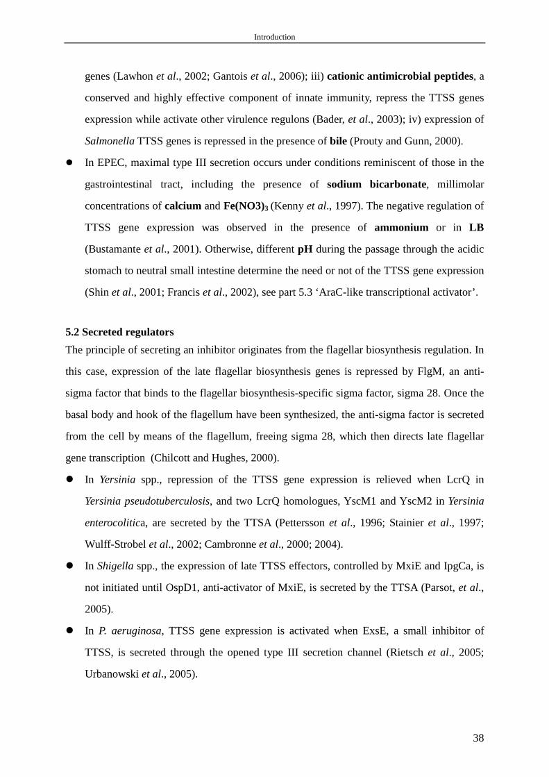
Introduction
38
genes (Lawhon et al., 2002; Gantois et al., 2006); iii) cationic antimicrobial peptides, a
conserved and highly effective component of innate immunity, repress the TTSS genes
expression while activate other virulence regulons (Bader, et al., 2003); iv) expression of
Salmonella TTSS genes is repressed in the presence of bile (Prouty and Gunn, 2000).
� In EPEC, maximal type III secretion occurs under conditions reminiscent of those in the
gastrointestinal tract, including the presence of sodium bicarbonate, millimolar
concentrations of calcium and Fe(NO3)3 (Kenny et al., 1997). The negative regulation of
TTSS gene expression was observed in the presence of ammonium or in LB
(Bustamante et al., 2001). Otherwise, different pH during the passage through the acidic
stomach to neutral small intestine determine the need or not of the TTSS gene expression
(Shin et al., 2001; Francis et al., 2002), see part 5.3 ‘AraC-like transcriptional activator’.
5.2 Secreted regulators
The principle of secreting an inhibitor originates from the flagellar biosynthesis regulation. In
this case, expression of the late flagellar biosynthesis genes is repressed by FlgM, an anti-
sigma factor that binds to the flagellar biosynthesis-specific sigma factor, sigma 28. Once the
basal body and hook of the flagellum have been synthesized, the anti-sigma factor is secreted
from the cell by means of the flagellum, freeing sigma 28, which then directs late flagellar
gene transcription (Chilcott and Hughes, 2000).
� In Yersinia spp., repression of the TTSS gene expression is relieved when LcrQ in
Yersinia pseudotuberculosis, and two LcrQ homologues, YscM1 and YscM2 in Yersinia
enterocolitica, are secreted by the TTSA (Pettersson et al., 1996; Stainier et al., 1997;
Wulff-Strobel et al., 2002; Cambronne et al., 2000; 2004).
� In Shigella spp., the expression of late TTSS effectors, controlled by MxiE and IpgCa, is
not initiated until OspD1, anti-activator of MxiE, is secreted by the TTSA (Parsot, et al.,
2005).
� In P. aeruginosa, TTSS gene expression is activated when ExsE, a small inhibitor of
TTSS, is secreted through the opened type III secretion channel (Rietsch et al., 2005;
Urbanowski et al., 2005).

Introduction
39
The underlying principle in all these systems is the same: a type III secreted negative
regulator is at the top of the subsequent regulatory cascade though the details of the regulation
appear to differ among the systems.
5.3 AraC-like transcriptional activators
AraC family, is known by virtue of their homology to a 99 amino acid segment of AraC, the
first transcriptional activator described. Proteins of AraC family contain two potential α-
helix-turn-α-helix DNA binding motifs and regulate diverse bacterial functions, including
sugar catabolism, responses to stress, and virulence (Gallegos et al., 1997; Martin and Rosner,
2001).
� In Yersinia, VirF from Y. enterocolitica (Cornelis et al., 1987), and LcrF from Y. pestis
(Yother et al., 1986) and Y. pseudotuberculosis (Forsberg and Wolf-Watz, 1988), belong
to the AraC family. VirF acts as a key transcriptional activator of the expression of TTSS
genes, such as yop and yscA-L, by specially binding to the promoters of these genes
(Hueck, 1998). As discussed in part 5.1.1, VirF is only active at 37°C and its expression
is modulated by a histone-like protein called Yersinia modulator A (YmoA) (Cornelis et
al., 1991).
� In S. flexneri, VirF is an AraC-like positive regulator of the secondary regulatory gene
virB and the structural gene icsA. The product of the virB gene in turn activates
transcription of the genes coding for the invasion proteins, and for the TTSA which
promotes export of the invasion proteins to the bacterial cell surface (Dorman et al.,
2001).
� In S. typhimurium, HilD and HilC, two AraC/XylS family members, activate both
indirectly, via HilA, and directly the TTSS gene expression (Schechter et al., 1999;
Akbar et al., 2003). InvF, a third AraC-like regulator encoded by the first gene of the inv-
spa gene cluster, is required for the optimal expression of several genes encoding SPI1-
secreted proteins (Kaniga et al., 1994; Darwin and Miller, 1999; Eichelberg and Galan,
1999). RtsA, a fourth AraC-like regulator, increases expression of the TTSS genes by
inducing hilA expression (Ellermeier and Slauch, 2003).

Introduction
40
� In EPEC, which is a major cause of infantile diarrhea in a number of developing
countries and is the prototype of pathogenic bacteria that cause attaching and effacing
(A/E) intestinal lesions (Nataro and Kaper, 1998), PerA (Gomez-Duarte and Kaper, 1995;
Porter et al., 2004) and GadX (Shin et al., 2001), two members of the AraC/XylR family
of bacterial transcriptional regulators, coordinately regulate the expression of acid
tolerance and TTSS genes, because in order to colonize or invade the intestinal
epithelium, EPEC must first overcome the hostile acidic environment encountered during
passage through the stomach (Francis, et al., 2002). In acidic conditions such as in the
stomach, GadX (glutamate decarboxylases) activates gadAB, genes expressing acid
tolerance, but represses PerA (plasmid encoded regulator), activator of Ler (LEE encoded
regulator) that is required for the TTSS genes expression (Mellies et al., 2006), to ensure
survival. After entry into the duodenum, where ingested food contents are deacidified and
digested by pancreatic juice, GadX represses gadAB and activates PerA to promote a
cascade of expression of TTSS genes, which are encoded by a pathogenicity island
termed locus of enterocyte effacement (LEE) and are required for the successful
colonization of the small intestine.
� In P. aeruginosa, AraC-like ExsA is the master activator for TTSS gene expression, see
below (6.1.1) for more details.
5.4 Regulatory chaperones
� In Yersinia spp., both chaperone LcrH and SycH play a role in the regulation of TTSS
genes expression together with their substrate YopD and LcrQ/YscM respectively
(Anderson et al., 2002; Wulff-Strobel et al., 2002; Cambronne et al., 2000; 2004).
� In S. flexneri, IpgC, chaperone of effectors IpaB and IpaC, binds to MxiE and allows
MxiE to bind DNA target sites and activate late TTSS genes when IpgC has been
liberated after the secretion of its substrates (Mavris et al., 2002; Parsot et al., 2005).
� In S. typhimurium, the type III secretion chaperone SicA binds and activates InvF, a
regulator that is responsible for the transcription of the SPI-1 type III secretion genes in
this organism (Darwin and Miller, 2001). This result implied a model whereby SicA is
bound to its secretion substrates, SipB and SipC, when S. typhimurium are not secreting.
When secretion is triggered, SipB and SipC are transported out of the cell, freeing SicA
to bind InvF and induce transcription of TTSS genes (Miller, 2002).

Introduction
41
� In P. aeruginosa, ExsC, chaperone of secreted regulator ExsE, acts as an anti-anti-
activator in the regulation of TTSS genes expression, see part 6.1.1 for more details.
5.5 Two-components regulatory system
Two-component system (TCS) is one of the most fundamental and widespread mechanisms of
signal perception/transduction in prokaryotes to respond to changes in their surroundings. A
canonical TCS consists of a histidine kinase sensor and a cognate response regulator.
Signaling through TCS begins with the autophosphorylation of a sensor kinase at a conserved
histidine residue, usually in response to an extracellular signal. This phosphate is ultimately
the substrate of a phosphotransfer reaction to a conserved aspartate residue in a cognate
response regulator domain/protein. Phosphorylation induces a conformational change in the
regulatory domain that results in activation of an associated domain that effects the response
(Stock et al., 2000).
� In Y. enterocolitica, YsrS/YsrR (Walker and Miller, 2004), a putative TCS, is required
for the NaCl-dependent expression of Ysa TTSS, which is the 2nd Y. enterocoliticais
TTSS encoded by chromosomal DNA and have only been detected in vitro when cells are
cultured at 26°C in a high-NaCl medium (Haller et al., 2000).
� In S. sonnei, CpxA/CpxR is involved in regulation of TTSS gene expression through
posttranscriptional processing of the regulator VirB, a master regulator of Shigella spp.
TTSS genes (Mitobe et al., 2005). OmpR/EnvZ, another TCS, appears to modulate TTSS
genes expression according to environmental conditions (Bernardini et al., 1990).
� In S. typhimurium, PhoP/PhoQ (Pegues et al., 1995), represses the synthesis of prgHIJK
(PhoP-repressed gene) transcript, which, together with InvG, is required for the assembly
of TTSA (Kubori et al., 2000, Marlovits et al., 2004). In fact, PhoP/PhoQ control
prgHIJK expression indirectly by regulation of the expression of HilA, which is the
master regulator of TTSS expression in Salmonella spp. (Bajaj et al., 1995; 1996;
Eichelberg and Galan, 1999; Lostroh and Lee, 2001). BarA/SirA, another two-component
sensor kinase and a response regulator, activates TTSS genes expression via hilA and
hilC (Teplitski et al., 2003).
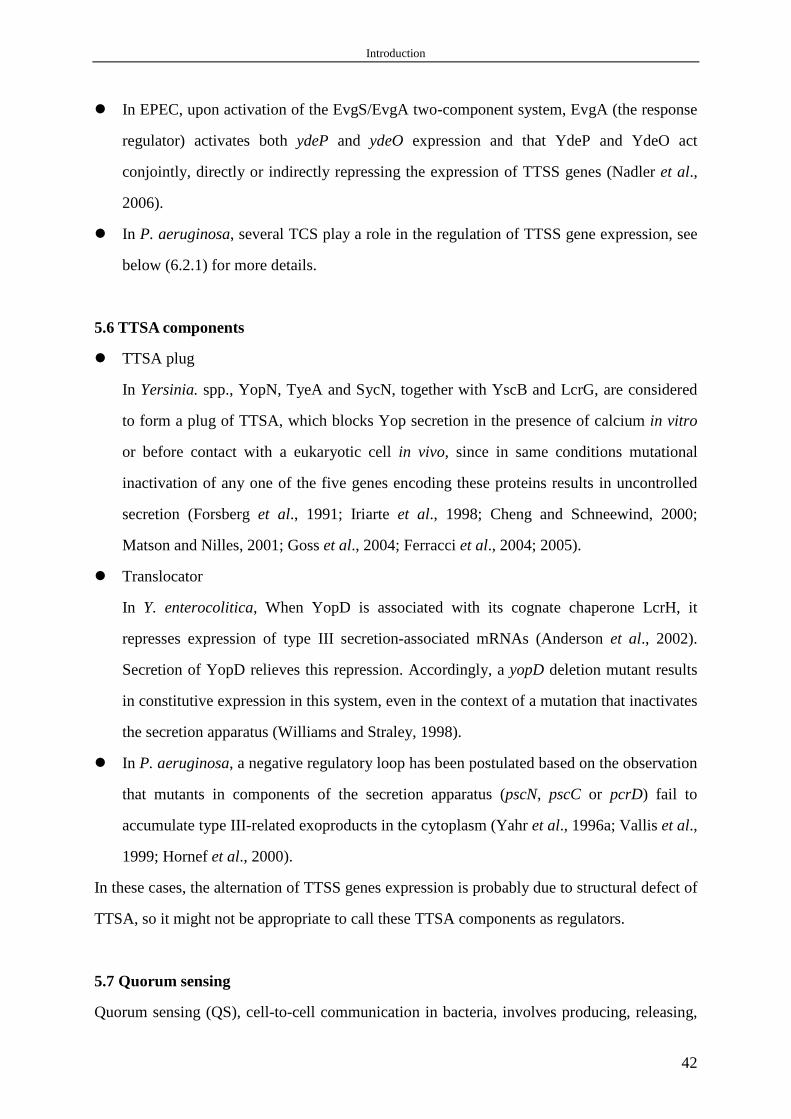
Introduction
42
� In EPEC, upon activation of the EvgS/EvgA two-component system, EvgA (the response
regulator) activates both ydeP and ydeO expression and that YdeP and YdeO act
conjointly, directly or indirectly repressing the expression of TTSS genes (Nadler et al.,
2006).
� In P. aeruginosa, several TCS play a role in the regulation of TTSS gene expression, see
below (6.2.1) for more details.
5.6 TTSA components
� TTSA plug
In Yersinia. spp., YopN, TyeA and SycN, together with YscB and LcrG, are considered
to form a plug of TTSA, which blocks Yop secretion in the presence of calcium in vitro
or before contact with a eukaryotic cell in vivo, since in same conditions mutational
inactivation of any one of the five genes encoding these proteins results in uncontrolled
secretion (Forsberg et al., 1991; Iriarte et al., 1998; Cheng and Schneewind, 2000;
Matson and Nilles, 2001; Goss et al., 2004; Ferracci et al., 2004; 2005).
� Translocator
In Y. enterocolitica, When YopD is associated with its cognate chaperone LcrH, it
represses expression of type III secretion-associated mRNAs (Anderson et al., 2002).
Secretion of YopD relieves this repression. Accordingly, a yopD deletion mutant results
in constitutive expression in this system, even in the context of a mutation that inactivates
the secretion apparatus (Williams and Straley, 1998).
� In P. aeruginosa, a negative regulatory loop has been postulated based on the observation
that mutants in components of the secretion apparatus (pscN, pscC or pcrD) fail to
accumulate type III-related exoproducts in the cytoplasm (Yahr et al., 1996a; Vallis et al.,
1999; Hornef et al., 2000).
In these cases, the alternation of TTSS genes expression is probably due to structural defect of
TTSA, so it might not be appropriate to call these TTSA components as regulators.
5.7 Quorum sensing
Quorum sensing (QS), cell-to-cell communication in bacteria, involves producing, releasing,

Introduction
43
detecting, and responding to small hormone-like molecules termed autoinducers allowing
bacteria to monitor the environment and to alter behavior in a population density-dependent
manner in response to changes in the number and/or species present in a community (Waters
and Bassler, 2005).
� In Y. enterocolitica, production of the Yop proteins in the wild type and in a QS
mutant is indistinguishable (Throup et al., 1995).
� In S. flexneri, maximal expression of TTSS gene and maximal activity of TTSA occur
at high cell density. However, autoinducer 2 (AI-2), a QS signaling molecule active in
late log phase of S. flexneri, does not influence TTSS gene expression (Day and
Maurelli, 2001).
� In EPEC, QS controls TTSS genes transcription and protein secretion (Sperandio et
al., 1999). Such known regulators include AI-2 (Sperandio et al., 1999), SdiA
(Kanamaru et al., 2000), an E. coli homologue of QS regulators, and QseA, quorum-
sensing E. coli regulator A (Sperandio et al., 2002). QseA shares homology with
several hypothetical P. aeruginosa proteins and PtxR, which positively regulates
exotoxin A (Hamood et al., 1996).
� In P. aeruginosa, QS has an important impact on the TTSS expression, see part 6.2.3
for more details.
Taken together, expression of TTSS genes is controlled by multicomponent regulatory
networks which integrate a diverse set of environmental cues, probably to restrict the energy-
consuming expression of 20 or more proteins to the correct place and time only when they are
really needed (Hueck, 1998). Additional regulatory factors implicated in the expression of
TTSSs of several animal pathogens are listed in Table 2.
6. Regulation of TTSS of P. aeruginosa
The TTSS of P. aeruginosa is induced by at least three environmental signals (Yahr and
Frank, 1994; Frank, 1997; Vallis et al., 1999, Hornef et al., 2000): i) in vivo contact with
eukaryotic host cells; ii) in vitro removal of calcium from medium; iii) the presence of serum
(due to the existance of type III secretion factors, which were recently identified as albumin
and casein (Kim et al., 2005). However, a subset of P. aeruginosa TTSA component, encoded

Introduction
44
by the operon exsDPscB-L, and transcriptional activator ExsA, are constitutively expressed,
indicating the existence of an expression hierarchy similar to the ordered assembly of the
highly related flagellar structure (Wolfgang et al., 2003). Such scenario may facilitate the
subsequent assembly and localization of additional TTSA components for rapid secretion of
presynthesized or cotranslational proteins under TTSS-inducing conditions.
TTSS of P. aeruginosa is tightly transcriptionally and posttranscriptionally regulated.
6.1 ExsA-dependent regulation
6.1.1 Regulatory operon exsCEBA
� ExsA, a member of the AraC family of transcriptional regulators, exhibits 56% overall
amino acid identity to the Yersinia VirF/LcrF proteins (Frank, 1997). ExsA functions as a
central transcriptional regulator of TTSS because it is required for the expression of all
TTSS genes, including itself, by binding to the consensus sequence TXAAAAXA, which
is located approximately 51 or 52 bp upstream of the transcriptional start site of four
operons (pscN-U, popNpcr1234DR, pcrGVHpopBD, exsDpscB-L) encoding the TTSA
and translocation machinery, the operon exsCEBA, and the effector proteins (Hovey and
Frank, 1995). ExsA is constitutively expressed (Wolfgang et al., 2003), but little
information is available about this control.
� ExsC, a type three secretion chaperone of ExsE, functions as an anti-anti-activator due to
the fact that ExsC binds to and sequesters the anti-activator, ExsD, under TTSS-inducing
conditions, thus freeing ExsA to activate the expression of TTSS genes (Dasgupta et al.,
2004).
� ExsE, identified by two independent groups, is a secreted regulator of TTSS in P.
aeruginosa (Rietsch et al., 2005; Urbanowski et al., 2005). When the secretion channel is
closed, ExsE is complexed with its chaperone, ExsC, in the cytoplasm, and transcription
of the TTSS is repressed by sequestration of ExsA by ExsD. On the contrary, when the
secretion channel is opened, ExsE is secreted, leaving ExsC free to interact with ExsD
and releasing ExsA, thereby allowing liberated ExsA to activate transcription of the
TTSS genes (Fig. 4).

Introduction
45
� exsB, 3rd gene of the operon exsCEBA, is not translated. Untranslated exsB region
mediates either the stability or the translation of exsA (Goranson et al., 1997).
FIG. 4. A model for the coupling of transcription with type III secretory activity in P. aeruginosa. All
proteins except ExsE (E) and PcrV (V) are indicated by name. The type III secretion apparatus is depicted
spanning the inner membrane (IM) and outer membrane (OM). Expression of exoS under inducing condition is
indicated by a wavy arrow. Reprinted from Rietsch et al., 2005.
6.1.2 ExsD
ExsD was identified as the first negative regulator of P. aeruginosa TTSS (McCaw et al.,
2002). Bacterial two-hybrid data indicated that ExsD interacts with the ExsA and functions as
an antiactivator to regulate the expression of TTSS genes. ExsD is essential to prevent
inappropriate expression of TTSS genes in conditions such as in the absence of the
appropriate environmental cue for secretion or in mutants lacking a functional TTSA.
6.1.3 PtrA
PtrA, Pseudomonas type III repressor A, can repress TTSS genes expression by inhibition of
the ExsA function through a direct binding. The expression of PtrA is highly and specifically
High Ca2+:Secretion channel is closed
A BLow Ca2+:Secretion channel is open
High Ca2+:Secretion channel is closed
A BLow Ca2+:Secretion channel is open

Introduction
46
induced by high copper signal, for instance during the infection of mouse burn wound,
through a CopR-CopS two-component regulatory system to shut down energy-expensive
TTSS (Ha et al., 2004).
6.2 ExsA-independent regulation
6.2.1 Two-components regulatory system
� RetS, regulator of exopolysaccharide and type III secretion (Goodman et al., 2004;
Zolfaghar et al., 2005), and RtsM, regulator of type III secretion (Laskowski et al., 2004),
are the same protein identified independently by different groups. RetS, an unusual
hybrid two-component signaling protein with a sensor kinase domain followed by two
response regulator receiver domains in tandem, is required for expression of TTSS genes
and for repression of genes responsible for exopolysaccharide components of the P.
aeruginosa biofilm matrix. Recent study also showed that, an intact phosphoacceptor site
within receiver domain 2, but not receiver domain 1, is absolutely required for RetS
activity, and periplasmic domain and phosphorylation of receiver domain 1 may inhibit
RetS activation. Furthermore, RetS may serve as a substrate for another sensor kinase
(Laskowski and Kazmierczak, 2006).
� LadS, another hybrid sensor kinase, and GacS/GacA, a canonical TCS, together with
RetS, are all involved in reciprocal regulation of TTSS genes expression and
exopolysaccharide synthesis (Ventre et al., 2006). Signals transduced by these three
sensor kinase proteins are hypothesized to ultimately control the levels of a small
regulatory RNA, RsmZ, which in turn regulates the reciprocal expression of genes
involved in TTSS and biofilm formation by titrating the amount of free, or active RsmA
in the cell (Goodman et al., 2004; Ventre et al., 2006; Burrowes et al., 2006). RsmA is a
global regulator, which appears to control gene expression at the posttranscriptional level
and positively regulates TTSS gene expression (see part 6.2.4) (Heurlier et al., 2004;
Burrowes et al., 2005; Mulcahy et al., 2006). Such a system would allow for P.
aeruginosa to switch between patterns of gene expression that characterize acute
infection versus chronic colonization; however, both the signals that are sensed by this
system as well as the biochemical relationships between these various regulators remain

Introduction
47
uncharacterized. It is possible that RetS and LadS may respond to carbohydrates of host
or bacterial origin, because the periplasmic domain of both proteins belongs to a class of
bacterial periplasmic sensor modules, 7TMR-DISMED2, which have been identified in a
variety of carbohydrates binding proteins (Ventre et al., 2006).
� SadARS, a three-component regulatory system, is comprised of a putative sensor
histidine kinase, SadS, and two response regulators, SadA and SadR. SadARS may either
promote or repress TTSS genes expression under different circumstances (Kuchma et al.,
2005).
� CopR-CopS, a canonical TCS, can sense the copper stress during the infection of mouse
burn wound and induce the expression of PtrA to shut down energy-expensive TTSS
genes expression in such special conditions (Ha et al., 2004).
Screening the P. aeruginosa genome has led to the identification of 64 response regulators and
63 histidine kinases, as well as 16 atypical kinases (Rodrigue et al., 2000). However, the
functions of the majority of these regulatory proteins have not been established yet.
6.2.2 Metabolic factors
� Mutants lacking pyruvate dehydrogenase (aceA or aceB), or a glucose transport regulator
(gltR) fail to induce the TTSS expression (Dacheux et al., 2002, Wolfgang et al., 2003).
� In the presence of histidine in the medium, excessive uptake and degradation of histidine
due to the overexpression of histidine utilization genes, for instance hutT, abolishes the
ability to induce exoS expression and rends the bacteria non-cytotoxic (Rietsch et al.,
2004). The cytotoxicity defect can be partially suppressed by an insertion in cbrA, gene
that encodes the sensor kinase of CbrAB two-component regulatory system involved in
sensing and responding to a carbon-nitrogen imbalance.
� A dose-dependent upregulation of TTSS gene expression is seen with increasing NaCl
concentrations (Hornef et al., 2000). Increasing the concentration of salt in the medium
correlates with an increase in the percentage of cells that are able to induce exoS
expression. It seems that exoS expression is in fact controlled by osmolarity because salt
could be replaced by sucrose in these experiments. Furthermore, one metabolite
controlling the TTSS expression might be derived from acetyl-CoA (Rietsch and

Introduction
48
Mekalanos, 2006). Though induction of exoS expression by removal of calcium does not
work in minimal glucose media, addition of caseamino acids but not just glutamate is
able to totally restore its induction (Rietsch and Mekalanos, 2006).
6.2.3 Quorum sensing
QS is crucial in the pathogenesis of P. aeruginosa infections and controls virulence factor
gene expression in the lungs of CF patients (Juhas et al., 2005; Erickson et al., 2002). The two
intimately linked QS systems in P. aeruginosa involve an interaction between a small
diffusible molecule, an acylhomoserine lactone (AHL), and a transcriptional activator. The las
system (Gambello and Iglewski, 1991), consists of the transcriptional activator LasR and the
AHL synthase LasI, which directs the synthesis of N-3-oxo-dodecanoyl-homoserine lactone
(3-oxo-C12-HSL); the rhl system (Latifi et al., 1995), consists of the transcriptional regulator
RhlR and the AHL synthase RhlI, which directs the synthesis of N-butanoyl-homoserine
lactone (C4-HSL). The third QS system in P. aeruginosa involves a non-AHL-related signal,
2-heptyl-3-hydroxy-4-quinolone, P. aeruginosa quinolone signal (PQS) (Pesci et al., 1999).
PQS adds a further level of complexity to the QS network and provides a link between the las
and rhl systems. QS systems in P. aeruginosa are hierarchically arranged with the las system
being on top of the signalling cascade and have been shown to activate the expression of a
wide variety of virulence genes (Juhas et al., 2005).
� Expression of TTSS genes, except operon exsCEBA, is negatively regulated by
RhlI/RhlR, but not LasI/LasR QS system (Hogardt et al., 2004; Bleves et al., 2005) as
well as by the stationary phase sigma factor RpoS (Hogardt et al., 2004). Although the
mechanism is unknown, it has been proposed that TTSS would not be necessary for
bacteria embedded in a biofilm during chronic infection, as most bacterial cells will not
have any contact with the host cells.
� Analysis of 5 QS-deficient clinical isolates producing either no or very low level of AHL
signals showed that, only one isolate produced normal TTSS effector proteins ExoS and
ExoT, two isolates produced ExoT only, and two isolates produced no effector proteins
(Schaber et al., 2004).

Introduction
49
6.2.4 Others
� RsmA, regulator of secondary metabolites, is a small RNA-binding protein and bind to,
and affect the translation rate of target mRNAs, a function that is further modulated by
one or more, small, untranslated competitive regulatory RNAs (Heeb et al., 2006). RsmA
plays an important role in the posttranscriptional regulation of a number of virulence-
related genes in P. aeruginosa (Pessi et al., 2001; Heurlier et al., 2004; Burrowes et al.,
2005) and positively regulates TTSS genes expression by controlling other TTSS
regulators (Mulcahy et al., 2006), because rsmA mutant results in decreased expression of
five positive regulators, including RetS, ExsA, ExsC, CyaB and Vfr, and one negative
regulator, ExsD (Mulcahy et al., 2006), and increased production of QS signal C4-HSL
(Pessi et al., 2001) and multidrug resistance efflux pump MexEF-OprN (Burrowes et al.,
2006). All these effects, except that on ExsD, make RsmA a positive regulator of the
expression of TTSS genes.
RsmA is highly homologous (85% identity) to Salmonella CsrA, which has been shown
to be a positive regulator of invasion genes and components of the TTSS (Lawhon et al.,
2003).
� A membrane-associated adenylcyclase (CyaB), responsible for cAMP synthesis, and a
cAMP-binding CRP homologue called Vfr are shown to be required for the expression of
TTSS genes (Wolfgang et al., 2003; Smith et al., 2004; Lory et al., 2004). Modulation of
intracellular cAMP seems to be an important mechanism controlling TTSS expression.
� truA, gene encoding pseudouridinase enzyme, is required for the expression of type III
secretory genes. Pseudouridination of tRNAs is proposed to be critical for the translation
of TTSS genes or their regulators (Ahn et al., 2004).
� dsbA, gene encoding a periplasmic thiol/disulphide oxidoreductase affects expression of
multiple virulence factors, including TTSS genes. This is probably a non-specific effect
resulting from abnormal protein folding caused by the lack of disulphide bonds (Ha et al.,
2003).
� FimL, homologous to the N-proximal domain of the complex chemosensory protein
ChpA, regulates TTSS genes expression by intersecting with Vfr-modulated pathways
(Whitchurch et al., 2005).

Introduction
50
� Overexpression of the MexCD-OprJ and MexEF-OprN, multidrug resistance efflux
pumps, is associated with a reduction of TTSS genes expression. Since these pumps can
extrude a wide range of compounds belonging to different structural family, it is possible
that intracellular signal needed for the activation of TTSS genes expression is extruded
(Linares et al., 2005).
The regulation of TTSS expression in P. aeruginosa is briefly summarized in Fig. 5.
FIG. 5. Regulation of TTSS expression in P. aeruginosa. The expression of the operons encoding TTSS
regulators, apparatus, translocon and effectors is under the control of the positive transcriptional regulator ExsA.
Other main factors implicated in the regulation of TTSS expression are indicated.
ExsApopNpcr1234DR
pcrGVHpopBD
exsDpscB-L
pscN-U
exoSTYU
Appara
tus
Translocon
Effectors
ExsD
ExsC
ExsE
PtrACopRS
[Cu2+]
SadARS
RpoS RhlI/R
RetS GacSA LadS
RsmA
CyaB
Vfr
exsCEBA
Re
gulators
PDH
Contact with host cell
Calcium depletion
Serum(albumin and casein)
Activation Repression
RsmZ
ExsApopNpcr1234DR
pcrGVHpopBD
exsDpscB-L
pscN-U
exoSTYU
Appara
tus
Translocon
Effectors
popNpcr1234DRpopNpcr1234DR
pcrGVHpopBDpcrGVHpopBD
exsDpscB-LexsDpscB-L
pscN-UpscN-U
exoSTYUexoSTYU
Appara
tus
Translocon
Effectors
ExsD
ExsC
ExsE
PtrACopRS
[Cu2+]
SadARS
RpoS RhlI/R
RetS GacSA LadS
RsmA
CyaB
Vfr
exsCEBAexsCEBA
Re
gulators
PDH
Contact with host cell
Calcium depletion
Serum(albumin and casein)
Activation Repression
RsmZ

Introduction
51
INTRODUCTION (Résumée en français)
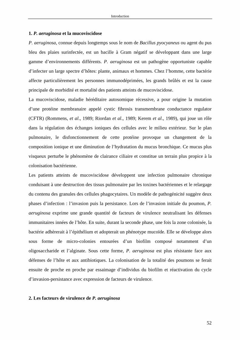
Introduction
52
1. P. aeruginosa et la mucoviscidose
P. aeruginosa, connue depuis longtemps sous le nom de Bacillus pyocyaneus ou agent du pus
bleu des plaies surinfectée, est un bacille à Gram négatif se développant dans une large
gamme d’environnements différents. P. aeruginosa est un pathogène opportuniste capable
d’infecter un large spectre d’hôtes: plante, animaux et hommes. Chez l’homme, cette bactérie
affecte particulièrement les personnes immunodéprimées, les grands brûlés et est la cause
principale de morbidité et mortalité des patients atteints de mucoviscidose.
La mucoviscidose, maladie héréditaire autosomique récessive, a pour origine la mutation
d’une protéine membranaire appelé cystic fibrosis transmembrane conductance regulator
(CFTR) (Rommens, et al., 1989; Riordan et al., 1989; Kerem et al., 1989), qui joue un rôle
dans la régulation des échanges ioniques des cellules avec le milieu extérieur. Sur le plan
pulmonaire, le disfonctionnement de cette protéine provoque un changement de la
composition ionique et une diminution de l’hydratation du mucus bronchique. Ce mucus plus
visqueux perturbe le phénomène de clairance ciliaire et constitue un terrain plus propice à la
colonisation bactérienne.
Les patients atteints de mucoviscidose développent une infection pulmonaire chronique
conduisant à une destruction des tissus pulmonaire par les toxines bactériennes et le relargage
du contenu des granules des cellules phagocytaires. Un modèle de pathogénicité suggère deux
phases d’infection : l’invasion puis la persistance. Lors de l’invasion initiale du poumon, P.
aeruginosa exprime une grande quantité de facteurs de virulence neutralisant les défenses
immunitaires innées de l’hôte. En suite, durant la seconde phase, une fois la zone colonisée, la
bactérie adhèrerait à l’épithélium et adopterait un phénotype mucoïde. Elle se développe alors
sous forme de micro-colonies entourées d’un biofilm composé notamment d’un
oligosaccharide et l’alginate. Sous cette forme, P. aeruginosa est plus résistante face aux
défenses de l’hôte et aux antibiotiques. La colonisation de la totalité des poumons se ferait
ensuite de proche en proche par essaimage d’individus du biofilm et réactivation du cycle
d’invasion-persistance avec expression de facteurs de virulence.
2. Les facteurs de virulence de P. aeruginosa
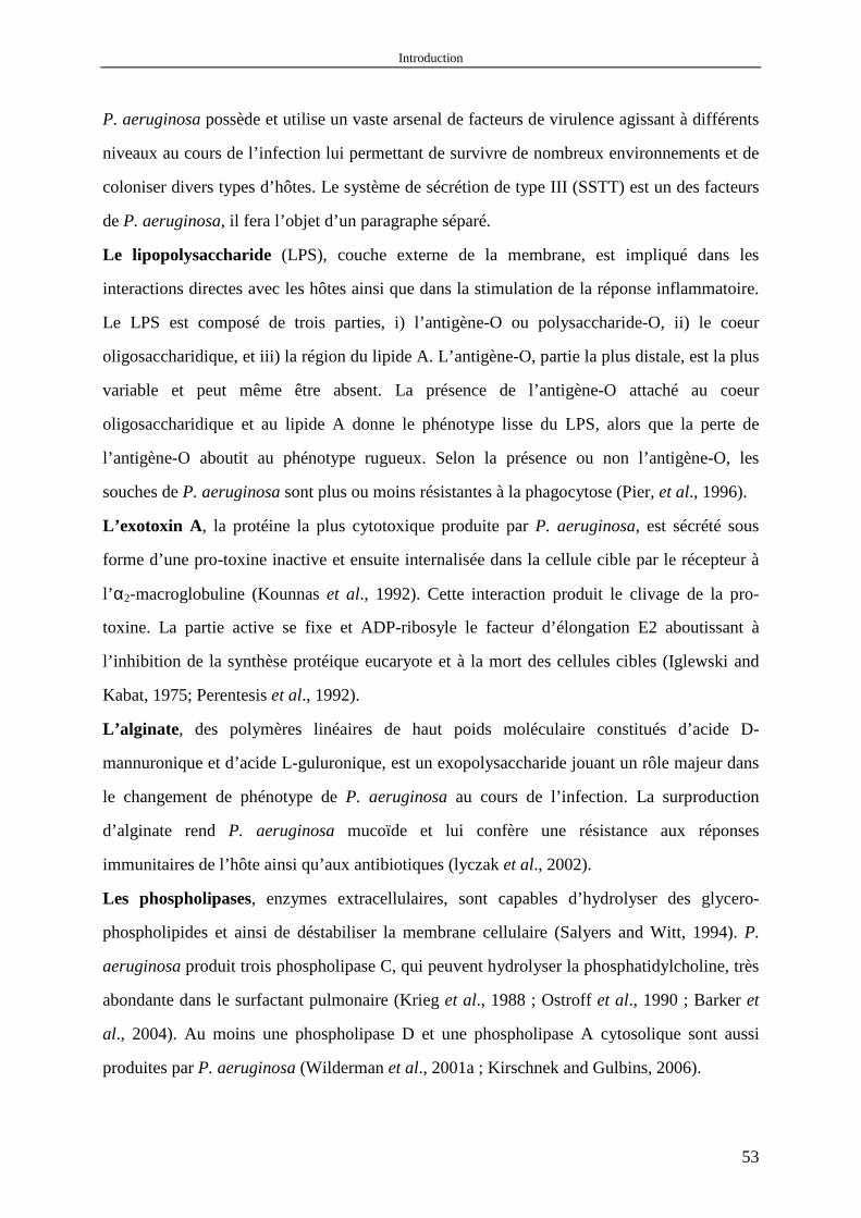
Introduction
53
P. aeruginosa possède et utilise un vaste arsenal de facteurs de virulence agissant à différents
niveaux au cours de l’infection lui permettant de survivre de nombreux environnements et de
coloniser divers types d’hôtes. Le système de sécrétion de type III (SSTT) est un des facteurs
de P. aeruginosa, il fera l’objet d’un paragraphe séparé.
Le lipopolysaccharide (LPS), couche externe de la membrane, est impliqué dans les
interactions directes avec les hôtes ainsi que dans la stimulation de la réponse inflammatoire.
Le LPS est composé de trois parties, i) l’antigène-O ou polysaccharide-O, ii) le coeur
oligosaccharidique, et iii) la région du lipide A. L’antigène-O, partie la plus distale, est la plus
variable et peut même être absent. La présence de l’antigène-O attaché au coeur
oligosaccharidique et au lipide A donne le phénotype lisse du LPS, alors que la perte de
l’antigène-O aboutit au phénotype rugueux. Selon la présence ou non l’antigène-O, les
souches de P. aeruginosa sont plus ou moins résistantes à la phagocytose (Pier, et al., 1996).
L’exotoxin A , la protéine la plus cytotoxique produite par P. aeruginosa, est sécrété sous
forme d’une pro-toxine inactive et ensuite internalisée dans la cellule cible par le récepteur à
l’ α2-macroglobuline (Kounnas et al., 1992). Cette interaction produit le clivage de la pro-
toxine. La partie active se fixe et ADP-ribosyle le facteur d’élongation E2 aboutissant à
l’inhibition de la synthèse protéique eucaryote et à la mort des cellules cibles (Iglewski and
Kabat, 1975; Perentesis et al., 1992).
L’alginate , des polymères linéaires de haut poids moléculaire constitués d’acide D-
mannuronique et d’acide L-guluronique, est un exopolysaccharide jouant un rôle majeur dans
le changement de phénotype de P. aeruginosa au cours de l’infection. La surproduction
d’alginate rend P. aeruginosa mucoïde et lui confère une résistance aux réponses
immunitaires de l’hôte ainsi qu’aux antibiotiques (lyczak et al., 2002).
Les phospholipases, enzymes extracellulaires, sont capables d’hydrolyser des glycero-
phospholipides et ainsi de déstabiliser la membrane cellulaire (Salyers and Witt, 1994). P.
aeruginosa produit trois phospholipase C, qui peuvent hydrolyser la phosphatidylcholine, très
abondante dans le surfactant pulmonaire (Krieg et al., 1988 ; Ostroff et al., 1990 ; Barker et
al., 2004). Au moins une phospholipase D et une phospholipase A cytosolique sont aussi
produites par P. aeruginosa (Wilderman et al., 2001a ; Kirschnek and Gulbins, 2006).

Introduction
54
Les rhamnolipides, biosurfactants glycolipidiques, peuvent inhiber la fonction ciliaire de
l’épithélium respiratoire humain et se trouver dans les crachats des patients atteints de
mucoviscidose (Hastie et al., 1986; Kownatzki et al., 1987). Cependant, le rôle exact des
rhamnolipides dans les infections aiguës ou chroniques reste obscur.
Les protéases, qui agissent sur un grand nombre de protéines dont l’élastine, un composant
majeur du tissu pulmonaire, sont impliquées dans l’invasion tissulaire (Cowell et al., 2003).
P. aeruginosa peut produire quatre protéases majeures, y compris LasB (Bever and Iglewski,
1988), lasA (Olson JC and Ohman DE, 1992), AprA (Okuda et al., 1990) et protéase IV
(Wilderman et al., 2001b). Il semble que l’ensemble de ces 4 protéases harmonise l’invasion
tissulaire (Matsumoto et al., 2004).
Les pili, situées sur la membrane externe, sont des structures filamenteuses et sont importants
pour l’adhésion aux cellules cibles (Woods et al., 1980). Les pili interviennent aussi dans une
mobilité de type ‘twiching’ (déplacement sur des surfaces) et dans le développement du
biofilm (O'Toole and Kolter, 1998; Mattick et al., 2002).
Le flagelle, organelle de mobilité, est constituée de trois principales parties, le filament, le
crochet et le corps basal (Fig. 2). Des travaux sur modèle murin montrent clairement qu’un
mutant dépourvu de flagelle est atténué dans sa virulence (Drake and Montie, 1988). Le
flagelle est aussi impliqué dans le développement du biofilm (O'Toole and Kolter, 1998 ;
Mattick et al., 2002).
3. Les différents systèmes de sécrétion bactériens
La sécrétion des facteurs de virulence est réalisée grâce à différents mécanismes qui
permettent le transport actif de protéines à travers les membranes bactériennes. On pourrait
diviser les différents systèmes de sécrétion en deux groupes selon l’utilisation ou non du
système sec, qui transporte des protéines contenant une séquence N-terminale clivable du
cytoplasme vers l’espace périplasmique (Economou, 1999).
3.1 La sécrétion sec-dépendante
Le système de sécrétion de type II, la mieux connue des voies de sécrétion, s’effectue en
deux étapes. La première étape consiste en un transport des protéines du cytoplasme vers le

Introduction
55
périplasme par le système sec. La deuxième étape correspond à l’exportation vers le milieu
extérieur à travers la membrane externe par une voie différente de la première. L’exemple le
plus étudié est la sécrétion de pullulanase par Klebsiella oxytoca (Fig. 1). Chez P. aeruginosa,
le système de sécrétion de type II, également appelé Xcp, est impliqué dans la sécrétion de
nombreuses enzymes et toxines, telles que l'élastase, l'exotoxine A, la lipase, les
phospholipases ou la phosphatase alcaline (Filloux et al., 1990; 1998).
L’autotransporteur , lui-même contenant non seulement la séquence N-terminale clivable
mais aussi l’information pour le passage à travers la membrane externe, peut passer la
membrane externe par lui-même (Kostakioti et al., 2005) (Fig. 1). Chez P. aeruginosa,
l'estérase EstA a été identifiée comme autotransporteur (Wilhelm et al., 1999) et deux autres
homologues ont été proposés (Ma et al., 2003).
Le système de sécrétion chaperonne/usher, dédié à l’assemblage et la sécrétion de pili,
réalise son rôle en harmonisant la fonction de 2 composants: une chaperonne et une protéine
membranaire, ‘usher’. L’exemple le plus étudié est l’assemblage les pili de type I par E. coli
(Fig. 1). Chez P. aeruginosa, ce système, appelé Cup, est impliqué dans la formation du
biofilm à la phase initiale (Vallet et al., 2001).
3.2 La sécrétion sec-indépendante
Système de sécrétion de type I, aussi appelé ABC-transporteur, permet le transport de
protéines directement du cytoplasme vers le milieu extérieur (Binet et al., 1997). Ce système
est constitué 3 complexes protéiques: une protéine de transport avec une activité ATPase dans
la membrane interne, une protéine formant un pore dans la membrane externe, et une protéine
liant les deux premières (Fig. 1). Chez P. aeruginosa, la protéase alcaline est sécrétée par le
système de sécrétion de type I ( Guzzo et al., 1991).
Système de sécrétion de type IV, absent chez P. aeruginosa, est capable de transporter une
grande variété de substrats, y compris protéines, DNA et de complexes nucléoprotéiques.
L’exemple le plus étudié est la sécrétion de fragment T du plasmide Ti (T-DNA)
d’Agrobacterium tumefaciens (Cascales and Christie, 2003) (Fig. 1).
Système de sécrétion de type III, par sa composition et son fonctionnement, est le plus
complexe des systèmes de sécrétion bactériens connus et est présent dans de nombreux genres

Introduction
56
de bacilles à Gram négatif dont Yersinia, Salmonella, Shigella, les Escherichia
entéropathogènes, etc (Hueck, 1998). La sécrétion de type III est indépendante du système
sec, cependant son assemblage requiert probablement le système sec, car plusieurs
composants possèdent des séquences signal similaires à celles trouvées dans le système sec
(Table 1). L’appareil de sécrétion de type III est composé de plus de vingtaine de protéines et
la plupart des protéines de la membrane interne sont homologues aux protéines de l’appareil
de biosynthèse du flagelle des bactéries Gram négatif et positif (Table 1). Activé par le
contact avec la cellule eucaryote, ce système est dédié à l’injection d’effecteurs directement
du cytoplasme de la bactérie dans le cytosol de la cellule cible. Ces effecteurs ont pour effets
la mort cellulaire ou le dysfonctionnement de la cellule en détruisant la membrane, en
perturbant les cascades de transduction des signaux ou en désorganisant le cytosquelette
(Hueck, 1998). Ils permettent aux bactéries pathogènes d’envahir les tissus de l’organisme
infecté ou de circonscrire ses défenses. Les SSTTs sont conservés au niveau structural entre
les bactéries à Gram négatif et présentent de grandes similitudes avec le corps basal du
flagelle (Fig. 2) (Tampakaki et al., 2004).
4. Le système de sécrétion de type III de P. aeruginosa
4.1 L’organisation génétique
A l’exception des gènes codant les exotoxines, les gènes codant les protéines du SSTT sont
regroupés en clusters sur chromosome contrairement au genre Yersinia pour lequel le SSTT
est situé sur un plasmide. Ils sont organisés en 5 opérons. L’opéron exsCEBA code les
principaux régulateurs du SSTT, les opéron pscN-U, popNpcr1234DR, exsDpscB-L codent la
structure de l’appareil de sécrétion, permettant le passage des effecteurs à travers les
membranes bactériennes, et l’opéron pcrGVHpopBD code des protéines indiquées dans
l’appareil de translocation, permettant l’injection des effecteurs directement dans le cytosol de
la cellule cible eucaryote (Table 1) (Frank, 1997).
4.2 Effecteurs
Quatre effecteurs sécrétés par le SSTT sont connus chez P. aeruginosa : ExoS, ExoT, ExoY
and ExoU. Toutes les souches ne possèdent pas tous les effecteurs (Feltman et al., 2001).
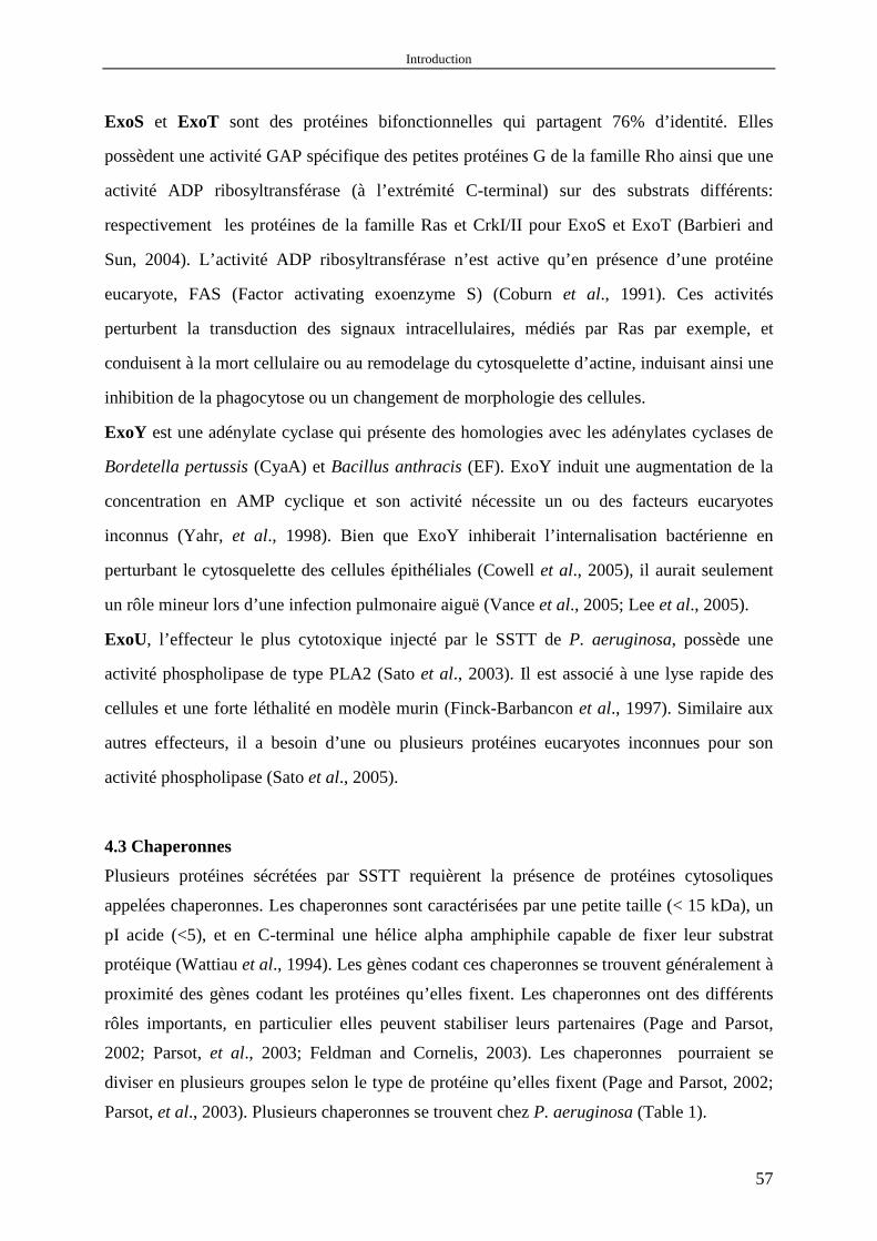
Introduction
57
ExoS et ExoT sont des protéines bifonctionnelles qui partagent 76% d’identité. Elles
possèdent une activité GAP spécifique des petites protéines G de la famille Rho ainsi que une
activité ADP ribosyltransférase (à l’extrémité C-terminal) sur des substrats différents:
respectivement les protéines de la famille Ras et CrkI/II pour ExoS et ExoT (Barbieri and
Sun, 2004). L’activité ADP ribosyltransférase n’est active qu’en présence d’une protéine
eucaryote, FAS (Factor activating exoenzyme S) (Coburn et al., 1991). Ces activités
perturbent la transduction des signaux intracellulaires, médiés par Ras par exemple, et
conduisent à la mort cellulaire ou au remodelage du cytosquelette d’actine, induisant ainsi une
inhibition de la phagocytose ou un changement de morphologie des cellules.
ExoY est une adénylate cyclase qui présente des homologies avec les adénylates cyclases de
Bordetella pertussis (CyaA) et Bacillus anthracis (EF). ExoY induit une augmentation de la
concentration en AMP cyclique et son activité nécessite un ou des facteurs eucaryotes
inconnus (Yahr, et al., 1998). Bien que ExoY inhiberait l’internalisation bactérienne en
perturbant le cytosquelette des cellules épithéliales (Cowell et al., 2005), il aurait seulement
un rôle mineur lors d’une infection pulmonaire aiguë (Vance et al., 2005; Lee et al., 2005).
ExoU, l’effecteur le plus cytotoxique injecté par le SSTT de P. aeruginosa, possède une
activité phospholipase de type PLA2 (Sato et al., 2003). Il est associé à une lyse rapide des
cellules et une forte léthalité en modèle murin (Finck-Barbancon et al., 1997). Similaire aux
autres effecteurs, il a besoin d’une ou plusieurs protéines eucaryotes inconnues pour son
activité phospholipase (Sato et al., 2005).
4.3 Chaperonnes
Plusieurs protéines sécrétées par SSTT requièrent la présence de protéines cytosoliques
appelées chaperonnes. Les chaperonnes sont caractérisées par une petite taille (< 15 kDa), un
pI acide (<5), et en C-terminal une hélice alpha amphiphile capable de fixer leur substrat
protéique (Wattiau et al., 1994). Les gènes codant ces chaperonnes se trouvent généralement à
proximité des gènes codant les protéines qu’elles fixent. Les chaperonnes ont des différents
rôles importants, en particulier elles peuvent stabiliser leurs partenaires (Page and Parsot,
2002; Parsot, et al., 2003; Feldman and Cornelis, 2003). Les chaperonnes pourraient se
diviser en plusieurs groupes selon le type de protéine qu’elles fixent (Page and Parsot, 2002;
Parsot, et al., 2003). Plusieurs chaperonnes se trouvent chez P. aeruginosa (Table 1).
Introduction
58
4.4 Signaux de sécrétion
Pour être sécrétés, les effecteurs de SSTT doivent d’abord être reconnus par l’appareil de
sécrétion. Trois différents types de signaux ont été proposés, y compris un 5’ mRNA, une
séquence N-terminale et les chaperonnes (Anderson and Schneewind, 1997; Cheng et al.,
1997; Lloyd et al., 2001; 2002; Birtalan et al., 2002; Ramamurthi and Schneewind, 2002;
2003a; 2003b; 2005). L’existence de plusieurs signaux de sécrétion pourraient rendre la
sécrétion plus efficace et déterminer la hiérarchie de la sécrétion, par exemple, les protéines
nécessaires à la formation de l’appareil de translocation devront être sécrétées avant les
effecteurs par l’appareil de sécrétion.
5. La régulation de l’expression du SSTT chez P. aeruginosa
Le SSTT de P. aeruginosa est activé in vivo lors du contact avec la cellule eucaryote, in vitro
lors de la déplétion calcique dans le milieu de culture et la présence de sérum (Yahr and
Frank, 1994; Frank, 1997; Vallis et al., 1999, Hornef et al., 2000; Kim et al., 2005). Pourtant,
même en absence des signaux d’activation, l’opéron exsDpscB-L (gènes codant l’appareil de
sécrétion) et exsA, le gène codant l’activateur transcriptionnel, ont une faible expression
(Wolfgang et al., 2003). En effet, l’ensemble des gènes constituant le SSTT de P. aeruginosa
est sous le contrôle d’un facteur transcriptionnel, ExsA, qui se trouve dans l’opéron exsCEBA.
ExsA coordonne l’expression des gènes de l’appareil de sécrétion, de l’appareil de
translocation et des effecteurs. Les réseaux de régulation contrôlant l’activation du SSTT
agissent principalement sur l’expression ou l’activité d’ExsA, de façon directe ou indirecte.
Par conséquent, on peut distinguer deux types de régulation: la régulation ExsA-dépendante
et la régulation ExsA-indépendante.
5.1 La régulation ExsA-dépendante
ExsA, homologue de VirF qui contrôle l’expression du SSTT chez Yersinia, est un membre
de famille des activateurs transcriptionnels de type AraC (Frank, 1997). Il possède une
activité de fixation ADN de type ‘α-hélice-tour-α-hélice’ et se fixe sur les promoteurs des
opérons codant les gènes du SSTT au niveau de la séquence consensus TXAAAAXA, à
environ 50 paires de base en amont du site d’initiation de la transcription (Hovey and Frank,
Introduction
59
1995). ExsA active sa propre synthèse en se fixant sur le promoteur de l’opéron exsCEBA,
créant ainsi une boucle de rétroaction positive. Une mutation du gène exsA conduisant à un
défaut d’expression ou une protéine non fonctionnelle, abolit l’expression des gènes du SSTT
et aboutit à une souche moins virulente. L’hypothèse d’une bistabilité dans le niveau
d’expression de ExsA a été récemment proposée (Filopon et al., 2006).
ExsD, codé par le première gène de l’opéron exsDpscB-L, est un anti-activateur du SSTT de
P. aeruginosa (McCaw et al., 2002). ExsD est capable de se fixer à ExsA et d’inhiber son
activité. Une surexpression d’ExsD induit une inhibition du SSTT et son absence une
dérépression de l’opéron exsCEBA et par conséquent de l’ensemble des gènes du SSTT.
ExsC, codé par le premier gène de l’opéron exsCEBA, est un anti-anti-activateur du SSTT de
P. aeruginosa par sa capacité à inhiber l’activité de ExsD en libérant ExsA (Dasgupta et al.,
2004). De plus, ExsC pourrait être la protéine chaperonne d’ExsE (Rietsch et al., 2005;
Urbanowski et al., 2005).
ExsE, positionné au sommet de la cascade de régulation centrée sur l’activité d’ExsA, est un
inhibiteur du SSTT de P. aeruginosa (Rietsch et al., 2005; Urbanowski et al., 2005). En effet,
les quatre protéines ExsA, ExsD, ExsC et ExsE forment une cascade de régulation sous la
dépendance d’ExsA. ExsE, au sommet de cette cascade, couple la transcription des gènes du
SSTT avec l’activation de la sécrétion. En absence d’activation, le canal de sécrétion est
fermé, ExsE est fixée à ExsC laissant ExsD jouer un rôle d’inhibiteur en se fixant à ExsA.
Lors de la présence des signaux d’activation, par exemple la déplétion calcique, ExsE est
sécrété, ExsC libéré fixe alors ExsD et permet à ExsA d’induire la transcription des gènes du
SSTT (Fig. 4). L’expression des gènes du SSTT doit dépendre d’un équilibre fin entre les
concentrations des différents constituants de cette cascade.
PtrA , répresseur du SSTT de P. aeruginosa, pourrait inhiber en s’y liant, l’activité d’ExsA et
ainsi l’expression des gènes du SSTT. PtrA lui-même est induit par la présence de cuivre en
concentration élevée, ainsi que cela est observé lors de l’infection secondaire à une brûlure
(Ha et al., 2004).
5.2 La régulation ExsA-indépendante
D’autre facteurs, y compris i) les régulateurs à deux composants, par exemple RetS
Introduction
60
(Goodman et al., 2004; Zolfaghar et al., 2005; Laskowski et al., 2004), LadS et GacS/GacA
(Ventre et al., 2006); ii) les facteurs métaboliques, par exemple la pyruvate déhydrogénase
(PDH) (Dacheux et al., 2002); iii) le quorum sensing, par exemple RhlI/RhlR (Hogardt et al.,
2004; Bleves et al., 2005); iv) les régulateurs globaux, par exemple Vfr (Wolfgang et al.,
2003); pourraient aussi être impliqués dans la régulation de l’expression des gènes du SSTT
chez P. aeruginosa (Fig. 5).
6. Objectif du travail
Les objectifs de ce travail sont d’une part de caractériser les protéines impliquées dans la
régulation transcriptionnelle de exsCEBA et, d’autre part d’identifier des signaux régulateurs
de type ‘quorum sensing’.

Materials and Methods
61
MATERIALS AND METHODS

Materials and Methods
62
1. Bacterial media • Luria-Bertani Medium (LB medium)
o Tryptone (GibcoBRL, Paisley, Scotland) 10g o Yeast extract (GibcoBRL) 5g o NaCla (VWR International, Fontenay sous Bois, France) 10g Add 1 liter of deionized H2O from Millipore, ultra-pure water system (Millipore SAS, Molsheim, France). Shake until the solutes have dissolved (adjust the pH to 7.0 with 10 N NaOH if necessary). Autoclave at 121°C for 15 minutes. Sterile media can be stored at room temperature. aIf not indicated, the chemical reagents used in this study were either from VWR International or from Sigma, St. Louis, MO, USA.
• Vogel-Bonner (VB) minimal medium (Vogel and Bonner, 1956) o D-(+)-Glucose (C6H12O6) 4.5g (25mM) o Citric acid (C6H8O7.H2O) 2g (9.5mM) o Sodium amnonium phosphate (NaNH5PO4.4H2O) 3.5g (16.7mM) Add 980 ml of tap water (not deionized H2O). Shake until the solutes have dissolved. Adjust the pH to 7.2 with 10 N NaOH ( ~ 3ml). Sterilize by filtration through a 0.22 µM filter (Millex GP, Cork, Ireland).
• LB agar plate o Tryptone 10g o Yeast extract 5g o NaCl 10g o Agar (GibcoBRL) 15g Add 1 liter of deionized H2O. Shake until the solutes have dissolved (adjust the pH to 7.0 with 5 N NaOH, if necessary). Autoclave at 121°C for 15 minutes. When the medium is recovered from the autoclave, swirl it gently to distribute the melted agar evenly throughout the solution. Allow the medium to cool to 50-60°C before adding appropriate antibiotics (Table 3). Plates can then be poured directly from the flask using about 20 ml of medium per 90-mm plate (Dominique Dutscher, Brumath, France). When the medium has hardened completely, invert the plates and store them at 4°C until needed (as fresh as possible, no more than 2 months). The plates should be incubated at 37°C for 1 hour before they are used.
• Pseudomonas Isolation Agar (PIA) plate Suspend 45g of the PIA (Difco laboratories, Detroit, USA) powder in 1 liter of deionized H2O supplemented with 20 ml of glycerol. Prepare the PIA plate as described for LB agar plate.
TABLE 3. Antibiotic solutions Stock solutiona Working concentration Pseudomonas aeruginosa, CHA Escherichia coli LB PIA plate LB/LB agar plate Carbenicillin (Cb) 300 mg ml-1 300 µg ml-1 600 µg ml-1 Gentamicin (Gm) 80 mg ml-1 200 µg ml-1 400 µg ml-1 10/20 µg ml-1 Tetracycline (Tc)b 20 mg ml-1 250 µg ml-1 250 µg ml-1 10/20 µg ml-1 Ampicillin (Amp) 100 mg ml-1 100 µg ml-1 Kanamycin (Kana) 50 mg ml-1 50 µg ml-1 aStock solutions of antibiotics dissolved in H2O (except tetracycline here) should be sterilized by filtration through a 0.22 µM filter. Store antibiotic solutions at -20°C. bTetracycline should be dissolved in ethanol and don’t need be sterilized. Store stock solution in light-tight containers. Magnesium ions are antagonists of tetracycline. Use media without magnesium salts (e.g., LB medium) for selection of bacterial resistant to tetracycline. 2. Bacterial strains and plasmids All bacterial strains and plasmids used in this study are listed in Table 4.

Materials and Methods
63
TABLE 4. Bacterial strains and plasmids used in this study Strains or plasmids Descriptiona Source Strains
E. coli DH5α used for plasmid transformation Stratagene E. coli BL21(DE3) used for recombinant protein production Stratagene E. coli S17-1 used for mating constructs into P. aeruginosa Simon et al.,1983 CHA mucoid CF isolate Toussaint et al., 1993
∆exsA exsA::Gm mutant of CHA Dacheux et al., 1999 CHA (pClux) lux fusion with exsCEBA promoter intergrated at the attB site of the CHA chromosome This study CHA (pSlux) lux fusion with exoS promoter at the attB site of the CHA chromosome This study ∆psrA CHA containing an in-frame deletion of amino acids 14-194 of psrA ORF This study ∆psrA (pClux) lux fusion with exsCEBA promoter at the attB site of the ∆psrA chromosome This study ∆psrA (pSlux) lux fusion with exoS promoter at the attB site of the ∆psrA chromosome This study ∆rpoS CHA containing an in-frame deletion of amino acids 1-207 of rpoS ORF This study ∆rpoS (pClux) lux fusion with exsCEBA promoter at the attB site of the ∆rpoS chromosome This study ∆psrA (pPsrA) (pClux) ∆psrA (pClux) complemented by psrA This study ∆psrA (pPsrA) (pSlux) ∆psrA (pSlux) complemented by psrA This study ∆psrA (pDD2) (pClux) ∆psrA (pClux) containing pDD2 vector This study ∆psrA (pDD2) (pSlux) ∆psrA (pSlux) containing pDD2 vector This study ∆lasI CHA containing an in-frame deletion of amino acids 1-155 of lasI ORF This study ∆lasI (pClux) lux fusion with exsCEBA promoter at the attB site of the ∆lasI chromosome This study ∆rhlI CHA containing an in-frame deletion of amino acids 20-201 of rhlI ORF This study ∆rhlI (pClux) lux fusion with exsCEBA promoter at the attB site of the ∆rhlI chromosome This study ∆lasI rhlI CHA containing two in-frame deletions of both lasI ORF and rhlI ORF This study ∆lasI rhlI (pClux) lux fusion with exsCEBA promoter at the attB site of the ∆lasIrhlI chromosome This study ∆phnAB CHA containing an in-frame deletion of amino acids 1-530 and 1-172 of phnA and
phnB ORF respectively This study
∆phnAB (pClux) lux fusion with exsCEBA promoter at the attB site of the ∆phnAB chromosome This study ∆trpA CHA containing an in-frame deletion of amino acids 36-247 of trpA ORF This study ∆trpA (pClux) lux fusion with exsCEBA promoter at the attB site of the ∆trpA chromosome This study ∆trpA (pSlux) lux fusion with exoS promoter at the attB site of the ∆trpA chromosome This study ∆virA CHA containing an in-frame deletion of amino acids 234-677 of virA ORF This study ∆virA (pClux) lux fusion with exsCEBA promoter at the attB site of the ∆virA chromosome This study ∆virA (pSlux) lux fusion with exoS promoter at the attB site of the ∆virA chromosome This study
Plasmids
pTOPO Blunt end cloning vector, Apr Invitrogen pIApC pUCP20-derived vector containing exsCEBA promoter Dacheux et al., 2001 pDD2 pUCP20 derived vector containing exsA gene; Apr Dacheux et al., 1999 pEX100Tlink Allelic replacement suicide vector; Apr(Cbr) Quenee et al., 2005 pUCGmlox pUC18-based vector containing the aacC1 gene flanked by lox; Apr, Gmr Quenee et al., 2005 pCM157 Cre recombinase; Tcr Marx and Lidstrom,
2002 Mini-CTX -lux mini-CTX-lux ; Tcr Laskowski et al., 2004 Mini-CTX-exsCp-lux mini-CTX-lux containing exsCEBA promoter ; Tcr Laskowski et al., 2004 mini-CTX-exoSp-lux mini-CTX-lux containing exoS promoter ; Tcr Laskowski et al., 2004 pFLP2 Flp recombinase; Apr(Cbr) Hoang et al., 1998 pUCP20 Escherichia-Pseudomonas shuttle vector ; Apr(Cbr) West et al., 1994 pUC18 For Pseudomonas genomic DNA cloning; Apr Yanisch-Perron et al.,
1985 pexsAind pUCP20-derived vector containing IPTG-inducilbe exsA; Apr(Cbr) Filopon et al., 2006 pppsrA pTOPO-derived vector containing psrA promoter This study ppfleQ pTOPO-derived vector containing fleQ promoter This study pphpA-Rec pET15b-derived vector containing phpA; Apr This study ppsrA-Rec pET15b-derived vector containing psrA; Apr This study pvfr-Rec pET15b-derived vector containing vfr; Apr This study
pPsrA pUCP20-derived vector containing psrA ORF as well as its promoter region; Apr(Cbr) This study pKO-psrA pEX100Tlink-derived vector containing up- and downstream region of psrA and
Gmlox; Apr, Gmr This study
pKO-rpoS pEX100Tlink-derived vector containing up- and downstream region of rpoS and Gmlox; Apr, Gmr
This study
pKO-lasI pEX100Tlink-derived vector containing up- and downstream regions of lasI and Gmlox; Apr, Gmr
This study
pKO-rhlI pEX100Tlink-derived vector containing up- and downstream regions of rhlI and Gmlox; Apr, Gmr
This study
pKO-phnAB pEX100Tlink-derived vector containing up- and downstream regions of phnAB and Gmlox; Apr, Gmr
This study
pKO-trpA pEX100Tlink-derived vector containing up- and downstream regions of trpA and Gmlox; Apr, Gmr
This study
pKO-virA pEX100Tlink-derived vector containing up- and downstream regions of virA and Gmlox; Apr, Gmr
This study
aApr, ampicillin resistance ; Gmr, gentamicin resistance ; Tcr, tetracycline resistance ; Cbr, carbenicillin resistance; KO, knock out.

Materials and Methods
64
2.1 Strain construction 2.1.1 Strain containing chromosomal transcriptional gene reporter In order to study precisely the level of transciption of exsCEBA and exoS promoters, we used the system developped by Laskowsky and colleagues to construct transcriptional gene reporter. In brief, the plasmids mini-CTX-exsCp-lux and mini-CTX-exoSp-lux were mobilized into Pseudomonas strain by mating and vector backbone sequences were excised by Flp recombinase as previously described (Fig. 6) (Hoang et al., 1998). Each reporter construct is therefore present as a single, unmarked copy integrated at the chromosomal attB site (Laskowski et al., 2004). FIG. 6. Construction of transcriptional gene reporter. Adapted from Hoang et al., 1998. MATERIALS_________________________________________________________ -Bacterial strains
• P. aeruginosa wild-type CHA • E.coli S17-1
-Plasmids • mini-CTX-exsCp-lux (a kind gift of Dr. Kazmierczak) • mini-CTX-exoSp-lux (a kind gift of Dr. Kazmierczak) • pFLP2 (a kind gift of Pr. Schweizer) Attention, genebank access N° for mini-CTX-lux and pFLP2 are AF140579 and 048702, respectively.
-Agar plate • LB (Tetracycline, 20 µg ml-1) • PIA (Tetracycline, 250 µg ml-1)
mini-CTX-pClux12838
luxE
luxB
luxA
luxD
lucC
int
tet
attP
pC
FRT
FRT
CHA chromosome
pClux
mini-CTX-pClux12838
luxE
luxB
luxA
luxD
lucC
int
tet
attP
pC
FRT
FRT
CHA chromosome
pClux
CHA chromosome
pCluxpClux

Materials and Methods
65
• PIA (Carbenicillin, 600 µg ml-1) • LB without salt
METHOD____________________________________________________________ -Preparation of E.coli S17-1 containing mini-CTX-exsCp-lux or mini-CTX-exoSp-lux
1. Transform E.coli S17-1 by mini-CTX-exsCp-lux and mini-CTX-exoSp-lux and select in LB agar plate containing tetracycline (20 µg ml-1)
-Mating between P. aeruginosa wild-type CHA and E.coli S17-1 2. Mix at 1:1 with 50 µl of exponential phase (OD600 ≈ 0.5) CHA and E.coli S17-1.
Deposit the mixture on the surface of LB agar without salt. Incubate the plate overnight in the incubator at 37°C.
3. Transfer the bacteria into a microfuge tube containing 1 ml of LB. 4. Spread 100 µl bacteria (OD600 is from 1 to 2 ) on the PIA containing tetracycline (250
µg ml-1). Incubate the plate overnight in the incubator at 37°C. -Excision of vector backbone sequences by Flp recombinase
5. Electroporate the mated bacteria with pFLP2. Select on PIA containing carbenicillin (600 µg ml-1). Incubate the plate overnight in the incubator at 37°C.
-Remove pFLP2 plasmid 6. Culture the bacteria (Cbr) in 2ml of LB without antibiotic overday. Inoculate again in
2 ml of LB overnight. Repeat another overday and overnight. 7. Select the bacteria (Tcs, Cbs). 8. Verify the construct by EGTA activation test. IMPORTANT : To obtain mutant containing pClux and pSlux, we begin by mating CHA
(pClux) [or CHA (pSlux)] with E. Coli S17-1 containing plasmid pKO-gene (Fig. 7), though we can also begin by mating mutant with E. Coli S17-1 containing gene reporter pClux or pSlux. 2.1.2 Mutants Study of gene function is often carried out by deleting genes in the strains of interest and observing the phenotypic differences between the parental and deleted strains. We used cre-lox system developped in our lab combining the sacB-based positive selection to construct mutants (Fig. 7) (Quenee et al., 2005). The advantage of cre-lox method is that lox-flanked antibiotic marker (aacC1, for gentamicin resistance) can be removed from the bacterial genome by site-specific recombinase encoded by the cre gene. MATERIALS_________________________________________________________ -Plasmids
• PKO-gene (Table 4) • pEX100Tlink (∆HindIII) (Quenee et al., 2005 ).
This vector contains 1) the counterselectable sacB marker, 2) an oriT for conjugation-mediated plasmid transfer and 3) multiple cloning sites. SacB gene allows for positive selection of the segregation of true mutants from the more fequently occuring merodiploids on medium containing 5% sucrose (Levansucrase , encoded by sacB, can produce toxic products from sucrose; mutants with simple combination could not survive in the medium containing sucrose due to the presence sacB; only mutants with double combination could survive in the 5% sucrose due to the lost of sacB, Schweizer and Hoang. 1995).
• pUCGmlox • pCM157
-Bacteria strains • CHA • E.coli S17-1
-Agar plate
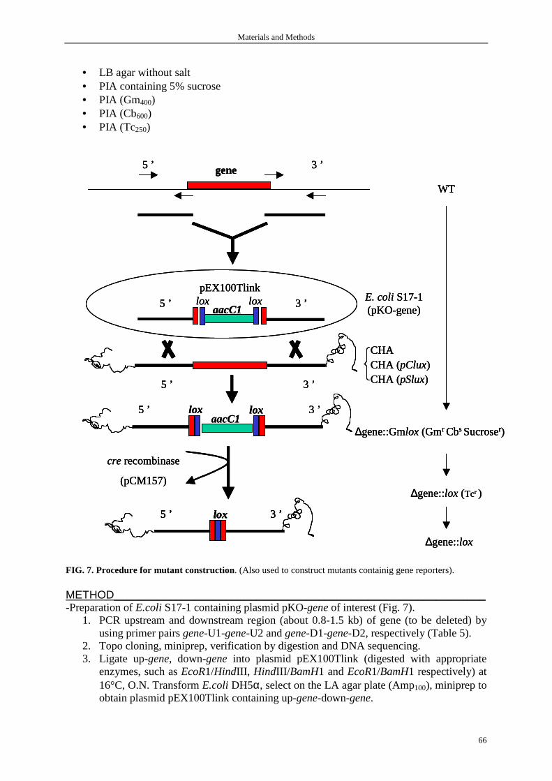
Materials and Methods
66
• LB agar without salt • PIA containing 5% sucrose • PIA (Gm400) • PIA (Cb600) • PIA (Tc250)
FIG. 7. Procedure for mutant construction. (Also used to construct mutants containig gene reporters). METHOD____________________________________________________________ -Preparation of E.coli S17-1 containing plasmid pKO-gene of interest (Fig. 7).
1. PCR upstream and downstream region (about 0.8-1.5 kb) of gene (to be deleted) by using primer pairs gene-U1-gene-U2 and gene-D1-gene-D2, respectively (Table 5).
2. Topo cloning, miniprep, verification by digestion and DNA sequencing. 3. Ligate up-gene, down-gene into plasmid pEX100Tlink (digested with appropriate
enzymes, such as EcoR1/HindIII, HindIII/ BamH1 and EcoR1/BamH1 respectively) at 16°C, O.N. Transform E.coli DH5α, select on the LA agar plate (Amp100), miniprep to obtain plasmid pEX100Tlink containing up-gene-down-gene.
∆gene::Gmlox (Gmr Cbs Sucroser)
WT
∆gene::lox (Tcr )
∆gene::lox
aacC1lox5 ’ 3 ’lox
5 ’ 3 ’gene
lox5 ’ 3 ’
cre recombinase
(pCM157)
5 ’ 3 ’
CHACHA (pClux)CHA (pSlux)
aacC1loxlox5 ’ 3 ’
pEX100TlinkE. coli S17-1 (pKO-gene)
∆gene::Gmlox (Gmr Cbs Sucroser)
WT
∆gene::lox (Tcr )
∆gene::lox
∆gene::Gmlox (Gmr Cbs Sucroser)
WT
∆gene::lox (Tcr )
∆gene::lox
aacC1lox5 ’ 3 ’lox
aacC1lox5 ’ 3 ’lox
5 ’ 3 ’gene5 ’ 3 ’gene
lox5 ’ 3 ’
cre recombinase
(pCM157)
lox5 ’ 3 ’lox5 ’ 3 ’
cre recombinase
(pCM157)
5 ’ 3 ’
CHACHA (pClux)CHA (pSlux)
aacC1loxlox5 ’ 3 ’
pEX100TlinkE. coli S17-1 (pKO-gene)
5 ’ 3 ’
CHACHA (pClux)CHA (pSlux)
aacC1loxlox5 ’ 3 ’
pEX100TlinkE. coli S17-1 (pKO-gene)

Materials and Methods
67
TABLE 5. Primer sequences used in this study Primer Sequences* Description psrA-U1 5’-AGAATTC CGCCGTCGTCATTCGCCGA-3’ Amplification of upstream region flanking psrA psrA-U2 5’-TAAGCTT CCGCATCGAGAATGCGTTCG-3’ psrA-D1 5’-TAAGCTT GGTGATGCACCTGATGGTGC-3’ Amplification of downstream region flanking psrA psrA-D2 5’-AGGATCCCATCGACAGCTCCCTCAATC-3’ rpoS-U1 5’-TAGGATCCAGACCCTGAAAGGCGCC-3’ Amplification of upstream region flanking rpoS rpoS-U2 5’-AGAAGCTTCGCTGGGTTTTCTGGC -3’ rpoS-D1 5’-ATAAGCTTCTCGTCCAGCATGATGCC -3’ Amplification of downstream region flanking rpoS rpoS-D2 5’-ATGAATTCGAAGACCTGCATCGCCG-3’ vfr-R1 5’-ACATATGGTAGCTATTACCCACACA-3’ Amplification of recombinant Vfr protein vfr-R2 5’-AGGATCCTCAGCGGGTGCCGAAGAC-3’ psrA-R1 5’-ACATATGGCCCAGTCGGAAACCG-3’ Amplification of recombinant PsrA protein psrA-R2 5’-AGGATCCTCAGGCCTTGGCGGGCGT-3’ psrA-C1 5’-AGAATTC ACTGGTGCGTCATCTGCCGC-3’ Amplification of fragment containing psrA ORF as well as its
promoter region psrA-C2 5’-AGGATCCTCAGGCCTTGGCGGGCGT-3’ ppsrA-F 5’-AGAATTCGGGGCGTCAGCTTCTGCAT-3’ Amplification of fragment containing psrA promoter region ppsrA-R 5’-AGAATTCAATGCGTTCGACGGTTTCCG-3’ pfleQ-F 5’-AGAATTCCTACCAGATGTTCGGATAAGT-3’ Amplification of fragment containing vfr promoter region pfleQ-R 5’-AGAATTCCGAATGGGTCTCGCTCGACC-3’ phpA-1 5’-ACATATGGAATTCCTTGTTAAAAGCGT-3’ Verification of ∆phpA phpA-2 5’-AGGATCCGGGTCACTTGGCCCGTTCC-3’ psrA-1 5’-ATGGCCCAGTCGGAAACCG-3’ Verification of ∆psrA psrA-2 5’-TCAGGCCTTGGCGGGCGT-3’ rpoS-1 5’-GCGAGTTGGTCATCATCAAAC-3’ Verification of ∆rpoS rpoS-2 5’-GCATCAAGTGCAAATACACCG-3’ lasI-U1 5’-ACGGATCCAAGTGGCTATGTCGCCG-3’ Amplification of upstream region flanking lasI lasI-U2 5’-ACAAGCTTCCGGACCCTTGCTAGGC-3’ lasI-D1 5’-AGAAGCTTGGCCTGGACGTATCGCG-3’ Amplification of downstream region flanking lasI lasI-D2 5’-GGGAATTCTCAGCCTGCCC-3’ rhlI -U1 5’-TATCTAGACGCGGTGCGCCGCAAGG-3’ Amplification of upstream region flanking rhlI rhlI -U2 5’-ATAAGCTTGCTCGGCGATCATGGCG-3’ rhlI -D1 5’-ATAAGCTTGCGTCATCGGGCGTTCC-3’ Amplification of downstream region flanking rhlI rhlI -D2 5’-CAGAGCTCTGTCCGGAAATCCTCATGC-3’ phnAB-U1 5’-ATGGATCCATGTTGAACCACGGTGTCC-3’ Amplification of upstream region flanking phnAB phnAB-U2 5’-ATAAGCTTTCGCCGCAATGGATGTCC-3’ phnAB-D1 5’-ATAAGCTTGATTCTCACCACCCACGG-3’ Amplification of downstream region flanking phnAB phnAB-D2 5’-ATGAATTCGGGAACTGACGATTGCAG-3’ trpA-U1 5’-AGAATTCTCAGATCAGCTCGTGGCGG-3’ Amplification of upstream region flanking trpA trpA-U2 5’-TAAGCTTCAGGCGGTCAAGGATGTCCT-3’ trpA-D1 5’-TAAGCTTCCAGGGAAGACGCGTAGT-3’ Amplification of downstream region flanking trpA trpA-D2 5’-TGGATCCGCACAAGATCAACAACTGC-3’ virA-U1 5’-AGGATCCCACACCGATGCAGAAGACCG-3’ Amplification of upstream region flanking virA virA-U2 5’-TAAGCTTGTCGTGGAGATAGTCGCGAT-3’ virA-D1 5’-TAAGCTTAAGGGCGTGGGCAAGGGCAC-3’ Amplification of downstream region flanking virA virA-D2 5’-AGAATTCTGTACCCATGCCTCGACCCT-3’ lasI-1 5’-GTGGATGCTCAAGAAGGACTAC-3’ Verification of ∆lasI lasI-2 5’-CTGATCGGCAACCTTACCC-3’ rhlI -1 5’-CGCGGCGCTGGGTCTCATC-3’ Verification of ∆rhlI rhlI -2 5’-GTAACCGCCCTCTTCCGTGCGG-3’ phnAB-1 5’-GATTCTCTCCCGCCAGGCG-3’ Verification of ∆phnAB phnAB -2 5’-CCATCGACGAGGAACTGAAG-3’ trpA-1 5’-TCAGCGTGCGTTGCGCACGC-3’ Verification of ∆trpA trpA-2 5’-AAGCATGAGCCGCCTGCAGA-3’ virA-1 5’-GTCGCGGATATTCTTCGGCG-3’ Verification of ∆virA virA-2 5’-TTGCCGATCTCCGAGTGGAT-3’ trpA-C1 5’-AGGATCCATGAGCCGCCTGCAGACCCG-3’ Amplification of complete trpA ORF for complementation trpA-C2 5’-TAAGCTTCAGCGTGCGTTGCGCACGC-3’ aacC1-F 5’-ATGTTACGCAGCAGCAACGA-3’ Amplification of aacC1 probe for southern blot aacC1-R 5’-CAAGTTCCCGAGGTAATCGG-3’ * Restriction enzyme sites are underlined
4. Ligate Gmlox (digested from plasmid pUCGmlox by HindIII) into HindIII digested
pEX100Tlink containing up-gene-down-gene at 16°C, O.N. Transform the ligate into E.coli DH5α and select on the LA agar plate (Gm10/Amp100). Miniprep to obtain plasmid pKOgene (pEX100Tlink-up-gene-Gmlox-down-gene).
5. Transform pKOgene (pEX100Tlink-up-gene-Gmlox-down-gene) into E.coli S17-1. -Mating between CHA/CHA (pClux)/CHA (pSlux) and E.coli S17-1 (pKO-gene)

Materials and Methods
68
6. Mix at 1:1 of 50 µl exponential phase (OD600 ≈ 0.5) CHA and E.coli S17-1 (pKO-gene). Deposit the mixture on the surface of LB agar without salt. Incubate the plate overnight in the incubator at 37°C.
7. Transfer the bacteria into a microfuge tube containing 1 ml of LB. 8. Spread 100 µl bacteria (OD600 is from 1 to 2 ) on the PIA (Gm400). Overnight, 37°C. 9. Inoculate 50 colonies on the PIA containing 5% sucrose. Overnight, 37°C.
In theory, only mutants with double combination could survive in the presence of 5% sucrose, but in fact all mutants survive after overnight’s incubation (with different size of the colonies).
10. Re-inoculate these 50 colonies on the PIA (Gm400) as well as PIA (Cb600). Overnight, 37°C.
11. Select colonies growing on the PIA(Gm400) but not on the PIA (Cb600) for further verification by PCR.
-Excision of Gmlox by Cre recombinase 12. Electroporate the PCR confirmed colonies (Gmr/Cbs) with plasmid pCM157 and select
on the PIA (Tc250). Overnight, 37°C. -Remove pCM157 plasmid
13. Culture the bacteria (Tcr) in 2ml of LB (without antibiotic) overday. Inoculate again in 2 ml of LB overnight. Repeat another overday and overnight.
14. Select the bacteria (Tcs, Gms). 15. Verify the construct by PCR.
2.1.3 Complementation construct A complemetation construct has to be constructed to confirm that the phenotype of the mutant is really due to the deletion of the gene but not due to the neighbouring genes that might be affected by a polar effect of this deletion. MATERIALS_________________________________________________________ -Primers
• psrA-C1 • psrA-C2
-plasmid pUCP20 -Materials needed for PCR, agarose gel electrophoresis, Topo cloning, Miniprep, digestion, ligation and transformation. METHOD____________________________________________________________
1. Carry out the PCR to produce a 782-bp fragment containing the psrA open reading frame (ORF) as well as 80 bp upstream encompassing the promoter region so that psrA is under the transcriptional control of its own promoter.
2. Topo cloning, miniprep, verification by digestion and DNA sequencing. 3. Digest and ligate this fragment inot pUCP20 (EcoR1-BamH1) to generate pPsrA. 4. Transform pPsrA into strain ∆psrA by electroporation.
2.2 Strain maintenance, storage, recovery, culture and quantitation MATERIALS_________________________________________________________ -Bacterial strains (Table 4) -Bacterial media (Materials and Methods 1) -Special equipments
• Protect Bacterial Preservers (TSC, Lancashire, UK) • Nunc Cryo Tube, 1.0 ml (NordicCell, Copenhagen, Denmark)
Materials and Methods
69
• Toothpick • Disposable bacterial loop/spreader (VWR International) • 14 ml of polypropylene round-bottom tube (Becton Dickinson labware, Meylan, France) • Microbiological safety cabinet class II (Claus Damm, Humleback, Denmark) • Incubator (Memmert, Schwabach, Germany) • Bacterial shaker: Novotron platform shaker (TLS, Auckland, New Zealand) • Spectrophotometer: Uvikon 860 (UVK-LAB, Trappes, France) • -86°C ULT Freezer (Thermo Forma, Marietta, OH, USA) • Autoclaver (Slli, Montpellier, France)
METHOD____________________________________________________________
• P.aeruginosa and E. coli strains were maintained on PIA plate and LB agar plate respectively with antibiotics as required (Table 3).
• P. aeruginosa strains were stored in Protect Bacterial Preservers at -80°C. Briefly, scrape enough P. aeruginosa from PIA palte and make a thich suspension in the cryopreservation fluid. Invert intensely the tube and stand it for several minutes. Withdraw as much as fluid as possible and store it at -80 °C.
• E. coli strains were stored at -80°C as 40% glycerol stocks in Nunc Cryo Tube. Briefly, add 0.5 ml of sterile 80 % glycerol (sterilized by autoclaving) to 0.5 ml of E.coli culture in Nunc Cryo Tube. Mix well and freeze the culture in ethanol-dry ice or in liquid nitrogen. Transfer the tube to -80 °C for long-term storage. 15% glycerol stock is proposed by Molecular Cloning (Sambrook and Russell, 2001).
• To recover the bacteria, scrape the frozen surface of E. coli (Nunc Cryo Tube) or pick a frozen bead of P. aeruginosa (Protect Bacterial Preservers) with a sterile bacterial spreader, and then immediately streak onto the surface of an LB agar plate or PIA plate containing the appropriate antibiotics. Incubate the plate overnight in the incubator at 37°C.
• Bacterial culture was usually performed in 2 ml of LB with a single colony picked from an agar plate. Grow the bacteria overnight in a shaker at 37°C with agitation of 260 rpm. Wash the overnight bacteria with LB and inoculate at 1:100 for another culture. The volume of culture is best not more than 1 tenth of that of the tube or the flask to assure enough air needed for the growth of the bacteria.
• Bacterial growth level or quantity were estimated by by optical density measuremnt at 600 nm (OD600) using spectrophotometer. We consider that there are 6×10⁸ P. aeruginosa in 1 ml of LB at an OD600 of 1.
2.3 Bacterial transformation Bacterial transformation is the process by which bacterial cells take up naked DNA molecules. DNA do not enter bacteria under their own power, but require assistance traversing the outer and inner cell membranes and in reaching an intracellular site where they can be expressed and replicated. Bacteria which are able to uptake foreign DNA are called ‘competent’ and are obtained either from a commercial source or by treatment with calcium chloride in the early log phase of growth. Transformation are carried either by chemical method, heat-shock method or by physical method, electroporation. 2.3.1 Chemical transformation of E.coli DH 5αααα by heat shock method MATERIALS_________________________________________________________ -DNA: ligated mixture,or plasmid, or PCR product, or digested genomic DNA, et al.

Materials and Methods
70
-Chemically competent DH5α (Invitrogen) -Thermomixer (Eppendorf, Hamburg, Germany) or a water bath preset to 42°C -SOC medium (Invitrogen) or LB medium -LB agar plate containing appropriate antibiotics METHOD____________________________________________________________
1. Add 1-3 µl of transforming DNA (up to 25 ng) in 50 µl of competent DH5α. Swirl the tube gently several times to mix their content. Do not mix by pipetting up and down. The DNA volume should not exceed 10 % of that of the competent cells.
2. Incubate on ice for 30 minutes. 3. Heat-shock the cells for 45 seconds at 42°C without shaking. 4. Transfer immediately the tube to ice and allow the cells to cool for 1-2 minutes. 5. Add 250 µl prewarmed SOC medium (or LB) and shake the tube at 37°C for 1 hour. 6. Spread 25 µl and 250 µl of transformed cells onto the prewarmed selective plates . 7. Incubate at 37°C overnight.
2.3.2 Transformation of P. aeruginosa by electroporation (Enderle and Farwell, 1998)
MATERIALS_________________________________________________________ -Electroporation cuvettes (BTX, San Diego, California,USA) -ECM 399 pulse generator (BTX) -Fresh P. aeruginosa from PIA (storing the plate at 4°C lowers the transformation efficiency) -Disposable bacterial loop/spreader (VWR) -Plasmid (1:10 diluted Miniprep in H2O or TE, 10 mM Tris-HCl, pH 8.0, 1mM EDTA) -PIA plate containing appropriate antibiotic -H2O (sterile, distilled and deionized) METHOD____________________________________________________________
1. Transfer the half loop of fresh P. aeruginosa to a 1.5 ml microcentrifuge tube containing 1 ml of H2O. Suspend the cells by pipetting up and down.
2. Centrifuge at 14 000 rpm for 1 min. Remove the supernatant and resuspend the cells in another 1 ml of H2O. Vortex to make a homogenos mixture.
3. Repeat step 2 for three times. Suspend the cells in 40 µl of ice H2O (OD600 is about 5, so there would be about 1 × 10⁸ bacteria). Keep on ice.
4. Add 1-3 µl plasmid (10-30 ng) and mix well with the cell. Incubate on ice for 30 minutes.
5. Add the mixture in the cold electroporation cuvette (-20°C for 30 minutes) and pulse the cuvette at 1800V in a ECM 399 pulse generator. Add immediately 1 ml of prewarmed LB and shake at 37°C for 1 hour. Cuvettes could be reused by rising in H2O and ethanol, air-drying and treating with UV light to inactivate any DNA.
6. Spread 50 µl and 500 µl of transformed cells onto the prewarmed PIA plates containing appropriate antibiotic. Transformation efficiency is about 1 transformant per 106 bacteria.
7. Incubate at 37°C overnight.

Materials and Methods
71
2.4 Plasmid manipulation 2.4.1 Miniprep Many methods have been developed to purify plasmids from bacteria. These methods invariably involve three steps: growth of the bacterial culture, harvesting and lysis of the bacteria and purification of the plasmid DNA. QIAprep Miniprep System provides a fast, simple, and cost-effective plasmid miniprep method for routine molecular biology laboratory applications. QIAprep Spin Miniprep kit using a microcentrifuge is designed for purification of up to 20 µg of high-copy plasmid DNA from 1-5 ml of overnight cultures of E. coli in LB medium. MATERIALS_________________________________________________________ -1-5 ml of overnight cultures of E. coli -QIAprep Spin Miniprep kit (Qiagen, Courtaboeuf, France) -Centrifuge (Eppendorf, Hamburg, Germany)
Relative centrifugal force (RCF)=1.1 × 10⁻⁶ (rpm)2r. The symbol r represents the radial distance (mm) from the center of the rotor to the point for which RCF is required. This is generally equivalent to rmax of the rotor. For example, rotor speed (rpm) = 80, 000 rpm and r = 20 mm, RCF can be calculated as 142, 080 g usig the equation given above.
METHOD____________________________________________________________ All procedures were performed according to the instructions of the manual of the kit. 2.4.2 Restriction enzyme digestion MATERIALS_________________________________________________________ -Restriction enzymes (Roche, Meylan, France or Invitrogen, Cergy pontoise, France) -10 × buffer for Restriction enzymes (Roche or Invitrogen) -Plasmids -Thermomixer (Eppendorf, Hamburg, Germany) or a water bath preset to 37°C METHOD____________________________________________________________
1. In a sterile 0.5 or 1.5 ml microfuge tube, mix: • Plasmid 0.1-0.5 µg (1-5 µl) • Restriction enzymes (10 U µl-1) 0.5 µl • 10 × buffer for restriction enzymes 1 µl • H2O to total volume of 10 µl
One unit is defined as the amount of enzyme required to hydrolyze 1 µg of λDNA to completion in 60 minutes in a total reaction volume of 50 µl, under optimal assay conditions (salt concentration, pH, temperature). Digestion is generally performed with a 5-fold excess of enzyme whose volume should not be more than 1 tenth of the total volume to avoid side effects, such as star activity.
2. Incubate at 37°C for 2h (mix 1-2 times during the incubation, centrifuge the tube if necessary).
3. Check the digestion by agarose gel electrophoresis (see Materials and Methods 6). 2.4.3 DNA ligation MATERIALS_________________________________________________________ -DNA (digested by same restriction enzyme(s) and purified from agarose gel)
• Plasmid • Insert DNA
-T4 DNA ligase (Invitrogen)

Materials and Methods
72
T4 DNA ligase joins fragments of DNA with compatible cohesive termini as well as blunt ended DNA fragments. DNA fragments with cohesive termini can be ligated at 4°C overnight by the addition of 0.01 to 0.1 Weiss units of enzyme per µg DNA. Blunt-ended DNA fragment ligation is usually carried out at 16°C with 1 to 2 Weiss units per µg DNA. DNA ligase catalyzed the formation of a phosphodiester bond between adjacent 3’-OH and 5’-P termini. This phosphodiester bond-forming reaction requires ATP hydrolysis. The 10 × incubation buffer provided in the kit containing ATP. Inorganic pyrophosphate and AMP are released as by-products.
-10 × buffer for T4 DNA ligase (Invitrogen) -Thermal cycler or a water bath preset to 16°C METHOD____________________________________________________________
1. In a sterile 0.5 or 1.5 ml microfuge tube, mix: • Plasmid 20-200 ng (often use 0.1 µl of Miniprep) • Insert DNA 3-fold molar excess (not mass) • T4 DNA ligase 1 µl • 10 × buffer for T4 DNA ligase 1 µl • H2O to total volume of 10 µl
2. Incubate at 16°C overnight. 3. Carry out a transformatin or conserve it at -20°C .
2.4.4 Transformation It was performed as described in Materials and Methods 2.3.1 and 2.3.2. 2.4.5 DNA sequencing It was performered at GENOME express, Meylan, France by delivering 1 µg of plasmid. (generally 10 µl of Miniprep). 2.4.6 Cloning in plasmid vectors In principle, cloning in plasmid vectors is very straightforward. Closed circular plasmid DNA is cleaved with one or two restriction enzymes and ligated in vitro to foreign DNA bearing compatible termini. The products of the ligation reaction are then used to transform an appropriate strain of E. coli. The resulting transformed colonies are screened by hybridization, by PCR or by digestion with restriction enzymes to identify those that carry the desired DNA sequences. To generate a high yield of circular recombinants containing a single insert in a predefined orientation, digestion with two restriction enzymes is a good choice which is known as directional cloning. 2.4.6.1 TOPO and PCR-Script cloning MATERIALS_________________________________________________________ -PCR cloning kit
• Zero Blunt TOPO PCR cloning kit (Invitrogen, San Diego, USA) Zero Blunt TOPO PCR cloning provides a highly efficient, 5 minutes, one-step cloning strategy for the direct insertin of blunt-end PCR products generated by a proofreading polymerase (i.e. Pfu) into a plasmid vector (pCR-Blunt II-TOPO).
• PCR-Script cloning kit (Stratagene, La Jolla, CA, USA) -PCR product amplified by Pfu polymerase -IPTG (for PCR-Script cloning)
Materials and Methods
73
IPTG is a galactose analogue not recognized by β-galactosidase. It is an inducer of the operon in E. Coli. IPTG can induce β-galactosidase gene (lacZ) expression allowing the selection of Lac+ recombinant bacteria in X-Gal containing medium. Make a 1M stock solution by dissolving IPTG in deionized H2O. Sterilize by filtration through a 0.22 µM filter (Millex GP). Dispense the solution into 1-ml aliquots and store them at -20°C. IPTG is generally used at a 0.5-1mM concentration in LB medium. For LB agar plate, spread 50 µl of 10 mM IPTG and 50 µl of 2% X-gal on the prewarmed plate 30 minutes before spreading transformed E. coli.
- X-Gal (for PCR-Script cloning) X-Gal is a β-galactosidase chromogenic substrate. In the presence of β-galactosidase, X-Gal is hydrolyzed and yields a blue precipitate. X-Gal is used for the identification and selection of β-galactosidase producing bacteria, they produce blue colonies on agar medium. Make a 2% (20 mg ml-1) stock solution by dissolving X-Gal in dimethylformamide. Use a glass or polypropylene tube. The tube containing the solution should be wrapped in aluminum foil to prevent damage by light and should be stored at -20°C. It is not necessary to sterilize X-Gal solutions by filtration. X-Gal is used at a 40 µg ml-1 concentration.
-LB agar plate containing • 50 µg ml-1 kanamycin (for Topo cloning) • 100 µg ml-1 ampicillin supplemented with IPTG and X-gal (for PCR-Scrpit cloning)
METHOD____________________________________________________________ All procedures were performed according to the instructions of the manual of the kit.
2.4.6.2 Expressing cloning MATERIALS_________________________________________________________ -DNA
• Plasmid, pET-15b (Novagen, Darmstadt, Germany) This vector allows the expression from a T7 promoter of the gene of interest cloned in frame with a sequence for poly His-tag used in immobilized metal affinity chromatography (IMAC) purification prodecure.
• Gene (phpA, psrA, vfr) digested by NdeI and BamHI -T4 DNA ligase and 10 × buffer (Invitrogen) -Chemically competent DH5α (Invitrogen) -E. coli BL21 (DE3) (Novagen) METHOD____________________________________________________________
1. Ligate pET-15b and insert DNA. 2. Transform the ligation mixture into DH5α and select on LB agar plate containing
Amp100. 3. Miniprep, verify by digestion and DNA sequencing. 4. Transform the pET-15b-gene into E. coli BL21 (DE3) as step 2 and select on LB agar
containing Amp100. Miniprep, verify by digestion. 5. Small-scale expression of the target protein, see Materials and Methods 16.
2.4.6.3 Genomic DNA cloning MATERIALS_________________________________________________________ -DNA
• Plasmid, 7.5 µg pUC18 • 2.5 µg genomic DNA of transposon insertion mutant
-Shrimp alkaline phosphatase (SAP) and 10 × buffer (Invitrogen) -T4 DNA ligase and 10 × buffer

Materials and Methods
74
-Chemically competent DH5α (Invitrogen) -E. coli BL21 (DE3) (Novagen, Darmstadt, Germany) METHOD____________________________________________________________
1. Digest plasmid pUC18 and genomic DNA with PstI. 2. Inactivate PstI by heating to 65°C for 15 minutes. 3. Incubate SAP (1 unit/ µg DNA) with restriciton-digested pUC18 at 37°C for 15 to 60
minutes in 10× SAP buffer in a final volume of 30 µl. After linearization with a single restriction enzyme, the pUC18 is dephosphorylated to reduce its ability to recircularize during ligation and to increase the proportion of recombinant clones.
4. Inactivate SAP by heating to 65°C for 15 minutes. 5. Use a 10 µl of dephosphorylated pUC18 aliquot (2.5 µg) for ligation with 2.5 µg
genomic DNA digested by PstI in a 30 µl volume. 6. Transform the ligation mixture into chemically competent DH5α and select on LB
agar plate containing Amp100 and Gm10. 7. Miniprep, verify by digestion, southern blot and DNA sequencing.
2.4.6.4 Other cloning For example, in complementation test, gene was cloned into plasmid pUCP20, a shuttle vector for E.coli and P.aeruginosa, the procedure is similar as described before, always including digestion, ligation, transformation and verification. 3. Preparation of genomic DNA from P.aeruginosa The standard way to remove proteins from nucleic acid solutions is to extract first with phenol:chloroform and then with chloroform. This procedure takes advantage of the fact that deproteinization is more efficient when two different organic solvents are used instead of one. Although phenol denatures proteins efficiently, it does not completely inhibit RNAase activity, and it is a solvent for RNA molecules that contain long tracts of poly(A). Both of these problems can be circumvented by using a mixture of phenol:chloroform:isoamyl alcohol (25:24:1). The subsequent extraction with chloroform:isoamyl alcohol removes any lingering traces of phenol from the nucleic acid preparation. MATERIALS_________________________________________________________ -2 ml of overnight cultures of P.aeruginosa -Buffers and solutions
• PBS o NaCl 8 g o KCl 0.2 g o Na2HPO4 1.44 g o KH2PO4 0.24 g Add 900ml of deionized H2O. Shake until the solutes have dissolved. Adjust the pH to 7.4 with HCl. Add H2O to 1 liter. Dispense the solution into aliquots and sterilize them by autoclaving.
• Resuspension buffer o 0.15 M NaCl o 0.1 M EDTA (pH 8.0)
• 10% SDS A 10 % SDS (w/v) should be prepared in deionized H2O and stored at room temperature. SDS is toxic, an irritant, and poses a risk of severe damage to the eyes. Wear appropriate gloves.
• 5M Sodium perchlorate, NaClO4 • Phenol:chloroform:isoamyl alcohol (QBIOgene, Illkirch, France)

Materials and Methods
75
• Chloroform:isoamyl alcohol (QBIOgene) • 100% ethanol (VWR International) • TE
o 10 mM Tris-HCl, pH8.0 o 1 mM EDTA, pH 8.0
-Special equipment • Vortexer (Bioblock Scientific, Illkirch, France) • Centrifuge (Eppendorf, Hamburg, Germany) • Thermomixer (Eppendorf) or a water bath preset to 60°C
METHOD____________________________________________________________
1. Collect 1.5 ml of overnigh cultures of P.aeruginosa in LB and centrifuge for 2 min at 9,000 rpm.
2. Wash pelleted bacteria by adding 1.5 ml of PBS to remove alginate and centrifuge for 1 min at 9,000 rpm.
3. Resuspend pelleted bacteria in 500 µl resuspension buffer and then add 100 µl 10% SDS to lysis the bacteria.
4. Mix gently and incubate at 60°C until the solution becomes clear (about 5 minutes). 5. Add 75 µl 5M NaClO4 and mix well to stop the lysis. 6. Add 675 µl phenol:chloroform:isoamyl alcohol and mix thoroughly by vortexing for 1
min. 7. Centrifuge for 15 min at 14,000 rpm and collect aqueous phase (about 600 µl). 8. Repeat steps 6 through 7 (or just repeat step 7) if there is visible protein contamination
in the collected aqueous phases. 9. Add an equal volume of chloroform:isoamyl alcohol and mix well. 10. Centrifuge for 15 min at 14,000 rpm and collect aqueous phase (about 500 µl). 11. Repeat step 10 if there is visible protein contamination. 12. Precipitate DNA by adding 2 volume of cold ethanol (Mix well and see cotton-like
pellet). 13. Centrifuge for 15 min at 14,000 rpm at 4°C. 14. Discard the ethanol and wash the pellet by adding 1 ml of 70% ethanol and 30% TE. 15. Centrifuge for 5 min at 14,000 rpm at 4°C. 16. Discard the supernatants and dry the pellet in the air. 17. Dissolve the pellet by adding 50 µl H2O and store them at -4°C.
4. Quantitation of nucleic acids Two types of methods are widely used to measure the amount of nucleic acid in a preparation. If the sample is pure (i.e., without significant amounts of contaminants such as proteins, phenol, agarose, or other nucleic acids), spectrophotometric measurement of the amount of ultraviolet irradiation absorbed by the bases is simple and accurte. Sometimes there is not sufficent DNA (<250 ng ml-1) to assay spectrophotometrically, or the DNA may be heavily contaminated with other substances that absorb ultraviolet irradiation and therefore impede accurate analysis. A rapid way to estimate the amout of DNA in such samples is to utilize the ultraviolet-induced fluorescence emitted by EB molecules intercalated into the DNA. Because the amount of fluorescence is proportional to the total mass of DNA, the quantity of DNA in the sample can be estimated by comparing the fluorescent yield of the sample with that of DNA molecular weight marker. As little as 1-5 ng of DNA can be detected by this method. This method is used to estimate the amount of DNA digested by restriction enzymes.
Materials and Methods
76
MATERIALS_________________________________________________________ -DNA samples, such as genomic DNA of P.aeruginosa. -Spectrophotometer (Beckman, DU640, Fullerton, California, USA) METHOD____________________________________________________________ Take readings at wavelength of 260 nm and 280 nm. The reading at 260 mm allows calculation of the concentration of nucleic acid in the sample. An OD of 1 corresponds to approximately 50 µg ml-1 for double-stranded DNA, 40 µg ml-1 for single-stranded DNA and RNA, and ~ 33 µg ml-1 for single-stranded oligonucleotides. The ratio between the readings at 260 nm and 280 nm (OD260/OD280) provides an estimate of the purity of the nucleic acid. Pure purifications of DNA and RNA have OD260/OD280 values of 1.8 and 2.0 respectively. If there is contamination with protein or phenol, the OD260/OD280
will be significantly less than the values given above, and accurate quantitation of the amount of nucleic acid will not be possible. 5. Polymerase chain reaction Polymerase chain reaction (PCR), is a common method of creating copies of specific fragments of DNA. PCR rapidlly amplifies a single DNA molecule into many billions of molecules. MATERIALS_________________________________________________________ -Template DNA. The template can be a fragment of DNA, a preparation of genomic DNA, a plasmid, or any other DNA-containing sample. In the case of mammalian genomic DNA, up to 1.0 µg of DNA is utilized per reaction, an amount that contains ~3 × 10⁵ copied of a single-copy autosomal gene. The typical amounts of yeast, bacterial, and plasmid DNAs used per reaction are 10 ng, 1 ng, and 1 pg, respectively. -Forward primer and reverse primer. Each primer should be 20-30 nucleotides in length and have no significant homology with other sequences on either strand of the target genome. G+C content should be between 40 and 60%, with a balanced distribution of all four bases along the length of the primer and a low propensity to form stable secondary structures. Restriction sites can be added to the 5’ termini of the primer and these primer-specific restriction sites are transferred to the termini of the target DNA during amplification (It should be sure that there is no primer-specific restriction sites within amplified DNAs). Oligonucleotide primers can be synthesized on an automated DNA synthesizer and can generally be used in standard PCR without further purification. It is recommended that primers be reconstituted at concentrations greater than 10 µM in TE (10 mM Tris-HCl, pH8.0; 1 mM EDTA pH 8.0) and stored at -20°C. We stored all primers at concentration of 100 µM in H2O at -20°C. Typically 0.1-0.5 µM of each primer is used and higher concentrations of primers favor mispriming, which may lead to nonspecifi amplification. All PCR primers used in this study are listed in Table 5. All primers were designed on the basis of PAO1 genome sequence (http:www.pseudomonas.com) and synthesizd by Invitrogen, Cergy pontoise, France. -Thermostable DNA polymerase. Taq DNA polymerase is the enzyme of choice for routine amplification of small segments of DNA. However, when greater fidelity is required, other thermostable DNA polymerase may have significant advantages. For example, if the goal is to make faithful copies of a gene, an enzyme with proofreading function is required, whereas if the goal is to clone an amplified product, an enzyme that generates blunt ends may be of advantage, Pfu polymerase is the choice in such case. Otherwises, mixtures of two or more DNA polymerases can significantly increase yield and enhence amplification, particularly of longer target DNAs.
• Taq DNA polymerase (QBIOgene, Illkirch, France) • Pfu polymerase (Promega, Madison, USA)
-Buffers and solutions. All thermostable DNA polymerases require free divalent cations, usually Mg2+, for activity. Increasing the concentration of Mg2+ to 4.5 mM or 6 mM has been reported to decrease nonspecific priming in some cases and to increase it in others. The optimal concentration of Mg2+ must therefore be determined empirically for each combination of primers and template. Optimization can be achieved by comparing the yield obtained from a series of ten PCRs containing concentration of Mg2+ ranging from 0.5 mM to 5.0 mM, in 0.5 mM increments. If

Materials and Methods
77
possible, preparations of template DNA should not contain significant amounts of chelating agents such as EDTA or negatively charged ions, such as PO4
3-, which can sequester Mg2+. DMSO has been shown to facilitate DNA strand separation (in GC rich difficult secondary structures) because it disrupts base pairing and has been shown to improve PCR efficiency (Sambrook and Russell, 2001). Sometimes 5% DMSO was added in our PCR.
• 10 × amplification buffer (QBIOgene) • dNTP solution containing all four dNTPs (QBIOgene)
-Additional reagents • Reagents needed for agarose gel electrophoresis (see Materials and Methods 6) -Special equipment
• Microfuge tubes (0.25 ml or 0.5 ml) • Thermal cycler (Mastercycler, Hamburg, Germany or MJ MiniCyclerTM, watertown,
USA) If the thermal cycler is not equipped with a heated lid (for example, MJ MiniCyclerM), use either mineral oil or parafin wax to prevent evaporation of liquid from the reaction mixture during PCR. If mineral oil was used to overlay the reaction, remove the oil from the sample by extraction with 150 µl of chloroform for the purpose such as cloning.
• Equipment needed for agarose gel electrophoresis METHOD____________________________________________________________
1. In a sterile 0.25 or 0.5 ml microfuge tube, mix in the following order: • 10 × amplification buffer 5 µl • 5 mM solution of four dNTPs 2 µl • 10 µM (pmol µl-1) forward primer 2 µl • 10 µM (pmol µl-1) reverse primer 2 µl • 5 unit µl-1 thermostable DNA polymerase 0.2 µl • H2O 33-38 µl • Template DNA 1-5 µl • Total volume 50 µl
TABLE 6. Standard reaction conditions for PCR Mg2+ KCl dNTPs Primers DNA polymerase Template DNAa 1.5 mM 50 mM 200 µM 0.1-1 µM 1-5 units 1 pg to 1 µg
aFor bacterial colony PCR, pick colony with a steril toothpick and dab the toothpick into a microfuge tube containing 100 µl water. Heat the tube at 95°C for 10 minutes to disrupt the cells. Centrifuge at 6000g for 5 min and use the supernanant as template DNA.
2. Programming PCR PCR is an iterative process, consisting of three elements: denaturation of the template by heat, annealing of the oligonucleotide primers to the single-stranded target sequence(s), and extension of the annealed primers by a thermostable polymerase. Double-stranded DNA templates denature at a temperature that is determined in part by their G+C content. The higher the proportion of G+C, the higher the temperature required to separate the strands of template DNA. 45 seconds at 94-95°C are recommended for routine amplification of linear DNA templates whose contents of G+C is 55% or less. In the first cycle of PCR, denaturation is sometimes carried out for 5 minutes to increase the probability that long molecules of templae DNA are fully denatured. The temperature used for the annealing step (Ta) is critical. If the annealing temperature is too high, the primers anneal poorly to the template and the yield of amplified DNA is very low. If the annealing temperature is too low, nonspecific annealing of primers may occur, resulting in the amplification of unwanted segments of DNA. Annealing is usually carried out 3-5°C lower that the calculating melting temperature at which the oligonucleotide primers dissociate from their templates. The melting temperature (Tm) of hybrids between oligonucleotide primers can be easily estimated using classical equation: Tm (in °C)=2 (A+T) + 4 (G+C), where (A+T) is the sum of the A and T residues in the oligonucleotide and (G+C) is the sum of G and C residues in the oligonucleotide.

Materials and Methods
78
Extension of primers is carried out at or near the optimal temperature for DNA synthesis catalyzed by the thermostable polymerase, which in the case of Taq DNA polymerase is 72-78°C. Extension is carried out for 1 minute for every 1000 bp of product. For the last cycle of PCR, an extension time that is generally three times longer than in the previous cycles is used to allow completion of all amplified products. The number of cycles required for amplification depends on the number of copies of template DNA present at the beginning of the reaction and the efficiency of primer extension and amplification. At least 25 cycles are required to achieve acceptabe levels of amplification of single-copy target sequences in mammalian DNA templates.
3. Amplify the nucleic acids using the desired program, an example listed below • 94°C 5 min • 94°C 45 sec • 60°C 45 sec • 72°C 1 min (for 1 kb of length of the target DNA) • repeat steps 2-4 for another 34 cycles • 72°C 7 min • 4°C up to overnight
6. Agarose Gel Electrophoresis Electrophoresis through agarose gel lies near the heart of molecular cloning and is used to separate, identify, and purify DNA fragments. The technique is simple, rapid to perform, and capable of separating fragments of DNA that cannot be separated adequately by other procedures, such as density gradient centrifugation. Furthermore, the location of DNA within the gel can be determined directly by staining with low concentrations of fluorescent intercalating dyes, such as ethidium bromide; bands containing as little as 20 pg of double-stranded DNA can be detected by direct examination of the gel in UV. If necessary, these bands of DNA can be recovered from the gel and used for a variety of purposes. The two major classes of agaroses are standard agaroses and low-melting temperature agaroses whose melting teperatures are near 95°C and about 65°C respectively. Agarose gels have a great range of separation. DNAs from 50 bp to several megabases in length can be separated on agarose gels of various concentrations and configuraions, see Table 7. TABLE 7. Range of separation of DNA fragments through different types of agarose Size range of DNA fragments separated by agaroses Agarose (%) Standard Low melting temperature 0.5 700 bp to 25 kb 0.8 500 bp to 15 kb 800 bp to 10 kb 1.0 250 bp to 12 kb 400 bp to 8 kb 1.2 150 bp to 6 kb 300 bp to 7 kb 1.5 80 bp to 4 kb 200 bp to 4 kb 2.0 100 bp to 3 kb 3.0 50 bp to 1 kb MATERIALS_________________________________________________________ -Agarose • Standard agarose, (SeaKemGTG agarose, Rockland, ME, USA) • Low melting temperature agarose (NuSieveGTG agarose, Rockland, ME, USA)
Agarose should be melted in the electrophoresis buffer that provide electrical conductivity and buffering capacity.
-Buffers and solutions • Electrophoresis buffer, 50 × TAE stock solution, per liter
Materials and Methods
79
o Tris base 242 g (2 M) o Glacial acetic acid 57.1 ml (2 M) o EDTA (0.5M, pH 8.0) 100 ml (50 mM)
• EDTA (0.5M, pH 8.0) Add 186.1g disodium ethylenediaminetetraacetate.2H2O to 800 ml of deionized H2O. Stir vigorously and adjust the pH to 8.0 with NaOH (~20g of NaOH pellets, the disodium salt of EDTA will not go into solution until the pH of the solution is adjusted to approximately 8.0). Dispense into aliquots and sterilize by autoclaving.
• Ethidium bromide: 10 mg ml-1 (20,000 ×) (Sigma). EB is a powerful mutagen and is toxic. Avoid breathing the dust. Wear appropriate gloves when working with solutions that contain this dye. Specific handling and disposal procedures are recommended by the laboratory.
• Gel-loading buffer: Blue/Orange 6 × loading dye (Promega, Madison, WI, USA). Gel-loading buffers are mixed with the samples before loading into the slots of the gel. These buffers serve three purposes: They increase the density of the sample, ensuring that the DNA sinks evenly into the well; they add color to the sample, thereby simplifying the loading process; and they contain dyes that, in an electric field, move toward the anode at predictable rates. Bromophenol blue migrates through agarose gels run in 0.5 × TAE at approximately the same rate as linear double-stranded DNA 300 bp in length, whereas xylene cyanol FF migrates at approximately the same rate as linear double-stranded DNA 4 kb in length.
-Nucleic acids and oligonucleotides • DNA samples, such as PCR product, digested plasmids. • Molecular weight marker: 1 kb DNA ladder (100 ng µl -1) ( Promega)
-Special equipment • Electrophoresis apparatus with chamber and comb (Embi Tec, San Diego, CA, USA) • Gel-sealing tape • Microwave oven (Bluesky, Meylan, France) • Electrophoresis power supply device (Apelex, Massy, France) • UV device (Bioblock Scientific, Illkirch, France) UV light and/or UV radiation is dangerous and can damage the retina of the eyes. Never look at an unshielded UV light source with naked eyes. View only through a filter or safety glasses that absorb harmful wavelengths. Wear protective appropriate gloves when holding materials under the UV light source.
• Imaging device (Vilber lourmat, Marne-La-Vallee, France) METHOD____________________________________________________________
1. Prepare sufficient electrophoresis buffer (0.5 × TAE) to to cast the gel and fill the electrophoresis tank.
2. Prepare a solution of agarose in 0.5 × TAE at an appropriate concentration (usually 1% (w/v)) in an Erlenmeyer flask (the gel should be between 3 mm and 5 mm thick).
3. Heat the slurry in a microwave oven until the agarose dissolves and add EB to a final concentration of 0.5 µg ml-1 when the molten gel has cooled (about 55°C).
4. Mix the gel solution thoroughly by gentle swirling and pour the agarose solution into the mold containing an appropriate comb for forming the sample slots in the gel.
5. Allow the gel to set completely (30-45 minutes at room temperature) and mount the gel in the electrophoresis tank.
6. Add electrophoresis buffer (without EB) to cover the gel to a depth of ~ 1 mm. 7. Mix the samples of DNA with 6 × gel-loading buffer. 8. Load the DNA marker and the sample mixture into the slots of the submerged gel. 9. Close the lid of the gel tank and attach the electrical leads so that the DNA will
migrate toward the positive anode (red lead). Apply a voltage of 1-5 V cm-1 (measured as the distance between the positive and negative electrodes). Bubbles should be generated at the anode and cathode (due to electrolysis).

Materials and Methods
80
10. Run the gel until the bromophenol blue and xylene cyanol EF have migrated an appropriate distance through the gel.
11. Turn off the electric current and remove the leads and lid from the gel tank. 12. Photograph the gel using the imaging device (or just examine the gel by UV device) 13. Imprint and save the image.
7. Recovery of DNA from agarose gel Many methods and many kits have been developed over the year to recovered DNA from agarose gels. Two such kits were used in our work. MATERIALS_________________________________________________________ -Gel Extraction kit
• QIAquick Gel Extraction kit (Qiagen, Courtaboeuf, France) QIAquick Gel Extraction kit is designed for extraction of DNA fragments (70 bp to 10 kb) from standard, or low-melting agarose gels in TAE or TBE buffer and DNA cleanup from enzymatic reaction.
• UltraCleanTM15 DNA purification kit (Mo Bio, Solana Beach, USA) UltraCleanTM15 kit is a 10-15 minute DNA purification kit. DNA can be recovered from TAE and TBE agarose gels, solutions, and enzymatic reactions. The DNA size range that can be purified is 60 bp to 50 kb.
-Agarose gel containing the DNA fragment of interest (at least 100 ng) -Additional regents
• Isopropanol (needed only for very big or small DNA fragment) -Special equipment
• Equipment needed for agarose gel electrophoresis • Scapel • Thermomixer (Eppendorf, Hamburg, Germany) or a water bath preset to 50°C • Centrifuge (Eppendorf) • UV device (Bioblock Scientific, Illkirch, France)
METHOD____________________________________________________________ All procedures were performed according to the instructions of the manual of the kit. 8. SDS-PAGE of proteins Almost all analytical electrophoresis of proteins are carried out in polyacrylamide gels under conditions that ensure dissociation of the proteins into their individual polypeptide subunits and that minimize aggregation. Most commonly, the strongly anionic detergent SDS is used in combination with a reducing agent and heat to dissociate the proteins before they are loaded onto the gel. The denatured polypeptides bind SDS and become negatively charged. Because the amount of SDS bounds almost always proportional to the molecular weigh of the polypeptide and is independent of its sequences, SDS-polypeptide complexes migrate through polyacrylamide gels in accordance with the size of the polypeptide. At saturaion, ~ 1.4 g of detergent is bound per gram of polypeptide. By using markers of known molecular weight, it is possible ot estimate the molecular weight of the polypeptide chain(s). Modifications to the polypeptide backbone, such as N- or O-linked glycosylaion, however, have a significant impact on the apparent molecular weight. Thus, the apparet molecular weith of glycosylated proteins is not a true reflection of the mass of the polypeptide chain. In most cases, SDS-PAGE is carried out with a dicontinuous buffer system. The sample and the stacking gel contain Tris-HCl (pH 6.8), the resolving gel contains Tris-HCl (pH8.8), and the electrophoresis buffer contains Tris-glycine (pH 8.3). All components of the system

Materials and Methods
81
contain 0.1 % SDS. The SDS-polypeptide complexes in the sample that is applied to the gel are swept along by a moving boundary created when an electric current is passed between the electrodes. After migrating through a stacking gel of high porosity, the complexes are deposited in a very thin zone (or stack) on the surface of the resolving gel. The ability of discontinuous buffer systems to concentrate all of the complexes in the sample into a very small volume greatly increases the resolution of SDS-polyacrylamide gels. The SDS-polypeptide complexes move through the resolving gel and are separated according to size by sieving. The effective range of separation of SDS-polyacrylamide gels depends on the concentration of polyacrylamide used to cast the gel and on the amount of cross-linking (see Table 8). TABLE 8. Effective range of separation of SDS-polyacrylamide gels Acrylamide concentration (%) Linear range of separation (kDa)
15 10-43 12 12-60 10 20-80 7.5 36-94 5.0 57-212
8.1 SDS-PAGE MATERIALS_________________________________________________________ -Acrylamide solution
• 30 % Acrylamide and N,N’-methylene-bis-acrylamide solution, 37.5 :1 (Bio-Rad, Hercules, CA, USA).
Monomers of acrylamide are polymerized into long chains in a reaction initiated by free radicals. In the presence of N,N’-methylene-bis-acrylamide, these chains become cross-linked to form a gel. The porosity of the resulting gel is determined by the length of chains and degree of cross-linking that occurs during the polymerization reaction. Acrylamide (unpolymerized) is a potent neurotoxin and is absorbed through the skin. Wear appropriate gloves. Polyacrylamide is considered to be nontoxic, but it should be handled with care because it might contain small quantities of unpolymerized acrylamide.
• 1.5 M Tris (pH 8.8) • 1.0 M Tris (pH 6.8) Dissolve 181.65 g or 121.1 g of Trizma base, minimum (Sigma) in 800 ml of H2O. Adjust the pH to the desired value by adding concentrated HCl (wear appropriate gloves and perform with great care in a chemical fume hood). Allow the solution to cool to room temperature before making final adjustments of the pH since the pH of Tris solutions is temperature-dependent and decreases ~0.03 pH units for each 1°C increase in temperature. Adjust the volume of the solution to 1 liter with H2O. Dspense into aliquots and sterilize by autoclaving. Store at room temperature. Tris may be harmful.
• 10% SDS Exclusive use of one brand of SDS is recommended since the pattern of migration of polypeptides may change quite drastically when one manufacturer’s SDS is substituted for another’s. • Ammonium persulfate (Bio-Rad) Ammonium persulfate (AP) provides the free radicls that drive polymerization of acrylamide and bisacrymamide. A small amount of a 10 % (w/v) stock solution should be prepared in deionized H2O and stored at 4°C. AP decomposes slowly, and fresh solutions should be prepared weekly.
• TEMED (Bio-Rad) TEMED accelerates the polymerization of acrylamide and bisacrylamide by catalyzing the formation of free radicals from ammonium persulfate. TEMED is extremely destructive to tissues of the mucous membranes and upper respiratory tract, eyes,and skin. Wear appropriate gloves and use in a chemical fume hood
See Table 9 for resolving gel recipes and Table 10 for stacking gel recipes.

Materials and Methods
82
TABLE 9. Solutions for resolving gels for SDS-PAGE Concentration 6 % 8 % 10 % 12 % 15 % Components /volume 10ml 30ml 10ml 30ml 10ml 30ml 10ml 30ml 10ml 30ml H2O 5.3 15.9 4.6 13.9 4.0 11.9 3.3 9.9 2.3 6.9 30 % crylamide/bis 2.0 6.0 2.7 8.0 3.3 10.0 4.0 12.0 5.0 15.0 1.5 M Tris (pH 8.8) 2.5 7.5 2.5 7.5 2.5 7.5 2.5 7.5 2.5 7.5 10 % SDS 0.1 0.3 0.1 0.3 0.1 0.3 0.1 0.3 0.1 0.3 10% AP 0.1 0.3 0.1 0.3 0.1 0.3 0.1 0.3 0.1 0.3 TEMED 0.008 0.024 0.006 0.018 0.004 0.012 0.004 0.012 0.004 0.012
TABLE 10. Solutions for 5 % stacking gels for SDS-PAGE Components/volume 1 ml 2 ml 5 ml 6 ml H2O 0.68 1.4 3.4 4.1 30 % acrylamide/bis 0.17 0.33 0.83 1.0 1.0 M Tris (pH 6.8) 0.13 0.25 0.63 0.75 10 % SDS 0.01 0.02 0.05 0.06 10% AP 0.01 0.02 0.05 0.06 TEMED 0.001 0.002 0.005 0.006
-Protein marker (Amersham Biosciences, Buckinghamshire, UK) -10 × SDS-PAGE buffer
• 250 mM Tris • 2.5 M glycine (pH 8.3) • 1 % (w/v) SDS
-5 × SDS gel-loading buffer • 250 mM Tris-HCl (pH 6.8) • 500 mM dithiothreitol • 10 % (w/v) SDS • 0.5 % bromophenol blue • 50 % glycerol
-Protein samples Samples to be resolved, for example, cell lysates, purified protein. -Special equipment
• Electrophoresis power supply device (Apelex, Massy, France) • Vertical electrophoresis apparatus (Amersham Biosciences, San Francisco, CA, USA) • Thermomixer (Eppendorf, Hamburg, Germany) preset to 99°C • 20 µl or 50 µl MicroliterTM syringe (Hamilton, Bonaduz, Switzerland)
METHOD____________________________________________________________
1. Assemble the glass plates. 2. Prepare the appropriate volume (5 ml for a minigel, 8 cm × 10 cm × 0.1 cm; and 30 ml
for a big gel, 15 cm × 18 cm × 0.15 cm) and concentration (often 12 %) of solution for resolving gel using values given in Table 9.
3. Mix the acrylamide solution and pour it into the gap between the glass plates as soon as the TEMED has been added. Use a Pasteur pipette to carefully overlay the acrylamide solution with H2O (best containing 0.1 % SDS) in order to prevent oxygen from diffusing into the gel and inhibiting polymerization.
Materials and Methods
83
4. Place the gel in a vertical position at room temperature until the polymerization is complete (about 30 minutes). Pour off the overlay and wash the top of the gel with H2O. Remove any remaining H2O with the edgy of a paper towel.
5. Prepare the stacking gel (1 ml for a minigel and 5 ml for a big gel) using values given in Table 10 and pour it directly onto the surface of the polymerized resolving gel.
6. Insert quickly a comb into the stacking gel and place the gel in a vertical position at room temperature.
7. Prepare the samples in the SDS gel-loading buffer and heat them, including protein marker, to 100°C for 5 minutes to denature the proteins.
8. Remove the comb carefully after the polymerization is complete (about 30 minutes). Wash the wells immediately with H2O to remove any unpolymerized acrylamide.
9. Mount the gel in the electrophoresis apparatus and add SDS-PAGE buffer to the top and bottom reservoirs.
10. Load up to 15 µl of each of the samples in a predetermined order into the bottom of the wells using microliter syringe.
11. Apply a voltage of 8V/cm to the stacking gel and then increase the voltage to 15 V/cm to the resolving gel. Run it until the bromophenol blue reaches the bottom of the gel.
12. Mark the orientation of the gel and then the gel can be stained or used to establish an immunoblot as described on the following pages.
8.2 Staining SDS-polyacrylamide gels by Coomassie Brilliant Blue Unlabeled proteins separated by SDS-PAGE typically are detected by staining, either with Coomassie Brilliant Blue or with silver salts. In a relatively rapid and straight-forward reaction, Coomassie Brilliant Blue binds nonspecifically to proteins but not to the gel, thereby allowing visualization of the proteins as discreet blue bands. Silver staining, although somewhat more difficult to perform, is nearly 100-fold more sensitive than staining with Coomassie Brilliant Blue and is capable of detecting as little as 0.1 ~ 1 ng of polypeptide in a single band. Kits for silver staining are commercially available from Pierce and Bio-Rad. MATERIALS_________________________________________________________ -Coomassie Brilliant Blue R 250 (Merck, Darmstadt, Germany). Dissolve 0.25 g of Coomassie Brilliant Blue R250 per 100 ml of methanol:acetic acid: H20 (5:1:4, v/v/v) solution. Filter the solution through a Whatman No. 1 filter to remove any particulate matter. -Methanol: glacial acetic acid:H2O (5:1:4, v/v/v) -SDS-polyacrylamide gel containg separated proteins (See Materials and Methods 8.1) METHOD____________________________________________________________
1. Immerse the gel in the Coomassie Brilliant Blue R 250 solution. 2. Rotate slowly for a minimum of 1 hour at room temperature. 3. Remove the staining solution and save it for future use. 4. Destain the gel by soaking it in the methanol: glacial acetic acid: H2O (5:1:4, v/v/v) on
a slowly rocking platform for 4-8 hours, changing the destaining solution several times (Use methanol: glacial acetic acid: H2O 3:1:6, v/vv, for a more rapid destainig).
5. Store indefinitely the fixed gel in H2O containg 20 % glycerol before drying. 8.3 Air drying SDS-polyacrylamide gels MATERIALS_________________________________________________________ -Owl Gel-Drying kits (Owl Sepration Systems, Portsmouth, NH, USA)
• Two sets of gel-drying frames

Materials and Methods
84
• 50 sheets of precut cellophane -Methanol (20 %) containing 3% glycerol -SDS-polyacrylamide gel from Materials and Methods 8.2. METHOD____________________________________________________________
1. Soak the fixed gel in 20 % methanol containing 3% glycerol for at least 1 h. 2. Wash the cellophane with H2O. 3. Place the gel between two sheets of cellophane and remove any bubble if possible. 4. Fix the cellophane on the gel-drying frame and place them in a vertical position at
room temperature for several days until the gel is dry. 9. Preparation of labeled DNA probes Radioactive isotopes, usually 32P and 35S, are used to detect hybridization between labeled probes and target sequences. Although radiolabeled probes can detect minimal quantities of immobilized target DNA (< 1 pg), they suffer from a low stability (always less than 6 weeks). In addition, the exposure to radiation, the storage and disposal of low-level radioactive may be significant. The most used nonradioactive method was based on the labeling of the nucleic acid probe with biotin. After hybridization, the biotinylated probe was detected via interaction with streptavidin that had been conjugated to a reporter enzyme, usually horse radish peroxidase (HRP). The membrane was then exposed to an enzyme capable of hydrolyzing a colorigenic, fluorogenic, or chemiluminescent substrate. The sensitivity of this systmem derives from the rapid enzymatic conversion of the substrate to a product that is colored or emits visible or UV light at a specific wavelength. The distribution and intensity of the product correspond to the spatial distribution and concentration of target sequences on the membrane. Digoxigenin (DIG), like biotin, can be chemically coupled to linkers and nucleotides. DIG-labeled hybrids are detected with an anti-DIG Fab fragment conjugated to a reporter enzyme, usually alkaline phosphatase (AP), whose best chromogenic substrates are 5-bromo-4-chloro-3-indolyl phosphate (BCIP) and nitroblue tetrazolium (NBT). 9.1 Labelig probes with 35S MATERIALS_________________________________________________________ -[35S]-dATP (PerkinElmerTM, Boston, MA, USA) -1 mM dGTP (Roche, Meylan, Rance) -5 unit µl-1 Klenow enzyme (Roche)
DNA polymerase I can be cleaved by mild treatment with subtilisin into two fragments, the larger fragment is known as the Klenow fragment and the smaller fragment is nameless. Klenow fragment, which retains the DNA polymerase and the 3’→5’exonuclease activities of the holoenzyme but lack its powerful 5’→3’ exonuclease activity, is used to fill recessed 3’ termini of double-stranded DNA.
-10 × SuRE/Cut Buffer H for Restriction enzyme (Roche) -Linearized DNA: a 280-bp exsCEBA promoter (pC), 3 µg plasmid pIApC of Miniprep digested by BamH1 and purified from agarose gel. -Thermomixer (Eppendorf, Hamburg, Germany) METHOD____________________________________________________________
1. Add the following in a sterile 1.5 ml microfuge tube: • 100 ng µl -1 linearized DNA 30 µl • 1 mM dGTP# 2.5 µl • 5U µl-1 Klenow enzyme 2 µl

Materials and Methods
85
• 10 × SuRE/Cut Buffer H 5 µl • H2O 8.5 µl • [35S]-dATP 2 µl • Totla volume 50 µl # dGTP were added, BamH1 5’-G OH3’ 3’-CCTAG P5’
2. Mix well. 3. Incubate for 2 hours at 37°C. 4. Stop reaction by incubation for 10 min at 80°C. 5. Decrease slowly the temperature to 25°C and then store the samples at 4°C.
All procedure was carried out in special room for radioactive manipulation. Always wear appropriate gloves, lab coat and safety goggles. Always monitor thoroughly after using radioisotopes.
9.2 Labeling probes with Biotin MATERIALS_________________________________________________________ -Biotin-16-dUTP (Roche, Meylan, Rance) -1 mM dATP (Roche) -1 mM dGTP (Roche) -5 unit µl-1 Klenow enzyme (Roche) -10 × SuRE/Cut Buffer H for Restriction enzyme (Roche) -Linearized DNA
• A 280-bp exsCEBA promoter (pC), 3 µg of plasmid pIApC of Miniprep digested by BamH1 and purified from agarose gel.
• A 280-bp psrA promoter (ppsrA), 3 µg of plasmid pppsrA of Miniprep digested by EcoR1 and purified from agarose gel.
• A 280-bp fleQ promoter (pfleQ), 3 µg of plasmid ppfleQ of Miniprep digested by EcoR1 and purified from agarose gel.
-Thermomixer (Eppendorf) METHOD____________________________________________________________
1. Add the following in a sterile 1.5 ml microfuge tube: • 100 ng µl -1 linearized DNA 30 µl • 1 mM Biotin-16-dUTP 2 µl • 1 mM dATP* 2.5 µl • 1 mM dGTP# 2.5 µl • 5U µl-1 Klenow enzyme 2 µl • 10 × SuRE/Cut Buffer H 5 µl • H2O 6 or 8.5 µl • Totla volume 50 µl * Only dATP was added for ppsrA and pfleQ/ EcoR1 5’-G OH3’ 3’-CTTAA P5’ # Both dGTP and dATP were added for pC/BamH1 5’-G OH3’ 3’-CCTAG P5’
2. Mix well. 3. Incubate for 2 hours at 37°C. 4. Stop reaction by incubation at 80°C for 10 min.

Materials and Methods
86
5. Decrease slowly the temperature to 25°C and then store the samples at -20°C. 6. Test the labeling efficiency using EMSA (see Materials and Methods 15)
9.3 Labeling probes with Digoxigenin MATERIALS_________________________________________________________ -10 × PCR DIG labeling Mix (Roche)
• 2 mM dATP • 2 mM dCTP • 2 mM dGTP • 1.9 mM dTTP • 0.1 mM DIG-11-dUTP
-Thermomixer (Eppendorf) -Other reagents and special equipment needed for PCR (See Materials and Methods 5) METHOD____________________________________________________________
1. PCR (See Materials and Methods 5). 2. Prepare low melting agarose gel (See Materials and Methods 6). 3. Agarose gel electrophoreis (See Materials and Methods 6) and cut the band. 4. Store the band (probe) at -20°C.
Attention, the PCR product amplified using PCR DIG labeling Mix is bigger than that using normal dNTP mix.
10. Southern hybridization Southern transfer and hybridization is used to study how genes are organized within genomes by mapping restriction sites in and around segments of genomic DNA for which specific probes are available. Genomic DNA is first digested with one or more restriction enzymes, and the resulting fragments are separated according to size by electrophoresis through a standard agarose gel. The DNA is then denatured in situ and transferred from the gel to a solid support (usually a nylon membrane, which is far more durable and has a higher binding capacity than a nitrocellulose membrane). The DNA attached to the membrane is hybridized to a labeled DNA probe (or RNA, or oligonucleotide probe) and bands complementary to the probe are located by an appropriate detection system, for example, by traditional autoradiography or by enzymatic methods. By estimating the size and number of the bands generated after digestion of the genomic DNA with different restriction enzymes, singly or in combination, it is possible to place the target DNA within a context of restriction sites. In our study, southern hybridization was performed to verify that the mutants were caused by a single transposition event (that is to say there is only one copy of aacC1 inserted in genomic DNA) and to obtain useful informations for subsequent cloning. MATERIALS_________________________________________________________ -Buffers and solutions
• Denaturation solution o 1.5 M NaCl o 0.5 M NaOH
• Neutralizing buffer o 0.5 M Tris (pH7.5) o 1.5 M NaCl
• 20 × SSC (neutral transfer buffer) o 3M NaCl

Materials and Methods
87
o 300 mM sodium citrate o Adjust the pH to 7.0 with NaOH and autoclave.
• Blocker (Roche, Meylan, France) Blocking agents prevent ligands from sticking to surfaces. They are used to decrese nonspecific binding of probes in Southern, Northern and Western blotting. If left to their own devices, these probes would bind tightly and nonspecifically to the supporting nitrocellulose or nylon membrane. Without blocking agents, it would be impossible to detect anything but the strongest target macromolecules. Two blocking agents in common use in nucleic acid hybridization are Denhardt’s reagent and BLOTTO (Bovine Lacto Transfer Technique Optimizer) (Sambrook and Russell, 2001).
• 10 % (w/v) blocking solution (10 g blocker in 100 ml of detection solution 1) • Prehybridization/hybridization solution (DIG Easy Hyb, Roche)
o 5 × SSC o 1 % (w/v) blocking solution o 0.02 % (w/v) SDS o % (w/v) N-lauroylsarcosine (detergent)
• Washing buffer 1: 2 × SSC containing 0.1% SDS • Washing buffer 2: 0.1 × SSC containing 0.1% SDS • Detection solution 1
o 150 mM NaCl o 100 mM maleic acid o Adjust the pH to 7.5 with NaOH and autoclave.
• Detection solution 2: detection solution 1 containing 1 % (w/v) blocking solution • Detection solution 3
o 100 mM Tris o 100 mM NaCl o 50 mM MgCl2 o Adjust the pH to 9.5 with HCl and autoclave.
• DIG Nucleic Acid Detection Kit (Roche) o Anti-DIG AP-conjugate o NBT-BCIP stock solution
-Enzymes and buffers • Pst1 (Invitrogen)
-Nucleic acids and oligonucleotides • Genomic DNA, ~ 5 µg • Digoxigenin labeled probe DNA: low-melting temperature agaroses containing 50 µl
PCR product of aacC1. -Materials needed for agarose (0.8 %) gel electrophoresis -Special equipments
• Transparent ruler • Nylon membranes, positively charged (Roche, Indianapolis, IN, USA) • Whatman 3MM (Whatman, Middlesex, UK) • Model 785 Vacuum blotter unit (Bio-Rad, Hercules, CA, USA) • Liquid trap • Cross-linking device: UV Stratalinker 1800 (Stratagene, La Jolla, CA, USA) • Water bath, boiling • Hybridization oven (Bioblock Scientific, Illkirch, France) • Roller bottles (Schott, Stafford, UK) • Rotary platform shaker (Stuart Scientific, Watford Herts, UK)

Materials and Methods
88
METHOD____________________________________________________________ -Digestion and electrophoresis of genomic DNA
1. Digest 5 µg genomic DNA with 30 units Pst1 (generally 5 units per µg DNA) at 37°C overnight in a total volume of 50 µl.
2. Incubate at 80°C for 10 minutes to inactivate the enzyme and wait until the temperature has decreased to 37°C.
3. Add 1 µg RNase to degrade RNA at 37°C for 10 minutes. Steps 2 and 3 are not obligatory.
4. Separate the DNA fragments by electrophoresis through an agarose gel (0.8%). 5. Photograph the gel by placing a transparent ruler alongside the gel.
A complete digestion should give a smear. 6. Place the gel in 0.25 N HCl for 15 minutes with agitation. 7. Remove the 0.25 N HCl and rince the gel twice with water.
Steps 6 and 7 are recommended by the instruction manuel of Bio-Rad, but we haven’t performed these two steps.
-Preparation for vaccum transfer 8. Cut a nylon membrane (cut off a small corner to simplify orientation) and Whatman
3MM according to the size of the gel and wet them with water. Use very clean forceps and powder-free gloves, and handle the membrane only at the corners.
9. Setup vaccum blotter. (See instruction manuel of Bio-Rad for more details and informations). -Vaccum transfer DNA from agarose gels to nylon membrane
10. Start slowly the vaccum source until the gauge reads at 5 inches of mercury. 11. Denature immediately the gel by pouring 15 ml of denaturation solution for 15
minutes. 12. Neutralize the gel by pouring 10 ml of neutralizing buffer for 5 minutes. 13. Repeat step 12. 14. Transfer the gel by pouring 20 × SSC (about 50 ml) for 1 hour. Keep the gel humide. 15. Turn off the vacuum source and soak the membrane in 5 × SSC for 5 minutes.
Several other methods, such as electrophoretic transfer, can be used to transfer DNA from agarose gels to nylon membrane. Although single-stranded DNA and RNA could be transferred directly, fragments of double-stranded DNA must first be denatured in situ as step 11, neutralized as step 12 and then soaked in electrophoresis buffer (0.5 × TAE) before electrophoretic transfer.
-Cross-linking DNA to the membrane by UV irradiation 16. Place the damp membrane on a dry piece of paper and cross-link the DNA to the
membrane by UV Stratalinker 1800 using ‘auto cross link’ . Cross-link once for each side of the membrane.
-Hybridization of probes to DNA immobilized on nylon membrane 17. Soak the membrane in 5 × SSC for 2 minutes. 18. Roll gently the wetted membrane into a hybridization roller bottle. Add 15 ml of
prehybridization solution (at least 0.1 ml per 1 cm2 of membrane). Close the bottle tightly.
19. Place the hybridization bottle inside a prewarmed hybridization oven at 68°C for 1 h. Attention, 42°C for solvents containing 50 % formamide; 65°C for phosphate-SDS solvents.
20. Denature the probe DNA (PCR product of aacC1 in low-melting temperature agarose) by heating at 100°C for 5 minutes and chilled rapidly on ice for 2 minutes. Store the probe DNA at 68°C (if not, the agarose will gel).
Single-stranded DNA or RNA probes need not be denatured. 21. Pour off the prehybridization solution from the hybridization bottle and replace with
fresh hybridization solution containing probe. Collect both prehybridization and hybridization solutions which could be reused and store them at -20°C.

Materials and Methods
89
22. Seal the bottle and replace it in hybridization oven overnight with rolling at 68°C. -Detection of hybridization
23. Pour off the hybridization buffer and wash the membrane with 25 ml of washing buffer 1 (2 × SSC containing 0.1% SDS, at least 1 ml per 1 cm2 of membrane) at room temperature for 15 minutes. Repeat another wash. Pour off the wahsing buffer 1.
24. Add 25 ml of washing buffer 2 (0.1 × SSC containing 0.1% SDS, at least 1 ml per 1 cm2 of membrane) at 68°C for 15 minutes. Repeat another wash.
25. Remove the membrane from the hybridization bottle and place it in a tray with appropriate size (as small as possible to economize the reagent).
26. Soak the membrane in 20 ml of detection buffer 1 (150 mM NaCl, 100 mM maleic acid, pH 7.5) and incubate at room temperature with gentle agitation for 2 minutes. Pour off the detection buffer 1.
27. Add 20 ml of detection buffer 2 (detection buffer 1 containing 1 % (w/v) blocking solution). Incubate at room temperature with gentle agitation for 1 hour. Pour off the detection buffer 2.
28. Add 10 ml of Anti-DIG AP-conjugate solution (1:5000 Anti-DIG AP-conjugate in dection buffer 2). Incubate at room temperature with gentle agitation for 30 minutes. Pour off the solution.
29. Add 20 ml of detection buffer 2 and incubate at room temperature with gentle agitation for 15 minutes. Repeat once again and pour off the solution.
30. Add 20 ml of detection buffer 3 and incubate at room temperature with gentle agitation for 2 minutes. Pour off the solution.
31. Add 10 ml of relevation solution (200 µl NBT-BCIP stock solution in 10 ml of detection buffer 3) and incubate at room temperature with gentle agitation until the bands have appeared (about 30 minutes). Prevent light during the incubation.
32. Remove the membrane and wash it with water for several times. 33. Dry the membrane between two sheets of paper towels. 34. Take a photo and restore the membrane in an envelope.
11. Western blotting Western blotting is used to identify and measure the size of macromolecular antigens (usually proteins) that react with a specific antibody. The proteins are first separated by electrophoresis through SDS-polyacrylamide gels and then transferred electrophoretically from the gel to a solid support, such as a nitrocellulose, cationic nylon membrane or polyvinylidene difluoride. After the unreacted binding sites of the membrane are blocked to suppress nonspecific adsorption of antibodies, the immobilized proteins are reacted with a specific polyclonal or monoclonal antibody. Antigen-antibody complexes are finally located by radiographic, chromogenic, or chemiluminescent reactions. As little as 1-5 ng of an average-sized protein can be detected by Western blotting. MATERIALS_________________________________________________________ -SDS-polyacrylamide gel containg separated proteins (See Materials and Methods 8) -Ponceau S (Sigma)
Ponceau S may be used to prepare a stain for rapid (5 minutes) reversible detection of protein bands on nitrocellulose membranes. 0.1% (w/v) ponceau S is used in 5 % (v/v) acetic acid.
-Electrophoretic transfer apparatus: semi-dry transfer unit (Amersham Biosciences) -Whatman 3MM paper (Whatman, Middlesex, UK) -Nitrocellulose membrane (Bio-Rad, Hercules, CA, USA)
Nitrocellulose binds proteins chiefly by hydrophobic interaction. The nitrocellulose can be stained with the removable but insensitive stain Ponceau S.
-Semi-dry buffer (pH 8.3)

Materials and Methods
90
• Tris base 24 mM • Glycine 192 mM • Methanol 20 % (v/v)
-1 × TBS (Tris-buffered saline) • 10 mM Tris-HCl, pH 8.0 • 150 mM NaCl • 0.05% Tween 20
-1 × TBST: 1 × TBS containing 0.05% Tween 20 -Blocking solution: 1 × TBST containing 5 % (w/v) nonfat dried milk -ECL Western blotting detection reagents and analysis system (Amersham Biosciences) -Autoradiography cassette (Amersham Biosciences) -X-ray film: HyperfilmTM ECLTM (Amersham Biosciences) -Dark room METHOD____________________________________________________________ -Transfer proteins from SDS-polyacrylamide gel to nitrocellulose using ‘semi-dry’ method.
1. Soak gel, nitrocellulose membrane and 6 pieces of Whatman 3MM paper in semi-dry buffer for several minutes.
2. Wet transfer apparatus with H2O. Place 3 pieces of Whatman 3MM paper on the platform; place nitrocellulose on 3MM paper; place gel on top of nitrocellulose; place another 3 pieces of prewet 3MM paper on top of gel; roll out air bubbles at each step.
3. Switch the transfer apparatus and run it at 1 milliamp/cm2 (equal to 3-15 V according to the size of the gel) for 60 minutes.
-Confirm transfer effeciency by Ponceau S staining 4. Soak the membrane in 0.1 % (w/v) Ponceau S solution for several minutes (less than 5
minutes) with gentle agitation till the appearance of bands. Wash with H2O; mark the positions of bands and take a photo if necessary.
5. Wash away the Ponceau S with H2O or TBS. (The membrane could then be wraped in serawrap and be stored at 4°C or -20°C)
-Western hybridization 6. Put the membrane into 10 ml of blocking solution and incubate at room temperature
for 45 min. 7. Pour off the blocking solution and add 10 ml of blocking solution containing 1:5000
anti-His6 HRP conjugates (=antibody). Incubate for 45 minutes at room temperature with gentle agitation. Collet the solution (containing antibody) which could be reused.
8. Wash the membrane 3 times with TBST for 5 min each time with agitation. 9. Briefly dry the membrane with the edgy of a paper towel and put it on the serawrap. 10. Mix 1 ml of the two ECL detection reagents together and pour off the mixture on the
membrane and incubate the membrane for 1 minute. 11. Briefly dry the membrane; wrap the membrane with serawrap and put it into cassette. 12. Expose the membrane in a dark room to ECL hyperfilm from 30 seconds to several
minutes. 12. Heparin-SepharoseTM Affinity Chromatography Ion exchange is a technique that is used to separate closely similar biological molecules from highly complex mixtures on the basis of the charges carried by solute molecules. Since almost all biological molecules are polar and can be charged, ion exchange is widely used and has a very high resolving power. Ion exchangers can base on Sepharose, Sephadex, DEAE-Sephacel, and so on. Polyanionique structure of heparin mimics DNA and binds transcriptional factors.
Materials and Methods
91
MATERIALS_________________________________________________________ -Chromatography system
• HiTrap-heparin column 5 ml and 1 ml (Amersham Biosciences, Buckinghamshire, UK)
• Chromatography accessories o Adaptor (Pharmacia Biotech, Sweden) o Pump, Pharmcia LKB Pump P-1 (Pharmacia Biotech) o Recorder, Pharmcia LKB REC 101 (Pharmacia Biotech) o Monitor, Optical unit UV-1 and Control unit UV-1 (Pharmacia Biotech)
-Cell extract from 200 ml of cultures (OD600 of 1.5) of mutant ∆exsA -Buffer A
• 25 mM HEPES (pH 7.3) • 5% Glycerol
-Elution buffer • NaCl (0.3, 0.42, 0.44, 0.46, 0.48, 0.50, 0.52, 0.54, 0.56, 0.6 M) • Buffer A
-Addtional equipment • Cell disruptor (Branson Sonifier, Danbury, CT, USA) • Centrifuge (Beckman avantiTM J-25, Ramsey, Minnesota, USA)
METHOD____________________________________________________________ -Cell extract preparations
1. Inoculate 200 ml of LB in a 1-liter flask with 2 ml of overnight cultures of ∆exsA and incubate the cultures with shaking at 37°C until the cultures have reached the OD600 of 1.5 (about 3-3.5 hours).
2. Harvest the cells by centrifugation at 5000g for 15 minutes at 4°C. 3. Resuspend the cell pellets in a 50-ml tube of polypropylene with 25 ml of buffer A
(generally 1/10 original culture volume). 4. Disrupt the cell by Branson Sonifier using a ‘2’ output, ‘30’ % duty cycle and
‘continous’ power for 1 minute avoiding any bubble and keeping cold during sonicaion and then put it on ice for 1 minute.
5. Repeat step 4 several times until the solution becomes clear. 6. Centrifuge the solution at 14,000 rpm for 30 minutes at 4°C and filtrate the
supernatant (cell lysate) through a 0.22 µM filter. 7. Collect 20 ml of (1.5 µg ml-1) solution in a new tube. 8. Adjust the NaCl concentration to 0.2 M by adding 10 ml of buffer A containing 0.6 M
NaCl and store it at 4°C (ready for step 11) or -20°C. -Equilibration of the HiTrap-heparin column (5 ml)
9. Wash the column with 5 column volumes of sterile H2O (25 ml). 10. Equilibrate the column with 5 volumes of buffer A containing 0.2 M NaCl.
-Fractination of the lysate 11. Load immediately the cell lysate (30 ml, 1 mg ml-1, from step 8), onto the column and
adjust the flow rate at 1 ml min-1 and collect the whole flowthrough. 12. Wash the column with at least 10 column volumes of buffer A containing 0.2 M NaCl
until the A280 of the flowthrough is <0.01 and adjust the flow rate at 2 ml min-1. Collect the fisrt 10 ml of wash soultion.
13. Elute the column with 5 volumes of elution buffer containing 0.3 M NaCl (25 ml) . 14. Collect the first 2-3 volumes of elution solution (about 10-15 ml) according to the A280
of the fraction.
Materials and Methods
92
15. Repeat steps 13 and 14 using elution buffer containing 0.6 M NaCl. 16. Wash the column and regenerate the column according to the instructions given by
Amersham. -Determine the concentration of each fraction (see Materials and Methods 14). -Assay each fraction for pC binding activity by EMSA (see Materials and Methods 15). -Fractionner the preliminarily purified fractions (about 12 ml, eluted from elution buffer containing 0.6 M NaCl) having pC binding activity by a 1 ml HiTrap-heparin column.
Adjust the NaCl concentration of preliminarily purified fractions to 0.3 M with buffer A. Equilibrate the column by the buffer A containing 0.3 M NaCl, wash the column by 10 column volumes of buffer A containing 0.3M NaCl at a flow rate of 0.5 ml min-1 and then load the preliminarily purified fractions. Wash the column with at least 10 volumn volumes of buffer A containing 0.3 M NaCl until the A280 is <0.01. Elute delicately the column with 10 volumes of elution buffer containing a series of 0.42-0.6 M NaCl at a flow rate of 0.5 ml min-1 and monitor the A280. Collect the first 2-3 volumes (about 2-3 ml) of flowthrough (0.1-0.3 mg of each fraction) according to the A280 of each fraction. Analyse each fraction for pC binding activity by EMSA. 13. Mass spectrometry analysis Mass spectrometry (MS) has emerged as an important tool for analyzing and characterizing large biomolecules of varying complexity. The matrix assisted laser desorption/ionization (MALDI) technique (Karas et al., 1987), developed in 1987, has increased the upper mass limit for mass spectrometric analysis of biomolecules to over 300,000 Da and has enabled the analysis of large biomolecules by mass spectrometry to become easier and more sensitive. An attractive feature of the time-of-flight (TOF) mass spectrometer is its simple instrumental design. TOF mass spectrometers operate on the principle that when a temporally and spacially well defined group of ions of differing mass/charge (m/z) ratios are subjected to the same applied electric field and allowed to drift in a region of constant electric field, they will traverse this region in a time which depends upon their m/z ratios. MALDI TOF-MS is now a valuable tool in the biosciences for obtaining both accurate mass determinations and primary sequence information. MATERIALS_________________________________________________________ -SDS-polyacrylamide gel stained by Coomassie Brilliant Blue -Sterile scalpel -10% alcohol METHOD____________________________________________________________ -MALDI sample preparation
1. Depose the gel on the glass plate which has been washed by soap liquid and then by methanol.
2. Cut the bande of interest (as small as possible). 3. Put the band in the eppendorf (biopure, keratin-free) containing 500 µl of 10% alcohol
(or 25 mM ammonium bicarbonate, also called ammonium hydrogen carbonate, NH4HCO3) and seal the eppendorf with parafilm. Store at 4°C. It is important to wear the gloves washed by soap and to avoid any contamination of keratin, such as the hand, the box of gloves.
-MALDI TOF-MS was perfomred by Kuhn L in the laboratory of Protein Chemistry (Director J. Garin), CEA, Grenoble, France. 14. Quantitation of protein Many methods have been used to determine the concentration of soluble protein. The Bio-Rad Protein Assay, based on the Bradford dye-binding procedure, is a simple and accurate

Materials and Methods
93
procedure. It involves the addition of an acidic dye, Coomassie Brilliant Blue G-250 whose absorbance maximum shifts from 465 nm to 595 nm when binding to protein occurs, and subsequent measurement at 595 nm with a spectrophotometer or microplate reader. The color change of dye is in response to various concentrations of protein. Comparison to a standard curve provides a relative measurement of protein concentration. MATERIALS_________________________________________________________ -Bio-Rad’s protein assay kit (Bio-Rad, Hercules, CA, USA) -Spectrophotometer:Beckman DU640 (Beckman coulter, Fullerton, CA, USA) -Protein sample METHOD____________________________________________________________ All procedures were performed according to the instructions of the manual of the kit. 15. Electrophoretic mobility shift assay Electrophoretic mobility shift assay (EMSA) has been used extensively for studying DNA-protein interactions. This assay is based on the fact hat DNA-protein complexes migrate slower than unbound DNA in a nondenaturing polyacrylamide or agarose gel, resulting in a ‘shift’ in migration of the labeled DNA band. To further determine the binding sequence of the protein, DNase I footprint assay should be performed. The LightShiftTM Chemiluminescent EMSA Kit uses a nonisotopic method to detect DNA-protein interactions. Biotin end-labeled DNA containing the binding site of interest is incubated with a nuclear extract, cell lysate or purified factor. This reaction is then subjected to gel electrophoresis on a nondenaturing polyacrylamide gel and transferred to a nylon membrane. The biotin end-labeled DNA is detected using the streptavidin-horseradish peroxidase conjugate and LightShiftTM chemiluminescent substrate. MATERIALS_________________________________________________________ -LightShiftTM Chemiluminescent EMSA Kit (Pierce, Rockford, IL, USA) -Biotin-labeled DNA probe (Materials and Methods 9.2)
• exsCEBA promoter fragment • psrA promoter fragment • fleQ promoter fragment
-Proteins of interest • bacterial extract • recombinant protein, such as rPsrA, rVfr
-Buffers and solutions • 5 × TBE stock solution
o Tris base 54 g (0.45 M) o Boric acid 27.5 g (0.45 M) o EDTA (0.5M, pH 8.0) 20 ml (10 mM)
A precipitate forms when concentrated stock solution of TBE are stored for long periods of time. To avoid problems, store the 5 × solution in glass bottles at room temperature and discard any batches that develop a precipitate.
• polydIdC (Amersham Biosciences, Buckinghamshire, UK) PolydIdC is the non-specific competitor DNA which is used to reduce nonspecific binding.
• 250 mM HEPES (pH 7.3) • 0.5 M NaCl • 50 % glycerol • 5 % nondenaturing polyacrylamide gel solution
Materials and Methods
94
o 30 % acrylamide/bis 5 ml o 5 × TBE 3 ml o 10 % ammonium persulfate 0.3 ml o TEMED 30 µl o H2O total volume 30 ml
• EMSA electrophoretic buffer: 0.5 × TBE -Materials needed for polyacrylamide gel electrophoresis -Additional equipments
• Nylon membranes, positively charged (Roche, Indianapolis, IN, USA) • Whatman 3MM paper (Whatman, Middlesex, UK) • Electrophoretic transfer apparatus: semi-dry transfer unit (Amersham Biosciences, San
Francisco, CA, USA) • Cross-linking device: UV Stratalinker 1800 (Stratagene, La Jolla, CA, USA) • Autoradiography cassette (Amersham Biosciences) • X-ray film: HyperfilmTM ECLTM (Amersham Biosciences) • X-ray room
METHOD____________________________________________________________ -Binding reactions
1. To a sterile 1.5-ml microfuge tube add: o 2 µg µl-1 polydIdC 1 µl o 200 pM biotin-labeled DNA probe 2 µl o 250 mM HEPES (pH 7.3) 2 µl o 0.5 M NaCl 2 µl o 50 % glycerol 2 µl o protein of interest 0.01-7.2 µg
$ o H2O to 20 µl MW of rPsrA ≈ PsrA + His-Tag-Thrombin site= 25784*+2012 = 27796 Da MW of rVfr ≈ Vfr + His-Tag-Thrombin site= 24225+2012 = 26237 Da Suppose 1 µg rPsrA is in the 20 µl reaction volume, the rPsrA concentration is equivalent of 1.7 µM (1/20/27796 M). On the contrary, 1 µM rPsrA in 20 µl reaction volume is equivalent of 0.6 (0.56) µg protein. $Various quantity of recombinant proteins was added, however, about 1µg of extract was added for each binding reaction as control; *calculated from www.infobiogen.fr. 20 µM cAMP was supplemented both in the gel and in the electrophoretic buffer for the binding reaction of rVfr.
2. Mix and incubate the solution at 37°C for 20 to 30 minutes. -Native polyacrylamide gel electrophoresis
3. Pre-run the gel for 30 to 60 minutes. Apply 120 V for a 15 cm × 18 cm × 0.1 cm gel. 4. Load 20 µl of each example onto the gel and run the gel at 180 V for 2 hours.
No difference was observed when the electrophoresis was performed at room temperature or at 4° C.
-Electrophoretic transfer binding reactions to positively charged nylon membrane 5. Soak Whatman 3MM paper and nylon membrane in 0.5 × TBE for 1 minute. 6. Sandwich the Whatman 3MM paper (2-3 sheets), nylon membrane, gel and Whatman
3MM paper (2-3 sheets) in the electrophoretic transfer apparatus according to the manufacturer’s instructions.
7. Transfer at 1 milliamp per cm2 of membrane (4-15 V) for 1 hour. -Cross-linking DNA to the membrane by UV irradiation

Materials and Methods
95
8. Place the damp membrane on a dry piece of paper towels and cross-link the DNA to the membrane by UV Stratalinker 1800 using ‘auto cross link’ . Cross-link once for each side of the membrane.
Membrane may be stored at -4°C for several days. -Detection of biotin-labeled DNA
9. Perform according to the instructions but use one third or half volume of reagent recommended by the manufacturer (Pierce).
16. Expression and purification of recombinant protein 16.1 Expression of recombinant protein Escherichia coli is the system of choice for expression of many heterologous proteins because of the vast fund of knowedge about its genetics, biochemistry and molecular biology; straightforward genetic manipulations; easy and inexpensive culture; high toleration and expression of many foreign proteins. The amount of heterologous proteins produced in E. coli will vary depending on the target protein, but it is typically in the range of 1-10 mg of protein per 100 ml of cell culture. However, E.coli is not always the host of choice. For example, proteins whose full biological activity requires posttranslational modification (e.g., glycosylation or cleavage at specific sites) may best be expressed in a eukaryotic host. MATERIALS_________________________________________________________ -Nucleic acids and oligonucleotides
• Primers phpA R1-R2 containing NdeI-BamHI respectively (Table 5). • Primers psrA R1-R2 containing NdeI-BamHI respectively (Table 5). • Primers vfr R1-R2 containing NdeI-BamHI respectively (Table 5).
- Bacterials strains and vectors • E. coli BL21 (DE3) (Novagen, Darmstadt, Germany)
E. coli BL21 (DE3) contains a chromosomal copy of the gene for T7 RNA polymerase (T7 gene 1) from bacteriophage DE3, a lambda derivative that also has the immunity region of phage 21 and carries a DNA fragment containing the lacI gene and the lacUV5 promoter. In the absence of inducer, T7 RNA polymease production is inhibited by the lac reprssor, encoded by lacI gene, which binds to the operator (lacO) and prevents synthesis of T7 gene 1 mRNA. Repression of T7 RNA polymerase is relieved by addition of IPTG, which binds to the repressor and causes a conformational change that lowers the affinity of the repressor for the lacO. T7 RNA polymease then in turn transcribes the target DNA in the plasmid containing bacteriophage T7 promoters. DE3 is inserted into the int gene of E. coli BL21, preventing DE3 from integrating into or excising from the chromosome without a helper phage. E. coli BL21 is deficient in the lon protease and lack the ompT outer membrane protease that can degrade proteins during purification. E. coli BL21 (DE3) is the most widely used host for target gene expression.
• pET-15b (Novagen) pET-15b vector carries bacteriophage T7 promoters and allows regulated expression (IPTG-inducible)
of foreign genes by bacteriophage T7 RNA polymerase recognizing only bacteriophage T7 promoters, which are not present in the E.coli chromosomal DNA. Their multiple cloning sites (BamH1, Xho1, Nde1) allow an inserted coding sequence to be placed under control of the ‘natural’ promoter for T7 RNA polymerase. Attaching target protein to an N-terminal His-Tag sequence provide a convenient method to ‘tag’ and purify the target protein by nickel affinity chromatography.
-Media • LB agar plates containing 100 µg ml-1 ampicillin • LB containing 100 µg ml-1 ampicillin
-Additional reagents • IPTG • Materials needed for PCR • Zero Blunt TOPO PCR cloning kit

Materials and Methods
96
• Materials needed for SDS-PAGE • Materials needed for Coomassie Brilliant staining
-Special equipment • Flask of 1 L • Centrifuge (Beckman avantiTM J-25, Ramsey, Minnesota, USA)
METHOD____________________________________________________________ -Preparation of E. coli BL21 (DE3) containing the recombinant expression vector
1. Amplify phpA, psrA and vfr by PCR and clone them into pCRII-Blunt-TOPO Sequence the constructs to ensure that no spurious mutations were induced during PCR.
2. Digest phpA-Topo, psrA-Topo and vfr-Topo of plasmid minipreparations as well as pET-15b by NdeI-BamHI
3. Ligate phpA, psrA and vfr into pET-15b 4. Transform the ligation mixutre into DH5α, select on LB agar plate containing Amp100,
and Miniprep. 5. Transform E. coli BL21 (DE3) with pET-15b containing gene of interest. 6. Screen the transformants by restriction enzyme analysis after Miniprep.
-Large-scale expression of the target protein 7. Inoculate 10 ml of LB containing 100 µg ml-1 ampicillin in a 100-ml flask with
individual colonies of E. coli BL21 (DE3) containing gene of interest and incubate the cultures with shaking overnight at 37°C.
8. Inoculate 200 ml of LB containing 100 µg ml-1 ampicillin in a 1-liter flask with 2 ml of overnight cultures and incubate the cultures with shaking at 37°C until the culture has reached the mid-log phase of growth (A600 = 0.5- 1.0).
9. Induce expression of recombinant protein by adding 0.5 mM IPTG with shaking but at 30°C for another 3 hours.
10. Harvest the cells by centrifugation at 5000g for 15 minutes at 4°C. 11. Store the cells at -20°C (or at -80°C) before purification.
An optimization of the induction of target protein expression should be performed in a small volume before large-scale expression, including optimal values of IPTG concentration, incubation temperature and incubation time. Target protein expression could be analysed by SDS-PAGE (resuspend directly each cell pellet in SDS gel-loading buffer) in combination with Coomassie brilliant blue staining and confirmed by Western blot using anti-His6 antibody.
16. 2 Purification of recombinant protein A protein carrying an exposed His-6 region will bind to a resin charged with divalent nickel ions. Because of its large binding capacity, as much as 10 mg of recombinant protein may be purified from 1 ml nickel resin. Contaminating proteins can be removed with appropriate washing, and the protein of interest can then be eluted by a soluble competing chelator such as imidazole. Efficient elution is usually achieved with 50-100 mM imidazole at pH 7-8. The lowest effective concentraion should be established using a linear gradient of 10-200 mM imidazole. Because few natural proteins bind with significant affinities to a resin charged with divalent nickel ions, His-6-labeled proteins generated by recombinant techniques can be purified substantially in a single step by metal chelate affinity chromatography.Those natural proteins that do bind to these matrices can almost always be removed in a second chromatography step. Because any chelator can interfere with metal affinity chromatography, buffers used for extraction and chromatography should be free of EDTA or EGTA. The nickel ions resin can be regenerated and reused many times.

Materials and Methods
97
MATERIALS_________________________________________________________ -E. coli BL21 (DE3) pellets (from 200 ml of cell cultures) expressing a polyhistidine-tagged protein, PhpA, PsrA or Vfr -0.22 µM filter (Millex GP) -Ni2+-charged chromatography resin: Ni SepharoseTM high performance (Amersham Biosciences). -Binding buffer (pH7.3)
• 25 mM HEPES (pH 7.3) • 250 mM NaCl • 5 mM imidazole
-Imidazole (5 M) -Elution buffer
• Imidazole (50, 100, 200, 500 mM) • Binding buffer (pH 7.3)
In fact, elution buffer of pH 6 is recommended by Molecular Cloning (Sambrook and Russell, 2001). -Special equipment
• Test-tube rotator (Labinco, Breda, Netherlands) • Cell disruptor (Branson Sonifier, Danbury, CT, USA) • Centricon YM-10 (Millipore, Bedford, MA, USA)
Centricon centrifugal filter units provides efficient concentration of even dilute (ng ml-1 to µg ml-1) protein solutions. Uses low binding Ultracel-YM membrance with five cut-offs available: 3,000, 10,000, 30,000, 50,000 and 100,000 nominal molecular weight limit. For instance, Centricon YM-10 can concentrate proteins having a molecular weigh more than 10 kDa.
METHOD____________________________________________________________ -Equilibration of the Ni2+-charged chromatography resin
1. Invert gently the bottle of Ni2+-charged chromatography resin to mix the slurry, and transfer 2 ml to a 15-ml tube of polypropylene. Allow the resin to pack under gravity flow.
2. Wash the resin with 25 ml of sterile H2O. 3. Centrifuge the solution at 1,000 g for 3 minutes at 4°C and discard the supernatant. 4. Equilibrate the solution with 25 ml of binding buffer, centrifuge and discard the
supernatant. 5. Repeat step 4 for another time. 6. Equilibrate the solution by rotating gently on test-tube rotator for 15 minutes at 4°C. 7. The resin is now ready for use in step 13.
Prepacked Ni2+-affinity column (HisTrapTM HP column) is available from Amersham Biosciences, in this case, we directly equilibrate the column and carry out the purification according to the instructions which is similar to that of chromatography. Monitor the A280 of each fraction. HisTrapTM HP column could bind at least 40 mg polyhistidine-tagged protein per ml medium.
-Preparation of cell extract 8. Resuspend the E. coli BL21 (DE3) cell pellets (Materials and Methods 16.1, step 11)
in a 50-ml tube of polypropylene with 25 ml (generally one tenth of original culture volume) of binding buffer (pH7.3) containing 1 mM 4-(2-aminoethyl) benzenesulfonyl-fluoride (AEBSF), and 10 µM pepstatin A, inhibitors of protease.
9. Disrupt the cell by Branson Sonifier using a ‘2’ output, ‘30’ % duty cycle and ‘continous’ power for 1 minute avoiding any bubble and keeping cold during sonicaion and then put it on ice for 1 minute.
10. Repeat step 9 several times until the solution becomes clear. 11. Centrifuge the solution at 14,000 rpm for 30 minutes at 4°C and collect the
supernatant (cell lysate) in a new tube.

Materials and Methods
98
12. Filtrate the cell lysate through a 0.22 µM filter and collect it in a new tube. -Purification of the polyhistidine-tagged protein
13. Add the equilibrated resin in the filtered supernatant and mix them by rotating gently on test-tube rotator for 30 minutes at 4°C.
14. Centrifuge at 1,000 g for 3 minutes at 4°C and collect the supernatant (flowthrough) 15. Wash the bound protein with 25 ml of binding buffer containing 10 mM imidazole and
mix them by rotating gently on test-tube rotator for 5 minutes at 4°C. 16. Centrifuge at 1,000 g for 3 minutes at 4°C and collect the supernatant (wash). 17. Elute the bound protein with 5 ml of elution buffer containing 50 mM imidazole and
mix them by rotating gently on test-tube rotator for 5 minutes at 4°C. 18. Centrifuge at 1,000 g for 3 minutes at 4°C and collect the supernatant (elution 1, 50
mM imidazole). 19. Repeat steps 17 and 18 using elution buffer containing increasing concentrations of
imidazole (100 mM, 200 mM and 500 mM). 20. Assay all the fractions for the presence of the polyhistidine-tagged protein by
analyzing 30-µl aliquots by SDS-PAGE in combination with Coomassie brillant blue staining and then Western blot using anti-His6 antibody.
-Concentration of the polyhistidine-tagged protein (if necessary) 21. Add purified polyhistidine-tagged protein (up to 2 ml) in Centricon YM-10. 22. Centrifuge at 4,000 g at 4°C ( for about 45 minutes) until the volume has greatly
decreased ( 90%) 23. Add 1.5 ml of binding buffer without imidazole and centrifuge to eliminate imidazole. 24. Repeat step 23. 25. Collect concentrated protein containing little imidazole and sotre it at -20°C.
17. Gene reporting analysis: TTSS transcriptional activation In order to study precisely the level of transcription of exsCEBA and exoS promoters, reporter strains containing one single copy of transcriptional fusions of pClux or pSlux were tested in different conditions. 17.1 Transcription in response to calcium depletion MATERIALS_________________________________________________________ -Bacteria containing pClux or pSlux -0.5M EGTA (pH7.2)
Add 19.02 g of EGTA to 80 ml of H2O. Mix well by vortexing and adjust the pH to 7.2 with NaOH. Add H2O to 100 ml. Filtrate through a 0.22 µM filter. EGTA does not dissolve until the pH of the solution is adjusted to ~7.2.
-2M MgCl2
-0.5M CaCl2 -spectrophotoluminometer (Luminoskan Ascent, Labsystems, Finland) -96 wells plate (Costar) METHOD____________________________________________________________
1. Grow bacteria in 2 ml of LB overnight at 37°C, with aeration and agitation of 260 rpm 2. Wash bacteria 2 times with LB and then inoculate them at 1:100 in 1 ml of LB
supplemented with 5mM EGTA/20mM MgCl2 (inducing condition) or LB in the presence of 5mM CaCl2 (non-inducing conditon), for 3-4 hours till an OD600 of 1-1.5.
3. Measure relative luciferase units (RLU) of 0.2 ml of culture (1:2, 1:4 and 1:8 dilution by LB) in 96 wells plate using a spectrophotoluminometer. Bacterial quantity was

Materials and Methods
99
estimated from OD600 (we consider that there is 6×10⁸ bacteria in 1ml of LB at OD600 of 1). Transcription is then expressed as RLU per 6×10⁸ bacteria.
17.2 Transcription in response to human serum MATERIALS_________________________________________________________ -Bacteria containing pClux or pSlux -Human serum AB -Modified HEPES-buffered saline (mHBS) (Dacheux et al., 1999)
• 15 mM HEPES • 8 mM glucose • 4 mM KCl • 140 mM NaCl • 0.5 mM MgCl2 • 0.9 mM CaCl2
-spectrophotoluminometer (Luminoskan) -96-well plate (Costar) METHOD____________________________________________________________
1. Wash O.N. bacteria 2 times with LB and preculture them at 1:100 in 1 ml of LB to OD600 of ~1 (about 2 hours).
2. Wash twice with mHBS and plate 5×10⁵ bacteria/well in a 96-well plate with or without 10% human serum AB.
3. Read RLU from the entire culture plate immediately after adding bacteriae (t = 0) and at 1 h intervals.
17.3 Transcription in response to cell contact MATERIALS_________________________________________________________ -Bacteria containing pClux or pSlux -Differentiated PLB-985 cells (Bionda et al., 2004) -mHBS
-spectrophotoluminometer (Luminoskan) -96-well plate (Costar) METHOD____________________________________________________________
1. Wash PLB-985 2 times with mHBS and plate 105 cell/well in a 96-well plate. 2. Add precultured bacteria at a MOI of 5. Control infections were set up under identical
conditions in the absence of PLB-985 cells. 3. Read RLU from the entire culture plate immediately after adding bacteriae (t = 0) and
at 1 h intervals.
18. Functionality of TTSS To examine the functionality of TTSS, we analysed the secreted proteins. MATERIALS_________________________________________________________ -Bacteria of interest -LB -LB containing 5mM EGTA and 20 mMMgCl2 -LB containing 5mM CaCl2

Materials and Methods
100
-Acetone -Trichloroacetic acid (TCA) 50 % solution
Dissolve 110.1 g TCA in 100 ml of deionized H2O. The resulting solution will contain 50% (w/v) TCA. -SDS-PAGE associated materials METHOD____________________________________________________________
1. Grow bacteria in 2 ml of LB containing appropriate antibiotics at 37°C for overnight. 2. Wash bacteria twice by LB, re-inoculated at 1:100 into inducing or non-inducing LB,
and culture at 37°C for 3 h. 3. Collect the supernatant of the bacterial culture (equivalent of 1 ml of OD600 1) by
centrifugation 13,000 rpm for 15 min . 4. Add 0.35 ml 50% TCA per ml supernatant (TCA final concentration is 13%) to
precipitate proteins at 4°C for 30 min. 5. Collect precipitated proteins by centrifugation 13,000 rpm for 15 min 6. Wash the pellet with cold acetone (-20°C) and centrifuge 5 min; repeat another wash. 7. Dry the pellet in air ( about 5 min) and resuspend them in 20 µl 1× SDS-PAGE
loading buffer. 8. Perform a 10% SDS-PAGE and stain the gel by Coomassie Brilliant Blue.
19. Resistance to phagocyte killing Differentiated poly-morphonuclear (PMN) leukocyte-like cells, PLB-985 were used to examine the ability of different strains of P.aeruginosa to resist the bacterial killing activity of PMN. MATERIALS_________________________________________________________ -PLB-985 cells (GREPI, CHU Grenoble, France, Bionda et al., 2004)
PLB-985 are single, round cells and are belong to human acute myeloid leukemia, derivative of HL-60. They are maintained in 90% RPMI 1640 with 10% fetal bovin serum (FBS), 100 units ml-1 penicillin, 100 µg ml-1 streptomycin, 2 mM L-glutamine at 37°C in a 5% CO2 atmosphere. Minimum density of subculture is about 0.2 × 10⁶ cells ml-1, doubling time is about 24 hours and maximal density is at about 1.5 × 10⁶ cells ml-1 (generally is 1 × 10⁶ cells ml-1). PLB-985 are stored in liquid nitrogen (-196°C) in 70% RPMI 1640, 20% FBS, 10% DMSO at about 5 × 10⁶ cells per ampoule.
-Cell culture incubator (Heraeus, Courtaboeuf, France) -RPMI 1640 (Invitrogen, San Diego, USA ) -FBS (Invitrogen) -10, 000 units ml-1 penicillin (Invitrogen) -10, 000 µg ml-1 streptomycin (Invitrogen) -200 mM (100 ×) L-glutamine (Invitrogen) -N,N-Dimethylformamide (DMF) (Sigma) -mHBS (See Material and Methods 17.2) METHOD____________________________________________________________
1. Expose 5 × 10⁵ PLB-985 cells per ml (20 ml volume) to 0.5% DMF for 5-7 days to obtain differentiated granulocytes whose activity could be estimated as described previously (Bionda et al., 2004).
2. Re-inoculate overnight P. aeruginosa at 1:100 in LB for 3 hours. 3. Wash bacteria twice with mHBS and adjust them to 2.5 × 10⁸ bac ml-1 in mHBS. 4. Wash differentiated PLB-985 twice with mHBS and resuspend 5 × 10⁶ cell in 800 µl
mHBS in a culture tube. 5. Add prepared 100 µl of P. aeruginosa (2.5 × 10⁷ bac, MOI= 5), 100 µl human serum
AB (1 ml total), mix well and then incubate the tube at 37°C wtih gentle agitation.

Materials and Methods
101
6. Prepare immediately (time [t]=0) and after different intervals two 1:105 interaction dilutions and plated 50 µl diluted solution onto the prewarmed PIA plates.
7. Count CFU after 16 h of incubation at 37°C and calculate the average value. The 1:105 dilution was prepared by adding 50 µl interaction solution in 5 ml of LB (1:102); then 50 µl in another 5 ml of LB (1:104) and at the end 100 µl in 900 ul LB (105). In theroy, in 50 µl interaction solution (time [t]=0), there should be 50 bacteria (=0.05×2.5×107/105).
20. Search of TTSS inhibitor(s) from stationary phase culture supernanant 20.1 Supernatant preparation and its activity on the transcription of exsCEBA MATERIALS_________________________________________________________ -Strains
• CHA wide type • Transposon insertion mutants • CHA (pClux)
-Medium • LB • VB
-Flask of 500 ml METHOD____________________________________________________________ -Supernatant preparation
1. Inoculate overnight bacteria (wash twice with LB) at 1:200 in 100 ml of LB in a flask of 500 ml and grow the bacteria at 37°C with agitation of 260 rpm for 6 hours (OD600 should be 3.0-4.0)
2. Centrifuge at 8,000 rpm for 10 min and pour off the supernatant. 3. Wash briefly the pellet with 10 ml of VB, centrifuge and pour off the supernatant. 4. Add 100 ml of VB and culture at 37°C with agitation overnight. 5. Centrifuge at 15,000 rpm for 45 min and collect the supernant which could be stored
at -20°C or 4°C. Otherwise, we could concentrate the supernatant by lyophilization using Freeze dry system/LYPH LOCK 4.5® (LABCONCO, Kansas, Missouri, USA); then suspend the powder in 10 ml of 70 % alcohol to precipate the salt and protein at -20°C for 1 hour; centrifuge at 15000 rpm at 4°C for 45 minutes; collect the supernatant and then eliminate alcohol by ISS110 Speed VAC® system (Thermo, Waltham, Manchester, USA) and finally resuspend the precipation in 1 ml of H2O. Such treatment does not influence the activity of supernatant on CHA (pClux)/(pSlux) (Thesis of Didier FILOPON, December 21, 2005, UJF Grenoble 1).
-Supernatant activity on the transcription of exsCEBA 6. Take 1.05 ml of supernatant and add 150 µl mixture as follows:
• 12 µl precultured CHA (pClux) at OD600≈1 • 120 µl 10 × LB • 12 µl/12 µl 0.5 M EGTA/2 M MgCl2*
7. Read RLU immediately after adding bacteriae (t = 0) and at 1 h intervals. *: replace 12 µl 0.5 M CaCl2 for the negative control.
20.2 Construction and screening transposon insertion mutant library Transposon insertion mutants were generated by mobilizing the suicidal plasmid pFAC from E. coli S17-1 to P. aeruginosa CHA as previously decribed (Wong and Mekalanos, 2000; Diaz-Perez et al., 2004).

Materials and Methods
102
MATERIALS_________________________________________________________ -Strains
• Wild-type CHA • E.coli S17-1 (pFAC) (pFAC is a gift from Dr. Campos-Garcia)
-Medium • LB agar without salt • PIA Gm400 • LB • VB
-Digital multichannel pipette 5-50 µl (Titertek, Titertek, Finland) -Digital multichannel pipette 50-200 µl (Titertek) -96 wells plate METHOD____________________________________________________________ -Mating between wild-type CHA and E.coli S17-1 (pFAC)
2. Mix at 1:1 of 100 µl exponential phase (OD600 ≈ 0.5) CHA and E.coli S17-1. Deposit the mixture on the surface of LB agar without salt. Incubate the plate overnight in the incubator at 37°C.
3. Transfer the bacteria into a microfuge tube containing 1 ml of LB and dilute OD600 to 0.02.
4. Spread 100 µl suspension on the PIA Gm400 and incubate the plate overnight in the incubator at 37°C (mating rate is about 1/104, there is about 120 colonies, 0.02×6×108/10/104).
-Screening mutants whose supernatant do not repress exoS promoter activity 5. Inoculate 84 colonies as well as CHA (in 6 different wells, positive control) in 96-well
plate containg 100 µl LB/well (conserve 6 empty wells as negative control). Incubate the plate overnight at 37°C with agitation.
6. Transfer 5 µl overnight culture using multichannel pipette into another 96-well plate containing 100 µl LB/well. Incubate the plate 6 hours at 37°C with agitation.
7. Centrifuge the plate at 1200 g at 25°C for 1 hour and pour off the supernatant. 8. Add 100 µl VB/well and incubate the plate overnight at 37°C with agitation. 9. Centrifuge the plate at 1200 g at 25°C for 1 hour and transfer carefully 80 µl
supernatant/well using multichannel pipette. 10. Add 20 µl mixture containing
• 5 µl precultured CHA (pSlux) at OD600≈1 • 10 µl 10 × LB • 1 µl/1 µl 0.5 M EGTA/2 M MgCl2* • 3 µl H2O
11. Read RLU immediately after adding bacteriae (t = 0) and at 1 h intervals. *: replace 1 µl 0.5 M CaCl2 for the negative control.
20.3 Mutant characterization Mutants whose stationary phase supernanants lost their ability to inhibit exoS promoter activity were characterized by genomic DNA cloning into pUC18 and then sequencing, see Material and Methods 2.4.6.3 and 2.4.5. A single transposition event of mutant was verified by southern hybridization, see Material and Methods 10.

Materials and Methods
103
21. High Performance Liquid Chromatography MATERIALS_________________________________________________________ -HPLC system
• Liquid chromatography: LC-10AD (Shimadzu, Kyoto, Japan) • UV spectrophotometric detector: SPD-6A (Shimadzu) • Chromatopac: C-R5A (Shimadzu) • Pump: Gastorr GT-103 (Laboc) • C18 cartridge column (Shimadzu)
-Supernatant (filtrated by 0.22 µM filter, see Materials and Methods 20.1) -Solvent A: 0.1% trifluoroacetic acid (TFA ) -Solvent B: 80% methanol and 20% solvent A METHOD____________________________________________________________ Stationary phase culture supernatant from the wild-type CHA and mutant ∆trpA were prepared as described in Materials and Methods 20.1 and were filtrated through a 0.22 µM filter. 1 ml of samples were injected into a LC-10AD device, washed with 10 ml of solvent A and eluted with 5 ml of solvent B at a flow rate of 0.5 ml min-1. The linear gradient elution was initially 100% solvent A, programmed to be 100% solvent B over 30 minutes and hold 100% solvent B for 5min. A C18 cartridge column was equilibrated prior and after each injection.

Results-Chapter 1
104
RESULTS
Results-Chapter 1
105
Chapter 1
PsrA is a positive transcriptional regulator of type
III secretion system in Pseudomonas aeruginosa

Results-Chapter 1
106
Résumé en français Le système de sécrétion de type III (SSTT) de Pseudomonas aeruginosa est induit in vivo lors
du contact avec la cellule eucaryote et in vitro lors de la déplétion calcique dans le milieu de
culture. Nous avons observé qu’une protéine précédemment identifiée, PsrA, est nécessaire
pour la pleine activation de l'expression des gènes du SSTT chez P. aeruginosa. Les analyses
de gel retard ont montré que la protéine recombinante PsrA peut se fixer sur la région du
promoteur d’exsCEBA, qui est l'opéron régulateur du SSTT chez P. aeruginosa. Un mutant
avec une délétion du gène psrA a été construit. En utilisant des fusions transcriptionnelles,
nous avons montré que PsrA est nécessaire pour la pleine activation de la transcription
d’exsCEBA et exoS, bien que le mutant ∆psrA ait toujours répondu à la déplétion calcique, au
sérum, et au contact de cellule hôte. Le mutant ∆psrA a montré une diminution marquée de la
sécrétion des effecteurs de type III et une faible résistance à la phagocytose par les cellules
PLB-985. Le défaut de la transcription des gènes et la sécrétion des effecteurs du SSTT dans
le mutant ∆psrA a pu être complémenté par l’expression in trans de psrA. PsrA a été
précédemment identifié comme un activateur transcriptionnel de RpoS, un régulateur central
pendant la phase stationnaire. Nous avons confirmé que RpoS a un effet négatif sur
l'expression des gènes du SSTT. L’ensemble des résultats suggère que PsrA soit un nouveau
activateur qui est impliqué directement dans l'expression du SSTT en augmentant le niveau de
la transcription d'exsCEBA.
En anglais 1. Background
In P. aeruginosa, expression of all TTSS genes is coordinately regulated by an AraC-like
transcriptional activator, ExsA, which binds to a consensus sequence (TNAAAANA)
approximately 50 bp upstream of the transcriptional start site of the TTSS genes (Hovey and
Frank, 1995). In recent years, a cascade of proteins has been discovered which may influence
posttranscriptionally the activity of ExsA. ExsD, an antiactivator, is a negative regulator that
binds ExsA and prevents it from activating TTSS gene transcription (McCaw et al., 2002).
ExsC has recently been shown to interact with ExsD and is thought to act as an anti-
Results-Chapter 1
107
antiactivator that releases ExsA from inhibition by ExsD (Dasgupta et al., 2004). ExsE, a
small TTSS secreted protein binding tightly to ExsC and acting as a repressor of the TTSS,
has been recently identified (Rietsch et al., 2005; Urbanowski et al., 2005). ExsE sequesters
ExsC when the TTSS channel is closed but is secreted when the channel is activated by a
signal such as low calcium concentration, leaving ExsC free to interact with ExsD and
releasing ExsA, thereby allowing ExsA to activate TTSS expression. It is interesting that,
except for exsD, the genes encoding these proteins, exsA, exsC, and exsE, are located in the
same operon, exsCEBA, but other genes, such as that for PtrA, pseudomonas type III repressor
A, may also affect the TTSS through direct interaction with ExsA. PtrA is highly and
specifically inducible by a high copper concentration signal and suppresses the TTSS by
inhibiting the function of ExsA through direct interaction, although it is not clear whether
PtrA blocks the DNA binding ability of ExsA or its ability to interact with the RNA
polymerase (Ha et al., 2004). Otherwise, defect of TTSS gene expression in mutants, such as
deletions of cyaAB or vfr (Wolfgang et al., 2003), aceAB (Dacheux et al., 2002), and
retS/rtsM (Goodman et al., 2004; Laskowski et al., 2004; Zolfaghar et al., 2005), could be
restored to normal level by in trans expression of exsA, indicating another important
transcriptional level of regulation of the TTSS through the activity of promoter of the
exsCEBA operon (pC). Therefore, it is interesting to study the regulation of exsCEBA itself to
explore the regulation of the TTSS in P. aeruginosa.
Previous work realised in our lab showed that, transcriptional activation of the TTSS is
defective in pyruvate dehydrogenase (PDH) mutants of P. aeruginosa strain CHA (Dacheux
et al., 2002). Work also showed that cell extracts of PDH mutant contain a protein able to bind
strongly to the promoter pC. Leucine aminopeptidase, PhpA, one clearly enriched protein in
the fraction having the strongest pC binding activity was indentified by Matrix Assisted Laser
Desorption/Ionization-Time of Flight (MALDI-TOF) (unpublished work realised by
Epaulard, et al). Furthermore, PhpA was reported to be involved in negative regulation of the
expression of algD, the first gene of the alginate biosynthetic operon (Woolwine and
Wozniak, 1999), and its homologous (56 % amino acid identity) protein in E. coli, PepA, is a
multifunctional protein, possessing independent aminopeptidase and DNA-binding activities
(Charlier et al., 2000). The regulation of algD transcription in P. aeruginosa is related to the
aminopeptidase activity of PhpA and the binding of PhpA to the algD promoter region has not
been studied (Woolwine et al., 2001).
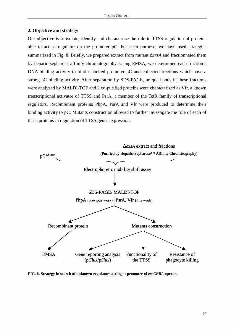
Results-Chapter 1
108
2. Objective and strategy
Our objective is to isolate, identify and characterize the role in TTSS regulation of proteins
able to act as regulator on the promoter pC. For such purpose, we have used strategies
summarized in Fig. 8. Briefly, we prepared extract from mutant ∆exsA and fractionnated them
by heparin-sepharose affinity chromatography. Using EMSA, we determined each fraction’s
DNA-binding activity to biotin-labelled promoter pC and collected fractions which have a
strong pC binding activity. After separation by SDS-PAGE, unique bands in these fractions
were analyzed by MALDI-TOF and 2 co-purified proteins were characterized as Vfr, a known
transcriptional activator of TTSS and PsrA, a member of the TetR family of transcriptional
regulators. Recombinant proteins PhpA, PsrA and Vfr were produced to determine their
binding activity to pC. Mutants construction allowed to further investigate the role of each of
these proteins in regulation of TTSS genes expression.
FIG. 8. Strategy in search of unknown regulators acting at promoter of exsCEBA operon.
Electrophoretic mobility shift assay
SDS-PAGE/ MALDI-TOF
pC*biotin
∆exsA extract and fractions
(Purified by Heparin-SepharoseTM Affinity Chromatography)
Recombinant protein
EMSA
Mutants construction
Gene reporting analysis(pClux/pSlux)
Resistance of phagocyte killing
PsrA, Vfr (this work)PhpA(previous work)
Functionality of the TTSS
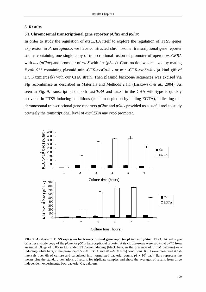
Results-Chapter 1
109
3. Results
3.1 Chromosomal transcriptional gene reporter pClux and pSlux
In order to study the regulation of exsCEBA itself to explore the regulation of TTSS genes
expression in P. aeruginosa, we have constructed chromosomal transcriptional gene reporter
strains containing one single copy of transcriptional fusion of promoter of operon exsCEBA
with lux (pClux) and promoter of exoS with lux (pSlux). Construction was realized by mating
E.coli S17 containing plasmid mini-CTX-exsCp-lux or mini-CTX-exoSp-lux (a kind gift of
Dr. Kazmierczak) with our CHA strain. Then plasmid backbone sequences was excised via
Flp recombinase as described in Materials and Methods 2.1.1 (Laskowski et al., 2004). As
seen in Fig. 9, transcription of both exsCEBA and exoS in the CHA wild-type is quickly
activated in TTSS-inducing conditions (calcium depletion by adding EGTA), indicating that
chromosomal transcriptional gene reporters pClux and pSlux provided us a useful tool to study
precisely the transcriptional level of exsCEBA ane exoS promoter.
FIG. 9. Analysis of TTSS exprssion by transcriptional gene reporter pClux and pSlux. The CHA wild-type carrying a single copy of the pClux or pSlux transcriptional reporter at its chromosome were grown at 37°C from an initial OD600 of 0.05 in LB under TTSS-noninducing (black bars, in the presence of 5 mM calcium) or -inducing (white bars, in the presence of 5 mM EGTA and 20 mM MgCl2) conditions. RLU were measured at 1-h intervals over 6h of culture and calculated into normalized bacterial counts (6 × 10⁸ bac). Bars represent the means plus the standard deviations of results for triplicate samples and show the averages of results from three independent experiments. bac, bacteria. Ca, calcium.
0500
10001500200025003000350040004500
1 2 3 4 5 6
Culture time (hours)
RLU
/6*1
08 ba
c ( p
Clu
x)
Ca
EGTA
0100200300400500600700800900
1 2 3 4 5 6
Culture time (hours)
RLU
/6*1
08 ba
c ( p
Slu
x)
Ca
EGTA
0500
10001500200025003000350040004500
1 2 3 4 5 6
Culture time (hours)
RLU
/6*1
08 ba
c ( p
Clu
x)
Ca
EGTA
0100200300400500600700800900
1 2 3 4 5 6
Culture time (hours)
RLU
/6*1
08 ba
c ( p
Slu
x)
Ca
EGTA
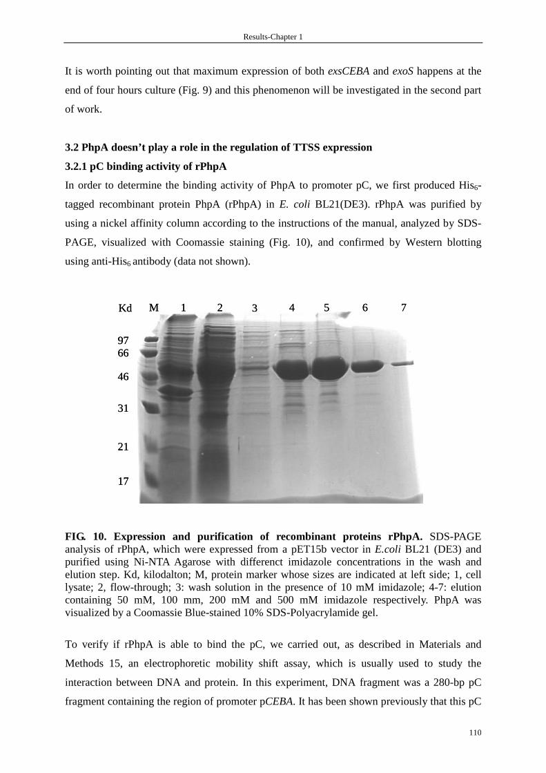
Results-Chapter 1
110
It is worth pointing out that maximum expression of both exsCEBA and exoS happens at the
end of four hours culture (Fig. 9) and this phenomenon will be investigated in the second part
of work.
3.2 PhpA doesn’t play a role in the regulation of TTSS expression
3.2.1 pC binding activity of rPhpA
In order to determine the binding activity of PhpA to promoter pC, we first produced His6-
tagged recombinant protein PhpA (rPhpA) in E. coli BL21(DE3). rPhpA was purified by
using a nickel affinity column according to the instructions of the manual, analyzed by SDS-
PAGE, visualized with Coomassie staining (Fig. 10), and confirmed by Western blotting
using anti-His6 antibody (data not shown).
FIG. 10. Expression and purification of recombinant proteins rPhpA. SDS-PAGE analysis of rPhpA, which were expressed from a pET15b vector in E.coli BL21 (DE3) and purified using Ni-NTA Agarose with differenct imidazole concentrations in the wash and elution step. Kd, kilodalton; M, protein marker whose sizes are indicated at left side; 1, cell lysate; 2, flow-through; 3: wash solution in the presence of 10 mM imidazole; 4-7: elution containing 50 mM, 100 mm, 200 mM and 500 mM imidazole respectively. PhpA was visualized by a Coomassie Blue-stained 10% SDS-Polyacrylamide gel.
To verify if rPhpA is able to bind the pC, we carried out, as described in Materials and
Methods 15, an electrophoretic mobility shift assay, which is usually used to study the
interaction between DNA and protein. In this experiment, DNA fragment was a 280-bp pC
fragment containing the region of promoter pCEBA. It has been shown previously that this pC
M
9766
46
31
21
17
1 2 3 4 5 6 7Kd M
9766
46
31
21
17
1 2 3 4 5 6 7Kd
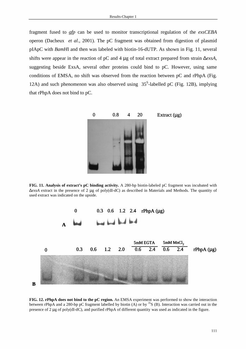
Results-Chapter 1
111
fragment fused to gfp can be used to monitor transcriptional regulation of the exsCEBA
operon (Dacheux et al., 2001). The pC fragment was obtained from digestion of plasmid
pIApC with BamHI and then was labeled with biotin-16-dUTP. As shown in Fig. 11, several
shifts were appear in the reaction of pC and 4 µg of total extract prepared from strain ∆exsA,
suggesting beside ExsA, several other proteins could bind to pC. However, using same
conditions of EMSA, no shift was observed from the reaction between pC and rPhpA (Fig.
12A) and such phenomenon was also observed using 35S-labelled pC (Fig. 12B), implying
that rPhpA does not bind to pC.
FIG. 11. Analysis of extract’s pC binding activity. A 280-bp biotin-labeled pC fragment was incubated with ∆exsA extract in the presence of 2 µg of poly(dI-dC) as described in Materials and Methods. The quantity of used extract was indicated on the upside.
FIG. 12. rPhpA does not bind to the pC region. An EMSA experiment was performed to show the interaction between rPhpA and a 280-bp pC fragment labelled by biotin (A) or by 35S (B). Interaction was carried out in the presence of 2 µg of poly(dI-dC), and purified rPhpA of different quantity was used as indicated in the figure.
0.8 4 20 Extract (µg)0 0.8 4 20 Extract (µg)0
0.3 0.6 1.2 2.4 rPhpA (µg)0
A
0.3 0.6 1.2 2.0 0.6 2.4 0.6 2.4 rPhpA (µg)
5mM EGTA 5mM MnCl2
0
B
0.3 0.6 1.2 2.4 rPhpA (µg)0
A
0.3 0.6 1.2 2.4 rPhpA (µg)0
A
0.3 0.6 1.2 2.0 0.6 2.4 0.6 2.4 rPhpA (µg)
5mM EGTA 5mM MnCl2
0
B
0.3 0.6 1.2 2.0 0.6 2.4 0.6 2.4 rPhpA (µg)
5mM EGTA 5mM MnCl2
0
B
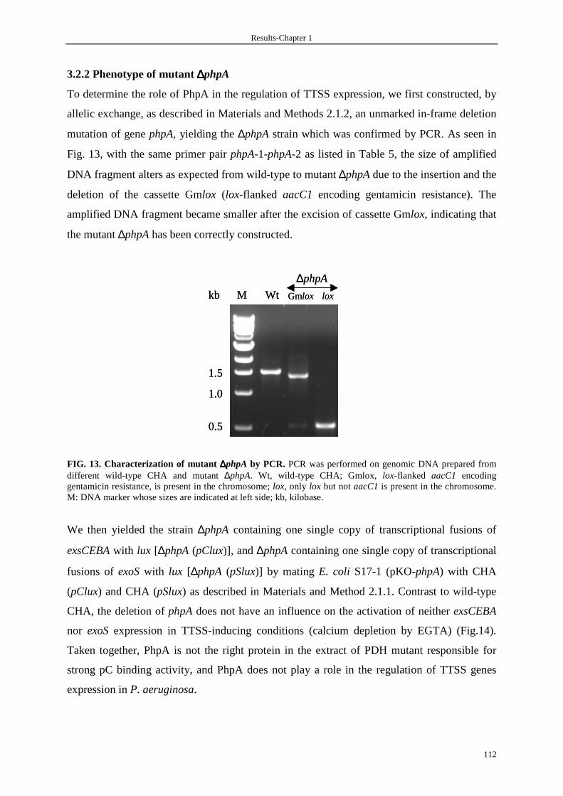
Results-Chapter 1
112
3.2.2 Phenotype of mutant ∆∆∆∆phpA
To determine the role of PhpA in the regulation of TTSS expression, we first constructed, by
allelic exchange, as described in Materials and Methods 2.1.2, an unmarked in-frame deletion
mutation of gene phpA, yielding the ∆phpA strain which was confirmed by PCR. As seen in
Fig. 13, with the same primer pair phpA-1-phpA-2 as listed in Table 5, the size of amplified
DNA fragment alters as expected from wild-type to mutant ∆phpA due to the insertion and the
deletion of the cassette Gmlox (lox-flanked aacC1 encoding gentamicin resistance). The
amplified DNA fragment became smaller after the excision of cassette Gmlox, indicating that
the mutant ∆phpA has been correctly constructed.
FIG. 13. Characterization of mutant ∆∆∆∆phpA by PCR. PCR was performed on genomic DNA prepared from different wild-type CHA and mutant ∆phpA. Wt, wild-type CHA; Gmlox, lox-flanked aacC1 encoding gentamicin resistance, is present in the chromosome; lox, only lox but not aacC1 is present in the chromosome. M: DNA marker whose sizes are indicated at left side; kb, kilobase.
We then yielded the strain ∆phpA containing one single copy of transcriptional fusions of
exsCEBA with lux [∆phpA (pClux)], and ∆phpA containing one single copy of transcriptional
fusions of exoS with lux [∆phpA (pSlux)] by mating E. coli S17-1 (pKO-phpA) with CHA
(pClux) and CHA (pSlux) as described in Materials and Method 2.1.1. Contrast to wild-type
CHA, the deletion of phpA does not have an influence on the activation of neither exsCEBA
nor exoS expression in TTSS-inducing conditions (calcium depletion by EGTA) (Fig.14).
Taken together, PhpA is not the right protein in the extract of PDH mutant responsible for
strong pC binding activity, and PhpA does not play a role in the regulation of TTSS genes
expression in P. aeruginosa.
1.5
1.0
0.5
M Wt
∆phpA
Gmlox loxkb
1.5
1.0
0.5
M Wt
∆phpA
Gmlox loxkb
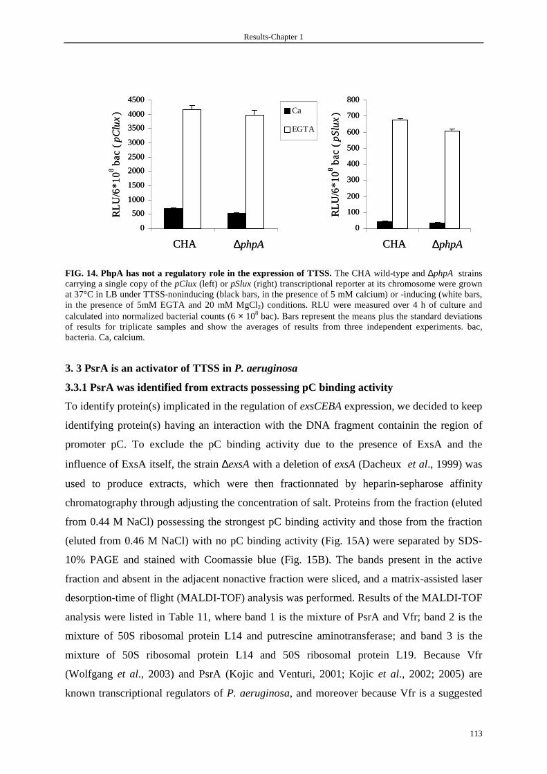
Results-Chapter 1
113
FIG. 14. PhpA has not a regulatory role in the expression of TTSS. The CHA wild-type and ∆phpA strains carrying a single copy of the pClux (left) or pSlux (right) transcriptional reporter at its chromosome were grown at 37°C in LB under TTSS-noninducing (black bars, in the presence of 5 mM calcium) or -inducing (white bars, in the presence of 5mM EGTA and 20 mM MgCl2) conditions. RLU were measured over 4 h of culture and calculated into normalized bacterial counts (6 × 10⁸ bac). Bars represent the means plus the standard deviations of results for triplicate samples and show the averages of results from three independent experiments. bac, bacteria. Ca, calcium.
3. 3 PsrA is an activator of TTSS in P. aeruginosa
3.3.1 PsrA was identified from extracts possessing pC binding activity
To identify protein(s) implicated in the regulation of exsCEBA expression, we decided to keep
identifying protein(s) having an interaction with the DNA fragment containin the region of
promoter pC. To exclude the pC binding activity due to the presence of ExsA and the
influence of ExsA itself, the strain ∆exsA with a deletion of exsA (Dacheux et al., 1999) was
used to produce extracts, which were then fractionnated by heparin-sepharose affinity
chromatography through adjusting the concentration of salt. Proteins from the fraction (eluted
from 0.44 M NaCl) possessing the strongest pC binding activity and those from the fraction
(eluted from 0.46 M NaCl) with no pC binding activity (Fig. 15A) were separated by SDS-
10% PAGE and stained with Coomassie blue (Fig. 15B). The bands present in the active
fraction and absent in the adjacent nonactive fraction were sliced, and a matrix-assisted laser
desorption-time of flight (MALDI-TOF) analysis was performed. Results of the MALDI-TOF
analysis were listed in Table 11, where band 1 is the mixture of PsrA and Vfr; band 2 is the
mixture of 50S ribosomal protein L14 and putrescine aminotransferase; and band 3 is the
mixture of 50S ribosomal protein L14 and 50S ribosomal protein L19. Because Vfr
(Wolfgang et al., 2003) and PsrA (Kojic and Venturi, 2001; Kojic et al., 2002; 2005) are
known transcriptional regulators of P. aeruginosa, and moreover because Vfr is a suggested
0
500
1000
1500
2000
2500
3000
3500
4000
4500
RLU
/6*1
0 8 b
ac
( pC
lux)
Ca
EGTA
0
100
200
300
400
500
600
700
800
RLU
/6*1
0 8 b
ac
( pS
lux)
CHA ∆phpA CHA ∆phpA
0
500
1000
1500
2000
2500
3000
3500
4000
4500
RLU
/6*1
0 8 b
ac
( pC
lux)
Ca
EGTA
0
100
200
300
400
500
600
700
800
RLU
/6*1
0 8 b
ac
( pS
lux)
CHA ∆phpA CHA ∆phpA

Results-Chapter 1
114
activator of the TTSS by an unknown mechanism, we were interested to know whether Vfr
and/or PsrA play a role in TTSS regulation by direct binding to the promoter pC.
FIG. 15. Isolation of protein(s) possessing pC binding activity. (A) Results of EMSA of ∆exsA crude extract or purified fractions on the pC. A 280-bp biotin-labeled pC fragment was incubated with ∆exsA extract or a fraction obtained from heparin-Sepharose affinity chromatography in the presence of 2 µg of poly(dI-dC) as described in Materials and Methods. Extract, total extract of ∆exsA; elution 1, preliminarily purified extract from 5-ml column; other lanes, fractions collected from second purification with elution at the indicated NaCl concentration. One microgram of protein was added in each lane. (B) SDS-PAGE analysis of fractions of ∆exsA extract purified using heparin-sepharose affinity chromatography. The fraction (0.44 M) possessing the pC binding activity and the adjacent fraction (0.46 M) not having any pC binding activity (A) were separated by SDS-10% PAGE and stained with Coomassie blue. The bands present only in the 0.44 M fraction indicated by arrows were sliced, and a MALDI-TOF analysis was performed.
Table 11. Analysis of MALDI-TOF
band Protein name Gene name *PA number
1 PsrA (Pseudomonas sigma regulator) psrA PA3006
Vfr (virulence factor regulator) vfr PA0652
2 RplN (50S ribosomal protein L14) rplN PA4253
SpuC (putrescine aminotransferase) spuC PA0299
3 RplN (50S ribosomal protein L14) rplN PA4253
RplS (50S ribosomal protein L19) rplS PA3742
* PA number refers to the P. aeruginosa assigned numbers in www.pseudomonas.com.
3.3.2 Recombinant protein PsrA binds to the pC region
To determine if PsrA and/or Vfr could effectively bind to the pC, we produced recombinant
protein PsrA (rPsrA) and rVfr in E. coli BL21(DE3). Recombinant proteins were purified by
using a nickel affinity column chromatography with imidazole gradient elution, analyzed by
Extract
Elution
1
Flowthr
ough
Wash[0.
42][0.
44][0.
46][0.
48][0.
50][0.
52][0.
54][0.
56][0.
60]
Elution (NaCl, mol/L)
Free pC
A B14
21
31
43
66
1
2
3
M 0.44M 0.46MkDa
Extract
Elution
1
Flowthr
ough
Wash[0.
42][0.
44][0.
46][0.
48][0.
50][0.
52][0.
54][0.
56][0.
60]
Elution (NaCl, mol/L)
Free pC
Extract
Elution
1
Flowthr
ough
Wash[0.
42][0.
44][0.
46][0.
48][0.
50][0.
52][0.
54][0.
56][0.
60]
Elution (NaCl, mol/L)
Free pC
Extract
Elution
1
Flowthr
ough
Wash[0.
42][0.
44][0.
46][0.
48][0.
50][0.
52][0.
54][0.
56][0.
60]
Elution (NaCl, mol/L)
Free pC
A B14
21
31
43
66
1
2
3
M 0.44M 0.46MkDa
14
21
31
43
66
1
2
3
M 0.44M 0.46MkDa

Results-Chapter 1
115
SDS-PAGE, visualized with Coomassie staining, and confirmed by Western blotting using
anti- His6 antibody (Fig. 16).
FIG. 16. Expression and purification of recombinant proteins rPsrA and rVfr. SDS-PAGE analysis of rPsrA (A) and rVfr (C), which were expressed from a pET15b vector in E.coli BL21 (DE3) and purified using Ni-NTA resin from Invitrogen with different imidazole concentrations in the wash and elution step. Kd, kilodalton; M: protein marker whose sizes are indicated at left side; 1: total cell lysate (5 mM imidazole); 2: flow-through; 3: wash (10 mM imidazole); 4-6: 50 mM, 100 mM and 200 mM imidazole elution respectively. Proteins were visualized by a Coomassie Blue-stained 15% SDS-Polyacrylamide gel. Arrows indicate the position of rPsrA (A) and rVfr (C). Western blot analysis of rPsrA (B) and rVfr by using anti-His6-HRP antibody, the bands present in (B) and (D) correspond to the bands presented at the level indicated by arrows in (A) and (C) respectively.
To verify if rPsrA and/or rVfr is able to bind the pC, an EMSA was performed. Purified rPsrA
and rVfr were tested for their ability to bind the 280-bp DNA pC fragment containing
promoter of exsCEBA operon. As shown in Fig. 17, the mobility of the pC was retarded in the
presence of 1.5 µM rPsrA. It has to be noted that the position at which this retardation
occurred is not exactly the same as that due to the preliminarily purified extract from which
PsrA was identified. In contrast, no retardation of the pC was observed in the presence of
rVfr, even in combination with 20 µM cyclic AMP, a known ligand of Vfr that increase its
B.
1 2 3 4 5 6MA.
94
66
43
31
21
Kd 1 2 3 4 5 6M
9466
43
31
21
14
Kd
D.
C.
B.
1 2 3 4 5 6MA.
94
66
43
31
21
Kd
B.
1 2 3 4 5 6MA.
94
66
43
31
21
Kd 1 2 3 4 5 6M
9466
43
31
21
14
Kd
D.
C. 1 2 3 4 5 6M
9466
43
31
21
14
Kd
D.
C.

Results-Chapter 1
116
capacity to bind DNA. The mixing of rPsrA and rVfr in an equimolar ratio did not modify the
retardation profile of rPsrA alone. This suggested that Vfr is not able to bind, at least in vitro,
to the pC. The binding of rPsrA needed relatively high concentrations of recombinant protein,
indicating either a low binding affinity for the pC or poor activity of the recombinant protein.
In order to determine the binding specificity of our rPsrA on the pC, we also produced a 280-
bp ppsrA, a known promoter for PsrA (Kojic et al., 2002), and a 280-bp pfleQ, a
proven promoter having binding sequences for Vfr (Dasgupta et al., 2002). As shown in
Fig. 17, no retardation was observed in the presence of pfleQ and rPsrA even with a high
concentration of rPsrA. In contrast, retardation was observed with pfleQ and rVfr (Fig. 17),
indicating (i) that the rVfr protein is correctly folded and active and (ii) that, for the first time,
we showed that Vfr is not able to bind the pC. In the same manner, ppsrA was retarded in the
presence of rPsrA at a concentration 10 times lower than that needed for the pC, indicating
that rPsrA is active and that, although the binding is specific, the in vitro binding affinity of
PsrA for the pC is low.
FIG. 17. rPsrA binds to the pC region. An EMSA experiment was performed to show that rPsrA interacts with the pC region. Biotin-labeled DNA fragments containing pC, pfleQ, or ppsrA were incubated in the presence of 2 µg of poly(dI-dC) with purified rPsrA of different concentrations as indicated in the figure. Elution 1, preliminarily purified extract (1 µg) fraction obtained from the first heparin-Sepharose affinity chromatography purification as described in Materials and Methods. *, rVFR was used for this lane to verify its pfleQ binding activity.
3.3.3 Mutant ∆∆∆∆psrA and ∆∆∆∆psrA containing gene reporter pClux or pSlux
As PsrA is able to bind the pC, we asked whether PsrA was indeed necessary for the
transcription of exsA and logically the TTSS effectors, such as exoS, in response to various
known stimuli of the TTSS. Therefore, from our CHA parental strain we constructed, by
pC
0
Elu
tion1
1.5 3 6 12
ppsrA
0 0.01
50.
15
0.3 (µM)
pfleQ
0 3 6 12 0.15*
pC
0
Elu
tion1
1.5 3 6 12
ppsrA
0 0.01
50.
15
0.3 (µM)
pfleQ
0 3 6 12 0.15*
pC
0
Elu
tion1
1.5 3 6 12
pC
0
Elu
tion1
1.5 3 6 12
ppsrA
0 0.01
50.
15
0.3 (µM)
ppsrA
0 0.01
50.
15
0.3 (µM)
pfleQ
0 3 6 12 0.15*
pfleQ
0 3 6 12 0.15*

Results-Chapter 1
117
allelic exchange, an unmarked in-frame deletion mutation of ∆psrA as described in Materials
and Methods, yielding the strain ∆psrA, and then ∆psrA containing one single copy of
transcriptional fusions of exsCEBA with lux [∆psrA (pClux)], and ∆psrA containing one single
copy of transcriptional fusions of exoS with lux [∆psrA (pSlux)], which were confirmed by
PCR (Fig. 18).
FIG. 18. Mutant analysis by PCR. PCR was performed on genomic DNA prepared from different strains. Wt, wild-type CHA; Gmlox, lox-flanked aacC1 encoding gentamicin resistance, is present in the chromosome; lox, only lox but not aacC1 is present in the chromosome. M: DNA marker whose sizes are indicated at left side; kb, kilobase.
3.3.4 PsrA is required for the full transcriptional activation of exsA and exoS
---PsrA is required for the full activation of exsA and exoS by calcium depletion
The different strains were cultured from an OD600 of 0.05 in TTSS-noninducing conditions (in
the presence of calcium) and in TTSS-inducing conditions (in the presence of calcium
chelator EGTA). As seen in Fig. 19, the transcriptional level for exoS and exsA in the CHA
wild-type was two- to threefold higher than that in the ∆psrA strain over 4 h of growth with
EGTA. However, the difference in the fold activation of gene expression between the wild-
type strain and the ∆psrA strain was quite low (4.7/3.3 and 11.1/10.4). Note that under these
culture conditions, no discernible growth difference was observed between the strains. Thus,
it seemed that PsrA is implicated in the regulation of the level of expression of exsA and
hence exoS but not in the regulation in response to calcium depletion. As calcium depletion
0.5
1.0
1.5
kb
M Wt Gmlox lox Gmlox lox
∆psrA(pClux) ∆psrA(pSlux)
0.5
1.0
1.5
kb
0.5
1.0
1.5
kb
M Wt Gmlox lox Gmlox lox
∆psrA(pClux) ∆psrA(pSlux)

Results-Chapter 1
118
activation occurs through the release of ExsA from its antiactivator ExsD, our result confirm
that PsrA is a positive transcriptional activator of TTSS independent of ExsA.
FIG. 19. Full activation of TTSS transcription requires PsrA. The CHA wild-type, ∆psrA , ∆psrA(pPsrA), ∆psrA(pDD2), and ∆rpoS strains carrying a single copy of the pClux (up) or pSlux (down) transcriptional reporter at its chromosome were grown at 37°C in LB under TTSS-noninducing (black bars) or -inducing (white bars) conditions. RLU were measured over 4 h of culture and calculated into normalized bacterial counts (6 × 10⁸ bac). Bars represent the means plus the standard deviations of results for triplicate samples and show the averages of results from three independent experiments. *, fold activation of gene expression under inducing conditions compared with that under noninducing conditions. bac, bacteria.
CHA ∆psrApPsrA
∆psrApDD2
∆psrA0
100
200
300
400
500
600
700
800
900
RL
U/6
*10
8 ba
c ( p
Slu
x)
Cacl2
EGTA
11.1*
10.4*
13.4*
2.8*
CHA ∆psrApPsrA
∆psrApDD2
∆psrA0
100
200
300
400
500
600
700
800
900
RL
U/6
*10
8 ba
c ( p
Slu
x)
Cacl2
EGTA
11.1*
10.4*
13.4*
2.8*
CHA ∆psrApPsrA
∆psrApDD2
∆rpoS∆psrA
4.7*
3.3*
3.3*
1.7*
4.7*
0
1000
2000
3000
4000
5000
6000
7000
8000
RLU
/6*1
0 8 b
ac
( pC
lux)
Cacl2
EGTA
CHA ∆psrApPsrA
∆psrApDD2
∆rpoS∆psrA
4.7*
3.3*
3.3*
1.7*
4.7*
0
1000
2000
3000
4000
5000
6000
7000
8000
RLU
/6*1
0 8 b
ac
( pC
lux)
Cacl2
EGTA
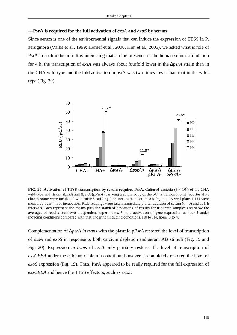
Results-Chapter 1
119
---PsrA is required for the full activation of exsA and exoS by serum
Since serum is one of the environmental signals that can induce the expression of TTSS in P.
aeruginosa (Vallis et al., 1999; Hornef et al., 2000, Kim et al., 2005), we asked what is role of
PsrA in such induction. It is interesting that, in the presence of the human serum stimulation
for 4 h, the transcription of exsA was always about fourfold lower in the ∆psrA strain than in
the CHA wild-type and the fold activation in psrA was two times lower than that in the wild-
type (Fig. 20).
FIG. 20. Activation of TTSS transcription by serum requires PsrA. Cultured bacteria (5 × 10⁵) of the CHA wild-type and strains ∆psrA and ∆psrA (pPsrA) carrying a single copy of the pClux transcriptional reporter at its chromosome were incubated with mHBS buffer (–) or 10% human serum AB (+) in a 96-well plate. RLU were measured over 4 h of incubation. RLU readings were taken immediately after addition of serum (t = 0) and at 1-h intervals. Bars represent the means plus the standard deviations of results for triplicate samples and show the averages of results from two independent experiments. *, fold activation of gene expression at hour 4 under inducing conditions compared with that under noninducing conditions. H0 to H4, hours 0 to 4.
Complementation of ∆psrA in trans with the plasmid pPsrA restored the level of transcription
of exsA and exoS in response to both calcium depletion and serum AB stimuli (Fig. 19 and
Fig. 20). Expression in trans of exsA only partially restored the level of transcription of
exsCEBA under the calcium depletion condition; however, it completely restored the level of
exoS expression (Fig. 19). Thus, PsrA appeared to be really required for the full expression of
exsCEBA and hence the TTSS effectors, such as exoS.
0
10
20
30
40
50
60
70
RLU
( p
Clu
x) H0
H1
H2
H3
H4
CHA- ∆psrApPsrA-
∆psrA- ∆psrApPsrA+
∆psrA+CHA+
20.2*
11.0*
25.6*
0
10
20
30
40
50
60
70
RLU
( p
Clu
x) H0
H1
H2
H3
H4
CHA- ∆psrApPsrA-
∆psrA- ∆psrApPsrA+
∆psrA+CHA+
20.2*
11.0*
25.6*

Results-Chapter 1
120
---PsrA is required for the full activation of exsA upon host cell contact
Besides calcium depletion growth conditions, the expression of the P. aeruginosa TTSS and
secretion of effector proteins could also be induced after contact with tissue culture cells
(Vallis et al., 1999; Hornef et al., 2000). Therefore, we asked whether PsrA is required for
such induction by measuring transcription of the pClux reporter construct in wild-type and
∆psrA strains following infection of PLB-985 polymorphonuclear leukocyte-like cells. As
seen in Fig. 21, exsCEBA transcription increased about fourfold at hour 4 in CHA in the
presence of PLB-985 cells as did that in the ∆psrA strain; on the contrary, the global
transcription level of exsCEBA in the ∆psrA mutant was twofold lower than that in the wild-
type strain.
FIG. 21. Transcriptional activation of exsCEBA during exposure of bacteria to differentiated PLB-985 cells requires PsrA. RLU produced by 5 × 10⁵ cultured bacteria of CHA and ∆psrA strains carrying a single copy of the pClux transcriptional reporter at its chromosome were measured over 4 h following bacterial interaction with 105 PLB-985 cells or mHBS buffer control in a 96-well plate. RLU readings were taken immediately after the addition of bacteria (t = 0) and at 1-h intervals. Bars represent the means plus the standard deviations of results for triplicate samples and show the averages of results from two independent experiments. *, fold activation of gene expression at hour 4 under inducing conditions compared with that under noninducing conditions. H0 to H4, hours 0 to 4.
0
2
4
6
8
10
12
14
16
RLU
( p
Clu
x) H0
H1
H2
H3
H4
CHA+Buffer CHA+PLB985 ∆psrA+PLB985∆psrA+Buffer
4.4*
4.4*
0
2
4
6
8
10
12
14
16
RLU
( p
Clu
x) H0
H1
H2
H3
H4
CHA+Buffer CHA+PLB985 ∆psrA+PLB985∆psrA+Buffer
4.4*
4.4*

Results-Chapter 1
121
3.3.5 Secretion of the type III effectors is decreased in the ∆∆∆∆psrA strain
Secretion of TTSS effectors is strongly correlated with P. aeruginosa virulence both in animal
models and in studies of human disease (Hauser et al., 2002). Therefore, we examined the
secretion of TTSS effector molecules in wild-type CHA, the ∆psrA strain, and the ∆psrA
strain carrying pPsrA [∆psrA (pPsrA)] under TTSS-noninducing or -inducing conditions. As
seen in Fig. 22, the ∆psrA mutant showed markedly decreased secretion of ExoS, ExoT, and
PopB proteins compared to the parental CHA strain. Conversely, complementation of ∆psrA
restored the strain's ability to secrete ExoS, ExoT, and PopB proteins whose amounts seemed
slightly higher than those of proteins from the wild-type CHA strain, confirming the positive
regulatory function of PsrA for the TTSS. However, ectopic expression of psrA cannot
overcome the requirement for a TTSS-inducing signal, such as calcium depletion, as
demonstrated by the absence of ExoS, ExoT, and PopB secretion when ∆psrA(pPsrA) was
grown under noninducing conditions, indicating that the presence of EGTA is essential for the
secretion (Fig. 22).
FIG. 22. Secretion of the type III effectors is decreased in the ∆∆∆∆psrA strain. Bacteria were grown under TTSS-noninducing (lanes 1) or TTSS-inducing (lanes 2) conditions. Supernatants normalized to bacterial counts (6 × 10⁸ bacteria lane-1) were subjected to trichloroacetic acid precipitation. The extracellular proteins were separated by SDS-10% PAGE and stained with Coomassie blue. M, protein marker. Sizes in kilodaltons are indicated at the left.
ExoT
∆psrA-pPsrA
1 2
31
43
66
94
kDa M
ExoS
PopB
∆psrA1 2
CHA1 2
ExoT
∆psrA-pPsrA
1 2
31
43
66
94
kDa M
ExoS
PopB
∆psrA1 2
CHA1 2

Results-Chapter 1
122
3.3.6 Strain ∆∆∆∆psrA is less resistant to the phagocyte-like PLB-985 cells
To examine the effect of the ∆psrA mutation on the ability of P. aeruginosa to resist the
bacterial killing activity of polymorphonuclear leukocyte-like cells from differentiated PLB-
985 cells, CHA, the ∆psrA strain, and ∆exsA, a TTSS-defective strain, were compared. Fig. 23
shows that the ∆psrA strain is less resistant to the phagocyte-like PLB-985 cells than its
parental strain, CHA, as the
number of viable bacteria after 80 min of coculture with PLB-985 was two times lower for the
∆psrA strain than for wild-type CHA. Nevertheless, the survival of ∆exsA in the presence of
phagocyte-like PLB-985 cells reveals that, besides the TTSS, other factor(s) may also play a
role in the resistance of P. aeruginosa to phagocytosis.
FIG. 23. The ∆∆∆∆psrA strain is less resistant to phagocyte-like PLB-985 cells. The numbers of viable bacteria were calculated from colony counts made from a 1:105 dilution over 3 h following bacterial interaction with differentiated PLB-985 cells at different intervals. Points represent the means plus the standard deviations of results for duplicate samples and show the averages of results from two independent experiments.
0
50
100
150
200
250
300
0 20 40 60 80 120 180
Interaction time (min)
CHA
∆psrA
∆exsA
105
via
ble
bac
teri
a
0
50
100
150
200
250
300
0 20 40 60 80 120 180
Interaction time (min)
CHA
∆psrA
∆exsA
105
via
ble
bac
teri
a

Results-Chapter 1
123
4. Discussion
The TTSS is known to be tightly controlled by the proteins encoded by the regulatory operon
exsCEBA, particularly by the central positive regulator ExsA, though many other proteins,
such as Vfr, RetS, PtrA, and pyruvate dehydrogenase, are implicated in the regulation of the
TTSS (Fig. 24).
FIG. 24. A model for the regulation of the TTSS. PsrA activates the TTSS through a positive effect (+) on the expression of ExsA and may also regulate another unknown protein(s) (X) which has a role in the TTSS. PsrA also has a positive effect on RpoS, which has a negative effect on the TTSS by an unknown mechanism. Other main molecules involved in the regulation of the TTSS that are mentioned in the text are also indicated. It is not surprising that P. aeruginosa has multiple regulatory networks, because 8% of its
genome codes for regulatory genes, indicating that P. aeruginosa has dynamic and
complicated regulatory mechanisms responding to various environmental signals (Greenberg,
2000). Because of the requirement for a large number of genes, the construction of the type III
secretion apparatus is an energy-expensive process for the bacteria, rendering so important the
ability to precisely regulate the TTSS in response to the environmental changes.
In this work, we describe a P. aeruginosa protein, PsrA, that is required for the full activation
of the TTSS regulatory operon exsCEBA and hence the expression of effector genes, although
the mutant still responds to depletion of calcium, exposure to serum, and contact with tissue
culture cells. PsrA was previously described as a regulator involved in positive regulation of
RpoS and in negative autoregulation by direct binding to the promoters (Kojic and Venturi,
2001; Kojic et al., 2002; 2005). RpoS, a central regulator during stationary phase (Loewen et
PsrA ExsA+
RpoS
+
+
X
? ?
TTSS
PtrA
ExsD
ExsC
CyaB
Vfr
+
RtsM
+
ExsE
PsrA ExsA+
RpoS
+
+
X
? ?
TTSS
PtrA
ExsD
ExsC
CyaB
Vfr
+
RtsM
+
ExsE

Results-Chapter 1
124
al., 1998), was identified also as an inhibitor of the TTSS (Hogardt et al., 2004) (Fig. 24). The
PsrA-dependent induction of the synthesis of RpoS has been shown to take place in the late
exponential and stationary phases (Kojic and Venturi, 2001). Here we measured the induction
of the TTSS by different stimuli in the first part of the exponential growth phase when no
RpoS induction of synthesis could occur. Furthermore, we confirmed the RpoS-dependent
inhibition of TTSS synthesis in our strain CHA ∆rpoS(pClux) (Fig. 19). This indicated that
the action of PsrA on TTSS gene transcription is independent of its role in RpoS regulation. A
previous study showed that a ∆psrA mutant had a 90% reduction in rpoS promoter activity
(Kojic and Venturi, 2001); considering also the opposite functions of PsrA and RpoS in TTSS
regulation, we could speculate that PsrA should have an even more important regulatory
ability for the TTSS than that measured here. It seemed that PsrA is implicated rather in the
regulation of the basic level of exsA and hence exoS expression than in the regulation in
response to environmental signals, at least calcium depletion and host cell contact, since there
was no or only a small difference in fold activation of gene expression under these inducing
conditions compared with that under noninducing conditions for the mutant and the wild-type
strain (Fig. 19 and Fig. 21). We also always observed that the growth curves of the CHA and
∆psrA strains were identical, indicating that the deletion of psrA has no metabolic
consequences for the CHA strain. Moreover, the complementation in trans by a multicopy
plasmid harboring the psrA gene controlled by its own promoter showed an augmentation of
the type III secretion. In addition, we showed that the decrease of exsCEBA expression due to
psrA deletion could not be overcome by expression in trans of exsA, while exoS expression
could be restored (Fig. 19). Taken altogether, these results suggest that PsrA is a ExsA-
independent positive regulator of exsCEBA and hence the whole TTSS genes.
Using the PsrA binding motif (G/CAAAC N2 to 4 GTTTG/C, where N2 to 4 is any two to four
nucleotides) to search against the P. aeruginosa genome sequence at
http://www.pseudomonas.com revealed that at least 26 genes may be controlled by PsrA
(Table 12), indicating that the PsrA is a global regulator. 15 genes were described as target
genes of the PsrA regulator (kojic et al., 2005) during the process of our submitted paper
concerning PsrA. It is reasonable to propose that PsrA could influence the TTSS by activating
or repressing genes having a potent regulatory role in TTSS expression (Fig. 24).
Identification of such unknown genes as well as the relationship with PsrA will help us to
better understand the mechanism by which PsrA affects TTSS expression.

Results-Chapter 1
125
TABLE 12. Possible promoter regions containing PsrA binding sites
PsrA binding site Region Type Gene name or N° of PA CAAAC AAAC GTTTG intergenic etfB-PA2953 CAAAC AGTG GTTTC intergenic groES-PA4387 CAAAC GCCT GTTTG intergenic PA0505-PA0506 GAAAC CAGC GTTTC intergenic PA3594-PA3595 CAAAC CCGC GTTTC intergenic PA4792-PA4793 GAAAC CG GTTTC intergenic PA4963-parC CAAAC ACTT GTTTG intergenic psrA-lexA (PA3006-3007) GAAAC CG GTTTC intergenic ptxR-ptxS (PA2258-2259) GAAAC CG GTTTC intergenic ptxS-PA2260 GAAAC GCT GTTTG PA_gene argJ (PA4402) GAAAC TG GTTTC PA_gene betB (PA5373) CAAAC CC GTTTG PA_gene cyoA (PA1317) GAAAC GTAT GTTTC PA_gene cysC (PA1393) CAAAC GAGC GTTTC PA_gene ligT (PA2861) GAAAC CGGG GTTTC PA_gene mmsA (PA3570) GAAAC CC GTTTC PA_gene PA0098 CAAAC CCG GTTTC PA_gene PA0112 GAAAC TGAA GTTTC PA_gene PA0805 CAAAC CGA GTTTC PA_gene PA1736 CAAAC TCC GTTTG PA_gene PA2674 CAAAC TTCC GTTTG PA_gene PA3623 GAAAC GCCC GTTTC PA_gene PA4421 GAAAC CG GTTTG PA_gene PA4489 GAAAC CC GTTTC PA_gene pilK (PA0412) CAAAC GT GTTTC PA_gene pilT (PA0395) GAAAC CG GTTTC PA_gene wbpW (PA5452)
C/GAAAC N2-4 GTTTG/C
We do not know the signal(s) to which PsrA responds, but it may play an important role
during infection, as ∆psrA bacteria are less resistant to phagocyte-like PLB-985 cells.
Furthermore, the presence of PsrA homologues in other pathogenic bacteria that employ a
TTSS, such as P. syringae, raises the possibility that these homologues also play roles in
regulating the other TTSS. If this proves to be the case, it will be interesting to ask whether
the signal(s) transduced by PsrA and its homologues is also conserved among plant and
human pathogens.
Though the rPsrA in vitro bound the pC with a relatively low affinity, it is still possible that
PsrA may in vivo activate the TTSS by directly binding onto it. In fact, pC sequence analysis
revealed no presence of a true consensus binding site for PsrA, but some sequences, such as
GAAAC at the position -56 from the transcriptional start site, resemble a partial binding site.
Considering the complicated regulatory network of the TTSS, it is possible that PsrA may
bind the pC in vivo at a higher affinity in the presence of another partner(s) or it may also help

Results-Chapter 1
126
another regulator(s) to bind the pC. The copurification of PsrA and Vfr, an known activator of
the TTSS (Wolfgang et al., 2003), suggests possible interaction of PsrA and Vfr during the
process of regulation of the expression of the TTSS. Exploring the interaction of PsrA with
another molecule(s) and the pC would enrich understanding of the adaptation of bacteria to
living conditions. The extract of the ∆psrA strain possessed the same pC binding activity as
the one of ∆exsA strain (Fig. 25), indicating that the protein(s) having a strong pC binding
activity has not been isolated in this work and is possibly the partner of PsrA in TTSS
regulation. Isolation and identification of this protein will provide us more information about
the regulation of TTSS genes expression via the transcription level of the regulatory operon
exsCEBA.
FIG. 25. Extract of ∆∆∆∆psr preserve the pC binding activity. Extracts of different strains as indicated were prepared from TTSS non inducing (-) and inducing conditions (+, EGTA). A 280-bp biotin-labeled pC fragment was incubate with 1 µg extract in the presence of 2 µg of poly(dI-dC).
pC - + - + - + EGTA
∆exsA ∆psrA ∆psrA-pPsrA
pC - + - + - + EGTA
∆exsA ∆psrA ∆psrA-pPsrA
pC - + - + - + EGTA
∆exsA∆exsA ∆psrA∆psrA ∆psrA-pPsrA

Results-Chapter 2
127
Chapter 2
Study of the inhibition of TTSS genes expression
during the stationary phase

Results-Chapter 2
128
Résumé en français P. aeruginosa est responsable d’infections aiguës à d’infections chroniques. Le système de
sécrétion de type III (SSTT) est un facteur de virulence qui semble être très important dans les
infections aiguës chez l’humain mais moins important pour maintenir les infections
chroniques causées par P. aeruginosa. Nous avons observé que l’expression du SSTT est, in
vitro, dépendante de la densité des bactéries dans le milieux de culture. En outre, le
surnageant venant d’une culture en phase stationnaire peut inhiber l'activation du SSTT. Ici
nous rapportons l'observation de PARST, un inhibiteur qui est produit dans la phase
stationnaire de la culture et réprime l’expression du SSTT in vitro. L'analyse des mutants
d'insertion de transposon a montré que la production de PARST pourrait dépendre du
tryptophane, qui est le précurseur de l'acide d'indole-3-acétic (IAA). IAA-Na et un analogue
fonctionnel, potassium acide 1-naphthalacétic (NAA-K) à des concentrations millimolaires
sont en effet capable d’inhiber l’expression et la sécrétion du SSTT. Une molécule de m/z
224.2, présent uniquement dans le surnageant stationnaire de la souche sauvage CHA mais
pas celui du mutant avec la délétion de gène trpA a été déterminé par LC-MS. Cette molécule
de m/z 224.2 a le même poids moléculaire de NAA-K pourrait être l'inhibiteur de
l’expression des gènes du SSTT quand les bactéries entrent la phase stationnaire. Ces
observations fournissent une autre voie régulatrice dans la transition de l’expression de gènes
entre un état avec des cellules à faible densité et celui avec des cellules à forte densité.

Results-Chapter 2
129
En anglais 1. Background
Several reports suggest that functional type III secretion of P. aeruginosa is far less common
in cystic fibrosis (CF) patient isolates than in isolates from patients with acute infections
(Dacheux et al., 2000; Roy-Burman et al., 2001). Following infection of cystic fibrosis patient
airways, P. aeruginosa strains evolved to reduce the expression of TTSS (Lee et al., 2005) or
gradually changed from a type III protein secretion-positive phenotype to a secretion-negative
phenotype (Jain et al., 2004). This theme seems to be common in bacterial infection, because
the expression of other TTSSs were also down-regulated during mammalian cell infection by
Salmonella enterica serovar Typhimurium, E. coli O157 and Shigella flexneri (Eriksson et al.,
2003; Dahan et al., 2004, Lucchini et al., 2005, Faucher et al., 2006). The consistency of
these data suggests that the down-regulation of TTSS genes may be a conserved phenomenon
that follows successful infection of mammalian cells by these gram-negative pathogens.
Presumably, a mechanism should exist for inactivating the TTSS once it has fulfilled its
mission.
In enterohemorrhagic and enteropathogenic E. coli, study showed that maximum expression
of TTSS happens during the transition from the late exponential to the stationary phase
(Sperandio et al., 1999). In P. aeruginosa, we observed that maximum expression of TTSS
genes such as exsCEBA and exoS, happens at 4h of culture (corresponding an OD600 of 1.5 to
2.0), and then decreased rapidly with the increase of OD600 (Fig. 27, page 131). Furthermore,
we observed the existance of unknown inhibitor(s) repressing the expression of TTSS genes
when bacteria enter stationary phase in the culture medium. This unknown inhibitor(s) was
termed as ‘PARST’ for ‘Pseudomonas autorepressor of secretion three’.

Results-Chapter 2
130
2. Objective and strategy
Our aim is to identify gene(s) involved in the production of PARST and finally the nature of
PARST. For such purpose, we have used strategy summarized in Fig. 26. Briefly, we first
produced a library of about 105 mutants using mariner-based transposon Himar1::Gmr from
plasmid pFAC (Wong and Mekalanos, 2000; Diaz-Perez et al., 2004). Then, we screened
about 2,000 mutants for the effect of their stationary phase culture supernatant on the
expression of exoS. We selected and characterized transposon insertion mutants whose
supernatants did lose the ability to inhibit the exoS expression. To investigate the nature of
PARST, further analysis such as liquid chromatography-mass spectrometry will be performed.
FIG. 26. Strategy in determination of the nature of PARST
Library of 10,000 transposon insertion mutants
Screening mutants whose stationnay phase supernanant do not repress exoSpromoter activity
CHA E.coli (pFAC)
Mutants characterization Molecular cloning
Southern blot
Sequencing
Deletion mutants construction
Further analysis
Library of 10,000 transposon insertion mutants
Screening mutants whose stationnay phase supernanant do not repress exoSpromoter activity
CHA E.coli (pFAC)
Mutants characterization Molecular cloning
Southern blot
Sequencing
Deletion mutants construction
Further analysis

Results-Chapter 2
131
3. Results
3.1 TTSS expression is cell density-dependent
Chromosomal transcriptional gene reporters composed of exsCEBA protmoter-lux fusion
(pClux), and exoS promoter-lux fusion (pSlux) were used to study precisely the transcriptional
level of exsCEBA and exoS (Shen et al., 2006). From the results shown in Fig. 27, it is evident
that both exsCEBA and exoS were expressed in a growth phase-dependent manner when
strains were initially cultured from an OD600 of 0.05 in LB without or with calcium depletion
by EGTA. ExsCEBA and exoS expression reached their maximums at 4h of culture
corresponding an OD600 of 1.5 to 2.0 and then decreased rapidly with the increase of OD600.
We were not sure why exsCEBA and exoS expression decline over time but this phenomenon
was reminiscent of quorum sensing (QS) regulation, in which a sufficient number of bacteria,
the bacterial ‘quorum’, is needed to induce or repress expression of a group of genes (Fuqua
et al., 1994). QS having an impact on TTSS expression in P. aeruginosa (Schaber et al.,
2004; Hogardt et al., 2004; Bleves et al., 2005; Juhas et al., 2005) plus report that the
maximum expression of TTSS happens during the transition from the late exponential to the
stationary phase in enterohemorrhagic and enteropathogenic E. coli (Sperandio et al., 1999)
prompted us to examine the possibility that the reduction of exsCEBA and exoS expression
over time was due to the regulation of QS, which control genes expression at cell density-
dependent manner by producing different QS signaling molecules.
FIG. 27. TTSS expression is cell density-dependent. The CHA wild type carrying a single copy of pClux (right) or pSlux (left) transcriptional reporter at its chromosome was grown at 37°C in LB under TTSS-noninducing (black bars, in the presence of 5 mM calcium) or inducing conditions (white bars, in the presence of 5mM EGTA and 20 mM MgCl2). RLU were measured over 6h of culture at 1-h intervals and calculated to normalized bacterial counts (6 × 10⁸ bac). Bars represent the mean plus the standard deviation of triplicate samples, and are the average of three independent experiments. Black angles and white squares represent the OD600 in noninducing and inducing conditions, respectively. bac, bacteria.
0
500
1000
1500
2000
2500
3000
3500
4000
4500
1 2 3 4 5 6
Culture time (hours)
RLU
/6*1
08 bac
(pC
lux)
0
0.5
1
1.5
2
2.5
3
3.5
4
OD
600
0
100
200
300
400
500
600
700
800
900
1 2 3 4 5 6
Culture time (hours)
RLU
/6*1
08 bac
(pS
lux)
0
0.5
1
1.5
2
2.5
3
3.5
4O
D6
00
0
500
1000
1500
2000
2500
3000
3500
4000
4500
1 2 3 4 5 6
Culture time (hours)
RLU
/6*1
08 bac
(pC
lux)
0
0.5
1
1.5
2
2.5
3
3.5
4
OD
600
0
100
200
300
400
500
600
700
800
900
1 2 3 4 5 6
Culture time (hours)
RLU
/6*1
08 bac
(pS
lux)
0
0.5
1
1.5
2
2.5
3
3.5
4O
D6
00
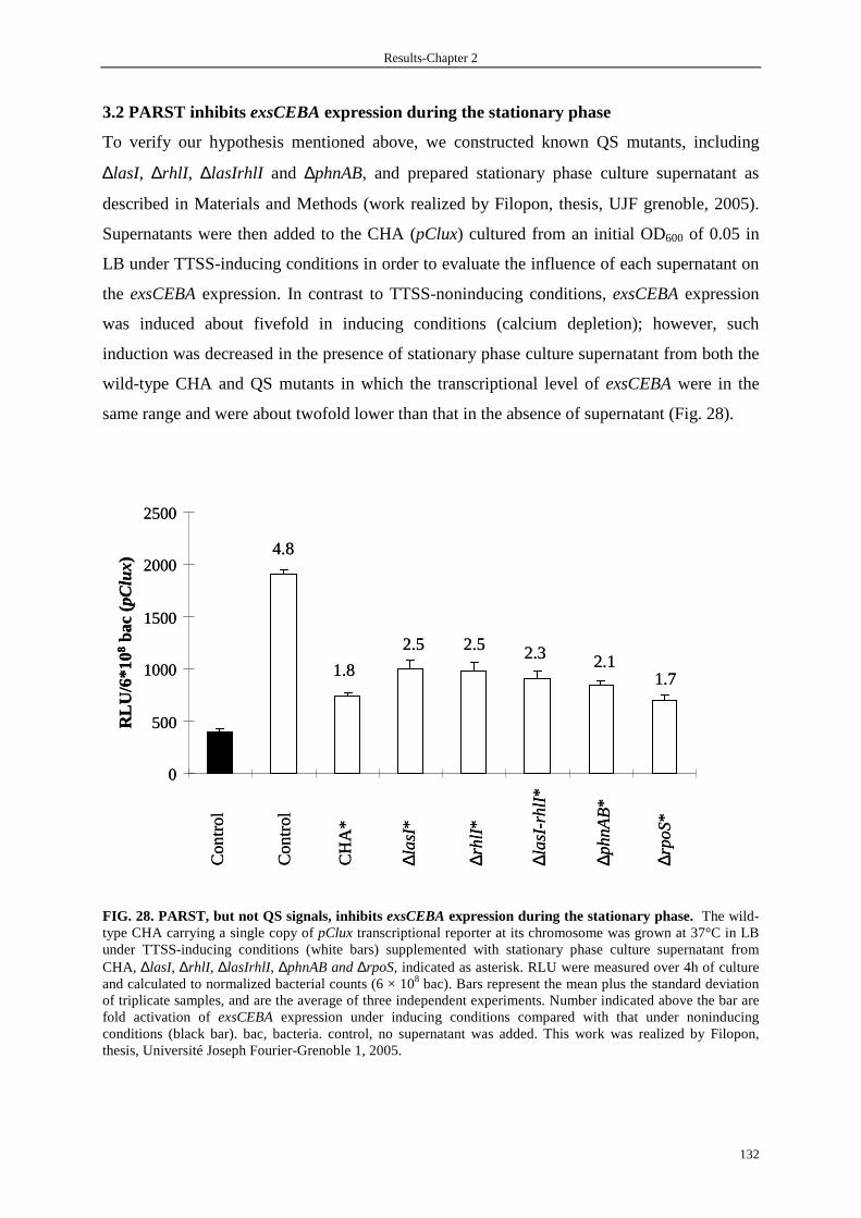
Results-Chapter 2
132
3.2 PARST inhibits exsCEBA expression during the stationary phase
To verify our hypothesis mentioned above, we constructed known QS mutants, including
∆lasI, ∆rhlI , ∆lasIrhlI and ∆phnAB, and prepared stationary phase culture supernatant as
described in Materials and Methods (work realized by Filopon, thesis, UJF grenoble, 2005).
Supernatants were then added to the CHA (pClux) cultured from an initial OD600 of 0.05 in
LB under TTSS-inducing conditions in order to evaluate the influence of each supernatant on
the exsCEBA expression. In contrast to TTSS-noninducing conditions, exsCEBA expression
was induced about fivefold in inducing conditions (calcium depletion); however, such
induction was decreased in the presence of stationary phase culture supernatant from both the
wild-type CHA and QS mutants in which the transcriptional level of exsCEBA were in the
same range and were about twofold lower than that in the absence of supernatant (Fig. 28).
FIG. 28. PARST, but not QS signals, inhibits exsCEBA expression during the stationary phase. The wild-type CHA carrying a single copy of pClux transcriptional reporter at its chromosome was grown at 37°C in LB under TTSS-inducing conditions (white bars) supplemented with stationary phase culture supernatant from CHA, ∆lasI, ∆rhlI , ∆lasIrhlI, ∆phnAB and ∆rpoS, indicated as asterisk. RLU were measured over 4h of culture and calculated to normalized bacterial counts (6 × 10⁸ bac). Bars represent the mean plus the standard deviation of triplicate samples, and are the average of three independent experiments. Number indicated above the bar are fold activation of exsCEBA expression under inducing conditions compared with that under noninducing conditions (black bar). bac, bacteria. control, no supernatant was added. This work was realized by Filopon, thesis, Université Joseph Fourier-Grenoble 1, 2005.
RLU
/6*1
08
bac
(pC
lux)
Con
trol
Con
trol
CH
A*
∆la
sI*
∆rh
lI*
∆la
sI-r
hlI
*
∆ph
nA
B*
∆rp
oS*
4.8
1.8
2.5 2.5 2.3 2.11.7
0
500
1000
1500
2000
2500
RLU
/6*1
08
bac
(pC
lux)
Con
trol
Con
trol
CH
A*
∆la
sI*
∆rh
lI*
∆la
sI-r
hlI
*
∆ph
nA
B*
∆rp
oS*
4.8
1.8
2.5 2.5 2.3 2.11.7
0
500
1000
1500
2000
2500
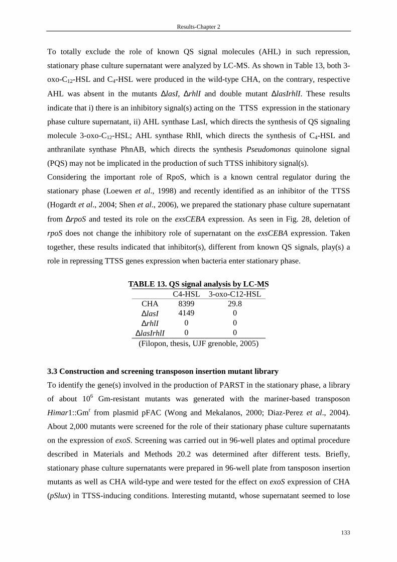
Results-Chapter 2
133
To totally exclude the role of known QS signal molecules (AHL) in such repression,
stationary phase culture supernatant were analyzed by LC-MS. As shown in Table 13, both 3-
oxo-C12-HSL and C4-HSL were produced in the wild-type CHA, on the contrary, respective
AHL was absent in the mutants ∆lasI, ∆rhlI and double mutant ∆lasIrhlI. These results
indicate that i) there is an inhibitory signal(s) acting on the TTSS expression in the stationary
phase culture supernatant, ii) AHL synthase LasI, which directs the synthesis of QS signaling
molecule 3-oxo-C12-HSL; AHL synthase RhlI, which directs the synthesis of C4-HSL and
anthranilate synthase PhnAB, which directs the synthesis Pseudomonas quinolone signal
(PQS) may not be implicated in the production of such TTSS inhibitory signal(s).
Considering the important role of RpoS, which is a known central regulator during the
stationary phase (Loewen et al., 1998) and recently identified as an inhibitor of the TTSS
(Hogardt et al., 2004; Shen et al., 2006), we prepared the stationary phase culture supernatant
from ∆rpoS and tested its role on the exsCEBA expression. As seen in Fig. 28, deletion of
rpoS does not change the inhibitory role of supernatant on the exsCEBA expression. Taken
together, these results indicated that inhibitor(s), different from known QS signals, play(s) a
role in repressing TTSS genes expression when bacteria enter stationary phase.
TABLE 13. QS signal analysis by LC-MS
C4-HSL 3-oxo-C12-HSL CHA 8399 29.8 ∆lasI 4149 0 ∆rhlI 0 0
∆lasIrhlI 0 0 (Filopon, thesis, UJF grenoble, 2005)
3.3 Construction and screening transposon insertion mutant library
To identify the gene(s) involved in the production of PARST in the stationary phase, a library
of about 106 Gm-resistant mutants was generated with the mariner-based transposon
Himar1::Gmr from plasmid pFAC (Wong and Mekalanos, 2000; Diaz-Perez et al., 2004).
About 2,000 mutants were screened for the role of their stationary phase culture supernatants
on the expression of exoS. Screening was carried out in 96-well plates and optimal procedure
described in Materials and Methods 20.2 was determined after different tests. Briefly,
stationary phase culture supernatants were prepared in 96-well plate from tansposon insertion
mutants as well as CHA wild-type and were tested for the effect on exoS expression of CHA
(pSlux) in TTSS-inducing conditions. Interesting mutantd, whose supernatant seemed to lose

Results-Chapter 2
134
their ability to inhibit the exoS expression as exemplified in Fig. 29 (arrowed ‘Interesting
mutant’), were selected and stored at -20°C for further analysis.
FIG. 29. High throughput screening of transposon insertion mutants that do not inhibit the exoS expression. Stationary phase culture supernatants were prepared in 96-well plate from tansposon insertion mutants as well as CHA wild-type and were tested for the effect on exoS expression by mesuring exoS promter activity in TTSS-inducing condtions (5mM EGTA and 20 mM MgCl2) except for CII. M, mutants; C, controls, I: no PARST (but same volume of VB was added in place of PARST); II, 5 mM CaCl2; III, PARST of wild-type CHA.
From about 2000 screened transposon insertion mutants, twenty mutants were selected in the
first round screening and stationary phase culture supernatants of these mutants were prepared
in normal culture tube (in replace of 96-well plate). Supernatants were then added to the CHA
(pSlux) cultured from an initial OD600 of 0.05 in LB under TTSS-inducing conditions to
evaluate if these mutants could still produce PARST. As seen in Fig. 30, most supernatants of
these twenty mutants have partially lost their inhibitory role on the exoS expression,
particularly three of them, including supernatants from mutants M7, M11 and M14, indicating
that these mutants could no longer produce (enough) PARST.
0
10
20
30
40
50
60
70
80
M M M M M M M C
1
2
3
4
5
6
7
8
9
10
11
12
Interesting mutant
Normal phenotype
I
IIIII
RLU
(pS
lux)
Mutants and controls
0
10
20
30
40
50
60
70
80
M M M M M M M C
1
2
3
4
5
6
7
8
9
10
11
12
Interesting mutant
Normal phenotype
II
IIIIIIIIII
RLU
(pS
lux)
Mutants and controls

Results-Chapter 2
135
FIG. 30. Second screening of 20 transposon insertion mutants selected from first-round screening. Stationary phase culture supernatant were prepared in normal tube from 20 mutants (numbered from 1 to 20) as well as CHA wild-type (CIII) and were tested for the effect on exoS expression by mesuring exoS promter activity in TTSS-inducing condtions (5mM EGTA and 20 mM MgCl2) except for CII. CI, control without PARST (but same volume of VB was added in replace of PARST); CII, TTSS-noninducing conditions (5 mM CaCl2); CIII, in the presence of supernatant (PARST) from wild-type CHA. 3.4 Isolation and characterization of mutants not producing PARST
To determine the transposon position in mutant M7, M11 and M14, genomic DNA of these
mutants were prepared, digested by Pst1 and cloned into plasmid pUC18 (Fig. 31).
FIG. 31. Molecular cloning of genomic DNA of transposon insertion mutants. Genomic DNA of transposon insertion mutants was digested bt Pst1 and ligated into plasmid pUC18 (P). P-M7, P-M11, P-14 mean pUC18 containing transposon Himar1::Gmr (encoded by aacC1) originated from mutant M7, M11 and M14 respectively. M, marker; Kb, kilobase.
0
50
100
150
200
250
300
350
400
450
500
1 2 3 4 5 6 7 8 9 10 11 12 13 14 15 16 17 18 19 20 CI CII CIII
RLU
/6*1
08 ba
c (p
Slu
x)
Mutants and controls
0
50
100
150
200
250
300
350
400
450
500
1 2 3 4 5 6 7 8 9 10 11 12 13 14 15 16 17 18 19 20 CI CII CIII
RLU
/6*1
08 ba
c (p
Slu
x)
Mutants and controls
P-M
11
3.02.01.5
1.0
P-M
7
P-14
Kb
0.5
M P P-M
11
3.02.01.5
1.0
P-M
7
P-14
Kb
0.5
M P

Results-Chapter 2
136
The flanking DNA fragments of transposon were then sequenced. Comparison of sequence to
PAO1genome sequence (http://www.pseudomonas.com, Stover et al., 2000) showed that
transposon insertion of M7 and M14 had occurred in the ORF of trpA (position of 37371,
PA0035), gene encoding tryptophan (Trp) synthase chain, which is implicated in the Trp
biosynthesis (Hadero and Crawford, 1986) and leuC (position of 3504252, PA3121), gene
encoding 3-isopropylmalate dehydratase large subunit, which is implicated in the leucine and
isoleucine biosynthesis (Kirino et al., 1994), respectively. Unfortunately, we haven’t obtain
the sequence information about M14 due to unknown reason.
To confirm that all mutants were caused by a single transposition event, southern blot
hybridization was performed using digoxigenin-labelled probe aacC1 (Fig. 32). As shown in
Fig. 33, each mutant has a unique hybridization band, indicating that the mariner-based
transposon Himar1::Gmr (encoded by aacC1) was indeed inserted in only one site.
FIG. 32. Preparation of digoxigenin-labelled probe aacC1. Digoxigenin-labelled probe aacC1 was prepared by PCR using dNTP mixture supplemented with DIG-11-dUTP. Lane 1, PCR with normal dNTP mixture; lane 2, PCR with DIG-11-dUTP-contained dNTP mixture. Attention, fragment amplified from DIG-11-dUTP-contained dNTP is a little bigger (lane2) than that amplified from normal dNTP mixture (lane 1).
FIG. 33. Characterization of transposon insertion mutants by southern blot. Genomic DNA from transposon insertion mutants (M7, M11 and M14) was digested by Pst1, seprated by agarose gel and transfered to nylon membrane. Southern blot was performed using digoxigenin-labelled probe aacC1. C, control.
1 2M 1 2M
M7 M11C M14M7 M11C M14
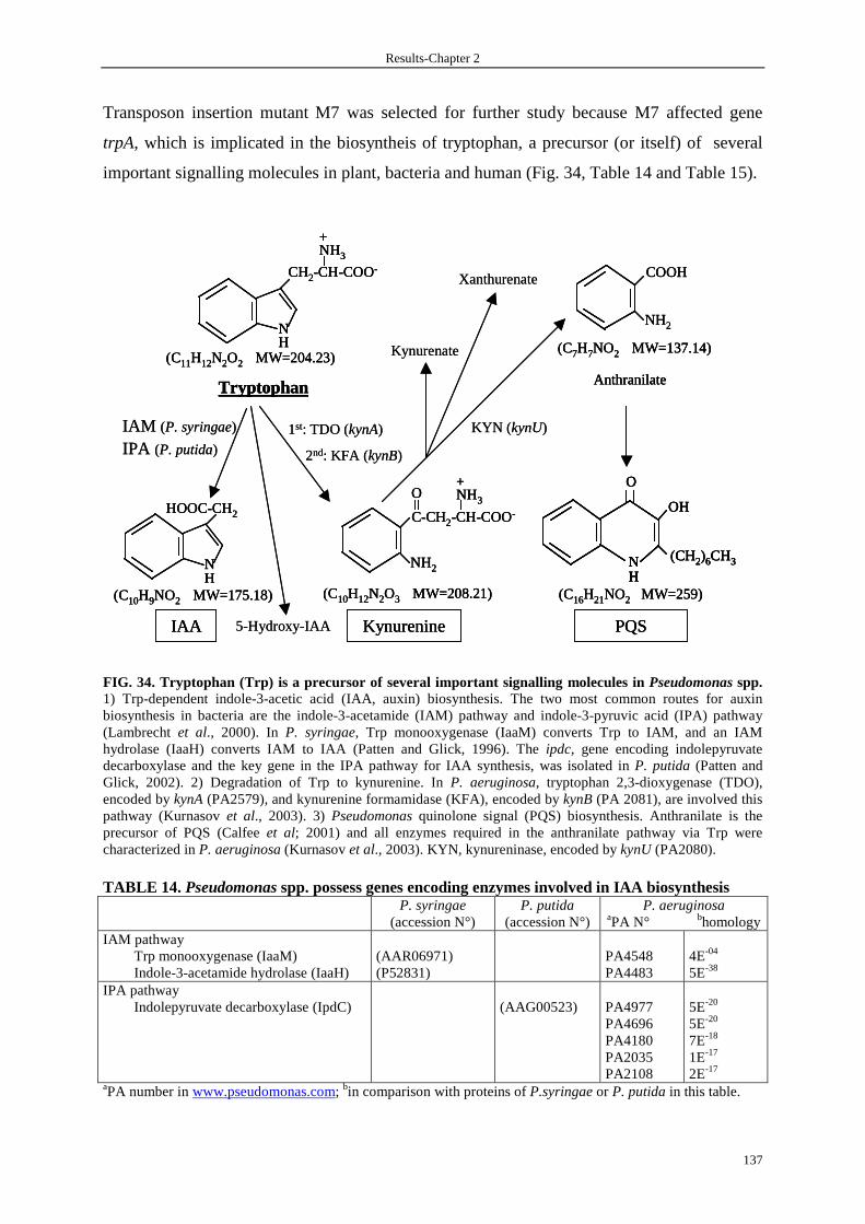
Results-Chapter 2
137
Transposon insertion mutant M7 was selected for further study because M7 affected gene
trpA, which is implicated in the biosyntheis of tryptophan, a precursor (or itself) of several
important signalling molecules in plant, bacteria and human (Fig. 34, Table 14 and Table 15).
FIG. 34. Tryptophan (Trp) is a precursor of several important signalling molecules in Pseudomonas spp. 1) Trp-dependent indole-3-acetic acid (IAA, auxin) biosynthesis. The two most common routes for auxin biosynthesis in bacteria are the indole-3-acetamide (IAM) pathway and indole-3-pyruvic acid (IPA) pathway (Lambrecht et al., 2000). In P. syringae, Trp monooxygenase (IaaM) converts Trp to IAM, and an IAM hydrolase (IaaH) converts IAM to IAA (Patten and Glick, 1996). The ipdc, gene encoding indolepyruvate decarboxylase and the key gene in the IPA pathway for IAA synthesis, was isolated in P. putida (Patten and Glick, 2002). 2) Degradation of Trp to kynurenine. In P. aeruginosa, tryptophan 2,3-dioxygenase (TDO), encoded by kynA (PA2579), and kynurenine formamidase (KFA), encoded by kynB (PA 2081), are involved this pathway (Kurnasov et al., 2003). 3) Pseudomonas quinolone signal (PQS) biosynthesis. Anthranilate is the precursor of PQS (Calfee et al; 2001) and all enzymes required in the anthranilate pathway via Trp were characterized in P. aeruginosa (Kurnasov et al., 2003). KYN, kynureninase, encoded by kynU (PA2080). TABLE 14. Pseudomonas spp. possess genes encoding enzymes involved in IAA biosynthesis
P. syringae (accession N°)
P. putida (accession N°)
P. aeruginosa aPA N° bhomology
IAM pathway Trp monooxygenase (IaaM) Indole-3-acetamide hydrolase (IaaH)
(AAR06971) (P52831)
PA4548 PA4483
4E-04
5E-38 IPA pathway
Indolepyruvate decarboxylase (IpdC)
(AAG00523)
PA4977 PA4696 PA4180 PA2035 PA2108
5E-20
5E-20
7E-18
1E-17
2E-17 aPA number in www.pseudomonas.com; bin comparison with proteins of P.syringae or P. putida in this table.
NH
HOOC-CH2
IAA
(C10H9NO2 MW=175.18)
C-CH2-CH-COO-
NH3
+O
NH2
Kynurenine
(C10H12N2O3 MW=208.21)
PQS
(C16H21NO2 MW=259)
OH
(CH2)6CH3NH
O
1st: TDO (kynA)
2nd: KFA (kynB)
KYN (kynU)IAM (P. syringae)
IPA (P. putida)
5-Hydroxy-IAA
Kynurenate
Xanthurenate COOH
NH2
(C7H7NO2 MW=137.14)
Anthranilate
NH
CH2-CH-COO-
NH3
+
(C11H12N2O2 MW=204.23)
Tryptophan
NH
HOOC-CH2
IAA
(C10H9NO2 MW=175.18)
NH
HOOC-CH2
IAA
(C10H9NO2 MW=175.18)
C-CH2-CH-COO-
NH3
+O
NH2
Kynurenine
(C10H12N2O3 MW=208.21)
C-CH2-CH-COO-
NH3
+O
NH2
Kynurenine
(C10H12N2O3 MW=208.21)
PQS
(C16H21NO2 MW=259)
OH
(CH2)6CH3NH
O
PQS
(C16H21NO2 MW=259)
OH
(CH2)6CH3NH
O
OH
(CH2)6CH3NHNH
O
1st: TDO (kynA)
2nd: KFA (kynB)
KYN (kynU)IAM (P. syringae)
IPA (P. putida)
5-Hydroxy-IAA
Kynurenate
Xanthurenate COOH
NH2
(C7H7NO2 MW=137.14)
Anthranilate
COOH
NH2
(C7H7NO2 MW=137.14)
Anthranilate
NH
CH2-CH-COO-
NH3
+
(C11H12N2O2 MW=204.23)
Tryptophan
NH
CH2-CH-COO-
NH3
+
(C11H12N2O2 MW=204.23)
Tryptophan

Results-Chapter 2
138
Table 15. Role of three Trp-derived signalling molecules, IAA, Kynurenine and PQS
Molecules Functions References
IAA 1. Phytohormone, plant growth and development (active at nM)
2. A reciprocal signal in bacteria-plant interactions
3. Inhibit the expression of genes involved in T-DNA transfer
(type IV secretion) in Agrobacterium tumefaciens
4. 0.5-1.0 mM IAA causes death of neutrophils
Woodward and Bartel, 2005
Lambrecht et al., 2000
Liu et al., 2006
Piers de melo et al., 1998
Kynurenine 1. Inhibit T cell proliferation
2. Inhibit NK cell proliferation (0.5 mM of L-kynurenine )
3. Inhibit NK cell activity (0.3-0.6 mM of L-kynurenine)
Hwu et al., 2000 ; Frumento et
al., 2002 ; Terness et al., 2002
Frumento et al., 2002
Della Chiesa et al., 2006
PQS Pseudomonas quorum-sensing signal Pesci et al., 1999
3.5 Tryptophan is implicated in the production of PARST
To determine the role of trpA in the production of PARST during the stationary phase, an
unmarked in-frame deletion mutation of trpA was constructed by allelic exchange as
described in Materials and Methods and was confirmed by PCR (Fig. 35).
FIG. 35. Mutant analysis by PCR. PCR was performed on genomic DNA prepared from different strains. Wt, wild-type CHA; mutant ∆trpA and ∆trpA carring a single copy of pClux[∆trpA (pClux)] or pSlux[∆trpA (pSlux)]. 1, cassette Gmlox, lox-flanked aacC1 encoding gentamicin resistance, is present in the chromosome; 2, only lox, but not aacC1, is present in the chromosome. M: DNA marker whose sizes are indicated at left side; Kb, kilobase.
0.5
1.0
1.5
Kb M Wt 1 2
∆trpA (pClux)
1 2
∆trpA (pSlux)
1 2
∆trpA
0.5
1.0
1.5
Kb M Wt 1 2
∆trpA (pClux)
1 2
∆trpA (pSlux)
1 2
∆trpA

Results-Chapter 2
139
Stationary phase culture supernatants were prepared from wild-type CHA and mutant ∆trpA,
and were added to the CHA (pClux) cultured from an initial OD600 of 0.05 in LB under TTSS-
inducing conditions. In contrast to the supernatant from the wild-type CHA, supernatant from
∆trpA lost its ability to inhibit exsCEBA expression and gave the same profile as ‘control’ in
which supernatant was replaced by VB (Fig. 36). Complementation of ∆trpA by adding 200
µg of Trp ml-1 in VB during the preparation of supernatant restored the inhibitory feature of
the supernatant from ∆trpA on the exsCEBA expression, on the contrarily, the addition of Trp
during the preparation of supernatant of the wild-type CHA didn’t change its inhibitory ability
on the exsCEBA expression compared to normal prepared supernatant (Fig. 36). These results
indicate that, trpA may be implicated in the production of PARST during the stationary phase
and PARST production seemed to be Trp-dependent.
FIG. 36. Trp is implicated in the production of PARST during the stationary phase. The wild-type CHA carrying a single copy of pClux transcriptional reporter at its chromosome was grown at 37°C in LB in the presence of stationary phase culture supernatants (indicated as asterisk), which were prepared in VB (CHA and ∆trpA) or VB supplemented with 200 µg of Trp ml-1 (CHA200 and ∆trpA200). RLU were measured over 4h of culture and calculated to normalized bacterial counts (6 × 10⁸ bac). Bars represent the mean plus the standard deviation of triplicate samples, and are the average of three independent experiments. Black bar and white bars represent TTSS-noninducing and inducing conditions, respectively. bac, bacteria. CI, control, TTSS-noninducing conditions (in the presence of 5 mM calcium); CII, control, TTSS-inducing conditions (5 mM EGTA and 20 mM MgCl2). 3.6 Tryptophan itself is not responsible for the TTSS inhibitory activity
Before investigating the role of Trp-derived metabolites in the regulation of TTSS expression,
we first we first determined the role of Trp. A serial dilution of L-Trp (Sigma) was added in
the CHA (pClux) cultured from an initial OD600 of 0.05 in LB under TTSS-noninducing and
inducing conditions, no difference was observed in culture with or without Trp (Fig. 37),
indicating that Trp itself is not responsible for the TTSS inhibitory activity.
0
200
400
600
800
1000
1200
1400
1600
1800
CI CII CHA* CHA200* ∆trpA* ∆trpA200*
RLU
/6*1
08ba
c (p
Clu
x)
0
200
400
600
800
1000
1200
1400
1600
1800
CI CII CHA* CHA200* ∆trpA* ∆trpA200*
RLU
/6*1
08ba
c (p
Clu
x)
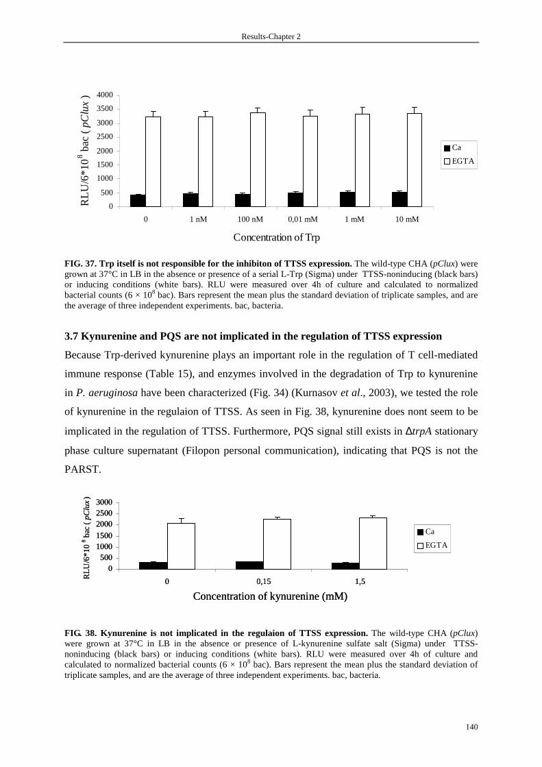
Results-Chapter 2
140
FIG. 37. Trp itself is not responsible for the inhibiton of TTSS expression. The wild-type CHA (pClux) were grown at 37°C in LB in the absence or presence of a serial L-Trp (Sigma) under TTSS-noninducing (black bars) or inducing conditions (white bars). RLU were measured over 4h of culture and calculated to normalized bacterial counts (6 × 10⁸ bac). Bars represent the mean plus the standard deviation of triplicate samples, and are the average of three independent experiments. bac, bacteria.
3.7 Kynurenine and PQS are not implicated in the regulation of TTSS expression
Because Trp-derived kynurenine plays an important role in the regulation of T cell-mediated
immune response (Table 15), and enzymes involved in the degradation of Trp to kynurenine
in P. aeruginosa have been characterized (Fig. 34) (Kurnasov et al., 2003), we tested the role
of kynurenine in the regulaion of TTSS. As seen in Fig. 38, kynurenine does nont seem to be
implicated in the regulation of TTSS. Furthermore, PQS signal still exists in ∆trpA stationary
phase culture supernatant (Filopon personal communication), indicating that PQS is not the
PARST.
FIG. 38. Kynurenine is not implicated in the regulaion of TTSS expression. The wild-type CHA (pClux) were grown at 37°C in LB in the absence or presence of L-kynurenine sulfate salt (Sigma) under TTSS-noninducing (black bars) or inducing conditions (white bars). RLU were measured over 4h of culture and calculated to normalized bacterial counts (6 × 10⁸ bac). Bars represent the mean plus the standard deviation of triplicate samples, and are the average of three independent experiments. bac, bacteria.
Concentration of kynurenine (mM)
0500
10001500
200025003000
0 0,15 1,5
RLU
/6*1
0 8 b
ac (
pClu
x)
Ca
EGTA
Concentration of kynurenine (mM)
0500
10001500
200025003000
0 0,15 1,5
RLU
/6*1
0 8 b
ac (
pClu
x)
Ca
EGTA
0
500
1000
1500
2000
2500
3000
3500
4000
0 1 nM 100 nM 0,01 mM 1 mM 10 mM
Concentration of Trp
RL
U/6
*10
8 bac
( pC
lux
)
Ca
EGTA
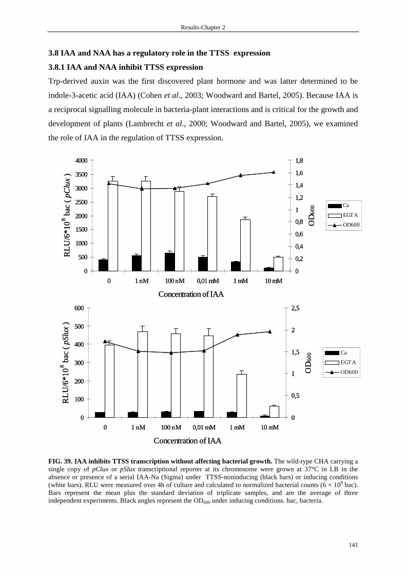
Results-Chapter 2
141
3.8 IAA and NAA has a regulatory role in the TTSS expression
3.8.1 IAA and NAA inhibit TTSS expression
Trp-derived auxin was the first discovered plant hormone and was latter determined to be
indole-3-acetic acid (IAA) (Cohen et al., 2003; Woodward and Bartel, 2005). Because IAA is
a reciprocal signalling molecule in bacteria-plant interactions and is critical for the growth and
development of plants (Lambrecht et al., 2000; Woodward and Bartel, 2005), we examined
the role of IAA in the regulation of TTSS expression.
FIG. 39. IAA inhibits TTSS transcription without af fecting bacterial growth. The wild-type CHA carrying a single copy of pClux or pSlux transcriptional reporter at its chromosome were grown at 37°C in LB in the absence or presence of a serial IAA-Na (Sigma) under TTSS-noninducing (black bars) or inducing conditions (white bars). RLU were measured over 4h of culture and calculated to normalized bacterial counts (6 × 10⁸ bac). Bars represent the mean plus the standard deviation of triplicate samples, and are the average of three independent experiments. Black angles represent the OD600 under inducing conditions. bac, bacteria.
0
500
1000
1500
2000
2500
3000
3500
4000
0 1 nM 100 nM 0,01 mM 1 mM 10 mM
Concentration of IAA
RL
U/6
*10 8 b
ac (
pClu
x)
0
0,2
0,4
0,6
0,8
1
1,2
1,4
1,6
1,8
OD
60
0 Ca
EGTA
OD600
0
100
200
300
400
500
600
0 1 nM 100 nM 0,01 mM 1 mM 10 mM
Concentration of IAA
RL
U/6
*10
8 bac
( pS
lux)
0
0,5
1
1,5
2
2,5O
D6
00 Ca
EGTA
OD600
0
500
1000
1500
2000
2500
3000
3500
4000
0 1 nM 100 nM 0,01 mM 1 mM 10 mM
Concentration of IAA
RL
U/6
*10 8 b
ac (
pClu
x)
0
0,2
0,4
0,6
0,8
1
1,2
1,4
1,6
1,8
OD
60
0 Ca
EGTA
OD600
0
100
200
300
400
500
600
0 1 nM 100 nM 0,01 mM 1 mM 10 mM
Concentration of IAA
RL
U/6
*10
8 bac
( pS
lux)
0
0,5
1
1,5
2
2,5O
D6
00 Ca
EGTA
OD600

Results-Chapter 2
142
As shown in Fig. 39, though IAA sodium salt (IAA-Na, Sigma) having no effect on TTSS
expression up to concentration of 0.01mM, 1 mM IAA-Na had an evident inhibition on both
exsCEBA expression and exoS expression and 10 mM IAA-Na inhibit totally the TTSS
expression with no inhibition of the growth of bacteria.
We also tested the other three commercial IAA derivatives (formula of sodium or potassium
salt, Sigma), including indole-3-butyric acid (IBA), 1-Naphthaleneacitic acid (NAA) and (2,4-
Dichlorophenoxy) acetic acid (2,4-D) (Woodward and Bartel, 2005), and only NAA-K had a
similar inhibitory role to IAA on the TTSS expression (Fig. 40).
FIG. 40. Both IAA and NAA inhibit TTSS transcriptio n induced by EGTA. The wild-type CHA carrying a single copy of pClux transcriptional reporter at its chromosome were grown at 37°C in LB under TTSS-noninducing (in the presence of 5 mM calcium) or inducing conditions (in the presence of 5 mM EGTA and 20 mM MgCl2) in supplemented with or not IAA-Na (Sigma) and derivatives , including IBA-K, NAA-K, 2,4-D-Na (Sigma), of dirrerent concentraions (0, 1 µM, 10 µM, 100 µM, 1 mM and 10 mM). RLU were measured over 4h of culture and calculated to normalized bacterial counts (6 × 10⁸ bac). Ca, calcium.
Besides in vitro calcium depletion growth conditions, growth in the presence of serum leads
to generalized TTSS expression and secretion (Vallis et al., 1999). Therefore, we asked
whether IAA could inhibit TTSS expression in the presence of human serum AB stimuli. As
seen in Fig. 41, exsCEBA transcription was partially inhibited in the presence of 1mM IAA-
Na and NAA-K but not IBA-K or 2,4-D-Na. No pH change was observed due to the use of
IAA-Na and its derivatives. Considering that IBA is identical to IAA except for two
0
500
1000
1500
2000
2500
3000
3500
4000
4500
0
10 uM 1 m
M 0
10 uM 1 m
M 0
10 uM 1 m
M 0
10 uM 1 m
M 0
10 uM 1 m
M 0
10 uM 1 m
M 0
10 uM 1 m
M 0
10 uM 1 m
M
RL
U/6
*108 b
ac (p
Clu
x)
Ca EGTA
IAA IBA NAA 2,4-D IAA IBA NAA 2,4-D
Concentration of IAA and its derivatives
0
500
1000
1500
2000
2500
3000
3500
4000
4500
0
10 uM 1 m
M 0
10 uM 1 m
M 0
10 uM 1 m
M 0
10 uM 1 m
M 0
10 uM 1 m
M 0
10 uM 1 m
M 0
10 uM 1 m
M 0
10 uM 1 m
M
RL
U/6
*108 b
ac (p
Clu
x)
Ca EGTA
IAAIAA IBAIBA NAANAA 2,4-D2,4-D IAAIAA IBAIBA NAANAA 2,4-D2,4-D
Concentration of IAA and its derivatives
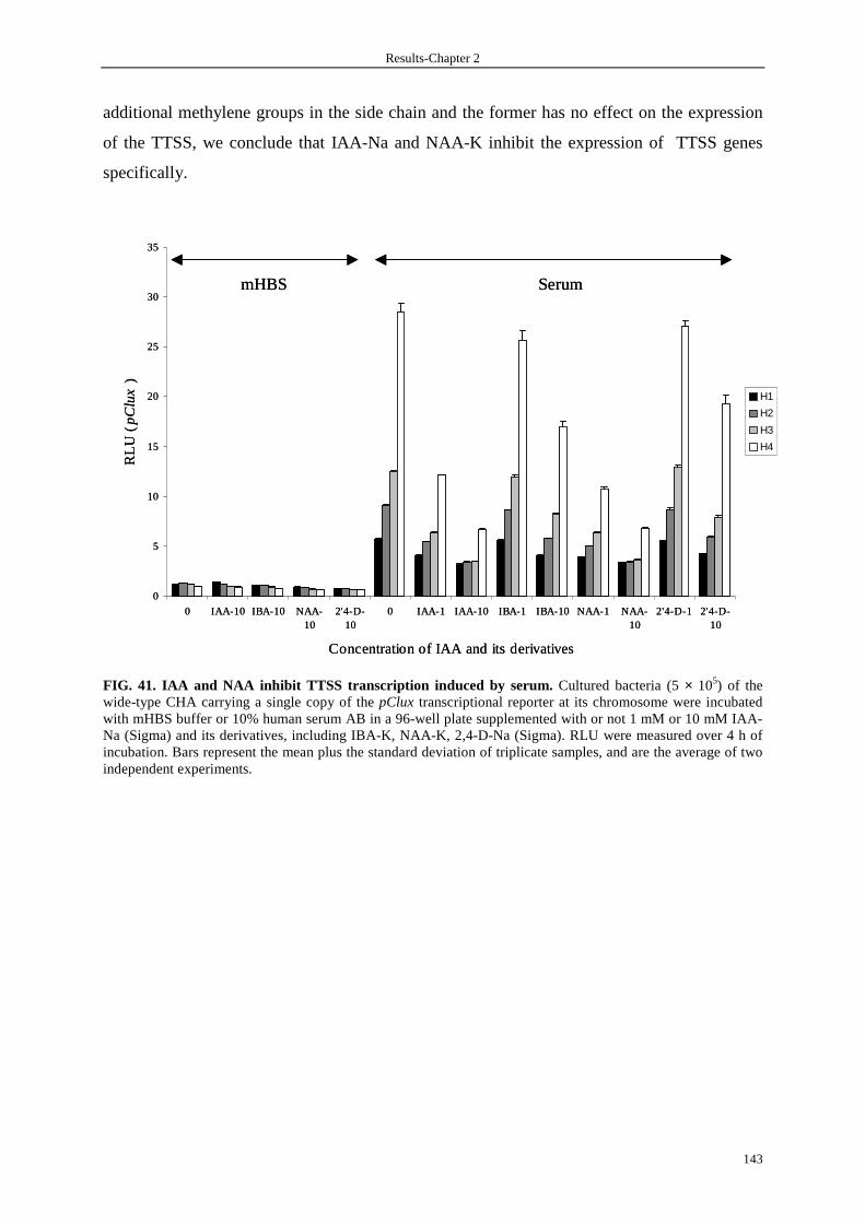
Results-Chapter 2
143
additional methylene groups in the side chain and the former has no effect on the expression
of the TTSS, we conclude that IAA-Na and NAA-K inhibit the expression of TTSS genes
specifically.
FIG. 41. IAA and NAA inhibit TTSS transcription ind uced by serum. Cultured bacteria (5 × 10⁵) of the wide-type CHA carrying a single copy of the pClux transcriptional reporter at its chromosome were incubated with mHBS buffer or 10% human serum AB in a 96-well plate supplemented with or not 1 mM or 10 mM IAA-Na (Sigma) and its derivatives, including IBA-K, NAA-K, 2,4-D-Na (Sigma). RLU were measured over 4 h of incubation. Bars represent the mean plus the standard deviation of triplicate samples, and are the average of two independent experiments.
0
5
10
15
20
25
30
35
0 IAA-10 IBA-10 NAA-10
2'4-D-10
0 IAA-1 IAA-10 IBA-1 IBA-10 NAA-1 NAA-10
2'4-D-1 2'4-D-10
Concentration of IAA and its derivatives
RL
U (
pC
lux
)
H1
H2
H3
H4
mHBS Serum
0
5
10
15
20
25
30
35
0 IAA-10 IBA-10 NAA-10
2'4-D-10
0 IAA-1 IAA-10 IBA-1 IBA-10 NAA-1 NAA-10
2'4-D-1 2'4-D-10
Concentration of IAA and its derivatives
RL
U (
pC
lux
)
H1
H2
H3
H4
mHBS Serum
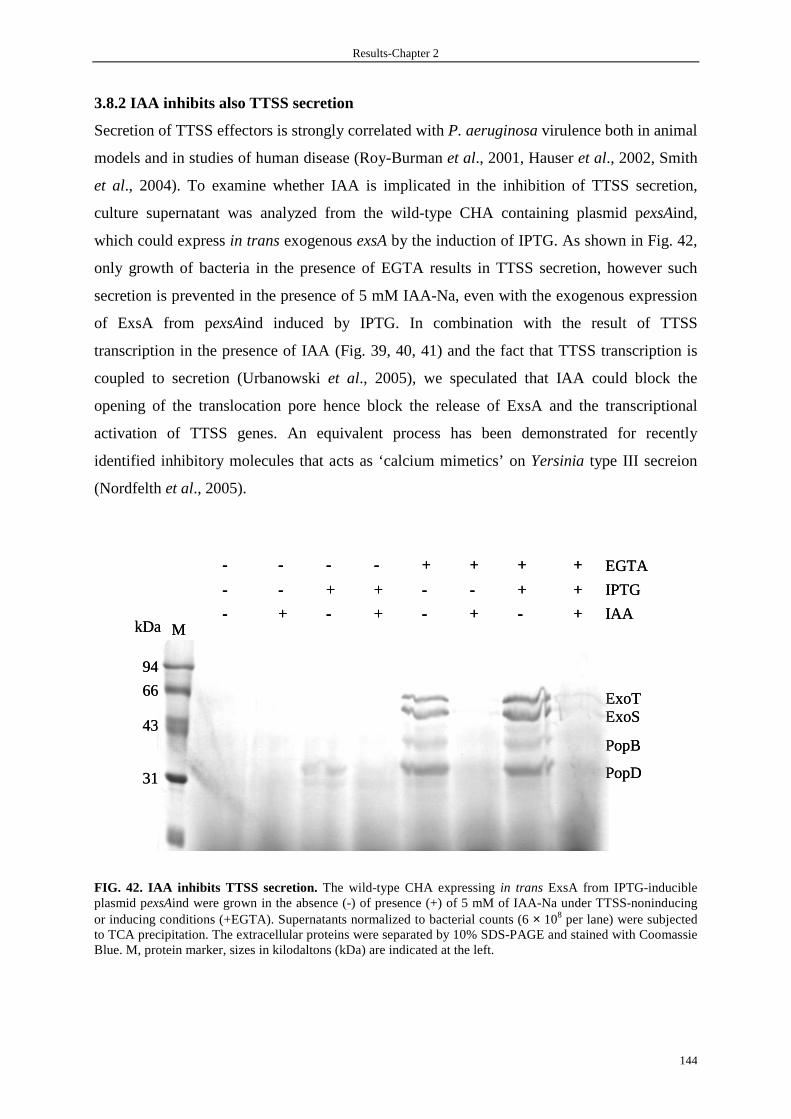
Results-Chapter 2
144
3.8.2 IAA inhibits also TTSS secretion
Secretion of TTSS effectors is strongly correlated with P. aeruginosa virulence both in animal
models and in studies of human disease (Roy-Burman et al., 2001, Hauser et al., 2002, Smith
et al., 2004). To examine whether IAA is implicated in the inhibition of TTSS secretion,
culture supernatant was analyzed from the wild-type CHA containing plasmid pexsAind,
which could express in trans exogenous exsA by the induction of IPTG. As shown in Fig. 42,
only growth of bacteria in the presence of EGTA results in TTSS secretion, however such
secretion is prevented in the presence of 5 mM IAA-Na, even with the exogenous expression
of ExsA from pexsAind induced by IPTG. In combination with the result of TTSS
transcription in the presence of IAA (Fig. 39, 40, 41) and the fact that TTSS transcription is
coupled to secretion (Urbanowski et al., 2005), we speculated that IAA could block the
opening of the translocation pore hence block the release of ExsA and the transcriptional
activation of TTSS genes. An equivalent process has been demonstrated for recently
identified inhibitory molecules that acts as ‘calcium mimetics’ on Yersinia type III secreion
(Nordfelth et al., 2005).
FIG. 42. IAA inhibits TTSS secretion. The wild-type CHA expressing in trans ExsA from IPTG-inducible plasmid pexsAind were grown in the absence (-) of presence (+) of 5 mM of IAA-Na under TTSS-noninducing or inducing conditions (+EGTA). Supernatants normalized to bacterial counts (6 × 10⁸ per lane) were subjected to TCA precipitation. The extracellular proteins were separated by 10% SDS-PAGE and stained with Coomassie Blue. M, protein marker, sizes in kilodaltons (kDa) are indicated at the left.
ExoT66
94
43
31
MkDa
EGTA
IPTG
IAA
ExoS
PopB
PopD
-
-
-
-
-
+
-
+
-
-
+
+
+
-
-
+
-
+
+
+
-
+
+
+
ExoT66
94
43
31
MkDa
EGTA
IPTG
IAA
ExoS
PopB
PopD
-
-
-
-
-
-
-
-
+
-
-
+
-
+
-
-
+
-
-
+
+
-
+
+
+
-
-
+
-
-
+
-
+
+
-
+
+
+
-
+
+
-
+
+
+
+
+
+

Results-Chapter 2
145
3.8.3 VirA is not involved in the IAA-dependent inhibition of TTSS expression
Since IAA can inhibit the expression of TTSS and might be an inhibitory signal during the
stationary phase, we looked for the inhibition of bacterial secretion systems by similar
molecules. Agrobacterium tumefaciens is a pathogen that cause crown gall tumors in a variety
of plants by transferring the oncogenic DNA fragments (T-DNA) into plant cells via its type
IV secretion system (Introduction 3.2.2). VirA/G, a two component regulatory system, senses
the plant signal molecules, typically phenolic compound acetosyringone, and activates the
expression of vir genes, which are responsible for T-DNA processing and transfer (Brencic
and Winans, 2005). It was recently shown that IAA inactivates vir genes expression by
competing with acetosyringone for interaction with VirA (Liu et al., 2006). By searching
Pseudomonas genome database, we found a protein (PA1243), annotated as a probable
sensor/response regulator hybrid, is 49% similar to VirA (www.pseudomonas.com, Stover et
al., 2000). To investigate if Pseudomonas VirA is the receptor of IAA and has a regulatory
role in genes expression, such as TTSS expression, we deleted the gene virA and yielded the
mutant ∆virA containing the chromosomal transcriptional gene reporter pClux and pSlux. As
seen in Fig. 43., the deletion of virA has no influence on the activation of exsCEBA expression
as 10 mM NAA still inhibit the activation of exsCEBA expression in TTSS-inducing
conditions in the absence of VirA sensor. This result indicates that VirA is not involved in the
NAA-dependent inhibition of TTSS expression in P. aeruginosa.
FIG. 43. VirA is not involved in the IAA-dependent inhibition of TTSS expression in P. aeruginosa. The wild-type CHA and mutant ∆virA carrying a single copy of pClux transcriptional reporter at its chromosome were grown at 37°C in LB in the absence or presence of 1mM IAA-Na (IAA-1) or 10 mM IAA-Na (IAA-10) under TTSS-noninducing (-) or inducing conditions (+). RLU were measured over 4h of culture and calculated to normalized bacterial counts (6 × 10⁸ bac). Bars represent the mean plus the standard deviation of triplicate samples, and are the average of three independent experiments. bac, bacteria.
0
500
1000
1500
2000
2500
3000
RL
U/6
*10
8 b
ac (
pC
lux)
0
IAA-1
IAA-10
CHA- CHA+ ∆virA- ∆virA+
0
500
1000
1500
2000
2500
3000
RL
U/6
*10
8 b
ac (
pC
lux)
0
IAA-1
IAA-10
CHA- CHA+ ∆virA- ∆virA+

Results-Chapter 2
146
FIG. 44. HPLC analysis of stationary phase culture supernatants. Stationary phase culture supernatants from wild-type CHA and ∆trpA were prepared in VB (CHA and ∆trpA) or VB supplemented with 200 µg of Trp ml-1 (CHA200 and ∆trpA200), and HPLC was performed as described in Materials and Methods. Arrow showed the peak, which was only present in the supernatant of the wild-type CHA, absent in the supernatant of ∆trpA but reappeared in the supernatant of ∆trpA when 200 µg of Trp ml-1 was added during the preparation of supernatants.
CHA
∆trpA
CHA200
∆trpA200
CHA
∆trpA
CHA200
∆trpA200

Results-Chapter 2
147
3.9 Tentative of determination of the nature of PARST
Based on the obtained results before, the production of PARST during the stationary phase
depends on Trp, precursor of IAA, which could inhibit both TTSS secretion and
expression. We asked here if IAA is indeed in the stationary phase culture supernatant. Using
Salkowski’s reagent as previously described (Patten and Glick, 2002), we haven’t detected
IAA in the stationary phase culture supernatant in the wild-type CHA and no difference was
observed between the supernatants of different phases (data not shown). It seemed that IAA
should not be PARST, however we could not rule out that the special analogs of IAA might
exist in the supernatant and regulate TTSS expression in vivo at very low concentration,
which could not be detected by Salkowski’s reagent. To identify the nature of PARST, the
wild-type CHA and the ∆trpA were collected from stationary phase and then grown overnight
in the VB or VB supplemented with 200 µg ml-1 Trp. Supernatants were collected by
centrifugation, filtrated through a 0.22 µM filter, and then were passed and analysed by HPLC
on C18 column as described in Materials and Methods 21. As seen in Fig. 44, peak 44.925
(min of elution, indicated by the arrow) presented in the wild-type CHA was absent from the
∆trpA and this same peak reappeared after supplementation by Trp during the culture of strain
∆trpA.
FIG. 45. Peak analysis on the activation of exsCEBA expression. During the procedure of HPLC, peak of CHA (Peak-CHA), indicated in Fig. 44, as well as that of trpA (‘peak’-∆trpA) appeared at the same time point were collected, evaporated and incubated with CHA carring pClux transcriptional report under TTSS-inducing conditions (white bars). RLU were measured over 4h of culture and calculated to normalized bacterial counts (6 × 10⁸ ). Bar represent the mean plus the standard deviation of 3 reads. CI, control, TTSS-noninducing conditions (black bar); CII, control, TTSS-inducing conditions. Bac, bacteria.
0
500
1000
1500
2000
2500
RL
U/6
*10 8 b
ac (
pClu
x)
CI CII Peak-CHA ‘Peak’-∆trpA0
500
1000
1500
2000
2500
RL
U/6
*10 8 b
ac (
pClu
x)
CI CII Peak-CHA ‘Peak’-∆trpA

Results-Chapter 2
148
To determine its role on the TTSS expression, peak 44.925 (CHA) was collected, evaporated
and resuspended in LB containing CHA (pClux) with an initial OD600 of 0.05 under TTSS-
inducing conditions. A weak inhibition of exsCEBA expression (Fig. 45) was observed in the
presence of resuspended peak 44.925 (CHA) compared with the presence of the solution
collected from ∆trpA during HPLC the same time point at which the peak 44.925 (CHA) was
collected. We could not exclude the possibility that PARST was not in peak 44.925 (CHA),
but even PARST was indeed in the peak, it might have lost a part of its activity during the
preparation.
FIG. 46. m/z 224 is uniquely present in the stationary phase culture supernatant of CHA. Fractions of the peak 44.925 CHA (as indicated in Fig. 44) and that from ∆trpA collected at the same time point during the separation by HPLC were further analyzed by LC-MS as described in Materials and Methods. The extracted ion chromatograms here showed the only detected differences in a scan range from m/z 100 to 1500 between two strains. m/z present only in the wild-type CHA were indicated by arrows.
1 0 0 1 2 0 1 4 0 1 6 0 1 8 0 2 0 0 2 2 0 2 4 0 2 6 00
%
103 .0 119 .32 57 .313 5 .2
21 7 .31 49 .1173 .3 229 .3
261 .2
∆trpA
m/z
100
80
60
40
20
Rel
ativ
e in
tens
ity
(%)
100 120 140 160 180 200 220 240 2600
%
207.3
103.2
119.2 135.2
137.2149.3
179.3
247.2
225.2
208.3
211.3
239.3
226.3248.2
249.2 263.2
[M+H] +
[M+Na]+CHA
m/z
100
80
60
40
20
Rel
ativ
e in
tens
ity
(%)
[M-H2O+H]+
1 0 0 1 2 0 1 4 0 1 6 0 1 8 0 2 0 0 2 2 0 2 4 0 2 6 00
%
103 .0 119 .32 57 .313 5 .2
21 7 .31 49 .1173 .3 229 .3
261 .2
∆trpA
m/z
100
80
60
40
20
Rel
ativ
e in
tens
ity
(%)
1 0 0 1 2 0 1 4 0 1 6 0 1 8 0 2 0 0 2 2 0 2 4 0 2 6 00
%
103 .0 119 .32 57 .313 5 .2
21 7 .31 49 .1173 .3 229 .3
261 .2
∆trpA
m/z
100
80
60
40
20
Rel
ativ
e in
tens
ity
(%)
100 120 140 160 180 200 220 240 2600
%
207.3
103.2
119.2 135.2
137.2149.3
179.3
247.2
225.2
208.3
211.3
239.3
226.3248.2
249.2 263.2
[M+H] +
[M+Na]+CHA
m/z
100
80
60
40
20
Rel
ativ
e in
tens
ity
(%)
[M-H2O+H]+
100 120 140 160 180 200 220 240 2600
%
207.3
103.2
119.2 135.2
137.2149.3
179.3
247.2
225.2
208.3
211.3
239.3
226.3248.2
249.2 263.2
[M+H] +
[M+Na]+CHA
m/z
100
80
60
40
20
Rel
ativ
e in
tens
ity
(%)
[M-H2O+H]+
Results-Chapter 2
149
3.10 PARST might be a molecule of m/z 224
Supposing PARST is indeed in the peak 44.925 (CHA) (Fig. 44), we collected this peak as
several fractions and that from ∆trpA at the same time point during the separation by HPLC.
LC-MS analysis showed that 4 molecular ions, including m/z 207.3 [M-H2O+H] +, m/z 225.2
[M+H] +, m/z 247.2 [M+Na]+ and m/z 239 (possibly [M+CH2+H]+), were uniquely present
in the peak of 44.925 (CHA) (Fig. 46).
Therefore, the molecule present in the stationary phase culture supernatant from the wild-type
CHA should be m/z 224.2 (225.2 minus 1, molecular weight of atom H) and it was
reminiscent of NAA-K simply due to the facts that NAA-K has a molecular weigh of 224.3
and can inhibits TTSS expression. So what’s the relationship between the m/z 224.2 and the
NAA-K? Mass spectra analysis of commercial NAA-K exhibited 3 m/z, includng m/z 187.07,
225.03 and 262.99 corresponding to [NAA+H]+, [NAA+K] + and [NAA+2K-H]+ respectively.
It is possible that m/z 207.3, 225.2 and 247.2 in the peak 44.925 (CHA) might correspond to
[NAA-H 2O+K]+, [NAA+K] + and [NAA+K+Na-H]+ respectively. It is normal that no NAA-
2K was detected because there is little K but rather Na in the culture medium. Taken together,
we speculated that NAA itself or its derivatives could be produced in the stationary phase and
inhibits TTSS expression.
4. Discussion
The expression of the TTSS is controlled by multicomponent regulatory networks which
integrate a diverse set of environmental cues, probably to restrict the energy-consuming
expression of 20 or more proteins to the correct place and time only when they are really
needed (Hueck, 1998). Although TTSS is very likely important in acute human infections, it
is not necessary for maintaining chronic infection (Dacheux et al., 2000; Roy-Burman et al.,
2001; Hauser et al., 2002; Eriksson et al., 2003; Jain et al., 2004; Dahan et al., 2004; Lucchini
et al., 2005; Lee et al., 2005; Faucher et al., 2006). In vitro culture condition, the expression
of the TTSS seems to be cell density-dependent (this study). Presumably, just as a mechanism
exists for activating the TTSS genes when they are needed, a mechanism should exist for
inactivating the same TTSS genes when they are not required. The data presented here
described PARST, an inhibitor of the expression of TTSS, that appears in the stationary phase
of culture (high cell density).
Considering that QS is a generic regulatory mechanism controlling in a population density-
dependent manner the expression of virulence genes, including TTSS genes (Juhas et al.,
2005), we ruled out the possibility that the PARST is associated with the QS system (Fig. 28).

Results-Chapter 2
150
The fact that the production of PARST is Trp-dependent (Fig. 36) in combination with the
fact that Trp is the precursor of IAA, which has several important features described below,
we tested the role of IAA and IAA-like compounds in the regulation of TTSS expression.
IAA, the first discovered plant hormone, is critical for plant growth and development and is
often potent at nanomolar concentrations in plants (Woodward and Bartel, 2005). Although it
is widely accepted that plants use both Trp-dependent and Trp-independent routes to
synthesize IAA, none of the pathways is yet defined to the level of knowing each relevant
gene, enzyme and intermediate. It is also recognized that many bacteria are able to produce
IAA mainly using Trp-dependent pathway (Lambrecht et al., 2000) and such pathway is
indeed observed in Pseudomonas (Patten and Glick, 1996; 2002) (Fig. 34 and Table 14).
Recent studies showed that several strains of P. aeruginosa isolated from soil (Bano et al.,
2003; Kumar et al., 2005; Ayyadurai et al., 2006) could produce IAA and maximum
production of IAA (19.1 µg ml-1) by strain P. aeruginosa FP10 was observed after 42-h
incubation in Dworkin and Foster (DF) salts medium supplemented with 500 µg ml-1 of L-
tryptophan (Ayyadurai et al., 2006).
IAA and one of IAA-like compounds, NAA, could indeed inhibit the expression and the
secretion of the TTSS at millimolar concentrations (Fig. 39, 40, 41, 42). However, no IAA
was detected from our stationary phase culture supernatant using Salkowski’s reagent. Thus,
IAA does not seem to be the component of PARST, though genes encoding enzymes needed
in the Trp-dependent biosynthesis of IAA do exist in the genome of P. aeruginosa PAO1
(Table 14) (www.pseudomonas.com). However, we still have reasons to believe that PARST
could be IAA-like compound, because m/z 225.2 (Fig. 46), corresponding to the molecular
weight of [NAA+K]+, is only present in the supernatant of the wild-type CHA but not the
mutant with the deletion of trpA whose product is the precursor in the biosynthesis of IAA. It
is possible that undetectable IAA in the culture supernatant might be due to the dilution of
IAA, if exist, and the limit of the method we used, in such case, it is possible that IAA or
IAA-like compound at very low concentration could be highly active due to its special form.
It is worth noting that 1mM of IAA has cytotoxic effect on the cultured neutrophils (Pires de
Melo et al., 1998). Therefore, it would be interesting if IAA could play a role in the regulation
of the transition between two totally different events: inhibiting the expression of the TTSS on
the one hand and activating the pathway to kill the neutrophils on the other hand.
Though one study showed that IAA can inhibit the expression of genes required for the
transfer of T-DNA (type IV secretion) in A. tumefaciens via VirA, a sensor kinase of a two-

Results-Chapter 2
151
component regulatory system (Liu et al., 2006), VirA homolog of P. aeruginosa is not
involved in the IAA-dependent inhibition of the expression of TTSS (Fig. 43).
Recently, one study showed that LadS and RetS, two hybrid sensor kinases of Pseudomonas,
are implicated in the transition between activation of the TTSS (acute infection) and the
biofilm (chronic infection) by sensing an unknown signal (Ventre et al., 2006). Therefore, it
would be interesting to investigate if IAA or NNA is the signal sensed by LadS/RetS
pathway.
We don’t know why PARST, probably Trp-derived metabolite, is implicated in the regulation
of TTSS, but it is true that metabolic signals/stresses have a profound influence on the TTSS
expression. Mutants lacking pyruvate dehydrogenase (aceA or aceB) (Dacheux et al., 2002) or
a glucose transport regulator (gltR) (Wolfgang et al., 2003) fail to induce the TTSS expression
under calcium depletion conditions. Similarly, overproduction of MDR efflux pumps (Linares
et al., 2005) or overexpression of genes involved in histidine transport and metabolism
(Rietsch et al., 2004) prevents the TTSS expression. A recent study suggests that one
metabolite controlling the TTSS expression is derived from acetyl-CoA (Rietsch and
Mekalanos, 2006).
It is not surprising that P. aeruginosa has multiple regulatory networks, because 8% of its
genome codes for regulatory genes, indicating that P. aeruginosa has dynamic and
complicated regulatory mechanisms responding to various environmental conditions
(Greenberg, 2000). In addition to its role in TTSS genes induction, IAA or NAA may serve as
a signal to Pseudomonas that the living environment is changing, from a low cell density to a
high cell density state, and that the bacterium should modify its gene expression to favarite its
survival. This possibility could be explored through the use of DNA microarrays or all
proteomic analysis under conditions in which the IAA is added or not.

Conclusion and perspectives
152
Conclusion and perspectives

Conclusion and perspectives
153
En français P. aeruginosa, comme beaucoup de bactéries Gram-négatives pathogènes de plante ou
d’animal, possède un facteur de virulence majeur, le système de sécrétion de type III (SSTT).
Il est composé de plus de trente protéines qui s’assemblent pour former un machinerie
complexe dont le rôle est de délivrer des effecteurs protéiques directement dans le cytoplasme
des cellules cibles (Yahr et al., 1995; Frank 1997) où ils agiront pour pervertir différents
mécanismes cellulaires de l’hôte, conférant finalement à la bactérie un avantage pour sa
survie (Hueck, 1998). La construction, et l’utilisation de ce système de virulence représente
un coût énergétique considérable pour la bactérie qui en contre partie a mis en place un
contrôle extrêmement précis tant au niveau de l’expression des gènes qu’à celui de
l’utilisation du SSTT.
PsrA est un activateur trancriptionnel du SSTT de P. aeruginosa Chez P. aeruginosa, l’expression de l’ensemble des gènes du SSTT est contrôlée par ExsA,
une protéine qui se fixe sur des séquences spécifiques situées dans les régions régulatrices des
différents opérons du SSTT, pour activer leur transcription (Hovey and Frank, 1995). Ceci
permet de supposer que la bactérie peut contrôler l’expression du SSTT soit en modifiant
l’activité de ExsA soit en contrôlant le niveau d’expression de ExsA lui-même. La première
situation a été très bien étudiée et a permis de caractériser les systèmes de contrôle de
l’activité de ExsA (McCaw et al., 2002; Dasgupta et al., 2004; Ha et al., 2004; Rietsch et al.,
2005; Urbanowski et al., 2005). Ici nous avons découvert le rôle d’un nouveau facteur de
transcription, PsrA, nécessaire pour que la transcription de l’opéron exsCEBA qui contient
exsA et par conséquent celle des autres opérons du SSTT soient maximales.
La recherche des régulateurs impliqués dans le contrôle du niveau d’expression de l’opéron
exsCEBA est basée sur l’observation faite au laboratoire que des extraits bactériens totaux
préparés à partir de culture d’un mutant de délétion de exsA contiennent toujours une activité
de fixation spécifique à l’ADN du promoteur de l’opéron (pC). Une protéine, PsrA, a pu être
identifiée à partir de l’analyse de fractions enrichies en activité de fixation sur pC. La protéine
recombinante PsrA (rPsrA) a été préparée pure et elle est effectivement capable de se fixer
spécifiquement sur pC. Bien que l’affinité de fixation sur pC soit relativement modeste, cela
suggère que PsrA peut être un régulateur de l’opéron exsCEBA et donc de l’ensemble des
gènes du SSTT. La démonstration du rôle joué par PsrA dans l’expression des gènes du SSTT
a été clairement établie par l’étude du phénotype d’un mutant de délétion de psrA (∆psrA)
obtenu au cours de ce travail. En effet, l’analyse par gène rapporteur a montré que PsrA est
nécessaire pour obtenir une activation totale de la transcription de exsA dans différentes
conditions d’induction comme la déplétion calcique, l’addition de sérum ou le contact
cellulaire. La délétion de psrA se traduit par une diminution d’un facteur 2 ou 3 du niveau
d’expression de exsA et peut être complémentée par l’addition, en trans du gène psrA.
La diminution du niveau d’expression de exsA se traduit d’une part par une diminution de
Conclusion and perspectives
154
l’expression des gènes du SSTT, en particulier de la toxine ExoS et, d’autre part par une
diminution de la sécrétion de ExoS et ExoT. Finalement, le rôle positif de PsrA dans
l’expression du SSTT et, par conséquent, dans la virulence de la bactérie a été confirmé par la
plus faible résistance à la phagocytose de la souche ∆psrA.
Curieusement, les extraits du mutant ∆psrA continuent d’avoir cette forte activité de fixation
au promoteur de l’opéron exsCEBA, indiquant que la protéine PsrA n’est pas la protéine
responsable de cette activité. Si on considère que c’est en condition de non activation du
SSTT que l’on mesure la plus forte activité de fixation dans les extraits (Figure 25), il est tout
à fait légitime d’émettre l’hypothèse que la protéine responsable agirait plutôt comme un
inhibiteur de transcription, alors que PsrA agit clairement comme activateur. La présence de
ces différentes activités de fixation au promoteur pC, indépendantes de ExsA et de PsrA
suggèrent bien que d’autres facteurs de transcription pourraient agir de concert pour donner à
la bactérie une grande flexibilité et une grande précision dans le niveau d’expression du
SSTT. La caractérisation de ces régulateurs doit se poursuivre afin non seulement d’obtenir
plus d’informations sur la capacité des bactéries à contrôler finement le niveau d’expression
de leurs gènes, mais aussi pour fournir des cibles potentielles permettant la conception de
nouveaux moyens de lutte contre les infections bactériennes.
Le PARST (Pseudomonas autorepressor of secretion three) inhibe l’expression du SSTT de P. aeruginosa en phase stationnaire Il avait déjà été observé au laboratoire que l’expression des gènes du SSTT chez P.
aeruginosa dépend de la densité cellulaire avec un maximum de l’expression des gènes
comme exsCEBA ou exoS vers le milieu de la phase exponentielle correspondant à une DO600
de 1.5 à 2 suivit d’une rapide diminution, voire annulation complète de l’expression lorsque
que la DO dépasse 2. Ceci indique qu’un mécanisme de type ‘quorum sensing’ intervient pour
inactiver l’expression du SSTT en phase stationnaire. De plus, les surnageants de culture de
notre souche ou d’autres souches de P.aeruginosa obtenus en fin de phase stationnaire
contiennent un signal, très fort, capable de provoquer le blocage de l’expression du SSTT.
Nous avons appelé ce signal PARST pour ‘Pseudomonas autorepressor of secretion three’.
Puisque les mécanismes de ‘quorum sensing’ (QS) sont bien connus chez P. aeruginosa pour
réguler l’expression de gènes de manière dépendante de la densité cellulaire et que le QS a en
effet un impact sur l’expression du SSTT (Sperandio et al., 1999; Schaber et al., 2004;
Hogardt et al., 2004; Bleves et al., 2005; Juhas et al., 2005), nous avons examiné si
l’inhibition de l’expression du SSTT constatée à haute densité cellulaire était due ou non aux
mécanismes du QS conventionnel. L’activité PARST est maintenue dans les surnageants des
différents mutants de délétion des gènes impliqués dans la synthèse des signaux connus du
QS. Ceci indique clairement que le PARST est différent des signaux classiques du QS, et que
sa synthèse implique des voies nouvelles. C’est la raison pour laquelle nous avons construit
une banque de plusieurs milliers de mutants de transposition de notre souche CHA pour
identifier les gènes impliqués dans la synthèse du PARST en phase stationnaire.
Le crible de 2000 mutants a révélé que le gène trpA codant la sous-unité A de la tryptophane

Conclusion and perspectives
155
synthétase ne peut plus produire l’activité PARST dans le surnageant. Un mutant de délétion
de trpA (∆trpA) a été construit pour préciser le rôle joué par TrpA dans la synthèse du PARST.
De manière similaire au mutant d’insertion, aucune activité PARST n’a pu être mesurée dans
les surnageants de cultures du mutant de délétion sauf si on ajoute dans le milieu de culture du
tryptophane ce qui suggère fortement que le tryptophane est un intermédiaire essentiel dans la
synthèse du PARST.
Puisque le tryptophane est le précurseur d’une grande famille de signaux connus chez les
plantes et les bactéries, les auxines, nous avons examiné le rôle des représentants principaux
de cette famille comme IAA, IBA, NAA et 2,4-D dans l’expression du SSTT chez P.
aeruginosa. Nous avons observé que l’IAA et aussi le NAA peuvent totalement réprimer
l’expression du SSTT et la sécrétion de toxines lorsqu’ils sont ajoutés à la concentration de 5
mM dans les milieux de culture, sans aucune inhibition de la croissance des bactéries.
Malheureusement nous n’avons pas pu détecter l’IAA dans les surnageants des cultures de nos
souches de P. aeruginosa.
Pour déterminer avec précision la nature du signal PARST, nous avons comparer le profil de
séparation par HPLC sur colonne C18 des surnageants contenant du PARST issus de la
souche CHA à ceux n’en contenant pas issus du mutant ∆trpA. Nous avons observé qu’un pic
évident était présent dans les surnageant de CHA, absent dans ceux du mutant ∆trpA et à
nouveaux présents lorsqu’on rajoute du tryptophane dans le milieu de culture du mutant
∆trpA. L’analyse par LC-MS de ces surnageants indique qu’une molécule de m/z 224.2 est
présent dans ce pic issu de CHA. Bien que ceci corresponde au m/z du NAA-K, il semble
difficile pour l’instant de pouvoir affirmer que le NAA est bien le PARST.
Dans un modèle murin d’infection aiguë à P. aeruginosa, le PARST agit positivement pour
améliorer la clairance des bactéries, ce qui indique que des inhibiteurs spécifiques du SSTT
peuvent être utilisés avec avantage pour lutter contre P. aeruginosa. En fait l’expression de
gènes de virulence représente désormais de bonnes cibles pour le développement
d’antibiotique nouveaux comme cela a été encore récemment démontré chez Vibrio cholera
où une petite molécule inhibant l’expression de la toxine et des pili s’est révélée capable de
protéger les jeunes souris de la colonisation par cette bactérie (Hung et al., 2005). Très
récemment 2 différentes molécules ont été proposées pour inhiber le SSTT de Chlamydia
trachomatis afin de lutter contre les fléaux causés par cette bactérie (Muschiol et al., 2006;
Wolf et al., 2006).
Bien que le NAA ne soit probablement pas le PARST, on pourrait examiner son rôle dans
l’infection pulmonaire aiguë à P. aeruginosa. Si cette molécule a bien un rôle protecteur, il
faudra alors identifier par quel récepteur le signal se transmet jusqu’aux systèmes de contrôle
de l’expression du SSTT.

Conclusion and perspectives
156
En anglais
P. aeruginosa, like many other Gram-negative animal and plant pathogens, possesses a major
virulence factor, the type III secretion system (TTSS), where over 30 proteins assemble into a
complex machinery designed to deliver bacterial protein (effectors) directly into eukaryotic
host cells (Yahr et al., 1995; Frank 1997). Injection of the bacterial effectors into the host cells
results in various phisiological changes, all of which seem to confer a survival advantage on
the bacterial pathogen within the host environment (Hueck, 1998). Because the construction
of the type III secretion apparatus is an energy-expensive process due to the requirement for a
large number of genes, it is important for the bacteria to tightly regulate the TTSS in response
to the environmental changes.
PsrA is a positive transcriptional regulator of type III secretion system in P. aeruginosa
In P. aeruginosa, expression (transcription) of the entire TTSS genes is controlled by ExsA, a
DNA-binding protein acting as a positive transcriptional regulator (Hovey and Frank, 1995),
suggesting that P. aeruginosa might control the expression of TTSS genes either by
influencing the activity of ExsA or by adjusting ExsA’s expression level. The first situation
has been extensively investigated (McCaw et al., 2002; Dasgupta et al., 2004; Ha et al., 2004;
Rietsch et al., 2005; Urbanowski et al., 2005). Here, we shed light of a new transcriptional
activator, PsrA, which is needed for the full transcription of the operon exsCEBA, and hence
the expression of other TTSS genes.
The research of regulator involved in the regulation of exsCEBA expression was based on the
observation, that the crude extract from a mutant ∆exsA (with a deletion of exsA gene) has a
strong binding activity to the DNA fragment containin the promoter of the operon exsCEBA
(pC). One protein, PsrA, was identified from the fraction having the strongest pC binding
activity. We further showed that the recombinant protein PsrA (rPsrA) could specifically bind
to the pC though the binding affinity is quite low, implying that PsrA might be a regulator of
the operon exsCEBA and hence interfere the expression of whole TTSS genes. Our hypothese
has been justified by analyzing the phenotype of the mutant ∆psrA (with a deletion of psrA
gene). Gene reporter analysis showed that PsrA is required for the full transcription of the
exsCEBA in different TTSS-inducing conditions, including calcium depletion from the culture
medium, contact with eukaryotic host cells and the presence of human serum. The deletion of
psrA decreased two- to threefold the transcription level of exsCEBA in different TTSS-
inducing conditions and such defect was restored by in trans complementation with psrA,
confirming that the decrease of the expression level of exsCEBA is indeed due to the deletion
of psrA. Furthermore, the decrease of exsCEBA expression could not be totally overcame by
the expression of exsA, indicating clearly that PsrA controls the transcrption level of
exsCEBA. As expected, the decrease of exsCEBA transcription resulted in a decrease of TTSS

Conclusion and perspectives
157
genes expression, such as exoS, and of the secretion of type III effectors, for instance ExoS
and ExoT. The positive role of PsrA in regulation of the TTSS genes expression was further
confirmed by phagocyte killing test, since the strain ∆psrA is less resistant to phagocyte-like
PLB985 cells.
In fact, we haven’t indeed identified the protein from the fraction having the strongest binding
activity to pC, since the extract of the strain ∆psrA possessed the same pC binding activity as
the strain ∆exsA. Considering that the pC binding acitvity is more stronger in TTSS-
noninducing conditions than in TTSS-inducing condition (Fig. 25), and that the expression of
TTSS is induced only in the presence of special enviromental signal, we speculated that the
protein to be identifed would more likely to be an inhibitor. Because the extrac of strain
∆exsA have different binding activity (Fig. 11 and Fig. 15), it is possible that, beside ExsA
and PsrA, other transcriptional regulators might act together on the level of the promoter of
the operon exsCEBA. In fact, such scenario give P. aeruginosa more flexibility in their
response to ever changing environmental conditions. The identification of such regulator will
provide us more information concerning the regulation of TTSS expression and may provide
us targets for new drugs which specially attenuate the bacterial pathogens by directly affecting
the function of the main virulence factor TTSS.
PARST inhibits the expression of TTSS of P. aeruginosa during the stationary phase
In P. aeruginosa, we observed that the expression of TTSS genes is cell density-dependent,
more precisely, maximum expression of TTSS genes, such as exsCEBA and exoS, happens at
an OD600 of 1.5 to 2.0, and then decreased rapidly with the increase of OD600, indicating that a
mechanism exists for inactivation of TTSS in response to the cell density increase.
Furthermore, we found an inhibitory signal produced from the stationary phase (high cell
density) culture supernatant and termed this unknown inhibitory signal as ‘PARST’ for
‘Pseusomonas autorepressor of secretion three’ in analogy to the other signals of quorum
sensing (QS), the ‘PAI’ for ‘Pseudomonas autoinducer’.
Because QS regulates genes expression at cell density-dependent manner and QS having an
impact on the expression of TTSS genes (Sperandio et al., 1999; Schaber et al., 2004;
Hogardt et al., 2004; Bleves et al., 2005; Juhas et al., 2005), we examined whether the
repression of TTSS genes expression at high cell-density was due to the regulation of the
known QS signals. However, inhibitory actvity still exists in the stationary phase culture
supernatant from mutants with the deletion of genes involving the production of known QS
signals, indicating that PARST is different from QS signal. A transposon insertion mutant
library was constructed to identify potential gene(s) involved in the production of PARST
during the stationary phase. After having screened about 2000 insertion mutants, we observed
that the mutant with the transposon insertion inside the gene trpA, coding the tryptophan (Trp)
synthase involved in the biosynthesis of Trp, could no longer produce PARST. A mutant with

Conclusion and perspectives
158
the deletion of trpA (∆trpA) was constructed to determine the role of trpA in the production of
PARST. Similiar to its transposon insertion mutant, no evident PARST was produced from
∆trpA and such defect could be restored by adding Trp during the preparation of stationary
phase culture supernatant, indicating trpA might be implicated in the productin of PARST.
Since Trp is the precursor in the biosynthesis of auxin, a plant hormone, we examined the role
of different auxins, including IAA, IBA, NAA and 2,4-D, in the regulation of TTSS genes
expression. We observed that both IAA and NAA could totally repress the expression and
secretion of TTSS genes at a concentration of 10 mM without inhibiting their growth.
However, no IAA was detected from the stationary phase culture supernatant.
Oxydative metabolism of Trp also produces kynurenine, but kynurenine was not able to
repress the expression of TTSS.
To determine the nature of PARST, we compared the HPLC profile between the supernatant
containing PARST, collected from CHA, and the supernatant without PARST, collected from
∆trpA. We observed that an evident peak was present in CHA and absent in ∆trpA, on the
contrary, this peak reappeared in ∆trpA when Trp was added during the preparation of
supernatant from ∆trpA. LC-MS analysis showed that a molecule with a m/z 224 was uniquely
present in that peak of the supernatant from CHA but not from ∆trpA. Though this unknown
molecule has the same molecular weight of NAA, one derivate of auxin, they are probably not
the same.
In fact, virulence gene expression represents a target for antibiotic discovery, as recently
shown for Vibrio cholerae where a small molecule inhibitor of both toxin and pili expression,
protecting infant mice from colonization (Hung et al., 2005). Very recently, 2 different
molecules inhibitors of TTSS were identified as potential drug in treatment of Chlamydia
trachomatis, a Gram-negative bacteria causing sexually transmitted disease and preventable
blindness worldwide (Muschiol et al., 2006; Wolf et al., 2006).
Though NAA is probably not the PARST, we could still examine its possible role in the acute
infection of P. aeruginosa. If NAA is able to decrease the virulence of P. aeruginosa in the
acute lung infection, it would be interesting to investigate its receptor on the surface of
bacteria.
Nevertheless, work needs to be done to further characterize PARST molecule. The influence
of PARST on global gene expression in P. aeruginosa should also be studied by proteomic
and/or transcriptomic approaches.

References
159
REFERENCES

References
160
Ahn KS, Ha U, Jia J, Wu D, Jin S. The truA gene of Pseudomonas aeruginosa is required for the expression of
type III secretory genes. Microbiology. 2004;150:539-47.
Akbar S, Schechter LM, Lostroh CP, Lee CA. AraC/XylS family members, HilD and HilC, directly activate
virulence gene expression independently of HilA in Salmonella typhimurium. Mol Microbiol. 2003;47:715-28.
Aldridge P, Hughes KT. How and when are substrates selected for type III secretion? Trends Microbiol.
2001;9:209-14.
Aldridge P, Hughes KT. Regulation of flagellar assembly. Curr Opin Microbiol. 2002;5:160-5.
Allaoui A, Schulte R, Cornelis GR. Mutational analysis of the Yersinia enterocoliticavirC operon:
characterization of yscE, F, G, I, J, K required for Yop secretion and yscH encoding YopR. Mol Microbiol.
1995;18:343-55.
Allewelt M, Coleman FT, Grout M, Priebe GP, Pier GB. Acquisition of expression of the Pseudomonas
aeruginosa ExoU cytotoxin leads to increased bacterial virulence in a murine model of acute pneumonia and
systemic spread. Infect Immun. 2000;68:3998-4004.
Allmond LR, Karaca TJ, Nguyen VN, Nguyen T, Wiener-Kronish JP, Sawa T. Protein binding between
PcrG-PcrV and PcrH-PopB/PopD encoded by the pcrGVH-popBD operon of the Pseudomonas aeruginosa type
III secretion system. Infect Immun. 2003;71:2230-3.
Anderson DM, Ramamurthi KS, Tam C, Schneewind O. YopD and LcrH regulate expression of Yersinia
enterocolitica YopQ by a posttranscriptional mechanism and bind to yopQ RNA. J Bacteriol. 2002;184:1287-95.
Anderson DM, Schneewind O. A mRNA signal for the type III secretion of Yop proteins by Yersinia
enterocolitica. Science. 1997;278:1140-3.
Arora SK, Ritchings BW, Almira EC, Lory S, Ramphal R. A transcriptional activator, FleQ, regulates mucin
adhesion and flagellar gene expression in Pseudomonas aeruginosa in a cascade manner. J Bacteriol.
1997;179:5574-81.
Ayyadurai N, Ravindra Naik P, Sreehari Rao M, Sunish Kumar R, Samrat SK, Manohar M, Sakthivel N.
Isolation and characterization of a novel banana rhizosphere bacterium as fungal antagonist and microbial
adjuvant in micropropagation of banana. J Appl Microbiol. 2006;100:926-37.
Bader MW, Navarre WW, Shiau W, Nikaido H, Frye JG, McClelland M, Fang FC, Miller SI. Regulation
of Salmonella typhimurium virulence gene expression by cationic antimicrobial peptides. Mol Microbiol.
2003;50:219-30.
Bahrani FK, Sansonetti PJ, Parsot C. Secretion of Ipa proteins by Shigella flexneri: inducer molecules and
kinetics of activation. Infect Immun. 1997;65:4005-10.
Bajaj V, Hwang C, Lee CA. hilA is a novel ompR/toxR family member that activates the expression of
Salmonella typhimurium invasion genes. Mol Microbiol. 1995;18:715-27.
Bajaj V, Lucas RL, Hwang C, Lee CA. Co-ordinate regulation of Salmonella typhimurium invasion genes by
environmental and regulatory factors is mediated by control of hilA expression. Mol Microbiol. 1996;22:703-14.
Bano N, Musarrat J. Characterization of a new Pseudomonas aeruginosa strain NJ-15 as a potential biocontrol
agent. Curr Microbiol. 2003;46:324-8.
Barbieri JT, Sun J. Pseudomonas aeruginosa ExoS and ExoT. Rev Physiol Biochem Pharmacol. 2004;152:79-
92.
Bardwell JC, McGovern K, Beckwith J. Identification of a protein required for disulfide bond formation in
vivo. Cell. 1991;67:581-9.
Barker AP, Vasil AI, Filloux A, Ball G, Wilderman P J, Vasil ML. A novel extracellular phospholipase C of
Pseudomonas aeruginosa is required for phospholipid chemotaxis. Mol Microbiol. 2004;53:1089-98.
Barve SS, Straley SC. lcrR, a low-Ca2(+)-response locus with dual Ca2(+)-dependent functions in Yersinia
pestis. J Bacteriol. 1990;172:4661-71.

References
161
Bayer AS, Speert DP, Park S, Tu J, Witt M, Nast CC, Norman DC. Functional role of mucoid
exopolysaccharide (alginate) in antibiotic-induced and polymorphonuclear leukocyte-mediated killing of
Pseudomonas aeruginosa. Infect Immun. 1991;59:302-8.
Beloin C, McKenna S, Dorman CJ. Molecular dissection of VirB, a key regulator of the virulence cascade of
Shigella flexneri. J Biol Chem. 2002;277:15333-44.
Bendig JW, Kyle PW, Giangrande PL, Samson DM, Azadian BS. Two neutropenic patients with multiple
resistant Pseudomonas aeruginosa septicaemia treated with ciprofloxacin. J R Soc Med. 1987;80:316-7.
Benjelloun-Touimi Z, Sansonetti PJ, Parsot C. SepA, the major extracellular protein of Shigella flexneri:
autonomous secretion and involvement in tissue invasion. Mol Microbiol. 1995;17:123-35.
Bergman T, Erickson K, Galyov E, Persson C, Wolf-Watz H. The lcrB (yscN/U) gene cluster of Yersinia
pseudotuberculosis is involved in Yop secretion and shows high homology to the spa gene clusters of Shigella
flexneri and Salmonella typhimurium. J Bacteriol. 1994;176:2619-26.
Bergman T, Hakansson S, Forsberg A, Norlander L, Macellaro A, Backman A, Bolin I, Wolf-Watz H.
Analysis of the V antigen lcrGVH-yopBD operon of Yersinia pseudotuberculosis: evidence for a regulatory role
of LcrH and LcrV. J Bacteriol. 1991;173:1607-16.
Bernardini ML, Fontaine A, Sansonetti PJ. The two-component regulatory system ompR-envZ controls the
virulence of Shigella flexneri. J Bacteriol. 1990;172:6274-81.
Bever RA, Iglewski BH. Molecular characterization and nucleotide sequence of the Pseudomonas aeruginosa
elastase structural gene. J Bacteriol. 1988;170:4309-14.
Binet R, Letoffe S, Ghigo JM, Delepelaire P, Wandersman C. Protein secretion by Gram-negative bacterial
ABC exporters--a review. Gene. 1997;192:7-11.
Birtalan S, Ghosh P. Structure of the Yersinia type III secretory system chaperone SycE. Nat Struct Biol.
2001;8:974-8.
Birtalan SC, Phillips RM, Ghosh P. Three-dimensional secretion signals in chaperone-effector complexes of
bacterial pathogens. Mol Cell. 2002;9:971-80.
Blaylock B, Riordan KE, Missiakas DM, Schneewind O. Characterization of the Yersinia enterocoliticatype
III secretion ATPase YscN and its regulator, YscL. J Bacteriol. 2006;188:3525-34.
Bleves S, Soscia C, Nogueira-Orlandi P, Lazdunski A, Filloux A. Quorum sensing negatively controls type III
secretion regulon expression in Pseudomonas aeruginosa PAO1. J Bacteriol. 2005;187:3898-902.
Blocker A, Gounon P, Larquet E, Niebuhr K, Cabiaux V, Parsot C, Sansonetti P. The tripartite type III
secreton of Shigella flexneri inserts IpaB and IpaC into host membranes. J Cell Biol. 1999;147:683-93.
Blocker A, Komoriya K, Aizawa S. Type III secretion systems and bacterial flagella: insights into their
function from structural similarities. Proc Natl Acad Sci U S A. 2003;100:3027-30.
Bodey GP, Bolivar R, Fainstein V, Jadeja L. Infections caused by Pseudomonas aeruginosa. Rev Infect Dis.
1983;5:279-313.
Boland A, Sory MP, Iriarte M, Kerbourch C, Wattiau P, Cornelis GR. Status of YopM and YopN in the
Yersinia Yop virulon: YopM of Y.enterocoliticais internalized inside the cytosol of PU5-1.8 macrophages by the
YopB, D, N delivery apparatus. EMBO J. 1996;15:5191-201.
Boucher RC. New concepts of the pathogenesis of cystic fibrosis lung disease. Eur Respir J. 2004;23:146-58.
Bradley DE. The adsorption of Pseudomonas aeruginosa pilus-dependent bacteriophages to a host mutant with
nonretractile pili. Virology. 1974;58:149-63.
Brencic A, Winans SC. Detection of and response to signals involved in host-microbe interactions by plant-
associated bacteria. Microbiol Mol Biol Rev. 2005;69:155-94.
Britigan BE, Rasmussen GT, Cox CD. Augmentation of oxidant injury to human pulmonary epithelial cells by
the Pseudomonas aeruginosa siderophore pyochelin. Infect Immun. 1997;65:1071-6.

References
162
Broms JE, Edqvist PJ, Forsberg A, Francis MS. Tetratricopeptide repeats are essential for PcrH chaperone
function in Pseudomonas aeruginosa type III secretion. FEMS Microbiol Lett. 2006;256:57-66.
Broms JE, Forslund AL, Forsberg A, Francis MS. PcrH of Pseudomonas aeruginosa is essential for secretion
and assembly of the type III translocon. J Infect Dis. 2003;188:1909-21.
Brubaker RR. Influence of Na(+), dicarboxylic amino acids, and pH in modulating the low-calcium response of
Yersinia pestis. Infect Immun. 2005;73:4743-52.
Budzikiewicz H. Secondary metabolites from fluorescent pseudomonads. FEMS Microbiol Rev. 1993;10:209-
28.
Burghout P, van Boxtel R, Van Gelder P, Ringler P, Muller SA, Tommassen J, Koster M. Structure and
electrophysiological properties of the YscC secretin from the type III secretion system of Yersinia enterocolitica.
J Bacteriol. 2004;186:4645-54.
Burrowes E, Abbas A, O'Neill A, Adams C, O'Gara F. Characterisation of the regulatory RNA RsmB from
Pseudomonas aeruginosa PAO1. Res Microbiol. 2005;156:7-16.
Burrowes E, Baysse C, Adams C, O'Gara F. Influence of the regulatory protein RsmA on cellular functions in
Pseudomonas aeruginosa PAO1, as revealed by transcriptome analysis. Microbiology. 2006;152:405-18.
Bustamante VH, Santana FJ, Calva E, Puente JL. Transcriptional regulation of type III secretion genes in
enteropathogenic Escherichia coli: Ler antagonizes H-NS-dependent repression. Mol Microbiol. 2001;39:664-
78.
Buttner CR, Cornelis GR, Heinz DW, Niemann HH. Crystal structure of Yersinia enterocoliticatype III
secretion chaperone SycT. Protein Sci. 2005;14:1993-2002.
Bye MR, Ewig JM, Quittell LM. Cystic fibrosis. Lung. 1994;172:251-70.
Cabral DA, Loh BA, Speert DP. Mucoid Pseudomonas aeruginosa resists nonopsonic phagocytosis by human
neutrophils and macrophages. Pediatr Res. 1987;22:429-31.
Caiazza NC, Shanks RM, O'Toole GA. Rhamnolipids modulate swarming motility patterns of Pseudomonas
aeruginosa. J Bacteriol. 2005;187:7351-61.
Calfee MW, Coleman JP, Pesci EC. Interference with Pseudomonas quinolone signal synthesis inhibits
virulence factor expression by Pseudomonas aeruginosa. Proc Natl Acad Sci U S A. 2001;98:11633-7.
Cambronne ED, Cheng LW, Schneewind O. LcrQ/YscM1, regulators of the Yersinia yop virulon, are injected
into host cells by a chaperone-dependent mechanism. Mol Microbiol. 2000;37:263-73.
Cambronne ED, Sorg JA, Schneewind O. Binding of SycH chaperone to YscM1 and YscM2 activates effector
yop expression in Yersinia enterocolitica. J Bacteriol. 2004;186:829-41.
Campos-Garcia J, Caro AD, Najera R, Miller-Maier RM, Al-Tahhan RA, Soberon-Chavez G. The
Pseudomonas aeruginosa rhlG gene encodes an NADPH-dependent beta-ketoacyl reductase which is
specifically involved in rhamnolipid synthesis. J Bacteriol. 1998;180:4442-51.
Cascales E, Christie PJ. Definition of a bacterial type IV secretion pathway for a DNA substrate. Science.
2004;304:1170-3.
Cascales E, Christie PJ. The versatile bacterial type IV secretion systems. Nat Rev Microbiol. 2003;1:137-49.
Chamnongpol S, Cromie M, Groisman EA. Mg2+ sensing by the Mg2+ sensor PhoQ of Salmonella enterica.
J Mol Biol. 2003;325:795-807.
Charlier D, Kholti A, Huysveld N, Gigot D, Maes D, Thia-Toong TL, Glansdorff N. Mutational analysis of
Escherichia coli PepA, a multifunctional DNA-binding aminopeptidase. J Mol Biol. 2000;302:411-26.
Cheng LW, Anderson DM, Schneewind O. Two independent type III secretion mechanisms for YopE in
Yersinia enterocolitica. Mol Microbiol. 1997;24:757-65.
Cheng LW, Schneewind O. Yersinia enterocoliticaTyeA, an intracellular regulator of the type III machinery, is
required for specific targeting of YopE, YopH, YopM, and YopN into the cytosol of eukaryotic cells. J Bacteriol.
2000;182:3183-90.

References
163
Chilcott GS, Hughes KT. Coupling of flagellar gene expression to flagellar assembly in Salmonella enterica
serovar typhimurium and Escherichia coli. Microbiol Mol Biol Rev. 2000;64:694-708.
Chitkara YK, Feierabend TC. Endogenous and exogenous infection with Pseudomonas aeruginosa in a burns
unit. Int Surg. 1981;66:237-40.
Chitnis CE, Ohman DE. Genetic analysis of the alginate biosynthetic gene cluster of Pseudomonas aeruginosa
shows evidence of an operonic structure. Mol Microbiol. 1993;8:583-93.
Christie PJ. Type IV secretion: intercellular transfer of macromolecules by systems ancestrally related to
conjugation machines. Mol Microbiol. 2001;40:294-305.
Cobb LM, Mychaleckyj JC, Wozniak DJ, Lopez-Boado YS. Pseudomonas aeruginosa flagellin and alginate
elicit very distinct gene expression patterns in airway epithelial cells: implications for cystic fibrosis disease. J
Immunol. 2004;173:5659-70.
Coburn J, Gill DM. ADP-ribosylation of p21ras and related proteins by Pseudomonas aeruginosa exoenzyme
S. Infect Immun. 1991;59:4259-62.
Coburn J, Kane AV, Feig L, Gill DM. Pseudomonas aeruginosa exoenzyme S requires a eukaryotic protein
for ADP-ribosyltransferase activity. J Biol Chem. 1991;266:6438-46.
Coburn J, Wyatt RT, Iglewski BH, Gill DM. Several GTP-binding proteins, including p21c-H-ras, are
preferred substrates of Pseudomonas aeruginosa exoenzyme S. J Biol Chem. 1989;264:9004-8.
Cohen JD, Slovin JP, Hendrickson AM. Two genetically discrete pathways convert tryptophan to auxin: more
redundancy in auxin biosynthesis. Trends Plant Sci. 2003;8:197-9.
Cornelis G, Vanootegem JC, Sluiters C. Transcription of the yop regulon from Y. enterocoliticarequires trans
acting pYV and chromosomal genes. Microb Pathog. 1987;2:367-79.
Cornelis GR, Sluiters C, Delor I, Geib D, Kaniga K, Lambert de Rouvroit C, Sory MP, Vanooteghem JC,
Michiels T. ymoA, a Yersinia enterocoliticachromosomal gene modulating the expression of virulence
functions. Mol Microbiol. 1991;5:1023-34.
Cornelis GR, Wolf-Watz H. The Yersinia Yop virulon: a bacterial system for subverting eukaryotic cells. Mol
Microbiol. 1997;23:861-7.
Cornelis GR. How Yops find their way out of Yersinia. Mol Microbiol. 2003;50:1091-4.
Cowell BA, Evans DJ, Fleiszig SM. Actin cytoskeleton disruption by ExoY and its effects on Pseudomonas
aeruginosa invasion. FEMS Microbiol Lett. 2005;250:71-6.
Cowell BA, Twining SS, Hobden JA, Kwong MS, Fleiszig SM. Mutation of lasA and lasB reduces
Pseudomonas aeruginosa invasion of epithelial cells. Microbiology. 2003;149:2291-9.
Dacheux D, Attree I, Schneider C, Toussaint B. Cell death of human polymorphonuclear neutrophils induced
by a Pseudomonas aeruginosa cystic fibrosis isolate requires a functional type III secretion system. Infect
Immun. 1999;67:6164-7.
Dacheux D, Attree I, Toussaint B. Expression of ExsA in trans confers type III secretion system-dependent
cytotoxicity on noncytotoxic Pseudomonas aeruginosa cystic fibrosis isolates. Infect Immun. 2001;69:538-42.
Dacheux D, Epaulard O, de Groot A, Guery B, Leberre R, Attree I, Polack B, Toussaint B. Activation of
the Pseudomonas aeruginosa type III secretion system requires an intact pyruvate dehydrogenase aceAB operon.
Infect Immun. 2002;70:3973-7.
Dacheux D, Toussaint B, Richard M, Brochier G, Croize J, Attree I. Pseudomonas aeruginosa cystic fibrosis
isolates induce rapid, type III secretion-dependent, but ExoU-independent, oncosis of macrophages and
polymorphonuclear neutrophils. Infect Immun. 2000;68:2916-24.
Dahan S, Knutton S, Shaw RK, Crepin VF, Dougan G, Frankel G. Transcriptome of enterohemorrhagic
Escherichia coli O157 adhering to eukaryotic plasma membranes. Infect Immun. 2004;72:5452-9.
Darwin KH, Miller VL. InvF is required for expression of genes encoding proteins secreted by the SPI1 type III
secretion apparatus in Salmonella typhimurium. J Bacteriol. 1999;181:4949-54.

References
164
Darwin KH, Miller VL. Type III secretion chaperone-dependent regulation: activation of virulence genes by
SicA and InvF in Salmonella typhimurium. EMBO J. 2001;20:1850-62.
Dasgupta N, Ferrell EP, Kanack KJ, West SE, Ramphal R. fleQ, the gene encoding the major flagellar
regulator of Pseudomonas aeruginosa, is sigma70 dependent and is downregulated by Vfr, a homolog of
Escherichia coli cyclic AMP receptor protein. J Bacteriol. 2002;184:5240-50.
Dasgupta N, Lykken GL, Wolfgang MC, Yahr TL. A novel anti-anti-activator mechanism regulates
expression of the Pseudomonas aeruginosa type III secretion system. Mol Microbiol. 2004;53:297-308.
Dasgupta N, Wolfgang MC, Goodman AL, Arora SK, Jyot J, Lory S, Ramphal R. A four-tiered
transcriptional regulatory circuit controls flagellar biogenesis in Pseudomonas aeruginosa. Mol Microbiol.
2003;50:809-24.
Davey ME, Caiazza NC, O'Toole GA. Rhamnolipid surfactant production affects biofilm architecture in
Pseudomonas aeruginosa PAO1. J Bacteriol. 2003;185:1027-36.
Day JB, Plano GV. A complex composed of SycN and YscB functions as a specific chaperone for YopN in
Yersinia pestis. Mol Microbiol. 1998;30:777-88.
Day WA Jr, Maurelli AT. Shigella flexneri LuxS quorum-sensing system modulates virB expression but is not
essential for virulence. Infect Immun. 2001;69:15-23.
DeBord KL, Lee VT, Schneewind O. Roles of LcrG and LcrV during type III targeting of effector Yops by
Yersinia enterocolitica. J Bacteriol. 2001;183:4588-98.
Della Chiesa M, Carlomagno S, Frumento G, Balsamo M, Cantoni C, Conte R, Moretta L, Moretta A,
Vitale M. The tryptophan catabolite L-kynurenine inhibits the surface expression of NKp46 and NKG2D
activating receptors and regulates NK cell function. Blood. 2006 Aug 10
Denning GM, Railsback MA, Rasmussen GT, Cox CD, Britigan BE. Pseudomonas pyocyanine alters
calcium signaling in human airway epithelial cells. Am J Physiol. 1998;274:L893-900.
Deretic V, Schurr MJ, Boucher JC, Martin DW. Conversion of Pseudomonas aeruginosa to mucoidy in
cystic fibrosis: environmental stress and regulation of bacterial virulence by alternative sigma factors. J
Bacteriol. 1994;176:2773-80.
Deziel E, Lepine F, Milot S, Villemur R. rhlA is required for the production of a novel biosurfactant promoting
swarming motility in Pseudomonas aeruginosa: 3-(3-hydroxyalkanoyloxy) alkanoic acids (HAAs), the
precursors of rhamnolipids. Microbiology. 2003;149:2005-13.
Diaz-Perez AL, Zavala-Hernandez AN, Cervantes C, Campos-Garcia J. The gnyRDBHAL cluster is
involved in acyclic isoprenoid degradation in Pseudomonas aeruginosa. Appl Environ Microbiol. 2004;70:5102-
10.
Ding Z, Atmakuri K, Christie PJ. The outs and ins of bacterial type IV secretion substrates. Trends Microbiol.
2003;11:527-35.
Doig P, Smith NR, Todd T, Irvin RT. Characterization of the binding of Pseudomonas aeruginosa alginate to
human epithelial cells. Infect Immun. 1987;55:1517-22.
Dorman CJ, McKenna S, Beloin C. Regulation of virulence gene expression in Shigella flexneri, a facultative
intracellular pathogen. Int J Med Microbiol. 2001;291:89-96.
Drake D, Montie TC. Flagella, motility and invasive virulence of Pseudomonas aeruginosa. J Gen Microbiol.
1988;134:43-52.
Dubnau D. DNA uptake in bacteria. Annu Rev Microbiol. 1999;53:217-44.
Dulon S, Leduc D, Cottrell GS, D'Alayer J, Hansen KK, Bunnett NW, Hollenberg MD, Pidard D,
Chignard M. Pseudomonas aeruginosa elastase disables proteinase-activated receptor 2 in respiratory epithelial
cells. Am J Respir Cell Mol Biol. 2005;32:411-9.

References
165
Durand JM, Dagberg B, Uhlin BE, Bjork GR. Transfer RNA modification, temperature and DNA
superhelicity have a common target in the regulatory network of the virulence of Shigella flexneri: the
expression of the virF gene. Mol Microbiol. 2000;35:924-35.
Economou A. Following the leader: bacterial protein export through the Sec pathway. Trends Microbiol.
1999;7:315-20.
Edqvist PJ, Broms JE, Betts HJ, Forsberg A, Pallen MJ, Francis MS. Tetratricopeptide repeats in the type
III secretion chaperone, LcrH: their role in substrate binding and secretion. Mol Microbiol. 2006;59:31-44.
Edqvist PJ, Olsson J, Lavander M, Sundberg L, Forsberg A, Wolf-Watz H, Lloyd SA. YscP and YscU
regulate substrate specificity of the Yersinia type III secretion system. J Bacteriol. 2003;185:2259-66.
Eichelberg K, Galan JE. Differential regulation of Salmonella typhimurium type III secreted proteins by
pathogenicity island 1 (SPI-1)-encoded transcriptional activators InvF and hilA. Infect Immun. 1999;67:4099-
105.
Elborn JS, Shale DJ, Britton JR. Cystic fibrosis: current survival and population estimates to the year 2000.
Thorax. 1991;46:881–885.
Ellermeier CD, Slauch JM. RtsA and RtsB coordinately regulate expression of the invasion and flagellar genes
in Salmonella enterica serovar Typhimurium. J Bacteriol. 2003;185:5096-108.
Enderle PJ, Farwell MA. Electroporation of freshly plated Escherichia coli and Pseudomonas aeruginosa cells.
Biotechniques. 1998;25:954-6
Engel LS, Hill JM, Caballero AR, Green LC, O'Callaghan RJ. Protease IV, a unique extracellular protease
and virulence factor from Pseudomonas aeruginosa. J Biol Chem. 1998a;273:16792-7.
Engel LS, Hill JM, Moreau JM, Green LC, Hobden JA, O'Callaghan RJ. Pseudomonas aeruginosa protease
IV produces corneal damage and contributes to bacterial virulence. Invest Ophthalmol Vis Sci. 1998b;39:662-5.
Engel LS, Hobden JA, Moreau JM, Callegan MC, Hill JM, O'Callaghan RJ. Pseudomonas deficient in
protease IV has significantly reduced corneal virulence. Invest Ophthalmol Vis Sci. 1997;38:1535-42.
Erickson DL, Endersby R, Kirkham A, Stuber K, Vollman DD, Rabin HR, Mitchell I, Storey DG.
Pseudomonas aeruginosa quorum-sensing systems may control virulence factor expression in the lungs of
patients with cystic fibrosis. Infect Immun. 2002;70:1783-90.
Eriksson S, Lucchini S, Thompson A, Rhen M, Hinton JC. Unravelling the biology of macrophage infection
by gene expression profiling of intracellular Salmonella enterica. Mol Microbiol. 2003;47:103-18.
Evans LR, Linker A. Production and characterization of the slime polysaccharide of Pseudomonas aeruginosa.
J Bacteriol. 1973;116:915-24.
Falconi M, Colonna B, Prosseda G, Micheli G, Gualerzi CO. Thermoregulation of Shigella and Escherichia
coli EIEC pathogenicity. A temperature-dependent structural transition of DNA modulates accessibility of virF
promoter to transcriptional repressor H-NS. EMBO J. 1998;17:7033-43.
Fan F, Macnab RM. Enzymatic characterization of FliI. An ATPase involved in flagellar assembly in
Salmonella typhimurium. J Biol Chem. 1996;271:31981-8.
Faucher SP, Porwollik S, Dozois CM, McClelland M, Daigle F. Transcriptome of Salmonella enterica serovar
Typhi within macrophages revealed through the selective capture of transcribed sequences. Proc Natl Acad Sci U
S A. 2006;103:1906-11.
Faudry E, Vernier G, Neumann E, Forge V, Attree I. Synergistic Pore Formation by Type III Toxin
Translocators of Pseudomonas aeruginosa. Biochemistry. 2006;45:8117-8123.
Feldman MF, Cornelis GR. The multitalented type III chaperones: all you can do with 15 kDa. FEMS
Microbiol Lett. 2003;219:151-8.
Feltman H, Schulert G, Khan S, Jain M, Peterson L, Hauser AR. Prevalence of type III secretion genes in
clinical and environmental isolates of Pseudomonas aeruginosa. Microbiology. 2001;147:2659-69.

References
166
Ferracci F, Day JB, Ezelle HJ, Plano GV. Expression of a functional secreted YopN-TyeA hybrid protein in
Yersinia pestis is the result of a +1 translational frameshift event. J Bacteriol. 2004;186:5160-6.
Ferracci F, Schubot FD, Waugh DS, Plano GV. Selection and characterization of Yersinia pestis YopN
mutants that constitutively block Yop secretion. Mol Microbiol. 2005;57:970-87.
Fields KA, Plano GV, Straley SC. A low-Ca2+ response (LCR) secretion (ysc) locus lies within the lcrB region
of the LCR plasmid in Yersinia pestis. J Bacteriol. 1994;176:569-79.
Filloux A, Bally M, Ball G, Akrim M, Tommassen J, Lazdunski A. Protein secretion in gram-negative
bacteria: transport across the outer membrane involves common mechanisms in different bacteria. EMBO J.
1990;9:4323-9.
Filloux A, Michel G, Bally M. GSP-dependent protein secretion in gram-negative bacteria: the Xcp system of
Pseudomonas aeruginosa. FEMS Microbiol Rev. 1998;22:177-98.
Filopon D, Merieau A, Bernot G, Comet JP, Leberre R, Guery B, Polack B, Guespin-Michel J. Epigenetic
acquisition of inducibility of type III cytotoxicity in P. aeruginosa. BMC Bioinformatics. 2006;7:272.
Finck-Barbancon V, Goranson J, Zhu L, Sawa T, Wiener-Kronish JP, Fleiszig SM, Wu C, Mende-Mueller
L, Frank DW. ExoU expression by Pseudomonas aeruginosa correlates with acute cytotoxicity and epithelial
injury. Mol Microbiol. 1997;25:547-57.
Finck-Barbancon V, Yahr TL, Frank DW. Identification and characterization of SpcU, a chaperone required
for efficient secretion of the ExoU cytotoxin. J Bacteriol. 1998;180:6224-31.
Fleiszig SM, Zaidi TS, Pier GB. Pseudomonas aeruginosa invasion of and multiplication within corneal
epithelial cells in vitro. Infect Immun. 1995;63:4072-7.
Forsberg A, Viitanen AM, Skurnik M, Wolf-Watz H. The surface-located YopN protein is involved in
calcium signal transduction in Yersinia pseudotuberculosis. Mol Microbiol. 1991;5:977-86.
Forsberg A, Wolf-Watz H. The virulence protein Yop5 of Yersinia pseudotuberculosis is regulated at
transcriptional level by plasmid-plB1-encoded trans-acting elements controlled by temperature and calcium. Mol
Microbiol. 1988;2:121-33.
Francis MS, Lloyd SA, Wolf-Watz H. The type III secretion chaperone LcrH co-operates with YopD to
establish a negative, regulatory loop for control of Yop synthesis in Yersinia pseudotuberculosis. Mol Microbiol.
2001;42:1075-93.
Francis MS, Wolf-Watz H, Forsberg A. Regulation of type III secretion systems. Curr Opin Microbiol.
2002;5:166-72.
Frank DW, Iglewski BH. Cloning and sequence analysis of a trans-regulatory locus required for exoenzyme S
synthesis in Pseudomonas aeruginosa. J Bacteriol. 1991;173:6460-8.
Frank DW. The exoenzyme S regulon of Pseudomonas aeruginosa. Mol Microbiol. 1997;26:621-9.
Franzetti F, Cernuschi M, Esposito R, Moroni M. Pseudomonas infections in patients with AIDS and AIDS-
related complex. J Intern Med. 1992;231:437-43.
Fraylick JE, La Rocque JR, Vincent TS, Olson JC. Independent and coordinate effects of ADP-
ribosyltransferase and GTPase-activating activities of exoenzyme S on HT-29 epithelial cell function. Infect
Immun. 2001;69:5318-28.
Fraylick JE, Riese MJ, Vincent TS, Barbieri JT, Olson JC. ADP-ribosylation and functional effects of
Pseudomonas exoenzyme S on cellular RalA. Biochemistry. 2002a;41:9680-7.
Fraylick JE, Rucks EA, Greene DM, Vincent TS, Olson JC. Eukaryotic cell determination of ExoS ADP-
ribosyltransferase substrate specificity. Biochem Biophys Res Commun. 2002b;291:91-100.
Friedman L, Kolter R. Two genetic loci produce distinct carbohydrate-rich structural components of the
Pseudomonas aeruginosa biofilm matrix. J Bacteriol. 2004;186:4457-65.

References
167
Frithz-Lindsten E, Holmstrom A, Jacobsson L, Soltani M, Olsson J, Rosqvist R, Forsberg A. Functional
conservation of the effector protein translocators PopB/YopB and PopD/YopD of Pseudomonas aeruginosa and
Yersinia pseudotuberculosis. Mol Microbiol. 1998;29:1155-65.
Frumento G, Rotondo R, Tonetti M, Damonte G, Benatti U, Ferrara GB. Tryptophan-derived catabolites are
responsible for inhibition of T and natural killer cell proliferation induced by indoleamine 2,3-dioxygenase. J
Exp Med. 2002;196:459-68.
Fu H, Coburn J, Collier RJ. The eukaryotic host factor that activates exoenzyme S of Pseudomonas
aeruginosa is a member of the 14-3-3 protein family. Proc Natl Acad Sci U S A. 1993;90:2320-4.
Fuqua WC, Winans SC, Greenberg EP. Quorum sensing in bacteria: the LuxR-LuxI family of cell density-
responsive transcriptional regulators. J Bacteriol. 1994;176:269-75.
Gacesa P. Bacterial alginate biosynthesis--recent progress and future prospects. Microbiology. 1998;144:1133-
43.
Galentine PG, Cohen EJ, Laibson PR, Adams CP, Michaud R, Arentsen JJ. Corneal ulcers associated with
contact lens wear. Arch Ophthalmol. 1984;102:891-4.
Gallegos MT, Schleif R, Bairoch A, Hofmann K, Ramos JL. Arac/XylS family of transcriptional regulators.
Microbiol Mol Biol Rev. 1997;61:393-410.
Gambello MJ, Iglewski BH. Cloning and characterization of the Pseudomonas aeruginosa lasR gene, a
transcriptional activator of elastase expression. J Bacteriol. 1991;173:3000-9.
Gambello MJ, Kaye S, Iglewski BH. LasR of Pseudomonas aeruginosa is a transcriptional activator of the
alkaline protease gene (apr) and an enhancer of exotoxin A expression. Infect Immun. 1993;61:1180-4.
Gantois I, Ducatelle R, Pasmans F, Haesebrouck F, Hautefort I, Thompson A, Hinton JC, Van Immerseel
F. Butyrate specifically down-regulates Salmonella pathogenicity island 1 gene expression. Appl Environ
Microbiol. 2006;72:946-9.
Garau J, Gomez L. Pseudomonas aeruginosa pneumonia. Curr Opin Infect Dis. 2003;16:135-43.
Garcia Vescovi E, Soncini FC, Groisman EA. Mg2+ as an extracellular signal: environmental regulation of
Salmonella virulence. Cell. 1996;84:165-74.
Genin S, Boucher CA. A superfamily of proteins involved in different secretion pathways in gram-negative
bacteria: modular structure and specificity of the N-terminal domain. Mol Gen Genet. 1994;243:112-8.
Goehring UM, Schmidt G, Pederson KJ, Aktories K, Barbieri JT. The N-terminal domain of Pseudomonas
aeruginosa exoenzyme S is a GTPase-activating protein for Rho GTPases. J Biol Chem. 1999;274:36369-72.
Goguen JD, Yother J, Straley SC. Genetic analysis of the low calcium response in Yersinia pestis mu d1(Ap
lac) insertion mutants. J Bacteriol. 1984;160:842-8.
Goldberg JB, Hatano K, Pier GB. Synthesis of lipopolysaccharide O side chains by Pseudomonas aeruginosa
PAO1 requires the enzyme phosphomannomutase. J Bacteriol. 1993;175:1605-11.
Goldberg JB, Pier GB. Pseudomonas aeruginosa lipopolysaccharides and pathogenesis. Trends Microbiol.
1996;4:490-4.
Gomez-Duarte OG, Kaper JB. A plasmid-encoded regulatory region activates chromosomal eaeA expression
in enteropathogenic Escherichia coli. Infect Immun. 1995;63:1767-76.
Goodman AL, Kulasekara B, Rietsch A, Boyd D, Smith RS, Lory S. A signaling network reciprocally
regulates genes associated with acute infection and chronic persistence in Pseudomonas aeruginosa. Dev Cell.
2004;7:745-54.
Goranson J, Hovey AK, Frank DW. Functional analysis of exsC and exsB in regulation of exoenzyme S
production by Pseudomonas aeruginosa. J Bacteriol. 1997;179:1646-54.
Gordon VM, Young WW Jr, Lechler SM, Gray MC, Leppla SH, Hewlett EL. Adenylate cyclase toxins
from Bacillus anthracis and Bordetella pertussis. Different processes for interaction with and entry into target
cells. J Biol Chem. 1989;264:14792-6.
References
168
Goss JW, Sorg JA, Ramamurthi KS, Ton-That H, Schneewind O. The secretion signal of YopN, a regulatory
protein of the Yersinia enterocoliticatype III secretion pathway. J Bacteriol. 2004;186:6320-4.
Goure J, Broz P, Attree O, Cornelis GR, Attree I. Protective anti-V antibodies inhibit Pseudomonas and
Yersinia translocon assembly within host membranes. J Infect Dis. 2005;192:218-25.
Goure J, Pastor A, Faudry E, Chabert J, Dessen A, Attree I. The V antigen of Pseudomonas aeruginosa is
required for assembly of the functional PopB/PopD translocation pore in host cell membranes. Infect Immun.
2004;72:4741-50.
Govan JR, Fyfe JA. Mucoid Pseudomonas aeruginosa and cystic fibrosis: resistance of the mucoid from to
carbenicillin, flucloxacillin and tobramycin and the isolation of mucoid variants in vitro. J Antimicrob
Chemother. 1978;4:233-40.
Granstrom M, Ericsson A, Strandvik B, Wretlind B, Pavlovskis OR, Berka R, Vasil ML. Relation between
antibody response to Pseudomonas aeruginosa exoproteins and colonization/infection in patients with cystic
fibrosis. Acta Paediatr Scand. 1984;73:772-7.
Gray GL, Smith DH, Baldridge JS, Harkins RN, Vasil ML, Chen EY, Heyneker HL. Cloning, nucleotide
sequence, and expression in Escherichia coli of the exotoxin A structural gene of Pseudomonas aeruginosa. Proc
Natl Acad Sci U S A. 1984;81:2645-9.
Green SK, Schroth MN, Cho JJ, Kominos SK, Vitanza-jack VB. Agricultural plants and soil as a reservoir
for Pseudomonas aeruginosa. Appl Microbiol. 1974;28:987-91.
Greenberg EP. Bacterial genomics. Pump up the versatility. Nature. 2000;406:947-8.
Groisman EA. The pleiotropic two-component regulatory system PhoP-PhoQ. J Bacteriol. 2001;183:1835-42.
Guzzo J, Pages JM, Duong F, Lazdunski A, Murgier M. Pseudomonas aeruginosa alkaline protease: evidence
for secretion genes and study of secretion mechanism. J Bacteriol. 1991;173:5290-7.
Ha UH, Kim J, Badrane H, Jia J, Baker HV, Wu D, Jin S. An in vivo inducible gene of Pseudomonas
aeruginosa encodes an anti-ExsA to suppress the type III secretion system. Mol Microbiol. 2004;54:307-20.
Ha UH, Wang Y, Jin S. DsbA of Pseudomonas aeruginosa is essential for multiple virulence factors. Infect
Immun. 2003;71:1590-5.
Hadero A, Crawford IP. Nucleotide sequence of the genes for tryptophan synthase in Pseudomonas
aeruginosa. Mol Biol Evol. 1986;3:191-204.
Hakansson S, Galyov EE, Rosqvist R, Wolf-Watz H. The Yersinia YpkA Ser/Thr kinase is translocated and
subsequently targeted to the inner surface of the HeLa cell plasma membrane. Mol Microbiol. 1996;20:593-603.
Haller JC, Carlson S, Pederson KJ, Pierson DE. A chromosomally encoded type III secretion pathway in
Yersinia enterocoliticais important in virulence. Mol Microbiol. 2000;36:1436-46.
Halter R, Pohlner J, Meyer TF. IgA protease of Neisseria gonorrhoeae: isolation and characterization of the
gene and its extracellular product. EMBO J. 1984;3:1595-601.
Hamood AN, Colmer JA, Ochsner UA, Vasil ML. Isolation and characterization of a Pseudomonas aeruginosa
gene, ptxR, which positively regulates exotoxin A production. Mol Microbiol. 1996;21:97-110.
Hamood AN, Iglewski BH. Expression of the Pseudomonas aeruginosa toxA positive regulatory gene (regA) in
Escherichia coli. J Bacteriol. 1990;172:589-94.
Hassan HM, Fridovich I. Mechanism of the antibiotic action pyocyanine. J Bacteriol. 1980;141:156-63.
Hastie AT, Hingley ST, Higgins ML, Kueppers F, Shryock T. Rhamnolipid from Pseudomonas aeruginosa
inactivates mammalian tracheal ciliary axonemes. Cell Motil Cytoskeleton. 1986;6:502-9.
Hatch RA, Schiller NL. Alginate lyase promotes diffusion of aminoglycosides through the extracellular
polysaccharide of mucoid Pseudomonas aeruginosa. Antimicrob Agents Chemother. 1998;42:974-7.
Hauser AR, Cobb E, Bodi M, Mariscal D, Valles J, Engel JN, Rello J. Type III protein secretion is associated
with poor clinical outcomes in patients with ventilator-associated pneumonia caused by Pseudomonas
aeruginosa. Crit Care Med. 2002;30:521-8.

References
169
Hauser AR, Fleiszig S, Kang PJ, Mostov K, Engel JN. Defects in type III secretion correlate with
internalization of Pseudomonas aeruginosa by epithelial cells. Infect Immun. 1998;66:1413-20.
Hauser AR, Kang PJ, Engel JN. PepA, a secreted protein of Pseudomonas aeruginosa, is necessary for
cytotoxicity and virulence. Mol Microbiol. 1998;27:807-18.
Hauser G, Karnovsky ML. Rhamnose and rhamnolipide biosynthesis by Pseudomonas aeruginosa. J Biol
Chem. 1957;224:91-105.
Hayashi F, Smith KD, Ozinsky A, Hawn TR, Yi EC, Goodlett DR, Eng JK, Akira S, Underhill DM,
Aderem A. The innate immune response to bacterial flagellin is mediated by Toll-like receptor 5. Nature.
2001;410:1099-103.
Heeb S, Kuehne SA, Bycroft M, Crivii S, Allen MD, Haas D, Camara M, Williams P. Functional analysis of
the post-transcriptional regulator RsmA reveals a novel RNA-binding site. J Mol Biol. 2006;355:1026-36.
Hendrixson DR, de la Morena ML, Stathopoulos C, St Geme JW 3rd. Structural determinants of processing
and secretion of the Haemophilus influenzae hap protein. Mol Microbiol. 1997;26:505-18.
Henriksson ML, Sundin C, Jansson AL, Forsberg A, Palmer RH, Hallberg B. Exoenzyme S shows selective
ADP-ribosylation and GTPase-activating protein (GAP) activities towards small GTPases in vivo. Biochem J.
2002;367:617-28.
Heurlier K, Williams F, Heeb S, Dormond C, Pessi G, Singer D, Camara M, Williams P, Haas D. Positive
control of swarming, rhamnolipid synthesis, and lipase production by the posttranscriptional RsmA/RsmZ
system in Pseudomonas aeruginosa PAO1. J Bacteriol. 2004;186:2936-45.
Hoang TT, Karkhoff-Schweizer RR, Kutchma AJ, Schweizer HP. A broad-host-range Flp-FRT
recombination system for site-specific excision of chromosomally-located DNA sequences: application for
isolation of unmarked Pseudomonas aeruginosa mutants. Gene. 1998;212:77-86.
Hobbs M, Collie ES, Free PD, Livingston SP, Mattick JS. PilS and PilR, a two-component transcriptional
regulatory system controlling expression of type 4 fimbriae in Pseudomonas aeruginosa. Mol Microbiol.
1993;7:669-82.
Hogardt M, Roeder M, Schreff AM, Eberl L, Heesemann J. Expression of Pseudomonas aeruginosa exoS is
controlled by quorum sensing and RpoS. Microbiology. 2004;150:843-51.
Holmstrom A, Olsson J, Cherepanov P, Maier E, Nordfelth R, Pettersson J, Benz R, Wolf-Watz H,
Forsberg A. LcrV is a channel size-determining component of the Yop effector translocon of Yersinia. Mol
Microbiol. 2001;39:620-32.
Honko AN, Mizel SB. Mucosal administration of flagellin induces innate immunity in the mouse lung. Infect
Immun. 2004;72:6676-9.
Hornef MW, Roggenkamp A, Geiger AM, Hogardt M, Jacobi CA, Heesemann J. Triggering the ExoS
regulon of Pseudomonas aeruginosa: A GFP-reporter analysis of exoenzyme (Exo) S, ExoT and ExoU
synthesis. Microb Pathog. 2000;29:329-43.
Hovey AK, Frank DW. Analyses of the DNA-binding and transcriptional activation properties of ExsA, the
transcriptional activator of the Pseudomonas aeruginosa exoenzyme S regulon. J Bacteriol. 1995;177:4427-36.
Hsueh PR, Teng LJ, Yang PC, Chen YC, Ho SW, Luh KT. Persistence of a multidrug-resistant Pseudomonas
aeruginosa clone in an intensive care burn unit. J Clin Microbiol. 1998;36:1347-51.
Hueck CJ. Type III protein secretion systems in bacterial pathogens of animals and plants. Microbiol Mol Biol
Rev. 1998;62:379-433.
Hung DT, Shakhnovich EA, Pierson E, Mekalanos JJ. Small-molecule inhibitor of Vibrio cholerae virulence
and intestinal colonization. Science. 2005;310:670-4.
Hunt TA, Peng WT, Loubens I, Storey DG. The Pseudomonas aeruginosa alternative sigma factor PvdS
controls exotoxin A expression and is expressed in lung infections associated with cystic fibrosis. Microbiology.
2002;148:3183-93.

References
170
Hussain AS, Bozinovski J, Maurice DH, McLaughlin BE, Marks GS, Brien JF, Nakatsu K. Inhibition of
the action of nitric oxide prodrugs by pyocyanin: mechanistic studies. Can J Physiol Pharmacol. 1997;75:398-
406.
Hwu P, Du MX, Lapointe R, Do M, Taylor MW, Young HA. Indoleamine 2,3-dioxygenase production by
human dendritic cells results in the inhibition of T cell proliferation. J Immunol. 2000;164:3596-9.
Ibrahim EH, Ward S, Sherman G, Kollef MH. A comparative analysis of patients with early-onset vs late-
onset nosocomial pneumonia in the ICU setting. Chest. 2000;117:1434-42.
Iglewski BH, Kabat D. NAD-dependent inhibition of protein synthesis by Pseudomonas aeruginosa toxin. Proc
Natl Acad Sci U S A. 1975;72:2284-8.
Iglewski BH, Sadoff J, Bjorn MJ, Maxwell ES. Pseudomonas aeruginosa exoenzyme S: an adenosine
diphosphate ribosyltransferase distinct from toxin A. Proc Natl Acad Sci U S A. 1978;75:3211-5.
Iriarte M, Cornelis GR. Identification of SycN, YscX, and YscY, three new elements of the Yersinia yop
virulon. J Bacteriol. 1999;181:675-80.
Iriarte M, Sory MP, Boland A, Boyd AP, Mills SD, Lambermont I, Cornelis GR. TyeA, a protein involved
in control of Yop release and in translocation of Yersinia Yop effectors. EMBO J. 1998;17:1907-18.
Ishimoto KS, Lory S. Formation of pilin in Pseudomonas aeruginosa requires the alternative sigma factor
(RpoN) of RNA polymerase. Proc Natl Acad Sci U S A. 1989;86:1954-7.
Jackson KD, Starkey M, Kremer S, Parsek MR, Wozniak DJ. Identification of psl, a locus encoding a
potential exopolysaccharide that is essential for Pseudomonas aeruginosa PAO1 biofilm formation. J Bacteriol.
2004;186:4466-75.
Jackson MW, Day JB, Plano GV. YscB of Yersinia pestis functions as a specific chaperone for YopN. J
Bacteriol. 1998;180:4912-21.
Jackson MW, Plano GV. Interactions between type III secretion apparatus components from Yersinia pestis
detected using the yeast two-hybrid system. FEMS Microbiol Lett. 2000;186:85-90.
Jain M, Ramirez D, Seshadri R, Cullina JF, Powers CA, Schulert GS, Bar-Meir M, Sullivan CL, McColley
SA, Hauser AR. Type III secretion phenotypes of Pseudomonas aeruginosa strains change during infection of
individuals with cystic fibrosis. J Clin Microbiol. 2004;42:5229-37.
Jin Q, He SY. Role of the Hrp pilus in type III protein secretion in Pseudomonas syringae. Science.
2001;294:2556-8.
Journet L, Agrain C, Broz P, Cornelis GR. The needle length of bacterial injectisomes is determined by a
molecular ruler. Science. 2003;302:1757-60.
Juhas M, Eberl L, Tummler B. Quorum sensing: the power of cooperation in the world of Pseudomonas.
Environ Microbiol. 2005;7:459-71.
Jyot J, Dasgupta N, Ramphal R. FleQ, the major flagellar gene regulator in Pseudomonas aeruginosa, binds to
enhancer sites located either upstream or atypically downstream of the RpoN binding site. J Bacteriol.
2002;184:5251-60.
Kanamaru K, Kanamaru K, Tatsuno I, Tobe T, Sasakawa C. SdiA, an Escherichia coli homologue of
quorum-sensing regulators, controls the expression of virulence factors in enterohaemorrhagic Escherichia coli
O157:H7. Mol Microbiol. 2000;38:805-16.
Kang PJ, Hauser AR, Apodaca G, Fleiszig SM, Wiener-Kronish J, Mostov K, Engel JN. Identification of
Pseudomonas aeruginosa genes required for epithelial cell injury. Mol Microbiol. 1997;24:1249-62.
Kaniga K, Bossio JC, Galan JE. The Salmonella typhimurium invasion genes invF and invG encode
homologues of the AraC and PulD family of proteins. Mol Microbiol. 1994;13:555-68.
Karas M, Bachmann D, Bahr U, Hillenkamp F. Matrix-assisted ultraviolet laser desorption of non-volatile
compounds. Int J Mass Spectrom Ion Proc. 1987;78:53-68

References
171
Karlinsey JE, Lonner J, Brown KL, Hughes KT. Translation/secretion coupling by type III secretion systems.
Cell. 2000;102:487-97.
Kenny B, Abe A, Stein M, Finlay BB. Enteropathogenic Escherichia coli protein secretion is induced in
response to conditions similar to those in the gastrointestinal tract. Infect Immun. 1997;65:2606-12.
Kerem B, Rommens JM, Buchanan JA, Markiewicz D, Cox TK, Chakravarti A, Buchwald M, Tsui LC.
Identification of the cystic fibrosis gene: genetic analysis. Science. 1989;245:1073-80.
Kielhofner M, Atmar RL, Hamill RJ, Musher DM. Life-threatening Pseudomonas aeruginosa infections in
patients with human immunodeficiency virus infection. Clin Infect Dis. 1992;14:403-11.
Kim JF, Wei ZM, Beer SV. The hrpA and hrpC operons of Erwinia amylovora encode components of a type III
pathway that secretes harpin. J Bacteriol. 1997;179:1690-7.
Kirino H, Aoki M, Aoshima M, Hayashi Y, Ohba M, Yamagishi A, Wakagi T, Oshima T. Hydrophobic
interaction at the subunit interface contributes to the thermostability of 3-isopropylmalate dehydrogenase from
an extreme thermophile, Thermus thermophilus. Eur J Biochem. 1994;220:275-81.
Kirschnek S, Gulbins E. Phospholipase A2 functions in Pseudomonas aeruginosa-induced apoptosis. Infect
Immun. 2006;74:850-60.
Knight DA, Finck-Barbancon V, Kulich SM, Barbieri J T. Functional domains of Pseudomonas aeruginosa
exoenzyme S. Infect Immun. 1995;63:3182-6.
Kohler T, Curty LK, Barja F, van Delden C, Pechere JC. Swarming of Pseudomonas aeruginosa is
dependent on cell-to-cell signaling and requires flagella and pili. J Bacteriol. 2000;182:5990-6.
Kojic M, Aguilar C, Venturi V. TetR family member psrA directly binds the Pseudomonas rpoS and psrA
promoters. J Bacteriol. 2002;184:2324-30.
Kojic M, Jovcic B, Vindigni A, Odreman F, Venturi V . Novel target genes of PsrA transcriptional regulator
of Pseudomonas aeruginosa. FEMS Microbiol Lett. 2005;246:175-81.
Kojic M, Venturi V. Regulation of rpoS gene expression in Pseudomonas: involvement of a TetR family
regulator. J Bacteriol. 2001;183:3712-20.
Kollef MH. Prevention of hospital-associated pneumonia and ventilator-associated pneumonia. Crit Care Med.
2004;32:1396-1405
Kopp E, Medzhitov R. Recognition of microbial infection by Toll-like receptors. Curr Opin Immunol.
2003;15:396-401.
Koronakis V, Koronakis E, Hughes C. Isolation and analysis of the C-terminal signal directing export of
Escherichia coli hemolysin protein across both bacterial membranes. EMBO J. 1989;8:595-605.
Kostakioti M, Newman CL, Thanassi DG, Stathopoulos C. Mechanisms of protein export across the bacterial
outer membrane. J Bacteriol. 2005;187:4306-14.
Kounnas MZ, Morris RE, Thompson MR, FitzGerald DJ, Strickland DK, Saelinger CB. The alpha 2-
macroglobulin receptor/low density lipoprotein receptor-related protein binds and internalizes Pseudomonas
exotoxin A. J Biol Chem. 1992;267:12420-3.
Kownatzki R, Tummler B, Doring G. Rhamnolipid of Pseudomonas aeruginosa in sputum of cystic fibrosis
patients. Lancet. 1987;1:1026-7.
Krall R, Schmidt G, Aktories K, Barbieri JT. Pseudomonas aeruginosa ExoT is a Rho GTPase-activating
protein. Infect Immun. 2000;68:6066-8.
Krall R, Sun J, Pederson KJ, Barbieri JT. In vivo rho GTPase-activating protein activity of Pseudomonas
aeruginosa cytotoxin ExoS. Infect Immun. 2002;70:360-7.
Krieg DP, Bass JA, Mattingly SJ. Phosphorylcholine stimulates capsule formation of phosphate-limited
mucoid Pseudomonas aeruginosa. Infect Immun. 1988;56:864-73.
Kubori T, Matsushima Y, Nakamura D, Uralil J, Lara-Tejero M, Sukhan A, Galan JE, Aizawa SI.

References
172
Supramolecular structure of the Salmonella typhimurium type III protein secretion system. Science.
1998;280:602-5.
Kubori T, Sukhan A, Aizawa SI, Galan JE. Molecular characterization and assembly of the needle complex of
the Salmonella typhimurium type III protein secretion system. Proc Natl Acad Sci U S A. 2000;97:10225-30.
Kuchma SL, Connolly JP, O'Toole GA. A three-component regulatory system regulates biofilm maturation
and type III secretion in Pseudomonas aeruginosa. J Bacteriol. 2005;187:1441-54.
Kumar RS, Ayyadurai N, Pandiaraja P, Reddy AV, Venkateswarlu Y, Prakash O, Sakthivel N.
Characterization of antifungal metabolite produced by a new strain Pseudomonas aeruginosa PUPa3 that
exhibits broad-spectrum antifungal activity and biofertilizing traits. J Appl Microbiol. 2005;98:145-54.
Kurnasov O, Jablonski L, Polanuyer B, Dorrestein P, Begley T, Osterman A. Aerobic tryptophan
degradation pathway in bacteria: novel kynurenine formamidase. FEMS Microbiol Lett. 2003;227:219-27.
Laibson PR. Cornea and sclera. Arch Ophthalmol. 1972;88:553-74.
Lambrecht M, Okon Y, Vande Broek A, Vanderleyden J. Indole-3-acetic acid: a reciprocal signalling
molecule in bacteria-plant interactions. Trends Microbiol. 2000;8:298-300.
Lang S, Wullbrandt D. Rhamnose lipids-biosynthesis, microbial production and application potential. Appl
Microbiol Biotechnol. 1999;51:22-32.
Laskowski MA, Kazmierczak BI. Mutational Analysis of RetS, an Unusual Sensor Kinase-Response Regulator
Hybrid Required for Pseudomonas aeruginosa Virulence. Infect Immun. 2006;74:4462-73.
Laskowski MA, Osborn E, Kazmierczak BI. A novel sensor kinase-response regulator hybrid regulates type
III secretion and is required for virulence in Pseudomonas aeruginosa. Mol Microbiol. 2004;54:1090-103.
Latifi A, Winson MK, Foglino M, Bycroft BW, Stewart GS, Lazdunski A, Williams P. Multiple homologues
of LuxR and LuxI control expression of virulence determinants and secondary metabolites through quorum
sensing in Pseudomonas aeruginosa PAO1. Mol Microbiol. 1995;17:333-43.
Lawhon SD, Frye JG, Suyemoto M, Porwollik S, McClelland M, Altier C. Global regulation by CsrA in
Salmonella typhimurium. Mol Microbiol. 2003;48:1633-45.
Lawhon SD, Maurer R, Suyemoto M, Altier C. Intestinal short-chain fatty acids alter Salmonella typhimurium
invasion gene expression and virulence through BarA/SirA. Mol Microbiol. 2002;46:1451-64.
Learn DB, Brestel EP, Seetharama S. Hypochlorite scavenging by Pseudomonas aeruginosa alginate. Infect
Immun. 1987;55:1813-8.
Leary SE, Williamson ED, Griffin KF, Russell P, Eley SM, Titball RW. Active immunization with
recombinant V antigen from Yersinia pestis protects mice against plague. Infect Immun. 1995;63:2854-8.
Lee CA. Type III secretion systems: machines to deliver bacterial proteins into eukaryotic cells? Trends
Microbiol. 1997;5:148-56.
Lee VT, Mazmanian SK, Schneewind O. A program of Yersinia enterocoliticatype III secretion reactions is
activated by specific signals. J Bacteriol. 2001;183:4970-8.
Lee VT, Schneewind O. Type III machines of pathogenic Yersiniae secrete virulence factors into the
extracellular milieu. Mol Microbiol. 1999;31:1619-29.
Lee VT, Smith RS, Tummler B, Lory S. Activities of Pseudomonas aeruginosa effectors secreted by the Type
III secretion system in vitro and during infection. Infect Immun. 2005;73:1695-705.
Lequette Y, Greenberg EP. Timing and localization of rhamnolipid synthesis gene expression in Pseudomonas
aeruginosa biofilms. J Bacteriol. 2005;187:37-44.
Letzelter M, Sorg I, Mota LJ, Meyer S, Stalder J, Feldman M, Kuhn M, Callebaut I, Cornelis GR. The
discovery of SycO highlights a new function for type III secretion effector chaperones. EMBO J. 2006;25:3223-
33.

References
173
Lewenza S, Gardy JL, Brinkman FS, Hancock RE. Genome-wide identification of Pseudomonas aeruginosa
exported proteins using a consensus computational strategy combined with a laboratory-based PhoA fusion
screen. Genome Res. 2005;15:321-9.
Li CM, Brown I, Mansfield J, Stevens C, Boureau T, Romantschuk M, Taira S. The Hrp pilus of
Pseudomonas syringae elongates from its tip and acts as a conduit for translocation of the effector protein HrpZ.
EMBO J. 2002;21:1909-15.
Linares JF, Lopez JA, Camafeita E, Albar JP, Rojo F, Martinez JL. Overexpression of the multidrug efflux
pumps MexCD-OprJ and MexEF-OprN is associated with a reduction of type III secretion in Pseudomonas
aeruginosa. J Bacteriol. 2005;187:1384-91.
Linker A, Jones RS. A new polysaccharide resembling alginic acid isolated from pseudomonads. J Biol Chem.
1966;241:3845-51.
Liu P, Nester EW. Indoleacetic acid, a product of transferred DNA, inhibits vir gene expression and growth of
Agrobacterium tumefaciens C58. Proc Natl Acad Sci U S A. 2006;103:4658-62.
Llosa M, O'Callaghan D. Euroconference on the Biology of Type IV Secretion Processes: bacterial gates into
the outer world. Mol Microbiol. 2004;53:1-8.
Lloyd SA, Norman M, Rosqvist R, Wolf-Watz H. Yersinia YopE is targeted for type III secretion by N-
terminal, not mRNA, signals. Mol Microbiol. 2001;39:520-31.
Lloyd SA, Sjostrom M, Andersson S, Wolf-Watz H. Molecular characterization of type III secretion signals
via analysis of synthetic N-terminal amino acid sequences. Mol Microbiol. 2002;43:51-9.
Locher M, Lehnert B, Krauss K, Heesemann J, Groll M, Wilharm G. Crystal structure of the Yersinia
enterocoliticatype III secretion chaperone SycT. J Biol Chem. 2005;280:31149-55.
Loewen PC, Hu B, Strutinsky J, Sparling R. Regulation in the rpoS regulon of Escherichia coli. Can J
Microbiol. 1998;44:707-17.
Lory S, Wolfgang M, Lee V, Smith R. The multi-talented bacterial adenylate cyclases. Int J Med Microbiol.
2004;293:479-82.
Lory S. Effect of iron on accumulation of exotoxin A-specific mRNA in Pseudomonas aeruginosa. J Bacteriol.
1986;168:1451-6.
Lostroh CP, Lee CA. The HilA box and sequences outside it determine the magnitude of HilA-dependent
activation of P(prgH) from Salmonella pathogenicity island 1. J Bacteriol. 2001;183:4876-85.
Lucchini S, Liu H, Jin Q, Hinton JC, Yu J. Transcriptional adaptation of Shigella flexneri during infection of
macrophages and epithelial cells: insights into the strategies of a cytosolic bacterial pathogen. Infect Immun.
2005;73:88-102.
Luo Y, Bertero MG, Frey EA, Pfuetzner RA, Wenk MR, Creagh L, Marcus SL, Lim D, Sicheri F, Kay C,
Haynes C, Finlay BB, Strynadka NC. Structural and biochemical characterization of the type III secretion
chaperones CesT and SigE. Nat Struct Biol. 2001;8:1031-6.
Lyczak JB, Cannon CL, Pier GB. Establishment of Pseudomonas aeruginosa infection: lessons from a
versatile opportunist. Microbes Infect. 2000;2:1051-60.
Lyczak JB, Cannon CL, Pier GB. Lung infections associated with cystic fibrosis. Clin Microbiol Rev.
2002;15:194-222.
Lynn WA, Golenbock DT. Lipopolysaccharide antagonists. Immunol Today. 1992;13:271-6.
Ma Q, Zhai Y, Schneider JC, Ramseier TM, Saier MH Jr. Protein secretion systems of Pseudomonas
aeruginosa and P fluorescens. Biochim Biophys Acta. 2003;1611:223-33.
Mahajan-Miklos S, Tan MW, Rahme LG, Ausubel FM. Molecular mechanisms of bacterial virulence
elucidated using a Pseudomonas aeruginosa-Caenorhabditis elegans pathogenesis model. Cell. 1999;96:47-56.
Maier RM, Soberon-Chavez G. Pseudomonas aeruginosa rhamnolipids: biosynthesis and potential
applications. Appl Microbiol Biotechnol. 2000;54:625-33.
References
174
Marlovits TC, Kubori T, Lara-Tejero M, Thomas D, Un ger VM, Galan JE. Assembly of the inner rod
determines needle length in the type III secretion injectisome. Nature. 2006;441:637-40.
Marlovits TC, Kubori T, Sukhan A, Thomas DR, Galan JE, Unger VM. Structural insights into the
assembly of the type III secretion needle complex. Science. 2004;306:1040-2.
Martin DW, Schurr MJ, Mudd MH, Govan JR, Holloway B W, Deretic V. Mechanism of conversion to
mucoidy in Pseudomonas aeruginosa infecting cystic fibrosis patients. Proc Natl Acad Sci U S A.
1993;90:8377-81.
Martin RG, Rosner JL. The AraC transcriptional activators. Curr Opin Microbiol. 2001;4:132-7.
Marx CJ, Lidstrom ME. Broad-host-range cre-lox system for antibiotic marker recycling in gram-negative
bacteria. Biotechniques. 2002;33:1062-7.
Mathee K, McPherson CJ, Ohman DE. Posttranslational control of the algT (algU)-encoded sigma22 for
expression of the alginate regulon in Pseudomonas aeruginosa and localization of its antagonist proteins MucA
and MucB (AlgN). J Bacteriol. 1997;179:3711-20.
Matson JS, Nilles ML. LcrG-LcrV interaction is required for control of Yops secretion in Yersinia pestis. J
Bacteriol. 2001;183:5082-91.
Matsukawa M, Greenberg EP. Putative exopolysaccharide synthesis genes influence Pseudomonas aeruginosa
biofilm development. J Bacteriol. 2004;186:4449-56.
Matsumoto K. Role of bacterial proteases in pseudomonal and serratial keratitis. Biol Chem. 2004;385:1007-16.
Mattick JS. Type IV pili and twitching motility. Annu Rev Microbiol. 2002;56:289-314.
Maurelli AT, Blackmon B, Curtiss R 3rd. Temperature-dependent expression of virulence genes in Shigella
species. Infect Immun. 1984;43:195-201.
Maurer J, Jose J, Meyer TF. Characterization of the essential transport function of the AIDA-I autotransporter
and evidence supporting structural predictions. J Bacteriol. 1999;181:7014-20.
Mavris M, Page AL, Tournebize R, Demers B, Sansonetti P, Parsot C. Regulation of transcription by the
activity of the Shigella flexneri type III secretion apparatus. Mol Microbiol. 2002;43:1543-53.
Mavrodi DV, Bonsall RF, Delaney SM, Soule MJ, Phillips G, Thomashow LS. Functional analysis of genes
for biosynthesis of pyocyanin and phenazine-1-carboxamide from Pseudomonas aeruginosa PAO1. J Bacteriol.
2001;183:6454-65.
Mayhall CG. The epidemiology of burn wound infections: then and now. Clin Infect Dis. 2003;37:543-50.
Mazzola M, Cook RJ, Thomashow LS, Weller DM, Pierson LS 3rd. Contribution of phenazine antibiotic
biosynthesis to the ecological competence of fluorescent pseudomonads in soil habitats. Appl Environ
Microbiol. 1992;58:2616-24.
McCarter LL. Regulation of flagella. Curr Opin Microbiol. 2006;9:180-6.
McCaw ML, Lykken GL, Singh PK, Yahr TL. ExsD is a negative regulator of the Pseudomonas aeruginosa
type III secretion regulon. Mol Microbiol. 2002;46:1123-33.
McGuffie EM, Frank DW, Vincent TS, Olson JC. Modification of Ras in eukaryotic cells by Pseudomonas
aeruginosa exoenzyme S. Infect Immun. 1998;66:2607-13.
Mellies JL, Barron AM, Haack KR, Korson AS, Oldridg e DA. The global regulator Ler is necessary for
enteropathogenic Escherichia coli colonization of Caenorhabditis elegans. Infect Immun. 2006;74:64-72.
Menard R, Sansonetti P, Parsot C. The secretion of the Shigella flexneri Ipa invasins is activated by epithelial
cells and controlled by IpaB and IpaD. EMBO J. 1994;13:5293-302.
Michiels T, Cornelis GR. Secretion of hybrid proteins by the Yersinia Yop export system. J Bacteriol.
1991a;173:1677-85.
Michiels T, Vanooteghem JC, Lambert de Rouvroit C, China B, Gustin A, Boudry P, Cornelis GR.
Analysis of virC, an operon involved in the secretion of Yop proteins by Yersinia enterocolitica. J Bacteriol.
1991b;173:4994-5009.
References
175
Michiels T, Wattiau P, Brasseur R, Ruysschaert JM, Cornelis G. Secretion of Yop proteins by Yersiniae.
Infect Immun. 1990;58:2840-9.
Miller VL. Connections between transcriptional regulation and type III secretion? Curr Opin Microbiol.
2002;5:211-5.
Mitobe J, Arakawa E, Watanabe H. A sensor of the two-component system CpxA affects expression of the
type III secretion system through posttranscriptional processing of InvE. J Bacteriol. 2005;187:107-13.
Miyata S, Casey M, Frank DW, Ausubel FM, Drenkard E. Use of the Galleria mellonella caterpillar as a
model host to study the role of the type III secretion system in Pseudomonas aeruginosa pathogenesis. Infect
Immun. 2003;71:2404-13.
Mueller CA, Broz P, Muller SA, Ringler P, Erne-Brand F, Sorg I, Kuhn M, Engel A, Cornelis GR. The V-
antigen of Yersinia forms a distinct structure at the tip of injectisome needles. Science. 2005;310:674-6.
Mulcahy H, O'Callaghan J, O'Grady EP, Adams C, O'Gara F. The posttranscriptional regulator RsmA plays
a role in the interaction between Pseudomonas aeruginosa and human airway epithelial cells by positively
regulating the type III secretion system. Infect Immun. 2006;74:3012-5.
Muschiol S, Bailey L, Gylfe A, Sundin C, Hultenby K, Bergstrom S, Elofsson M, Wolf-Watz H, Normark
S, Henriques-Normark B. A small-molecule inhibitor of type III secretion inhibits different stages of the
infectious cycle of Chlamydia trachomatis. Proc Natl Acad Sci U S A. 2006;103:14566-71
Nadler C, Shifrin Y, Nov S, Kobi S, Rosenshine I. Characterization of enteropathogenic Escherichia coli
mutants that fail to disrupt host cell spreading and attachment to substratum. Infect Immun. 2006;74:839-49.
Nanao M, Ricard-Blum S, Di Guilmi AM, Lemaire D, Lascoux D, Chabert J, Attree I, Dessen A. Type III
secretion proteins PcrV and PcrG from Pseudomonas aeruginosa form a 1:1 complex through high affinity
interactions. BMC Microbiol. 2003;3:21.
Nataro JP, Kaper JB. Diarrheagenic Escherichia coli. Clin Microbiol Rev. 1998;11:142-201.
Neely AN, Holder IA, Wiener-Kronish JP, Sawa T. Passive anti-PcrV treatment protects burned mice against
Pseudomonas aeruginosa challenge. Burns. 2005;31:153-8.
Nilles ML, Williams AW, Skrzypek E, Straley SC. Yersinia pestis LcrV forms a stable complex with LcrG
and may have a secretion-related regulatory role in the low-Ca2+ response. J Bacteriol. 1997;179:1307-16.
Nivens DE, Ohman DE, Williams J, Franklin MJ. Role of alginate and its O acetylation in formation of
Pseudomonas aeruginosa microcolonies and biofilms. J Bacteriol. 2001;183:1047-57.
Nordfelth R, Kauppi AM, Norberg HA, Wolf-Watz H, El ofsson M. Small-molecule inhibitors specifically
targeting type III secretion. Infect Immun. 2005;73:3104-14.
Nouwen N, Ranson N, Saibil H, Wolpensinger B, Engel A, Ghazi A, Pugsley AP. Secretin PulD: association
with pilot PulS, structure, and ion-conducting channel formation. Proc Natl Acad Sci U S A. 1999;96:8173-7.
O'Callaghan RJ, Engel LS, Hobden JA, Callegan MC, Green LC, Hill JM. Pseudomonas keratitis. The role
of an uncharacterized exoprotein, protease IV, in corneal virulence. Invest Ophthalmol Vis Sci. 1996;37:534-43.
Ochsner UA, Fiechter A, Reiser J. Isolation, characterization, and expression in Escherichia coli of the
Pseudomonas aeruginosa rhlAB genes encoding a rhamnosyltransferase involved in rhamnolipid biosurfactant
synthesis. J Biol Chem. 1994a;269:19787-95.
Ochsner UA, Koch AK, Fiechter A, Reiser J. Isolation and characterization of a regulatory gene affecting
rhamnolipid biosurfactant synthesis in Pseudomonas aeruginosa. J Bacteriol. 1994b;176:2044-54.
Ochsner UA, Reiser J. Autoinducer-mediated regulation of rhamnolipid biosurfactant synthesis in
Pseudomonas aeruginosa. Proc Natl Acad Sci U S A. 1995;92:6424-8.
Okuda K, Morihara K, Atsumi Y, Takeuchi H, Kawamoto S, Kawasaki H, Suzuki K, Fukushima J.
Complete nucleotide sequence of the structural gene for alkaline proteinase from Pseudomonas aeruginosa IFO
3455. Infect Immun. 1990;58:4083-8.

References
176
Olson JC, Ohman DE. Efficient production and processing of elastase and LasA by Pseudomonas aeruginosa
require zinc and calcium ions. J Bacteriol. 1992;174:4140-7.
Orenstein DM, Winnie GB, Altman H. Cystic fibrosis: a 2002 update. J Pediatr. 2002;140:156-64.
Ostroff RM, Vasil AI, Vasil ML. Molecular comparison of a nonhemolytic and a hemolytic phospholipase C
from Pseudomonas aeruginosa. J Bacteriol. 1990;172:5915-23.
O'Toole GA, Kolter R. Flagellar and twitching motility are necessary for Pseudomonas aeruginosa biofilm
development. Mol Microbiol. 1998;30:295-304.
Page AL, Parsot C. Chaperones of the type III secretion pathway: jacks of all trades. Mol Microbiol. 2002;46:1-
11.
Page AL, Sansonetti P, Parsot C. Spa15 of Shigella flexneri, a third type of chaperone in the type III secretion
pathway. Mol Microbiol. 2002;43:1533-42.
Park PW, Pier GB, Hinkes MT, Bernfield M. Exploitation of syndecan-1 shedding by Pseudomonas
aeruginosa enhances virulence. Nature. 2001;411:98-102.
Parsot C, Ageron E, Penno C, Mavris M, Jamoussi K, d'Hauteville H, Sansonetti P, Demers B. A secreted
anti-activator, OspD1, and its chaperone, Spa15, are involved in the control of transcription by the type III
secretion apparatus activity in Shigella flexneri. Mol Microbiol. 2005;56:1627-35.
Parsot C, Hamiaux C, Page AL. The various and varying roles of specific chaperones in type III secretion
systems. Curr Opin Microbiol. 2003;6:7-14.
Patten CL, Glick BR. Bacterial biosynthesis of indole-3-acetic acid. Can J Microbiol. 1996;42:207-20.
Patten CL, Glick BR. Role of Pseudomonas putida indoleacetic acid in development of the host plant root
system. Appl Environ Microbiol. 2002;68:3795-801.
Pedersen SS, Hoiby N, Espersen F, Koch C. Role of alginate in infection with mucoid Pseudomonas
aeruginosa in cystic fibrosis. Thorax. 1992;47:6-13.
Pederson KJ, Krall R, Riese MJ, Barbieri JT. Intracellular localization modulates targeting of ExoS, a type III
cytotoxin, to eukaryotic signalling proteins. Mol Microbiol. 2002;46:1381-90.
Pederson KJ, Vallis AJ, Aktories K, Frank DW, Barbieri JT. The amino-terminal domain of Pseudomonas
aeruginosa ExoS disrupts actin filaments via small-molecular-weight GTP-binding proteins. Mol Microbiol.
1999;32:393-401.
Pegues DA, Hantman MJ, Behlau I, Miller SI. PhoP/PhoQ transcriptional repression of Salmonella
typhimurium invasion genes: evidence for a role in protein secretion. Mol Microbiol. 1995;17:169-81.
Perentesis JP, Miller SP, Bodley JW. Protein toxin inhibitors of protein synthesis. Biofactors. 1992;3:173-84.
Persson C, Nordfelth R, Holmstrom A, Hakansson S, Rosqvist R, Wolf-Watz H. Cell-surface-bound
Yersinia translocate the protein tyrosine phosphatase YopH by a polarized mechanism into the target cell. Mol
Microbiol. 1995;18:135-50.
Pesci EC, Milbank JB, Pearson JP, McKnight S, Kende AS, Greenberg EP, Iglewski BH. Quinolone
signaling in the cell-to-cell communication system of Pseudomonas aeruginosa. Proc Natl Acad Sci U S A.
1999;96:11229-34.
Pessi G, Williams F, Hindle Z, Heurlier K, Holden MT, Camara M, Haas D, Williams P. The global
posttranscriptional regulator RsmA modulates production of virulence determinants and N-acylhomoserine
lactones in Pseudomonas aeruginosa. J Bacteriol. 2001;183:6676-83.
Pettersson J, Nordfelth R, Dubinina E, Bergman T, Gustafsson M, Magnusson KE, Wolf-Watz H.
Modulation of virulence factor expression by pathogen target cell contact. Science. 1996;273:1231-3.
Phan J, Tropea JE, Waugh DS. Structure of the Yersinia pestis type III secretion chaperone SycH in complex
with a stable fragment of YscM2. Acta Crystallogr D Biol Crystallogr. 2004;60:1591-9.
Phillips LG, Heggers JP, Robson MC, Boertman JA, Meltzer T, Smith DJ Jr. The effect of endogenous skin
bacteria on burn wound infection. Ann Plast Surg. 1989;23:35-8.

References
177
Pier GB, Coleman F, Grout M, Franklin M, Ohman DE. Role of alginate O acetylation in resistance of
mucoid Pseudomonas aeruginosa to opsonic phagocytosis. Infect Immun. 2001;69:1895-901.
Pier GB, Grout M, Zaidi TS, Olsen JC, Johnson LG, Yankaskas JR, Goldberg JB. Role of mutant CFTR in
hypersusceptibility of cystic fibrosis patients to lung infections. Science. 1996;271:64-7.
Pier GB. CFTR mutations and host susceptibility to Pseudomonas aeruginosa lung infection. Curr Opin
Microbiol. 2002;5:81-6.
Pindar DF, Bucke C. The biosynthesis of alginic acid by Azotobacter vinelandii. Biochem J. 1975;152:617-22.
Pires de Melo M, Curi TC, Miyasaka CK, Palanch AC, Curi R. Effect of indole acetic acid on oxygen
metabolism in cultured rat neutrophil. Gen Pharmacol. 1998;31:573-8.
Plano GV, Barve SS, Straley SC. LcrD, a membrane-bound regulator of the Yersinia pestis low-calcium
response. J Bacteriol. 1991;173:7293-303.
Plano GV, Straley SC. Multiple effects of lcrD mutations in Yersinia pestis. J Bacteriol. 1993;175:3536-45.
Plano GV, Straley SC. Mutations in yscC, yscD, and yscG prevent high-level expression and secretion of V
antigen and Yops in Yersinia pestis. J Bacteriol. 1995;177:3843-54.
Pollack C, Straley SC, Klempner MS. Probing the phagolysosomal environment of human macrophages with a
Ca2+-responsive operon fusion in Yersinia pestis. Nature. 1986;322:834-6.
Porter ME, Mitchell P, Roe AJ, Free A, Smith DG, Gally DL. Direct and indirect transcriptional activation of
virulence genes by an AraC-like protein, PerA from enteropathogenic Escherichia coli. Mol Microbiol.
2004;54:1117-33.
Possot O, Pugsley AP. Molecular characterization of PulE, a protein required for pullulanase secretion. Mol
Microbiol. 1994;12:287-99.
Prouty AM, Gunn JS. Salmonella enterica serovar typhimurium invasion is repressed in the presence of bile.
Infect Immun. 2000;68:6763-9.
Quenee L, Lamotte D, Polack B. Combined sacB-based negative selection and cre-lox ntibiotic marker
recycling for efficient gene deletion in Pseudomonas aeruginosa. Biotechniques. 2005;38:63-7.
Quinaud M, Chabert J, Faudry E, Neumann E, Lemaire D, Pastor A, Elsen S, Dessen A, Attree I. The
PscE-PscF-PscG complex controls type III secretion needle biogenesis in Pseudomonas aeruginosa. J Biol
Chem. 2005;280:36293-300.
Radke J, Pederson KJ, Barbieri JT. Pseudomonas aeruginosa exoenzyme S is a biglutamic acid ADP-
ribosyltransferase. Infect Immun. 1999;67:1508-10.
Rahim R, Ochsner UA, Olvera C, Graninger M, Messner P, Lam JS, Soberon-Chavez G. Cloning and
functional characterization of the Pseudomonas aeruginosa rhlC gene that encodes rhamnosyltransferase 2, an
enzyme responsible for di-rhamnolipid biosynthesis. Mol Microbiol. 2001;40:708-18.
Ramamurthi KS, Schneewind O. A synonymous mutation in Yersinia enterocoliticayopE affects the function
of the YopE type III secretion signal. J Bacteriol. 2005;187:707-15.
Ramamurthi KS, Schneewind O. Substrate recognition by the Yersinia type III protein secretion machinery.
Mol Microbiol. 2003b;50:1095-102.
Ramamurthi KS, Schneewind O. Yersinia enterocoliticatype III secretion: mutational analysis of the yopQ
secretion signal. J Bacteriol. 2002;184:3321-8.
Ramamurthi KS, Schneewind O. Yersinia yopQ mRNA encodes a bipartite type III secretion signal in the first
15 codons. Mol Microbiol. 2003a;50:1189-98.
Ramos HC, Rumbo M, Sirard JC. Bacterial flagellins: mediators of pathogenicity and host immune responses
in mucosa. Trends Microbiol. 2004;12:509-17.
Riese MJ, Barbieri JT. Membrane localization contributes to the in vivo ADP-ribosylation of Ras by
Pseudomonas aeruginosa ExoS. Infect Immun. 2002a;70:2230-2.
Riese MJ, Goehring UM, Ehrmantraut ME, Moss J, Barbieri JT, Aktories K, Schmidt G. Auto-ADP-

References
178
ribosylation of Pseudomonas aeruginosa ExoS. J Biol Chem. 2002b;277:12082-8.
Rietsch A, Mekalanos JJ. Mol Microbiol. Metabolic regulation of type III secretion gene expression in
Pseudomonas aeruginosa. 2006;59:807-20.
Rietsch A, Vallet-Gely I, Dove SL, Mekalanos JJ. ExsE, a secreted regulator of type III secretion genes in
Pseudomonas aeruginosa. Proc Natl Acad Sci U S A. 2005;102:8006-11.
Rietsch A, Wolfgang MC, Mekalanos JJ. Effect of metabolic imbalance on expression of type III secretion
genes in Pseudomonas aeruginosa. Infect Immun. 2004;72:1383-90.
Riordan JR, Rommens JM, Kerem B, Alon N, Rozmahel R, Grzelczak Z, Zielenski J, Lok S, Plavsic N,
Chou JL, et al. Identification of the cystic fibrosis gene: cloning and characterization of complementary DNA.
Science. 1989;245:1066-73.
Rocchetta HL, Burrows LL, Lam JS. Genetics of O-antigen biosynthesis in Pseudomonas aeruginosa.
Microbiol Mol Biol Rev. 1999;63:523-53.
Rocha CL, Coburn J, Rucks EA, Olson JC. Characterization of Pseudomonas aeruginosa exoenzyme S as a
bifunctional enzyme in J774A.1 macrophages. Infect Immun. 2003;71:5296-305.
Rodrigue A, Quentin Y, Lazdunski A, Mejean V, Foglino M. Two-component systems in Pseudomonas
aeruginosa: why so many? Trends Microbiol. 2000;8:498-504.
Rohde JR, Fox JM, Minnich SA. Thermoregulation in Yersinia enterocoliticais coincident with changes in
DNA supercoiling. Mol Microbiol. 1994;12:187-99.
Rohde JR, Luan XS, Rohde H, Fox JM, Minnich SA. The Yersinia enterocoliticapYV virulence plasmid
contains multiple intrinsic DNA bends which melt at 37 degrees C. J Bacteriol. 1999;181:4198-204.
Rommens JM, Iannuzzi MC, Kerem B, Drumm ML, Melmer G, Dean M, Rozmahel R, Cole JL, Kennedy
D, Hidaka N, et al. Identification of the cystic fibrosis gene: chromosome walking and jumping. Science.
1989;245:1059-65.
Rosenshine I, Ruschkowski S, Finlay BB. Expression of attaching/effacing activity by enteropathogenic
Escherichia coli depends on growth phase, temperature, and protein synthesis upon contact with epithelial cells.
Infect Immun. 1996;64:966-73.
Rosqvist R, Magnusson KE, Wolf-Watz H. Target cell contact triggers expression and polarized transfer of
Yersinia YopE cytotoxin into mammalian cells. 1: EMBO J. 1994;13:964-72.
Roy-Burman A, Savel RH, Racine S, Swanson BL, Revadigar NS, Fujimoto J, Sawa T, Frank DW,
Wiener-Kronish JP. Type III protein secretion is associated with death in lower respiratory and systemic
Pseudomonas aeruginosa infections. J Infect Dis. 2001;183:1767-74.
Russel M. Macromolecular assembly and secretion across the bacterial cell envelope: type II protein secretion
systems. J Mol Biol. 1998;279:485-99.
Sage AE, Vasil ML. Osmoprotectant-dependent expression of plcH, encoding the hemolytic phospholipase C, is
subject to novel catabolite repression control in Pseudomonas aeruginosa PAO1. J Bacteriol. 1997;179:4874-81.
Saier MH Jr. Evolution of bacterial type III protein secretion systems. Trends Microbiol. 2004;12:113-5.
Salyers A, Witt D. Virulence factors that damage the host In: Bacterial pathogenesis: a molecular approach.
Salyers, A. and Witt, D., Eds. 1994. pp 47–62. ASM Press, Washington, DC.
Sambrook J, Russell DW. Molecular Cloning. Cold Spring Harbor Laboratory Press. 2001. 3rd edition.
Sandkvist M. Biology of type II secretion. Mol Microbiol. 2001;40:271-83.
Sato H, Feix JB, Hillard CJ, Frank DW. Characterization of phospholipase activity of the Pseudomonas
aeruginosa type III cytotoxin, ExoU. J Bacteriol. 2005;187:1192-5.
Sato H, Frank DW, Hillard CJ, Feix JB, Pankhaniya RR, Moriyama K, Finck-Barbancon V, Buchaklian
A, Lei M, Long RM, Wiener-Kronish J, Sawa T. The mechanism of action of the Pseudomonas aeruginosa-
encoded type III cytotoxin, ExoU. EMBO J. 2003;22:2959-69.

References
179
Sauer K, Camper AK, Ehrlich GD, Costerton JW, Davies DG. Pseudomonas aeruginosa displays multiple
phenotypes during development as a biofilm. J Bacteriol. 2002;184:1140-54.
Sawa T, Corry DB, Gropper MA, Ohara M, Kurahashi K, Wiener-Kronish JP. IL-10 improves lung injury
and survival in Pseudomonas aeruginosa pneumonia. J Immunol. 1997;159:2858-66
Sawa T, Yahr TL, Ohara M, Kurahashi K, Gropper MA, Wiener-Kronish JP, Frank DW. Active and
passive immunization with the Pseudomonas V antigen protects against type III intoxication and lung injury. Nat
Med. 1999;5:392-8.
Schaber JA, Carty NL, McDonald NA, Graham ED, Cheluvappa R, Griswold JA, Hamood AN. Analysis
of quorum sensing-deficient clinical isolates of Pseudomonas aeruginosa. J Med Microbiol. 2004;53:841-53.
Schechter LM, Damrauer SM, Lee CA. Two AraC/XylS family members can independently counteract the
effect of repressing sequences upstream of the hilA promoter. Mol Microbiol. 1999;32:629-42.
Schmitt W, Haas R. Genetic analysis of the Helicobacter pylori vacuolating cytotoxin: structural similarities
with the IgA protease type of exported protein. Mol Microbiol. 1994;12:307-19.
Schoehn G, Di Guilmi AM, Lemaire D, Attree I, Weissenhorn W, Dessen A. Oligomerization of type III
secretion proteins PopB and PopD precedes pore formation in Pseudomonas. EMBO J. 2003;22:4957-67.
Schubot FD, Cherry S, Austin BP, Tropea JE, Waugh DS. Crystal structure of the protease-resistant core
domain of Yersinia pestis virulence factor YopR. Protein Sci. 2005;14:1679-83.
Schubot FD, Jackson MW, Penrose KJ, Cherry S, Tropea JE, Plano GV, Waugh DS. Three-dimensional
structure of a macromolecular assembly that regulates type III secretion in Yersinia pestis. J Mol Biol.
2005;346:1147-61.
Schurr MJ, Martin DW, Mudd MH, Hibler NS, Boucher J C, Deretic V. The algD promoter: regulation of
alginate production by Pseudomonas aeruginosa in cystic fibrosis. Cell Mol Biol Res. 1993;39:371-6.
Schweizer HP, Hoang TT. An improved system for gene replacement and xylE fusion analysis in Pseudomonas
aeruginosa. Gene. 1995;158:15-22.
Scotti PA, Urbanus ML, Brunner J, de Gier JW, von Heijne G, van der Does C, Driessen AJ, Oudega B,
Luirink J. YidC, the Escherichia coli homologue of mitochondrial Oxa1p, is a component of the Sec
translocase. EMBO J. 2000;19:542-9.
Sekiya K, Ohishi M, Ogino T, Tamano K, Sasakawa C, Abe A. Supermolecular structure of the
enteropathogenic Escherichia coli type III secretion system and its direct interaction with the EspA-sheath-like
structure. Proc Natl Acad Sci U S A. 2001;98:11638-43.
Shankar S, Ye RW, Schlictman D, Chakrabarty AM. Exopolysaccharide alginate synthesis in Pseudomonas
aeruginosa: enzymology and regulation of gene expression. Adv Enzymol Relat Areas Mol Biol. 1995;70:221-
55.
Shen DK, Filopon D, Kuhn L, Polack B, Toussaint B. PsrA is a positive transcriptional regulator of the type
III secretion system in Pseudomonas aeruginosa. Infect Immun. 2006 ;74:1121-9.
Shin S, Castanie-Cornet MP, Foster JW, Crawford JA, Brinkley C, Kaper JB. An activator of glutamate
decarboxylase genes regulates the expression of enteropathogenic Escherichia coli virulence genes through
control of the plasmid-encoded regulator, Per. Mol Microbiol. 2001;41:1133-50.
Simon R, Priefer U, Puhler A. A broad host range mobilization system for in vivo genetic engineering:
transposon mutagenesis in gram negative bacteria. Biotechnology. 1983;1:784-91.
Singer AU, Desveaux D, Betts L, Chang JH, Nimchuk Z, Grant SR, Dangl JL, Sondek J. Crystal structures
of the type III effector protein AvrPphF and its chaperone reveal residues required for plant pathogenesis.
Structure. 2004;12:1669-81.
Skryzpek E, Straley SC. LcrG, a secreted protein involved in negative regulation of the low-calcium response
in Yersinia pestis. J Bacteriol. 1993;175:3520-8.
Smith RS, Wolfgang MC, Lory S. An adenylate cyclase-controlled signaling network regulates Pseudomonas

References
180
aeruginosa virulence in a mouse model of acute pneumonia. Infect Immun. 2004;72:1677-84.
Sory MP, Cornelis GR. Translocation of a hybrid YopE-adenylate cyclase from Yersinia enterocoliticainto
HeLa cells. Mol Microbiol. 1994;14:583-94.
Sperandio V, Li CC, Kaper JB. Quorum-sensing Escherichia coli regulator A: a regulator of the LysR family
involved in the regulation of the locus of enterocyte effacement pathogenicity island in enterohemorrhagic E.
coli. Infect Immun. 2002;70:3085-93.
Sperandio V, Mellies JL, Nguyen W, Shin S, Kaper JB. Quorum sensing controls expression of the type III
secretion gene transcription and protein secretion in enterohemorrhagic and enteropathogenic Escherichia coli.
Proc Natl Acad Sci U S A. 1999;96:15196-201.
Stainier I, Iriarte M, Cornelis GR. YscM1 and YscM2, two Yersinia enterocoliticaproteins causing
downregulation of yop transcription. Mol Microbiol. 1997;26:833-43.
Stapper AP, Narasimhan G, Ohman DE, Barakat J, Hentzer M, Molin S, Kharazmi A, Hoiby N, Mathee
K. Alginate production affects Pseudomonas aeruginosa biofilm development and architecture, but is not
essential for biofilm formation. J Med Microbiol. 2004;53:679-90.
Stebbins CE, Galan JE. Maintenance of an unfolded polypeptide by a cognate chaperone in bacterial type III
secretion. Nature. 2001;414:77-81.
Stein M, Kenny B, Stein MA, Finlay BB. Characterization of EspC, a 110-kilodalton protein secreted by
enteropathogenic Escherichia coli which is homologous to members of the immunoglobulin A protease-like
family of secreted proteins. J Bacteriol. 1996;178:6546-54.
Stock AM, Robinson VL, Goudreau PN. Two-component signal transduction. Annu Rev Biochem.
2000;69:183-215.
Stover CK, Pham XQ, Erwin AL, Mizoguchi SD, Warrener P, Hickey MJ, Brinkman FS, Hufnagle WO,
Kowalik DJ, Lagrou M, Garber RL, Goltry L, Tolentin o E, Westbrock-Wadman S, Yuan Y, Brody LL,
Coulter SN, Folger KR, Kas A, Larbig K, Lim R, Smith K, Spencer D, Wong GK, Wu Z, Paulsen IT,
Reizer J, Saier MH, Hancock RE, Lory S, Olson MV. Complete genome sequence of Pseudomonas
aeruginosa PA01, an opportunistic pathogen. Nature. 2000;406:959-64.
Sun J, Barbieri JT. Pseudomonas aeruginosa ExoT ADP-ribosylates CT10 regulator of kinase (Crk) proteins. J
Biol Chem. 2003;278:32794-800.
Surana NK, Cutter D, Barenkamp SJ, St Geme JW 3rd. The Haemophilus influenzae Hia autotransporter
contains an unusually short trimeric translocator domain. J Biol Chem. 2004;279:14679-85.
Suter S. The role of bacterial proteases in the pathogenesis of cystic fibrosis. Am J Respir Crit Care Med.
1994;150:S118-22.
Tamano K, Aizawa S, Katayama E, Nonaka T, Imajoh-Ohmi S, Kuwae A, Nagai S, Sasakawa C.
Supramolecular structure of the Shigella type III secretion machinery: the needle part is changeable in length and
essential for delivery of effectors. EMBO J. 2000 ;19:3876-87.
Tampakaki AP, Fadouloglou VE, Gazi AD, Panopoulos NJ, Kokkinidis M. Conserved features of type III
secretion. Cell Microbiol. 2004;6:805-16.
Teplitski M, Goodier RI, Ahmer BM. Pathways leading from BarA/SirA to motility and virulence gene
expression in Salmonella. J Bacteriol. 2003;185:7257-65.
Terness P, Bauer TM, Rose L, Dufter C, Watzlik A, Simon H, Opelz G. Inhibition of allogeneic T cell
proliferation by indoleamine 2,3-dioxygenase-expressing dendritic cells: mediation of suppression by tryptophan
metabolites. J Exp Med. 2002;196:447-57.
Thanassi DG, Saulino ET, Lombardo MJ, Roth R, Heuser J, Hultgren SJ. The PapC usher forms an
oligomeric channel: implications for pilus biogenesis across the outer membrane. Proc Natl Acad Sci U S A.
1998;95:3146-51.

References
181
Throup JP, Camara M, Briggs GS, Winson MK, Chhabra SR, Bycroft BW, Williams P, Stewart GS.
Characterisation of the yenI/yenR locus from Yersinia enterocoliticamediating the synthesis of two N-
acylhomoserine lactone signal molecules. Mol Microbiol. 1995;17:345-56.
Toder DS, Gambello MJ, Iglewski BH. Pseudomonas aeruginosa LasA: a second elastase under the
transcriptional control of lasR. Mol Microbiol. 1991;5:2003-10.
Toussaint B, Delic-Attree I, Vignais PM. Pseudomonas aeruginosa contains an IHF-like protein that binds to
the algD promoter. Biochem Biophys Res Commun. 1993;196:416-421.
Traidej M, Marquart ME, Caballero AR, Thibodeaux BA , O'Callaghan RJ. Identification of the active site
residues of Pseudomonas aeruginosa protease IV. Importance of enzyme activity in autoprocessing and
activation. J Biol Chem. 2003;278:2549-53.
Umanski T, Rosenshine I, Friedberg D. Thermoregulated expression of virulence genes in enteropathogenic
Escherichia coli. Microbiology. 2002;148:2735-44.
Urbanowski ML, Lykken GL, Yahr TL. A secreted regulatory protein couples transcription to the secretory
activity of the Pseudomonas aeruginosa type III secretion system. Proc Natl Acad Sci U S A. 2005;102:9930-5.
Vallet I, Olson JW, Lory S, Lazdunski A, Filloux A. The chaperone/usher pathways of Pseudomonas
aeruginosa: identification of fimbrial gene clusters (cup) and their involvement in biofilm formation. Proc Natl
Acad Sci U S A. 2001;98:6911-6.
Vallis AJ, Yahr TL, Barbieri JT, Frank DW. Regulation of ExoS production and secretion by Pseudomonas
aeruginosa in response to tissue culture conditions. Infect Immun. 1999;67:914-20
van Eerde A, Hamiaux C, Perez J, Parsot C, Dijkstra BW. Structure of Spa15, a type III secretion chaperone
from Shigella flexneri with broad specificity. EMBO Rep. 2004;5:477-83.
Vance RE, Rietsch A, Mekalanos JJ. Role of the type III secreted exoenzymes S, T, and Y in systemic spread
of Pseudomonas aeruginosa PAO1 in vivo. Infect Immun. 2005;73:1706-13.
Vasseur P, Vallet-Gely I, Soscia C, Genin S, Filloux A. The pel genes of the Pseudomonas aeruginosa PAK
strain are involved at early and late stages of biofilm formation. Microbiology. 2005;151:985-97.
Ventre I, Goodman AL, Vallet-Gely I, Vasseur P, Soscia C, Molin S, Bleves S, Lazdunski A, Lory S,
Filloux A. Multiple sensors control reciprocal expression of Pseudomonas aeruginosa regulatory RNA and
virulence genes. Proc Natl Acad Sci U S A. 2006;103:171-6.
Viitanen AM, Toivanen P, Skurnik M. The lcrE gene is part of an operon in the lcr region of Yersinia
enterocoliticaO:3. J Bacteriol. 1990;172:3152-62.
Vogel HJ, Bonner DM. Acetylornithinase of Escherichia coli: partial purification and some properties. J Biol
Chem. 1956;218:97-106.
Vukomanovic DV, Zoutman DE, Stone JA, Marks GS, Brien JF, Nakatsu K. Electrospray mass-
spectrometric, spectrophotometric and electrochemical methods do not provide evidence for the binding of nitric
oxide by pyocyanine at pH 7. Biochem J. 1997;322:25-9.
Walker JE, Saraste M, Gay NJ. The unc operon. Nucleotide sequence, regulation and structure of ATP-
synthase. Biochim Biophys Acta. 1984;768:164-200.
Walker KA, Miller VL. Regulation of the Ysa type III secretion system of Yersinia enterocoliticaby
YsaE/SycB and YsrS/YsrR. J Bacteriol. 2004;186:4056-66.
Warren JB, Loi R, Rendell NB, Taylor GW. Nitric oxide is inactivated by the bacterial pigment pyocyanin.
Biochem J. 1990;266:921-3.
Watarai M, Tobe T, Yoshikawa M, Sasakawa C. Contact of Shigella with host cells triggers release of Ipa
invasins and is an essential function of invasiveness. EMBO J. 1995;14:2461-70.
Waters CM, Bassler BL. Quorum sensing: cell-to-cell communication in bacteria. Annu Rev Cell Dev Biol.
2005;21:319-46.
References
182
Wattiau P, Bernier B, Deslee P, Michiels T, Cornelis GR. Individual chaperones required for Yop secretion
by Yersinia. Proc Natl Acad Sci U S A. 1994;91:10493-7.
Wattiau P, Cornelis GR. SycE, a chaperone-like protein of Yersinia enterocolitica involved in Ohe secretion of
YopE. Mol Microbiol. 1993;8:123-31.
Weissman BA, Mondino BJ, Pettit TH, Hofbauer JD. Corneal ulcers associated with extended-wear soft
contact lenses. Am J Ophthalmol. 1984;97:476-81.
West SE, Schweizer HP, Dall C, Sample AK, Runyen-Janecky LJ. Construction of improved Escherichia-
Pseudomonas shuttle vectors derived from pUC18/19 and sequence of the region required for their replication in
Pseudomonas aeruginosa. Gene. 1994;148:81-6.
Whitchurch CB, Beatson SA, Comolli JC, Jakobsen T, Sargent JL, Bertrand JJ, West J, Klausen M,
Waite LL, Kang PJ, Tolker-Nielsen T, Mattick JS, Engel JN. Pseudomonas aeruginosa fimL regulates
multiple virulence functions by intersecting with Vfr-modulated pathways. Mol Microbiol. 2005;55:1357-78.
Wilderman PJ, Vasil AI, Johnson Z, Vasil ML. Genetic and biochemical analyses of a eukaryotic-like
phospholipase D of Pseudomonas aeruginosa suggest horizontal acquisition and a role for persistence in a
chronic pulmonary infection model. Mol Microbiol. 2001a;39:291-303.
Wilderman PJ, Vasil AI, Johnson Z, Wilson MJ, Cunliffe HE, Lamont IL, Vasil ML. Characterization of an
endoprotease (PrpL) encoded by a PvdS-regulated gene in Pseudomonas aeruginosa. Infect Immun.
2001b;69:5385-94.
Wilhelm S, Tommassen J, Jaeger KE. A novel lipolytic enzyme located in the outer membrane of
Pseudomonas aeruginosa. J Bacteriol. 1999;181:6977-86.
Williams AW, Straley SC. YopD of Yersinia pestis plays a role in negative regulation of the low-calcium
response in addition to its role in translocation of Yops. J Bacteriol. 1998;180:350-8.
Williams PA, Worsey MJ. Ubiquity of plasmids in coding for toluene and xylene metabolism in soil bacteria:
evidence for the existence of new TOL plasmids. J Bacteriol. 1976;125:818-28.
Wilson R, Sykes DA, Watson D, Rutman A, Taylor GW, Cole PJ. Measurement of Pseudomonas aeruginosa
phenazine pigments in sputum and assessment of their contribution to sputum sol toxicity for respiratory
epithelium. Infect Immun. 1988;56:2515-7.
Woestyn S, Allaoui A, Wattiau P, Cornelis GR. YscN, the putative energizer of the Yersinia Yop secretion
machinery. J Bacteriol. 1994;176:1561-9.
Wolf K, Betts HJ, Chellas-Gery B, Hower S, Linton CN, Fields KA. Treatment of Chlamydia trachomatis
with a small molecule inhibitor of the Yersinia type III secretion system disrupts progression of the chlamydial
developmental cycle. Mol Microbiol. 2006;61:1543-55.
Wolfgang MC, Lee VT, Gilmore ME, Lory S. Coordinate regulation of bacterial virulence genes by a novel
adenylate cyclase-dependent signaling pathway. Dev Cell. 2003;4:253-63.
Wong SM, Mekalanos JJ. Genetic footprinting with mariner-based transposition in Pseudomonas aeruginosa.
Proc Natl Acad Sci U S A. 2000;97:10191-6.
Woods DE, Straus DC, Johanson WG Jr, Berry VK, Bass JA. Role of pili in adherence of Pseudomonas
aeruginosa to mammalian buccal epithelial cells. Infect Immun. 1980;29:1146-51.
Woodward AW, Bartel B. Auxin: regulation, action, and interaction. Ann Bot (Lond). 2005;95:707-35.
Woolwine SC, Sprinkle AB, Wozniak DJ. Loss of Pseudomonas aeruginosa PhpA aminopeptidase activity
results in increased algD transcription. J Bacteriol. 2001;183;4674-9.
Woolwine SC, Wozniak DJ. Identification of an Escherichia coli pepA homolog and its involvement in
suppression of the algB phenotype in mucoid Pseudomonas aeruginosa. J Bacteriol. 1999;181:107-16.
Wozniak DJ, Wyckoff TJ, Starkey M, Keyser R, Azadi P, O'Toole GA, Parsek MR. Alginate is not a
significant component of the extracellular polysaccharide matrix of PA14 and PAO1 Pseudomonas aeruginosa
biofilms. Proc Natl Acad Sci U S A. 2003;100:7907-12.

References
183
Wulff-Strobel CR, Williams AW, Straley SC. LcrQ and SycH function together at the Ysc type III secretion
system in Yersinia pestis to impose a hierarchy of secretion. Mol Microbiol. 2002;43:411-23.
Wurtele M, Wolf E, Pederson KJ, Buchwald G, Ahmadian MR, Barbieri JT, Wittinghofer A. How the
Pseudomonas aeruginosa ExoS toxin downregulates Rac. Nat Struct Biol. 2001;8:23-6.
Yahr TL, Barbieri JT, Frank DW. Genetic relationship between the 53- and 49-kilodalton forms of
exoenzyme S from Pseudomonas aeruginosa. J Bacteriol. 1996b;178:1412-9.
Yahr TL, Frank DW. Transcriptional organization of the trans-regulatory locus which controls exoenzyme S
synthesis in Pseudomonas aeruginosa. J Bacteriol. 1994;176:3832-38.
Yahr TL, Goranson J, Frank DW. Exoenzyme S of Pseudomonas aeruginosa is secreted by a type III pathway.
Mol Microbiol. 1996;22:991-1003.
Yahr TL, Mende-Mueller LM, Friese MB, Frank DW. Identification of type III secreted products of the
Pseudomonas aeruginosa exoenzyme S regulon. J Bacteriol. 1997;179:7165-8.
Yahr TL, Vallis AJ, Hancock MK, Barbieri JT, Frank DW. ExoY, an adenylate cyclase secreted by the
Pseudomonas aeruginosa type III system. Proc Natl Acad Sci U S A. 1998;95:13899-904.
Yanisch-Perron C, Vieira J, Messing J. Improved M13 phage cloning vectors and host strains: nucleotide
sequences of the M13mp18 and pUC19 vectors. Gene. 1985;33:103-19.
Yen MR, Peabody CR, Partovi SM, Zhai Y, Tseng YH, Saier MH. Protein-translocating outer membrane
porins of Gram-negative bacteria. Biochim Biophys Acta. 2002;1562:6-31.
Yip CK, Finlay BB, Strynadka NC. Structural characterization of a type III secretion system filament protein
in complex with its chaperone. Nat Struct Mol Biol. 2005;12:75-81.
Yother J, Chamness TW, Goguen JD. Temperature-controlled plasmid regulon associated with low calcium
response in Yersinia pestis. J Bacteriol. 1986;165:443-7.
Zahorchak RJ, Brubaker RR. Effect of exogenous nucleotides on Ca2+ dependence and V antigen synthesis
in Yersinia pestis. Infect Immun. 1982;38:953-9.
Zaidi TS, Lyczak J, Preston M, Pier GB. Cystic fibrosis transmembrane conductance regulator-mediated
corneal epithelial cell ingestion of Pseudomonas aeruginosa is a key component in the pathogenesis of
experimental murine keratitis. Infect Immun. 1999;67:1481-92.
Zielinski NA, Chakrabarty AM, Berry A. Characterization and regulation of the Pseudomonas aeruginosa
algC gene encoding phosphomannomutase. J Biol Chem. 1991;266:9754-63.
Zierler MK, Galan JE. Contact with cultured epithelial cells stimulates secretion of Salmonella typhimurium
invasion protein InvJ. Infect Immun. 1995;63:4024-8.
Zolfaghar I, Angus AA, Kang PJ, To A, Evans DJ, Fleiszig SM. Mutation of retS, encoding a putative hybrid
two-component regulatory protein in Pseudomonas aeruginosa, attenuates multiple virulence mechanisms.
Microbes Infect. 2005;7:1305-16.
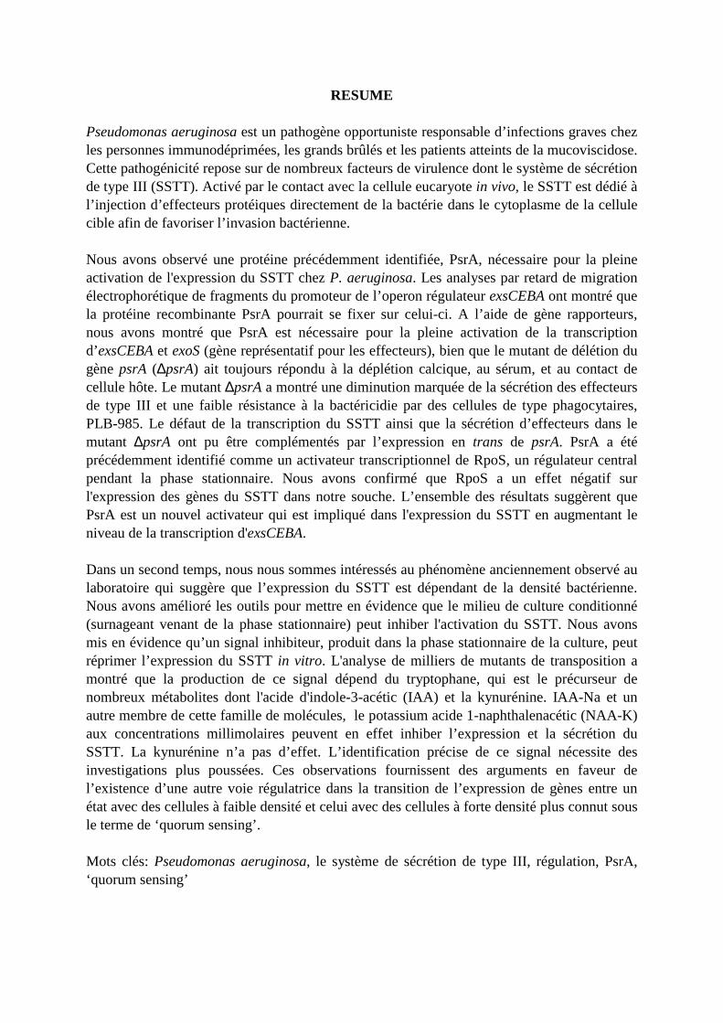
RESUME
Pseudomonas aeruginosa est un pathogène opportuniste responsable d’infections graves chez les personnes immunodéprimées, les grands brûlés et les patients atteints de la mucoviscidose. Cette pathogénicité repose sur de nombreux facteurs de virulence dont le système de sécrétion de type III (SSTT). Activé par le contact avec la cellule eucaryote in vivo, le SSTT est dédié à l’injection d’effecteurs protéiques directement de la bactérie dans le cytoplasme de la cellule cible afin de favoriser l’invasion bactérienne. Nous avons observé une protéine précédemment identifiée, PsrA, nécessaire pour la pleine activation de l'expression du SSTT chez P. aeruginosa. Les analyses par retard de migration électrophorétique de fragments du promoteur de l’operon régulateur exsCEBA ont montré que la protéine recombinante PsrA pourrait se fixer sur celui-ci. A l’aide de gène rapporteurs, nous avons montré que PsrA est nécessaire pour la pleine activation de la transcription d’exsCEBA et exoS (gène représentatif pour les effecteurs), bien que le mutant de délétion du gène psrA (∆psrA) ait toujours répondu à la déplétion calcique, au sérum, et au contact de cellule hôte. Le mutant ∆psrA a montré une diminution marquée de la sécrétion des effecteurs de type III et une faible résistance à la bactéricidie par des cellules de type phagocytaires, PLB-985. Le défaut de la transcription du SSTT ainsi que la sécrétion d’effecteurs dans le mutant ∆psrA ont pu être complémentés par l’expression en trans de psrA. PsrA a été précédemment identifié comme un activateur transcriptionnel de RpoS, un régulateur central pendant la phase stationnaire. Nous avons confirmé que RpoS a un effet négatif sur l'expression des gènes du SSTT dans notre souche. L’ensemble des résultats suggèrent que PsrA est un nouvel activateur qui est impliqué dans l'expression du SSTT en augmentant le niveau de la transcription d'exsCEBA. Dans un second temps, nous nous sommes intéressés au phénomène anciennement observé au laboratoire qui suggère que l’expression du SSTT est dépendant de la densité bactérienne. Nous avons amélioré les outils pour mettre en évidence que le milieu de culture conditionné (surnageant venant de la phase stationnaire) peut inhiber l'activation du SSTT. Nous avons mis en évidence qu’un signal inhibiteur, produit dans la phase stationnaire de la culture, peut réprimer l’expression du SSTT in vitro. L'analyse de milliers de mutants de transposition a montré que la production de ce signal dépend du tryptophane, qui est le précurseur de nombreux métabolites dont l'acide d'indole-3-acétic (IAA) et la kynurénine. IAA-Na et un autre membre de cette famille de molécules, le potassium acide 1-naphthalenacétic (NAA-K) aux concentrations millimolaires peuvent en effet inhiber l’expression et la sécrétion du SSTT. La kynurénine n’a pas d’effet. L’identification précise de ce signal nécessite des investigations plus poussées. Ces observations fournissent des arguments en faveur de l’existence d’une autre voie régulatrice dans la transition de l’expression de gènes entre un état avec des cellules à faible densité et celui avec des cellules à forte densité plus connut sous le terme de ‘quorum sensing’. Mots clés: Pseudomonas aeruginosa, le système de sécrétion de type III, régulation, PsrA, ‘quorum sensing’