Embed Size (px)
Citation preview

RESEARCH ARTICLE
The putative ceramide-conjugation protein Cwh43 regulatesG0 quiescence, nutrient metabolism and lipid homeostasisin fission yeastNorihiko Nakazawa1,‡, Takayuki Teruya1, Kenichi Sajiki1, Kazuki Kumada1, Alejandro Villar-Briones1,*,Orie Arakawa1, Junko Takada1, Shigeaki Saitoh2 and Mitsuhiro Yanagida1,‡
ABSTRACTCellular nutrient states control whether cells proliferate, or whetherthey enter or exit quiescence. Here, we report characterizations offission yeast temperature-sensitive (ts) mutants of the evolutionarilyconserved transmembrane protein Cwh43, and explore its relevanceto utilization of glucose, nitrogen source and lipids. GFP-taggedCwh43 localizes at ER associated with the nuclear envelopeand the plasma membrane, as in budding yeast. We found thatcwh43 mutants failed to divide in low glucose and lost viabilityduring quiescence under nitrogen starvation. In cwh43 mutants,comprehensive metabolome analysis demonstrated dramaticchanges in marker metabolites that altered under low glucose and/or nitrogen starvation, although cwh43 cells apparently consumedglucose in the culture medium. Furthermore, we found that cwh43mutant cells had elevated levels of triacylglycerols (TGs) andcoenzyme A, and that they accumulated lipid droplets. Notably, TGbiosynthesis was required to maintain cell division in the cwh43mutant. Thus, Cwh43 affects utilization of glucose and nitrogensources, as well as storage lipid metabolism. These results may fit anotion developed in budding yeast stating that Cwh43 conjugatesceramide to glycosylphosphatidylinositol (GPI)-anchored proteinsand maintains integrity of membrane organization.
KEY WORDS: Cwh43, Ceramide, Fission yeast, Low glucose,Nitrogen starvation, Lipid droplet
INTRODUCTIONNutrition is a major determinant of whether cells divide orundergo cell cycle arrest to enter quiescence. Principal nutrientsinclude carbohydrates, proteins and lipids, although vitaminsand minerals are also essential. Carbohydrates are hydrolyzed tomonosaccharides, such as glucose, the essential carbon and energysource for all organisms. Amino acids are a critical nitrogensource for synthesis of numerous biological substances, such asproteins, nucleotides and neurotransmitters, and are also necessaryfor gluconeogenesis under low-glucose conditions. Underenvironmental conditions in which carbon and/or nitrogen sources
are not available, cells have the ability to utilize lipids as analternative energy source. Most eukaryotic cells are capable ofstoring lipid droplets (LDs), comprised primarily of triacylglycerols(TGs) and sterols, in specialized intracellular organelles (Meyerset al., 2017; Pol et al., 2014; Thiam et al., 2013). Remarkably,changes in the cellular regulation of LDs and LD-associatedproteins are linked to human diseases including obesity, diabetesand atherosclerosis (Krahmer et al., 2013). Therefore, metabolicprocesses involving principal nutrients are tightly integrated.
Mechanisms controlling metabolism of principal nutrients areassumed to be evolutionarily conserved. Because the fission yeastSchizosaccharomyces pombe is a suitable model organism for thestudy of cell division and quiescence, we investigated geneticallycontrolled mechanisms in this organism that respond to fasting orstarvation. Nitrogen starvation induces entry into the quiescent G0phase, in which cells remain alive for months without growth ordivision (Su et al., 1996; Yanagida, 2009), demonstrating S. pombeG0 quiescence provides a model system to study chronologicallifespan. Using a collection of temperature-sensitive (ts) mutants, weidentified more than 80 ‘super housekeeping genes’ required for bothproliferation and nitrogen starvation-induced G0 quiescence (Sajikiet al., 2009 and our unpublished data). For example, the stress-responsive mitogen-activated protein kinase (MAPK) Sty1 is requiredfor G0 entry. The 26S proteasome maintains G0 quiescence byminimizing reactive oxygen species (ROS) in collaboration withautophagy (Takeda et al., 2010). By contrast, the quiescent state hasalso been studied under limited glucose conditions. In mediacontaining low glucose (4.4 mM, 0.08%), S. pombe wild-type cellsare able to divide as well as in normal glucosemedia (111 mM, 2%). Itis reported that the critical glucose transporter Ght5, the zinc-fingertranscription factor Scr1, the calmodulin kinase kinase (CaMKK)-likeSsp1, the PP2A phosphatase inhibitor Sds23 and the TORC2-mediated signaling pathway are essential for cell division under low-glucose conditions (Hanyu et al., 2009; Saitoh et al., 2015). Inresponse to a wide range of environmental stresses, a limited numberof key signaling pathways including protein kinase C (PKC) (Levinet al., 1990; Toda et al., 1993), MAPKs (Chen and Thorner, 2007;Perez and Cansado, 2010; Shiozaki and Russell, 1995; Sugiura et al.,1999, 1998) and the Ca2+/calmodulin-dependent phosphatasecalcineurin (Cyert, 2003; Sugiura et al., 2002; Yoshida et al., 1994),become activated. Thus, identification of factors involved in utilizationof both carbon and nitrogen sources should help to illuminatecommon signaling pathways of principal nutrients.
In this study, we isolated eight temperature-sensitive (ts) mutantalleles of the fission yeast cwh43 gene from our ts mutantcollections, and found that seven of these mutants showedsensitivity to both nitrogen starvation and glucose limitation.Cwh43 encodes a conserved transmembrane protein that is thoughtReceived 1 March 2018; Accepted 20 July 2018
1G0 Cell Unit, Okinawa Institute of Science and Technology Graduate University,Tancha 1919-1, Onna-son, Okinawa 904-0495, Japan. 2Institute of Life Science,Kurume University, Hyakunen-Kohen 1-1, Kurume, Fukuoka 839-0864, Japan.*Present address: Okinawa Institute of Science and Technology GraduateUniversity, Instrumental Analysis Section, Tancha 1919-1, Onna-son, Okinawa,Japan 904-0495.
‡Authors for correspondence ([email protected]; [email protected])
N.N., 0000-0001-8386-0812; M.Y., 0000-0003-0293-5654
1
© 2018. Published by The Company of Biologists Ltd | Journal of Cell Science (2018) 131, jcs217331. doi:10.1242/jcs.217331
Journal
ofCe
llScience

to incorporate the sphingolipid, ceramide, into a lipid moiety ofglycosylphosphatidylinositol (GPI)-anchored proteins (GPI-APs) inbudding yeast plasma membranes (Ghugtyal et al., 2007; Martin-Yken et al., 2001; Reggiori et al., 1997; Umemura et al., 2007;Yoko-o et al., 2013). Ceramide is present in all eukaryotic cells andserves structural functions in lipid bilayers (Chaurasia andSummers, 2015). In addition, ceramide acts as a signalingmolecule in response to stresses such as UV irradiation andchemotherapy, which induce cell cycle arrest, apoptosis and cellsenescence (Hannun, 1996; Hannun and Obeid, 2008). To addressthe novel, nutrient-related phenotypes in S. pombe cwh43 mutantcells, we comprehensively measured the levels of metabolites andlipids using metabolomic and lipidomic approaches. Strikingly,Cwh43-deficient cells had changed levels of nutrient stress-inducible metabolic compounds and they also over-accumulatedTGs (neutral lipids) in LDs. We present evidence that Cwh43, aputative ceramide-conjugation protein, is intimately involved incellular metabolism of principal nutrients and storage lipids.
RESULTSIsolation of S. pombe cwh43 temperature-sensitive mutantsWe previously developed 1015 ts haploid S. pombe strains (Hayashiet al., 2004), and identified genes responsible for their ts phenotypes(Hanyu et al., 2009; Hayashi et al., 2004; Nakamura et al., 2012). To
identify the mutant gene, plasmids that rescued the ts phenotype ofthe mutant strain were isolated by transformation, using an S. pombegenomic DNA library. Plasmid DNAs recovered from Ts+
transformants were then subcloned and sequenced. Nevertheless,the colony number of obtained transformants was occasionallyinsufficient to identify the high-copy suppressor gene owing to lowplasmid transformation efficiency. To overcome this technicaldifficulty for determination of mutation sites in eight ts mutantstrains (ts200, ts202, ts285, ts609, ts724, ts824, ts833, ts941), weperformed whole-genome sequencing of the mutant strains. Uponcompletion, all mutation sites responsible for these eight tsphenotypes turned out to be located in cwh43+ (SPAC589.12),encoding the putative glycosylceramide biosynthesis protein Cwh43(PomBase, https://www.pombase.org/) (Fig. 1A). We backcrossedthese ts strains with the wild-type multiple times and confirmed thatthe amino acid changes co-segregated with the ts phenotypes.
Cwh43 is a conserved eukaryotic transmembrane protein. Itcontains the Frag1/DRAM/Sfk1 family domain at its N-terminusand an endonuclease/exonuclease/phosphatase superfamily domainat its C-terminus (Dlakic, 2000; Ghugtyal et al., 2007; Martin-Ykenet al., 2001; Umemura et al., 2007) (Fig. 1A). Nineteentransmembrane helix motifs occur in the N-terminal 700 aminoacids. The mutation sites of four cwh43 ts mutants, K273stop(ts609), G300E (ts824), N418K (ts285) and W605stop (ts202), are
Fig. 1. Isolation of S. pombetemperature-sensitive mutants ofthe gene encoding the conservedtransmembrane protein Cwh43.(A)S. pombeCwh43 (SpCwh43) is drawnschematically along with S. cerevisiaeCWH43 (Sc CWH43) protein. Mutationsites of eight S. pombe cwh43 ts mutantsare indicated with amino acid changes.Asterisks indicate stop codons. The N-and C-terminal conserved regions of theFrag1/DRAM/Sfk1 and endonuclease/exonuclease/phosphatase superfamiliesare indicated with red and green boxes,respectively. Horizontal green lines showpositions of transmembrane helices in SpCwh43 protein. Cwh43 comprises twodistinct proteins in mammalian cells.Human PGAP2 and CWH43/FLJ21511are homologous to the N- and C-terminalregions of yeast Cwh43, respectively(Ghugtyal et al., 2007; Umemura et al.,2007). (B) Amino acid sequences of theconserved C-terminal regions thatsurround the G753R mutation in theS. pombe cwh43-833 ts strain. Aminoacid sequences of Cwh43 orthologs inS. pombe (Sp), Aspergillus niger (An),Saccharomyces cerevisiae (Sc), Homosapiens (Hs), and Mus musculus (Mm)are aligned. (C) DAPI-stained wild-typeand cwh43-G753R mutant cells areshown. Mild and severe defects in cellmorphology were observed in cwh43-G753R mutant cells at 26°C and 36°Cfor 6 h, respectively. Scale bar: 10 μm.(D) Wild type, the cwh43-G753R mutantand the deletion mutant lacking thecwh43+ gene (cwh43Δ) were spotted onEMM2 solid medium.
2
RESEARCH ARTICLE Journal of Cell Science (2018) 131, jcs217331. doi:10.1242/jcs.217331
Journal
ofCe
llScience

located in the region that contains transmembrane motifs. In theC-terminal domain, the evolutionarily conserved F715, G753 andG803 were mutated to leucine (ts724), arginine (ts833) and arginine(ts941) residues, respectively (Fig. 1B; Fig. S1). T914I (ts200) islocated just behind the C-terminal domain. In contrast, none of ourmutants resulted from mutations in the conserved N-terminaldomain of the Frag1/DRAM/Sfk1 family. Thus, the transmembraneregion and conserved C-terminal domain appear to be indispensablefor S. pombe Cwh43 function, although the N-terminal domain mayhave an additive role, consistent with the more-severe phenotype ofthe deletion mutant of the full-length cwh43+ gene (see below).
Construction of cwh43-G753R and cwh43Δ mutant strainsAll eight cwh43 mutants produced partly elongated, swollen cellshapes at the permissive temperature (26°C), and thismorphological abnormality was more severe at the restrictivetemperature (36°C) (Fig. 1C; Fig. S2A). Abnormal cell morphologywas indistinguishable among the eight mutant strains. We selectedthe G753R mutation (ts833) for further phenotypic analyses. It islocated in the C-terminally conserved region and loses viability invegetative and G0 quiescent cells (see below).To confirm that the ts phenotype is caused by single amino acid
changes in the Cwh43 protein, we introduced the G753R mutationinto the wild-type strain under the native promoter, along with thehemagglutinin (HA) tag. The resulting transformants, designatedCwh43-WT-HA:KanR and cwh43-G753R-HA:KanR (hereafter,termed wild type and cwh43-G753R, respectively), were used in thisstudy. The HA-tagged wild-type strain showed normal cell growth,as did the non-tagged wild-type; however, the cwh43-G753Rmutant and the original cwh43-833mutant failed to form colonies atthe restrictive temperature (36°C) (Fig. 1D; Fig. S2B). After thetemperature shift from 26°C to 36°C, the cell count of the cwh43-G753R mutant hardly increased, while viability decreased from55% to 20% (Fig. S2C). Thus, cell number increase and viability ofcwh43-G753R mutant cells were impaired at 36°C. The cellmorphological defect was evident even at 26°C, suggesting that thecell morphological abnormality itself was not a lethal attribute. Theproportion of cells with septa was significantly higher in the cwh43-G753R mutant at both 26°C and 36°C, while the numbers withmultiple septa increased at 36°C, suggesting that this mutant isdefective in cytokinesis (Fig. S2D). These phenotypes of theintegrated strain were indistinguishable from those of the originalcwh43-833 mutant.Next, we constructed a deletion mutant of the cwh43+ gene by
replacing it with the hygromycin-resistance gene. The resultingcwh43 deletion mutant (cwh43Δ) failed to form colonies at 36°C, asdid the cwh43-G753R ts mutant (Fig. 1D), indicating that deletionof cwh43+ is indispensable for cell proliferation at high temperature.The colony-forming capacity of the cwh43Δmutant is less than thatof the cwh43-G753R mutant at all tested temperatures. Consistentwith this, the morphological abnormality of the cwh43Δmutant wasmore severe than that of the cwh43-G753Rmutant at both 26°C and36°C (Fig. S2E). Thus, cwh43-G753Rmutant cells may be not fullydevoid of Cwh43 function.
Monitoring defective cytokinesis in living cwh43mutant cellsTo examine cell division of cwh43-G753Rmutant cells in detail, wemade time-lapse observations of living cells. Wild-type and cwh43-G753R cells were cultivated at 26°C and then observed using amicrofluidic perfusion system at 26°C or 36°C (Materials andMethods). At 26°C, DIC images showed that most wild-type cells
divided within 4 h, and septum formation occurred in one or twomovie frames (15–30 min) just before cell division (Fig. 2A, top;Movie 1). However, in the cwh43mutant, cell division required 2 to8 h and septum formation took >2 h, demonstrating that completionof cytokinesis was significantly delayed (Fig. 2A, bottom; Movie 2).After the temperature shift to 36°C, division of cwh43-G753Rmutant cells was blocked but this did not occur in wild-type cells(Fig. 2B; Movies 3,4). Mutant cells gradually became elongatedand swelled without cell division, resulting in multiple septa andprotrusion of cytosol. In addition, Aniline Blue staining of cwh43-G753R mutant cells showed abnormally high concentrations of thecell wall component 1,3-β-glucan, particularly at the septum,compared with dividing wild-type cells (Fig. 2C). This abnormalaccumulation of 1,3-β-glucan was obvious at 36°C. As reported in alarge number of mutant strains defective in cell wall integrity, the tsphenotype of cwh43mutant cells was suppressed in the presence ofthe osmotic stabilizer sorbitol (1.2 M) (Fig. S2F). Concomitantly,the glucan accumulation phenotype was slightly alleviated bysorbitol treatment (Fig. S2G), implying a correlation between cellwall defects and excessive glucan deposition. Taken together,these data indicate that impaired Cwh43 delayed completion ofcytokinesis, presumably by causing excess 1,3-β-glucan toaccumulate at the septum.
cwh43 mutant shows low-glucose sensitivity and improperlocalization of glucose transporters, in spite of glucoseconsumption from the culture mediumTo test whether the cwh43+ gene is required for cell proliferationunder nutrient deficiency, we examined the colony-forming abilityof cwh43-G753R mutant cells on solid EMM2 medium containing0.04–2% glucose (2.2-111 mM; low to high levels of glucose). Aspreviously described for the authentic low-glucose-sensitive mutantstrains defective in Ssp1 kinase (Hanyu et al., 2009) and for adeletion mutant of hexose transporter Ght5 (Saitoh et al., 2015), thecwh43-G753Rmutant failed to form colonies on low-glucose media(0.04–0.08%) at 26–33°C (Fig. 3A). Seven of the eight cwh43original ts mutant strains failed to divide under low-glucoseconditions, exhibiting the low-glucose sensitivity (LGS)phenotype (Table S1).
Under low-glucose conditions, expression of specific hexosetransporters is induced, and these transporters localize at the cellsurface (Saitoh et al., 2015).We examined intracellular localization ofthe GFP-tagged hexose transporter Ght5 in cwh43mutant cells. Aftera shift from 2% to 0.08% glucose at 26°C, Ght5–GFP localization atthe cell surfacewas not intense and showed cytoplasmic signals in themutant (Fig. 3B). Expression of Ght5–GFP itself was properlyinduced in cwh43mutant cells, as well as in wild-type cells (Fig. 3C).In wild-type cells, another GFP-tagged hexose transporter, Ght8, waslocated at the plasma membrane exclusively at the poles of the cells(Saitoh et al., 2015) (Fig. S3). By contrast, cwh43 mutant cellsshowed cytoplasmic accumulations of Ght8–GFP similar to those ofGht5-GFP, suggesting that Cwh43 affects the localization of hexosetransporters under glucose limitation.
To examine glucose consumption by wild-type and cwh43mutant cells, we measured glucose concentrations in the culturemedium. Cells initially cultivated in high-glucose (2%) mediumwere transferred to high (2%)- or low (0.08%)-glucose medium andincubated at 26°C, along with ght5 deletion mutant cells (ght5Δ) asa control (Saitoh et al., 2015). Unexpectedly, glucose consumptionwas not significantly decreased in cwh43mutant cultures comparedwith wild-type, in either high or low glucose (Fig. 3D), although thecell number increase of cwh43mutant was decidedly slow (Fig. 3E).
3
RESEARCH ARTICLE Journal of Cell Science (2018) 131, jcs217331. doi:10.1242/jcs.217331
Journal
ofCe
llScience

These data suggest that the cwh43 mutant consumes glucose inculture media, even if cell division is declined. Although subcellularlocalization of Ght5 and Ght8 were affected in the cwh43 mutant,these transporters, which were partially retained at the plasmamembrane, may be sufficient for glucose uptake.
Cwh43 is required to maintain cell viability during G0quiescence induced by nitrogen starvationWe next determined whether the cwh43+ gene is required formaintaining viability during G0 quiescence under nitrogen starvation.Wild-type and cwh43-G753R mutant cells grown in EMM2 medium(EMM2+N) were transferred to nitrogen-deficient EMM2-N at 26°Cfor 1 and 4 days, and the resulting quiescent G0 cells were spottedonto YPD solid medium in order to count the cells that were able toform colonies. In comparison with the wild type, under nitrogenstarvation, the viability of cwh43-G753R mutants plunged to 27%after 1 day and to only 5% after 4 days, indicating severe defects in
regeneration capacity, even after replenishment of the nitrogen source(Fig. 3F). The cell number of wild-type cells increased 3.7× after 1 dayunder nitrogen starvation, indicating that two rounds of cell divisionoccurred. In contrast, numbers of cwh43 mutant cells increased only1.5×. Wild-type S. pombe cells have a spherical shape during G0quiescence (Sajiki et al., 2009; Su et al., 1996); however, cwh43-G753R mutant cells presented non-spherical, deformed shapes(Fig. 3G). Six of the seven remaining cwh43 ts mutant strains alsodecreased in viability under nitrogen starvation (Table S1). Theseresults suggest that cwh43 mutant cells failed to enter a normal andviable G0 quiescence in response to nitrogen deficiency.
Intracellular localization of Cwh43To observe the intracellular localization of Cwh43 protein, theC-terminus of cwh43+ or cwh43-G753R mutant genes wastagged with GFP. At 26°C, Cwh43-WT–GFP apparently localizedat the nuclear membrane and the plasma membrane, accompanying
Fig. 2. Monitoring defectivecytokinesis and excessive β-glucanaccumulation at cell septa in cwh43mutant cells. (A,B) Left, time-lapseimages of wild-type and cwh43-G753Rstrains. Cells were first cultivated at 26°C,and then either incubated further at 26°C(A) or shifted to 36°C (B) in a microfluidicperfusion chamber with a continuoussupply of EMM2 medium. Differentialinterference contrast (DIC) images werepresented at 2-h intervals (also seeMovies 1–4). Arrows and arrowheads in Bindicate the positions of multiple septaand a protruded cytosolic structure incwh43-G753R cells, respectively. Right,division time and duration of septumformation were monitored for individualcells. (C) Left, fluorescence and bright-field images of wild-type and cwh43-G753R cells were captured after stainingfor the cell wall component 1,3-β-glucanusing the fluorescent dye Aniline Blue.Cells were cultured at 26°C or 36°C for6 h in liquid medium. Right, Aniline Blue-stained cells were spotted on filter paper.Scale bars: 10 μm.
4
RESEARCH ARTICLE Journal of Cell Science (2018) 131, jcs217331. doi:10.1242/jcs.217331
Journal
ofCe
llScience

granule-like cytoplasmic signals (Fig. 4A, top). At 36°C,localization of Cwh43–GFP was similar to that at 26°C, althoughGFP-tagged Cwh43 proteins were possibly unstable in hightemperature, unlike the HA-tagged proteins (Fig. S4A,B).GFP-tagged Cwh43-G753R mutant proteins also showed similarlocalization as observed in wild type at both temperatures, implyingthat the G753R mutation does not affect Cwh43 localizationitself (Fig. 4A, bottom; Fig. S4A). We also demonstrated thatCwh43–GFP localized in close proximity to plasma membranes byperforming a protoplast preparation with enzymatic digestion of cellwalls (Fig. 4B).In budding yeast, CWH43 protein is reported to localize at the
endoplasmic reticulum (ER) (Umemura et al., 2007). Consideringthat the ER is continuous with the nuclear envelope and isassociated with the plasma membrane (Pidoux and Armstrong,1993; Zhang et al., 2010), S. pombe Cwh43 proteins are also
predicted to enrich in the ER. To test this possibility, we observedCwh43–GFP localization in an scs2Δ scs22Δ double-deletionmutant strain in which the cortical ER dissociates from plasmamembrane (Zhang et al., 2012). In wild-type cells, the Cwh43–GFPsignals colocalized with the artificial luminal ER marker AHDL–mCherry (Zhang et al., 2010) at the plasma membrane and nuclearperiphery (Fig. 4C, top). However, in scs2Δ scs22Δ double deletionmutant cells, localization of both Cwh43–GFP and AHDL–mCherry at the plasma membrane disappeared, and, instead,accumulated in the cytoplasm (Fig. 4C, bottom). This resultindicates that Cwh43, as in budding yeast CWH43, enriches at ERassociated with the plasma membrane.
Immunodetection of Cwh43 proteinProtein extracts of exponentially growing wild-type and cwh43-G753R strains were examined by immunoblotting using antibodies
Fig. 3. cwh43 mutants are sensitiveto both low glucose and nitrogenstarvation. (A) Aliquots (5×10⁴ cells)of wild-type and cwh43-G753R strainswere serially diluted 5×, spotted ontoEMM2 medium containing theindicated concentrations (mM andpercentage) of glucose at 26–33°C.(B) Expression and localization ofGFP-tagged Ght5 in wild-type (top)and cwh43-G753R (bottom) mutantcells. Cells were cultivated in EMM2liquidmedium at 26°C and the glucoseconcentration was switched from 111to 4.4 mM at time 0 h. GFPfluorescence microscopy imageswere taken at 2-h intervals. Insertscorrespond to the areas of the whitedashed boxes. (C) The protein level ofGht5–GFP was measured byimmunoblotting. Total protein stainedwith Ponceau S is shown as a loadingcontrol. (D) Glucose consumption wasexamined in the wild-type, cwh43 andght5Δ mutant strains. Cells weretransferred to fresh EMM2 suppliedwith 2% (left) or 0.08% (right) glucose(G-shift) at time 0 and cultivated at 26°C. The glucose concentrationremaining in the medium wasmeasured after 0, 6 and 24 h. Mean oftwo independent experiments areshown. (E) Cell proliferation wasexamined for the same conditions asin D. (F) Cell viability was measured at1 and 4 days after nitrogen starvation(EMM2-N) at 26°C alongwith changesin cell number after 1 day. (G) Bright-field images of wild-type and cwh43-G753R mutant cells cultured after1 day in EMM2-N medium at 26°C.Mutant cells showed non-spherical,deformed cell shapes. Scale bars:10 μm.
5
RESEARCH ARTICLE Journal of Cell Science (2018) 131, jcs217331. doi:10.1242/jcs.217331
Journal
ofCe
llScience
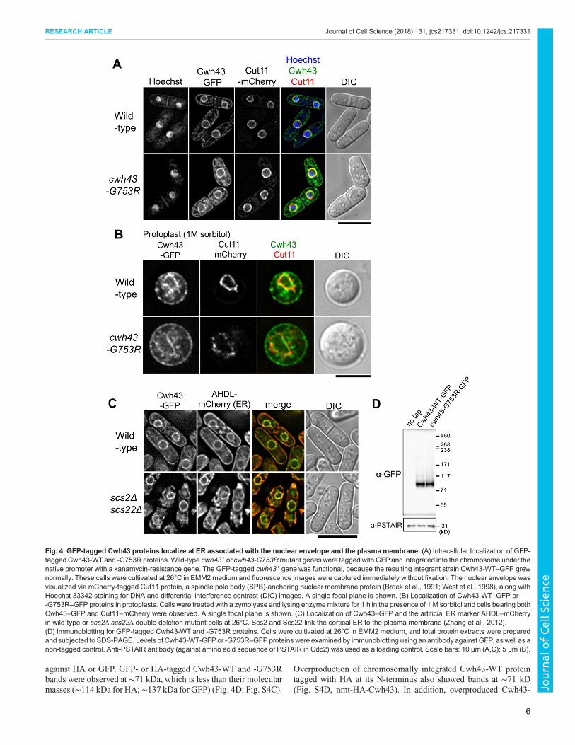
against HA or GFP. GFP- or HA-tagged Cwh43-WT and -G753Rbands were observed at ∼71 kDa, which is less than their molecularmasses (∼114 kDa for HA; ∼137 kDa for GFP) (Fig. 4D; Fig. S4C).
Overproduction of chromosomally integrated Cwh43-WT proteintagged with HA at its N-terminus also showed bands at ∼71 kD(Fig. S4D, nmt-HA-Cwh43). In addition, overproduced Cwh43-
Fig. 4. GFP-tagged Cwh43 proteins localize at ER associated with the nuclear envelope and the plasma membrane. (A) Intracellular localization of GFP-tagged Cwh43-WTand -G753R proteins.Wild-type cwh43+ or cwh43-G753Rmutant genes were tagged with GFPand integrated into the chromosome under thenative promoter with a kanamycin-resistance gene. The GFP-tagged cwh43+ gene was functional, because the resulting integrant strain Cwh43-WT–GFP grewnormally. These cells were cultivated at 26°C in EMM2 medium and fluorescence images were captured immediately without fixation. The nuclear envelope wasvisualized via mCherry-tagged Cut11 protein, a spindle pole body (SPB)-anchoring nuclear membrane protein (Broek et al., 1991; West et al., 1998), along withHoechst 33342 staining for DNA and differential interference contrast (DIC) images. A single focal plane is shown. (B) Localization of Cwh43-WT–GFP or-G753R–GFP proteins in protoplasts. Cells were treated with a zymolyase and lysing enzymemixture for 1 h in the presence of 1 M sorbitol and cells bearing bothCwh43–GFP and Cut11–mCherry were observed. A single focal plane is shown. (C) Localization of Cwh43–GFP and the artificial ER marker AHDL–mCherryin wild-type or scs2Δ scs22Δ double deletion mutant cells at 26°C. Scs2 and Scs22 link the cortical ER to the plasma membrane (Zhang et al., 2012).(D) Immunoblotting for GFP-tagged Cwh43-WT and -G753R proteins. Cells were cultivated at 26°C in EMM2 medium, and total protein extracts were preparedand subjected to SDS-PAGE. Levels of Cwh43-WT-GFPor -G753R–GFP proteins were examined by immunoblotting using an antibody against GFP, as well as anon-tagged control. Anti-PSTAIR antibody (against amino acid sequence of PSTAIR in Cdc2) was used as a loading control. Scale bars: 10 μm (A,C); 5 μm (B).
6
RESEARCH ARTICLE Journal of Cell Science (2018) 131, jcs217331. doi:10.1242/jcs.217331
Journal
ofCe
llScience

WT–HA protein derived from cwh43+ cDNA migrated faster thanits expected molecular mass (Fig. S4D, pCwh43-HA). Hence, S.pombe Cwh43 presents a fast-migrating band in SDS-PAGE, aproperty often reported in ER- or membrane-associated proteins(Shirai et al., 2008).
Metabolic changes of biomarkers for nutrient stress incwh43 mutant cellsTo further characterize the cwh43 mutant phenotype, we appliedour established metabolomic approach to cwh43-G753R mutantcells. Since cell viability of the cwh43-G753Rmutant at 36°C and ofthe the cwh43Δ deletion mutant at both 26°C and 36°C wasremarkably low, the cwh43-G753R strain was cultivated at 26°C inliquid EMM2 medium to mid-log phase (5×10⁶ cells/ml) alongwith wild-type cells, and these exponentially growing vegetativecells were collected. Then, cellular metabolites were extracted in50% methanol and metabolites were analyzed using an LC-MS(LTQ Orbitrap mass spectrometer), as described previously(Pluskal et al., 2010b; Pluskal et al., 2011; see Materials andMethods). 119 metabolites were detected in wild-type and cwh43-G753R mutant strains (Fig. 5A; Table S2). In cwh43 mutant cells,the levels of 26 metabolites increased more than 3-fold, while tendecreased more than 3-fold compared with wild-type cells (Fig. 5B,C; Table S3 and S4). Most of the compounds displayingsignificantly altered abundances in the mutant have beenpreviously reported as marker metabolites that change undernitrogen starvation or low-glucose conditions (Fig. 5A) (Pluskalet al., 2011; Sajiki et al., 2013). The cwh43mutant also accumulatedcompounds that commonly change in both types of nutritionalstarvation.The most strikingly accumulated compound in cwh43 mutant
cells was deferriferrichrome, which increased 19,000-fold relativeto wild-type cells (Fig. 5B). Deferriferrichrome is an iron storagecompound that increases with ferrichrome under low-glucoseconditions (Pluskal et al., 2011). The iron-free form (deferri-form)of ferrichrome is produced under iron deficiency (Labbé et al.,2007; Schrettl et al., 2004), suggesting that cwh43mutant cells weredeficient in iron.Another group of drastically increased compounds includes
ergothioneine and S-methyl-ergothioneine. Ergothioneine acts as aphysiological antioxidant (Cheah and Halliwell, 2012) that hasbeen reported to increase under both low-glucose and nitrogenstarvation conditions (Pluskal et al., 2011; Sajiki et al., 2013).Hercynylcysteine sulfoxide is a precursor of ergothioneine.Consistent with the fact that ergothioneine is a sulfur-containingderivative of trimethyl histidine, tri- and di-methylated histidinealso accumulated in cwh43 mutant cells. Additionally, trehaloseincreased over 60-fold in the mutant. This compound is produced tostabilize proteins and membranes under various physiologicstresses, including heat shock, low glucose and nitrogen starvation(Cansado et al., 1998; Elbein et al., 2003; Pluskal et al., 2010b).2-Oxoglutarate and succinate, intermediates of the TCA cycle,
increased 89-fold and 6-fold, respectively, in cwh43-G753Rmutant cells (Fig. 5B; Table S3). These compounds are specificbiomarkers that increase during the nitrogen starvation response(Sajiki et al., 2013). By contrast, the purine biosynthesispathway intermediates, 5-phospho-ribose 1-diphosphate (PRPP),aminoimidazole-4-carboxamide ribonucleotide (AICAR), N-succinocarboxamide-5-aminoimidazole ribonucleotide (SAICAR)and N-formylglycinamide ribonucleotide (FGAR) decreasedremarkably in mutant cells (Fig. 5C; Table S4). These compoundshave also been reported as marker metabolites that decrease
immediately under nitrogen starvation (Sajiki et al., 2013). Takentogether, the metabolomic analysis indicated that the cwh43mutantis apparently defective in utilization of both carbon and nitrogensources, producing massive nutritional stresses, even when thesenutrients are present in the culture medium.
In wild-type S. pombe cells, the level of free-form coenzyme A(CoA) is normally extremely low relative to other metabolites(Nakamura et al., 2012). However, in cwh43mutant cells, we foundthat the level of CoA increased more than 4000-fold compared towild-type cells (Fig. 5A,B). This drastic increase in CoA content inthe absence of Cwh43 function is a unique metabolic property thathas not been reported under low glucose or nitrogen starvation. CoAis an essential cofactor in the metabolism of carboxylic acids andlipids, implying that cwh43 mutant cells have altered lipidabundance.
cwh43 mutant cells overproduce triacylglycerolsTo examine whether Cwh43 influences cellular lipid metabolism,we performed lipidomic analysis in wild-type and cwh43-G753Rmutant cells. Cells were cultivated in EMM2 at 26°C and total lipidswere extracted with tert-butylmethylether (Matyash et al., 2008).Lipid extracts were then subjected to LC/MS analysis andcompounds were identified using Lipidomics Gateway Software(seeMaterials andMethods). We identified 124 lipids in both strains(Fig. 6A; Table S5). 15 lipids increased ≥3-fold in abundance whilenine others diminished ≥3-fold in cwh43-G753R mutant cellscompared to thewild type (Table S6). Notably, in mutant cells, 14 of15 increased lipids were triacylglycerols (triglycerides, TGs) andthe levels of 14 TG species were∼3–5-fold higher than in wild-typecells (Fig. 6B). TG species containing unsaturated fatty acidsincreased, although TGs composed of only saturated fatty acidsdecreased in cwh43 mutant (Fig. 6C).
Thin-layer chromatography (TLC) confirmed that TGs wereenriched in cwh43 mutant cells compared to wild-type cells(Fig. 6D). In dga1Δ plh1Δ double-mutant cells lacking the TGbiosynthetic enzymes Dga1 and Plh1 (Meyers et al., 2016) (seebelow), TGs were not detected, as expected. Sterol esters alsoincreased in cwh43 mutant, though levels of diacylglycerols (DGs)were comparable to those in wild-type and dga1Δ plh1Δ mutantcells. Taken together, these results provide clear evidence thatdefects in Cwh43 function raise levels of TGs along with levels ofsterol esters.
Lipid droplets accumulate in cwh43-G753R mutant cellsTo further validate elevated levels of TGs in cwh43mutant cells, westained them with BODIPY 493/503, a fluorescent dye thatspecifically binds to TGs, and we observed LDs, consisting ofTGs and cholesterol (Long et al., 2012; Pol et al., 2014; Thiam et al.,2013). In cwh43 mutants, the number and size of BODIPY-stainedLDs was substantially greater than in wild-type cells (Fig. 6E–G),confirming that impaired function of Cwh43 causes excessaccumulation of storage lipids in vegetative cells.
Next, we constructed deletion mutant strains of two TG synthesisenzymes, Dga1 and Plh1, to determine whether the increasednumber of LDs in cwh43 mutant cells depends on TG syntheticpathways (Meyers et al., 2016). Both Dga1 (a diacylglycerolO-acyltransferase) and Plh1 (a phospholipid-diacylglycerolacyltransferase) conjugate acyl chains to diacylglycerol, usingdistinct acyl chain donors, acyl-CoA and glycerophospholipid,respectively (Oelkers et al., 2002, 2000; Zhang et al., 2003). Aspreviously reported (Meyers et al., 2016), dga1Δ plh1Δ doublemutant cells barely produce LDs (Fig. 6E,F). Furthermore, cwh43-
7
RESEARCH ARTICLE Journal of Cell Science (2018) 131, jcs217331. doi:10.1242/jcs.217331
Journal
ofCe
llScience
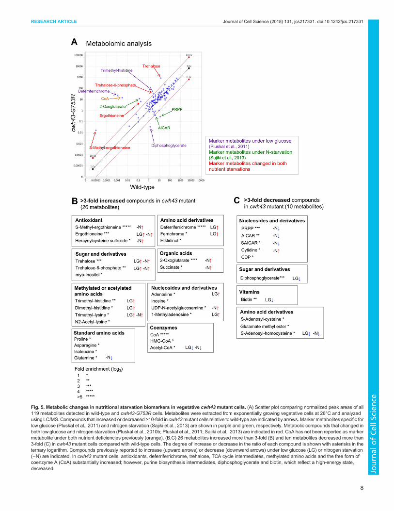
Fig. 5. Metabolic changes in nutritional starvation biomarkers in vegetative cwh43 mutant cells. (A) Scatter plot comparing normalized peak areas of all119 metabolites detected in wild-type and cwh43-G753R cells. Metabolites were extracted from exponentially growing vegetative cells at 26°C and analyzedusing LC/MS. Compounds that increased or decreased >10-fold in cwh43mutant cells relative towild-type are indicated by arrows.Markermetabolites specific forlow glucose (Pluskal et al., 2011) and nitrogen starvation (Sajiki et al., 2013) are shown in purple and green, respectively. Metabolic compounds that changed inboth low glucose and nitrogen starvation (Pluskal et al., 2010b; Pluskal et al., 2011; Sajiki et al., 2013) are indicated in red. CoA has not been reported as markermetabolite under both nutrient deficiencies previously (orange). (B,C) 26 metabolites increased more than 3-fold (B) and ten metabolites decreased more than3-fold (C) in cwh43 mutant cells compared with wild-type cells. The degree of increase or decrease in the ratio of each compound is shown with asterisks in theternary logarithm. Compounds previously reported to increase (upward arrows) or decrease (downward arrows) under low glucose (LG) or nitrogen starvation(−N) are indicated. In cwh43 mutant cells, antioxidants, deferriferrichrome, trehalose, TCA cycle intermediates, methylated amino acids and the free form ofcoenzyme A (CoA) substantially increased; however, purine biosynthesis intermediates, diphosphoglycerate and biotin, which reflect a high-energy state,decreased.
8
RESEARCH ARTICLE Journal of Cell Science (2018) 131, jcs217331. doi:10.1242/jcs.217331
Journal
ofCe
llScience

Fig. 6. Increased triacylglycerols and LDs in cwh43mutant cells. (A) Lipidomic analysis of cellular extracts in wild-type and cwh43-G753R cells. Normalizedpeak areas of 124 lipid compounds are presented as a scatterplot. Cells were cultivated in EMM2 medium at 26°C and extracted lipid samples were analyzed byLC/MS. Plots for TGs are marked in orange. (B) Levels of 14 TG species increased ∼3–5-fold in mutant cells compared to wild-type cells. MIPC is a complexsphingolipid (mannosylinositol phosphorylceramide). (C) TG species composed of unsaturated fatty acids were increased in cwh43mutant cells. The degrees ofchange (the ratios of cwh43 to WT) of triacylglycerols (TG), diacylglycerols (DG), phosphatidylcholine (PC), phosphatidylethanolamine (PE), phosphatidylinositol(PI), phosphatidylserine (PS) and phytosphingosine (Sph) are shown with numbers of unsaturated bonds. The sum of detected lipids in cwh43 mutant wascompared with wild-type in each category. (D) The cwh43-G753Rmutant accumulated TGs while dga1Δ plh1Δ double-mutant failed to produce them, as shownvia TLC of total lipids. Positions of TGs, DGs and sterol esters are indicated with the origin of sample spot and the solvent front. (E) LDs were stained with thefluorescent dye BODIPY 493/503 in wild-type, cwh43-G753R, dga1Δ plh1Δ and cwh43-G753R dga1Δ plh1Δ mutant strains at 26°C. Dga1 and Plh1 catalyzeproduction of TGs from DGs through distinct reactions (Meyers et al., 2016). Differential interference contrast (DIC) images were merged with fluorescenceimages. Inserts correspond to the area of thewhite dashed boxes. Scale bar: 10 μm. (F) Mean±s.d. (n=81, 85, 62 and 67 cells for wild type, cwh43-G753R, dga1Δplh1Δ and cwh43-G753R dga1Δ plh1Δ strains, respectively) of the LD number in a cell. (G) Distribution of LD size in wild-type and cwh43mutant cells. The meanLD size in each cell is shown in arbitrary units. (H) Severe synthetic growth defect of triple mutants between cwh43-G753R and dga1Δ plh1Δ double mutants.
9
RESEARCH ARTICLE Journal of Cell Science (2018) 131, jcs217331. doi:10.1242/jcs.217331
Journal
ofCe
llScience

G753R dga1Δ plh1Δ triple mutant cells also scarcely showed LDs,demonstrating that elevated LD formation in cwh43 mutant cellsrequires the TG biosynthesis pathway.The dga1Δ plh1Δ double mutant grew normally; however,
cwh43-G753R and dga1Δ plh1Δ mutants were additively defectivein colony formation at 26–36°C (Fig. 6H). This negative geneticinteraction was striking and indicated that LD formation isindispensable for cell division in Cwh43-deficient cells. Since thissynthetic phenotype remained in the presence of 1.2 M sorbitol(Fig. S5A), the positive effect of TG synthesis on cwh43 mutantcells may be independent of morphological recovery. Simultaneousdepletion of dga1+ and plh1+ genes in cwh43 mutant apparentlyabolished sensitivity to glucose limitation, implying that the LGSphenotype of cwh43 mutant depends on TG synthesis (Fig. S5B).
DISCUSSIONIn the present study, we identified multiple ts mutant strainsdefective in S. pombe Cwh43 and characterized their phenotypes.Among the eight ts strains, we identified seven cwh43 ts mutantsthat showed sensitivities to both nitrogen starvation and glucose-limited conditions. Identification of these mutant phenotypes isunique in suggesting that Cwh43 contributes to coordinatedutilization of distinct major nutrients. Metabolomic analysisindicates that Cwh43-defective cells suffer from nutrient stressesand decline in metabolic activity required for cell division, even inthe presence of nutrients. Our results provide the first evidence thatCwh43, which is a proposed ceramide-conjugating protein, stronglyaffects cellular metabolism of principal nutrients and maintainsstorage lipid homeostasis.What is the physiological significance of TG accumulation and its
requirement for cell division in cwh43 mutant cells? First, theseresults suggest that TGs may function as a critical energy reserveunder nutritional stress caused by carbon and nitrogen starvation incells with defective Cwh43. In fact, wild-type S. pombe cells undernitrogen starvation significantly increase the cellular level of TGsaccompanying formation of massive LDs (Fig. S6A,B). Consistentwith this, both the protein and mRNA levels of two TG syntheticenzymes, Dga1 and Plh1, were upregulated in nitrogen-starved G0quiescent cells (Fig. S6C,D,E). These levels were not significantlychanged between wild-type and the cwh43 mutant, suggesting thatthe mechanism of TG accumulation in cwh43 mutant cells differsfrom that in wild-type G0 cells.An alternative possibility for the significance of TG accumulation
is that TGs preserve altered plasma membranes in cwh43 mutantcells. The activity of storage lipid synthesis affects membrane lipidmetabolism in budding and fission yeast (Gaspar et al., 2011; Heet al., 2014; Péter et al., 2017). Considering that dga1Δ plh1Δdouble mutants grew as well as wild-type cells under normal cultureconditions, TG synthesis is deduced to be non-essential for cellgrowth in the presence of functional Cwh43. In sharp contrast, withimpaired Cwh43, TGs may reinforce the disorganized membranes,in which ceramide incorporation is probably defective,compensating for the loss of cell division ability. The increasedlevel of TGs containing unsaturated fatty acids may confer fluidityon the plasma membrane in cwh43 mutant cells. However, becauseLDs originate at the ER, where Dga1 and Plh1 proteins localize(Meyers et al., 2016; Thiam et al., 2013), we cannot exclude thepossibility that Cwh43 directly affects formation of LDs at the ER.cwh43 mutant cells are defective in cell division under low
glucose and alter the level of metabolic markers for glucoselimitation, despite the fact that mutant cells consume glucose fromthe culture medium. These facts appear inconsistent, but suggest the
possibility that cwh43 mutants may fail to utilize glucose properlyafter its uptake. Furthermore, we presented genetic evidence thatcwh43 mutant shows sensitivity to glucose limitation only in thepresence of dga1+ and plh1+ genes (Fig. S5B). Thus, wehypothesize that cwh43 mutant cells preferentially consume theprincipal nutrients for TG production. In nutritionally rich media,carbon and nitrogen sources may be converted into TGs formaintaining cell division in cwh43mutant; however, TG productionmay be decreased in mutant cells under nutritionally limitingconditions.
The drastic increase of the iron-storage compounds, ferrichromeand deferriferrichrome, in cwh43 mutant implies cellular irondeficiency. S. pombe has two separate iron uptake pathways. Thefirst is reductive iron transport, employing Frp1 (an Fe3+ reductase),Fio1 (an Fe2+ oxidase) and Fip1 (an Fe3+ permease). The second isnon-reductive transport, which acquires ferrichrome-bound iron viathe specific transporters Str1, Str2 and Str3. Hence, one possibilityis that localization of these iron transporters at the plasma membranemight be adversely affected in cwh43 mutant cells. Alternatively,we assume that the transcriptional response to iron deficiency maybe coupled with glucose limitation. Transcriptional repressors forgenes encoding iron transporters and ferrichrome synthetases areactually shared with those of genes involved in the response to lowglucose (Janoo et al., 2001; Labbé et al., 2007; Mercier and Labbe,2010).
We found that a drastically increased CoA level is characteristicof the metabolic phenotype of Cwh43-defective cells. CoA isbiosynthesized from pantothenate in five reactions and acts as anacyl carrier involved in numerous metabolic reactions, includingfatty acid synthesis (Leonardi et al., 2005; Srinivasan and Sibon,2014). As an initial reaction, CoA is utilized for production ofacetyl-CoA. Therefore, our results raise the possibility that elevatedlevels of CoA in cwh43 mutants accelerate acetyl-CoA synthesis.Generally, excessive acetyl-CoA enables fatty acid synthesis andincreases triacylglycerol production. In contrast, the S. pombe ppc1-537 mutant, defective in phosphopantothenoylcysteine synthetase,which catalyzes the second step of CoA synthesis frompantothenate, decreases acetyl-CoA levels and suppresses LDformation (Nakamura et al., 2012). Cwh43 deficiency may thuscreate a cellular environment in which CoA levels increase to agreater extent than in wild-type cells, promoting triacylglycerolaccumulation via acetyl-CoA and fatty acid synthesis. Hydrolysis ofthioester bonds in CoA-conjugated metabolites releases high freeenergy (ΔG°′=−31.4 kJ/mol), which is comparable to that of ATPhydrolysis (ΔG°′=−30.5 kJ/mol), implying that cwh43mutant cellsmay consume the cellular energy stored in numerous CoAderivatives and maintain their survival under nutrient stresses.Further study is definitively required to understand metabolicchanges implicated in CoA accumulation in Cwh43-deficient cellsand its relationship to availability of principal nutrients.
The broad phenotypes in cwh43mutant cells can be explained byhypothesizing that Cwh43 properly organizes plasma membranes toprovide functional platforms for nutrient signaling and also forcytokinesis. S. cerevisiae CWH43 is proposed to be essential forconjugation of ceramide to GPI-anchored proteins (GPI-APs).Indeed, the S. pombe Cwh43 C-terminal amino acid sequencearound the G753R mutation is highly conserved in both buddingand fission yeasts. GPI-APs are modified at the ER and thenrecruited to the plasma membrane, where they act as receptors, celladhesion factors or enzymes (Fujita and Jigami, 2008; Kinoshitaand Fujita, 2016). Ceramide-enriched micro-domains at the plasmamembrane, such as lipid rafts, are believed to be centers for signal
10
RESEARCH ARTICLE Journal of Cell Science (2018) 131, jcs217331. doi:10.1242/jcs.217331
Journal
ofCe
llScience

transduction in various biological processes (Lingwood andSimons, 2010). Although further analysis is required, wespeculate that metabolic and lipidomic alterations in S. pombecwh43-G753R mutant cells are associated with defects in ceramideconjugation.
MATERIALS AND METHODSStrains and plasmidsS. pombe strains used in this study were derived from haploid wild-typestrains 972 (h−) and 975 (h+). A collection of temperature-sensitive (ts)strains made by random mutagenesis was used (Hayashi et al., 2004).Mutation sites and amino acid substitutions in cwh43 ts mutant strains weredetermined by whole-genome sequencing (Illumina) and confirmed bySanger dideoxy sequencing. Genetic linkage between the ts phenotype andmutation sites was verified by tetrad analysis. Strains expressing aC-terminal 3HA (hemagglutinin antigen)- or GFP-tagged Cwh43-WT or-G753R were made by chromosomal integration under the native promoterwith the kanamycin-resistance gene. Strains expressing GFP-tagged Dga1and Plh1 were constructed in the same manner as Cwh43–GFP with thehygromycin-resistance gene. Strains overproducing HA-tagged Cwh43proteins were constructed by replacing the native promoter with the nmt1+
promoter using the pFA6a-kanMX6-P3nmt1 plasmid (Bähler et al., 1998).The plasmid pRep1 carrying the nmt1+ promoter and the cwh43+ cDNAsequence tagged with HA at its C-terminus was also used foroverproduction. Deletion of the cwh43+, dga1+, plh1+, scs2+ and scs22+
genes was performed by replacing the entire genomic locus with thehygromycin or nourseothricin (clonNAT)-resistance genes in a haploidstrain. C-terminal mCherry-tagged Cut11 strains were made bychromosomal integration under the native promoter. GFP-tagged Ght5and Ght8 proteins and deletion of the ght5+ were previously described(Saitoh et al., 2015). The strain expressing mCherry-tagged AHDL, anartificial ER marker, was a gift from Dr Snezhana Oliferenko (King’sCollege London, UK).
Growth conditionsS. pombe cells were cultivated in YPD (rich medium) or EMM2 (minimalmedium) with modified glucose concentrations as indicated (Moreno et al.,1991). For nitrogen starvation, cells were first cultivated in EMM2 at 26°Cand then transferred to nitrogen-deficient EMM2-N medium at 26°C for24 h (Sajiki et al., 2009). Cell viability was calculated as a percentage of thenumber of colonies formed versus the number of plated cells. Liquid-cultured cells were counted using a Multisizer 3 (Beckman Coulter).
Fluorescence microscopy and live-cell analysisDAPI staining was carried out as previously described (Adachi andYanagida, 1989). Fluorescent staining of 1,3-β-glucan was performed usingAniline Blue, as previously described (Okada and Ohya, 2016). For lipiddroplet staining, 107 cells were stained in EMM2 medium containing100 nM BODIPY 493/503 (Thermo Fisher Scientific, D3922) (Meyerset al., 2016). Procedures for live-cell analysis were carried out using aDeltaVision Elite Microscopy System (GE Healthcare) as describedpreviously (Nakazawa et al., 2016). Oil immersion (60× and 100×; NA1.4; Olympus) or silicon objective lenses (UPLSAPO 60XS2 and 100XS;NA 1.3 and 1.35; Olympus) were used. For time-lapse imaging, cells wereloaded into an ONIX microfluidic perfusion chamber (CellASIC, Hayward,CA) and cultivated with a continuous medium supply. All-in-onemicroscopes BZ9000 and BZ-X700 (Keyence, Osaka, Japan), were usedto observe fixed cells.
Protoplast preparationS. pombe cells, cultured in 10 ml EMM2 medium at 1×10⁷ cells/ml, werecollected by centrifugation (1700 g for 3 min) and washed twice with anequal volume of SCS buffer (20 mM sodium citrate pH 5.8, 1 M sorbitol).Harvested cells were re-suspended in 1 ml SCS containing 2 mg/mlzymolyase 100T (Seikagaku, Tokyo, Japan) and 2 mg/ml lysing enzymes(Sigma, L1412), and incubated for 60 min at 36°C. After cell wall digestion,protoplasts were washed once with SCS buffer.
ImmunochemistryProtein extracts were prepared by cell breakage with trichloroacetic acid(TCA) and glass beads. Cell cultures were harvested by adding a quartervolume of 100% TCA and extracts were prepared in 10% TCA. Precipitatedcell extracts were boiled with LDS sample buffer and loaded onto a custom-made 4–12% gradient Bis-Tris gel with MOPS buffer (NuPAGE,Invitrogen). Immunoblotting was performed using antibodies against HA(Roche, catalog number 11 666 606 001, 1:500), GFP (Roche, catalognumber 11 814 460 001, 1:500) and PSTAIR (a gift from Dr YoshitakaNagahama, National Institute for Basic Biology, Japan, 1:200).
Measurement of glucose consumptionGlucose consumption was measured as previously described (Saitoh et al.,2015). An aliquot of cell culture was obtained and cell pellets were removedby centrifugation. The amount of glucose remaining in the medium wasmeasured using a Glucose HK Assay Kit (Sigma-Aldrich).
RNA extraction and reverse transcription-quantitative PCRTotal RNA from S. pombe cells was extracted using a MasterPure yeastRNA purification kit (Epicentre). Purified RNA was reverse-transcribedusing a PrimeScript RT reagent kit (TaKaRa) with oligo dT and randomprimers according to the manufacturer’s instructions. The genomic DNAeraser supplied with the above reagent was used to remove contaminatedgenomic DNA in the RNA sample. cDNA was quantified using real-timePCR (ExiCycler; Bioneer) with a SYBR Premix Ex Taq II solution(TaKaRa). PCR primer sequences are available upon request.
Metabolomic sample preparationMetabolomic samples were prepared using procedures describedpreviously (Pluskal et al., 2010b). Briefly, cultured cells (40 ml persample, 5×10⁶ cell/ml) were harvested by vacuum filtration andimmediately quenched in −40°C methanol. After cells were collected bycentrifugation, internal standards (10 nmol of HEPES and PIPES) wereadded to each sample. Cells were disrupted using a Multi-Beads Shocker(Yasui Kikai, Osaka, Japan) in 50% methanol. Proteins were removed byfiltration with an Amicon Ultra 10-kDa cut-off filter (Millipore). Sampleswere then concentrated by vacuum evaporation and re-suspended in 40 μl of50% acetonitrile; 1 μl of sample was used for each LC-MS injection.
Lipidomic analysis and thin-layer chromatographyCultured cells (10 ml per sample, 5×10⁶ cell/ml) were harvested anddisrupted in 50% methanol as performed in metabolome samplepreparation. Two independent S. pombe cell cultures were prepared foreach strain. For lipid extraction of S. pombe cells, non-polar lipids were firstextracted with 750 μl tert-butyl methyl ether (TBME) (Matyash et al., 2008)and 250 μl of 40 mM NaCl. Second, polar lipids were extracted with 500 μlTBME with water-saturated-1-butanol (3:2) and mixed with non-polar lipidextracts. Samples were then concentrated by vacuum evaporation and re-suspended in 100 μl of the mixed solvent (water-saturated-1-butanol/2-Propanol/H2O, 1:1:1); 1 μl of sample was used for each LC-MS injection.For thin-layer chromatography, 40 μl of sample were used as describedpreviously (Meyers et al., 2016).
LC-MS analysisLC-MS data were acquired using a Paradigm MS4 HPLC system (MichromBioresources, Auburn, USA) coupled to an LTQ Orbitrap massspectrometer (Thermo Fisher Scientific), as described previously (Pluskalet al., 2010b). For metabolome analysis, LC separation was performed on aZIC-pHILIC column (150 mm×2.1 mm, 5 μm particle size, Merck,Darmstadt, Germany). Acetonitrile (A) and 10 mM ammonium carbonatebuffer (pH 9.3) (B) were used as the mobile phase, with gradient elutionfrom 80% (A) to 20% (A) in 30 min, at a flow rate of 100 μl/min.Each sample was analyzed twice, once in negative and once in positiveionization mode. Raw LC-MS data were analyzed using MZmine 2(version 2.21) software. Data analytical procedures and parameters havebeen described previously (Pluskal et al., 2010a). Compounds wereidentified using either commercially available standards (STD) or analysis
11
RESEARCH ARTICLE Journal of Cell Science (2018) 131, jcs217331. doi:10.1242/jcs.217331
Journal
ofCe
llScience

of MS/MS spectra (MS/MS) (Table S2). Peak areas were normalizedby a weighted contribution of the internal standards (PIPES andHEPES) using MZmine 2. For lipidomic analysis, LC separation wasperformed on a Hypersil Gold C18 column (100 mm×2.1 mm, 3 μm particlesize, Thermo Fisher Scientific). 60% acetonitrile, 40% water, 10 mMammonium formate, 0.1% formic acid (A) and 90% isopropanol,10% acetonitrile, 10 mM ammonium formate and 0.1% formic acid (B)was used as the mobile phase, with gradient elution from 70% (A) to 0% (A)in 20 min followed by isocratic elution for 10 min, at a flow rate of 100 μl/min. The mass spectrometer was operated in full scan mode with ascan range of 200–1200 m/z. Peak identification by mass search wasperformed using Lipidomics Gateway (http://www.lipidmaps.org/) andverified using accurate mass, MS/MS fragmentation patterns and retentiontimes (Table S5). We defined the ‘relative peak area’ ratio as: relative peakarea (ppm)=raw peak area/total raw peak area×106. Raw LC-MS datain mzML format are accessible via the MetaboLights repository (http://www.ebi.ac.uk/metabolights). Metabolomic and lipidomic analysis dataare available under accession numbers MTBLS577 and MTBLS578,respectively.
AcknowledgementsWe are indebted to Dr Yukinobu Nakaseko for analyzing the mutation sites of theoriginal cwh43 ts mutant strains in an initial stage of this work and to Dr SnezhanaOliferenko for providing the mCherry-tagged AHDL strain. We thank Dr TakeshiHayashi for providing the mCherry-tagged Cut11 strain, Ms Risa Uehara forassistance with glucose consumption assay and Dr Steven D. Aird for editing themanuscript.
Competing interestsThe authors declare no competing or financial interests.
Author contributionsConceptualization: N.N.; Methodology: N.N., T.T., K.S., K.K., A.V., O.A., J.T., S.S.;Software: T.T.; Validation: N.N., S.S.; Formal analysis: N.N., T.T., K.S., K.K., S.S.;Investigation: N.N.; Resources: N.N.; Data curation: N.N., T.T.; Writing - originaldraft: N.N., M.Y.; Writing - review & editing: N.N., T.T., K.S., S.S., M.Y.; Visualization:N.N.; Supervision: M.Y.; Project administration: N.N., M.Y.
FundingThis study was supported by grants from the Japan Society for the Promotion ofScience (KAKENHI grant JP 17K07394 to S.S.) and by the MEXT-SupportedProgram for the Strategic Research Foundation at Private Universities from theMinistry of Education, Culture, Sports, Science and Technology, Japan (to S.S.).We are also grateful for the generous support of Okinawa Institute of Scienceand Technology Graduate University.
Data availabilityMetabolomic and lipidomic analysis data are available in the MetaboLightsrepository (https://www.ebi.ac.uk/metabolights/) under accession numbersMTBLS577 and MTBLS578, respectively.
Supplementary informationSupplementary information available online athttp://jcs.biologists.org/lookup/doi/10.1242/jcs.217331.supplemental
ReferencesAdachi, Y. and Yanagida, M. (1989). Higher order chromosome structure isaffected by cold-sensitive mutations in a Schizosaccharomyces pombe genecrm1+ which encodes a 115-kD protein preferentially localized in the nucleus andits periphery. J. Cell Biol. 108, 1195-1207.
Bahler, J., Wu, J.-Q., Longtine, M. S., Shah, N. G., McKenzie, A., Steever, A. B.,Wach, A., Philippsen, P. and Pringle, J. R. (1998). Heterologous modules forefficient and versatile PCR-based gene targeting in Schizosaccharomycespombe. Yeast 14, 943-951.
Broek, D., Bartlett, R., Crawford, K. andNurse, P. (1991). Involvement of p34cdc2in establishing the dependency of S phase on mitosis. Nature 349, 388-393.
Cansado, J., Vicente-Soler, J., Soto, T., Fernandez, J. and Gacto, M. (1998).Trehalose-6P synthase is essential for trehalase activation triggered by glucose,nitrogen source or heat shock, but not by osmostress, in Schizosaccharomycespombe. Biochim. Biophys. Acta 1381, 271-278.
Chaurasia, B. and Summers, S. A. (2015). Ceramides–lipotoxic inducers ofmetabolic disorders. Trends Endocrinol. Metab. 26, 538-550.
Cheah, I. K. and Halliwell, B. (2012). Ergothioneine; antioxidant potential,physiological function and role in disease. Biochim. Biophys. Acta 1822, 784-793.
Chen, R. E. and Thorner, J. (2007). Function and regulation in MAPK signalingpathways: lessons learned from the yeast Saccharomyces cerevisiae. Biochim.Biophys. Acta 1773, 1311-1340.
Cyert, M. S. (2003). Calcineurin signaling in Saccharomyces cerevisiae: how yeastgo crazy in response to stress. Biochem. Biophys. Res. Commun. 311,1143-1150.
Dlakic, M. (2000). Functionally unrelated signalling proteins contain a fold similar toMg2+-dependent endonucleases. Trends Biochem. Sci. 25, 272-273.
Elbein, A. D., Pan, Y. T., Pastuszak, I. and Carroll, D. (2003). New insights ontrehalose: a multifunctional molecule. Glycobiology 13, 17R-27R.
Fujita, M. and Jigami, Y. (2008). Lipid remodeling of GPI-anchored proteins and itsfunction. Biochim. Biophys. Acta 1780, 410-420.
Gaspar, M. L., Hofbauer, H. F., Kohlwein, S. D. and Henry, S. A. (2011).Coordination of storage lipid synthesis and membrane biogenesis. J. Biol. Chem.286, 1696-1708.
Ghugtyal, V., Vionnet, C., Roubaty, C. and Conzelmann, A. (2007). CWH43 isrequired for the introduction of ceramides into GPI anchors in Saccharomycescerevisiae. Mol. Microbiol. 65, 1493-1502.
Hannun, Y. A. (1996). Functions of ceramide in coordinating cellular responses tostress. Science 274, 1855-1859.
Hannun, Y. A. and Obeid, L. M. (2008). Principles of bioactive lipid signalling:lessons from sphingolipids. Nat. Rev. Mol. Cell Biol. 9, 139-150.
Hanyu, Y., Imai, K. K., Kawasaki, Y., Nakamura, T., Nakaseko, Y., Nagao, K.,Kokubu,A., Ebe,M., Fujisawa,A., Hayashi, T. et al. (2009). Schizosaccharomycespombecell division cycle under limited glucose requires Ssp1 kinase, the putativeCaMKK,andSds23,aPP2A-relatedphosphatase inhibitor.GenesCells14, 539-554.
Hayashi, T., Fujita, Y., Iwasaki, O., Adachi, Y., Takahashi, K. and Yanagida, M.(2004). Mis16 and Mis18 are required for CENP-A loading and histonedeacetylation at centromeres. Cell 118, 715-729.
He, Y., Yam, C., Pomraning, K., Chin, J. S. R., Yew, J. Y., Freitag, M. andOliferenko, S. (2014). Increase in cellular triacylglycerol content and emergenceof large ER-associated lipid droplets in the absence of CDP-DG synthasefunction. Mol. Biol. Cell 25, 4083-4095.
Janoo, R. T., Neely, L. A., Braun, B. R., Whitehall, S. K. and Hoffman, C. S.(2001). Transcriptional regulators of the Schizosaccharomyces pombe fbp1 geneinclude two redundant Tup1p-like corepressors and the CCAAT binding factoractivation complex. Genetics 157, 1205-1215.
Kinoshita, T. and Fujita, M. (2016). Biosynthesis of GPI-anchored proteins: specialemphasis on GPI lipid remodeling. J. Lipid Res. 57, 6-24.
Krahmer, N., Farese, R. V. and Walther, T. C. (2013). Balancing the fat: lipiddroplets and human disease. EMBO Mol. Med. 5, 973-983.
Labbe, S., Pelletier, B. and Mercier, A. (2007). Iron homeostasis in the fissionyeast Schizosaccharomyces pombe. Biometals 20, 523-537.
Leonardi, R., Zhang, Y.-M., Rock, C. O. and Jackowski, S. (2005). Coenzyme a:back in action. Prog. Lipid Res. 44, 125-153.
Levin, D. E., Fields, F. O., Kunisawa, R., Bishop, J. M. and Thorner, J. (1990). Acandidate protein kinase C gene, PKC1, is required for the S. cerevisiae cell cycle.Cell 62, 213-224.
Lingwood, D. and Simons, K. (2010). Lipid rafts as a membrane-organizingprinciple. Science 327, 46-50.
Long, A. P., Manneschmidt, A. K., VerBrugge, B., Dortch, M. R., Minkin, S. C.,Prater, K. E., Biggerstaff, J. P., Dunlap, J. R. and Dalhaimer, P. (2012). Lipiddroplet de novo formation and fission are linked to the cell cycle in fission yeast.Traffic 13, 705-714.
Martin-Yken, H., Dagkessamanskaia, A., De Groot, P., Ram, A., Klis, F. andFrancois, J. (2001). Saccharomyces cerevisiae YCRO17c/CWH43encodes aputative sensor/transporter protein upstream of the BCK2branch of the PKC1-dependent cell wall integrity pathway. Yeast 18, 827-840.
Matyash, V., Liebisch, G., Kurzchalia, T. V., Shevchenko, A. and Schwudke, D.(2008). Lipid extraction by methyl- tert-butyl ether for high-throughput lipidomics.J. Lipid Res. 49, 1137-1146.
Mercier, A. and Labbe, S. (2010). Iron-dependent remodeling of fungal metabolicpathways associated with ferrichrome biosynthesis. Appl. Environ. Microbiol. 76,3806-3817.
Meyers, A., del Rio, Z. P., Beaver, R. A., Morris, R. M., Weiskittel, T. M., Alshibli,A. K., Mannik, J., Morrell-Falvey, J. and Dalhaimer, P. (2016). Lipid dropletsform from distinct regions of the cell in the fission yeast Schizosaccharomycespombe. Traffic 17, 657-669.
Meyers, A., Weiskittel, T. M. and Dalhaimer, P. (2017). Lipid droplets: formation tobreakdown. Lipids 52, 465-475.
Moreno, S., Klar, A. and Nurse, P. (1991). Molecular genetic analysis of fissionyeast Schizosaccharomyces pombe. Meth. Enzymol. 194, 795-823.
Nakamura, T., Pluskal, T., Nakaseko, Y. and Yanagida, M. (2012). Impairedcoenzyme A synthesis in fission yeast causes defective mitosis, quiescence-exitfailure, histone hypoacetylation and fragile DNA. Open Biol. 2, 120117-120117.
Nakazawa, N., Mehrotra, R., Arakawa, O. and Yanagida, M. (2016). ICRF-193, ananticancer topoisomerase II inhibitor, induces arched telophase spindles thatsnap, leading to a ploidy increase in fission yeast. Genes Cells 21, 978-993.
12
RESEARCH ARTICLE Journal of Cell Science (2018) 131, jcs217331. doi:10.1242/jcs.217331
Journal
ofCe
llScience

Oelkers, P., Tinkelenberg, A., Erdeniz, N., Cromley, D., Billheimer, J. T. andSturley, S. L. (2000). A lecithin cholesterol acyltransferase-like gene mediatesdiacylglycerol esterification in yeast. J. Biol. Chem. 275, 15609-15612.
Oelkers, P., Cromley, D., Padamsee, M., Billheimer, J. T. and Sturley, S. L.(2002). The DGA1Gene determines a second triglyceride synthetic pathway inyeast. J. Biol. Chem. 277, 8877-8881.
Okada, H. and Ohya, Y. (2016). Fluorescent labeling of yeast cell wall components.Cold Spring Harb. Protoc. 2016, pdb.prot085241–5.
Perez, P. and Cansado, J. (2010). Cell integrity signaling and response to stress infission yeast. Curr. Protein Pept Sci. 11, 680-692.
Peter, M., Glatz, A., Gudmann, P., Gombos, I., Torok, Z., Horvath, I., Vıgh, L. andBalogh, G. (2017). Metabolic crosstalk between membrane and storage lipidsfacilitates heat stress management in Schizosaccharomyces pombe. PLoS ONE12, e0173739.
Pidoux, A. L. and Armstrong, J. (1993). The BiP protein and the endoplasmicreticulum of Schizosaccharomyces pombe: fate of the nuclear envelope duringcell division. J. Cell Sci. 105, 1115-1120.
Pluskal, T., Castillo, S., Villar-Briones, A. and Oresic, M. (2010a). MZmine 2:modular framework for processing, visualizing, and analyzingmass spectrometry-based molecular profile data. BMC Bioinformatics 11, 395.
Pluskal, T., Nakamura, T., Villar-Briones, A. and Yanagida, M. (2010b). Metabolicprofiling of the fission yeast S. pombe: quantification of compounds under differenttemperatures and genetic perturbation. Mol. BioSyst. 6, 182-198.
Pluskal, T., Hayashi, T., Saitoh, S., Fujisawa, A. and Yanagida, M. (2011).Specific biomarkers for stochastic division patterns and starvation-inducedquiescence under limited glucose levels in fission yeast. FEBS J. 278, 1299-1315.
Pol, A., Gross, S. P. and Parton, R. G. (2014). Review: biogenesis of themultifunctional lipid droplet: lipids, proteins, and sites. J. Cell Biol. 204, 635-646.
Reggiori, F., Canivenc-Gansel, E. and Conzelmann, A. (1997). Lipid remodelingleads to the introduction and exchange of defined ceramides on GPI proteins inthe ER and Golgi of Saccharomyces cerevisiae. EMBO J. 16, 3506-3518.
Saitoh, S., Mori, A., Uehara, L., Masuda, F., Soejima, S. andYanagida, M. (2015).Mechanisms of expression and translocation of major fission yeast glucosetransporters regulated by CaMKK/phosphatases, nuclear shuttling, and TOR.Mol. Biol. Cell 26, 373-386.
Sajiki, K., Hatanaka, M., Nakamura, T., Takeda, K., Shimanuki, M., Yoshida, T.,Hanyu, Y., Hayashi, T., Nakaseko, Y. and Yanagida, M. (2009). Genetic controlof cellular quiescence in S. pombe. J. Cell Sci. 122, 1418-1429.
Sajiki, K., Pluskal, T., Shimanuki, M. and Yanagida, M. (2013). Metabolomicanalysis of fission yeast at the onset of nitrogen starvation. Metabolites 3,1118-1129.
Schrettl, M., Winkelmann, G. and Haas, H. (2004). Ferrichrome inSchizosaccharomyces pombe–an iron transport and iron storage compound.Biometals 17, 647-654.
Shiozaki, K. and Russell, P. (1995). Counteractive roles of protein phosphatase 2C(PP2C) and a MAP kinase kinase homolog in the osmoregulation of fission yeast.EMBO J. 14, 492-502.
Shirai, A., Matsuyama, A., Yashiroda, Y., Hashimoto, A., Kawamura, Y., Arai, R.,Komatsu, Y., Horinouchi, S. and Yoshida, M. (2008). Global analysis of gel
mobility of proteins and its use in target identification. J. Biol. Chem. 283,10745-10752.
Srinivasan, B. and Sibon, O. C. M. (2014). Coenzyme A, more than “just” ametabolic cofactor. Biochm. Soc. Trans. 42, 1075-1079.
Su, S. S., Tanaka, Y., Samejima, I., Tanaka, K. and Yanagida, M. (1996). Anitrogen starvation-induced dormant G0 state in fission yeast: the establishmentfrom uncommitted G1 state and its delay for return to proliferation. J. Cell Sci. 109,1347-1357.
Sugiura, R., Sio, S. O., Shuntoh, H. andKuno, T. (2002). Calcineurin phosphatasein signal transduction: lessons from fission yeast. Genes Cells 7, 619-627.
Sugiura, R., Toda, T., Shuntoh, H., Yanagida, M. and Kuno, T. (1998). pmp1+, asuppressor of calcineurin deficiency, encodes a novel MAP kinase phosphatasein fission yeast. EMBO J. 17, 140-148.
Sugiura, R., Toda, T., Dhut, S., Shuntoh, H., Kuno, T. and Kuno, T. (1999). TheMAPK kinase Pek1 acts as a phosphorylation-dependent molecular switch.Nature 399, 479-483.
Takeda, K., Yoshida, T., Kikuchi, S., Nagao, K., Kokubu, A., Pluskal, T.,Villar-Briones, A., Nakamura, T. and Yanagida, M. (2010). Synergistic roles ofthe proteasome and autophagy for mitochondrial maintenance and chronologicallifespan in fission yeast. Proc. Natl Acad. Sci. USA 107, 3540-3545.
Thiam, A. R., Farese, R. V. and Walther, T. C. (2013). The biophysics and cellbiology of lipid droplets. Nature Publishing Group 14, 775-786.
Toda, T., Shimanuki, M. and Yanagida, M. (1993). Two novel protein kinase C-related genes of fission yeast are essential for cell viability and implicated in cellshape control. EMBO J. 12, 1987-1995.
Umemura, M., Fujita, M., Yoko-o, T., Fukamizu, A. and Jigami, Y. (2007).Saccharomyces cerevisiae CWH43 is involved in the remodeling of the lipidmoiety of GPI anchors to ceramides. Mol. Biol. Cell 18, 4304-4316.
West, R. R., Vaisberg, E. V., Ding, R., Nurse, P. and McIntosh, J. R. (1998).cut11(+): a gene required for cell cycle-dependent spindle pole body anchoring inthe nuclear envelope and bipolar spindle formation in Schizosaccharomycespombe. Mol. Biol. Cell 9, 2839-2855.
Yanagida, M. (2009). Cellular quiescence: are controlling genes conserved? TrendsCell Biol. 19, 705-715.
Yoko-o, T., Ichikawa, D., Miyagishi, Y., Kato, A., Umemura, M., Takase, K., Ra,M., Ikeda, K., Taguchi, R. and Jigami, Y. (2013). Determination andphysiological roles of the glycosylphosphatidylinositol lipid remodelling pathwayin yeast. Mol. Microbiol. 88, 140-155.
Yoshida, T., Toda, T. and Yanagida, M. (1994). A calcineurin-like gene ppb1+ infission yeast: mutant defects in cytokinesis, cell polarity, mating and spindle polebody positioning. J. Cell Sci. 107, 1725-1735.
Zhang, Q., Chieu, H. K., Low, C. P., Zhang, S., Heng, C. K. and Yang, H. (2003).Schizosaccharomyces pombe cells deficient in triacylglycerols synthesis undergoapoptosis upon entry into the stationary phase. J. Biol. Chem. 278, 47145-47155.
Zhang, D., Vjestica, A. and Oliferenko, S. (2010). The cortical ER network limitsthe permissive zone for actomyosin ring assembly. Curr. Biol. 20, 1029-1034.
Zhang, D., Vjestica, A. and Oliferenko, S. (2012). Plasma membrane tethering ofthe cortical ER necessitates its finely reticulated architecture. Curr. Biol. 22,2048-2052.
13
RESEARCH ARTICLE Journal of Cell Science (2018) 131, jcs217331. doi:10.1242/jcs.217331
Journal
ofCe
llScience