Embed Size (px)
Citation preview
Research ArticleThe Protective Effect of New Compound XH-103 onRadiation-Induced GI Syndrome
Yinping Dong , Ying Cheng , Qinlian Hou, Jing Wu, Deguan Li , and Hongqi Tian
Tianjin Key Laboratory of Radiation Medicine and Molecular Nuclear Medicine, Institute of Radiation Medicine, Chinese Academyof Medical Science and Peking Union Medical College, Tianjin 300192, China
Correspondence should be addressed to Deguan Li; [email protected] and Hongqi Tian; [email protected]
Received 12 March 2018; Revised 1 May 2018; Accepted 21 May 2018; Published 4 July 2018
Academic Editor: Kum Kum Khanna
Copyright © 2018 Yinping Dong et al. This is an open access article distributed under the Creative Commons Attribution License,which permits unrestricted use, distribution, and reproduction in any medium, provided the original work is properly cited.
Background. Radiation-induced intestinal injury is one of the side effects in patients receiving radiotherapy. The aim of the presentstudy was to investigate the protective effect of XH-103 on radiation-induced small intestinal injury and to explore its mechanism.Methods. C57BL/6N mice were irradiated and treated with XH-103. Firstly, the survival rate of mice exposed to 9.0 Gy and 11.0Gytotal body irradiation (TBI) was examined. Subsequently, at 3.5 d after IR, the small intestinal morphological changes wereexamined by HE. The numbers of crypt cells, the villus height, the expression of Ki67 and Lgr5, and the apoptotic cells in theintestinal crypts were examined by immunohistochemistry. Furthermore, the expression of p53 and Bax was analyzed by WB.Results. Compared to the irradiation group, XH-103 improved the mice survival rate, protected the intestinal morphology ofmice, decreased the apoptotic rate of intestinal crypt cells, maintained cell regeneration, and promoted crypt proliferation anddifferentiation. XH-103 also reduced the expression of p53 and Bax in the small intestine compared to the IR group. Conclusion.These data demonstrate that XH-103 can prevent radiation-induced intestinal injury, which is beneficial for the protection ofradiation injuries.
1. Introduction
Currently, radiation therapy is widely used in a variety ofcancer treatments. The small intestine is one of the mostsensitive organs to ionizing radiation (IR) in the human body.High doses of ionizing radiation induce acute damage to epi-thelial cells of the intestines and produce death within 10 daysreflecting toxicity to the gastrointestinal (GI) tract [1]. In con-trast to radiation-induced bonemarrow damage which can beprevented by bone marrow transplantation, there are noapproved methods to prevent or treat radiation-induced gas-trointestinal syndrome (RIGS) [2]. The main symptoms ofradiation-induced intestinal damage include anorexia, vomit-ing, diarrhea, dehydration, systemic infection, and in extremecases, septic shock and death [3]. Radiation-induced intestinaldamages seriously affect the treatment of patients withabdominal or pelvic tumors, reducing the quality of life ofpatients. Therefore, the development of efficient radiologicalintestinal drugs is an important area of radiation protection.
Natural antioxidant agents and aminothiol compoundshave been intensively investigated for the radiation protec-tion application [4–6]. Flavonoids belong to natural antioxi-dant agents that can suppress the formation of free radicalsand have antioxidant effects. However, it has disadvantageof poor stability and bioavailability [7]. Aminothiol com-pound possesses good efficacy profile but short half-life andhigh toxicity [8].
In this study, we take the natural antioxidation agentquercetin which belongs to flavonoid family derived fromplants as a lead compound and modify the molecularstructure. At the same time, we also try to combine theaminothiol analogue and the natural antioxidation agenttogether with different linkers in order to retain the efficacyof aminothiol and the safety property of natural antioxidationagent, respectively. The quercetin group and aminothiolgroup can modulate pharmacokinetic (PK) profile mutually.Finally, we designed and synthesized the compound TZ(XH-103) (Scheme 1).
HindawiOxidative Medicine and Cellular LongevityVolume 2018, Article ID 3920147, 9 pageshttps://doi.org/10.1155/2018/3920147

In this study, to define the effect of XH-103 on intestinalrepair and regeneration following radiation injury, we useda mouse model of radiation-induced intestinal damage byexposure to 9.0Gy total body irradiation (TBI). We foundthat the XH-103 could improve the survival rate of mice andintestinal epithelium cells (IECs). We also found that thecrypt-villous structure injuries of the small intestines andthe apoptosis of IECs induced by TBI were mitigated byXH-103. And XH-103 could protect the proliferation and dif-ferentiation of intestinal stem cells (ISCs). Here, we evaluatedthe possibility and mechanisms of the radiation protectiveeffects of XH-103 on radiation-induced intestinal injury.
2. Materials and Methods
2.1. Synthesis of Thiazolidine-3-carbonyl Chloride. To a solu-tion of bis(trichloromethyl) carbonate (4.79 g, 53.82mmol)(Energy Chemical, Shanghai, China) in anhydrous tetra-hydrofuran (35mL) were added thiazolidine (4 g,44.94mmol) (Energy Chemical, Shanghai, China) in partsand triethylamine (8.1mL) dropwise, the mixture was stirredunder nitrogen atmosphere and in ice water bath for 1 h andthen at room temperature for 10 h. The reaction mixture wasfiltered, and the residue was washed with dichloromethane(10mL) for three times. The filtrate phase was combinedand evaporated in vacuo. The resultant residue was useddirectly for the next step.
2.2. Synthesis of 2-(3,4-Bis((thiazolidine-3-carbonyl)oxy)phenyl)-4-oxo-4H-chromene-3,5,7-triyl Tris(thiazolidine-3-carboxylate).To a solution of 2-(3,4-dihydroxyphenyl)-3,5,7-trihydroxy-4H-chromen-4-one (1.6 g, 5.3mmol) (Shuya Chemical Sci-ence and Technology, China) in DMF (30mL) were addedtriethylamine (7mL), 4-dimethylaminopyridine (192mg,1.57mol), and thiazolidine-3-carbonyl chloride, the mixturewas stirred under 0°C for 1 h and at room temperature for10 h. The reaction mixture was poured into MeOH/H2O/DMSO 8/1.5/0.5 v/v/v (500mL), and then water was addeddropwise until the product precipitated. The residue was thenfiltered and washed with water (20mL) for three times. Thefilter cake was then dried under vacuo to give the title com-pound as a yellow solid (3.35 g, 72%): LC-MS: RT = 4 5 min;[M+H]+= 879.20, calculated 878.99, 1H NMR (400MHz,DMSO) δ ppm 7.95–7.85 (m, 2H), 7.68 (s, 1H), 7.58(d, J = 8 3 Hz, 1H), 7.25 (s, 1H), 4.58 (dd, J = 52 9, 22.8Hz,10H), 3.97–3.65 (m, 10H), and 3.14 (d, J = 20 2 Hz, 10H).
2.3. Animals. Male C57BL/6 mice (8–10 weeks) werepurchased from Beijing HFK Bioscience Co. Ltd. (Beijing,China). Animals were bred in the certified animal facilityin the Institute of Radiation Medicine (IRM) of ChineseAcademy of Medical Sciences (CAMS).
2.4. Ethics Approval and Consent to Participate. All experi-mental procedures were carried out in accordance with theNIH Guidelines for the Care and Use of Laboratory Animalsand were approved by the Institutional Animal Care and UseCommittee of the Institute of Radiation Medicine (IRM),Chinese Academy of Medical Sciences (CAMS) (PermitNumber 2017053). The animals were cared for in accor-dance with the dictates of the National Animal Welfare Lawof China.
2.5. Irradiation and Treatment. The mice were exposed toionizing radiation by using a 137Cs source following anExposure Instrument Gammacell-40 (Atomic Energy ofCanada Lim, Chalk River, ON, Canada) at a dose rate of1.0Gy/min. The mice were exposed to 9.0Gy and 11.0GyTBI in the survival experiments (n = 10).
In the remaining experiments, the animals were dividedrandomly into three groups (n = 5): (a) control, (b) IR+ vehi-cle, and (c) IR+103, and received 9.0Gy TBI. XH-103 wasdissolved in 4% DMSO; 96% of PEG400 was added afterheating for a final concentration of 20mg/mL. Individualmice in the IR+ 103 group received a dose of 200mg/kgXH-103 administered by gavage 1 hour before irradiation.The mice of the groups control and IR+ vehicle were treatedwith vehicle similarly to the procedure described for theXH-103 treatment.
2.6. Histological Analysis. At three days after IR, the micewere sacrificed, and the small intestines were collected andstained with hematoxylin-eosin (H&E) and analyzed undera microscope. For morphological analysis, six circular trans-verse sections were analyzed per mice in a blind manner fromcoded digital HE-stained photographs to measure the villilength and crypt number by using the ImageJ 1.37 software.
2.7. Immunohistochemistry Analysis. The 4μm-thick sectionsof paraffin-embedded small intestine sections were dewaxedand rehydrated with citrate buffer. Then, the sections wereboiled in 10mM/L citrate buffer solution (pH9.0) for antigenretrieval according to the standard procedures. After antigenretrieval, the sections were incubated with serum for 1 h atroom temperature to block nonspecific antigen-binding sitesand then with anti-Lgr5 antibody (1 : 50 dilution, Abcam,Cambridge, MA, USA), anti-Ki67 antibody (1 : 300 dilution,Novus a biotechne brand), anti-lysozyme (1 : 800 dilution,Abcam, Cambridge, MA, USA), or anti-villi (1 : 800 dilution,Abcam, Cambridge, MA, USA) overnight at 4°C. Sectionswere then incubated in secondary antibody for 30min at37°C. Positive cells were detected using DAB kit (Sigma-Aldrich). The images were captured, and positive stainingwas quantified objectively by the IPP software as describedpreviously in a blinded fashion.
SN O O
OO
O
OO
O
O
O
O N
N
NN
S
S
STZ
S
O
Scheme 1
2 Oxidative Medicine and Cellular Longevity

2.8. TUNEL Assay. The 3μm-thick sections were treatedaccording to the manufacturer’s protocols (Roche, Mannheim,Germany). Sections were analyzed by light microscope.
2.9. Isolation of Intestinal Crypt Cells. The method of isolat-ing intestinal crypts was described [9, 10]. Briefly, after flush-ing with ice-cold PBS, the small intestines were chopped intosmall pieces and then placed into cold PBS containing 2mMEDTA for 30min. After rinsing twice with ice-cold PBS, thefragments were resuspended in cold dissociation buffer. Thesolution was filtered through a 70 μm strainer to removethe villus fraction and collect the crypt fraction. The cryptfraction was centrifuged to isolate the single cells.
2.10. Western Blot Analysis. Protein was extracted fromsmall intestinal crypt cells with ice-cold lysis buffer (SolarbioScience and Technology, Beijing, China). Protein concentra-tion was quantified using the bicinchoninic acid proteinassay kit (Beyotime, Shanghai, China), and equal amountsof protein were resolved by SDS-PAGE gel. The blockedmembrane was incubated using anti-Bax antibody (1 : 1000dilution, Ruiying Bio, Suzhou, China) and antibodies againstβ-tubulin (1 : 2500 dilution, Proteintech, Wuhan, China)overnight at 4°C. Then, the membranes were incubatedin suitable horseradish peroxide-conjugated secondary anti-body for 1-2 hours at room temperature. Finally, the chemi-luminescent substrate (Millipore Corporation, Billerica, MA01821, USA) is used to detect protein.
2.11. Immunofluorescence Analysis. The paraffin-embeddedsections of the small intestine were subjected to antigenretrieval as described above and then washing thoroughlywith PBS. The sections were blocked with 5% goat serumfor 30min at room temperature and then incubated withanti-caspase-8 (1 : 100 dilution, CST, MA, USA), anti-caspase-9 (1 : 1000 dilution, CST, MA, USA), anti-γH2AX(1 : 1000 dilution, BD biosciences, NJ, USA), and anti-p53(1 : 1000 dilution, Ruiying Bio, Suzhou, China) overnight at4°C. After washing with PBS, the sections were incubated in
the secondary antibody for 40min at 37°C avoiding light.The sections were finally sealed with DAPI-containingsealing agent. The images were captured by laser scanningconfocal microscope (LSCM).
2.12. Statistical Analysis. Mice survival curves were analyzedby Kaplan-Meier method using GraphPad Prism 6.0 softwarefor Mac. The data were expressed as mean± standard devia-tion (SD). Analysis of variance (ANOVA) test was used toanalyze differences among the groups, and t-test was usedto analyze the difference between the two groups.
3. Results
3.1. Synthesis and Characterization of XH-103. Based ondesign concept, we designed and prepared the com-pound XH-103, of which the synthetic routes were shownin Figure 1. The synthetic procedures were depicted inSupplementary Materials (available here). Firstly, the thiazo-lidine was reacted with bis(trichloromethyl) carbonate withtriethylamine as base to prepare thiazolidine-3-carbonylchloride. Then, the as-prepared intermediate was furthercoupled with quercetin molecule in the presence oftriethylamine and 4-dimethylaminopyridine to affordthe 2-(3,4-bis((thiazolidine-3-carbonyl) oxy)phenyl)-4-oxo-4H-chromene −3,5,7-triyl tris(thiazolidine-3-carboxylate)(XH-103) as products with the isolation yield of 72%.The structure was characterized by NMR and ESI-MS.The 1H NMR and ESI-MS spectrum of XH-103 is shownin Figures S1a and S1b. The results indicated that the newcompound XH-103 was successfully prepared with facilesynthetic approach.
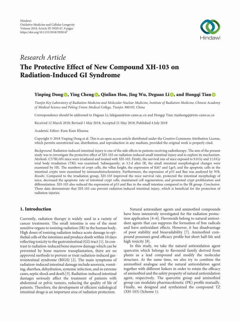
3.2. XH-103 Improves the Survival Rate of Mice after TBI.To determine the protective effect of XH-103 on miceexposure to radiation, we first observed the survival rateof mice after 9.0Gy TBI (Figure 2(a)). The mice were treatedwith XH-103 in three dosages (100mg/kg, 200mg/kg, and400mg/kg); we found that all doses could improve the
S
NH
+ CICI
CI
O O
O CI CICI THF
S
N
OCIRt3N
(a)
HO
HO
HO
O OH
OHO
+ CI
O
N
S
NN S
N O
O
O
S N O
TZ
O
OO
O O
O
O
O
N S
N
N
S
S
Et3N, DMF
(b)
Figure 1: Synthesis and structure of TZ (XH-103). (a) The thiazolidine was reacted with bis(trichloromethyl) carbonate with triethylamine asbase to prepare thiazolidine-3-carbonyl chloride. (b) Thiazolidine-3-carbonyl chloride was coupled with quercetin in the presence oftriethylamine and 4-dimethylaminopyridine to afford the product TZ.
3Oxidative Medicine and Cellular Longevity
survival rate of mice compared with the vehicle-treatedgroup. In the following study, there was 80% mortality invehicle-treated mice within 6 days of 11.0Gy TBI(Figure 2(b)), compared with XH-103-treated mice having60% survival, suggesting that XH-103 may have a protective
effect on RIGS in mice. These results indicate that XH-103effectively mitigates the TBI-induced lethality in mice.
3.3. XH-103 Reduces the Damages of Intestinal Morphology inMice after TBI. To determine the effect of XH-103 on
IR + VehicleIR + 103 (100 mg/kg)
IR + 103 (200 mg/kg)IR + 103 (400 mg/kg)
00
50
100
Surv
ival
(%)
5 10 15Days after 9 Gy TBI
(a)
IR + VehicleIR + 103
00 2 4 6 8 10
Days after 11 Gy TBI
50
100
Surv
ival
(%)
(b)
Figure 2: XH-103 enhances the survival rate of mice after TBI. Kaplan-Meier survival analysis of mice exposed to 9.0Gy or 11.0GyTBI. (a) Three doses of XH-103-treated mice show reduced mortality following lethal doses of TBI (9.0 Gy) within 13 days,compared with IR + vehicle group 100% mortality within 5 days (p < 0 05, n = 10 per group). (b) The Kaplan-Meier survival curve ofvehicle- and XH-103-treated mice (p < 0 05, n = 10 per group) after 11.0Gy total body irradiation. The data were expressed as the percentof surviving mice.
Control
H&
E
Vehicle XH-103IR
(a)
Num
ber o
f cry
pts/
cm
Control Vehicle XH-103IR
0.08
0.06
0.04
0.02
0.00
⁎
(b)
Villi
Control Vehicle XH-103IR
(c)
800
Villu
s hei
ght (�휇
m)
600
400
200
0Control Vehicle XH-103
IR
⁎
(d)
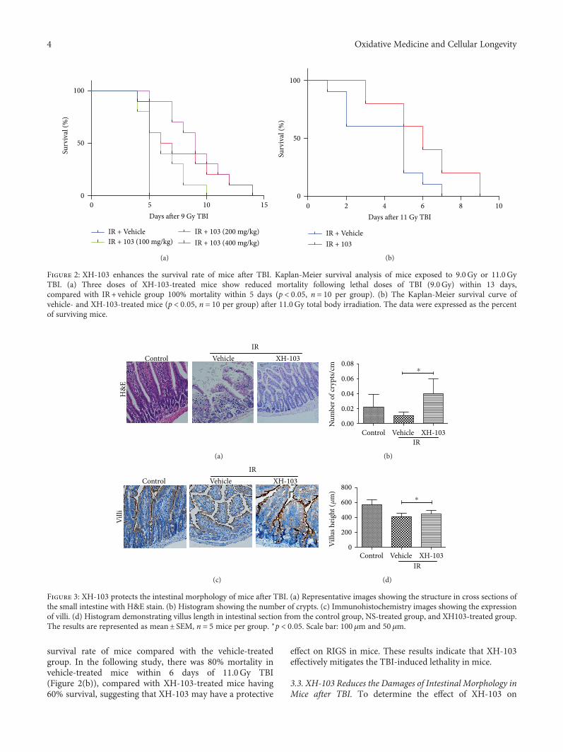
Figure 3: XH-103 protects the intestinal morphology of mice after TBI. (a) Representative images showing the structure in cross sections ofthe small intestine with H&E stain. (b) Histogram showing the number of crypts. (c) Immunohistochemistry images showing the expressionof villi. (d) Histogram demonstrating villus length in intestinal section from the control group, NS-treated group, and XH103-treated group.The results are represented as mean± SEM, n = 5 mice per group. ∗p < 0 05. Scale bar: 100 μm and 50μm.
4 Oxidative Medicine and Cellular Longevity
radiation-induced intestinal injuries, the morphologicalchanges of mouse small intestine are shown in Figure 3. At3 d after 9.0Gy TBI, the irradiated mice showed significantlyshorter villous length and fewer crypts than the control group(p < 0 05). In comparison to mice in the IR group, XH-103-treated mice showed more survival crypts and increasedvillus height (p < 0 05). The expression of villi+ enterocytewas also decreased by IR (Figure 3(c)) then was significantlyincreased by XH-103 compared to mice in the IR group.These results indicate that XH-103 treatment can preventpostradiation damage of the intestinal crypt-villus structureof mice.
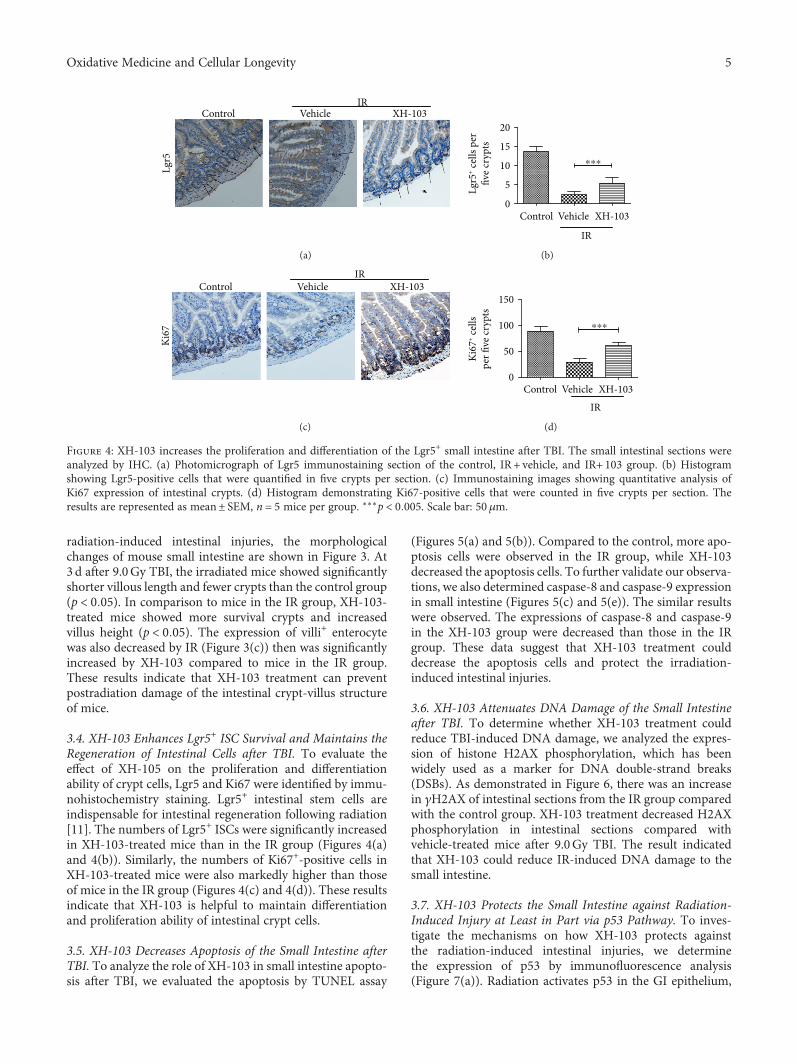
3.4. XH-103 Enhances Lgr5+ ISC Survival and Maintains theRegeneration of Intestinal Cells after TBI. To evaluate theeffect of XH-105 on the proliferation and differentiationability of crypt cells, Lgr5 and Ki67 were identified by immu-nohistochemistry staining. Lgr5+ intestinal stem cells areindispensable for intestinal regeneration following radiation[11]. The numbers of Lgr5+ ISCs were significantly increasedin XH-103-treated mice than in the IR group (Figures 4(a)and 4(b)). Similarly, the numbers of Ki67+-positive cells inXH-103-treated mice were also markedly higher than thoseof mice in the IR group (Figures 4(c) and 4(d)). These resultsindicate that XH-103 is helpful to maintain differentiationand proliferation ability of intestinal crypt cells.
3.5. XH-103 Decreases Apoptosis of the Small Intestine afterTBI. To analyze the role of XH-103 in small intestine apopto-sis after TBI, we evaluated the apoptosis by TUNEL assay
(Figures 5(a) and 5(b)). Compared to the control, more apo-ptosis cells were observed in the IR group, while XH-103decreased the apoptosis cells. To further validate our observa-tions, we also determined caspase-8 and caspase-9 expressionin small intestine (Figures 5(c) and 5(e)). The similar resultswere observed. The expressions of caspase-8 and caspase-9in the XH-103 group were decreased than those in the IRgroup. These data suggest that XH-103 treatment coulddecrease the apoptosis cells and protect the irradiation-induced intestinal injuries.
3.6. XH-103 Attenuates DNA Damage of the Small Intestineafter TBI. To determine whether XH-103 treatment couldreduce TBI-induced DNA damage, we analyzed the expres-sion of histone H2AX phosphorylation, which has beenwidely used as a marker for DNA double-strand breaks(DSBs). As demonstrated in Figure 6, there was an increasein γH2AX of intestinal sections from the IR group comparedwith the control group. XH-103 treatment decreased H2AXphosphorylation in intestinal sections compared withvehicle-treated mice after 9.0Gy TBI. The result indicatedthat XH-103 could reduce IR-induced DNA damage to thesmall intestine.
3.7. XH-103 Protects the Small Intestine against Radiation-Induced Injury at Least in Part via p53 Pathway. To inves-tigate the mechanisms on how XH-103 protects againstthe radiation-induced intestinal injuries, we determinethe expression of p53 by immunofluorescence analysis(Figure 7(a)). Radiation activates p53 in the GI epithelium,
Control
Lgr5
Vehicle XH-103IR
(a)
20
15
10
5
0Control Vehicle XH-103
Lgr5
+ cells
per
five c
rypt
s
IR
⁎⁎⁎
(b)
Control
Ki67
Vehicle XH-103IR
(c)
Ki67
+ cells
per fi
ve cr
ypts
Control Vehicle XH-103
IR
150
100
50
0
⁎⁎⁎
(d)
Figure 4: XH-103 increases the proliferation and differentiation of the Lgr5+ small intestine after TBI. The small intestinal sections wereanalyzed by IHC. (a) Photomicrograph of Lgr5 immunostaining section of the control, IR + vehicle, and IR+ 103 group. (b) Histogramshowing Lgr5-positive cells that were quantified in five crypts per section. (c) Immunostaining images showing quantitative analysis ofKi67 expression of intestinal crypts. (d) Histogram demonstrating Ki67-positive cells that were counted in five crypts per section. Theresults are represented as mean± SEM, n = 5 mice per group. ∗∗∗p < 0 005. Scale bar: 50 μm.
5Oxidative Medicine and Cellular Longevity

IRControl
TUN
EL
Vehicle XH-103
(a)
150
100
50
0TUN
EL-p
ositi
ve ce
llspe
r fiel
d
Control VehicleIR
XH-103
⁎⁎⁎
(b)
IRControl Vehicle XH-103
Casp
ase-
8/D
API
(c)
Control VehicleIR
XH-103
20
15
10
5
0Casp
ase-
8-po
sitiv
e cel
lspe
r fiel
d
⁎⁎
(d)
IRControl Vehicle XH-103
Casp
ase9
/DA
PI
(e)
Control VehicleIR
XH-103
40
30
20
10
0Casp
ase-
9-po
sitiv
e cel
lspe
r fiel
d⁎
(f)
Figure 5: XH-103 reduces the apoptosis of the small intestine after TBI. (a) Apoptosis was assayed by TUNEL staining. (b) The number ofTUNEL-positive cells was quantified per field. The paraffin-embedded sections of the small intestine were analyzed by immunofluorescence.(c) Representative DAPI and caspase-8-staining images of the small intestine (red, caspase-8; blue, DAPI). (d) Caspase-8-positive cells in asingle field of view were quantified. (e) Photomicrograph of caspase-9-staining images of the small intestine (red, caspase-9; blue, DAPI).(f) Bar graph showing quantitative analysis of caspase-9-positive cells per field of view. The results are represented as mean± SEM, n = 5mice per group. ∗p < 0 05, ∗∗p < 0 01, ∗∗∗p < 0 005. Scale bar: 50 μm and 10μm.
Control VehicleIR
XH-103
�훾H
2AX/
DA
PI
(a)
30
20
10
0
�훾H
2AX-
posit
ive c
ells
per fi
eld
Control Vehicle XH-103IR
⁎
(b)
Figure 6: XH-103 attenuates DNA damage of mice after TBI. The small intestines of control mice, vehicle-treated mice, and XH-103-treatedmice were obtained at 3 d after 9.0Gy TBI. (a) Representative immunofluorescence images for the expression of γH2AX of the small intestines(red, γH2AX; blue, DAPI). (b) Histogram demonstrating quantitative analysis of γH2AX-positive cells per view field. The results arerepresented as mean± SEM, n = 5 mice per group. ∗p < 0 05. Scale bar: 10μm.
6 Oxidative Medicine and Cellular Longevity

and p53-mediated apoptosis has been implicated in regulatingthe intestinal radiation injuries [12, 13].We also evaluated theexpression of Bax in the small intestine crypts by Westernblotting at 3 d after 9.0Gy TBI (Figure 7(c)). IR increasedthe expression of p53 in the small intestine compared withthe control group. In contrast, mice treated with XH-103downregulate the expression of p53 and Bax (Figures 7(b)and 7(c)). These findings suggest that XH-103 protects thesmall intestine from IR at least by p53 pathway.
4. Discussion
The development of the effective method and drug to miti-gate the radiation-induced intestinal injuries is an importantarea in cancer therapy, nuclear accident, and terrorism.Many studies have reported that Chinese herbal medicineor extracts may reduce TBI-induced injuries in the brain,esophagus, and hematopoietic system of irradiated animals[14–18]; the study of protective drugs on IR-induced intesti-nal injuries still needs to be improved. In the present study,we synthesized a new compound XH-103 and evaluated theprotective effects on radiation-induced intestinal injuries.
In this study, we observe that XH-103 improved thesurvival rate of mice exposed to the lethal dose TBI, whichindicates that XH-103 could protect the mice from irradia-tion. Under physiological conditions, epithelial homeostasisis maintained by proliferative cells in crypts, and the smallintestinal crypt cells are particularly sensitive to IR due totheir high proliferative rate [19]. The intestinal epitheliumis one of the most rapidly self-renewing organizations inmammals, which is continuously renewed by intestinal
epithelium stem cells (IESCs) located in the crypts. Intestinalepithelium cell renewal is identified by expression of Lgr5[20, 21]. We found that the numbers of Lgr5+ intestinal stemcells were increased in the XH-103 group after 9.0Gy TBI,and the Lgr5+ intestinal stem cells differentiated into morePaneth cells and villus cells. Thus, XH-103 may play the pro-tective role on IR-induced intestinal injuries by improvingthe proliferation and differentiation of Lgr5+ intestinal stemcells. The increased expression of Ki67, another proliferativemarker in the small intestine, in the XH-103 mice alsosuggested the protective effects of XH-103 on the intestinalradiation injury. After radiation, various degrees of villusblunting and fusion, villous epithelial cell attenuation andhypertrophy, and severe loss of crypts may occur, leadingto destruction of epithelial cell homeostasis and epithelialintegrity [13]. Incomplete epithelial cells cannot easily main-tain intestinal absorption and defense functions. Our resultsdemonstrated that the intestinal crypt-villus structure fromthe XH-103-treated mice was well preserved after 9.0GyTBI. These results indicate that XH-103 may have a protec-tive effect on irradiation-induced intestinal injury.
Apoptosis is programmed cell death that involves thecontrolled breakdown of intracellular components. Manystudies have shown that IR induced tissue damage, such assmall intestinal injuries, with increasing apoptosis cells[22–24]. Caspases are a family of genes important formain-taining homeostasis through regulating apoptosis andinflammation [25, 26]. Caspases involved in apoptosis havebeen subclassified by their mechanism of action, initiatorcaspase (caspase-8 and -9) and executioner caspase (cas-pase-3, 6, and 7). In this study, we investigated that treatment
Control Vehicle XH-103IR
p53/
DA
PI
(a)
25201510
5
p53-
posit
ive c
ells
per fi
eld
0Control Vehicle XH-103
IR
⁎⁎⁎
(b)
Bax
Con
trol
IR +
veh
icle
IR +
XH
103
Tubulin
(c)
Figure 7: XH-103 decreases the expression of p53 and Bax after TBI. The small intestinal sections of the control, IR + vehicle, and IR+ 103mice were gained at 3 d after 9.0Gy TBI. (a) Representative immunofluorescence images for the expression of p53 of the small intestines(red, p53; blue, DAPI). (b) Histogram showing quantitative analysis of p53-positive cells per field of view. (c) Western blot for Bax andtubulin in the intestinal crypts from non-IR mice, vehicle-treated mice, and XH-103-treated mice at 3 d after 9.0Gy TBI. The results arerepresented as mean± SEM, n = 5 mice per group. ∗∗∗p < 0 005. Scale bar: 10μm.
7Oxidative Medicine and Cellular Longevity
with XH-103 reduces the apoptosis cells compared with theIR group. The expression of caspase-8 and caspase-9 in thesmall intestines of XH-103 was also decreased compared tothat of the IR group. These results show the inhibitory effectsof XH-103 on radiation-induced apoptosis.
Radiation induces DNA damages [27] and destroysthe expression of proteins in cells [28], activating p53[23, 27, 29, 30]. H2AX phosphorylation is an indicator forquantifying DNA double-strand breaks [31–33]. In thisstudy, we found that the expression of γH2AX was decreasedin the XH-103-treated mice compared with the vehicle-treated mice.
It is well known that p53 activates genes that regulate cellcycle checkpoints, DNA damage and repair, and apoptosis[34–36]. p53 can promote apoptosis through interactionswith Bcl-2 family proteins in the cytoplasm. Studies reportedthat Bax−/− and Bak1−/− mice reduced the epithelial cellapoptosis exposure to irradiation [37, 38]. Therefore, weexamined whether XH-103 inhibits apoptosis by p53 path-way. For these experiments, treatment with XH-103 coulddecrease the level of p53 and Bax expression. The data sug-gested that XH-103 might mitigate the radiation-inducedintestinal injuries by p53-dependent apoptosis pathway.
Our studies synthesize a new compound XH-103 andshow protective effects of XH-103 against radiation-inducedintestinal injury. The results also suggest that XH-103 mayattenuate radiation-induced intestinal damage via the p53pathway. However, XH-103 is a novel compound that needsfurther optimization.
Data Availability
The data used to support the findings of this study areavailable from the corresponding author upon request.
Conflicts of Interest
The authors declare that they have no conflicts of interest.
Authors’ Contributions
Deguan Li and Hongqi Tian conceived and designed theexperiments. Ying Cheng and Hongqi Tian designed andsynthesized the new compound XH-103. Deguan Li, YinpingDong, Qinlian Hou, and Jing Wu carried out the follow-upexperiments, analyzed the data, and interpreted theresults. Deguan Li and Yinping Dong contributed to dataanalysis and manuscript preparation. Yinping Dong andYing Cheng contributed equally to this work and areco-first author.
Acknowledgments
This study was supported by the National Natural ScienceFoundation of China (no. 81673106, 81601411); the NaturalScience Foundation of Tianjin (15JCZDJC35200); TianjinHealth Industry Key Research Projects (15KG138); the inno-vation team funding (1649) from the Institute of RadiationMedicine, Chinese Academy of Medical Sciences and Peking
Union Medical College, and Fundamental Research Fundfor CAMS&PUMC (2016ZX310199, 2016ZX310070); andCAMS Innovation Fund for Medical Sciences (CIFMS,2017-I2M-3-019) from the Chinese Academy of MedicalSciences and Peking Union Medical College.
Supplementary Materials
Figure S1a: 1H NMR spectra of TZ. 1H NMR (400MHz,DMSO-d6) δ ppm 7.95–7.85 (m, 2H), 7.68 (s, 1H), 7.58(d, J = 8 3 Hz, 1H), 7.25 (s, 1H), 4.58 (dd, J = 52 9, 22.8Hz,10H), 3.97–3.65 (m, 10H), 3.14 (d, J = 20 2 Hz, 10H). FigureS1b: LC-MS spectra of TZ. LC-MSmethod. Experiments per-formed on a Waters 3100 Mass Detector. Chromatographiccolumn: Agilent, ZORBAX, C18 2.1∗50mm 3.5μ. Flow rate:0.6mL/min; mobile phase A: 0.1% formic acid in water;mobile phase B: 100% acetonitrile. Gradient elution program:t = 0 min, A% = 10%; t = 5 5 min, A% = 95%; t = 6 5 min,A% = 95%; t = 7 min, A% = 10%; t = 10 min, A% = 10%;RT = 4 50 min, MS (ESI, positive ion) m/z: 878.15 (M+1).(Supplementary Materials)
References
[1] N. H. A. Terry and E. L. Travis, “The influence of bone marrowdepletion on intestinal radiation damage,” InternationalJournal of Radiation Oncology∗Biology∗Physics, vol. 17,no. 3, pp. 569–573, 1989.
[2] M. E. Berger, D. M. Christensen, P. C. Lowry, O. W. Jones, andA. L. Wiley, “Medical management of radiation injuries:current approaches,” Occupational Medicine, vol. 56, no. 3,pp. 162–172, 2006.
[3] P. Monti, J. Wysocki, A. van der Meeren, and N. M. Griffiths,“The contribution of radiation-induced injury to the gastroin-testinal tract in the development of multi-organ dysfunctionsyndrome or failure,” The British Journal of Radiology, no. 1,Supplement_27, pp. 89–94, 2005.
[4] D. D. Peebles, C. M. Soref, R. R. Copp, A. L. Thunberg, andW. E. Fahl, “ROS-scavenger and radioprotective efficacy ofthe new PrC-210 aminothiol,” Radiation Research, vol. 178,no. 1, pp. 57–68, 2012.
[5] W. Liu, Q. Chen, S. Wu et al., “Radioprotector WR-2721 andmitigating peptidoglycan synergistically promote mouse sur-vival through the amelioration of intestinal and bone marrowdamage,” Journal of Radiation Research, vol. 56, no. 2,pp. 278–286, 2015.
[6] P. Bansal, P. Paul, A. Kunwar et al., “Radioprotection by quer-cetin-3-O-rutinoside, a flavonoid glycoside – a cellular andmechanistic approach,” Journal of Functional Foods, vol. 4,no. 4, pp. 924–932, 2012.
[7] A. R. Bilia, B. Isacchi, C. Righeschi, C. Guccione, and M. C.Bergonzi, “Flavonoids loaded in nanocarriers: an opportunityto increase oral bioavailability and bioefficacy,” Food andNutrition Sciences, vol. 05, no. 13, pp. 1212–1327, 2014.
[8] D. M. Brizel, T. H. Wasserman, M. Henke et al., “Phase IIIrandomized trial of amifostine as a radioprotector in headand neck cancer,” Journal of Clinical Oncology, vol. 18,no. 19, pp. 3339–3345, 2000.
[9] M. M. Mahé, E. Aihara, M. A. Schumacher et al., “Establish-ment of gastrointestinal epithelial organoids,” Current Proto-cols in Mouse Biology, vol. 3, no. 4, pp. 217–240, 2013.
8 Oxidative Medicine and Cellular Longevity
[10] W. Yang, Z. Sun, B. Yang, and Q. Wang, “Nrf2-knockout pro-tects from intestinal injuries in C57BL/6J mice followingabdominal irradiation with γ rays,” International Journal ofMolecular Sciences, vol. 18, no. 8, 2017.
[11] C. Metcalfe, N. M. Kljavin, R. Ybarra, and F. J. de Sauvage,“Lgr5+ stem cells are indispensable for radiation-inducedintestinal regeneration,” Cell Stem Cell, vol. 14, no. 2,pp. 149–159, 2014.
[12] W. Qiu, E. B. Carson-Walter, H. Liu et al., “PUMA regulatesintestinal progenitor cell radiosensitivity and gastrointestinalsyndrome,” Cell Stem Cell, vol. 2, no. 6, pp. 576–583, 2008.
[13] A. J. Merritt, C. S. Potten, C. J. Kemp et al., “The role of p53 inspontaneous and radiation-induced apoptosis in the gastroin-testinal tract of normal and p53-deficient mice,” CancerResearch, vol. 54, no. 3, pp. 614–617, 1994.
[14] I. Kindekov, M. Mileva, D. Krastev et al., “Radioprotectiveeffect ofRapana thomasianahemocyanin in gamma inducedacute radiation syndrome,” Biotechnology & BiotechnologicalEquipment, vol. 28, no. 3, pp. 533–539, 2014.
[15] L. Lu, Y.-Y. Wang, J.-L. Zhang, D.-G. Li, and A.-M. Meng,“p38 MAPK inhibitor insufficiently attenuates HSC senes-cence administered long-term after 6 Gy total body irradiationin mice,” International Journal of Molecular Sciences, vol. 17,no. 6, p. 905, 2016.
[16] D. Li, Z. Tian, W. Tang et al., “The protective effects of5-methoxytryptamine-α-lipoic acid on ionizing radiation-induced hematopoietic injury,” International Journal of Molec-ular Sciences, vol. 17, no. 6, p. 935, 2016.
[17] S. Suryavanshi, D. Sharma, R. Checker et al., “Amelioration ofradiation-induced hematopoietic syndrome by an antioxidantchlorophyllin through increased stem cell activity and modu-lation of hematopoiesis,” Free Radical Biology & Medicine,vol. 85, pp. 56–70, 2015.
[18] J. Li, J. Xu, W. Xu et al., “Protective effects of Hong Shan cap-sule against lethal total-body irradiation-induced damage inWistar rats,” International Journal of Molecular Sciences,vol. 16, no. 8, pp. 18938–18955, 2015.
[19] C. S. Potten and H. K. Grant, “The relationship between ioniz-ing radiation-induced apoptosis and stem cells in the smalland large intestine,” British Journal of Cancer, vol. 78, no. 8,pp. 993–1003, 1998.
[20] H. Clevers, “Identification of stem cells in small intestine andcolon by a single marker gene Lgr5,” European Journal ofCancer Supplements, vol. 6, no. 9, p. 1, 2008.
[21] I. Stzepourginski, G. Nigro, J. M. Jacob et al., “CD34+ mesen-chymal cells are a major component of the intestinal stem cellsniche at homeostasis and after injury,” Proceedings of theNational Academy of Sciences of the United States of America,vol. 114, no. 4, pp. E506–E513, 2017.
[22] H. Zhang, H. Yan, X. Zhou et al., “The protective effects of res-veratrol against radiation-induced intestinal injury,” BMCComplementary and Alternative Medicine, vol. 17, no. 1,p. 410, 2017.
[23] D. G. Kirsch, P. M. Santiago, E. di Tomaso et al., “p53 controlsradiation-induced gastrointestinal syndrome in mice indepen-dent of apoptosis,” Science, vol. 327, no. 5965, pp. 593–596,2010.
[24] A. Morita, I. Takahashi, M. Sasatani et al., “A chemicalmodulator of p53 transactivation that acts as a radioprotectiveagonist,” Molecular Cancer Therapeutics, vol. 17, no. 2,pp. 432–442, 2018.
[25] D. R. McIlwain, T. Berger, and T. W. Mak, “Caspase functionsin cell death and disease,” Cold Spring Harbor Perspectives inBiology, vol. 5, no. 4, article a008656, 2013.
[26] A. Negroni, S. Cucchiara, and L. Stronati, “Apoptosis, necrosis,and necroptosis in the gut and intestinal homeostasis,”Media-tors of Inflammation, vol. 2015, Article ID 250762, 10 pages,2015.
[27] X. Han, E. Mann, S. Gilbert et al., “Loss of guanylyl cyclase C(GCC) signaling leads to dysfunctional intestinal barrier,”PLoS One, vol. 6, no. 1, article e16139, 2011.
[28] X. Sun, Q. Wang, Y. Wang, L. Du, C. Xu, and Q. Liu, “Brusatolenhances the radiosensitivity of A549 cells by promoting ROSproduction and enhancing DNA damage,” InternationalJournal of Molecular Sciences, vol. 17, no. 7, p. 997, 2016.
[29] J. M. Sullivan, L. B. Jeffords, C.-L. Lee, R. Rodrigues, Y. Ma,and D. G. Kirsch, “p21 protects “super p53” mice fromthe radiation-induced gastrointestinal syndrome,” RadiationResearch, vol. 177, no. 3, pp. 307–310, 2012.
[30] B. J. Leibowitz, W. Qiu, H. Liu, T. Cheng, L. Zhang, and J. Yu,“Uncoupling p53 functions in radiation-induced intestinaldamage via PUMA and p21,” Molecular Cancer Research,vol. 9, no. 5, pp. 616–625, 2011.
[31] Y. Wang, A. Meng, H. Lang et al., “Activation of nuclear factorκB in vivo selectively protects the murine small intestineagainst ionizing radiation-induced damage,” Cancer Research,vol. 64, no. 17, pp. 6240–6246, 2004.
[32] Y. Shiloh, “ATM and related protein kinases: safeguardinggenome integrity,” Nature Reviews. Cancer, vol. 3, no. 3,pp. 155–168, 2003.
[33] M. Podhorecka, A. Skladanowski, and P. Bozko, “H2AXphosphorylation: its role in DNA damage response and cancertherapy,” Journal of Nucleic Acids, vol. 2010, Article ID920161, 9 pages, 2010.
[34] S. S. Foster, S. De, L. K. Johnson, J. H. J. Petrini, and T. H.Stracker, “Cell cycle- and DNA repair pathway-specific effectsof apoptosis on tumor suppression,” Proceedings of theNational Academy of Sciences of the United States of America,vol. 109, no. 25, pp. 9953–9958, 2012.
[35] J. R. Jeffers, E. Parganas, Y. Lee et al., “Puma is an essentialmediator of p53-dependent and -independent apoptotic path-ways,” Cancer Cell, vol. 4, no. 4, pp. 321–328, 2003.
[36] M.-J. Halaby, Y. Li, B. R. Harris et al., “Translational controlprotein 80 stimulates IRES-mediated translation of p53mRNA in response to DNA damage,” BioMed Research Inter-national, vol. 2015, Article ID 708158, 9 pages, 2015.
[37] M. Garcia-Barros, F. Paris, C. Cordon-Cardo et al., “Tumorresponse to radiotherapy regulated by endothelial cell apopto-sis,” Science, vol. 300, no. 5622, pp. 1155–1159, 2003.
[38] A. J. Merritt, T. D. Allen, C. S. Potten, and J. A. Hickman,“Apoptosis in small intestinal epithelia from p53-null mice:evidence for a delayed, p53-indepdendent G2/M-associatedcell death after γ-irradiation,” Oncogene, vol. 14, no. 23,pp. 2759–2766, 1997.
9Oxidative Medicine and Cellular Longevity

Stem Cells International
Hindawiwww.hindawi.com Volume 2018
Hindawiwww.hindawi.com Volume 2018
MEDIATORSINFLAMMATION
of
EndocrinologyInternational Journal of
Hindawiwww.hindawi.com Volume 2018
Hindawiwww.hindawi.com Volume 2018
Disease Markers
Hindawiwww.hindawi.com Volume 2018
BioMed Research International
OncologyJournal of
Hindawiwww.hindawi.com Volume 2013
Hindawiwww.hindawi.com Volume 2018
Oxidative Medicine and Cellular Longevity
Hindawiwww.hindawi.com Volume 2018
PPAR Research
Hindawi Publishing Corporation http://www.hindawi.com Volume 2013Hindawiwww.hindawi.com
The Scientific World Journal
Volume 2018
Immunology ResearchHindawiwww.hindawi.com Volume 2018
Journal of
ObesityJournal of
Hindawiwww.hindawi.com Volume 2018
Hindawiwww.hindawi.com Volume 2018
Computational and Mathematical Methods in Medicine
Hindawiwww.hindawi.com Volume 2018
Behavioural Neurology
OphthalmologyJournal of
Hindawiwww.hindawi.com Volume 2018
Diabetes ResearchJournal of
Hindawiwww.hindawi.com Volume 2018
Hindawiwww.hindawi.com Volume 2018
Research and TreatmentAIDS
Hindawiwww.hindawi.com Volume 2018
Gastroenterology Research and Practice
Hindawiwww.hindawi.com Volume 2018
Parkinson’s Disease
Evidence-Based Complementary andAlternative Medicine
Volume 2018Hindawiwww.hindawi.com
Submit your manuscripts atwww.hindawi.com