Embed Size (px)
Citation preview

Sturgill et al. Molecular Cancer 2010, 9:104http://www.molecular-cancer.com/content/9/1/104
Open AccessR E S E A R C H
ResearchThe promoter for intestinal cell kinase is head-to-head with F-Box 9 and contains functional sites for TCF7L2 and FOXA factorsThomas W Sturgill*1, Paul B Stoddard3, Steven M Cohn2 and Marty W Mayo3
AbstractBackground: Intestinal cell kinase (ICK; GeneID 22858) is a conserved MAPK and CDK-like kinase that is widely expressed in human tissues. Data from the Cancer Genome Anatomy Project indicated ICK mRNA is increased in cancer, and that its expression correlated with expression of mRNA for an uncharacterized F-box protein, FBX9 (GeneID: 26268). ICK and FBX9 genes are arranged head-to-head on opposite strands, with start sites for transcription separated by ~3.3 kb. We hypothesized ICK and FBX9 are potentially important genes in cancer controlled by a bidirectional promoter.
Results: We assessed promoter activity of the intergenic region in both orientations in cancer cell lines derived from breast (AU565, SKBR3), colon (HCT-15, KM12), and stomach (AGS) cancers, as well as in embryonic human kidney (HEK293T) cells. The intergenic segment was active in both orientations in all of these lines, and ICK promoter activity was greater than FBX9 promoter activity. Results from deletions and truncations defined a minimal promoter for ICK, and revealed that repressors and enhancers differentially regulate ICK versus FBX9 promoter activity. The ICK promoter contains consensus motifs for several FOX-family transcription factors that align when mouse and human are compared using EMBOSS. FOXA1 and FOXA2 increase luciferase activity of a minimal promoter 10-20 fold in HEK293T cells. Consensus sites for TCF7L2 (TCF4) (Gene Id: 6934) are also present in both mouse and human. The expression of β-catenin increased activity of the minimal promoter ~10 fold. ICK reference mRNAs (NM_014920.3, NM_016513) are expressed in low copy number and increased in some breast cancers, using a ten base tag 5'-TCAACCTTAT-3' specific for both ICK transcripts.
Conclusion: ICK and FBX9 are divergently transcribed from a bidirectional promoter that is GC-rich and contains a CpG island. A minimal promoter for ICK contains functional sites for β-cateinin/TCF7L2 and FOXA. These data are consistent with functions that have been proposed for ICK in development and in proliferation or survival of some breast and colon cancers.
BackgroundThe ICK gene encodes an evolutionarily conserved Ser/Thr kinase in the CMGC group of the kinome, clusteringin a subgroup with closely related MAK (male germ cell-associated protein kinase) and more distantly relatedMOK (MAPK/MAK/MRK overlapping kinase) [1]. ICKwas first identified and named MRK (MAK-related pro-tein kinase) after cloning of its cDNA from heart [2]).ICK expression was higher in the embryonic myocardium
during organogenesis than in the adult tissue [2].Decreasing expression of ICK in Colo205 cells stops pro-liferation and causes cell cycle arrest in G1 due to anincrease in p21Cip [3]. Colo205 cells greatly overexpressICK mRNA in comparison to other lines in the NCI60,suggesting an acquired addiction to ICK for proliferationin this line. ICK mRNA is detectable in normal intestinalepithelium only in the region for lineage specification andproliferation [4].
ICK has to be phosphorylated in a TDY motif (residues157-159) within the activation loop to be fully active.Phosphorylation of Y159 can occur by autophosphoryla-tion, but at least phosphorylation of T157 requires trans-
* Correspondence: [email protected] Departments of Pharmacology and Internal Medicine, University of Virginia, 1300 Jefferson Park Avenue, Charlottesville, Virginia, 22908, USAFull list of author information is available at the end of the article
BioMed Central© 2010 Sturgill et al; licensee BioMed Central Ltd. This is an Open Access article distributed under the terms of the Creative CommonsAttribution License (http://creativecommons.org/licenses/by/2.0), which permits unrestricted use, distribution, and reproduction inany medium, provided the original work is properly cited.

Sturgill et al. Molecular Cancer 2010, 9:104http://www.molecular-cancer.com/content/9/1/104
Page 2 of 13
phosphorylation by another kinase [1]. ICK is a substratefor a T157-kinase related to CDK-activating kinase withgene name CCRK (cell cycle regulated kinase, [GenBank:NM_001039803]). CCRK (NM_001039803) unequivo-cally has T157 kinase activity because wild type but not akinase-defective mutant phosphorylates T157 in cells [1].Decreasing CCRK expression ~80% markedly inhibitedproliferation of HCT116 and U2OS cells without a signif-icant, specific change in G1, M, or G2/M populations butmodestly increased the population with sub-G1 DNAcontent, suggesting increased apoptosis [5]. Otherreports support a role for CCRK in molecular carcino-genesis of ovarian cancer [6]. CCRK-specific gene silenc-ing causes ovarian cancer cells to arrest in G1 [6].Recently, CCRK was identified as an interactor of Broad-minded in Sonic hedgehog pathways [7].
ResultsFBX9 and ICK expression are correlated genesThe NCI60 is a panel of cancer cell lines for the CancerGenome Anatomy Project (CGAP). FBX9 expression cor-relates (R = 0.45, P = 1.5 e-04) with ICK expression in theNCI60 amongst genes present in microarrays from a verylarge collection of cDNAs. We took note because FBX9 isthe neighboring gene to ICK. FBX9 encodes an uncharac-terized F-box protein [8]. The two genes are on oppositestrands, arranged head-to-head with their predicted startsites separated by only ~3.3 kb. These data suggested theintergenic segment might have bidirectional promoteractivity.
We also were interested in using the intergenic segmentto gain insights to ICK regulation that in turn might sug-gest functions. Expression of ICK mRNA is confined tothe region in normal mouse epithelium where prolifera-tion and lineage specifications occur and where β-catenin/TCF7L2 (referred to herein as TCF4) is mostactive. Loss of a tumor suppressor causes activation of β-catenin/TCF4 in colon cancers [9]. We hypothesized thatICK promoter activity may be increased in colon cancercell lines (KM12, HCT-15) and in stomach cancer cells(AGS) because of this correlation. We also studied breastcancer cell lines (AU565 and SK-BR3-3) because β-catenin/TCF4 is highly active in breast cancers [10].
The FBX9-ICK intergenic segment has bidirectional promoter activityWe obtained a clone (RP3-341E18, [GenBank:AL031178]) for a portion of the p12.3-p11.2 region ofhuman chromosome 6 from the Sanger Institute. OneXhoI restriction fragment contains the intergenic regionand the start sites for transcription of both genes. This 4.5kilobase fragment and portions thereof were placed intothe promoterless pGL3-luciferase plasmid so as to gener-ate constructs (pGL3-PICK: 1-12 and pGL3-PFBX9: 1-5),
shown schematically in (Fig. 1). We refer to constructs asICK-1 to 12 and as FBX9-1 to 5. We used these constructsto study the promoter in five human cancer cell lines aswell as in HEK293T.
The full intergenic segment (constructs ICK-1 andFBX9-1) was active in both orientations in all six of thelines, suggesting that ICK and FBX9 share a bidirectionalpromoter. Analyses in the different lines (Figs. 2, 3, 4)show elements in the common SspIb to PstIb fragment areimportant for bidirectional activity, and may account forthe correlated expression of FBX9 and ICK in microarraydata that motivated this study. Our analyses (Figs. 2, 3,&4) show that the intergenic segment is not a constitu-tive, bidirectional promoter because the FBX9 activityrelative to ICK activity is variable.
Promoter activity in HER2-overexpressing breast cancer cellsICK promoter activity was 10-20 fold higher than FBX9promoter activity in AU565 and SKBR3 cells, using con-structs ICK-1 and FBX9-1 which contain the full inter-genic segment (Fig. 2). Moreover, AU565 and SKBR3 gavesimilar patterns of relative activity between the differentconstructs derived from ICK-1 and FBX9-1. This mayrelate to the fact that AU565 and SKBR3 were obtainedfrom pleural effusions from the same patient [11]. The
Figure 1 Restriction map of genomic DNA between FBX9 and ICK and pGL3 constructs. Indicated fragments were cloned into promot-er-less pGL3 for luciferase (Luc) expression. Arrows, start of transcrip-tion for reference ICK and FBX9 mRNAs.
4521bpPstI
ApaI
SmaI
ApaI
FBX9
NcoI
SmaI
EcoRISspI
BamHIHindIII
SspI
EcoRV
HindIII
EcoRIPacI
EcoRV PstI
ICK
ApaI
123456789101112
12345
Luc
Luc
Luc
Luc
Luc
Luc
Luc
Luc
Luc
Luc
Luc
Luc
Luc
Luc
Luc
Luc
Luc
ICK
FBX9
a a
a a
ab
bb
bb
c
a b

Sturgill et al. Molecular Cancer 2010, 9:104http://www.molecular-cancer.com/content/9/1/104
Page 3 of 13
results obtained with the truncation constructs (ICK-2 to7) reveal enhancer elements within the SspIb-EcoRVa seg-ment and a suppressor element within the unique EcoRV-EcoRV fragment. The internal deletions (ICK-10 to 12)indicate another enhancer element for ICK lies inEcoRVb-PstIb close to the ICK start site. Removal of thissegment reduces ICK promoter activity 40% in bothAU565 and SKBR3 cells. Extending the internal deletionfrom Pst1b back to SspIb (ICK-11), or further back to SspIa
(ICK-12), had modest and opposite effects. The regionfrom SspIb to PstIb is particularly complex, and appearslikely to have several important elements. This conclu-sion is borne out by data obtained from the other lines.
Promoter activity in colon cancer cellsKM12 and HCT-15 are two colon cancer cell lines. Bothare near diploid, and have relatively few structural rear-rangements confined to 7 chromosomes [12]. The pat-terns of luciferase activity created by the constructs inthese two cancer cell lines are quite different (Fig. 3).KM12 has homozygous loss for a lysine (K)-specific dem-ethylase 6A (KDM6A); a ubiquitously transcribed X
chromosome tetratricopeptide repeat protein; homozy-gous loss of PTEN; and heterozygous loss of p53 func-tions. (Some mutations in the NCI60 are available onlinehttp://www.sanger.ac.uk/). HCT-15 is null for function ofAPC, BRAC2, and FAM123 tumor suppressors, and hashomozygous loss of p53 along with oncogenic mutationsin KRAS, PI3KCα, and MSH6. The results for truncationsfor ICK in KM12 suggest an enhancer in SspIb-EcoRVa,and a suppressor in the unique EcoRV-EcoRV segment,and provide strong evidence for an enhancer in EcoRVb -PstIb. The internal deletions for ICK (ICK-10 to 12) alsostrongly support this enhancer. Specific removal ofEcoRVb - PstIb with ICK-10 caused a large decrease inactivity, and this phenomenon was observed to differentdegrees in all six lines. Extending the internal deletion toSspIb (ICK-11) or to SspIa (ICK-12) resulted in modestchanges by comparison. The largest change in activity inHCT-15 occurred with deletion of EcoRVb-PstIb.
Promoter activity in AGS gastric cancer and HEK293T kidney cellsAGS is a human gastric cancer line that robustlyexpresses ICK mRNA [4]. HEK293T cells are humanembryonic fibroblasts that were originally immortalizedby transformation with sheared adenovirus [13], andmuch later made to express the large T antigen of SV40.AGS is similar to KM12 in pattern of luciferase activitybetween constructs, and HEK293 is similar to HCT-15(Fig. 4). Results from AGS, like KM12 discussed above,
Figure 2 FBX9 and ICK are divergently transcribed from a bidirec-tional promoter. Equal numbers of SKBR3 and AU565 cells were seed-ed into 96-well plates, transfected with the indicated constructs (Figure 1), then assayed for luciferase activity in each well by the meth-ods described. Data in figures 2 and 3 were obtained by the same pro-cedures. Bar, ± S.D.
AU565
1
3
5
7
9
1110
2
4
6
8
12
1
3
5
2
4
ICK
FBX9
0 20 40 60 80 100 120Relative Light Units
0 20 40 60 80 100 1201
3
5
7
9
1110
2
4
6
8
12
1
3
5
2
4 SKBR3
ICK
FBX9
Relative Light Units
Figure 3 The SspIb to PstIb segment contains enhancer and sup-pressor elements. Equal numbers of KM12 and HCT-15 colon cancer cells were seeded into 96-well plates, transfected with the indicated constructs (Figure 1), then assayed for luciferase activity in each well by the methods described. Bar, ± S.D.
HCT-15
1
3
5
7
9
1110
2
4
6
8
12
ICK
FBX91
3
5
2
4
0 20 40 60 80 100 120Relative Light Units
1
3
5
7
9
1110
2
4
6
8
12
ICK
FBX91
3
5
2
4
0 20 40 60 80 100 120 140 160
KM12
Relative Light Units

Sturgill et al. Molecular Cancer 2010, 9:104http://www.molecular-cancer.com/content/9/1/104
Page 4 of 13
support regulatory elements within ApaIa-ApaIb, andconfirm the enhancer in SspIb-EcoRVa and the suppressorin the unique EcoRV-EcoRV. Overall, both the trunca-tions and the internal deletions in AGS and HEK293strongly support importance of EcoRVb-PstIb.
Conserved FOX binding motifs in human and mouse ICK promotersPromoters for ICK and FBX9 are similarly configured onmouse Chr9 in a head-to-head fashion with starts fortranscription on opposite strands. Because prediction oftranscription factor sites is difficult at best and there aremany false positive, we looked for conserved motifs pres-ent in both mouse and human that are well characterizedin literature.
A striking finding was a number of consensus motifsfor fork head box (FOX) proteins. Many FOX proteinsbind a conserved motif with a core of TGTTTR, where Ris (G, A) [14]. Also striking was the presence of a numberof aligned, conserved TG motifs (TTTGTT, TTT-GTTTT, TTTTGTTTGTTTT). FOX is a large family ofsequence-specific factors. Its members regulate expres-sion of many genes involved in cell growth, proliferation,
differentiation and development [15,16]. The first proteinin the FOX family was the Drosophila gene named forkhead. (Prior to year 2000, certain human FOX proteinshad several aliases, as winged helix protein proteins, ashepatocyte nuclear factors (HNFs), or forkhead-relatedclones (FREAC). For example, FOXA1 and 2 were HNF3αand β.) The winged helix domain of FOXA (HNF3) bindsoptimally to a consequence WWTRTTTRYWYDsequence [17], where W is (A/T), R is (A/G), Y is (C/T),and D is (A/C/T).
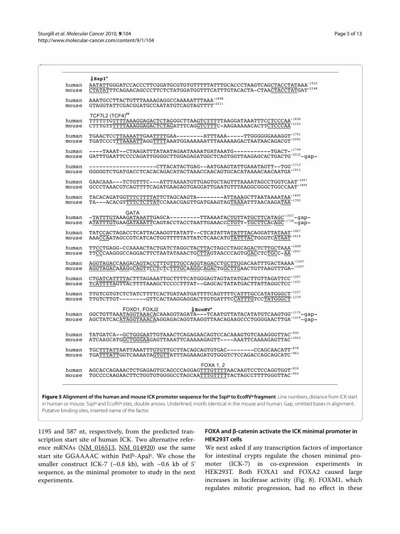
A motif, 5'-ATAGGTAAACA-3', near -1217 nt inhuman ICK, is predicted to bind FOX proteins, possiblyFOXD1 and FOXJ2 (Fig. 5). This motif has a conservedGTAAACA core known to bind FOXD1 (FREAC-4) andFOXF2 (FREAC-2). Bases that differentiate between fam-ily members lie near this core [14]. FOXJ2 functions ingametogenesis and early embryonic development [18].FOXD1 functions in development of the retina [19]. Amotif, 5'-GCCTTTTGTTTGTTTT-3' (near -30 nt inhuman), is conserved between mouse and human andcontains a consensus match to FOX proteins expressed inembryonic tissues, possibly FOXJ1 or FOXJ2 (Fig. 6). Thismotif also matches the core for FOXA.
Beginning near PstIb (Fig. 6) is a region of near identitythat surrounds the transcription start sites for ICK. Thisregion is GC-rich, and has conserved CpG sites concen-trated as a CpG island. This region [NCBI gi: 1038509]was isolated in a genome-wide purification of un-methy-lated CpG islands [20]. CpG islands overlap the 5'end ofgenes, and often contain the promoter and one or moreexons of genes [20]. Methylations can differentially regu-late recognition by transcription factors [21]. Methyla-tions at CpG can also change gene expression indevelopment in set programs of activation and silencing[22], and remain as a source of epigenomic variation [23].The putative activator of ICK, CCRK, is transcribed froma 5' start in a CpG island that is variably methylated inadult brain tissues [23].
Minimal ICK promoter in HEK293T and HCT-15 cellsTo enable initial studies of transcription factors, we chosea minimal ICK promoter for use in HEK293T cells. Activ-ity in HEK293T (Fig. 3) and HCT-15 (Fig. 4) cells did notdepend greatly on SspIa-SspIb and SspIb-EcoRVa frag-ments. To compare data from these lines, we normalizedour promoter data for ICK constructs to ICK-9 (Fig. 7).Activity of the full ICK promoter (ICK-1) is increased 13-14 fold in both of these lines. The normalized results fortruncations from the 5' end show that elements requiredfor luciferase activity in HEK293T and HCT-15 cellsreside in the EcoRVa-EcoRVb (611 nt) fragment and theEcoRVb-Pst1 (503 nt) fragment. ICK-6 and ICK-7 alsoretain the majority of reporter activity for ICK in theother cell lines. The first and second EcoRV cut sites are
Figure 4 The GC-rich EcoRVb-PstIb segment is required for ICK re-porter activity. Equal numbers of AGS stomach cancer cells and HEK293T cells were seeded into 96-well plates, transfected with the in-dicated constructs (Figure 1), then assayed for luciferase activity in each well by the methods described. Bar, ± S.D.
HEK293T
1
3
5
7
9
1110
2
4
6
8
12
ICK
FBX91
3
5
2
4
0 20 40 60 80 100 120Relative Light Units
0 20 40 60 80 100 120
AGS
1
3
5
7
9
1110
2
4
6
8
12
ICK
FBX91
3
5
2
4
Relative Light Units
Sturgill et al. Molecular Cancer 2010, 9:104http://www.molecular-cancer.com/content/9/1/104
Page 5 of 13
1195 and 587 nt, respectively, from the predicted tran-scription start site of human ICK. Two alternative refer-ence mRNAs (NM_016513, NM_014920) use the samestart site GGAAAAC within PstIb-ApaIc. We chose thesmaller construct ICK-7 (~0.8 kb), with ~0.6 kb of 5'sequence, as the minimal promoter to study in the nextexperiments.
FOXA and β-catenin activate the ICK minimal promoter in HEK293T cellsWe next asked if any transcription factors of importancefor intestinal crypts regulate the chosen minimal pro-moter (ICK-7) in co-expression experiments inHEK293T. Both FOXA1 and FOXA2 caused largeincreases in luciferase activity (Fig. 8). FOXM1, whichregulates mitotic progression, had no effect in these
Figure 5 Alignment of the human and mouse ICK promoter sequence for the SspIa to EcoRVa fragment. Line numbers, distance from ICK start in human or mouse. SspIa and EcoRVa sites, double arrows. Underlined, motifs identical in the mouse and human. Gap, omitted bases in alignment. Putative binding sites, inserted name of the factor.
human AATATTGGGATCCACCCTTCGGATGCGTGTGTTTTTATTTGCACCCTAAGTCAGCTACCTATAAA-1935 mouse CTATATTTCAGAACAGCCCTTCTCTATGGATGGTTTCATTTGTACACTA-CTAACTACCTATGAT-2248 human AAATGCCTTACTGTTTAAAAGAGGCCAAAAATTTAAA-1898 mouse GTAGGTATTCGACGGATGCCAATATGTCAGTAGTTTT-2211 human TTTTTTGTTTTAAAGGAGACTCTAGGGCTTAAGTCTTTTTAAGGATAAATTTCCTCCCAA-1838 mouse CTTTGTTTTTTAAAGGAGACTCTAGATTTCAGGTCTTTC-AAGAAAAACACTTCTCCCAA-2152 human TGAACTCCTTAAAATTGAATTTTGAA--------ATTTAAA-----TTGGGGGGAAAGGT-1791 mouse TGATCCCTTTAAAATTAGGTTTTAAATGGAAAAAATTTAAAAAAGACTAATAACAGACGT-2092 human ----TAAAT--CTAAGATTTATAATAGAATAAAATGATAAATG-----------TGACT--1749 mouse GATTTGAATTCCCCAGATTGGGGCTTGGAGAGATGGCTCAGTGGTTAAGAGCACTGACTG-2032-gap- human --------------------CTTACATACTGAG--AATGAAGTATTGAAATAGTT--TGG-1713 mouse GGGGGTCTGATGACCTCACACAGACATACTAAACCAACAGTGCACATAAAACAACAATGA-1912 human GAACAAA---TCTGTTTC---ATTTAAAATGTTGAGTGCTAGTTTAAAATAGCCTGGTCAAT-1657 mouse GCCCTAAACGTCAGTTTTCAGATGAAGAGTGAGGATTGAATGTTTAAGGCGGGCTGGCCAAT-1850 human TACACAGATGGTTTCTTTTATTCTAGCAAGTA--------ATTAAAGCTTAATAAAATAA-1605 mouse TA---ACACGTTTTCTCTTATCCAAACGAGTTGATGAAGTAGTAAAATTTAACAAGATAA-1793 human -TATTTGTAAAGATAAATTGAGCA---------TTAAAATACTGTTATGCTTCATAGC-1557-gap- mouse ATATTTGTGAAGATAAATTCAGTACCTACCTAATTGAAACCCTGTT-TGCTTCACAGC-1736-gap- human TATCCACTAGACCTCATTACAAGGTTATATT--CTCATATTATATTTACAGGATTATAAT-1467 mouse AAACCAATAGCCGTCATCACTGGTTTTTATTATCTCAACATGTATTTACTGGGTCATAAT-1616 human TTCCTGAGG-CCAAAACTACTGATCTAGGCTACTTACTAGCCTAGCAGACTCTTGCTAAA-1408 mouse TTCCCAAGGGCCAGGACTTCTAATATAAACTGCTTAGTAACCCAGTGGACCTCTGCC-AA-1557 human AGGTAGACCAAGACAGTACCTTTGTTTGCCAGGTAGACCTGCTTGGACAATTTGACTAAAA-1347 mouse AGGTAGACAAAGGCAGTTCCTCTCTTTGCAAGGCAGACTGGCTTGAACTGTTAAGTTTGA--1497 human CTGATCATTTTACTTTAGAAATTGCTTTTCATGGGAGTAGTATATGACTTGTTAGATTCC-1287 mouse TCATTTTAGTTACTTTTAAAGCTCCCCTTTAT--GAGCACTATATGACTTATTAGGCTCC-1431 human TTGTCGTGTCTCTATCTTTTCACTGATAATGATTTTCAGTTTTCATTTGCCATATGGGCT-1227 mouse TTGTCTTGT--------GTTCACTAAGGAGGACTTGTGATTTCCATTTGTCCTATGGGCT-1379 human GGCTGTTAAATAGGTAAACACAAAGGTAGATA---TCAATGTTATACATATGTCAAGTGG-1170-gap- mouse AGCTATCACATAGGTAAACAAGGAGACAGGTAAGGTTAACAGAAGCCCTGGGGAACTTGA-1319-gap-
human TATGATCA--GCTGGGAATTGTAAACTCAGAGAACAGTCCACAAAGTGTCAAAGGGTTAC-930 mouse ATCAAGCATGGCTGGGAAGAGTTAAATTCAAAAAGAGTT----AAATTCAAAAGAGTTAC-1023 human TGCTTTATTAATTAAATTTGTGTTGCTTACAGCAGTGTGAC--------CCAGCAACATT-878 mouse TGATTTATTGGTCAAAATAGTGTTATTTAGAAAGATGTGGGTCTCCAGACCAGCAGCATC-963
human AGCACCAGAAACTCTGAGAGTGCAGCCCAGGAGTTTGTTTTAACAAGTCCTCCAGGTGGT-818 mouse TGCCCCAAGAACTTCTGGTGTGGGGCCTAGCAATTTGTTTTTACTAGCCTTTTGGGTTAC-904
GATA
��SspIa
FOXA 1, 2
��EcoRVa
TCF7L2 (TCF4)##
FOXD1, FOXJ2
Sturgill et al. Molecular Cancer 2010, 9:104http://www.molecular-cancer.com/content/9/1/104
Page 6 of 13
experiments. Western blot analyses were performed toensure that cells expressed the transcription factors (seeadditional file 1). β-catenin also significantly enhancedICK-7 activity (Fig. 8). This helps explain the presence ofICK mRNA in crypts and absence of message in the dif-ferentiated cells of the epithelium [4], but more definitivestudies are necessary. Expression of a dominant-negative
form of TCF4 caused a small increase in basal activity inthese experiments, indicating that basal luciferase activityof the minimal reporter is not driven by β-catenin inHEK293T cells. This mutant lacks a binding site to part-ner with β-catenin [24].
Given the importance of TCF7L2 (TCF4) for crypt biol-ogy and colon cancer [24], we had looked for conserved
Figure 6 Alignment of the human and mouse ICK promoter sequence for the EcoRVb-PstIb fragment. Double arrows, EcoRVb and PstIb sites. Single arrow, start sites of indicated reference mRNAs. Putative binding sites, inserted name of the factor.
human TCTGATGCTTCCCGCTAATGTTTAAGAACTGTTGGCTTAGAACAATTATGTAAATTTAAAAT-756 mouse ------------------------------CACCCTGTGGAACGATAGAGT---------882 human CAGGGACCTGGAAGCAACTT--------CTTTATTCAAACCGCTCTAGCCTTATA-----709
mouse CCAGAGCCGGGTGGTAATTTGTTTTTTTTTTTTTTCAAAATGCTTCAAAGCTATCCTTT-823 human AGGATGATTTAAGGTAATGTCTACAATAT-680 mouse AAGATGATTTAAGGTGGGTCCCACAATAG-794 human CACTTATG-ACATAAAT-CAATGTGTTATAAAATTACGTGTTAGT--GTCAGATTCCCTA-624 mouse TATATATGTATGTAGGTATGACTTATTTTAGAGAGGTTTGAGACTTGGTCTCATTACATA-558 human TAACTAGTTACAGAGGACGATTGTTGTGTAGA---GGATATCT---GACTAGAGTCAGTT-570 mouse GTCCTAGCTGTCCTTGA-ATTCACTATGTAGACCAGGCTGTCCCAGAACTCACATAGATT-558 human TCC--CATCTGGGTTAAGAGTCCAGTCTTTACA----------ACTACTTG---CTAACA-525 mouse CACTGCATCTGCCTCCTAAGTGCTGTGATTAAAGGTGTACACCACCACTCAGGCTTTACA-498 human ACATAAAAACTCTTAAGAGTTCAAAGGAATG-CTAACAGGCAATCAGTTGCGTTTTCATT-466 mouse ATATAATTTATGTTGTGAGTTTGTTCTAGTGGTTCTCTGCCTTCCTAATACAGTTCC---- human TTATTTTTCTGTAAAGGT-AACATGAAATTTGCTGTT-TAACCACTT--TGAATACAATT-410 mouse TCATGTTGTGGTAATCACCAACTTTAAAATTATTTTTGTTGCTACTTCATAACTGTAATT-498 human CAGTGGCATTGATTACTTTCACAATGTCGTGCCACCTCAACTACGTTTTTTCAAATCAGA-350 mouse TTGCTACTGTTATGAATCATAATGTAAATATCTGTGTCTTCTG-ATGGTCTTAGGCCACC-439 human AAAATGTTTTCACAACGATTATCTTTAATTTAATGTGCTCAG--AACGTCCATCGACAGC-292
mouse TTTGAATCCCCAAAAGGGTTAACCACAGGTTGAGAACCTCTGCAAACAGTGTTAGATTTC-379 human CT-CCATTTCTAAGTCTTGATGGTTG-ATCGAGCCGATCTGCAAC-----GAGGCGTTCG-239 mouse CTACCACTGCATAGAAAGGAGTGTTGCATCTCCTTGGTTTCTCACTTCGGAAGCTATCCA-319 human TTCGGAGTCGGAGA-TCGCCTTGCGGT----------------------------------213 mouse TTCAGTCTTTCAAACTTACGTTGCAATAATGCTGAAATCATTTTTTCTACTGTGGTTAAA-gap- human CAGCGGGGGTTAGGGGTCCCTGGGCGCTGTGCTGCCC-TCGGCC---CAGAGCTGCGCCTG-156 mouse -AGCGGGGGCTTCCCTTATTTGAGTATTTTGGGGTCAATCAGCCTGTCAGTGGCAAGTCTG-109 human GCGGCTCGGCCGCGCGAGCCAGG-GCGAACAGG--------------------------124 mouse -CGGCTCCCCCGGGTGGGTTCGCCGCAGCCCAGaagctcaacgctgccctctgccgagca-50 �PstIb huma ACGCAGTGCAGGGACGCGCAGGGCCAGCGAGGGGAGCTGCAGGGTAACTTTTCCCAGGGA-64 mouse CGGTGGTGCAGGGCCGCGCGGGGCCAGCGAGGGGAGCTGCAGGGCAGCTTTTC-CAGGGA+21 �NM_019987(mouse) human AAGCGGCCGGTCGTCCCCAGTAGAAGCCACCCAGCCTTTTGTTTGTTTTCCTGCCGGAGG-4 mouse AAGCGGCTGGTCGTCCCCACTAGAAGCCACTCTGCCTTTTGTTTGTTTTGCTGCCGGAGG+84 ��NM_014920.3 (human) human CTGGAAAACGGCCGGGTTCCTGCTGCACTACCATGCGCCGTGCGGCCCGTGCGACTCGCC+56
mouse CCGCGGAGCGGC-GGGTTCCTGCTGGA-TGCCACGCGCTGACGGGTGTGGACTTGCCGGG+142
FOXJ1, FOXA SP1
TCF7L2 (TCF4)##
TCF7L2 (TCF4)##
�EcoRVb
-259
TCF7L2 (TCF4)##
CDX2##
CDX2##
CDX2, CDX1##
CDX2##
CDX2##
CDX1##

Sturgill et al. Molecular Cancer 2010, 9:104http://www.molecular-cancer.com/content/9/1/104
Page 7 of 13
TCF4 sites and failed to identify them because noTCAAG motifs were aligned by EMBOSS betweenhuman and mouse. Recently, a genome wide study forbinding sites defined the majority of the in vivo-occupied
TCF7L2-binding sites in LS174T colon cancer cells asevolutionarily conserved A-C/G-A/T-T-C-A-A-A-Gmotifs [25]. The motif 5'-AGTTCAAAG-3' at -539 nt(Fig. 6) is a perfect match for TCF7L2 in the human ICKpromoter. The motif, 5'-CACTTTGAAT-3', at -456 nt isalso a perfect match. There are also close matches forTCF7L2 motifs in the mouse ICK promoter in the sameregions. These are 5'-TGCTTCAAAG-3' at -1471 nt (in agap and not shown in Fig. 5.) and 5'-CTTTGAATC-3'(Fig. 6).
CDX1 or CDX2 plasmids increased activity insignifi-cantly under the conditions of our experiments. CDX1and CDX2 are distinct genes encoding related homeoboxtranscription factors known to have overlapping, but alsodistinct functions. Both CDX1 and CDX2 are expressedin crypts. Differential display identified MOK as a geneupregulated by CDX2 in stably engineered IEC-6 cellswith integrated Tet/Off™ [26]. CDX1 was a much weakeractivator of MOK reporter. CDX2 strongly activated aluciferase construct for the MOK promoter, and CDX2bound to the 5' untranslated region of MOK in cells.These data prove that CDX2 regulates expression of aprotein kinase related to ICK in vivo.
ICK was also characterized in sufficient detail to sug-gest, but not prove, that switching on CDX2 expression inalso induced ICK mRNA in IEC-6 cells. This requiresrestudy. There are four TTTA(C/T) motifs (at -544, -522,-466, and -455) in the minimal promoter for human ICKfor CDX2. Three TTTA(C/T) motifs for CDX2 are in thesame region (at -275, -341, and -366 nt). A longer binding
Figure 7 A minimal promoter for ICK in HEK293TT and HCT-15 cells. Data for ICK were normalized to ICK-9 construct lacking the ICK start site.
4521bpPstI
ApaI
SmaI
ApaINcoI
SmaI
EcoRISspI
BamHIHindIII
SspI
EcoRV
HindIII
EcoRI
PacI
EcoRV PstI
ICK
ApaI
4
123
56
8
= Luciferase
Fold, Luciferase
9 1.02.98.9
11.410.310.911.114.112.9
HEK-293T HCT-15
1.01.9
11.8
15.9
11.614.1
13.514.114.1
7
Figure 8 Expression of FOXA 1, FOXA2, and β-catenin significant-ly increase ICK reporter activity. HEK-293T cells in 12-well plates were transiently co-transfected with the ICK-7 luciferase reporter, the control CMV-β-galactosidase reporter, and with expression vectors en-coding the various transcription factors or vector control (VC) essential-ly as described for NFκB reporter assays [64]. The β-galactosidase activities were used to normalize the luciferase values. Co-transfec-tions were also performed with the promoterless ICK-9 luciferase re-porter, which served as a negative transcription control. Western blot analyses were performed to ensure that cells expressed the transcrip-tion factors (see additional file 1). No suitable antibody was available for CDX1. Data represents the mean + SD of two independent experi-ments performed in triplicate.
0
500
1500
4000
Vect
or
FO
XA
1
FO
XA
2
FO
XM
1
dnTC
F4
CD
X1
CD
X2
-Cate
nin
β
0
500
1000
1500
4000
EcoR
Vb-A
paIc
IC
K R
eport
er

Sturgill et al. Molecular Cancer 2010, 9:104http://www.molecular-cancer.com/content/9/1/104
Page 8 of 13
consensus was determined for chicken CDXA (CDX1) as5'-AWTWAR-3', where W is (A/T) and D is (A/G). Motif5'-CATAAAA-3' at -523 nt, overlaps a consensus forCDX2, and closely matches the CDX1 consensus, and isnear a site for TCF7L2 (TCF4). CDX1 can interact withLEF1 on promoters [27]. An exact match for CDX1, 5'-AATAATG-3' is present at -294 nt in mouse but is notadjacent to a consensus mouse TCFL2 site. The roles ofCDX1 or CDX2 if any on ICK expression in vivo are yet tobe defined.
A known caveat with co-expression experiments is thatactivation may arise at motifs that are not motif used inthe endogenous promoter. Thus, our conclusion that ICKpromoter is regulated by a FOX-family protein, β-catenin,and CDX remains an hypothesis, albeit a stronger onegiven our data, until gel shift and site mutations in vitroand ChIP and knock-down experiments in vivo can beperformed.
ICK mRNA is increased in human cancerSerial analysis of gene expression (SAGE) is a quantitativemethod to estimate copy number of a specific mRNA[28]. The SAGE method depends on identification ofsequence tag(s) with high specificity for a gene. Tags frommany mRNAs are isolated from polyA+ mRNA, linkedtogether, and the linked tags are sequenced. Tags appear-ing in the sequence are counted. The ICK 10-base tag(TCAACCTTAT) maps to the ICK gene locus and to noother locus, and is found near the 3' end of the mRNAsencoding ICK, such as isolated cDNA BC152464.1 andthe reference mRNAs NM_014920.3, and NM_016513.4.ICK mRNA is >6 kb and has a 3.5 kb 3'UTR, making ICKmRNA among the top 5% in length in the humangenome.
Several high quality SAGE data sets for normal breasttissue and breast cancer were available to us (Table 1). Wesearched each of these data sets for the ICK specific tag.The ICK transcripts are very non-abundant in breast tis-sue and are greatly increased in some breast cancers. Nocomparable studies are available with a newer 17-baseSAGE tag. Microarray data for the NCI60 cancer celllines show ICK is higher in the breast, colon, and lungcancer derived lines http://biogps.gnf.org.
Segments important for FBX9 promoter activityThe 4.5 kb XhoI-XhoI segment (Fig. 1) contains start sitesfor two (viz. NM_016513, NM_014920) of the three refer-ence FBX9 mRNAs. (The start site for the third(NM_012347) is ~5.5 kb distant.) Construct FBX9-2,missing the PstIa-ApaIb fragment, was slightly more activeor unchanged in comparison to FBX9-1 in four of fivelines, and serves as the reference for comparison with thetwo 5' end deletions we were able to obtain.
HCT-15 has the highest relative FBX9 promoter activ-ity of all six lines. Removal of the ICK half of the pro-moter caused a large and significant decrease (>75%) inFBX9 activity in breast (AU565, SKBR3), colon (HCT-15,KM12) as well as in HEK293T cells. Compare constructFBX9-3 to FBX9-2. Although FBX9 activities were lowerthan ICK activities, FBX9 activities greatly exceededbackground. Since the ICK half removed contains posi-tive cis-acting elements for ICK as well, this result is con-sistent with co-regulation of FBX9 and ICK. Interestingly,extending the end deletion by removal of SmaIb-HindIIIa
reverses part of this loss in all the lines except AGS, sug-gesting repressing elements for FBX9 exist in HindIIIa-XhoI. A repressor is one hypothesis for the differentialregulation of FBX9 versus ICK in the cancer cell lines.Another is that the products (ICK and FBX9) feedback atthe promoter to regulate each others expression, depen-dent upon the kinase activity of ICK and/or the ubiquitinligase activity of a hypothesized SCF complex containingFBX9.
DiscussionThe full intergenic segment (constructs ICK-1 and FBX9-1, respectively) was active in both orientations in all six ofthe lines, suggesting that ICK and FBX9 share a bidirec-tional promoter. Analyses in the different lines show ele-ments in the common SspIb to PstIb fragment areimportant for bidirectional activity, and may account forthe correlated expression of FBX9 and ICK in microarraydata that motivated this study. Our analyses show that theintergenic segment is not a constitutive, bidirectionalpromoter because the FBX9 activity relative to ICK isvariable. This report extends our knowledge of ICK regu-lation: (i) ICK shares a bi-directional promoter with anuncharacterized F-box protein, (ii) the putative ICK 5'start is in a GC-rich region containing a CpG island thatis active as a promoter, (iii) a minimal promoter can beregulated by expression of FOXA and β-catenin.
ICK is conserved and almost all metazoans and someunicellular species (including S. pombe) have homologs ofboth MAK and ICK. Human ICK/MRK and human MAKare nearly identical in the kinase domain. Danio rerio hasone gene that encodes a protein more similar to ICK thanMAK. This genome is an anomaly, as other teleost fisheshave both ICK and MAK genes. ICK message is highlyexpressed in developing retina in zebra fish (ZDB-GENE-030131-7279). Interestingly, ICK or MAK expression isgreatly increased in retinal cancer compared to normalretina (SAGE/cDNA Virtual Northern) according to dataat the Cancer Genome Anatomy Project).
Our prior work established ICK as the prototype for agroup of CDK and MAPK like protein kinases regulatedby phosphorylation in a TDY motif [1,29]. No canonicalMAP kinase cascades have yet emerged for activation of

Sturgill et al. Molecular Cancer 2010, 9:104http://www.molecular-cancer.com/content/9/1/104
Page 9 of 13
ICK, in its limited study. An alternative mechanism istranscriptional regulation followed by activation by activeprotein kinases. The ICK homolog in S. cerevisiae is regu-lated by transcription, and is subsequently phosphory-lated in the TXY motifs dependent upon yeast CAK [29].
In an insightful commentary, Adachi and Lieber [30]noted that of twenty, functional bidirectional promotersreported in the literature at the time, several directedtranscription of genes implicated in DNA repair: includ-ing BRCA1/NBR2, DNA-PKcs/MCM4, ATM/NPAT,DHFR/MSH3, and Ku86/TERP. While not unique to thisclass, they concluded placement of genes into bidirec-tional promoters is a common scenario for DNA repairgenes. Clearly, this correlation does not imply anythingabout function of FBX9 or ICK. Nevertheless, this is ofinterest since ICK has interactors that may have somerole in DNA repair [29].
FBX9 is predicted to encode an F-box protein [31]. F-box proteins contain a conserved domain that interactsdirectly with Skp1 as one of the components of a SCF
(Skip, Cullin, F-box) ubiquitin ligase. The F-box proteinprovides a specific interaction that specifically recruits asubstrate, possibly in a specific form (phosphorylated orun-phosphorylated) for degradation by linkage to ubiq-uitin. The substrate specificity of FBX9 is unknown.
FBX9 could produce three forms (403-447 residues)based on predicted transcripts. FBX9 has a possiblehomolog in S. cerevisiae named Hrt3p (E = 2.9e-14 over334 residues), discovered in a single genome search of S.cerevisiae using SSEARCH http://fasta.bioch.vir-ginia.edu. Reciprocally, a search of NCBI human refer-ence proteins with Hrt3p using SSEARCH finds FBX9 asthe very first hit. Hrt3p is a putative nuclear ubiquitinligase component based on large-scale studies (see Sac-charomyces Genome Database (Stanford University,Leland, CA). Hrt3p interacts with Cdc53p and Skp1p byaffinity capture mass spectrometry [32], and shows dos-age lethality with cdc34.
The intestinal epithelium has advantages for studies ofdifferentiation, one being the segregation of the epithe-
Table 1: ICK-specific tag TCAACCTTAT in SAGE Data sets for Breast Tissuesa
Data Setb Descriptionc Number, TCAACCTTAT
SAGE Tagsd
Data Set TCAACCTTAT,
TAGS/Millione
Data Set, Total Tags
in SAGE Libraryf
GSM383793 Mammary gland, DCIS-4, High Grade, Comedo
10 165 60605
GSM383790 Mammary gland, IDC, Node+, Gr3, ER+, PR+
8 119 67070
GSM383827 Metastasis to Node, from Primary Gr3, ER-, PR-
5 111 45087
GSM383796 Mammary gland, IDC-4 High Gr, Node-, ER-, erbB2-, p53+
7 109 64095
GSM383794 Mammary gland, DCIS, High Grade, Comedo
4 93 43098
GSM383795 Mammary gland, IDC-3, Low Gr, Node-, ER+, erbB2-, p53-
5 73 68891
GSM383797 Breast CA, IDC-5, Low Grade, ER+, erbB2-, p53-
4 66 60451
GSM383789 Mammary gland, IDC, Node+, Gr3 ER-, PR-,
1 25 39364
GSM383828 Metastasis to Node, from Primary Gr3, ER+, PR+
1 17 60343
GSM383824 Metastasis to Lung, Primary Gr3, ER+, PR-low, Her2-
0 N/A 49794
GSM383829 Mammary gland, Gestational Hyperplasia
1 16 61704
a SAGE data sets for libraries from tissue.b Data set names provided by NCBI.c Notes from clinical annotation for source and pathology.d Absolute tag count for TCAACCTTAT in the data set.e TAGs per million non-linker tags.f Total tags.

Sturgill et al. Molecular Cancer 2010, 9:104http://www.molecular-cancer.com/content/9/1/104
Page 10 of 13
lium into defined zones containing stem cells, zones forproliferating transit cells, and a zone of non-proliferatingdifferentiated enterocytes [33]. Other differentiated prog-eny, enteroendocrine cells, goblet cells and Paneth cells,derive from the same stem cells and assume characteristicpositions in the epithelium. The epithelium is also con-stantly turned over during adult life. Since transcriptionfactors regulate differentiation and are relatively easy tostudy, a large fund of knowledge existed for transcriptionfactors in the gut that could suggest functions for ICK.This was a major motivation for our study. We found thatFOXA1 and FOXA2, β-catenin activate an ICK reporter.These factors are known to regulate proliferation and dif-ferentiation in the intestinal epithelium [24,34,35].
Recently, mutation of ICK was linked to neonataldeaths in humans. A study of a cohort of malformed new-borns in Old Order Amish families revealed R272Qmutation of ICK as the probable cause of a severe reces-sive, endocrine/cerebro/osteodysplasia (ECO) syndrome[36]. R272Q mutation causes loss of nuclear localizationand kinase activity of ICK [1,36]. Abnormalities occurredin multiple systems, including bone, brain, and endocrinetissues [36].
If the R272Q mutation in ICK can be confirmed ascausally related to the ECO syndrome, ICK is unequivo-cally required for normal development. The finding war-rants testing a similar knock-in mutation in mouse. MAKhas been knocked out in mice with no phenotype notedexcept for reduced fertility and reduced sperm motility[37]. Lack of a clear phenotype for a MAK knockout maybe due to presence of ICK. However, the mild motilityphenotype mentioned for sperm may be significant.
A single ICK/MAK homolog (LmxMPK9) in Leishma-nia mexicana regulates morphogenesis of the flagella[38]. Loss of LmxMP9 causes elongated flagella whereasoverexpression of LmxMPK9 causes shortened or no fla-gella [38]. Genetic studies of flagella morphogenesis inChlamydomonas reinhardtii identified a CCRK homologas well as a homolog of MOK [39] as having function inflagellar morphogenesis.
These links to flagella phenotypes seem abstruse forhuman disease except for the fact that there is a majordevelopmental pathway in cells that respond to Sonichedgehog that depends on primary cilia [7]. CCRK inter-acts with Broadminded (Bromi) in the Sonic hedgehogpathway. We believe the cluster of genes ICK, MAK, andMOK may be regulated by CCRK and play a role in Sonichedgehog signaling that was preceded in evolution byroles in flagellar morphogenesis in unicellular eukaryotes.
Another possible function for ICK is cell cycle regula-tion. The related kinase in budding yeast Ime2p (inducerof meiosis 2) controls a checkpoint that times meiotic S-phase and controls meiotic progression [40,41]. ICK can
affect the cell cycle since reducing its expression inColo205 cells causes arrest in G1 [3].
The interactors suggest leads for ICK function to thedegree that the functions of the interactors are under-stood [29]. One interactor is multifunctional PP5, a pro-tein phosphatase that recognizes substrates by a dockingdomain. The best established roles of PP5 are in controlof apoptosis by inhibition of ASK1 [42]; in the cell cycleby suppressing a pathway regulating the expression ofp21(waf1) [43]; in DNA repair by dephosphorylation ofsubstrate DNA-PK [44]; and in ATR-mediated check-point activation via an unknown substrate [45].
The second ICK interactor we identified is the proteinin literature BAT3 (NCBI designation) or Scythe orBAG6, whose functional roles are becoming clearer evenif its names are not. All three names are common. ICKphosphorylates BAT3/Scythe at T1080 in vitro and in situ[29]. BAT3 functions demands more study. The nameScythe came from ability of the protein to bind reaper inin vitro capture experiments [46], leading to severalreports supporting the idea that BAT3 functions in apop-tosis [47]. BAT3, for example, can interact with an inter-membrane mitochondrial protein apoptosis-inducingfactor, which seemed to fit the apoptosis-functionhypothesis [47]. A Deletion of BAT3 (-, -) does causelethality and major abnormalities in development, andnot surprisingly increased apoptosis in tissues. This isalso consistent, but increased apoptosis may result indi-rectly, not because of a proposed model that BAT3 is adirect apoptotic regulator. BAT3 (-, -) fibroblasts are notvery different from wild type fibroblasts in propensity toapoptose except to a very few stimuli. BAT3 is notdirectly functioning in any known apoptosis cascades. Asecond literature supports function of BAT3 as a co-chaperone with Hsp70 and regulation of protein stabilityand ubiquitin-dependent degradation [48]. The kinasesICK, MAK, and MOK bind a chaperone Cdc37/p50, anonexclusive partner of Hsp90 [49].
Finding many interactions for BAT3 suggests a scaf-folding domain. We believe a unifying hypothesis for thedefects in development in the BAT3 (-, -) mouse maycome in the future from vigorous study of its nuclearfunctions. BAT3 contains a nuclear localization sequence[50]. Recent work establishes that nuclear retention ofBAT3 can be dependent upon cellular transformation[51]. In the nucleus, BAT3 and SET1A form a complexwith Boris to modulate H3K4 histone dimethylationmarks and gene expression [52]. The latter discovery fitsnicely with nuclear localization of BAT3 and transforma-tion, abnormalities in development, and the high expres-sion of BAT3 and MAK that occurs duringspermatogenesis [53,54]. H3K and H3K4 methylationinterplay to regulate gene activation [55]. Nuclear func-tion of BAT3 is also indicated by its requirement for p53

Sturgill et al. Molecular Cancer 2010, 9:104http://www.molecular-cancer.com/content/9/1/104
Page 11 of 13
acetylation in response to DNA damage [56]. CertainBAT3 genetic variations are strongly linked to suscepti-bility to lung cancer [57].
ConclusionICK is transcribed from a GC-rich promoter that con-tains a CpG island, and shares a bidirectional promoterwith FBX9. A minimal ICK promoter is activated by tran-scription factors (FOXA and β-catenin) that regulate pro-liferation and differentiation in the intestinal epithelium,motivating additional studies in vivo. Several of the can-didate motifs for FOX-family proteins are conservedbetween mouse and human.
MethodsCell linesAll of the cell lines were obtained from the AmericanType Culture Collection (ATCC) in Manassas, VA exceptthe AGS cells (gift of Dr. Anil Rustgi, University of Penn-sylvania). Cells were maintained in flasks (37°C) in Dul-becco's modified Eagle's medium (DMEM) supplementedwith 5% fetal calf serum in an atmosphere containing 5%CO2. For experiments, cells were seeded into 96-wellplates and allowed to attach and recover prior to transfec-tion.
Plasmids and cloningBAC clone RP3-341E18 was obtained from the SangerGene Institute (UK). The 4.5 kbp XhoI fragment of RP3-341E18 containing the ICK and FBX9 promoter regionwas subcloned into the XhoI site in pBSII KS. A portionof XhoI-XhoI in pBSII KS plasmid was cloned into thepromoter-less pGL3 fire fly reporter plasmid (Promega)to generate constructs shown (Fig. 1), and all of the con-structs obtained were verified by sequencing or diagnos-tic restriction digests. Robert Costa (deceased, see tributeand commentary on his contributions to the field [58])sent plasmids (FOXA1, FOXA2, and FOXM) [59]. JuanIovanna (INSERM, Marseille, France) provided plasmidsfor human CDX1 and CDX2 [60]. Marc van de Wetering(Hubrecht Institute, Utrecht, Netherlands) gave us plas-mids to express β-catenin and dominant-negative TCF4.We used MacVector software for analyses of DNA. Weused Qiagen™ kits to purify DNA for transfection, anddetermined DNA concentration by optical density.
Western blottingAnti-FOXA1/HNF3α and anti-FOXA2/HNF3β were rab-bit polyclonal antibodies [61]. Anti-FOXM1 was fromCell Signaling Technology. Anti-hemagglutinin antigen(HA) peptide antibody used for detection of HA-taggedβ-catenin and HA-tagged dominant-negative TCF4 wasobtained from Santa Cruz Biotechnology. Anti-CDX2was a generous gift from Nathalie Rivard [62]. Anti-tubu-
lin (Sigma Aldrich) was used as a control. No suitableantibody was available for untagged CDX1.
Alignment of promoters and transcription factor bioinformaticsEMBOSS in setting 'needle' was used to align the humanand mouse FBX9-ICK intergenic regions promotershttp://www.ebi.ac.uk. We used literature and three onlinesites for analysis of transcription factor motifs free to aca-demicians: Consite [63] from the Karolinska Institute,TESS [63] from the University of Pennsylvania, and Patchfor TransFac™ 6 at http://www.gene-regulation.com.
AssaysFor 96-well assays, equal numbers of cells (1-2 × 10⁴)were seeded into wells and allowed to recover in 200microliters of medium per well. Each luciferase construct(200 ng DNA/well), along with 10 ng DNA/well of controlSV40-Renila luciferase plasmid (phRL-SV40 (Promega),was transfected into cells (after seeding and recovery in200 microliters of medium per well) using TransIt™-LT1reagent (Mirus Corp.). Two days after transfection, bothluciferase activities were detected with Dual-Gloluciferase assay reagent (Promega), and measured by aVeritas™ micro luminometer (Turner) that has a dynamicrange of greater than nine decades. The values of fire flyluciferase activity were normalized by control Renillaluciferase activity for each well. Each measurement wasshown as means +/- SD of triplicate cultures and trans-fections. Relative Light Unit (RLU) is defined as fireflyluciferase activity divided by renilla luciferase activitytimes ten. Data in RLU were normalized to constructICK-1 (100%) for most comparisons; exceptions aredescribed in the figure legends. Data are representative ofmultiple experiments.
Additional material
Competing interestsThe authors declare that they have no competing interests.
Authors' contributionsTWS conceived the project, obtained the Sanger clone, directed the project,performed all of the bioinformatics analyses, interpreted the data, and wrotethe manuscript. PBS and MWW performed the co-expression experiments inHEK293T cells. SMC provided the motivation to embark on studies of ICK, pro-vided expertise on roles of β-catenin in intestinal epithelium, and helpedsupervise the project. All authors read and approved the final manuscript.
AcknowledgementsWe thank Dr. Yukiko Misawa, an excellent postdoctoral fellow lost to contact. Dr. Misawa made the constructs and performed the 96-well luciferase experi-ments. Dr. Misawa presented a preliminary version of her work in poster form at the 2004 Oncogene Meeting at Frederick, MD. Lisa Gray and Cun-Yu Wang performed Western blotting. This work was supported by NIH grants GM62890 (to T.W.S), DK064751 (to S.M.C.), and CA104397 to (M.W.M). YM was supported
Additional file 1 Western Blot. Whole cell lysates of HEK293 cells were analyzed for protein expression from the transfected plasmids.

Sturgill et al. Molecular Cancer 2010, 9:104http://www.molecular-cancer.com/content/9/1/104
Page 12 of 13
by local funds (to T.W.S) from the Mellon Trust in the Cancer Center, the Depart-ment of Pharmacology, and by the Beirne B. Carter Center for Immunology.
Author Details1Departments of Pharmacology and Internal Medicine, University of Virginia, 1300 Jefferson Park Avenue, Charlottesville, Virginia, 22908, USA, 2Digestive Health Center of Excellence, University of Virginia, 409 Lane Road, Charlottesville, Virginia, 22908, USA and 3Department of Biochemistry and Molecular Genetics, University of Virginia, 1300 Jefferson Park Avenue Charlottesville, Virginia, 22908, USA
References1. Fu Z, Schroeder MJ, Shabanowitz J, Kaldis P, Togawa K, Rustgi AK, Hunt DF,
Sturgill TW: Activation of a nuclear Cdc2-related kinase within a mitogen-activated protein kinase-like TDY motif by autophosphorylation and cyclin-dependent protein kinase-activating kinase. Mol Cell Biol 2005, 25:6047-6064.
2. Abe S, Yagi T, Ishiyama S, Hiroe M, Marumo F, Ikawa Y: Molecular cloning of a novel serine/threonine kinase, MRK, possibly involved in cardiac development. Oncogene 1995, 11:2187-2195.
3. Fu Z, Kim J, Vidrich A, Sturgill TW, Cohn SM: Intestinal Cell Kinase, a MAP Kinase-Related Kinase, Regulates Proliferation and G1 Cell Cycle Progression of Intestinal Epithelial Cells. Am J Physiol Gastrointest Liver Physiol 2009 in press.
4. Togawa K, Yan YX, Inomoto T, Slaugenhaupt S, Rustgi AK: Intestinal cell kinase (ICK) localizes to the crypt region and requires a dual phosphorylation site found in map kinases. J Cell Physiol 2000, 183:129-139.
5. Wohlbold L, Larochelle S, Liao JC, Livshits G, Singer J, Shokat KM, Fisher RP: The cyclin-dependent kinase (CDK) family member PNQALRE/CCRK supports cell proliferation but has no intrinsic CDK-activating kinase (CAK) activity. Cell Cycle 2006, 5:546-554.
6. Wu GQ, Xie D, Yang GF, Liao YJ, Mai SJ, Deng HX, Sze J, Guan XY, Zeng YX, Lin MC, Kung HF: Cell cycle-related kinase supports ovarian carcinoma cell proliferation via regulation of cyclin D1 and is a predictor of outcome in patients with ovarian carcinoma. Int J Cancer 2009, 125:2631-2642.
7. Ko HW, Norman RX, Tran J, Fuller KP, Fukuda M, Eggenschwiler JT: Broad-minded links cell cycle-related kinase to cilia assembly and hedgehog signal transduction. Dev Cell 18:237-247.
8. Cardozo T, Pagano M: The SCF ubiquitin ligase: insights into a molecular machine. Nat Rev Mol Cell Biol 2004, 5:739-751.
9. Korinek V, Barker N, Morin PJ, van Wichen D, de Weger R, Kinzler KW, Vogelstein B, Clevers H: Constitutive transcriptional activation by a beta-catenin-Tcf complex in APC-/- colon carcinoma. Science 1997, 275:1784-1787.
10. Lin SY, Xia W, Wang JC, Kwong KY, Spohn B, Wen Y, Pestell RG, Hung MC: Beta-catenin, a novel prognostic marker for breast cancer: its roles in cyclin D1 expression and cancer progression. Proc Natl Acad Sci USA 2000, 97:4262-4266.
11. Bacus SS, Kiguchi K, Chin D, King CR, Huberman E: Differentiation of cultured human breast cancer cells (AU-565 and MCF-7) associated with loss of cell surface HER-2/neu antigen. Mol Carcinog 1990, 3:350-362.
12. Roschke AV, Tonon G, Gehlhaus KS, McTyre N, Bussey KJ, Lababidi S, Scudiero DA, Weinstein JN, Kirsch IR: Karyotypic complexity of the NCI-60 drug-screening panel. Cancer Res 2003, 63:8634-8647.
13. Graham FL, Smiley J, Russell WC, Nairn R: Characteristics of a human cell line transformed by DNA from human adenovirus type 5. The Journal of general virology 1977, 36:59-74.
14. Perez-Sanchez C, Gomez-Ferreria MA, de La Fuente CA, Granadino B, Velasco G, Esteban-Gamboa A, Rey-Campos J: FHX, a novel fork head factor with a dual DNA binding specificity. J Biol Chem 2000, 275:12909-12916.
15. Tuteja G, Kaestner KH: Forkhead transcription factors II. Cell 2007, 131:192.
16. Tuteja G, Kaestner KH: SnapShot: forkhead transcription factors I. Cell 2007, 130:1160.
17. Overdier DG, Porcella A, Costa RH: The DNA-binding specificity of the hepatocyte nuclear factor 3/forkhead domain is influenced by amino-acid residues adjacent to the recognition helix. Mol Cell Biol 1994, 14:2755-2766.
18. Granadino B, Arias-de-la-Fuente C, Perez-Sanchez C, Parraga M, Lopez-Fernandez LA, del Mazo J, Rey-Campos J: Fhx (Foxj2) expression is activated during spermatogenesis and very early in embryonic development. Mech Dev 2000, 97:157-160.
19. Takahashi H, Sakuta H, Shintani T, Noda M: Functional mode of FoxD1/CBF2 for the establishment of temporal retinal specificity in the developing chick retina. Dev Biol 2009, 331:300-310.
20. Cross SH, Charlton JA, Nan X, Bird AP: Purification of CpG islands using a methylated DNA binding column. Nat Genet 1994, 6:236-244.
21. Gaston K, Fried M: CpG methylation has differential effects on the binding of YY1 and ETS proteins to the bi-directional promoter of the Surf-1 and Surf-2 genes. Nucleic Acids Res 1995, 23:901-909.
22. Bogdanovic O, Veenstra GJ: DNA methylation and methyl-CpG binding proteins: developmental requirements and function. Chromosoma 2009, 118:549-565.
23. Farcas R, Schneider E, Frauenknecht K, Kondova I, Bontrop R, Bohl J, Navarro B, Metzler M, Zischler H, Zechner U, et al.: Differences in DNA methylation patterns and expression of the CCRK gene in human and nonhuman primate cortices. Mol Biol Evol 2009, 26:1379-1389.
24. Wetering M van de, Sancho E, Verweij C, de Lau W, Oving I, Hurlstone A, Horn K van der, Batlle E, Coudreuse D, Haramis AP, et al.: The beta-catenin/TCF-4 complex imposes a crypt progenitor phenotype on colorectal cancer cells. Cell 2002, 111:241-250.
25. Hatzis P, Flier LG van der, van Driel MA, Guryev V, Nielsen F, Denissov S, Nijman IJ, Koster J, Santo EE, Welboren W, et al.: Genome-wide pattern of TCF7L2/TCF4 chromatin occupancy in colorectal cancer cells. Mol Cell Biol 2008, 28:2732-2744.
26. Uesaka T, Kageyama N: Cdx2 homeodomain protein regulates the expression of MOK, a member of the mitogen-activated protein kinase superfamily, in the intestinal epithelial cells. FEBS Lett 2004, 573:147-154.
27. Beland M, Pilon N, Houle M, Oh K, Sylvestre JR, Prinos P, Lohnes D: Cdx1 autoregulation is governed by a novel Cdx1-LEF1 transcription complex. Mol Cell Biol 2004, 24:5028-5038.
28. Velculescu VE, Zhang L, Vogelstein B, Kinzler KW: Serial analysis of gene expression. Science 1995, 270:484-487.
29. Fu Z, Larson KA, Chitta RK, Parker SA, Turk BE, Lawrence MW, Kaldis P, Galaktionov K, Cohn SM, Shabanowitz J, et al.: Identification of Yin-Yang Regulators and a Phosphorylation Consensus for Male Germ Cell-Associated Kinase (MAK)-Related Kinase. Mol Cell Biol 2006, 26:8639-8654.
30. Adachi N, Lieber MR: Bidirectional gene organization: a common architectural feature of the human genome. Cell 2002, 109:807-809.
31. Jin J, Cardozo T, Lovering RC, Elledge SJ, Pagano M, Harper JW: Systematic analysis and nomenclature of mammalian F-box proteins. Genes Dev 2004, 18:2573-2580.
32. Ho Y, Gruhler A, Heilbut A, Bader GD, Moore L, Adams SL, Millar A, Taylor P, Bennett K, Boutilier K, et al.: Systematic identification of protein complexes in Saccharomyces cerevisiae by mass spectrometry. Nature 2002, 415:180-183.
33. Vidrich A, Buzan JM, Cohn SM: Intestinal stem cells and mucosal gut development. Curr Opin Gastroenterol 2003, 19:583-590.
34. Ang SL, Wierda A, Wong D, Stevens KA, Cascio S, Rossant J, Zaret KS: The formation and maintenance of the definitive endoderm lineage in the mouse: involvement of HNF3/forkhead proteins. Development 1993, 119:1301-1315.
35. Gao N, White P, Kaestner KH: Establishment of intestinal identity and epithelial-mesenchymal signaling by Cdx2. Dev Cell 2009, 16:588-599.
36. Lahiry P, Wang J, Robinson JF, Turowec JP, Litchfield DW, Lanktree MB, Gloor GB, Puffenberger EG, Strauss KA, Martens MB, et al.: A multiplex human syndrome implicates a key role for intestinal cell kinase in development of central nervous, skeletal, and endocrine systems. American journal of human genetics 2009, 84:134-147.
37. Shinkai Y, Satoh H, Takeda N, Fukuda M, Chiba E, Kato T, Kuramochi T, Araki Y: A testicular germ cell-associated serine-threonine kinase, MAK, is dispensable for sperm formation. Mol Cell Biol 2002, 22:3276-3280.
Received: 22 October 2009 Accepted: 11 May 2010 Published: 11 May 2010This article is available from: http://www.molecular-cancer.com/content/9/1/104© 2010 Sturgill et al; licensee BioMed Central Ltd. This is an Open Access article distributed under the terms of the Creative Commons Attribution License (http://creativecommons.org/licenses/by/2.0), which permits unrestricted use, distribution, and reproduction in any medium, provided the original work is properly cited.Molecular Cancer 2010, 9:104

Sturgill et al. Molecular Cancer 2010, 9:104http://www.molecular-cancer.com/content/9/1/104
Page 13 of 13
38. Bengs F, Scholz A, Kuhn D, Wiese M: LmxMPK9, a mitogen-activated protein kinase homologue affects flagellar length in Leishmania mexicana. Mol Microbiol 2005, 55:1606-1615.
39. Miyata Y, Nishida E: Distantly related cousins of MAP kinase: biochemical properties and possible physiological functions. Biochem Biophys Res Commun 1999, 266:291-295.
40. Schindler K, Winter E: Phosphorylation of Ime2 regulates meiotic progression in Saccharomyces cerevisiae. J Biol Chem 2006, 281:18307-18316.
41. Clifford DM, Marinco SM, Brush GS: The meiosis-specific protein kinase Ime2 directs phosphorylation of replication protein A. J Biol Chem 2004, 279:6163-6170.
42. Morita K, Saitoh M, Tobiume K, Matsuura H, Enomoto S, Nishitoh H, Ichijo H: Negative feedback regulation of ASK1 by protein phosphatase 5 (PP5) in response to oxidative stress. Embo J 2001, 20:6028-6036.
43. Urban G, Golden T, Aragon IV, Cowsert L, Cooper SR, Dean NM, Honkanen RE: Identification of a functional link for the p53 tumor suppressor protein in dexamethasone-induced growth suppression. J Biol Chem 2003, 278:9747-9753.
44. Wechsler T, Chen BP, Harper R, Morotomi-Yano K, Huang BC, Meek K, Cleaver JE, Chen DJ, Wabl M: DNA-PKcs function regulated specifically by protein phosphatase 5. Proc Natl Acad Sci USA 2004, 101:1247-1252.
45. Zhang J, Bao S, Furumai R, Kucera KS, Ali A, Dean NM, Wang XF: Protein phosphatase 5 is required for ATR-mediated checkpoint activation. Mol Cell Biol 2005, 25:9910-9919.
46. Thress K, Henzel W, Shillinglaw W, Kornbluth S: Scythe: a novel reaper-binding apoptotic regulator. Embo J 1998, 17:6135-6143.
47. Desmots F, Russell HR, Michel D, McKinnon PJ: Scythe regulates apoptosis-inducing factor stability during endoplasmic reticulum stress-induced apoptosis. J Biol Chem 2008, 283:3264-3271.
48. Minami R, Shimada M, Yokosawa H, Kawahara H: Scythe regulates apoptosis through modulating ubiquitin-mediated proteolysis of the Xenopus elongation factor XEF1AO. Biochem J 2007, 405:495-501.
49. Miyata Y, Nishida E: CK2 controls multiple protein kinases by phosphorylating a kinase-targeting molecular chaperone, Cdc37. Mol Cell Biol 2004, 24:4065-4074.
50. Manchen ST, Hubberstey AV: Human Scythe contains a functional nuclear localization sequence and remains in the nucleus during staurosporine-induced apoptosis. Biochem Biophys Res Commun 2001, 287:1075-1082.
51. Tsukahara T, Kimura S, Ichimiya S, Torigoe T, Kawaguchi S, Wada T, Yamashita T, Sato N: Scythe/BAT3 regulates apoptotic cell death induced by papillomavirus binding factor in human osteosarcoma. Cancer Sci 2009, 100:47-53.
52. Nguyen P, Bar-Sela G, Sun L, Bisht KS, Cui H, Kohn E, Feinberg AP, Gius D: BAT3 and SET1A form a complex with CTCFL/BORIS to modulate H3K4 histone dimethylation and gene expression. Mol Cell Biol 2008, 28:6720-6729.
53. Wang R, Liew CC: The human BAT3 ortholog in rodents is predominantly and developmentally expressed in testis. Mol Cell Biochem 1994, 136:49-57.
54. Jinno A, Tanaka K, Matsushime H, Haneji T, Shibuya M: Testis-specific mak protein kinase is expressed specifically in the meiotic phase in spermatogenesis and is associated with a 210-kilodalton cellular phosphoprotein. Mol Cell Biol 1993, 13:4146-4156.
55. Hublitz P, Albert M, Peters AH: Mechanisms of transcriptional repression by histone lysine methylation. Int J Dev Biol 2009, 53:335-354.
56. Sasaki T, Gan EC, Wakeham A, Kornbluth S, Mak TW, Okada H: HLA-B-associated transcript 3 (Bat3)/Scythe is essential for p300-mediated acetylation of p53. Genes Dev 2007, 21:848-861.
57. Wang Y, Broderick P, Webb E, Wu X, Vijayakrishnan J, Matakidou A, Qureshi M, Dong Q, Gu X, Chen WV, et al.: Common 5p15.33 and 6p21.33 variants influence lung cancer risk. Nat Genet 2008, 40:1407-1409.
58. Raychaudhuri P, Lau LF, Grayson D, Kaplan J: Robert H. Costa: 1957-2006. Hepatology 2006, 44:1364.
59. Ye H, Kelly TF, Samadani U, Lim L, Rubio S, Overdier DG, Roebuck KA, Costa RH: Hepatocyte nuclear factor 3/fork head homolog 11 is expressed in proliferating epithelial and mesenchymal cells of embryonic and adult tissues. Mol Cell Biol 1997, 17:1626-1641.
60. Mallo GV, Soubeyran P, Lissitzky JC, Andre F, Farnarier C, Marvaldi J, Dagorn JC, Iovanna JL: Expression of the Cdx1 and Cdx2 homeotic
genes leads to reduced malignancy in colon cancer-derived cells. J Biol Chem 1998, 273:14030-14036.
61. Costa RH, Grayson DR: Site-directed mutagenesis of hepatocyte nuclear factor (HNF) binding sites in the mouse transthyretin (TTR) promoter reveal synergistic interactions with its enhancer region. Nucleic Acids Res 1991, 19:4139-4145.
62. Boulanger J, Vezina A, Mongrain S, Boudreau F, Perreault N, Auclair BA, Laine J, Asselin C, Rivard N: Cdk2-dependent phosphorylation of homeobox transcription factor CDX2 regulates its nuclear translocation and proteasome-mediated degradation in human intestinal epithelial cells. J Biol Chem 2005, 280:18095-18107.
63. Sandelin A, Wasserman W, Lenhard B: ConSite:web-based prediction of regulatory elements using cross-species comparison. Nucleic Acids Res 2004, 32:W249-252.
64. Mayo MW, Denlinger CE, Broad RM, Yeung F, Reilly ET, Shi Y, Jones DR: Ineffectiveness of histone deacetylase inhibitors to induce apoptosis involves the transcriptional activation of NF-kappa B through the Akt pathway. J Biol Chem 2003, 278:18980-18989.
doi: 10.1186/1476-4598-9-104Cite this article as: Sturgill et al., The promoter for intestinal cell kinase is head-to-head with F-Box 9 and contains functional sites for TCF7L2 and FOXA factors Molecular Cancer 2010, 9:104