Embed Size (px)
Citation preview
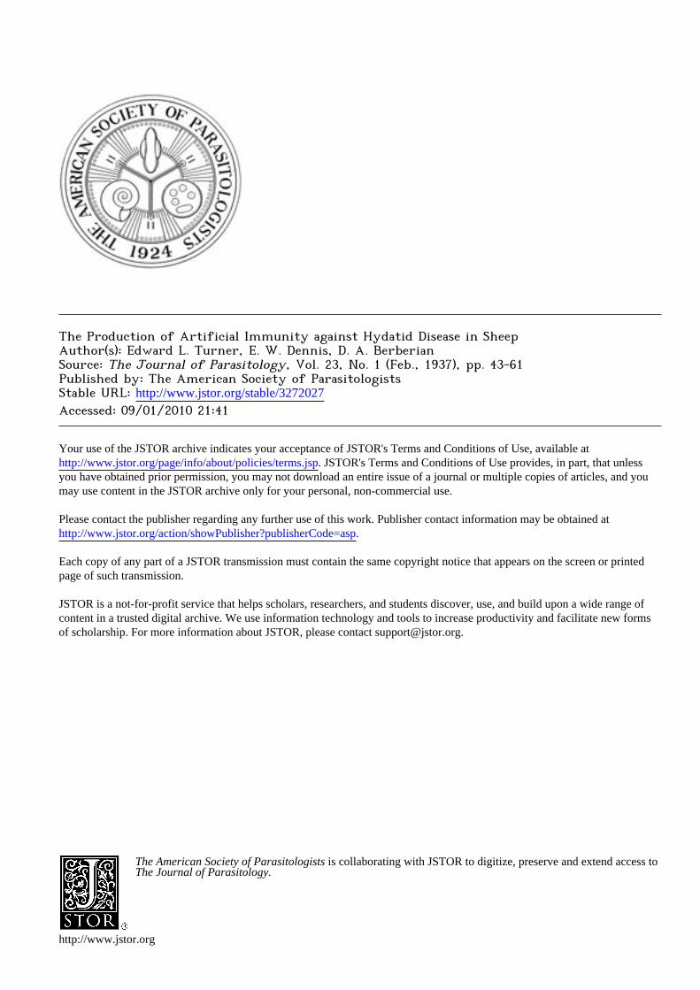
The Production of Artificial Immunity against Hydatid Disease in SheepAuthor(s): Edward L. Turner, E. W. Dennis, D. A. BerberianSource: The Journal of Parasitology, Vol. 23, No. 1 (Feb., 1937), pp. 43-61Published by: The American Society of ParasitologistsStable URL: http://www.jstor.org/stable/3272027Accessed: 09/01/2010 21:41
Your use of the JSTOR archive indicates your acceptance of JSTOR's Terms and Conditions of Use, available athttp://www.jstor.org/page/info/about/policies/terms.jsp. JSTOR's Terms and Conditions of Use provides, in part, that unlessyou have obtained prior permission, you may not download an entire issue of a journal or multiple copies of articles, and youmay use content in the JSTOR archive only for your personal, non-commercial use.
Please contact the publisher regarding any further use of this work. Publisher contact information may be obtained athttp://www.jstor.org/action/showPublisher?publisherCode=asp.
Each copy of any part of a JSTOR transmission must contain the same copyright notice that appears on the screen or printedpage of such transmission.
JSTOR is a not-for-profit service that helps scholars, researchers, and students discover, use, and build upon a wide range ofcontent in a trusted digital archive. We use information technology and tools to increase productivity and facilitate new formsof scholarship. For more information about JSTOR, please contact [email protected].
The American Society of Parasitologists is collaborating with JSTOR to digitize, preserve and extend access toThe Journal of Parasitology.
http://www.jstor.org
THE PRODUCTION OF ARTIFICIAL IMMUNITY AGAINST HYDATID DISEASE IN SHEEP*
EDWARD L. TURNER, E. W. DENNIS AND D. A. BERBERIAN
There now seems to be little room for doubt that protective reactions of a degree can be produced against certain cestode parasites by means of artificial immunization. Miller (I930, 1931, 1932, 1934) and his collaborators Massie (1932), Gardiner (1932) and Kerr (1932) demon- strated the production of immunity against Cysticercus fasciolaris in rats. These investigators were able to demonstrate the production of immunity by means of active immunization with prepared antigen, by attempts to
super-infest and by the use of specific immune sera. Evidence for the transmission of immunity from parent to offspring was also observed in some of their experiments. Kerr (1935) has recently published evi- dence of the production of immunity against another cestode parasite, Cysticerus pisiformis, in the rabbit. Kerr and Petkovich (1935) have
produced immunity in rabbits against the liver fluke Fasciola hepatica. Four years ago the authors began a study of the possibilities of the
production of artificial immunity against Echinococcus granulosus. At-
tempts were made to study the production of immunity in the definitive and intermediate hosts. Data has been reported (Turner, Berberian and Dennis, 1933, 1935, 1936) in evidence of the fact that the definitive host
(dog) shows a marked decrease in incidence and degree of intestinal in- fection with Echinococcus granulosus following the use of antigens pre- pared from hydatid cyst material. Partial, rather than complete protec- tive reactions were observed in the definitive host.
D ve (1927) was unable to produce experimental artificial immunity against Echinococcus granulosus in rabbits. In his experiments rabbits were immunized by the injection of sheep hydatid fluid and "hydatid sand." Following the injections of antigens living "hydatid sand" was introduced subcutaneously and three months later growing cysts were found on microscopic observation. Deve (1933) has also reported failure in the experimental production of immunity in white rats by means of
specific serotherapy and has been unable (Deve, 1934) to produce any curative or preventative reaction in white rats or rabbits by means of sunbcutaneous injections of hydatid membrane material.
The present paper deals with the experimental immunization of sheep against the larval stage of the life cycle of Echinococcus granulosus.
* From the Departments of Medicine and Parasitology, American University of Beirut, Beirut, Syria. The studies and observations on which this paper is based were conducted with the support of a grant from the Rockefeller Foun- dation.
43
44 THE JOURNAL OF PARASITOLOGY
EXPERIMENTAL DATA
Material and Methods
A. Animals In order to insure working with clean animals a group of pregnant
ewes was purchased in the winter of 1932. These animals were placed on the Near East Foundation Farm at Talabaya where they were housed in specially constructed quarters.* Seventy ewes were under the care of a shepherd who was responsible solely to the authors. As the young lambs were born they were cared for separately by a second attendant, but were allowed to feed from their own mothers. Each lamb was marked with metal ear tags and with an adjustable leather collar on which a duplicate number was placed. The collars were secured with lead seals. Within one to three weeks after birth initial injections of
antigen were given.
B. Preparation and Dosage of Antigen
The antigen was prepared by obtaining viable scolices and germi- nating membrane from fertile cysts of the lungs and livers of both sheep and cattle collected from the city abattoir. The scolices and germinating membrane were placed in an incubator at 370 C. and when thoroughly dry reduced to a fine powder. This powder was stored in tightly stop- pered bottles. Immediately prior to use a I per cent suspension in 0.5 per cent phenol saline was prepared. There was some tendency for the
suspension to precipitate so that it was necessary to agitate it frequently in order to obtain relatively uniform dosages in the syringe.
All injections of antigen were given into the muscles of the thigh. In all cases the initial dosage of antigen consisted of 0.5 cc. Subsequently injections were given at approximately weekly intervals. In order to determine the influence of the quantity of antigen received certain ani- mals were given only one injection; others were given two, three, four or five doses. When given, the second dose consisted of I.o cc., the third of 2.0 cc., the fourth and fifth of 2.5 cc.
C. Determination of Degree of Immunity, Experimental Infection
I. MATERIAL USED FOR INFECTION
Following the completion of the injections of antigen, intervals of from 21 to 170 days were allowed before attempts were made to infect the animals. Infectious material was obtained from two sources, (a) experimentally infected dogs which gave us known positive stools, and
(b) selected material from street dogs delivered to the laboratory. Ma- terial used for feeding consisted of a suspension of feces containing
* We are especially indebted to the Near East Foundation for their cooperation during the experiment.
TURNER, DENNIS AND BERBERIAN-HYDATID IN SHEEP 45
known numbers of onchospheres per cubic centimeter. By feeding ma- terial which was one week, two weeks or one month old, an attempt was made to determine whether fresh material was as infectious as that in which the onchospheres had been passed for some time. The animals were given from 500 to Iooo onchospheres at the time of their infective
feeding. The first few animals were given their infected feeding in milk from nursing bottles. This was found to be totally unsatisfactory as some of the material was lost through drooling, which tended to con- taminate the vicinity. Bottle feedings were therefore replaced by direct
feeding through a funnel and stomach tube. In this way a measured amount of the onchosphere suspension could be placed in the funnel and washed directly into the stomach by a small amount of water. There was thus relatively little danger of contamination from the mouth of the animal, and we do not believe any of the material was regurgitated.
2. CONTROLS
The efficiency of immunization and the viability of the onchospheres was controlled by giving appropriate amounts of each lot of feeding material to non-immunized lambs.
3. DETERMINATION OF INFECTION
Periods of 331 to 374 days were allowed to elapse between the feed- ing of infected material and the autopsy. As soon as the animals were killed, the viscera were completely removed. Careful gross examina- tions were made of the surface of the lungs, heart, liver, kidneys and
spleen. Anterior and posterior aspects of the lungs, and the dorsal and ventral surfaces of each liver were photographed. Surface counts of
cysts in each organ were then made. Each organ was then subjected to
sectioning in strips from two to three millimeters in width so that any internally located cysts could also be counted. As is usually the case, the
large majority of the cysts were located superficially. Representative and questionable cysts were removed from the organs of each animal in which infection was present, and fixed in Zenker's solution for subsequent histological study.
All animals in these groups were subjected to intradermal Casoni tests some time before autopsy. 0.5 cc. of hydatid fluid was injected intradermally on the wool-free region of the inner aspect of the thigh. Immediate reactions were read after 15 to 30 minutes, and delayed reactions were checked after 24 hours.
D. Experimental Protocols
A total of 115 animals have been studied in these experiments. These animals were divided into groups of from three to seven in number.
46 THE JOURNAL OF PARASITOLOGY
Each group consisted of one or more controls and lambs which had received a varied amount of antigen; all animals in each group received the same feeding material and were fed on the same day. The majority of the experiments were nullified and had to be discarded because of failure of feeding material. However, in other groups successful infec- tion of the controls gave validity to the results. The results of the obser- vations on the three groups where all factors were controlled and where there were no complications are presented in the following protocols.
GROUP I Animal no. 15 (female) IMMUNIZED.
Antigen injections (1% antigen suspension intramuscularly). March 21, 1933, 0.5 cc. March 28, 1933, I.o cc. April 4, 1933, 2.0 cc. April II, 1933, 2.5 cc.
Feeding on May 2, 1933, consisted of 500 onchospheres in suspension (material two weeks old). Feeding was 31 days after the last injection of antigen.
Casoni, March 3, 1934, immediate 2 plus, delayed I plus. Autopsy, May 27, 1934 (390 days following feeding).
Liver, 25 cysts, all but one 2-3 millimeters diameter, one liver cyst 12 millimeters in diameter. All of these cysts were hard and the walls exceedingly well calcified.
Lungs, kidneys and spleen, negative.
Animal no. 22 (female) IMMUNIZED. Antigen injections (1% antigen suspension intramuscularly).
March 21, 1933, 0.5 cc. March 28, 1933, I.o cc. April 4, 1933, 2.0 cc.
Feeding, May 2, 1933, consisted of 500 onchospheres of material identical in character with that given to animal no. 15. (28 days following the last antigen injection.)
Casoni not performed. Autopsy, March I, 1934 (303 days following feeding). This animal died fol-
lowing an attack of "staggers" and on autopsy was found to have Coenurus cerebellaris as the cause of death.
Liver, 10 cysts, 1-3 mm. in diameter, well calcified. Lungs, 6 cysts, 1-4 mm. in diameter, well calcified. Kidneys, spleen, brain, negative for hydatid cysts.
Animal no. 62 (male) CONTROL. Feeding, May 2, 1933, consisted of material identical with that given to animals
no. 15 and 22. Casoni, March 3, 1934, immediate 2 plus, delayed I plus. Autopsy, March 8, 1934 (331 days following feeding).
Liver, 68 cysts, 1-8 mm. diameter, no calcification. Lungs, 48 cysts, 3-8 mm. diameter, no calcification. Kidney, I cyst, 4 X 12 mm. diameter. Spleen, I cyst, 6 x Io mm. diameter.
GROUP II
Animal no. 3 (male) IMMUNIZED. Antigen injections (1% antigen suspension intramuscularly).
February 18, 1933, 0.5 cc.
TURNER, DENNIS AND BERBERIAN--HYDATID IN SHEEP 47
February 28, 1933, I.o cc. March 7, 1933, 2.0 cc. March 14, 1933, 2.5 cc. March 21, 1933, 2.5 cc.
Feeding, May 23, 1933, consisted of Iooo onchospheres in suspension (mixture of material I month and I week old). 67 days following the last injection of antigen.
Casoni, March 3, 1934, immediate 4 plus, delayed 3 plus. Autopsy, May 30, 1934 (372 days after feeding).
Liver, 190 cysts, 2-4 mm. diameter, mostly calcified. Lungs, 50 cysts, 4-6 mm. diameter, mostly calcified. Kidneys, I cyst, 3 mm. diameter, calcified. Spleen, 2 cysts, 2 and 3 mm. diameter, calcified.
Animal no. II (female) IMMUNIZED. Antigen injections (1% antigen suspension intramuscularly).
February 18, 1933, 0.5 cc. February 28, 1933, Io cc. March 7, 1933, 2.0 cc. March 14, 1933, 2.5 cc.
Feeding, May 23, 1933, consisted of Iooo onchospheres in suspension (mixture of material I month and I week old). 60 days following last injection of antigen.
Casoni, March 3, 1934, immediate I plus, delayed negative. Autopsy, May 30, 1934 (372 days following feeding).
Liver, 137 cysts, 1-3 mm. diameter, 80% heavily calcified. Lungs, 68 cysts, 2-3 mm. diameter, calcified. Kidneys, 2 cysts. Spleen, I cyst.
Animal no. 23 (female) IMMUNIZED. Antigen injections (1% antigen suspension intramuscularly).
March 14, 1933, 0.5 cc. March 21, 1933, I.o cc. March 28, 1933, 2.0 cc.
Feeding, May 23, 1933, consisted of Iooo onchospheres in suspension (mixture of material I week and I month old), 56 days following last injection of antigen.
Casoni, March 3, 1934, immediate 3 plus, delayed I plus. Autopsy, May 31, 1934, (373 days following feeding).
Liver, 18 cysts 3-5 mm. diameter, I cyst Io mm. in diameter (all heavily calcified).
Lungs, 26 cysts about the same diameter and condition. Kidneys and spleen negative.
Animal no. 32 (female) IMMUNIZED. Antigen injection (1% antigen suspension intramuscularly).
March 21, 1933, 0.5 cc. March 28, 1933, I.o cc.
Feeding, May 23, 1933, consisted of Iooo onchospheres in suspension (mixture of material I week and I month old). 56 days following last antigen injection.
Casoni, March 3, 1934, immediate 2 plus, delayed 2 plus. Autopsy, May 31, 1934 (373 days following feeding).
Liver, 25 cysts, 2-5 mm.-diameter, highly calcified. Lungs, 66 cysts, 2-6 mm. diameter, highly calcified. Kidneys, negative. Spleen, I cyst 3 mm., highly calcified.
48 THE JOURNAL OF PARASITOLOGY
Animal no. 42 (male) IMMUNIZED. Antigen injection (1% antigen suspension intramuscularly).
March 21, 1933, 0o.5 cc. Feeding, May 23, 1933, consisted of Iooo onchospheres in suspension (mixture
of material I week and I month old), 67 days following the injection of antigen.
Casoni, March 3, 1934, immediate 3 plus, delayed 3 plus. Autopsy, June I, 1934 (374 days following feeding).
Liver, 49 cysts, 1-4 mm. diameter, highly calcified. Lungs, 17 cysts, 2-5 mm. diameter, highly calcified. Kidneys and spleen, negative.
Animal no. 51 (female) IMMUNIZED. Antigen injections (1% antigen suspension intramuscularly).
March 21, 1933, 0.5 cc. April 4, 1933, I.0 cc. April I I, 1933, 2.0 cc. April 18, 1933, 2.5 cc. May 2, 1933, 2.5 cc.
Feeding, May 23, 1933, consisted of Iooo onchospheres in suspension (mixture of material I week and I month old), 21 days following last injection of antigen.
Casoni, March 3, 1934, immediate 3 plus, delayed negative. Autopsy, June I, 1934 (374 days following feeding).
Liver, 45 cysts, 2-4 mm. diameter, 80% highly calcified. Lungs, 51 cysts, 2-5 mm. diameter, highly calcified. Kidneys and spleen, negative.
Animal no. 63 (male) CONTROL. Feeding, May 23, 1933, consisted of material identical in source and character
to that fed to animals nos. 3, II, 23, 32, 42 and 51. Casoni, March 3, 1934, immediate 3 plus, delayed I plus. Autopsy, May 29, 1934 (371 days following feeding).
Liver, 252 cysts, 2-7 mm. diameter. Lungs, 96 cysts, 3-8 mm. diameter. Kidneys, I cyst in each kidney approximately 4 mm. diameter. Spleen, negative None of the cysts showed evidence of calcification and they all appeared to
be normally growing hydatid cysts.
GROUP III Animal no. 14 (male) IMMUNIZED.
Antigen injections (1% antigen suspension intramuscularly). February 18, 1933, 0.5 cc. February 28, 1933, 2.0 cc. March 7, 1933, I.o cc. March 14, 1933, 2.5 cc.
Feeding, August 31, 1933, consisted of 500 onchospheres in suspension (freshly collected material), 170 days following last injection of antigen.
Casoni, March 3, 1934, immediate 3 plus, delayed negative. Autopsy, August 7, 1934 (341 days following feeding).
Liver, 53 cysts, 2-4 mm. diameter, all highly calcified. Lungs, 13 cysts, 2-4 mm. diameter. Kidneys and spleen, negative.
Animal no. 26 (male) IMMUNIZED. Antigen injections (1% antigen suspension intramuscularly).
March 14, 1933, 0.5 cc.
TURNER, DENNIS AND BERBERIAN-HYDATID IN SHEEP 49
March 21, 1933, I.o cc. March 28, 1933, 2.0 cc.
Feeding, August 31, 1933, consisted of 500 onchospheres in suspension (freshly collected material), 157 days following last injection of antigen.
Casoni, March 3, 1934, immediate 3 plus, delayed 2 plus. Autopsy, August 7, 1934 (341 days following feeding).
Liver, 49 cysts, 2-4 mm. diameter, well calcified. Lungs, 12 cysts, calcified and obviously degenerating. Kidneys and spleen, negative.
Animal no. 34 (male) IMMUNIZED. Antigen injections (Io antigen suspension intramuscularly).
March 21, 1933, 0.5 cc. March 28, 1933, I.o cc.
Feeding, August 31, 1933, consisted of 500 onchospheres in suspension (freshly collected material), 156 days following last injection of antigen.
Casoni, March 3, 1934, immediate 2 plus, delayed 2 plus. Autopsy, August 13, 1934 (337 days following feeding).
Liver, 1o cysts, 2-3 mm. diameter, highly calcified. Lungs, 2 cysts. Kidneys and spleen, negative.
Animal no. 67 (female) CONTROL. Feeding, August 31, 1933, consisted of material identical in source and character
to that fed to animals nos. 14, 26, and 34. Casoni, March 3, 1933, immediate 3 plus, delayed negative. Autopsy, August I, 1934 (335 days following feeding).
Liver, 32 cysts, 2-8 mm. diameter, evidence of some calcification. Lungs, 22 cysts. Kidneys and spleen, negative.
E. Results
In Group I the control animal had a total of 118 cysts in the liver, lungs, kidneys and spleen. Animal no. 15 which received four injections of antigen, totalling 6.0 cc. of the I per cent antigen suspension, had only 25 cysts in the liver and none elsewhere. Animal no. 22 which received three injections of antigen (3-5 cc.) had a total count of 16 cysts. The maximum cyst diameter seen in the control animal was from 8-12 mm. The largest cysts observed in the two immunized animals of this group were not over 4 mm. in diameter. The cysts in the control animal were not calcified while those seen in the two immunized sheep were hard and, when cut open with a knife, were obviously calcified. Immunized sheep no. 15 and control sheep no. 62 developed similar Casoni reactions.
Animals in Group II received twice as many onchospheres in their infected feedings as were given to those in Group I. The total number of cysts developing in these two groups showed a close correlation be- tween the size of the infected feeding and the subsequent infection. Control no. 62 of Group I received 500 onchospheres in its feeding and developed 118 cysts while control no. 63 of Group II received Iooo oncho- spheres and developed a total of 349 cysts. The average number of cysts developing in the immunized animals of Group I was 20 while in
50 THE JOURNAL OF PARASITOLOGY
Group II the average number of cysts found in the immunized sheep was 125. All of the animals that had received injections of antigen showed marked evidence of cyst calcification while the cysts of the con- trols of Group II were thinner walled and lacked gross evidence of calci- fication. The largest cysts in Group II were found in the control no. 63 where a maximum diameter of 8 mm. was observed in lung cysts. The maximum diameter of the cysts in the immunized animals in this group varied between 4 and 6 mm. All of the immediate Casoni reactions in
Group II were positive. However, immunized animal no. i i with a total of 208 cysts gave only I plus immediate reaction and a negative delayed reaction.
In Group III the control animal no. 67 developed only 54 cysts while two of the immunized animals (no. 14 and no. 26) developed 66 and 61
cysts. The maximum diameter of the cysts observed in the control was 8 mm. while the immunized animals did not develop cysts greater in width than 4 mm. Calcification was much more marked in the cyst walls of the immunized animals. All immediate Casoni reactions in this Group were positive.
One consistent observation in these protocols is the fact that sheep receiving one, two or three injections of antigen developed relatively fewer cysts than those receiving four or five injections.
Microscopic studies of the cysts revealed marked differences in the structures of the cysts of the control and immunized animals. Control animals in all instances developed cysts that showed normal walls and an active germinal layer while in most instances the cyst structures in the immunized animals showed marked degenerative changes (see discussion and plates).
In addition to the detailed protocols presented above, the results of one entire series of experiments (Series 1935-1936) are given in con- densed tabular form (Table I) to illustrate the types of variation in results encountered during the course of our study. The complement fixation test, Casoni test, and precipitin test were used to obtain a certain amount of data pertaining to the specific immunological responses. How- ever, it was not until this series was nearly finished that we were able to
prepare an antigen which we considered satisfactory for a quantitative study, consequently the data on this series is qualitative and designed to show immunological differences toward the termination of the experi- ment. The antigen used for obtaining the data given in Table I was a solution of hydatid protein obtained by treatment of hydatid cyst fluid with acetic acid. The preparation and properties of this purified antigen will be described separately by one of us (Dennis, I937).
In the 1935-1936 series, 26 lambs which were maintained under the same conditions as those reported in the protocols above were immunized.
TURNER, DENNIS AND BERBERIAN-HYDATID IN SHEEP 51
Each lamb received a total of 11-15 cc. of scolex suspension adminis- tered in 3 doses given intramuscularly. These animals were experimen- tally infected, by the stomach tube method, from 35 to 69 days after the last injection of antigen (for economy of space the time between the in-
jections and the feedings are not shown since there appeared to be no relationship between these periods and the results). Nonimmunized lambs were given comparable feedings of echinococcus ova as controls. Variation in the age and number of ova administered is indicated in the table.
The results of this experiment are shown in the table. In the im- munized sheep (nos. 301-31o inclusive) the average number of cysts per animal was 19.2 as compared with 5.25 for each of the control group. In this case it would appear that the injections of antigen actually in- creased susceptibility until one compares the condition of the cysts. In the immunized animals the cysts were uniformly small, calcified and degenerate; in the control group the cysts, although few, were normal when examined microscopically. In the immunized sheep (nos. 311-328 inclusive) there were but 2.6 cysts per animal as compared with an aver-
age of 5 cysts per animal in the control group. Again the microscopic examination of the cysts from these animals showed calcification and
degeneration of the cysts in the immunized sheep, whereas the cysts in the control group were entirely normal. On analysis of the data, it would appear that a greater viability of the 12-day-old ova included within the
feeding material of sheep 301-31o might explain the increased number of cysts per animal, although we hesitate to interpret data showing such a high degree of variability. The most significant and always constant finding is the degenerate nature of the cysts in the immunized animals as compared with the controls.
DISCUSSION AND CONCLUSIONS
Several very clear cut evidences of a protective reaction resulting from immunization are to be noted on analysis of the foregoing proto- cols. In general the total number of cysts counted in the immunized animals is far below that in the controls. This is clearly illustrated in the animals of Group I. The total count in the control is 118 cysts while animals no. 15 and no. 22 (immunized) have total counts of 25 and 16 cysts respectively. A similar difference is noted in Group II. In Group III the total number of cysts in the control is less than the count in some of the immunized animals but this feature is counterbalanced by the marked difference in the condition of the cyst structures on gross and microscopic examination.
Invariably the diameter of the cysts in the control animals is greater than that of the cysts in sheep that have received injections of antigen.
52 THE JOURNAL OF PARASITOLOGY
This difference in diameter varies from 50 to Ioo per cent. Dew (1928) states that at five months the hydatid follicle may be about I.o cm. in diameter. The growth rate of cysts observed in our studies is obviously much slower as we rarely observed cysts I.o cm. in diameter at eleven or twelve months after infection. This must be interpreted as representing a definitely slower growth rate in our series than that seen by the Australian observer.
Cysts in immunized animals are firmer and more calcified than those in the controls. This difference is not only apparent on gross examina- tion but is also obvious on cutting through the organs containing the
cysts. Another constant observation on gross examination is the difference
in the numbers of deeply seated and superficial cysts in the control and immunized animals. Deep liver cysts are abundant in the control ani- mals while most of the cysts counted in the immunized animals are
superficially located. This tendency is probably the result of less re- sistance in the liver surface as compared to the interior of the organ. Hexacanth embryos lodging deeply in the liver tissue of immunized ani- mals encounter a more active resistance than those lodging peripherally. The more superficially placed ones encounter resistance from only the
deeper surface when growth is once started.
Although the gross differences are marked, and in most cases con-
vincing, the microscopic differences observed on studying the cyst struc- tures in the control and immunized animals are far more conclusive. In the control animals the cyst wall structures are clearly differentiated and normal in appearance. A normal active germinal layer is present. Cysts from immunized animals have much thicker walls. The laminated mem- brane and adventitial structures are not clearly differentiated and in many instances appear to be an undifferentiated "hyaline-like" mass. The ger- minal layer is absent or shows marked evidences of degeneration and there is a tendency towards obliteration of the lumen of the cysts. The difference in calcification of the cysts is also clearly defined microscopi- cally. Scattered patches of calcification are observed in a few of the control cysts. This calcification is at least twice as marked in the most
poorly calcified cysts of immunized animals. The process of calcification
apparently starts in small islands around and in the cyst wall structures
(adventitia). These islands of calcification then spread circularly around the cyst wall and tend to coalesce as the cyst grows older. In Group III where the control animal (no. 67) has a lower cyst count than the im- munized animals (no. 14 and no. 26) these microscopic differences are
fully as marked as they are in the other groups. In fact, these micro- scopic differences are constant throughout the entire series of experi-
TURNER, DENNIS AND BERBERIAN-HYDATID IN SHEEP 53
ments as well as in the three groups reported in the protocols of this paper (see Plates).
As indicated in the results, one of the most interesting features in studying the protocols is the relationship of the number of injections of antigen to the number of cysts developed. Consistently, animals receiv- ing only one, two or three injections of antigen developed fewer cysts than those receiving four and five injections.
Despite the fact that the accumulated data is inadequate for the formulation of any sweeping conclusions concerning the mechanism of resistance against Echinococcus granulosus, certain aspects of the problem are worthy of presentation.
Certain nonspecific factors probably play a significant r6le in deter- mining whether or not infection occurs. Experimental evidence is accu-
mulating in our laboratory indicating that the hatching of the onchosphere from the egg is greatly influenced by the hydrogen ion concentration and by the quality of the digestive juices to which they are exposed. It has been shown that qualitative differences exist between the intestinal juices of naturally suitable and unsuitable hosts which may reasonably account for differences in susceptibility of certain species of hosts under natural conditions. Furthermore individual differences in amount of free acid in the stomach and in the rate of passage of food through the ali- mentary tract at the time ova are ingested are probably important factors influencing the establishment of the infection. An additional nonspecific factor which appears to have been responsible for our failure to establish infection in certain experiments is that of the age and viability of the ova. Material which was fresh (24-48 hours old), and material kept in aqueous suspension at room temperature for one month or longer were unsatisfactory for establishing infection; material which was between 7 and 14 days old was usually more effective. The factors controlling the viability of the ova of echinococcus under natural and experimental conditions require further study.
Specific humoral antibodies and/or the increased tissue response which is the result of specific sensitization are alternative or correlative factors which may be responsible for the increased resistance to Echino- coccus granulosus which results from the parenteral introduction of echinococcus antigen. It is well known (Fleig and Lisbonne, 1907; Welsh, Chapman and Storey, 19o8; Fairley, 1923) that specific precipi- tins occur in the sera of individuals suffering from hydatid cysts. Also Ghedini (1906), Weinberg (1909) and Fairley (1922) amply demon- strated the presence of complement fixing antibodies in the sera of hosts suffering from hydatid disease. Clinical experience indicates that hel- minthicidal antibodies either are not produced or are unable to penetrate a normal hydatid cyst. Casoni (1911) was able to demonstrate specific
54 THE JOURNAL OF PARASITOLOGY
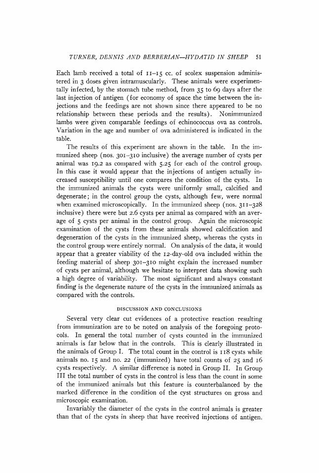
TABLE I.--Results of Series 1935-1936
Unit Cysts in No. Age of No. of Casoni of com- Ppt. ova ova test plement test Condition of cysts
fixed Liver Lungs 301 1: 4 : 12 3/1300 +++ 8 ++ 12 8 calcified : 2-4 mm. 302 " 3/1300 ++ 8 +++ 58 27 " 1-4 mm. 303 " 3/1300 ++ 6 +++ 8 5 3-4 mm. 304 " 3/1300 + 6 + 23 7 " 2-4 mm. 305 " 3/2300 +++ 6 ++ 23 0 " 2-4 mm. 306 " 3/2300 + 6 +++ 8 0 " 2-4 mm. 307 3/3300 ++ 6 +++ 0 5 " 2-4 mm.; 1 was 7 308 " 3/3300 ++ ac + 4 0 " 3-4 mm. 309 " 3/3300 + 2 +++ 4 0 " 2-3 mm. 310 " 3/3300 +++ 8 +++ 0 0
330c " 3/1300 0 ac ++ 2 3 not calcif.; 5-20 mm. 331c 3/2300 + 6 +++ 0 0 332c 3/3300 +++ 8 +++ 11 2 normal 3-6 mm. 333c 15 1/1500 + 2 ++ 0 3 " 3-4 mm.
311 1 : 3 2/1800 + 8 +4+ 4 0 calcified ; 3-5 mm. 312 " 2/1800 ++ 8 +++ 6 1 " 4-5 mm. 314 1 2/3800 +++ 8 +++ 3 1 " 2-5 mm. 315 1 3/3800 + 8 ++++ 2 3 " 4-5 mm. 316 1 2/2800 ++ 8 +++ 1 0 " 10 mm. 317 1 2/2800 +++ 3 +++ 0 2 " 3 mm. 318 1 2/2800 + 6 ++ 5 3 " 3-6 mm. 319 1 1/2000 + 8 + 2 2 " 2 mm. 320 1 1/3000 +++ 6 ++ 0 0 321 1 1/3000 ++ 6 + 1 0 " 8 mm. 322 1 1/3000 + 3 +++ 0 0 324 1 1/10,000 ++ 6 ++ 0 0 325 1 1/10,000 ++ 6 +++ 1 0 " 4 mnm. 326 1 1/10,000 + 6 +++ 0 0 327 1 1/10,000 +++ 8 ++++ 328 1 1/3000 +++ 8 +++ 00 0
334c 1 : 3 2/1800 + 6 +++ 2 partial calcif ; 5-6 mm. 335c 1 : 3 2/1800 ++ 6 +++ 3 5 normal; 6-10 mm. 336c 1 1/3000 0 ac +++ 1 0 ,t 4 mm. 337c 1 1/3000 + 0 +++ 6 2 " 4-6 mm. 338c 1 1/10,000 +++ 8 ++ 1 5 " 4-8 mm.
In the first column the letter c indicates the control of sheep for the group above. In the second column the numerals indicate the age of the fecal suspension in days; where more than one number is present the feeding material was a mixture. In the third column the numerator indicates the number of feedings one week apart, the denominator indicates the total number of echinococcus ova fed. Negative Casoni reaction indicated by 0. The complement fixations tests were quantitative; ac indicates that the serum was anticomplementary. The precipitin tests were qualitative, not quantitative, using purified antigen 1:1000. Animals sacrificed 11-12 months after infection.
tissue "hypersensitivity" in hydatid infection by the intradermal injection of small quantities of hydatid fluid as the eliciting antigen.
It is our opinion that such degree of immunity as may result from artificial immunization under the conditions of our experiment is the expression of induced specific tissue sensitivity to hydatid antigen. This opinion is supported by the following observations:
I. The immunizing procedure did not prevent infection. 2. Immunization reduced the number of cysts, as compared with
controls, only in certain experiments. 3. There were no significant differences in antibody content of
immunized and control sheep under the stimulus of infection (Table I).
4. In all experiments immunized animals have shown marked ability to defend themselves against the growth of the parasites by a
TURNER, DENNIS AND BERBERIAN-HYDATID IN SHEEP 55
rapid and highly efficient walling off of the invaders. The greatly thickened adventitia and thorough calcification of the wall of the hydatid cyst at an early period produces an efficient and eventually impermeable mechanical barrier, leading to death of the parasite as the result of starvation and the accumulation of toxic metabolites.
5. Hypersensitivity in resistant animals could always be demon- strated by the intradermal Casoni test. Control animals which were successfully infected usually but not always gave a positive Casoni test. It would seem that the previous sensitization (immu- nization) of the experimental sheep gave them the advantage of a more intense early response which was responsible for the observed differences in susceptibility.
BIBLIOGRAPHY
Casoni, T. 19II.-La diagnosi biologica dell'echinococcosi umana mediante l'intra- dermoreazione. Folia Clinica Chem. Micros., 4: 5-16.
D've, F. 1927.-Essai de Vaccination Anti chinococcique par de Sable Hydatique Tyndalise. Compt. rend. Soc. de biol., 97: 1130-1131.
1933.-La Souris Blanche, Animal RPactif pour les inoculations Echinococcique. Un essai de serotherapie anti chinococcique aspecifique. Compt. rend. Soc. de biol., 113: 1443-1445.
I934.-"L'anatoxine hydatique de Ymaz Apphatie ne possede pas de propriet~s curative A l'agard de l'&chinococcose experimentale." Compt. rend. Soc. de biol., 115: 954-956.
I934.-Essai d'immunisation anti-&chinococcique par injection sous-cutanees de membranes hydatique Broy~es a L'6tat frais. Compt. rend. Soc. de biol., 115: 1025-1026.
Dew, H. R. 1928.-Hydatid Disease. Its Pathology, Diagnosis and Treatment. The Australian Medical Publishing Co., Limited, Sydney. pp. 429.
Fairley, K. D. 1923.-The investigation of the immunity reactions in hydatid dis- ease. Preliminary report. Med. Jour. Australia, Io (II) : 27-37.
Fairley, N. H. 1922.-Researches on the complement fixation reaction in hydatid disease. Quart. Jour. Med., 15: 244-267.
Fleig, C., and Lisbonne, M. 1907.-Reserches sur un serodiagnostic du kyste hyda- tique par la methode des precipitines. Compt. rend. Soc. de biol., 62: 1198- 120I.
Ghedini, G. 1906.-Ricerche sul siero di sangue di individuo affetto da cisti da echinococco e sul liquido in essa contenuto. Gax. d. ospedali e d. cliniche, 27: 1616-1617.
Kerr, K. B. 1935.-Immunity against a Cestode Parasite, Cysticercus Pisiformis. Amer. Jour. Hyg., 22 (I): 169-182.
Kerr, K. B., and Petkovich, O. L. I935.-Active Immunity in Rabbits to the liver fluke, Fasciola hepatica. Jour. Parasit., 21 (4).
Miller, Jr., H. M. 193o.-Experiments on Immunity of the White Rat to Cysti- cercus Fasciolarus. Proc. Soc. Exper. Biol. and Med., 27: 926.
193I.-Immunity of the White Rat to Superinfestation with Cysticercus Fasci- olaris. Proc. Soc. Exper. Biol. and Med., 28: 467-468.
193I.-The Production of Artificial Immunity in the Albino Rat to a Metazoan Parasite. Jour. Prev. Med., 5 (6): 429-452.
193I.-Immunity of the Albino Rat to Superinfestation with Cysticercus Fasci- olaris. Jour. Prev. Med., 5 (6): 453-464.
56 THE JOURNAL OF PARASITOLOGY
193I.-Further Experiments on Artificial Immunity to a Larval Cestode. Proc. Soc. Exper. Biol. and Med., 28: 884-885.
1932.-Superinfection of Cats with Taeniae Taeniaeformis. Jour. Prev. Med., 6: 17-30.
1932.-Further Studies on Immunity to a Metazoan Parasite, Cysticercus Fasci- olaris. Jour. Prev. Med., 6: 37-46.
1932.-Transmission to Offspring of Immunity against Infection with Metazoan (cestode) Parasite. Proc. Soc. Exper. Biol. and Med., 29: 1124.
I932.-Acquired Immunity against a Metazoan Parasite by Use of Non-specific Worm Materials. Proc. Soc. Exper. Biol. and Med., 29: 1125-1126.
1932.-Therapeutic Effect of Specific Immune Serums against a Metazoan Parasite (Cysticercus Fasciolaris). Proc. Soc. Exper. Biol. and Med., 30: 82-83.
Miller, Jr., H. M., and Massie, E. 1932.-Persistence of Acquired Immunity to Cysticercus Fasciolaris after Removal of the Worms. Jour. Prev. Med., 6: 31-36.
Miller, Jr., H. M., and Gardiner, M. L. 1932.-Passive Immunity to Infection with a Larval Tapeworm of the Albino Rat. Science, 75: 270.
1932.-Protection of the Rat against Infection with a Larval Tapeworm by Serum from Immune Rats. Proc. Soc. Exper. Biol. and Med., 29: 779- 780.
1932.-Passive Immunity to Infection with a Metazoan Parasite, Cysticercus Fasciolaris, in the Albino Rat. Jour. Prev. Med., 6: 476-496.
Miller, Jr., H. M., and Kerr, K. B. 1932.-Attempts to Immunize Rabbits against a Larval Cestode, Cysticercus Pisiformis. Proc. Soc. Exper. Biol. and Med., 29: 67o-67I.
Turner, E. L., Berberian, D. A., and Dennis, E. W. 1933.-Successful Artificial Immunization of Dogs against Taeniae Echinococcus. Proc. Soc. Exper. Biol. and Med., 30: 618-619.
1935.-The Value of the Casoni Test in Dogs. Jour. Parasit., 21: 18O-I82. 1936.-The Production of Artificial Immunity in Dogs against Echinococcus
granulosus. Jour. Parasit., 22 (I) : 14-28. Weinberg, M. 1909.-Sero-diagnostic de l'echinococcose. Ann. de l'Institut Pas-
teur., 23: 472-502. Welsh, D., Chapman, H. G., and Storey, J. C. 19o8.-Some applications of the
precipitin reaction in the diagnosis of hydatid. Australian Med. Gaz., 27: 653-657.
EXPLANATION OF PLATES
All illustrations are photomicrographs taken with a Leica. Sections stained with haematoxylin-eosin. All cysts shown are approximately I year old.
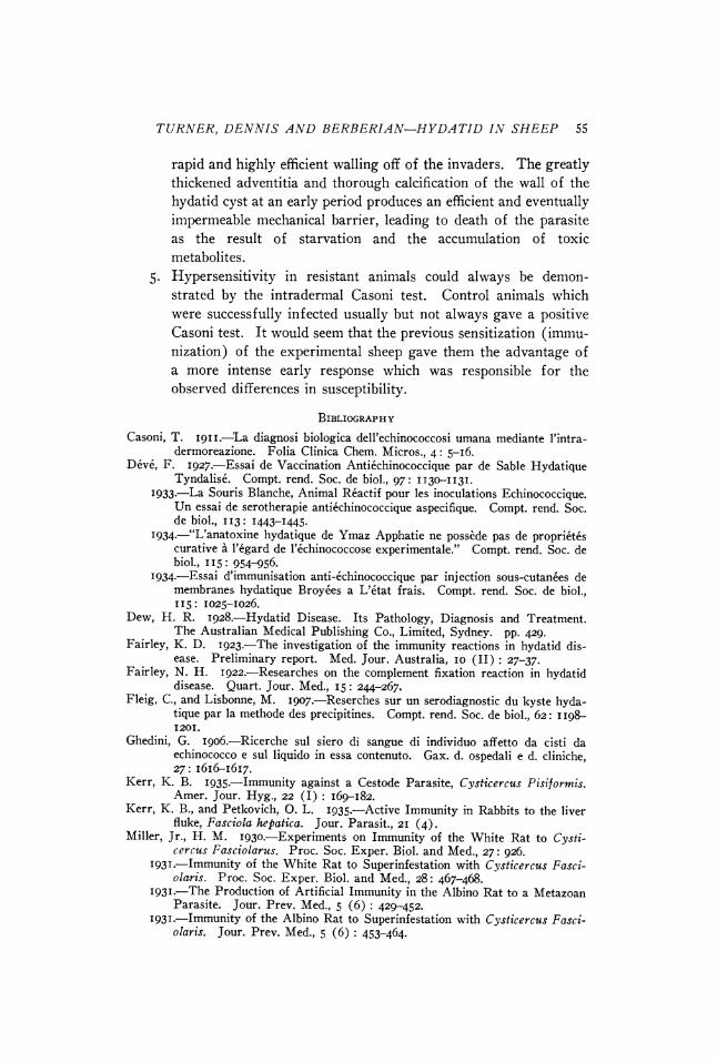
PLATE I
Fig. I. Section through two continuous hydatid cysts in the lung of control sheep no. 66. Note the relatively thin cyst wall and absence of calcification in a normal cyst. x 20.
Fig. 2. Enlargement of a portion of fig. I to show normal structure of cyst wall and active germinal layer. x 50.
Figs. 3 and 4. Hydatid cysts in lung of immunized sheep no. 32. Note the greatly thickened wall, early calcification, and tendency toward obliteration of the lumina of the cysts. x 30.
TURNER, DENNIS AND BERBERIAN-HYDATID IN SHEEP 57
PLATE I
~too
gigi loci loan 0, M
;*&,4 A*
oil
Ail
if U
9? ~I A
ioil f"`
1~~14ti:pe "i~'' ik :pd~ :2sAL. ~ :i~X4 is...........
.........
Pl ........
J"~ ~ ..........
AMP.,
fal
Of :1~
AMI& ii
58 THE JOURNAL OF PARASITOLOGY
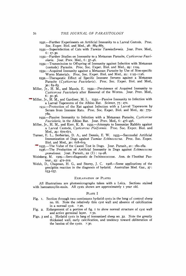
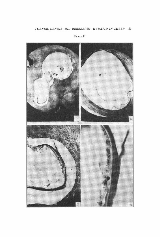
PLATE II
Fig. 5. Cyst deep in liver of control sheep no. 63. Cyst has unusually thick wall and a small point of calcification in the adventitia. Germinative mem- brane normal and active. x 20.
Fig. 6. Normal thin walled cyst in the liver of control sheep no. 63 showing active germinative membrane. No calcifications. x 20.
Fig. 7. Germinative membrane and laminated layer of cyst in Fig. 5. Fig. 8. Active germinative membrane of cyst shown in Figs. 5 and 7. x 200.
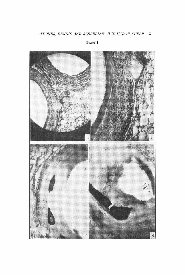
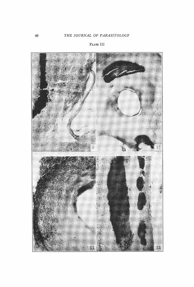
PLATE III
Figs. 9, Io, II and 12. Illustrating the greatly thickened cyst walls, calcifications, and degeneration of germinative membrane in cysts of immunized sheep nos. 3, 23, 42 and 51 respectively
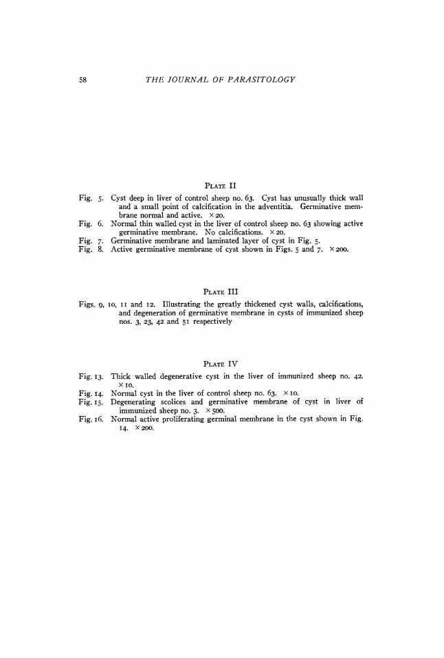
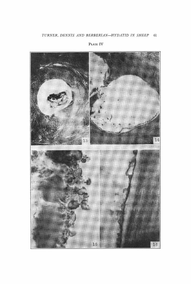
PLATE IV
Fig. 13. Thick walled degenerative cyst in the liver of immunized sheep no. 42. X IO.
Fig. 14. Normal cyst in the liver of control sheep no. 63. x 10. Fig. 15. Degenerating scolices and germinative membrane of cyst in liver of
immunized sheep no. 3. x 500. Fig. 16. Normal active proliferating germinal membrane in the cyst shown in Fig.
14. x 200.
TURNER, DENNIS AND BERBERIAN-HYDATID IN SHEEP 59
PLATE II
'i,
;
.-
D
..... ? :
i :
"1
x.; i
.•ri .... i•7
. ,:
60 THE JOURNAL OF PARASITOLOGY
PLATE III
6x-i: . i
""
J i ••'•!"ii•iiii
.iliii .....
•
1.
. .•
ito
::a
....
!i~
.: " i??
t. SO
:1 • T • •::•?•i
: ?$::.•:
"" ?:
:•
10
OL
:?: F I~ai-:' l ?:12
TURNER, DENNIS AND BERBERIAN-HYDATID IN SHEEP 61
PLATE IV
?::. .:
,or
.............. ::i.: !i!• :'•' : .. :-:-:ii
:l,:':r
?.
riii
I-P
2 ? e1-
.Alo: