Embed Size (px)
Citation preview

The prespliceosome components SAP 49 and SAP 145 interact in a complex implicated in tethering U2 snRNP to the branch site
Patrick C h a m p i o n - A r n a u d and Robin Reed 1
Department of Cell Biology, Harvard Medical School, Boston, Massachusetts 02115 USA
The mammalian spliceosome-associated protein, SAP 49, is associated specifically with U2 snRNP and is the most efficiently UV cross-linked protein in the spliceosomal complexes A, B, and C. We show here that SAP 49 cross-links to a region in the pre-mRNA immediately upstream of the branchpoint sequence in the prespliceosomal complex A. In addition to the RNA-binding activity of SAP 49, we show that this protein interacts directly and highly specifically with another U2 snRNP-associated spliceosomal protein, SAP 145. We have isolated a cDNA-encoding SAP 49 and find that it contains two amino-terminal RNA-recognition motifs (RRMs), consistent with the observation that SAP 49 binds directly to pre-mRNA. The remainder of the protein is highly proline-glycine rich (39% proline and 17% glycine). Unexpectedly, the SAP 49-SAP 145 protein-protein interaction requires the amino-terminus of SAP 49 that contains the two RRMs. The observation that SAP 49 and SAP 145 interact directly with both U2 snRNP and the pre-mRNA suggests that this protein complex plays a role in tethering U2 snRNP to the branch site.
[Key Words: U2 snRNP; prespliceosome; branchpoint sequence; proline domain; protein-protein interaction]
Received
The sequential transesterification reactions that gener- ate spliced pre-mRNA are thought to be carried out by an RNA-catalyzed mechanism, and U2, U5, and U6 small nuclear RNAs (snRNAs) are key spliceosomal RNAs in- volved in these reactions (for review, see Moore et al. 1993; Newman 1994). U2 snRNA forms an essential base-pairing interaction with the branchpoint sequence (BPS) in the intron (Parker et al. 1987; Wu and Manley 1989; Zhuang and Weiner 1989). U6 snRNA is thought to be base paired simultaneously to both the 5' splice site and U2 snRNA; this would position the putative cata- lytic domain of U6 snRNA near both the BPS and the 5' splice site (Madhani and Guthrie 1992; Sawa and Abel- son 1992; Sawa and Shimura 1992; Wassarman and Steitz 1992; Lesser and Guthrie 1993; Kandel-Lewis and S6raphin 1993; Sontheimer and Steitz 1993). Finally, U5 snRNA interacts with exon sequences adjacent to both the 5' and 3' splice sites and may play roles both in specifying the cleavage sites at the splice junctions and in holding the exons together for ligation (Newman and Norman 1991, 1992; Wassarman and Steitz 1992; Wyatt et al. 1992; Cortes et al. 1993, Sontheimer and Steitz 1993).
Although a direct role for snRNA-pre-mRNA interac- tions in catalysis is likely, little is known about how
~Corresponding author.
each of these RNA-RNA interactions are first formed, or how they are stabilized sufficiently to carry out their functions. Two observations indicate that additional fac- tors other than the snRNAs are needed to establish and maintain the critical base-pairing interactions. First, the U2, U5, and U6 snRNA-pre-mRNA interactions usually involve only a few nucleotides and therefore, would not be expected to form readily, or be stable, under splicing conditions. Second, in metazoa, the sequence elements in the pre-mRNA that participate in these interactions often deviate significantly from the consensus se- quences. One consequence of this is that even fewer than normal snRNA-pre-mRNA base pairs are formed. An- other consequence is that the specificity is further de- creased, which obviously hinders recognition of the ele- ments by snRNAs alone.
In mammals the BPS is highly degenerate. Thus, pro- teins are almost certainly vital for establishing and sta- bilizing U2 snRNA-BPS interactions. As yet, however, no spliceosomal proteins have been identified that inter- act directly with the BPS. A set of nine proteins interacts in an ATP-independent and reversible manner with 12S U2 small nuclear ribonucleoprotein particle (snRNP) to generate a 17S U2 snRNP particle that is active in splic- ing (Behrens et al. 1993a). Purified A, B, and C spliceo- somal complexes contain at least seven of these U2 snRNP proteins [spliceosome-associated proteins (SAPs)
1974 GENES & DEVELOPMENT 8:1974-1983 �9 1994 by Cold Spring Harbor Laboratory Press ISSN 0890-9369/94 $5.00
Cold Spring Harbor Laboratory Press on February 4, 2022 - Published by genesdev.cshlp.orgDownloaded from

A SAP 49-145 complex implicated in U2 snRNP binding
49, 61, 62, 114, 130, 145, and 155] (Gozani et al. 1994; Staknis and Reed 1994). Of these, all but SAP 130 can be UV cross-linked to pre-mRNA in purified A, B, and C complexes, and cross-linking requires the 3' but not the 5' splice site (Gozani et al. 1994; Staknis and Reed 1994). Three of the proteins U2 snRNP correspond to the es- sential splicing factor SF3a, whereas most, if not all, of the others are likely to correspond to the essential splic- ing activity SF3b (Brosi et al. 1993a, b; for review, see Hodges and Beggs 1994). The U2 snRNP-associated SAPs that cross-link to pre-mRNA have been proposed (Stak- nis and Reed 1994) to correspond to the so-called "branchpoint protection factor" identified in RNase pro- tection studies (Black et al. 1985; Ruskin and Green, 1985). This factor protects a 20- to 40-nucleotide region containing the branch site from RNase digestion during spliceosome assembly and catalytic step I of the splicing reaction (Black et al. 1985; Ruskin and Green, 1985).
In this study we demonstrate that SAP 49 UV cross- links to pre-mRNA immediately upstream of the branchpoint sequence in the prespliceosomal complex A. We also show that SAP 49 participates in direct pro- tein-protein interactions with SAP 145. Thus, our data, together with previous observations, suggest that SAP 49 and SAP 145 interact in a U2 snRNP-associated complex that functions to tether U2 snRNP to the branch site. Isolation of a cDNA encoding SAP 49 shows that it con- tains two RNA-binding domains that are required for the SAP 145 interaction and are likely to be required for the pre-mRNA-binding activity.
R e s u l t s
Isolation of a cDNA encoding SAP 49
SAP 49 UV cross-links efficiently to the pre-mRNA in the A, B, and C spliceosomal complexes, and thus, is likely to play an important role in the splicing reaction (Gozani et al. 1994; 8taknis and Reed 1994). To further
characterize SAP 49, we isolated a cDNA encoding this protein. In previous work peptide sequence information was obtained from individual spliceosomal proteins frac- tionated on two-dimensional gels (Bennett and Reed 1993). The spliceosomal proteins were obtained using a two-step purification procedure in which spliceosomes are assembled on biotinylated pre-mRNA, fractionated by gel filtration, and then affinity-purified by binding to avidin agarose (Reed 1990; Bennett et al. 1992a}. Using this strategy, we obtained the sequences of two tryptic digestion products of SAP 49 (data not shown). Degener- ate oligonucleotides were then used as PCR primers to generate a partial cDNA from total HeLa cell mRNA. This partial cDNA was used as a probe to screen a cDNA library (see Materials and methods), and two positive clones were isolated. In vitro translation of these clones generated a product (IVT-SAP 49) that cofractionates pre- cisely with native SAP 49 present in affinity-purified A3' complex (Fig. 1A). Northern analysis of poly (A) + RNA using the SAP 49 cDNA as a probe detects a single RNA of 2 kb (Fig. 1B).
The deduced amino acid sequence of the SAP 49 cDNA clone is shown in Figure 2. The initiator methi- onine was identified on the basis of the observations that a translation initiation consensus sequence is adjacent to the methionine (consensus: CACC, SAP 49: CGCC; Kozak 1986) and that IVT-SAP 49 comigrates precisely with native SAP 49 on two-dimensional gels (see Fig. 1A). The SAP 49 cDNA encodes a 424-amino-acid pro- tein with a calculated molecular mass of 44 kD. Thus, SAP 49 fractionates anomalously by SDS-PAGE. SAP 49 has an estimated isoelectric point of 9.1. Two well-con- served RNA recognition motifs (RRMs) are present in the amino terminus of the protein (Fig. 2), and the re- mainder of the protein is highly proline and glycine rich (39% proline and 17% glycine). SAP 49 has no obvious sequence homology to any known proteins, except sim- ilarities to proteins containing proline-rich regions or RRMs.
Figure 1. In vitro translation of the SAP 49 cDNA and Northern analysis. (A) Puri- fied A3' complex (A3' COMPLEX) or the in vitro translation product generated from the SAP 49 clone (IVT-SAP 49) was frac- tionated by two-dimensional gel electro- phoresis. The silver-stained U2 snRNP- specific proteins (A', B", and SAPs 155, 145, 130, 114, 62, 61, and 49) and the snRNP core proteins B and B' are indicated in the A3' complex and SAP 49 is designated in the IVT-SAP 49 sample. The acidic end of the gel is on the left. (B) Northern analysis. Poly(A) + RNA (10 ~g) was isolated from HeLa cells, transferred to nitrocellulose, and probed with 3zp-labeled DNA encod- ing SAP 49. The size of the mRNA was estimated from molecular mass markers (data not shown).
GENES & DEVELOPMENT 1975
Cold Spring Harbor Laboratory Press on February 4, 2022 - Published by genesdev.cshlp.orgDownloaded from

Champion-Arnaud and Reed
Figure 2. Deduced amino acid sequence of SAP 49. The two RNA recognition motifs are in the shaded boxes (RRM 1 and RRM 2), and the highly conserved residues in these motifs are indicated in bold italics. The proline-glycine domain is boxed. The proline residues in this domain are indi- cated in bold and the glycine residues are underlined. The two tryptic peptides ob- tained from sequence analysis are VSEPLL- WELFLQAGPVVNT (amino acids 24-42) and QHQGYGFVEFLSEEDA (amino acids 52-671.
SAP 49 interacts directly with SAP 145
Previous work showed that SAP 61 and SAP 62 each interact directly and highly specifically wi th SAP 114 (Bennett and Reed 1993; Chiara et al. 1994). To deter- mine whether SAP 49 also interacts directly with pre- spliceosomal proteins, we carried out Farwestern analy- sis (Fig. 3). 3ss Met-labeled IVT-SAP 49 was used to probe a blot containing total HeLa nuclear extract and two- dimensional gel-separated A3' complex (Fig. 3A). Signif- icantly, WT-SAP 49 detects a single high-molecular mass band in total nuclear extract (Fig. 3A; NE). One band of the same molecular mass was also detected in the two-dimensional Farwestem of total A3' complex proteins (Fig. 3A; A3' complex). Staining the two-dimen- sional blot wi th India ink revealed that this protein cor- responds to SAP 145 (data not shown). We conclude that
SAP 49 and SAP 145 interact wi th one another through protein-protein interactions. As observed previously with the SAP 61-SAP 114 and SAP 62-SAP 114 interac- tions, the SAP 49-SAP 145 interaction is highly specific and is readily detected on a blot containing only silver- stainable levels of protein.
To identify the region of SAP 49 involved in contact- ing SAP 145, we carried out Farwestem analyses using two mutan ts of SAP 49. The structures of these proteins are shown in Figure 3B, and the 3SS-labeled proteins used as probes are shown in Figure 3C. SAP 49 ARRM lacks the two RRMs (amino acids 41-209); whereas SAP 49 APG lacks the proline-glycine domain (amino acids 210- 424). Significantly, we found that deletion of the pro- l ine-glycine domain has no effect on the SAP 49-SAP 145 interaction (Fig. 3D, APG). In contrast, this interac- tion is abolished when the RRMs are deleted (Fig. 3D,
Figure 3. Protein-protein interactions between SAP 49 and SAP 145. {A) Total A3' complex assembled on 1.5 ~g Ad3' RNA was fractionated by two-dimen- sional gel electrophoresis, and total nu- clear extract (10 gl) was fractionated on the second dimension of the same gel (6% SDS-polyacrylamide gel). Proteins were transferred to nitrocellulose and probed with asS Met-labeled IVT-SAP 49. The po- sition of SAP 145 on the two-dimensional gel was identified by staining the blot with India ink (data not shown). (B) Schematic diagram of SAP 49 and the deletion mu- tants used for Farwestem analysis. Posi- tions of amino acids encompassing each deletion are indicated as well as the loca- tion of the RRMs. (C) 3sS Met-labeled IVT- SAP 49, WT-SAP 49 ARRM, and IVT-SAP 49 APG fractionated on a 10% SDS gel. {D) Total nuclear extract {10 gl) was fraction- ated on an 9% SDS gel and transferred to nitrocellulose. Blots were probed with the proteins shown in C.
1976 GENES & DEVELOPMENT
Cold Spring Harbor Laboratory Press on February 4, 2022 - Published by genesdev.cshlp.orgDownloaded from
A SAP 49-145 complex implicated in U2 snRNP binding
ARRM). Thus, unexpectedly, our data indicate that the RRM domain, and not the proline-rich domain, is in- volved in the SAP 49-SAP 145 interaction. Interestingly, although SAP 49 efficiently UV cross-links to pre- mRNA in spliceosomal complexes, we have not detected cross-linking to naked pre-mRNA or to naked U1 or U2 snRNAs {data not shown). Thus, these data indicate that SAP 49 must interact with other factors in the spliceo- some to cross-link to pre-mRNA. Our data indicate that SAP 145 may be one such factor.
SAP 49 UV cross-links near the BPS m the A3' complex
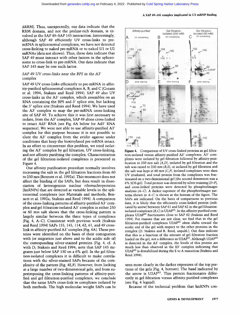
SAP 49 UV cross-links efficiently to pre-mRNA in affin- ity-purified spliceosomal complexes A, B, and C (Gozani et al. 1994; Staknis and Reed 1994). SAP 49 also UV cross-links in the A3' complex, which assembles on an RNA containing the BPS and 3' splice site, but lacking the 5' splice site (Staknis and Reed 1994). We have used the A3' complex to map the pre-mRNA cross-linking site of SAP 49. To achieve this it was first necessary to isolate, from the A3' complex, SAP 49 alone cross-linked to intact Ad3' RNA (see Fig. 6A below for Ad3' RNA sequence). We were not able to use affinity-purified A3' complex for this purpose because it is not possible to elute the A3' complex from the avidin agarose under conditions that keep the biotinylated pre-mRNA intact. In an effort to circumvent this problem, we tried isolat- ing the A3' complex by gel filtration, UV cross-linking, and not affinity purifying the complex. Characterization of the gel filtration-isolated complexes is presented in Figure 4.
Our affinity purification procedure normally involves increasing the salt in the gel filtration fractions from 60 to 250 mM (Bennett et al. 1992a). This treatment does not affect the binding of the SAPs, but does result in disso- ciation of heterogenous nuclear ribonucleoproteins (hnRNPs) that are detected at variable levels in the spli- ceosomal complexes (see Materials and methods; Ben- nett et al. 1992a; Staknis and Reed 1994). A comparison of the cross-linking patterns of affinity-purified A3' com- plex and gel filtration-isolated A3' complex in either 250 or 60 mM salt shows that the cross-linking pattern is largely similar between the three types of complexes (Fig. 4, A-C}. Consistent with previous work (Staknis and Reed 1994) SAPs 155, 145, 114, 62, 61, and 49 cross- link in affinity-purified A3' complex (Fig. 4A). These pro- teins were identified on the basis of their comigration with (or migration just above and to the acidic side of) the corresponding silver-stained proteins (Fig. 4, cf. A with D; Staknis and Reed 1994; note that SAP 145 mi- grates just below SAP 130 on a 6% gel). In the gel filtra- tion-isolated complexes it is difficult to make correla- tions with the silver-stained SAPs because of the com- plexity of the pattern (Fig. 4E, F). However, from looking at a large number of two-dimensional gels, and from su- perimposing the cross-linking patterns of affinity-puri- fied and gel filtration-purified complexes, we conclude that the same SAPs cross-link in complexes isolated by both methods. The high molecular weight SAPs can be
Figure 4. Comparison of UV cross-linked proteins in gel filtra- tion-isolated versus affinity-purified A3' complexes. A3' com- plexes were isolated by gel filtration followed by affinity-puri- fication in 250 mM salt {A,D), isolated by gel filtration and the salt was raised to 250 mM {B,E), or isolated by gel filtration and the salt was kept at 60 mM (C,F). Isolated complexes were then UV irradiated, and total protein from the complexes was frac- tionated on a two-dimensional gel {the second dimension was a 6% SDS gell. Total protein was detected by silver staining (D-F), and cross-linked proteins were detected by phosphorlmager analysis (A-C). A darker exposure of the phosphorlmager pat- terns shown in A-C is shown at the bottom of the figure. The SAPs are indicated. On the basis of comparisons to previous data, it is likely that the efficiently cross-linked protein (indi- cated by arrowl between SAP 61 and SAP 62 in the gel filtration- isolated complexes (B, C) is U2AF 65. In the affinity-purified com- plexes U2AF 65 fractionates close to SAP 62 (Staknis and Reed 19941. For reasons that are not clear, we find that in the gel filtration-purified complexes U2AF 6s often shifts toward the acidic end of the gel with respect to the other proteins in the complex (D. Staknis and R. Reed, unpubl.). Our data indicate that this is a function of the amount of gel filtration fraction loaded on the gel, not a difference in U2AF 65. Although U2AF 65 is detected in the A3' complex, the levels of this protein are much less than observed in the E3' complex indicating that U2AF 65 is destabilized during the E to A transition (Staknis and Reed 1994).
seen more clearly in the darker exposures of the top por- tions of the gels {Fig. 4, bottom). The band indicated by the arrow is U2AF 65. This protein fractionates differ- ently in gel filtration versus affinity-purified complexes {see Fig. 4 legend}.
Because of the technical problem that hnRNPs con-
GENES & DEVELOPMENT 1977
Cold Spring Harbor Laboratory Press on February 4, 2022 - Published by genesdev.cshlp.orgDownloaded from

Champion-Arnaud and Reed
taminate spliceosomal complexes to varying degrees (see Materials and methods), we chose to use gel filtration- purified A3' complex treated with 250 mM salt to map the SAP 49 cross-linking site. Under these conditions we found that it was possible to isolate reproducibly, from the A3' complex, SAP 49 alone cross-linked to pre- mRNA. The strategy used for this is shown in Figure 5 using representative data from one of the experiments.
The A3' complex was assembled on 32P-labeled Ad3' RNA in a large-scale splicing reaction (2.4 ml), fraction- ated by gel filtration, and the indicated fractions were pooled. To minimize contamination with the H com- plex, we use a low level of Ad3' RNA for the assembly reaction because this results in a high A3' to H complex ratio. After raising the salt from 60 to 250 raM, the pooled fractions were irradiated with UV light. SDS was then added, the pooled fractions were acetone precipitated and fractionated on an SDS gel. As expected from the two-dimensional gel analysis (see Fig. 4B), we observe a prominent band {designated SAP 49-Ad3' RNA), in ad- dition to a band containing naked Ad3' RNA. To elimi- nate the contaminating naked RNA from the SAP 49- Ad3' RNA band, we eluted this band and fractionated it on another SDS gel. The SAP 49-Ad3' RNA band eluted from this gel lacks any contaminating free Ad3' RNA (see below).
Previous work showed that, because of the presence of the cross-linked RNase A digestion product, cross-linked SAP 49 fractionates just above native SAP 49 present in the prespliceosome (e.g., see Fig. 4A, D; Staknis and Reed
1994). Consistent with this, we find that RNase A treat- ment of the eluted SAP 49-Ad3' RNA band generates a protein that fractionates just above native SAP 49 on an SDS gel [Fig. 5; the SAP 49 marker was IVT-SAP 49 that cofractionates with native SAP 49 present in the spliceo- some (see Fig. 1)]. We note that two-dimensional gel analysis shows that there are no other major cross-linked proteins of the molecular size of SAP 49 (e.g., Fig. 4B; Staknis and Reed 1994).
To determine whether the Ad3' RNA in the eluted SAP 49-Ad3' RNA band was intact, we treated the band with proteinase (Fig. 6B, lane +) and fractionated it on a denaturing gel. This analysis revealed that the RNA was intact as it largely comigrates with the 125 nucleotide naked Ad3' RNA (Fig. 6B, lane N). The portion of the Ad3' RNA (Fig. 6B, lane +) that is shifted above the naked Ad3' RNA (Fig. 6B, lane N) is attributable to in- complete proteinase digestion. When the SAP 49-Ad3' RNA band is not treated at all with proteinase, the com- plex remains in the well of the denaturing gel {Fig. 6B, lane - ) .
Representative data from oligonucleotide-directed RNase H cleavage of the SAP 49-Ad3' RNA band are shown in Figure 6C. The oligonucleotides used for the analysis are shown in Figure 6A. For each oligonucle- otide, we digested naked Ad3' RNA (N) and cross-linked SAP 49-Ad3' RNA treated (+) or not ( - ) with protein- ase. Digestion of naked Ad3' RNA with oligonucleotide C generates 63- and 45-nucleotide bands (Fig. 6C, oligo C, lane N). In contrast, digestion of SAP 49-Ad3' RNA
Figure 5. Isolation of SAP 49 UV cross-linked to Ad3' pre-mRNA. The A3' complex was isolated by gel filtration, the indicated fractions were pooled, raised to 250 mM salt and UV irradiated. The pooled fractions were then acetone precipitated and run on a 6% SDS gel. SAP 49 cross-linked to Ad3' RNA {SAP 49-Ad3' RNA) was eluted from the gel, acetone precipitated, and fractionated on another 6% SDS gel. The SAP 49-Ad3' RNA was then eluted from the second gel and acetone precipitated. The eluted SAP 49-Ad3' RNA was treated with RNase A and ffactionated on a 9% SDS gel next to IVT-SAP 49 or used for RNase H mapping with or without treatment with proteinase K {PK} {see Fig. 6}.
1978 GENES & DEVELOPMENT
Cold Spring Harbor Laboratory Press on February 4, 2022 - Published by genesdev.cshlp.orgDownloaded from

A SAP 49-145 complex implicated in U2 snRNP binding
with oligonucleotide C generates the 45-nucleotide band, but the 63 nucleotide band is either shifted (pro- teinase-treated sample, Fig. 6C, oligo C, lane +) or de- tected in the well of the gel (proteinase-untreated sam- ple, Fig. 6C, oligo C, lane -) . Similarly, with oligonu- cleotide D, naked RNA is cleaved to generate 45- and 72-nucleotide bands, whereas SAP 49-Ad3' RNA gener- ates the 45-nucleotide band but the 72-nucleotide band is either shifted {proteinase-treated sample, Fig. 6C, oligo D, lane + ) or detected in the well of the gel (proteinase- untreated sample, Fig. 6C, oligo D, lane -) . These data indicate that SAP 49 does not cross-link in exon 2.
Digestion of naked Ad3' RNA with oligonucleotide A generates 17- and 91-nucleotide products (Fig. 6C, oligo A, lane N). With SAP 49-Ad3' RNA, the 17-nucleotide product is detected, whereas the 91-nucleotide product is either shifted or is present in the well of the gel when treated or untreated with proteinase, respectively (Fig. 6C, oligo A, lanes +, - ). In other experiments, in which the proteinase treatment is less complete, the shift of the 91-nucleotide band is more apparent (data not shown). These data indicate that SAP 49 does not cross-link to the 5' portion of the intron. Digestion of naked Ad3' RNA with oligonucleotides A and D together generates 17-, 38-, and 45-nucleotide bands. In contrast, digestion
Figure 6. SAP 49 UV cross-links near the branch site in the A3' complex. (A) Se- quence of Ad3' RNA. The branch-site ade- nosine is indicated by an arrow and the exon sequence is boxed. The positions of the different oligonucleotides used for the RNase H mapping are indicated. The length of each oligonucleotide (bold line) and the size of the two RNA fragments gen- erated by RNase H cleavage of the naked Ad3' RNA are shown. The bracket delimits the region of SAP 49 binding, and the sche- matic of SAP 49 indicates the site within this region where SAP 49 is likely to bind (see text). (B,C) Naked RNA {N), SAP 49- Ad3' RNA treated with proteinase K { + ) or untreated {-) were fractionated on a dena- turing gel (B) or first digested with RNase H and the indicated oligonucleotides (C). The numbers indicate the lengths of the RNase digestion products for naked RNA. The wells of the gels are indicated by an arrow.
of SAP 49-Ad3' RNA generates the 17- and 45-nucle- otide products, but the 38-nucleotide product is shifted in the proteinase-treated sample or present in the well of the gel without the proteinase treatment. These data in- dicate that SAP 49 is present on the 38-nucleotide frag- ment between oligonucleotides A and D.
To localize the site of SAP 49 cross-linking more pre- cisely we used oligonucleotides B and E (see Fig. 6A). In addition, we tested oligonucleotides to sequences be- tween E and C, but found that these were not useful because naked Ad3' RNA could not be digested com- pletely with them (data not shown). With oligonucle- otides B and E, digestion of SAP 49-Ad3' RNA did not yield results as clear as those obtained with oligonucle- otides A, C, and D. We believe that this may be because SAP 49 cross-links to the binding sites of these oligonu- cleotides (see below). In addition, it is possible that SAP 49 cross-links to more than one site in the vicinity of these oligonucleotides. The observation that SAP 49 contains two RNA recognition motifs supports the no- tion that SAP 49 may have two cross-linking sites. In any case, digestion of SAP 49-Ad3' RNA with oligonucle- otide B generates a 27-nucleotide band that comigrates with the naked RNA digestion product, whereas the 85- nucleotide band is shifted (Fig. 6C, oligo B, lanes N, + ).
GENES & DEVELOPMENT 1979
Cold Spring Harbor Laboratory Press on February 4, 2022 - Published by genesdev.cshlp.orgDownloaded from

Champion-Arnaud and Reed
Using oligonucleotide E to digest SAP 49-Ad3' RNA, the 41-nucleotide band shifts and the 71-nucleotide band does not.
When SAP 49-Ad3' RNA was digested with oligonu- cleotides B or E in the absence of proteinase treatment, we could not identify clearly which of the two RNase digestion products for each oligonucleotide was detected in the well of the gel. An example of this is shown with oligonucleotide E (Fig. 6C, oligo E, cf. lanes N, +, - ). A band is detected in the well of the gel, but both the 71- and 41-nucleotide products are present at about equal levels in the gel. One interpretation of this result is that SAP 49 cross-links to sequences with which the oligo- nucleotide hybridizes; thus, neither of the two digestion products are detected in the well of the gel.
Summarizing these data, we have obtained evidence that SAP 49 cross-links to a 29-nucleotide region be- tween oligonucleotides A and C (indicated by bracket in Fig. 6A). Within this region, the data are less clear, but suggest the possibility that SAP 49 cross-links to two sites, one in the region of oligonucleotide B and the other in the region of oligonucleotide E (indicated by the Xs in the SAP 49 schematic in Fig. 6A). This region is located immediately upstream of the branch site.
Discussion
A great deal of previous work has shown that the branch- point sequence plays an essential role in the splicing reaction in both yeast (Domdey et al. 1984; Langford et al. 1984; Rodriguez et al. 1984) and mammals (Raut- mann and Breathnach 1985; Reed and Maniatis 1985; Ruskin et al. 1985; Hornig et al. 1986; Reed and Maniatis 1988; Zhuang et al. 1989). The BPS forms a duplex with a region at the 5' end of U2 snRNA, and this interaction is required for splicing (Parker et al. 1987; Wu and Man- ley 1989; Zhuang and Weiner 1989). The U2-BPS duplex appears to be established at or near the time that U2 snRNP first binds stably to the pre-mRNA to form the prespliceosomal complex A (Wassarman and Steitz 1992; Sawa and Shimura 1992; Sontheimer and Steitz 1993). One of the key functions of the duplex is thought to be bulging the branch-site adenosine that carries out the nucleophilic attack on the 5' splice site (Query et al. 1994). The duplex has been proposed to persist at the time of catalytic step I (Query et al. 1994), which is con- sistent with cross-linking data (Sawa and Shimura 1992; Wassarman and Steitz 1992; Sontheimer and Steitz 1993).
In addition to the role in specifying the nucleophile, the U2-BPS duplex is thought to be involved in the ini- tial recognition of the BPS by the specific base-pairing interaction. However, it is highly unlikely that U2 sn- RNA alone is responsible for recognizing and maintain- ing the U2-BPS duplex, primarily because the duplex is so short. For this reason, it is clear that other factors are involved in mediating U2 snRNA-BPS interactions. In this study we have identified a factor, SAP 49, that binds directly to the pre-mRNA near the BPS. Significantly, this interaction is first established at the time that the
U2-BPS duplex forms, in the A complex. Further paral- leling the U2-BPS duplex interactions, SAP 49 cross- linking is maintained in the B complex and persists through catalytic step I (Gozani et al. 1994; Staknis and Reed 1994). Further studies are required, however, to rule out the possibility that the site of SAP 49 cross- linking changes after A complex assembly. In any case, previous work showed that SAP 49 interacts specifically with U2 snRNP (Staknis and Reed 1994), and our data show that SAP 49 interacts by protein-protein interac- tions with another U2 snRNP-associated protein, SAP 145. These observations, together with the data dis- cussed below, suggest that SAP 49 and SAP 145 form a complex that plays a role in tethering U2 snRNP to the branch site.
Biochemical studies of U2 snRNP and of the two es- sential splicing acitivites, SF3a and SF3b, indicate that at least two other proteins (SAP 130 and SAP 155) associate with the SAP 49-SAP 145 complex. The sequential bind- ing of the essential splicing factors SF3b and SF3a con- verts U2 snRNP from an inactive 12S particle to a 17S particle functional in the splicing reaction (Behrens et al. 1993a, b). SAPs 61, 62, and 114 correspond to the U2 sn- RNPs that comprise SF3a, whereas SAPs 49, 130, 145, and 155 correspond to the U2 snRNPs thought to be components of SF3b (U2 snRNP s3, 12o, 15o, and 160, re- spectively; SAPs corresponding to U2 snRNP 3s and U2 snRNP 92 have not been identified; Staknis and Reed 1994; see Hodges and Beggs 1994). Thus, SAP 130 and SAP 155 are likely to associate with SAP 145 and SAP 49 in a heteromultimer. All three subunits of SF3a also UV cross-link to the pre-mRNA and thus may mediate direct U2 snRNP-BPS interactions as well. The RNA-protein and protein-protein interactions of these proteins are de- picted in Figure 7.
Specific protein-protein interactions occur between SAP 61 and SAP 114 (Bennett and Reed 1993) and be- tween SAP 62 and SAP 114 (Chiara et al. 1994). As indi- cated in Figure 7, the amino terminus of SAP 61 is re- quired for the SAP 114 interaction (Chiara et al. 1994). The carboxy-terminal half of SAP 62, which is highly proline rich and contains 22 heptapeptide repeats of a proline-rich sequence, is not required for the SAP 114 interaction, but no further mapping of the interaction domain has been done (M. Bennett and R. Reed, unpubl.). Both SAP 61 and SAP 62 contain zinc finger-like motifs that may be involved in the pre-mRNA binding (Bennett and Reed 1993; Chiara et al. 1994). In this study we found that SAP 49 interacts directly with SAP 145. The amino terminus of SAP 49 containing two RNA-binding domains (RRMs) is required for this interaction. A role for RRMs in protein-protein interactions has been sug- gested previously for the U1 snRNP-specific protein A (Nagai et al. 1990) and for the alternative splicing factor, transformer 2 (Amrein et al. 1994). On the basis of stud- ies of these and other proteins containing RRMs (Query et al. 1989; Scherly et al. 1989), it is likely that the SAP 49 RRMs are also involved in the pre-mRNA-binding activity. Similar to SAP 62, the carboxy-terminal half of SAP 49 is highly proline rich. As the proline-rich do-
1980 GENES & DEVELOPMENT
Cold Spring Harbor Laboratory Press on February 4, 2022 - Published by genesdev.cshlp.orgDownloaded from

A SAP 49-145 complex implicated in U2 snRNP binding
Figure 7. RNA-protein and protein-protein interactions in the A3' complex. The U2 snRNP-specific SAPs and U2 snRNA base-paired to the BPS in Ad3' RNA are indicated. The exact positions of the proteins on U2 snRNA and on the pre-mRNA were drawn arbitrarily. All of the proteins interact directly with the pre-mRNA except SAP 130. SAP 49 contacts SAP 145 and the amino terminus of SAP 49 (N +) containing the RRMs is required for the interaction. SAP 61 interacts directly with SAP 114 and the amino terminus of SAP 61 (N +) is required for the interaction. Both SAP 61 and SAP 62 contain zinc finger-like motifs (zn) that could be involved in the RNA binding. The carboxy-terminal halves of both SAP 62 and SAP 49 consist of a proline-rich domain (pro) that is not required for the protein- protein interactions depicted. In addition to the seven 17S U2 snRNP-specific proteins shown here, 35 and 92 kD proteins have been detected in 17S U2 snRNP (Behrens et al. 1993a). The relationship between these proteins and the SAPs is not known. SAPs 61, 62, and 144 comprise SF3a, whereas the other proteins are thought to be components of SF3b (Brosi et al. 1993a, b).
mains are not required for the respective strong protein- protein interactions between SAP 62 and SAP 114 and between SAP 49 and SAP 145, it is possible that these domains are involved in other protein-protein interac- tions wi th in the spliceosomal complexes, such as asso- ciation of SF3a and SF3b or association wi th other spli- ceosomal components such as U2AF or U6 snRNP (see below}. As indicated in Figure 7, SAP 155 cross-links to the pre-mRNA, whereas SAP 130 does not (Staknis and Reed 1994). We have not yet detected protein-protein interactions wi th these proteins.
The 17S U2 snRNP-specific proteins are thought to associate wi th the 5' end of U2 snRNA (see Fig. 7; Beh- rens et al. 1993a). This portion of U2 snRNA forms the duplex wi th the BPS. Thus, one or more of the SAPs that cross-link may be involved in the formation or stabili- zation of the U2-BPS duplex. Immediate ly upstream on U2 snRNA is a region that participates in a base-pairing interaction wi th U6 snRNA (essential in yeast; Madhani and Guthrie 1992). Thus, in addition to mediat ing bind- ing of U2 snRNP to the BPS, the 17S U2 snRNP proteins may be involved in U2-U6 snRNP interactions.
Our data indicate that, as proposed previously (Staknis and Reed 1994), SAP 49 is a component of the U2 sn- RNP-associated branchpoint protection factor that pro- tects a 30- to 40-nucleotide region surrounding the BPS from RNase digestion. The branchpoint protection fac- tor, s imilar to SAP 49, interacts wi th the pre-mRNA from the t ime of A complex assembly through catalytic step I (Black et al. 1985; Ruskin and Green 1985; Ruskin et al. 1988; Zamore and Green 1989). The other 17S U2 snRNP-specific SAPs that cross-link are also l ikely to be components of the branchpoint protection factor. Con- sistent wi th a role for these proteins in mediat ing U2 snRNP branch-site interactions, we find that a muta t ion of the BPS that blocks A complex assembly also blocks UV cross-linking of the U2 snRNP-associated SAPs (P. Champion-Arnaud, O. Gozani, and R. Reed, in prep.). It is unlikely, however, that we have detected all of the proteins that interact wi th the branch site throughout the splicing reaction. For example, our study would not detect proteins that interact t ransient ly wi th the branch site. An example of such a factor in yeast is PRP16, an RNA-dependent ATPase required for catalytic step II and thought to be involved in a branch-site proofreading mechan i sm (Schwer and Guthrie 1991, 1992; Burgess and Guthrie 1993}. In addition to 17S U2 snRNP (12S U2 snRNP, SF3a, and SF3b), assembly of the A complex re- quires U2AF, SF1, U1 snRNP, and SR proteins (Kramer 1988, 1992; Zamore and Green 1989; Barabino et al. 1990; Fu and Maniat is 1990; Krainer et al. 1990; Kramer and Utans 1991; Brosi et al. 1993a). Assembly of the B and C complexes involves the binding of U4, U5, and U6 snRNAs together wi th a large number of proteins (Kon- arska and Sharp 1987; Bennett et al. 1992a; Gozani et al. 1994). As spliceosomal complexes are highly dynamic particles, some of these factors may interact wi th the branch site during the course of the reaction. However, the observation that the 17S U2 snRNP-specific SAPs cross-link to the pre-mRNA when U2 snRNA first inter- acts with the branch site and then remain bound in a cross-linkable manner through catalytic step I suggest that these proteins are key factors involved in tethering U2 snRNP to the pre-mRNA.
Mater ia l s and m e t h o d s
Isolation of SAP 49 cDNA, in vitro translation, and Northern analysis
Prespliceosomes were isolated in large scale, fractionated on preparative two-dimensional gels, and peptide sequence was ob- tained from two tryptic digestion products of SAP 49 (Bennett and Reed 1993). Degenerate PCR primers were used to generate a probe encoding the SAP 49 peptide QHQGYGFVEFLSEEDA by amplification of total HeLa cell mRNA. The DNA probe encoding the peptide was then kinased and used to screen an HeLa hZAP eDNA library. Two positive clones encoding full- length SAP 49 cDNAs were isolated. In vitro translation of the SAP 49 eDNA clones was carried out using the reticulocyte lysate system from Promega. For Northern analysis, total poly(A) + RNA was isolated, transferred to nitrocellulose, and probed with 32P-labeled SAP 49 cDNA using standard proce- dures (Sambrook et al. 1989). Splicing reactions, affinity purifi-
GENES & DEVELOPMENT 1981
Cold Spring Harbor Laboratory Press on February 4, 2022 - Published by genesdev.cshlp.orgDownloaded from
Champion-Arnaud and Reed
cation, and analysis of splicing complexes were carried out as described (Bennett et al. 1992a).
Protein-protein interactions
For Farwestem analysis, the A3' complex (assembled on 1.5 lag Ad3' pre-mRNA) was fractionated by two-dimensional gel elec- trophoresis and total nuclear extract (10 ~1) was fractionated on the second dimension of the same gel or on separate SDS gels as indicated. Proteins were transferred to nitrocellulose and probed with aSS-labeled in vitro-translated SAP 49, SAP 49 ARRM, or SAP 49 APG. The blots were blocked and probed according to the methods of Zhang et al. (1992). The SAP 49 mutants were obtained by deleting the appropriate sequence in the SAP 49 cDNA. To identify SAP 145 on the two-dimensional blot, the nitrocellulose filter was stained with India ink as de- scribed in Sambrook et al. (1989).
Mapping the SAP 49 cross-linking site
Ad3' RNA was synthesized in transcription reactions (100 ~1) containing 50 ~Ci a2P-ATP, CTP, GTP, and UTP (3000 Ci/ mmole), and 100 ~M cold ATP, CTP, GTP, and UTP. The A3' complex assembled on Ad3' RNA (2.4-ml reaction, containing 2.4 ~g Ad3' RNA and 30% nuclear extract, incubated at 30~ for 30 rain) was isolated by gel filtration and the salt was either raised to 250 mM or kept at 60 mM as indicated. Isolated com- plexes were irradiated at 254 nm with a Sylvania light for 5 min on ice at a distance of 5.5 cm from the light source (Staknis and Reed 1994}. [Note that we have found variability in the levels of hnRNP cross-linking in the 60 mM gel filtration-isolated com- plexes (data not shown). This is because of variable levels of contamination of the spliceosomal complexes with the H com- plex that consists of hnRNPs and fractionates close to the spli- ceosomal complexes (Bennett et al. 1992b).] After irradiation, SDS was added to the gel filtration-isolated complexes to a final concentration of 2%, and they were acetone precipitated as de- scribed (Bennett et al. 1992a). The strategy for isolating SAP 49 cross-linked to pre-mRNA is presented in Figure 5. The oligo- nucleotides used for RNase H mapping are described in Figure 6A. RNase H digestion was carried out in a 20-~1 reaction for 30 rain at 30~ using 200 ng of each oligonucleotide. For each eluted sample, one-half was digested directly with RNase H, and the other half was first treated with proteinase K, phenol extracted, ethanol precipitated, and then digested with RNase H. The RNase digestion products were fractionated on 12% denaturing polyacrylamide gels.
Acknowledgments
We are grateful to R. Feld for excellent technical assistance and to D. Staknis and M. Chiara for comments on the manuscript. HeLa cells for nuclear extracts were provided by the National Institutes of Health (NIH) cell culture facility at Endotronics (MN). C.A.P. is supported in part by a French ARC fellowship. R. R. is a Lucille P. Markey Scholar. This work was supported by a grant from the Lucille P. Markey Charitable Trust and a grant from NIH.
The publication costs of this article were defrayed in part by payment of page charges. This article must therefore be hereby marked "advertisement" in accordance with 18 USC section 1734 solely to indicate this fact.
Note added in proof
The SAP 49 sequence data have been deposited to GenBank under accession number L35013.
References
Amrein, H., M.L. Hedley, and T. Maniatis. 1994. The role of specific protein-RNA and protein-protein interactions in positive and negative control of pre-mRNA splicing by trans- former 2. Cell 76: 735-746.
Barabino, S.M., B.J. Blencowe, U. Ryder, B.S. Sproat, and AT Lamond. 1990. Targeted snRNP depletion reveals an addi- tional role for mammalian U1 snRNP in spliceosome assem- bly. Cell 63: 293-302.
Behrens, S-E., K. Tyc, B. Kastner, J. Reichelt, and R. Luhrmann. 1993a. Small nuclear ribonucleoprotein (RNP) U2 contains numerous additional proteins and has a bipartite KNP struc- ture under splicing conditions. Mol. Cell Biol. 13: 307-319.
Behrens, S-E., F. Galisson, P. Legrain, and R. Luhrmann. 1993b. Evidence that the 60 kD protein of 17S U2 snRNP is immu- nologically and functionally related to the yeast PRP9 splic- ing factor and is required for efficient formation of prespli- ceosomes. Proc. Natl. Acad. Sci. 90: 8229-8233.
Bennett, M. and R. Reed. 1993. Correspondence between a mammalian spliceosome component and an essential yeast splicing factor. Science 262: 105-108.
Bennett, M., S. Michaud, J. Kingston, and R. Reed. 1992a. Pro- tein components specifically associated with prespliceo- some and spliceosome complexes. Genes & Dev. 6: 1986- 2000.
Bennett, M., S. Pifiol-Roma, D. Staknis, G. Dreyfuss, and R. Reed. 1992b. Transcript-dependent packaging of pre-mRNA in hnRNP complexes prior to spliceosome assembly in vitro. Mol. Cell. Biol. 12: 3165-3175.
Black, D., B. Chabot, and I.A. Steitz. 1985. U2 as well as U1 small nuclear ribonucleoproteins are involved in premessen- ger RNA splicing. Cell 42: 737-750.
Brosi, R., H.P. Hauri, and A. Kramer. 1993a. Separation of splic- ing factor SF3a into two components and purification of SF3a activity. Biol. Chem. 268: 17640-17646.
Brosi, R., K. Groning, S-E. Behrens, R. Luhrmann, and A. Kramer. 1993b. Interaction of mammalian splicing factor SF3a with U2 snRNP and relationship of its 60-kD subunit to yeast PRP9. Science 262: 102-105.
Burgess, S.M. and C. Guthrie. 1993. A mechanism to enhance mRNA splicing fidelity: The RNA-dependant ATPase PRP16 governs usage of a discard pathway for aberrant lariat intermediates. Cell 73: 1377-1391.
Chiara, M.D., P. Champion-Amaud, M. Buvoli, B. Nadal-Gi- nard, and R. Reed. 1994. Specific protein-protein interac- tions between the essential prespliceosome components SAPs 61 and 114. Proc. Natl. Acad. Sci. 91: 6403-6407.
Cortes, J.J., E.J. Sontheimer, S.D. Seiwert, and ].A. Steitz. 1993. Mutations in the conserved loop of human U5 snRNA gen- erate use of novel cryptic 5' splice sites in vivo. EMBO J. 12: 5181-5189.
Domdey H., B. Apostol, R.J. Lin, A. Newman, E. Brody, and ]. Abelson. 1984. Lariat structures are in vivo intermediates in yeast pre-mRNA splicing. Cell 39: 611-621.
Fu, X-D. and T. Maniatis. 1990. Factor required for mammalian spliceosome assembly is localized to discrete regions in the nucleus. Nature 343: 437-441.
Gozani, O., J.G. Patton, and R. Reed. 1994. A novel set of spli- ceosome-associated proteins (SAPs) and the essential splic- ing factor PSF bind stably to pre-mRNA prior to catalytic
1982 GENES & DEVELOPMENT
Cold Spring Harbor Laboratory Press on February 4, 2022 - Published by genesdev.cshlp.orgDownloaded from

A SAP 49-145 complex implicated in U2 snRNP binding
step II of the splicing reaction. EMBO J. (in press). Hodges, P.E. and J.D. Beggs. 1994. U2 fulfills a commitment.
Curt. Biol. 4: 264-267. Hornig, H., M. Aebi, and C. Weissmann. 1986. Effect of muta-
tions at the lariat branch acceptor site on beta-globin pre- mRNA splicing in vitro. Nature 324: 589-591.
Kandel-Lewis, S. and B. S6raphin. 1993. Role of U6 snRNAs in 5' splice site selection. Science 262: 2035-2039.
Konarska, M.M. and P.A. Sharp 1987. Interactions between small nuclear ribonucleoprotein particles in formation of spliceosomes. Cell 49: 763-774.
Kozak, M. 1986. Point mutations define a sequence flanking the AUG initiator codon that modulates translation by eukary- otic ribosomes. Cell 44: 283-292.
Krainer, A.R., G.C. Conway, and D. Kozak. 1990. Purification and characterization of pre-mRNA splicing factor SF2 from HeLa cells. Genes & Dev. 4: 1158-1171.
Kramer, A. 1988. Presplicing complex formation requires two proteins and U2 snRNP. Genes & Dev. 2: 1155-1167.
Kramer, A. 1992. Purification of splicing factor SF1, a heat-sta- ble protein that functions in the assembly of a presplicing complex. Mol. Cell. Biol. 12: 4545--4552.
Kramer, A. and U. Utans. 1991. Three protein factors (SF1, SF3, and U2AF) function in pre-splicing complex formation in addition to snRNPs. EMBO J. 10: 1503-1509.
Langford, C.J., F.J. Klinz, C. Donath, and D. Gallwitz. 1984. Point mutations identify the conserved, intron-contained TACTAAC box as an essential splicing signal sequence in yeast. Cell 36: 645-653.
Lesser, C.L. and C. Guthrie. 1993. Mutations in U6 snRNA that alter splice site specificity: Implications for the active site. Science 262: 1982-1988.
Madhani, H.D. and C. Guthrie. 1992. A novel base-pairing in- teraction between U2 and U6 snRNAs suggests a mecha- nism for the catalytic activation of the spliceosome. Cell 71: 803-817.
Moore, M.J., C.C. Query, and P.A. Sharp. 1993. Splicing of pre- cursors to messenger RNAs by the spliceosome. In R N A world (ed. R.F. Gestland and J.F. Atkins), pp. 303-357. Cold Spring Harbor Laboratory Press, Cold Spring Harbor, New York.
Nagai, K., C. Oubridge, T.H. Jessen, J. Li, and P.R. Evans. 1990. Crystal structure of the RNA binding domain of the U1A small nuclear ribonuclear protein A. Nature 348: 515-520.
Newman, A.J. 1994. Pre-mRNA splicing. Curr. Opin. Genet. Devl. 4: 298-304.
Newman, A.J. and C. Norman. 1991. Mutations in yeast U5 snRNA alter the specificity of 5' splice-site cleavage. Cell 6 5 : 1 1 5 - 1 2 3 .
- - . 1992. U5 snRNA interacts with exon sequences and 5' and 3' splice sites. Cell 68: 743-754.
Parker, R., P.G. Siliciano, and C. Guthrie. 1987. Recognition of the TACTAAC box during mRNA splicing in yeast involves base pairing to the U2-1ike snRNA. Cell 49: 229-239.
Query, C.C., R.C. Bentley, and J.D. Keene. 1989. A common RNA recognition motif identified within a defined U 1 RNA binding domain of the 70K U1 snRNP protein. Cell 57: 89- 101.
Query, C.C., M. Moore, and P. Sharp. 1994. Branch nucleophile selection in pre-mRNA splicing: Evidence for the bulged du- plex model. Genes & Dev. 8: 587-597.
Rautmann, G. and R. Breathnach. 1985. A role for branchpoints in splicing in vivo. Nature 315: 430--432.
Reed, R. 1990. Protein composition of mammalian spliceo- somes assembled in vitro. Proc. Natl. Acad. Sci. 87: 8031- 8035.
Reed, R. and T. Maniatis. 1985. Intron sequences involved in lariat formation during pre-mRNA splicing. Cell 41: 95-105.
~ . 1988. The role of the mammalian branchpoint sequence in pre-mRNA splicing. Genes & Dev. 2: 1268-1276.
Rodriguez, J.R., C.W. Pikielny, and M. Rosbash. 1984. In vivo characterization of yeast mRNA processing intermediates�9 Cell 39: 603-610.
Ruskin, B. and M.R. Green. 1985. Specific and stable intron- factor interactions are established early during in vitro pre- mRNA splicing. Cell 43: 131-142.
Ruskin, B., J.M. Greene, and M.R. Green. 1985. Cryptic branch point activation allows accurate in vitro splicing of human beta-globin intron mutants�9 Cell 41: 833-844.
Ruskin, B., P.D. Zamore, and M.R. Green. 1988. A factor, U2AF, is required for U2 snRNP binding and splicing complex as- sembly. Cell 52: 207-219.
Sambrook, J., E.F. Fritsch, and T. Maniatis. 1989. Molecular cloning: A laboratory manual, 2nd ed. Cold Spring Harbor Laboratory Press, Cold Spring Harbor, New York.
Sawa, H. and J. Abelson. 1992. Evidence for a base-pairing in- teraction between U6 small nuclear RNA and the 5' splice site during the splicing reaction in yeast. Proc. Natl. Acad. Sci. 89: 11269-11273.
Sawa, H., and Y. Shimura. 1992. Association of U6 snRNA with the 5'-splice site region of pre-mRNA in the spliceosome. Genes & Dev. 6: 244-254.
Schwer, B. and C. Guthrie. 1991. PRP16 is an RNA-dependent ATPase that interacts transiently with the spliceosme. Na- ture 349: 494-499.
�9 1992. A dominant negative mutation in a spliceosomal ATPase affects ATP hydrolysis but not binding to the spli- ceosome. Mol. Cell. Biol. 12: 3540-3547.
Scherly, D., W. Boelens, W.J. van Venrooij, N.A. Dathan, J. Hamm, and I.W. Mattaj. 1989. Identification of the RNA binding domain segment of human U1A protein and defini- tion of its binding site in U1 snRNA. EMBO J. 8: 4163--4170.
Sontheimer, E.J. and J.A. Steitz. 1993. The U5 and U6 small nuclear RNAs as active site components of the spliceosome. Science 262: 1989-1996.
Staknis, D. and R. Reed. 1994. Direct interactions between pre- mRNA and six U2 snRNP proteins during spliceosome as- sembly. Mol. Cell Biol. 14: 2994-3005.
Wassarman, D.A. and J.A. Steitz. 1992. Interactions of small nuclear RNAs with precursor messenger RNA during in vitro splicing. Science 257: 1918-1925.
Wu, J.Y. and J.L. Manley. 1989. Mammalian pre-mRNA branch site selection by U2 snRNP involves base pairing. Genes & Dev. 3: 1553-1561.
Wyatt, J.R., E.J. Sontheimer, and J.A. Steitz. 1992. Site-specific cross-linking of mammalian U5 snRNP to the 5' splice site prior to the first step of premessenger RNA splicing. Genes & Dev. 6: 2542-2553.
Zamore, P.D. and M.R. Green. 1989. Identification, purification and biochemical characterization of U2 small nuclear ribo- nucleoprotein auxilary factor. Proc. Natl. Acad. Sci. 86: 9243-9247.
Zhang, M., P.D. Zamore, M. Carmo-Fonesca, A.I. Lamond, and M.R. Green. 1992. Cloning and intracellular localization of the U2 small nuclear ribonucleoprotein auxiliary factor small unit. Proc. Natl. Acad. Sci. 89: 8769-8773.
Zhuang, Y. and A.M. Weiner. 1989. A compensatory base change in human U2 snRNA can suppress a branch site mu- tation. Genes & Dev. 3: 1545-1552.
Zhuang, Y., A.M. Goldstein, and A.M. Weiner. 1989. UAC- UAAC is the preferred branch site for mammalian pre- mRNA splicing. Proc. Natl. Acad. Sci. 86: 2752-2756.
GENES & DEVELOPMENT 1983
Cold Spring Harbor Laboratory Press on February 4, 2022 - Published by genesdev.cshlp.orgDownloaded from

10.1101/gad.8.16.1974Access the most recent version at doi: 8:1994, Genes Dev.
P Champion-Arnaud and R Reed complex implicated in tethering U2 snRNP to the branch site.The prespliceosome components SAP 49 and SAP 145 interact in a
References
http://genesdev.cshlp.org/content/8/16/1974.full.html#ref-list-1
This article cites 56 articles, 27 of which can be accessed free at:
License
ServiceEmail Alerting
click here.right corner of the article or
Receive free email alerts when new articles cite this article - sign up in the box at the top
Copyright © Cold Spring Harbor Laboratory Press
Cold Spring Harbor Laboratory Press on February 4, 2022 - Published by genesdev.cshlp.orgDownloaded from