Embed Size (px)
Citation preview

The Pathophysiologyof Septic Shock
O. Okorie Nduka, MDa,b,*, Joseph E. Parrillo, MDc
KEYWORDS
� Sepsis � Severe sepsis � Septic shock� Myocardial depression � Vasodilation
The word ‘‘sepsis’’ comes from the Greek word sepo meaning decay or putrefaction,and its original usage described the decomposition of organic matter in a manner thatresulted in decay and death.1 In the Hippocratic model of health and disease, livingtissues broke down by 1 of 2 processes. Pepsis was the process through whichfood was digested, leading to health. Sepsis, however, denoted tissue breakdownthat resulted in disease. Hippocrates used this term to describe the process ofabnormal tissue breakdown that resulted in a foul odor, pus-formation, and some-times dead tissue.2 This usage of the term sepsis persisted for almost 3 millennia,and subsequent work establishing a causal link between microbes and suppurativeinfections, or systemic symptoms from infection, did not change the use of the termas a description of a constellation of clinical findings, but rather established infectionas the underlying cause.3 The term ‘‘shock’’ comes from the French word choquermeaning ‘‘to collide with,’’ and aptly describes the body’s response to invadingmicrobes and, to a large extent, its disruptive effect on normal physiology. Initiallyused in the medical literature in the 1700s, its earliest uses connoted a sudden joltthat often led to death (the initial physical injury). This definition evolved to describewidespread circulatory dysfunction following injury.4
Sepsis is the systemic maladaptive response of the body to the invasion of normallysterile tissue by pathogenic, or potentially pathogenic, microorganisms. Shock may bedefined as a ‘‘state in which profound and widespread reduction of effective tissueperfusion leads first to reversible, and then, if prolonged, to irreversible cellularinjury.’’5 From a clinical standpoint, this progressive cellular dysfunction manifestsas a continuum from sepsis, to severe sepsis, and finally to septic shock (Box 1).
a Division of Critical Care Medicine, Department of Internal Medicine, Cooper UniversityHospital, Camden, NJ, USAb Altru Hospital, Grand Forks, ND 58206-6003, USAc Robert Wood Johnson Medical School, Department of Medicine, University of Medicine andDentistry of New Jersey, NJ, USA* Corresponding author. Division of Critical Care Medicine, Department of Internal Medicine,Cooper University Hospital, Camden, NJ.E-mail address: [email protected] (O.O. Nduka).
Crit Care Clin 25 (2009) 677–702doi:10.1016/j.ccc.2009.08.002 criticalcare.theclinics.com0749-0704/09/$ – see front matter ª 2009 Elsevier Inc. All rights reserved.

Box1Definitions of sepsis, severe sepsis, and septic shock
Sepsis: sepsis is defined as infection plus systemic manifestations of infection
Severe sepsis: sepsis with sepsis-induced organ dysfunction or tissue hypoperfusion
Sepsis-induced hypotension: a systolic blood pressure (SBP) less than 90 mm Hg or mean arterialpressure less than 70 mm Hg, or an SBP decrease of greater than 40 mm Hg or greater than 2 SDless than normal for age in the absence of other causes of hypotension
Septic shock: sepsis-induced hypotension persisting despite adequate fluid resuscitation.
Sepsis-induced tissue hypoperfusion: septic shock, lactate elevation beyond the upper limits ofnormal or oliguria
Acute oliguria: urine output less than 0.5 mL/kg/h for at least 2 hours, despite adequate fluidresuscitation
Data from Dellinger RP, Levy MM, Carlet JM, et al. Surviving Sepsis Campaign: internationalguidelines for management of severe sepsis and septic shock: 2008. Crit Care Med 2008;36:296–27.
Nduka & Parrillo678
Severe sepsis and septic shock are the end result of complex interactions betweeninfecting organisms and several elements of the host response, and reflect a primarilyinappropriate response by the host to a microbial pathogenic insult. The key term thatdescribes the pathophysiologic events in septic shock at any point in time is the‘‘mismatch’’ of the host response to the intensity of the pathogenic stimuli ultimatelyleading to organ injury or dysfunction with or without hypotension. This mismatchresults in, amongst other derangements, an immune profile that could be predomi-nantly proinflammatory (systemic inflammatory response syndrome [SIRS]), mixed(mixed antagonistic response syndrome [MARS]), or predominantly anti-inflammatory(compensatory anti-inflammatory syndrome [CARS]). The nature of the interactionsbetween the microbial pathogen and the host is complex and, at the tissues, resultsin excessive inflammation or immunosuppression, abnormal coagulation and bloodflow, and microcirculatory dysfunction leading to organ injury and cell death (Fig. 1).
The complex events that occur in septic shock can be broadly divided into micro-organism-related components and host-related components. The broad categoriesare further subdivided into cellular and humoral components. Pathogen-related eventsin the pathophysiology of septic shock include the mechanisms by which microbesevade host defenses and subvert aspects of the host immune response, resulting insignificantly increased morbidity. Concerning the host-related events in septic shock,multiple derangements involving several biologic systems contribute to differentdegrees to the development of septic shock. A meaningful review of every provenor proposed pathogenetic mechanism for septic shock is near impossible, and thisarticle focuses on selected dysfunctions believed to play more significant roles inthe development of septic shock. These are outlined below, and include aspects ofmicrobial pathogenicity, key cellular and humoral aspects of the maladaptive immu-noinflammatory response, the interactions between the immunoinflammatory andcoagulation systems, and their cardiocirculatory consequences, resulting in the clin-ical picture of septic shock.
- The role of the pathogen- Immunoinflammatory dysfunction leading to severe sepsis� Pathogen recognition� Pro- and anti-inflammatory cellular signaling/signal transduction

Fig. 1. Pathophysiology of severe sepsis and septic shock. (Adapted from Cinel I, Opal SM.Molecular biology of inflammation and sepsis: a primer. Crit Care Med 2009;37(1):293;with permission.)
The Pathophysiology of Septic Shock 679
� Release of pro-and anti-inflammatory mediators� Immune and non-immune effector cell dysfunction� Interactions with other biologic systems in sepsis
- Cardiocirculatory dysfunction in severe sepsis resulting in progression to septic shock
THE ROLE OF THE PATHOGEN
The initial event in severe sepsis and septic shock involves the invasion of normallysterile tissue by pathogenic microbes. The interactions between the pathogens andthe host immune system may result in either a contained infectious process withminimal tissue injury or severe sepsis and septic shock. The abnormal host responseseen in septic shock can be triggered by bacteria, viruses, or fungi.
Historically, our understanding of the pathophysiologic events in sepsis has focusedon the maladaptive responses by the host, minimizing the role, if any, played by thepathogen invaders. Emerging evidence suggests a more significant role for pathogensthan was previously believed.6 The development of modern imaging techniques withdifferential spatial and temporal resolution has provided the means to study thecomplex interactions between pathogens and their mammalian hosts, leading toadvances in our understanding of bacterial pathogenicity. It is known that bacterialand nonbacterial pathogens possess an array of specific mechanisms (virulencefactors) that confer the ability to evade host defense mechanisms and proliferate inhost tissues (Box 2, Fig. 2). Bacterial virulence factors are better studied than theirnonbacterial counterparts, and these mechanisms vary across species, classes,and strains of bacteria. Despite wide variation in the nature of the pathogens and theirvirulence factors, some common themes have emerged with regard to how pathogenssubvert the early immune response and exert their full pathogenic potential.

Box 2Virulence characteristics of bacterial pathogens
Mechanisms of bacterial adherence to host epithelial surfaces
Adhesins: bacterial protein products (integral or secreted) that enable pathogenic organisms tobind onto host tissue elements (eg, collagen fibers)
Flagella, fimbrae, and pili: bacterial appendages whose primary role is mobility, but they enablepathogens to directly attach themselves to host cells and extracellular matrix components
Type III secretion system: functions as a molecular conduit, enabling bacteria to introduceproteins into host cells, altering their function to enhance bacterial survival
Ligand mimicry: by producing proteins similar to host-derived proteins, bacteria are able tobind to relevant ligand receptors
Mechanisms of bacterial invasion following adherence
Bacterial protein secretion systems I, II, III, IV: specialized transporter systems that enable thedelivery of bacterial products into the extracellular matrix (I and II) or intracellularly (III and IV),facilitating tissue invasion and intracellular infection
Lipidrafts: avoiding the apical areas of epithelial cells exposed to commensals and resistant topathogen invasion, pathogenic bacteria bind to the baso-lateral aspects of the host cell plasmamembrane. This area is rich in cholesterol and pathogen recognition receptors. Followingbinding to plasma membrane cholesterol, the pathogens form intracellular vacuoles,rendering them immune to lysosomal endocytosis.
Bacterial host defense evasion mechanisms
Anti-phagocytosis: bacterial pathogens have several mechanisms to avoid phagocytosis: (1)inhibition of opsonization by encapsulation; (2) surface antigenic variation that preventsrecognition as pathogens; (3) inhibition of uptake via the release of toxic protein effectors; (4)intracelluar survival and replication in the cytosol and also within lysosomes; (5) induction ofimmune effector cell apoptosis.
Biofilm formation: a biofilm is a polysaccharide matrix that encapsulates entire bacterialcolonies. In addition to being protected from phagocytosis, these bacterial colonies are largelyimmune to antibiotic drug action by existing in a dormant state. Dead tissue and foreign bodiesprovide optimal conditions for biofilm formation.
Virulence factor-mediated host immune dysfunction
Discussed in section on immunoinflammatory dysfunction
Virulence factor-induced host tissue injury
Discussed in sections on immunoinflammatory dysfunction and host organ/cellular injury/dysfunction in septic shock
Nduka & Parrillo680
Quorum-sensing, Cell-to-cell Signaling and Coordinated Gene Expression
Following pathogen adherence to an epithelial surface, specific mucosal defensemechanisms are triggered by the host to suppress pathogen proliferation and preventinvasion of the epithelial barrier. These include secretion of a mucus layer, epithelialcell shedding, and secretion of enzymes such as lysozyme. To establish infection,bacteria must be able not only to evade these additional host defense mechanismsbut also to produce virulence factors to facilitate invasion. Expression of virulencefactors by a single bacterium is highly unlikely to lead to established infection, muchless tissue damage. Therefore the bacterial innoculum or population density tosome extent affects the development and severity of infection. The critical bacterialdensity needed to initiate an infectious process is referred to as a quorum. Bacteriahave developed systems of cell-to-cell communication that enable them to assess

Fig. 2. Sequence of events leading to established infection in human hosts.
The Pathophysiology of Septic Shock 681
their population density and react to their environment as a population, increasing theirchances of overwhelming host defense mechanisms and establishing infection.
These bacterial cell-to-cell signaling systems are called quorum-sensing systems(QSSs), and result in coordinated gene activation and expression of high concentrationsof extracellular virulence factors by the entire bacterial population. QSSs are describedin Gram-positive and Gram-negative bacteria involved in human sepsis, and involve thesecretion of signaling molecules called autoinducers, with the autoinducer concentra-tion tightly linked to the regulation of key aspects of genetic expression.7,8
Quorum sensing allows both intra- and interspecies bacterial cell-to-cell communi-cation. Animal experiments have demonstrated loss of microbial virulence with dele-tion of bacterial quorum-sensing genes and restoration of virulence following plasmidinsertion. The ability to have virulence gene expression regulated by a global controlsystem (ie, the QSS) prevents virulence factor expression or excessive proliferationwhen population densities are low, preventing premature pathogen detection. ThusQSSs play a major role in the regulation of biofilm synthesis.9 Once the critical popu-lation density is attained, virulence genes are expressed along with cellular prolifera-tion signals, with swift tissue invasion and establishment of infection.
Recent experiments have shown that QSSs are capable of facilitating host-path-ogen communication leading to pathogen-mediated modulation of host immuneresponses. Some QSSs can recognize and bind to human interferon-g leading tosubsequent expression of QSS genes.10 This suggests that the critical threshold forQSS gene expression may be somewhat host-dependent, with earlier activation ifthe host is sensed as being more susceptible.11
There are 2 main bacterial QSSs. Gram-positive bacteria synthesize cytosolicautoinducers that are actively transported to the extracellular environment, wherethey bind to specific receptor proteins on neighboring bacteria, initiating a signalingcascade resulting in QSS control of relevant aspects of cellular function.12

Nduka & Parrillo682
Gram-negative autoinducers are termed acyl-homoserine lactones (AHL) and areproduced by a different enzyme system (LuxI enzyme). After they are synthesizedthey diffuse passively between intra- and extracellular environments until critical pop-ulation density (high signal molecule concentration) is achieved. At this point, the AHLproteins bind to the intracellular LuxR enzymes, forming a complex that acts on thepromoter regions of QSS genes, leading to relevant gene expression.
Virulence Gene Upregulation and Increased Expression of Virulence Factors
QSS-regulated gene expression results in the synthesis and release of a variety of viru-lence factors. Despite coordinated gene expression, the ability of a given pathogen toinvade host tissue is dependent on the quantity and quality of the virulence factors itproduces. Given the heterogeneity of the host immune response, pathogens need tobe able to express a variety of virulence factors in large quantities following QSS acti-vation of transcriptional regulators.13 Given that virulence factors act synergistically,the pathogen must be able to coordinate the transcription of individual genes to maxi-mize virulence potential. Finally, it must be able to maintain virulence despite changesin the host response. To achieve all of the above, genes responsible for the expressionof microbial virulence are housed in discrete genetic units in close proximity to specificsequences of chromosomal DNA (direct repeats, insertion sequences, tRNA genes).These genetic units differentiate pathogenic bacteria from their nonpathogenic coun-terparts. They are the products of lateral gene transfer and are referred to as pathoge-nicity islands. These pathogenicity islands represent unstable DNA regions, andchanges in their genetic sequences can result in huge clinical consequences.Recently, a genetic alteration involving the pathogenic locus of Clostridium difficileresulted in severe cases of Clostridium difficile-associated colitis in North Americaby increasing the strain’s toxigenic potential. In addition, these islands may possessgene capture systems (integrons), facilitating the incorporation and disseminationby lateral transfer of antibiotic-resistance genes. A clinically relevant example is thedevelopment of a clone of community-acquired methicillin-resistant Staphylococcusaureus (MRSA) with genetic alterations leading to increased toxigenic potential andan epidemic of necrotizing soft tissue infections.
With the aid of virulence factors, pathogens penetrate extraepithelial and epithelialbarriers and invade host tissue, establishing infection. Further innate immune systemactivation occurs with recruitment of immune effector cells to the site of infection, withsignificant host-pathogen interaction. This recruitment represents the initial significantinteraction between the host immune cells and the invading pathogen.
IMMUNOINFLAMMATORY DYSFUNCTION IN SEPTIC SHOCK
Although the dysfunctional events that lead to septic shock involve multiple biologicsystems, immune response remains central to the development of septic shock.
Normal Immune Response
The immune system includes a structural component consisting of mucosal barriers tohost tissue invasion, a nonspecific early response system (the innate immuneresponse) and a more pathogen-specific response system (the adaptive immuneresponse) activated later following the presence of pathogenic stimuli. Normalimmune function requires the coordinated action of these components, resulting inearly recognition of a potential pathogen and its subsequent elimination with minimalhost tissue damage or disruption to physiologic processes. The structural barriersconsist of mucocutaneous membranes (including appendages) and the endogenous

The Pathophysiology of Septic Shock 683
colonizing flora on these surfaces. Optimal function requires proper appendage func-tion and stability of the endogenous flora population.
The innate immune system must be able to recognize invading pathogens earlyfollowing tissue invasion and mount a response of sufficient intensity to contain thethreat. It must also be able to regulate this intense nonspecific response to protecthost tissue from injury and facilitate repair.
The adaptive response is charged with ‘‘fine-tuning’’ the later aspects of the immuneresponse. This fine-tuning ensures that, for any given stimulus, the immune responseis focused and measured. To understand the degree of host dysfunction and, thus, thepathophysiology of septic shock, one must appreciate certain features of a normalhost immune response to microbial infection.
Temporal variationThe normal immune response may be characterized as an initially nonspecific, highlyproinflammatory phase, with a subsequent complementary anti-inflammatoryresponse necessary for the restoration of immune homeostasis and prevention ofcollateral immune-medicated host tissue injury.
Biologic redundancyIn experimental situations, a single pathogenic stimulus triggers the transcription ofproinflammatory genetic material, producing a few proinflammatory mediators. Ina 1:1 transmission system, the inactivity of any 1 of these pathways can seriouslyaffect the ability of the host to respond adequately to microbial pathogens. To mounteffective immune responses a single stimulus to the mammalian innate immunesystem results in the transcription of hundreds of proinflammatory genes. In addition,different immune effector pathways exhibit pathogenic cross-reactivity with markedlydifferent types of injury stimulating the same pathways. Furthermore, it is likely that, inthe clinical setting, there may be multiple injurious stimuli of different durations. Theexpression of innate immunity becomes biologically redundant and not prone todysfunction by the inhibition of a few mediators, which protects the system as a wholefrom being paralyzed by otherwise trivial subunit dysfunction.
Interaction with other biologic systems (cross talk)The host response to infection extends beyond the immune system to include otherbiologic systems (coagulation system, autonomic nervous system [ANS]) that interactwith the immune system to reduce the potential for host tissue injury, despite a robustimmune response, by maintaining organ perfusion (coagulation system) or by appro-priately down-regulating the immune response (ANS).
Heterogeneity (genetic and nongenetic)The immune response to a given pathogen in a given individual is determined by manyfactors including, but not limited to, the virulence of the pathogen, the individual’sgenetic composition, and pre-existing comorbidities. Staphylococcal infection ofnative cardiac valves should elicit a different host immune response from that inresponse to the common cold, although they both might be febrile illnesses witha cough. After an invading pathogen triggers an immune response, its severitydepends on the degree to which the innate immune system is expressed, which inturn depends on genetic and acquired factors. The physiologic response to ongoinginfection in the setting of pre-existing comorbidities differs from the response in theotherwise healthy host. Evidence for genetic differences in the immune response issupported by the observation that, with regard to dying from infection, a strong asso-ciation exists between adoptees and their natural, but not adoptive, parents.5 Genetic

Nduka & Parrillo684
polymorphisms in septic shock are discussed elsewhere in the issue of the critical careclinics.
Septic shock is often characterized by dysfunction involving all aspects of theimmune response. From a structural standpoint, the disruption may be modifiableand transient (intestinal bacterial overgrowth) versus nonmodifiable factors (mucocilli-ary dysfunction in cystic fibrosis). Immunodysfunction in sepsis may present as anuncontrolled (too intense or too long) early response with subsequent host tissueinjury, or as an inadequate response later in the course of the disease. This dysfunc-tion involves cellular and humoral components of the innate and adaptive immuneresponse systems.
Pathogen Recognition
Pattern recognition receptors, pathogen-associated molecular patterns,and danger-associated molecular patternsThe initial event in the innate immune response is the recognition of an invading path-ogenic threat. Bacteria and viruses (prokaryotic life forms) have molecular structuresthat are (largely) not shared with their host, are common to related pathogens, and areinvariant. These molecular signatures are also expressed by nonpathogenic andcommensal bacteria and, depending on the context, may be referred to as path-ogen-associated molecular patterns (PAMPs), or microbial-associated molecularpatterns (MAMPs).14 From a functional standpoint, the endogenous equivalents ofthese PAMPs are intracellular proteins expressed or released following host tissueinjury. These proteins are known as alarmins and, together with PAMPs, are referredto as damage-associated molecular patterns (DAMPs).15
Immune cells express a set of receptors known as pattern recognition receptors(PRRs) that can recognize and bind to DAMPs expressed by invading pathogens andinjured host tissue. At least 4 families of PRRs are recognized: toll-like receptors(TLRs); nucleotide oligomerization domain leucine-rich repeat (NOD-LRR) proteins;cytoplasmic caspase activation and recruiting domain helicases such as retinoic-acid-inducible gene I (RIG-I)-like helicases (RLHs); and C-type lectin receptors ex-pressed on dendritic and myeloid cells.16,17 These receptors initiate the innate immuneresponse and regulate the adaptive immune response to infection or tissue injury.
In humans, the TLRs are a family of 10 cell receptors expressed on immune effectorcell surfaces and constitute the prototype PRRs; their structure and function illustratemany of the steps involved in initial host-pathogen interaction in sepsis. The TLRs aretransmembrane proteins with leucine-rich repeat extracellular domains and an intracel-lular (cytoplasmic)domain composed of the toll interleukin-1 receptor resistance domain(TIR domain).PAMPs and DAMPs bind to PRRs, such as TLRs, expressedon the surfaceof host cells. In addition, intracellular PRRs exist and interact with intracellular patho-gens, viral particles, and proteins released from damaged tissue (Fig. 3, Table 1).
In sepsis, there is a full-blown activation of immune responses due to the release ofhigh levels of DAMPs from invading microorganisms or damaged host tissue, whichleads to upregulation of TLR expression. This response has been noted in experi-mental models and in septic patients.18,19 TLR interaction with DAMPs from hosttissue injury primes the innate immune system for enhanced TLR reactivity, resultingin excess lipopolysaccharide (LPS)-induced mortality.20 Positive feedback loopsbetween DAMPs/PAMPs and their respective receptors may lead to excessive immu-noactivation, characterized by a markedly imbalanced cytokine response with resul-tant tissue injury. In contrast, polymorphisms in the TLRs have been linked toincreased risks of infection. This association applies equally to polymorphisms inthe downstream signaling cascades. Single nucleotide polymorphisms (SNPs)
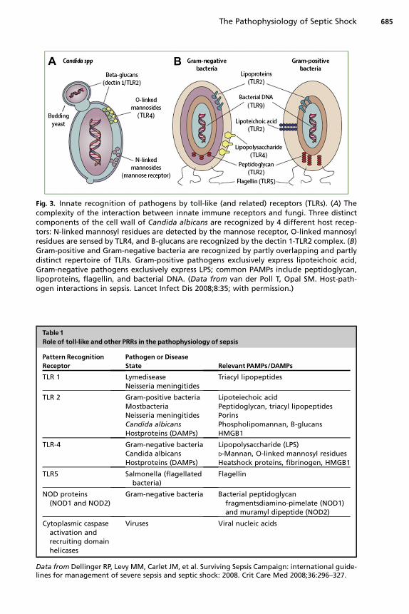
Fig. 3. Innate recognition of pathogens by toll-like (and related) receptors (TLRs). (A) Thecomplexity of the interaction between innate immune receptors and fungi. Three distinctcomponents of the cell wall of Candida albicans are recognized by 4 different host recep-tors: N-linked mannosyl residues are detected by the mannose receptor, O-linked mannosylresidues are sensed by TLR4, and B-glucans are recognized by the dectin 1-TLR2 complex. (B)Gram-positive and Gram-negative bacteria are recognized by partly overlapping and partlydistinct repertoire of TLRs. Gram-positive pathogens exclusively express lipoteichoic acid,Gram-negative pathogens exclusively express LPS; common PAMPs include peptidoglycan,lipoproteins, flagellin, and bacterial DNA. (Data from van der Poll T, Opal SM. Host-path-ogen interactions in sepsis. Lancet Infect Dis 2008;8:35; with permission.)
Table 1Role of toll-like and other PRRs in the pathophysiology of sepsis
Pattern RecognitionReceptor
Pathogen or DiseaseState Relevant PAMPs/DAMPs
TLR 1 LymediseaseNeisseria meningitides
Triacyl lipopeptides
TLR 2 Gram-positive bacteriaMostbacteriaNeisseria meningitidesCandida albicansHostproteins (DAMPs)
Lipoteiechoic acidPeptidoglycan, triacyl lipopeptidesPorinsPhospholipomannan, B-glucansHMGB1
TLR-4 Gram-negative bacteriaCandida albicansHostproteins (DAMPs)
Lipopolysaccharide (LPS)D-Mannan, O-linked mannosyl residuesHeatshock proteins, fibrinogen, HMGB1
TLR5 Salmonella (flagellatedbacteria)
Flagellin
NOD proteins(NOD1 and NOD2)
Gram-negative bacteria Bacterial peptidoglycanfragmentsdiamino-pimelate (NOD1)and muramyl dipeptide (NOD2)
Cytoplasmic caspaseactivation andrecruiting domainhelicases
Viruses Viral nucleic acids
Data from Dellinger RP, Levy MM, Carlet JM, et al. Surviving Sepsis Campaign: international guide-lines for management of severe sepsis and septic shock: 2008. Crit Care Med 2008;36:296–327.
The Pathophysiology of Septic Shock 685

Nduka & Parrillo686
identified in TLR4-CD14 have been linked to LPS hyporesponsiveness.21 Higher ratesof Gram-negative septic shock were noted in carriers of these SNPs in a medical inten-sive unit population.22 It is believed that these SNPs predispose affected individuals toendotoxin tolerance with inadequate early expression of proinflammatory cytokines.
Pro- and Anti-inflammatory Cellular Signaling/signal Transduction
Signal transduction describes the sequence of intracellular events in response to theengagement of ligands to their specific receptors (eg, bacterial LPS, cytokines) orchanges in the immediate extracellular environment. These molecular interactionstrigger the induction of specific cellular responses ranging from the expression ofspecific gene products (ie, protein production) to adhesion and chemotaxis.
Virtually all intracellular signaling pathways have, as their initial event, activation byphosphorylation of a target protein. Their target proteins could be transcription factorsor regulatory (cytoplasmic or nuclear) proteins. This activation is typically accom-plished by the binding of an enzyme (a kinase) to the target protein, and can leadto: (1) activation or alteration of its enzymatic activity; (2) changes in the stability ofthe target protein; (3) subcellular localization of the protein; and (4) interactions withother proteins. Many signaling pathways involve a cascade of 2 or more kinases inseries (signaling cascades), involving an upstream kinase involved in enzyme activa-tion and a downstream kinase whose substrate(s) are the protein products ofupstream kinase-substrate interaction. Kinase cascades are activated followingengagement of ligand-specific receptors (eg, Gram-negative LPS 1 TLR-4). In addi-tion, certain disruptions of cellular homeostasis (eg, changes in state of oxidation)can lead to activation of kinase cascades.
Kinases are able to phosphorylate other kinases, leading to signal amplification.Thus, a given stimulus may activate multiple kinase cascades, and several kinasecascades may be activated by different stimuli, leading to some measure of redun-dancy in a cellular signaling pathway. The natural consequence of amplification andredundancy in cellular signaling is a significant degree of overlap and lack of specificityin downstream effects and a requirement for intracellular regulation for effective, host-protective, stimulus-appropriate signal transduction. This negative regulation withresultant transcriptional downregulation is accomplished by dephosphorylation ofrelevant enzymes leading to a return to baseline levels of activity. Interactions occurbetween kinase cascades in such a way that increased activity of a given cascadeproduces suppression of activity in ‘‘opposite’’ cascades. This effect is termed crosstalk. In addition, efficient kinase-kinase interaction is facilitated by co-localization ofkinases on anchoring or adaptor proteins at relevant intracellular sites. An under-standing of the above aspects of cellular signaling is essential to understanding theinitial events that occur following host-pathogen interaction in sepsis.
Following the attachment of DAMPs/PAMPs to their specific ligands (TLRS) insevere sepsis, and the subsequent activation of signaling cascades (Fig. 4), there ismodification of the activity of key intracellular proteins, primarily transcription factorsand nuclear and cytoplasmic regulating proteins. For the TLRs, signaling dependsprimarily on 4 adaptor proteins: the myeloid differentiation primary-response protein88 (MyD88); and 3 non-MyD88 proteins: (1) toll/interleukin 1 (IL-1) receptor homology(TIR) domain-containing adaptor protein (TIRAP); (2) TIR domain-containing adaptorprotein–inducing interferon-b (TRIF); and (3) TRIF-related adaptor molecule (TRAM).These MyD88-dependent and MyD88-independent signal-transduction pathwaysresult in the activation of the prototypical transcription factor, nuclear factor-kB(NFkB). In addition, important enzyme systems regulating several key cellular

Fig. 4. Binding of toll-like receptors (TLRs) activates intracellular signal-transduction path-ways that lead to the activation of transcriptional activators such as interferon regulatorfactorsp13K/Akt, activator protein-1, and cytosolic nuclear factor-kappa[b] (NF-[k][b]). Acti-vated NF-[k][b] moves from the cytoplasm to the nucleus, binds to transcription sites andinduces activation of an array of genes for acute phase proteinsiNOS, coagulation factors,proinflammatory cytokinesand enzymatic activation of cellular proteases. TLR9 DNA, TLR3 dsRNA, and TLR7/8 ssRNA are endosomal. TLR10 ligand is not defined and TLR1 forms het-erodimers with TLR2. LPS, lipopolysaccharide; IRF, interferon regulatory factor; JNK; c-JunN- terminal kinase. (Adapted from Cinel I, Opal SM. Molecular biology of inflammationand sepsis: a primer. Crit Care Med 2009;37(1):291–304; with permission.)
The Pathophysiology of Septic Shock 687
functions and aspects of cellular signaling (the caspases, phosphoinositide 3-kinase[PI3K] and Rho GTPases) are activated.
NFkBNFkB is the prototypical transcription factor involved in modulating the expression ofmany of the inflammatory responses associated with severe sepsis and septic shock.It exists as homo- or heterogenous dimers composed of members of the Rel family ofproteins (P50, P105, P52, P100, P65 [Rel A], C-Rel). The Rel family of proteins playspivotal roles in inflammation, and various combinations of NFkB differ in their degreeof transcriptional activity (NFkB1-P50 1 P105, NFKB2-P52 1 P100, Rel A [P65]).
In the absence of cellular activation, NFkB exists in the cytoplasm maintained in alatent form by interacting with inhibitors of the IkB family (IkB-a, IkB-b, IkB-g, IkB-3,Bcl-3, p100, p105). MyD88-dependent and MyD88-independent kinase pathwaysactivate NFkB following TLR ligation by diverse stimuli, including bacterial products(LPS, peptidoglycans), cytokines (tumor necrosis factor-a [TNF-a], IL-8, IL-1b), reac-tive oxygen species, and changes in the cellular environment, such as ischemia.Regardless of the nature of the stimuli, activation occurs by phosphorylation of IkBmolecules followed by their degradation by the 26S proteosome. This step resultsin nuclear translocation of NFkB, its binding to specific gene promoter regions, andgene transcription. NFkB is involved in the induction of several predominantly proin-flammatory gene products (Table 2).
The degree of NFkB activation in septic patients seems to correlate with patientsurvival and outcome in septic shock.23–25 In addition to the pathophysiologic

Table 2NFkB-inducible genes involved in sepsis
Class NFkB-Dependent GenesAcute phase proteins C-reactive protein
LPS-binding protein
Cytokines TNF-aG-CSF, GM-CSFIL-1a, IL-1B, IL-2, IL-6, IL-12IFN-b
Chemokines MIP-1a
MIP-2
Coagulation factors Tissue factorTissue factor pathway inhibitor
Adhesion molecules ICAM-1VCAM-1E-selectinELAM-1
Enzymes Inducible nitric oxide synthaseCyclo-oxygenase-2C3 complementPhospholipase A2Matrix metalloproteinases
Immunoreceptors IL-2 receptor-aMajor histocompatibility complex class 1
Nduka & Parrillo688
consequences of excessive NFkB activation, inadequate stimulation can also lead toincreased morbidity in septic shock. Studies support the observation that defectiveNFkB signaling leads to immunosuppression in sepsis, favoring apoptosis in immuneeffector cells with undesirable consequences.26,27 Thus, excessive activation, espe-cially early in the disease course, or excessive negative regulation in sepsis mayproduce either excess inflammation with collateral host tissue damage or immunopar-alysis and consequent direct tissue damage.
The caspasesCaspases are a family of cysteine proteases synthesized as proenzymes and acti-vated by proteolysis. They are further subdivided into initiator caspases, activatedby autocleavage, and executioner caspases, activated by cleavage induced by theirinitiator counterparts. The caspases play important roles in the cellular processes ofinflammation and apoptosis following PAMP/DAMP-PRRs interaction. Followingcleavage, caspases produce many of the phenotypic changes seen in apoptosis,including cytoskeletal disintegration, DNA fragmentation, and disruption of cellularDNA repair molecular machinery. Although the TLRs are the most studied PRPs, thecytoplasmic NOD-like receptors (NLRs) are the most ubiquitous. Three members ofthe NLR family (NALP3, ICE protease-activating factor [IPAF], and apoptosis-associ-ated speck-like protein [ASC]) are involved in caspase activation. It is believed that,following pathogen recognition by TLRs, a signal is communicated intracellularlythat is recognized by the nucleotide-binding domains of the NLRs. This recognitionresults in the activation of a multiprotein complex termed an inflammasome. The in-flammasomes are multienzyme complexes (>70 KDa) that serve as molecular plat-forms for the activation of caspases 1 and 5, resulting in caspase-mediatedactivation and secretion of the proinflammatory cytokines IL-1b and IL-18. The signals

The Pathophysiology of Septic Shock 689
and mechanisms leading to inflammasome assembly/activation are in general stillpoorly understood. The inflammasome complexes assembled in sepsis are well char-acterized and consist of 2 different multiprotein complexes, the NALP1 and NALP3 in-flammasomes (Fig. 5). IL-1b is a highly potent proinflammatory cytokine, requiringinflammasome complex assembly as a prerequisite for caspase-1 activation beforeits precursor, pro-IL-1b (p35), released following TLR ligation, can be converted toits active form, which represents a mechanism to prevent uncontrolled expressionof IL-1b. In addition to the release of proinflammatory cytokines, the caspases targetthe enzyme, caspase-activated DNase (CAD). CAD activation induces DNA fragmen-tation leading to apoptosis. Cytoskeletal caspase targets include spectrin, nuclearlamin, and the enzyme gelosin, which cleaves actin.28,29 All these play roles in cyto-skeletal disintegration. Inflammation-mediated caspase activation contributes to thehost response in septic shock. Experiments in murine sepsis models show that thedeletion of caspase-1 genes prevents the development of sepsis in affected mice.30
Phosphoinositide 3-kinasesPhosphatidylinositol 3-kinases (PI3Ks) are a group of enzymes that, when activated,catalyze the production of membrane phosphatidylinositol triphosphate (PIP3). PI3K
Fig. 5. The NALP3 inflammasome complex. The NLAP3 inflammasome is composed of NALP3,ASC, and caspase-1 (a second adaptor protein CADD is present in NALP 3 but lacking inNALP). ASC interacts with 1 of the NALP proteins through Cognate pyrin domain (PYD)interactions and with procaspase-1 through homotypic caspase recruitment domain(CARD) interactions. The human inflammasome complex brings 2 molecules of procas-pase-1 (the second via CARDINAL) into close proximity, leading to autocatalysis and thesubsequent release of the active catalytic p20 and p10 domains of caspase-1. NALP3 bindsATP via the NACHT (nucleoside triphosphatase [NTPase] domain), is a precursor of IL-1Binto its biologically active fragment, and a potent mediator of fever and inflammation.There is no CARDINAL homolog in the mouse and, hence, murine NALP3 is believed torecruit only a single caspase-1 molecule. (TLRs, toll-like receptors; ATP, adenosine triphos-phate; NLRs, nucleotide-binding oligomerization domain (NOD)-like receptors; ASC,apoptosis-associated specklike protein containing a CARD; NALP, NACHT-, LRR-, andPYD-containing protein; LRRs, leucine-rich repeats). (From Trendelenburg G. Acute neurode-generation and the inflammasome: central processor for danger signals and the inflamma-tory response? J Cereb Blood Flow Metab 2008;28:867–81; with permission).

Nduka & Parrillo690
is activated by a variety of growth factor, hormonal, and chemokine receptors and,together with its downstream counterpart, the serine/threonine kinase Akt (proteinkinase B), regulates key aspects of cell proliferation and survival. The downstreamtargets of PI3K/Akt signaling include direct regulators of neutrophil functioning,including chemotaxis, adhesion, and apoptosis.26 Three isoforms of PI3Ks exist(PI3K-a, PI3K-b, and PI3K-y) with PI3K-y found exclusively in leukocytes. PI3K canfunction either as a positive or negative regulator of TLR signaling and, dependingon the cell type or specific TLR involved, activate either the NFKB or mitogen-acti-vated protein kinase (MAPK) signaling pathways.31 The NFKB signaling pathwayhas already been reviewed. The MAPK signaling pathway consists of 3 distinct path-ways described in leukocytes: p38, extracellular signal-regulated kinase (ERK), andc-jun NH2-terminal kinase (JNK). These are serine/threonine enzymes that act asthe final kinase in a 3-kinase cascade.
The p38 pathway is activated by a broad range of PAMPs (LPS, peptidoglycan)and inflammatory cytokines (tumor necrosis factor [TNF], IL-1, platelet-activatingfactor), and plays a role in neutrophil cytokine production, adhesion, chemotaxis,respiratory burst, and apoptosis.32,33 The ERK pathway is activated by several mito-gens (platelet-derived growth factor, insulin, epidermal growth factor, angiotensin II),and in monocyte LPS and cellular adherence. Its primary role is as a regulator ofcellular proliferation and differentiation, but it plays significant roles in cytokine(TNF-a) production and chemotaxis to C6a and IL-8. As a positive mediator ofTLR signaling, PI3K, together with p38 and ERK, mitogen-activated phosphoki-nases, leads to production of proinflammatory cytokines IL-1a, IL-6, and IL-8 onmicrobial challenge.34,35 The JNK pathway is activated by ligand receptor GTPases,cytokines (IL-1), and ultraviolet radiation. This pathway is important in cellular prolif-eration and apotosis.
In addition to enabling expression of the immune response, the PI3K/Akt signalingpathway acts as an endogenous negative feedback mechanism that serves to limitproinflammatory and apoptotic events, as seen in monocytes in response to endo-toxin.36 It can also promote the generation of anti-inflammatory cytokine IL-10,37
and helps balance Th1 versus Th2 responses.38
The ability of PI3K to modulate events in sepsis in a bidirectional fashion suggeststhat it could play a role in enhancing the efficacy of the innate immune response andlimiting excessive inflammation. This perspective is supported by recent experimentalevidence in which, following PI3K gene deletion, mice exposed to a pneumococcalvirulence factor developed more neutrophil-mediated alveolar injury despite inade-quate alveolar macrophage recruitment.39
Rho GTPasesCell surface ligand receptors consist of an extracellular ligand-binding domain con-nected by a single transmembrane region to an intracellular domain that possesseseither intrinsic enzyme activity or enzyme activation capabilities. The G-protein–linkedfamily of receptors are the most ubiquitous and are referred to as GTPases (GTPhydrolysis is required for receptor activation and signal transduction). The Rho andRac subfamilies of GTPases play a central role in the regulation of cell motility bycontrolling actin cytoskeleton rearrangement following the binding of specific prote-ases to the cell surface. Following DAMP-PRP interaction, Rho- and Rac-GTPaseshelp regulate the mechanical aspects of the cellular response by the innate immunesystem. These include cellular migration, pathogen uptake, phagocytosis, and main-tenance of endothelial integrity.40,41 Rac1 activation is required for PI3K activation onTLR stimulation.42

The Pathophysiology of Septic Shock 691
Release of Pro- and Anti-Inflammatory Mediators
One of the immediate consequences of cellular signaling in severe sepsis and septicshock is the synthesis and release of increased amounts of mediators into thesystemic circulation in an attempt to activate more immune effector cells and recruitthem to the site of infection. These highly potent molecules are normally present inthe circulation in low concentrations, but in high concentrations, or with prolongedexposure, they can exert potentially harmful biologic effects. Over-expression ofinflammatory mediators early in the course of sepsis plays a significant role in theeventual development of septic shock. This sequential release of mediators hasbeen termed the cytokine cascade. The availability of precise molecular tools, andthe ability to measure cytokine levels, has shed light on the patterns of cytokinerelease in sepsis, with the earliest cytokines, highly proinflammatory TNF-a and IL-1b, being responsible for the earliest clinical events in sepsis. The subsequent,and sometimes concomitant, release of counterinflammatory mediators has alsobeen observed. In addition, a clearer understanding of the source, structure, andactions of specific cytokines in the development of septic shock has emerged(Table 3), and novel mediators have been discovered. Selected novel cytokine medi-ators and new ideas regarding the role of otherwise well-known mediators are re-viewed below.
High-mobility group box 1 proteinHigh-mobility group box 1 protein (HMGB1) is a nuclear and cytoplasmic protein origi-nally discovered 3 decades ago as a nuclear binding protein, and was so namedbecause of its rapid mobility on electrophoresis gels. HMGB1 is amongst the most ubiq-uitous, abundant proteins in eukaryotes, and plays a major role in facilitating gene tran-scription by stabilizing nucleosome formation. More recent findings suggest thatHMGB1 is active in DNA recombination, repair, replication, and gene transcription, facil-itated by internal repeats of positively charged domains of the N terminus (HMG boxes).HMGB1 is released passively by necrotic (but not apoptotic) cells, and from macro-phages, dendritic cells, and natural killer cells, on activation by microbial pathogens.
The known biologic effects of HMGB1 are based on data obtained from cellcultures. HMGB1 stimulates the release of proinflammatory cytokines, includingTNF and IL-8, in macrophages/monocytes and endothelial cells. HMGB1 can alsobind to and induce the expression of the cellular receptor for advanced glycationend products (RAGE) and adhesion molecules (vascular cell adhesion molecule-1[VCAM-1], intercellular adhesion molecule-1 [ICAM-1]) in human endothelial cells.The induced expression of RAGE facilitates activation of the transcription factorNFKB and MAPKs. These observations suggest a role for HMGB1 as a proinflamma-tory cytokine, with significant adverse effects on gut barrier function, and as a regulatorof the coagulation system.
At present, HMGB1 does not seem to contribute significantly to the development ofseptic shock.
Macrophage migration inhibitory factorOriginally described as a T cell product, macrophage migration inhibitory factor(MMIF) is a cytokine produced by various cell types including other immune effectorand neuroendocrine cells. MMIF is capable of activating T cells and inducing proin-flammatory cytokine production in macrophages. Serum MMIF concentrations areincreased in septic patients and, in severe sepsis, elevated MMIF concentrationsseem to be an early indicator of poor outcome of septic patients in intensive care.
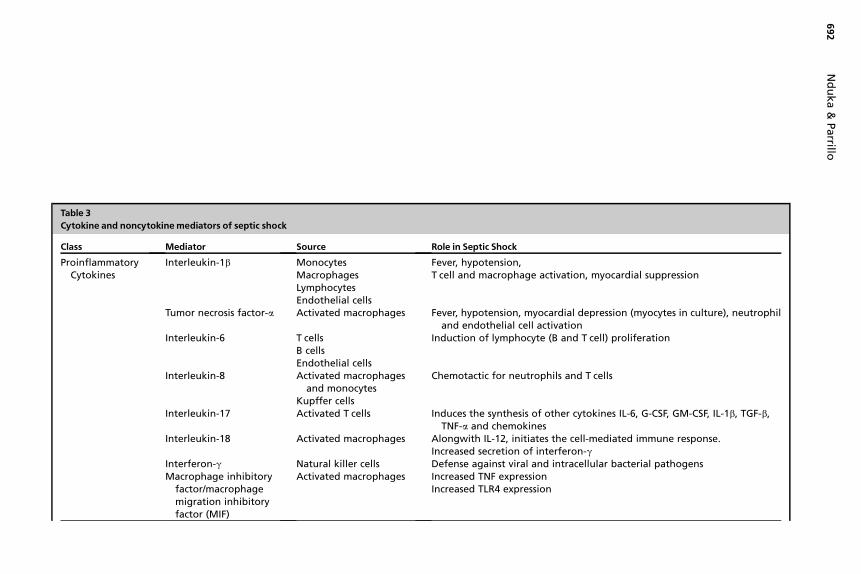
Table 3Cytokine and noncytokinemediators of septic shock
Class Mediator Source Role in Septic ShockProinflammatory
CytokinesInterleukin-1b Monocytes
MacrophagesLymphocytesEndothelial cells
Fever, hypotension,T cell and macropha e activation, myocardial suppression
Tumor necrosis factor-a Activated macrophages Fever, hypotension, yocardial depression (myocytes in culture), neutrophiland endothelial c ll activation
Interleukin-6 T cellsB cellsEndothelial cells
Induction of lymph yte (B and T cell) proliferation
Interleukin-8 Activated macrophagesand monocytes
Kupffer cells
Chemotactic for ne rophils and T cells
Interleukin-17 Activated T cells Induces the synthes of other cytokines IL-6, G-CSF, GM-CSF, IL-1b, TGF-b,TNF-a and chemo ines
Interleukin-18 Activated macrophages Alongwith IL-12, in ates the cell-mediated immune response.Increased secretion f interferon-g
Interferon-g Natural killer cells Defense against vir and intracellular bacterial pathogensMacrophage inhibitory
factor/macrophagemigration inhibitoryfactor (MIF)
Activated macrophages Increased TNF expre sionIncreased TLR4 expr ssion
Nd
uka
&Parrillo
692
g
meoc
ut
iskitio
alse

Anti-inflammatorycytokines
Interleukin-10 Epithelial cellsMonocytesLymphocytes
Downregulation of macrophage function leading to decreased TNF-a
IL-4 ? Induces differentiation of naive helper T cells to Th2 cellsIL-1Ra Monocytes Block IL-1 activityTGF-B Various host cells Interferes with phagocytic activation
Endothelial Factors Nitric oxide Increased microvascular permeabilityLossof vasomotor toneMyocardial depressionPeripheral venous pooling
Hormones VasopressinGlucocorticoids
Posterior pituitary glandHypothamic-pituitary axis
Relative deficiency may cause or worsen circulatory failure
Arachidonic acidmetabolites
ProstaglandinsLeukotrienesThromboxanes
Immune effector cellsPancreas
Airway reactivityVasoconstrictionPlatelet aggregationIncreased vascular permeability
Others Platelet-activating factor Endothelial cellsMacrophagesNeutrophils
Histamine release from plateletsActivation of endothelial cells
Complement proteinsC3a-C5a
Histamine release,Increased capillary permeability, vasodilation
Myocardial depressantfactors
Pancreas Negative inotropyImpaired phagocytosis
Th
ePath
op
hysio
log
yo
fSe
ptic
Sho
ck693

Nduka & Parrillo694
Based on our present understanding, MMIF does not seem to play a significant role inthe development of septic shock.
Immune and Nonimmune Effector Cell Dysfunction
The normal immune effector response in response to cytokine release is lost in severesepsis. The dysfunctions can involve every cell type from antigen-presenting cells(dendritic cells) to neutrophils and macrophages. The nature of cellular dysfunctionis similar to the associated cytokine homeostatic imbalance with elements ofincreased cellular activity and cellular hyporesponsiveness.
Neutrophil dysfunction in severe sepsisNeutrophils are key cells in the innate immune system and act primarily by recognizingand destroying pathogens by a coordinated series of steps including adhesion,chemotaxis, phagocytosis, and the release of cytotoxic molecules, followed byapoptotic cell death with the neutrophils spending approximately 7 hours in thesystemic circulation. In severe sepsis the tight regulation of neutrophil function islost, leading to excessive neutrophil activation and prolonged survival. These acti-vated neutrophils induce endothelial dysfunction, release cytotoxic molecules, andlead to inflammatory host organ injury.
Accelerated lymphocytic apoptosisThe T-helper lymphocytes play key roles in the adaptive immune response followingactivation by antigen-presenting cells of the innate immune system. Following activa-tion, the initial lymphocyte response is proinflammatory, with the emergence of a regu-latory phenotype after several days. Severe sepsis is characterized by acceleratedapoptotic death of lymphocytes leading to lymphocyte depletion and loss of the Tlymphocyte regulatory function.43
In addition to immune effector dysfunction, there is widespread nonimmune cellulardysfunction in severe sepsis. The alterations relevant to the development of cardiocir-culatory failure in the setting of severe sepsis are discussed in a later section.
Interactions with Other Biologic Systems in Severe Sepsis
The coagulation cascade in severe sepsisAs much as 4 decades ago, it was apparent that the coagulation system was abnor-mally activated in septic patients,44–46 and, by 1970, Dr James Corrigan47 had pub-lished on the potential use of anticoagulant therapy (heparin) to treat disorderedcoagulation in sepsis. Almost all patients with septic shock have coagulation abnor-malities. The nature and degree of coagulation dysfunction ranges from clinically silentbiochemical evidence of dysfunction to full-blown disseminated intravascular coagu-lation (DIC). An understanding of the hemostatic system, including the blood coagula-tion pathways and the natural anticoagulant pathways, is necessary for anymeaningful review of sepsis-induced coagulation dysfunction.
Although the classic model of coagulation (extrinsic and intrinsic pathways) maybe helpful in the interpretation of commonly used laboratory tests of coagulationdisorders, it does not represent a clinically relevant model of hemostasis in phys-iologic and, more importantly, pathophysiologic states such as sepsis. Currentunderstanding of hemostasis describes the coagulation pathway as a 3-phaseprocess (initiation, amplification, and thrombin action), with considerable overlapbetween the phases. These phases are counterbalanced by active natural antico-agulant systems targeting key steps in the coagulation cascade. The result isa hemostatic system that begins to form a clot less than 30 seconds following
The Pathophysiology of Septic Shock 695
vascular injury, with the process of thrombolysis initiated as soon as a thrombus isformed.
Initiation occurs shortly after vascular injury and is the result of increased tissuefactor (TF) expression by TF-bearing adventitial cells and platelets. Limited amountsof TF bind to and activate factor VII (FVII). The TF-FVIIa complex leads to productionof a limited amount of thrombin.
Amplification consists of thrombin activating platelets and other coagulation cofac-tors during the amplification phase. There is release of significant amounts ofprothrombin (factor X) and, together with platelets and the other coagulation factors,a prothrombinase complex (factor Xa and coagulation factors bound to activatedplatelets) is formed and is primarily responsible for the burst of thrombin productionleading to the third phase of clot formation.
The final phase of coagulation involves thrombin-dependent recruitment of addi-tional coagulation factors (FV and FXIII) into the coagulation process, maintainingplatelet activation and facilitating the conversion of fibrinogen to fibrin.
The natural anticoagulant systems serve to prevent clot formation in the absence ofvascular injury and prompt lysis of clots formed following vascular injury.
The balance between procoagulant and anticoagulant arms of the hemostaticprocess is disrupted in severe sepsis. The inflammatory response to severe infectionresults in a systemic dysfunction of the coagulation system. The events that constitutecoagulation dysfunction in septic shock can be divided into an initial activation, fol-lowed by a largely dysregulated response with suppression of the antifibrinolyticsystems. Cytokines released as part of the inflammatory response mediate many ofthe hypercoagulable responses triggered in severe sepsis and septic shock, andthe available evidence suggests contributory roles for immune effector cells, endothe-lial dysfunction, and metabolic alterations in the tissue. It is now understood that, insepsis, the interaction between the coagulation and inflammatory systems is bidirec-tional (Fig. 6). Binding of coagulation proteases (thrombin or TF) or anticoagulantproteins (activated protein C [APC]) to specific cell receptors on mononuclear cellsor endothelial cells may affect cytokine production or inflammatory cell apoptosis.Endothelial cells respond to the cytokines expressed and released by activated leuko-cytes, but they can also release cytokines themselves. Endothelial cells are able toexpress adhesion molecules and growth factors that promote the inflammatoryresponse and also affect the coagulation response. Endothelial cells play a prominentrole in all 3 major pathogenetic pathways associated with coagulopathy in sepsis: TF-mediated thrombin generation, dysfunctional anticoagulant pathways, and inhibitionof fibrinolysis.
APC acts in conjunction with the cofactor protein S to deactivate clotting factors Vaand VIIIa, preventing ongoing thrombin generation by the prothrombinase complex.APC may also inhibit inflammation by inhibiting cytokine production, preventing neutro-phil activation, and inhibiting leukocyte adhesion and rolling. Other key mediators in thehypercoagulable cascade in sepsis include antithrombin and TF pathway inhibitor.
This complex cross talk between the inflammatory and coagulation cascades repre-sents a vicious cycle which, if uninterrupted, results in tissue injury, organ dysfunction,and cellular death.
Neural regulation of the immunoinflammatory response: the vagal inflammatoryreflex and sympathetic effects on the host responseIn addition to the humoral mechanisms that act to prevent tissue injury fromexcessive release of proinflammatory mediators, emerging research suggestsa role for neural modulation of inflammation. One of the earliest documented

Fig. 6. Schematic overview of the major pathways involved in the interrelation betweencoagulation, anticoagulant pathways, and fibrinolysis and inflammation. 1, stimulatoryeffect; –, inhibitory effect; IL, interleukin; PAI-1, plasminogen activator inhibitor-1; TAFI,thrombin activatable fibrinolysis inhibitor; TNF, tumor necrosis factor; u-PA, urinary plasmin-ogen activator; u-PAR, urokirlase-type plasminogen activator receptor. (From Levi M, vander Poll T. The central role of the endothelium in the crosstalk between coagulation andinflammation in sepsis. Adv Sepsis 2004;3:93; with permission).
Nduka & Parrillo696
observations supporting the existence of central autonomic interaction with the im-munoinflammatory response involved a serendipitous finding that, with centraladministration of a TNF inhibitor, efferent vagal activity was stimulated withsystemic anti-inflammatory action.48
Subsequent work in animal sepsis models demonstrated significant inhibition ofTNF expression following vagal stimulation and improved disease end points in thesemodels.49,50 In addition, rendering these animals immune to vagal stimulation either bygenetic manipulation or vagotomy led to an exaggerated proinflammatory cytokineresponse.51 It is now understood that these cytokine-suppressive effects of vagalstimulation are mediated by the release of its neurotransmitter acetylcholine (ACh)and its subsequent interaction with ACh receptors expressed by macrophages andother immune effector cells.52,53 The best characterized of these cholinergic receptorsthat suppress cytokines is the a7 subunit of the nicotinic acetylcholine receptor (a7nAChR).
This autonomic parasympathetic-mediated immune modulation system has beentermed the inflammatory reflex (Fig. 7), with an immunosensing afferent arm (cytokinestimulation of vagal afferents) and an efferent immunosuppressing arm (the cholinergicanti-inflammatory pathway).54 Recent studies suggest that the spleen plays a signifi-cant role as an effector organ for this pathway.

Fig. 7. Wiring of the inflammatory reflex. Inflammatory products produced in damagedtissues activate afferent signals that are relayed to the nucleus tractus solitarius; subsequentactivation of vagus efferent activity inhibits cytokine synthesis through the cholinergic anti-inflammatory pathway (‘the inflammatory reflex’). Information can also be relayed to thehypothalamus and the dorsal vagal complex to stimulate the release of ACTH, thereby acti-vating the humoral anti-inflammatory pathway. Activation of the sympathetic outflow byflight-or-fight responses or pain, or through direct signalling, can increase local concentra-tions of adrenaline and noradrenaline, which can suppress inflammation further. (Data fromTracey KJ. The inflammatory reflex. Nature 2002;420:857).
The Pathophysiology of Septic Shock 697
The sympathetic ANS consists of sympathetic neurons and the adrenal medulla,with catecholamines as the neurotransmitter. In addition to the adrenal medulla andsympathetic neurons, immune effector cells are also a source of catecholamines insevere sepsis.55 Early uncomplicated sepsis is characterized by high circulating cate-cholamine levels with significant metabolic (catabolic state), immunomodulaory(excessive inflammation), and cardiocirculatory (increased cardiac output) conse-quences. Prolonged elevation of circulating catecholamines is toxic to host cells, pre-disposing the patient to cardiocirculatory failure with hypotension resulting fromperipheral vasodilatation and compromised myocardial contractility.56 Septic shockis more often characterized by depletion of endogenous catecholamine stores and,possibly, catecholamine resistance.
Nduka & Parrillo698
CARDIOCIRCULATORY DYSFUNCTION IN SEVERE SEPSIS RESULTING IN PROGRESSIONTO SEPTIC SHOCK
The widespread disruptions in severe sepsis can result in cardio-circulatory dysfunc-tion manifesting as shock. The dysfunction involves the cardiac, peripheral vascular(macrovascular) and microcirculatory elements of the circulation and, depending onthe degrees of cardiac or vascular dysfunction and the volume status of the patient,a clinical picture ranging from cold, clammy and under-perfused to one of hyperdy-namic shock, may be seen, although, in clinical medicine, hyperdynamic shock isseen much more frequently. The situation in septic shock is further complicated bywidespread microcirculatory dysfunction, further impairing tissue oxygen delivery,and diminished mitochondrial activity resulting in impaired oxygen extraction. A reviewof characteristics and pathogenetic mechanisms that underlie cardiac and macrovas-cular dysfunction in septic shock follows. The microcirculatory alterations are dis-cussed elsewhere in this issue.
Over 5 decades, multiple methods of myocardial function assessment have beenused to study ventricular performance in severe sepsis and septic shock.57–60 Theresults have been largely similar and the characteristic pattern of cardiac performanceduring septic shock has been proved to be one of reduced left and right ventricularejection fractions, increased end-diastolic and end-systolic volumes of both ventri-cles, and normal stroke volume; heart rate and cardiac output are elevated, andsystemic vascular resistance is reduced. The reduction in the ejection fraction andthe biventricular dilatation occur 24 to 48 hours after the onset of sepsis and, likemost other organs affected by the septic process, it is reversible with restoration ofmyocardial function if patients survive up to 10 days after their onset. An inability tomaintain cardiac output during this critical period is associated with a poor outcome,61
and ventricular dilatation allows for an increased end-diastolic volume and helps main-tain cardiac output. Thus, the decrease in ejection fraction with ventricular dilatation inseptic shock may be an appropriate adaptive response to myocardial dysfunction.
Myocardial depression results from the direct or indirect effects of 1 or more circu-lating myocardial depressant substances. In experiments, ultrafiltrates from patientswith severe sepsis show cardiotoxic effects.62 Several of the cytokines released insevere sepsis probably contribute to this dysfunction.
TNF-a and IL-1
TNF-a and IL-1 are associated with myocyte dysfunction and may explain the earlymyocardial depression seen in sepsis. In one series of studies using in vitro myocardialcells and human septic shock serum, TNF and IL-1 were shown to be responsible forthe myocardial depressant activity present in human sera.63 These cytokines arepotent inducible nitric oxide synthase (iNOS) inducers, and this probably representsone of the direct mechanisms for myocardial depression.
Nitric Oxide
Severe sepsis is associated with increased expression of iNOS and increased nitricoxide production. NO interferes with myocyte calcium metabolism and may impaircontractile function. In addition, reactive nitrogen species such as peroxynitrite,produced by NO interacting with superoxide ions, are directly toxic to myocytes.Experimental observations support a role for NO-mediated myocardial depressionin sepsis. Cardiac function was preserved following LPS challenge in iNOS-deficientmice.64
The Pathophysiology of Septic Shock 699
Vascular Dysfunction
Vascular alterations in septic shock are mainly due to the effects of mediators onvascular smooth muscle and endothelial dysfunction.
The NO released seems to be primarily responsible for vascular smooth muscledysfunction in sepsis. NO causes a hyperpolarization of smooth muscle plasmamembranes, rendering them unresponsive to catecholamines and causing a vasodila-tory state. In addition to the above, these patients may have relative vasopressin orcortisol deficiencies, leading to further catecholamine unresponsiveness and refrac-tory shock.
Endothelial dysfunction leads to an inability of the endothelial cells to maintainvascular tone with loss of blood pressure. In addition, endothelial damage leads tocapillary leak with intravascular volume depletion and edema formation in involvedorgans.
SUMMARY
Septic shock remains a significant challenge for clinicians. Recent advances in cellularand molecular biology have significantly improved our understanding of its pathoge-netic mechanisms. These improvements in understanding should translate to bettercare and improved outcomes for these patients.
REFERENCES
1. Geroulanos S, Douka ET. Historical perspective of the word ‘‘sepsis.’’ IntensiveCare Med 2006;32:2077.
2. Vincent JL, Abraham E. The last 100 years of sepsis. Am J Respir Crit Care Med2006;173:256–63.
3. Schottmueller H. Wesen und Behandlung der Sepsis [The nature and therapy ofsepsis]. Inn Med 1914;31:257–80 [in German].
4. Cannon WB. Traumatic shock. New York: D Appleton and Co; 1923.5. Kumar A, Parrillo J. Shock: classification, pathophysiology, and approach to
management. In: Parillo JE, Dellinger RP, editors. Critical care medicine: princi-ples of diagnosis and management in the adult. 3rd edition. Philadelphia: MosbyElsevier; 2008. p. 377–422.
6. Campisi Laura, Brau Fr�ed�eric, Glaichenhaus Nicolas. Imaging host–pathogeninteractions. Immunol Rev 2008;221(1):188–99.
7. Bassler BL. Small talk. Cell-to-cell communication in bacteria. Cell 2002;109:421–4.
8. Taga ME, Bassler BL. Chemical communication among bacteria. Proc Natl AcadSci U S A 2003;100(Suppl 2):14549–54.
9. Davies DG, Parsek MR, Pearson JP, et al. The involvement of cell-to-cell signals inthe development of a bacterial biofilm. Science 1998;280:295–8.
10. Shiner EK, Rumbaugh KP, Williams SC. Inter-kingdom signaling: deciphering thelanguage of acyl homoserine lactones. FEMS Microbiol Rev 2005;29:935–47.
11. Hooi DS, Bycroft BW, Chhabra SR, et al. Differential immune modulatory activityof Pseudomonas aeruginosa quorum-sensing signal molecules. Infect Immun2004;72:6463–70.
12. Miller MB, Bassler BL. Quorum sensing in bacteria. Annu Rev Microbiol 2001;55:165–99.

Nduka & Parrillo700
13. Whiteley M, Lee KM, Greenberg EP. Identification of genes controlled by quorumsensing in Pseudomonas aeruginosa. Proc Natl Acad Sci U S A 1999;96:13904–9.
14. Cinel I, Dellinger RP. Advances in pathogenesis and management of sepsis. CurrOpin Infect Dis 2007;20:345–52.
15. van der Poll T, Opal SM. Host-pathogen interactions in sepsis. Lancet Infect Dis2008;8:32–43.
16. Creagh EM, O’Neill LA. TLRs, NLRs and RLRs: a trinity of pathogen sensors thatco-operate in innate immunity. Trends Immunol 2006;27:352–7.
17. Granucci F, Foti M, Ricciardi-Castagnoli P. Dendritic cell biology. Adv Immunol2005;88:193–233.
18. Uematsu S, Akira S. Toll-like receptors and innate immunity. J Mol Med 2007;84:712–25.
19. Mollen KP, Anand RJ, Tsung A, et al. Emerging paradigm: toll-like receptor4-sentinel for the detection of tissue damage. Shock 2006;26:430–7.
20. Paterson HM, Murphy TJ, Purcell EJ, et al. Injury primes the innate immunesystem for enhanced toll-like receptor reactivity. J Immunol 2003;171:1473–83.
21. Arcaroli J, Fessler MB, Abraham E. Genetic polymorphisms and sepsis. Shock2005;24:300–12.
22. Gibot S, Cariou A, Drouet L, et al. Association between a genomic polymorphismwithin the CD14 locus and septic shock susceptibility and mortality rate. Crit CareMed 2002;30:969–73.
23. Arcaroli J, Silva E, Maloney JP, et al. Variant IRAK-1 haplotype is associated withincreased nuclear factor-kappaB activation and worse outcomes in sepsis. Am JRespir Crit Care Med 2006;173:1335–41.
24. Zaph C, Troy AE, Taylor BC, et al. Epithelial cell-intrinsic IKK-beta expressionregulates intestinal immune homeostasis. Nature 2007;446:552–6.
25. Liu YJ, Soumelis V, Watanabe N, et al. TSLP: an epithelial cell cytokine thatregulates T cell differentiation by conditioning dendritic cell maturation. AnnuRev Immunol 2007;25:193–219.
26. Peck-Palmer OM, Unsinger J, Chang KC, et al. Deletion of MyD88 markedlyattenuates sepsis-induced T and B lymphocyte apoptosis but worsens survival.J Leukoc Biol 2008;83:1009–18.
27. Adib-Conquy M, Moine P, Asehnoune K, et al. Toll-like receptor-mediated tumornecrosis factor and interleukin-10 production differ during systemic inflammation.Am J Respir Crit Care Med 2003;168:158–64.
28. Ayscough KR, Gourlay CW. The actin cytoskeleton: a key regulator of apoptosisand aging. Nat Rev Mol Cell Biol 2005;6:583–9.
29. Fischer U, Janicke RU, Schulze-Osthoff K. Many cuts to ruin: a comprehensiveupdate of caspase substrates. Cell Death Differ 2003;10(1):76–100.
30. Sarkar A, Hall MW, Exline M, et al. Caspase-1 regulates E. coli sepsis and splenicB cell apoptosis independently of IL-1[beta] and IL-18. Am J Respir Crit CareMed 2006;174:1003–10.
31. Cantley LC. The phosphoinositide 3-kinase pathway. Science 2002;296:1655–7.32. Nick JA, Young SK, Arndt PG, et al. Selective suppression of neutrophil accumu-
lation in ongoing pulmonary inflammation by systemic inhibition of p38 MAPkinase. J Immunol 2002;169(9):5260–9.
33. Yum HK, Arcaroli J, Kupfner J, et al. Involvement of phosphoinositide 3-kinases inneutrophil activation and the development of acute lung injury. J Immunol 2001;167(11):6601–8.
The Pathophysiology of Septic Shock 701
34. Ojaniemi M, Glumoff V, Harju K, et al. Phosphatidylinositol kinase is involved inToll-like receptor 4-mediated cytokine expression in mouse macrophages. EurJ Immunol 2003;335:597–605.
35. Guillot L, Le Goffic R, Bloch S, et al. Involvement of toll-like receptor 3 in theimmune response of lung epithelial cells to double-stranded RNA and influenzaA virus. J Biol Chem 2005;280:5571–80.
36. Guha M, Mackman N. The PI3K-Akt pathway limits LPS activation of signalingpathways and expression of inflammatory mediators in human monocytic cells.J Biol Chem 2002;277:32124–32.
37. Pengal RA, Ganesan LP, Wei G, et al. Lipopolysaccharide-induced production ofinterleukin-10 is promoted by the serin threonine kinase Akt. Mol Immunol 2006;43:1557–64.
38. Fukao T, Koyasu S. PI3K and negative regulation of TLR signaling. Trends Immu-nol 2003;24:358–63.
39. Maus UA, Backi M, Winter C, et al. Importance of phosphoinositide 3-kinasegamma in the host defense against pneumococcal infection. Am J Respir CritCare Med 2007;175(9):958–66.
40. Hall A, Rho GT. Pases and the actin cytoskeleton. Science 1998;279:509–14.41. Ruse M, Knaus UG. New players in TLR-mediated innate immunity. Immunol Res
2006;34:33–48.42. Arbibe L, Mira JP, Teusch N, et al. Toll-like receptor 2-mediated NF-kappa B acti-
vation requires a Racl dependent pathway. Nat Immunol 2000;1:533–54.43. Hotchkiss RS, Karl IE. The pathophysiology and treatment of sepsis. N Engl J
Med 2003;348:138–50.44. Rapaport SI, Tatter D, Coeur-Baron N. Pseudomonas septicemia with intravas-
cular clotting leading to the generalized Shwartzman reaction. N Engl J Med1964;271:80.
45. Corrigan JJ, Ray WL, May N. Changes in the blood coagulation system associ-ated with septicemia. N Engl J Med 1968;279:851–6.
46. Beller FK. The role of endotoxin in disseminated intravascular coagulation.Thromb Diath Haemorrh Suppl 1969;36:125–49.
47. Corrigan JJ, Jordan CM. Heparin therapy in septicemia with disseminated intra-vascular coagulation. N Engl J Med Shock 1970;283:778–82.
48. Bernik TR, Friedman SG, Ochani M, et al. Pharmacological stimulation of thecholinergic antiinflammatory pathway. J Exp Med 2002;195:781–8.
49. Borovikova LV, Ivanova S, Zhang M, et al. Vagus nerve stimulation attenuates thesystemic inflammatory response to endotoxin. Nature 2000;405:458–62.
50. Altavilla D, Guarini S, Bitto A, et al. Activation of the cholinergic anti-inflammatorypathway reduces NF-kappab activation, blunts TNF-alpha production, andprotects against splanchic artery occlusion shock. Shock 2006;25:500–6.
51. Huston JM, Ochani M, Rosas-Ballina M, et al. Splenectomy inactivates the cholin-ergic antiinflammatory pathway during lethal endotoxemia and polymicrobialsepsis. J Exp Med 2006;203:1623–8.
52. Wang H, Yu M, Ochani M, et al. Nicotinic acetylcholine receptor alpha7 subunit isan essential regulator of inflammation. Nature 2003;421:384–8.
53. Wang H, Liao H, Ochani M, et al. Cholinergic agonists inhibit HMGB1 release andimprove survival in experimental sepsis. Nat Med 2004;10:1216–21.
54. Tracey KJ. Physiology and immunology of the cholinergic antiinflammatorypathway. J Clin Invest 2007;117:289–96.
55. Flierl MA, Rittirsch D, Nadeau BA, et al. Phagocyte-derived catecholaminesenhance acute inflammatory injury. Nature 2007;449:721–5.

Nduka & Parrillo702
56. Annane D, de la Grandmaison G, Brouland JP, et al. Inappropriate sympatheticactivation at onset of septic shock: a spectral analysis approach. Am J RespirCrit Care Med 1999;160:458–65.
57. Calvin JE, Driedger AA, Sibbald WJ. An assessment of myocardial function inhuman sepsis utilizing ECG gated cardiac scintigraphy. Chest 1981;80:579–86.
58. Parker MM, McCarthy KE, Ognibene FP, et al. Right ventricular dysfunction anddilatation, similar to left ventricular changes, characterize the cardiac depressionof septic shock in humans. Chest 1990;97:126–31.
59. Munt B, Jue J, Gin K, et al. Diastolic filling in human severe sepsis: an echocar-diographic study. Crit Care Med 1998;26:1829–33.
60. Poelaert J, Declerck C, Vogelaers D, et al. Left ventricular systolic and diastolicfunction in septic shock. Intensive Care Med 1997;23:553–60.
61. Parker MM, Shelhamer JH, Natanson C, et al. Serial cardiovascular variables insurvivors and nonsurvivors of human septic shock: heart rate as an earlypredictor of prognosis. Crit Care Med 1987;15:923–9.
62. Parrillo JE, Burch C, Shelhamer JH, et al. A circulating myocardial depressantsubstance in humans with septic shock: septic shock patients with a reducedejection fraction have a circulating factor that depresses in vitro myocardial cellperformance. J Clin Invest 1985;76:1539–53.
63. Kumar A, Thota V, Dee L, et al. Tumor necrosis factor-alpha and interleukin-1 betaare responsible for depression of in vitro myocardial cell contractility induced byserum from humans with septic shock. J Exp Med 1996;183:949–58.
64. Ullrich R, Scherrer-Crosbie M, Bloch KD, et al. Congenital deficiency of nitricoxide synthase 2 protects against endotoxin-induced myocardial dysfunction inmice. Circulation 2000;102:1440–6.


![Septic Shock [EDocFind.com]](https://img.dokumen.tips/doc/110x75/55cf8fb1550346703b9edc7d/septic-shock-edocfindcom.jpg)


