Embed Size (px)
DESCRIPTION
patofisiologi
Citation preview

Review
J. Clin. Biochem. Nutr. | March 2011 | vol. 48 | no. 2 | 107–111doi: 10.3164/jcbn.10�79©2011 JCBN
JCBNJournal of Clinical Biochemistry and Nutrition0912-00091880-5086the Society for Free Radical Research JapanKyoto, Japanjcbn10-7910.3164/jcbn.10-79ReviewThe pathophysiology of non�steroidal anti�inflammatory drug (NSAID)�induced mucosal injuries in stomach and small intestineHirofumi Matsui,* Osamu Shimokawa, Tsuyoshi Kaneko, Yumiko Nagano, Kanho Rai and Ichinosuke Hyodo
Graduate School of Comprehensive Human Sciences, University of Tsukuba, 1�1�1 Tennodai, Tsukuba, Ibaraki 305�8575, Japan
*To whom correspondence should be addressed. E�mail: [email protected]
3(Received 1 July, 2010; Accepted 13 July, 2010; Published online 26 February, 2011)
Copyright © 2011 JCBN2011This is an open access article distributed under the terms of theCreative Commons Attribution License, which permits unre-stricted use, distribution, and reproduction in any medium, pro-vided the original work is properly cited.Non�steroidal anti�inflammatory drugs are the most commonly
prescribed drugs for arthritis, inflammation, and cardiovascular
protection. However, they cause gastrointestinal complications.
The pathophysiology of these complications has mostly been
ascribed to non�steroidal anti�inflammatory drugs’ action on the
cyclooxygenase inhibition and the subsequent prostaglandin defi�
ciency. However, recent clinical demonstrated the prevalence of
non�steroidal anti�inflammatory drugs�induced small intestinal
mucosal injury is more often than previously expected. In this
review, we discuss the defense mechanisms of stomach, and the
pathophysiology of non�steroidal anti�inflammatory drugs�induced
injury of stomach and small intestine, especially focused on non�
steroidal anti�inflammatory drugs’ action on mitochondria.
Key Words: Non�steroidal anti�inflammatory drugs,
gastrointestinal mucosal injury, mitochondria,
lipid peroxidation, reactive oxygen species
IntroductionNon-steroidal anti-inflammatory drugs (NSAIDs) such asaspirin and indomethacin are the most commonly prescribed
drugs for arthritis, inflammation, and cardiovascular protection.However, they cause gastrointestinal complications such as ulcersand erosions. The pathophysiology of these complications hasmostly been ascribed to NSAID’s action on the cyclooxygenase(COX) inhibition and the subsequent prostaglandin (PG) defi-ciency.(1) In 1970s and 1980s, under the concept of cytoprotection,extensive researches have revealed the role of PG in gastricmucosal defense system.(1)
Recent clinical researches shed a light on the NSAID-inducedsmall intestinal mucosal injury. Capsule endoscopic examinationsrevealed that NSAID induced mucosal damages including erosionsand ulcerations in small intestines more often than previouslyexpected.(2) Although gastric mucosal injuries are considered tobe associated with the gastric acid, these results suggested thatNSAIDs induced mucosal injuries independently with the acid,since there is no acid-secreting cell in small intestines. Therefore,the treatment for NSAID-induced small intestinal mucosal injuriesshould include medications other than conventional acid-reducingagents such as histamine 2 receptor antagonists (H2RAs) andproton pump inhibitors (PPIs).(3–6) The pathophysiological conceptthat will explain the NSAID-induced injuries of both stomach andsmall intestines is needed.
In this review, we discuss the defense mechanisms of stomach,and the pathophysiology of NSAID-induced injury of stomach andsmall intestine.
Gastric Mucosal Defense Mechanisms
Gastric mucosal injury occurs when the causative agents such
as gastric acid and NSAIDs overwhelm the mucosal defense. Thegastric defense mechanisms can be divided into three majorcomponents: pre-epithelial, epithelial, and post-epithelial defensemechanism (Table 1).(7,8)
The pre-epithelial defense mechanism consists mainly of amucous layer that contains mucous, bicarbonate and surface activephospholipids, and prevents epithelial cells from the contactwith luminal noxious agents such as gastric acid. Mucous andbicarbonate are secreted by gastric epithelial cells. As a result, thepH at the surface of gastric mucosal epithelial cells normally ismaintained in the neutral range when the pH at the gastric luminalsurface reaches 1 to 2.
The surface epithelial cells are served as the second line ofdefense. Not only their tight junction complexes to limit thediffusion of hydrogen ions and their secretion of mucous andbicarbonate, but they migrate into a site of injuries to restore adamaged region (restitution).
Mucosal blood flow within the gastric submucosal layer com-prises the post-epithelial defense mechanism. Blood flow providesan adequate supply of micronutrients and oxygen in order forepithelial cells to secrete mucous and bicarbonate. It also removesacid and other toxic metabolic by-products.
PGs play a key role in gastric epithelial defense by enhancingthe pre-epithelial, epithelial, post-epithelial defense mechanisms:PGs regulate the secretion of bicarbonate and mucous, inhibitgastric acid secretion, and are important in maintaining epithelialcell restitution and mucosal blood flow.
N
Table 1. Gastric mucosal defense mechanisms
1. Pre�epithelial mechanisms
• Mucous
• Bicarbonates
• Surface active phospholipids
2. Epithelial mechanisms
• Tight junction complex
• Restitution
• Growth factors
• Cell proliferation
3. Sub�epithelial mechanisms
• Microcirculation of blood
• Leukocytes

doi: 10.3164/jcbn.10�79©2011 JCBN
108
Proposed Mechanisms of NSAID�Induced Stomach Injury
The defense mechanisms of stomach against NSAID can bedivided into 2 categories: PG-dependent mechanism and non-PG-dependent mechanism.
The pathophysiology of PG�dependent NSAID�inducedstomach injury. Since PGs play a critical role in maintaininggastric mucosal defense system, the inhibition of COX leading todecreased mucosal PGs is considered as the most important in thepathogenesis of NSAID-induced gastric damage. Aspirin inhibitsCOX irreversibly, while other NSAIDs inhibit COX in a reversible,concentration-dependent manner.(8) Two COX isoforms wereidentified in early 1990s in mammalian cells, COX-1 and COX-2.COX-1 is constitutively expressed in most of tissues includingstomach and responsible for maintaining gastric mucosal integrityat base line, whereas COX-2 participates in inflammation. Gastricinjury was thus considered to be ascribed to gastric mucosal PGdeficiency by COX-1 inhibition. In human stomach, little or noCOX-2 protein and activity was demonstrated, while abundantCOX-1 protein and activity was demonstrated.(9) Therefore,NSAIDs that selectively inhibit COX-2 were developed andapplied in a clinical setting.
More recently, experimental data in animal demonstrated thatfor gastric ulceration to occur, both COX-1 and COX-2 must beinhibited.(10) The result challenges the concept that only COX-1plays a housekeeping role in the stomach. Recent researchessuggested that both COX-1 and COX-2 may play a role in PGsynthesis and maintenance of gastric mucosal integrity, and thatCOX-2 plays a “back-up” role by alleviating PG deficiency whichis induced by COX-1 inhibition.(1)
The pathophysiology of PG independent NSAID�inducedstomach injuryThe ‘trapping’ theory. The mechanism of NSAID-induced muco-sal injury that is not dependent with systemic PG deficiencyincludes local injuries of these agents. Most NSAIDs are weakorganic acids. In gastric juice, they are non-ionized and lipidsoluble. These NSAIDs diffuse across gastric mucosal epithelialcell membranes into the cytoplasm, where pH is neutral. In neutralpH, NSAIDs are converted into the re-ionized and relativelylipophobic form. Therefore NSAIDs are trapped and accumulatewithin cells, leading to the cellular injury.(11) The mechanismsthat NSAID induced local injury to the mucosal cell remain to beelucidated, but in vitro studies including ours proposed mito-chondria are the primary target of NSAIDs, which is discussed inlater sections.
This ‘trapping’ theory, however, may not be applied to smallintestinal mucosa, where luminal pH is almost neutral. Consid-ering the in vitro experimental results that NSAIDs are indeedabsorbed into small intestinal cells in neural pH, we think the“trapping” may not be essential for inducing small intestinalinjuries, but may accelerate the extent of injury by increasing theabsorption of NSAIDs.Mitochondria, lipid peroxidation, and apoptosis. A mitochon-drion is a membrane-enclosed organelle with 0.5 to 10 micro-meters range. In eukaryotic cells, mitochondria generate most ofthe cell’s chemical energy supply of adenosine triphosphate(ATP). In addition to energy metabolism, the regulation of celldeath has recently considered as a second major function ofmitochondria.(12) Mitochondrial respiratory chain is the majorsource of reactive oxygen species (ROS), which are mainlygenerated at Complex I and III of the respiratory chain. Moreimportantly, the mitochondrial respiratory chain is, at the sametime, an important target for the damaging effects of ROS. ROSfrom mitochondria play an important role in the release ofcytochrome c and other pro-apoptotic proteins, which can triggercaspase activation and apoptosis.(12)
Mitochondria are considered as the target intracellular organelleof absorbed NSAIDs. NSAIDs inhibit, or uncouple, oxidative
phosphorylation to dissipate the mitochondrial transmembranepotential (MTP), leading to the liberation of cytochrome c frommitochondrial intermembranous space into cytosol and to therelease of ROS such as superoxide (O2
•−) and hydrogen peroxide(H2O2), thereby causing caspase 9 and caspase 3 activation andcellular lipid peroxidation, all resulting in cellular apoptosis.(13–16)
The uncoupling of mitochondria also decreased the intracellularATP concentration, leakage of Ca2+ out of mitochondria, cellularosmotic imbalance and a loss of control over intracellular junc-tions, resulting in increased permeability and subsequent mucosaldamages.(17)
The Pathophysiology of NSAID�Induced Small IntestinalInjuries
The pathophysiology of NSAID-induced small intestinal injurieswas less well understood than that of gastric injuries. For thepathophysiology of NSAID-induced small intestinal injuries, athree step hypothesis was proposed (Fig. 1).(18) Firstly, NSAIDsthat were absorbed into the enterocytes inhibit the mitochondrialoxidative phosphorylation. Secondly, the inhibition of oxidativephosphorylation causes dysfunction of the tight intracellularjunctions and increases the intestinal permeability.(2) Thirdly,through the mucosal barrier whose permeability was increased,the enterocytes are exposed to luminal contents, such as bileacids, hydroeolytic/proteolytic enzymes, pancreatic secretions,and finally intestinal bacteria, resulting in neutrophil chemotaxiswith activation of neutrophils causing nonspecific inflammationand ulceration.(19)
The uncoupling of mitochondrial oxidative phosphoryal�tion. In these pathophysiological processes, the NSAID-inducedinhibition of oxidative phosphorylation in mitochondria isconsidered as the main underlying mechanism.(2) The electronmicroscopic study of small intestine of rats treated with indo-methacin revealed, within 1 h of treatment, swelling, vacuolation,ballooning and disruption of enterocyte mitochondria, which isconsisted with the presence of the uncoupling of mitochondrialoxidative phosphorylation.(20)
The exact biochemical mechanism how NSAIDs inhibit, or
Fig. 1. The proposed pathophysiology of NSAID�induced small intesi�tinal mucosal injury. NSAIDs were absorbed into the enterocytes, anduncouples the mitochondrial oxidative phosphorylation. This un�coulping causes dysfunction of the tight intracellular junctions andincreases the intestinal permeability. Through the mucosal barrierwhose permeability was increased, the enterocytes is exposed toluminal aggressive contents such as bile acids, hydroeolytic/proteolyticenzymes, pancreatic secretions, and finally intestinal bacteria, resultingin neutrophil chemotaxis with activation of neutrophils causing non�specific inflammation and ulcerations.
J. Clin. Biochem. Nutr. | March 2011 | vol. 48 | no. 2 | 109
©2011 JCBNH. Matsui et al.
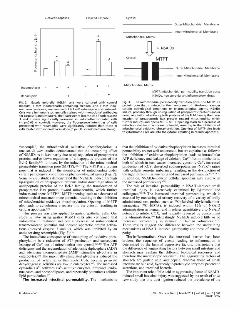
“uncouple”, the mitochondrial oxidative phosphorylation isunclear. In vitro studies demonstrated that the uncoupling effectof NSAIDs is at least partly due to up-regulation of proapoptoticproteins and/or down regulation of antiapoptotic proteins of theBcl-2 family,(21) followed by the induction of the mitochondrialpermeability transition pore (MPTP).(22,23) The MPTP is a proteinpore that is induced in the membranes of mitochondria undercertain pathological conditions or pharmacological agents (Fig. 2).Some in vitro studies demonstrated that NSAIDs induce, throughup-regulation of proapoptotic proteins and/or down regulation ofantiapoptotic proteins of the Bcl-2 family, the translocation ofproapoptotic Bax protein toward mitochondria, which furtherinduces and opens MPTP.(21) MPTP opening leads to a decrease ofmitochondrial transmembrane potential, resulting in the inhibitionof mitochondrial oxidative phosphorylation. Opening of MPTPalso leads to cytochrome c realase into the cytosol, resulting incellular apoptosis.(21)
This process was also applied to gastric epithelial cells. Ourstudy in vitro using gastric RGM1 cells also confirmed thatindomethacin treatment induced a decrease of mitochondrialtransmembrane potential and induced apoptotic protein activa-tions (cleaved caspase 3 and 9), which was inhibited by anantiulcer drug rebamipide (Fig. 3).(16)
The immediate consequence of uncoupling of oxidative phos-phorylation is a reduction of ATP production and subsequentleakage of Ca2+ out of mitochondria into cytosol.(24,25) The ATPdeficiency and the accumulation of adenosine diphosphate (ADP)and adenosine monophosphate (AMP) stimulate glycolysis inenterocytes.(26) The maximally stimulated glycolysis induced theproduction of lactate rather than acelyl CoA, because pyruvatedehydrogenase activities are low in enterocytes.(17) The increasedcytosolic Ca2+ activates Ca2+-sentitive enzymes, proteases, endo-nucleases, and phospholipases, and reportedly potentiates cellularlipid peroxidation.(27)
The increased intestinal permeability. The mechanisms
that the inhibition of oxidative phophorylation increases intestinalpermeability are not well understood, but are explained as follows:the inhibition of oxidative phophorylation leads to intracellularATP deficiency and leakage of calcium (Ca2+) from mitochondria,both of which in turn causes increased cytosolic Ca2+, increasedproduction of ROS, disturbed sodium/potassium (Na+/K+) ratioswith cellular osmotic imbalance, resulting in the dysfunction ofthe tight intracellular junctions and increased permeability.(2,17,19,28)
In addition, NSAIDs-induced cellular apoptosis may involve inthe increased permeability.(29)
The role of intestinal permeability in NSAID-induced smallintestinal injury is extensively examined by Bjarnason andcolleagues.(18,28) The increased intestinal permeability, which isassessed by measuring of urinary recovery of a variety of orallyadministered test probes such as 51Cr-labeled ethylenediamine-tetraacetate (51Cr-EDTA), is induced within 12 h of NSAIDadministration in human, and it relates quantitatively to NSAIDpotency to inhibit COX, and is partly reversed by concomitantPG administration.(30) Interestingly, NSAIDs induced little or noincreased permeability in stomach of human volunteers.(31,32)
These results suggest the difference between the underliningmechanisms of NSAID-induced gastropathy and those of entero-pathy.
The inflammation. Once the intestinal barrier has beenbroken, the sequence of events leading to inflammation isdetermined by the luminal aggressive factors. It is notable thatthe difference of aggravating factors between small intestine andstomach may explain the different biological responses andtherefore the macroscopic lesions.(17) The aggravating factors ofstomach are gastric acid and pepsin, whereas those of smallintestine are bile acid, hydroeolytic/proteolytic enzymes, pancreaticsecretions, and intestinal bacteria.
The important role of bile acid an aggravating factor of NSAID-induced small intestinal injury was suggested by the result of an invivo study that bile duct ligation reduced the prevalence of the
Fig. 2. Gastric epithelial RGM�1 cells were cultured with controlmedium, 1 mM indomethacin containing medium, and 1 mM indo�methacin containing medium with 1 h 1 mM rebamipide pretreatment.Cells were immunohistochemically stained with monoclonal antibodiesfor caspase 3 and caspase 9. The fluorescence intensities of both caspase3 and 9 were significantly increased in indomethacin�treated cells(*: p<0.05 vs control). However, the fluorescence intensities of cellspretreated with rebamipide were significantly reduced than those incells treated with indomethacin alone (#: p<0.05 vs indomethacin alone).
Fig. 3. The mitochondrial permeability transition pore. The MPTP is aprotein pore that is induced in the membranes of mitochondria undercertain pathological conditions or pharmacological agents. NSAIDsinduce, probably through up�regulation of proapoptotic proteins and/ordown regulation of antiapoptotic proteins of the Bcl�2 family, the trans�location of proapoptotic Bax protein toward mitochondria, whichfurther induces and opens MPTP. MPTP opening leads to a decrease ofmitochondrial transmembrane potential, resulting in the inhibition ofmitochondrial oxidative phosphorylation. Opening of MPTP also leadsto cytochrome c realase into the cytosol, resulting in cellular apoptosis.
doi: 10.3164/jcbn.10�79©2011 JCBN
110
lesions significantly.(33) Although the exact biological mechanismswere unclear, it is supposed that bile acid may further damagecellular membranes to increase intestinal permeability, or NSAIDsconjugated in bile may be converted into free, active form throughthe bacterial deconjugation in small intestine.
The role of bacteria as an aggravating factor of NSAID-inducedsmall intestinal injury is suggested by the experimental results thatNSAIDs induced very few intestinal lesions in germ-free animalsand that pretreatment with antimicrobials reduced NSAID-induced small intestinal damages.(34–36)
The neutrophil infiltration is essential in the development ofmacroscopic lesion. The microbial invasion and the damagedmucosa are considered as the main neutrophil chemoattractant.Generated ROS from activated neutrophils results in the mucosaldamage, the finding experimentally confirmed that the preventionof neutrophil infiltration by induction of neutropenia inhibitedNSAID-induced macroscopic mucosal damage.(37)
The role of PG deficiency in NSAID�induced small intestinalinjury. The role of PG deficiency in NSAID-induced smallintestinal injury is poorly understood, and there is controversyregarding whether PG deficiency is considered as the main causesof NSAID-induced small intestinal injury. Some studies demon-strated the protective effects of PG on NSAID-induced smallintestinal injuries in animal models(38–40) and the PG deficiency insmall intestine is proposed as the main cause.(11,41)
However, recent experimental studies demonstrated that thePG deficient does not have a major role in the small intestineinjuries.(42,43) Somasundram et al. studied in a rat model that wasdesigned to allow dissociation of the effects and pathophysio-logical consequences of the ‘topical’ mitochondrial inhibition andthe ‘systemically’ mediated COX inhibition.(44) They demonstratedthat the treatment of parenteral aspirin decreased mucosal PGswithout affecting mitochondria, cellular permeability and causedno inflammation, and that the treatment of dinitrophenol, anuncoupling agent, increased intestinal permeability and causedinflammation without affecting intestinal PG levels. They con-cluded that the deficient endogenous PG production is notsufficient to alter intestinal permeability in short term.(44)
Conclusion
This review is focused on the pathophysiology of NSAID-induced gastroenteropathy, especially on PG-independent, mito-
chondria-dependent small intestinal injury.It seems that the framework of pathophysiology of NSAID-
induced mucosal injury may differ in stomach and in smallintestine. The relative physiological importance of PGs in main-taining mucosal defense system between stomach and smallintestine may contribute to the experimental and clinical resultsof NSAID-induced mucosal injuries. In stomach, because of theessential role of PG, NSAID-induced mucosal injury can beexplained by the PG deficiency, and the exogenous PG treatmenthad protective effects in vitro and in vivo. On the other hand, insmall intestine, NSAID-induced mucosal injury cannot beexplained, although controversy exists, by the PG deficiencyalone. The difference in aggressive factors of mucosal epitheliummay affect the pathophysiology of NSAID-induced mucosalinjury: gastric acid in stomach and luminal bacteria in smallintestine. The reduction of gastric acid secretion in stomach andthe sterilization of bacteria in small intestine had a protectiveeffect on NSAID-induced mucosal injuries, and not vice versa.
However, we emphasize the importance of the uncoupling ofmitochondrial oxidative phosphorylation as a common first stepin NSAID-induced mucosal injury both in stomach and in smallintestine. We think a novel therapeutic approach against NSAID-induced gatrointestinal mucosal injuries should include agents thatprevent the uncoupling oxidative phosphorylation of mitochondriain epithelial cells.
Abbreviations
NSAIDs non-steroidal anti-inflammatory drugsCOX-1 cyclooxygenase-1COX-2 cyclooxygenase-2PG ProstaglandinH2RAs histamine 2 receptor antagonistsPPIs proton pump inhibitorsATP adenosine triphosphateROS reactive oxygen speciesMTP mitochondrial transmembrane potentialO2
•− superoxideH2O2 hydrogen peroxideMPTP mitochondrial permeability transition poreAMP adenosine monophosphateADP adenosine diphosphateEDTA ethylenediaminetetraacetate
References
1 Laine L, Takeuchi K, Tarnawski A. Gastric mucosal defense and cytoprotec-
tion: bench to bedside. Gastroenterology 2008; 135: 41–60.
2 Higuchi K, Umegaki E, Watanabe T, and et al. Present status and strategy of
NSAIDs-induced small bowel injury. J Gastroenterol 2009; 44: 879–888.
3 Higuchi K, Yoda Y, Amagase K, and et al. Prevention of NSAID-induced
small intestinal mucosal injury: prophylactic potential of lansoprazole. J Clin
Biochem Nutr 2009; 45: 125–130.
4 Ono S, Kato M, Imai A, and et al. Preliminary trial of rebamipide for preven-
tion of low-dose aspirin-induced gastric injury in healthy subjects: a random-
ized, double-blind, placebo-controlled, cross-over study. J Clin Biochem Nutr
2009; 45: 248–253.
5 Yamamoto T, Isono A, Mishina Y, and et al. Gastroduodenal mucosal injury
in patients taking low-dose aspirin and the role of gastric mucoprotective
drugs: possible effect of rebamipide. J Clin Biochem Nutr 2010; 47: 27–31.
6 Takahashi T, Otaka M, Odashima M, and et al. Correlation of heat shock
protein expression to gender difference in development of stress-induced
gastric mucosal injury in rats. J Clin Biochem Nutr 2010; 47: 64–73.
7 Del Valle J. Peptic ulcer disease and related disorders. In: Fauci AS, Kasper
DL, Longo DL, and et al, eds. Harrison’s Principles of Internal Medicine
17th Edition, New York: McGraw-Hill Professional, 2008; 1855–1872.
8 Cryer B, Spechler SJ. Peptic ulcer disease. In: Feldman M, Friedman LS,
Brandt LJ, eds. Sleisenger and Fordtran’s Gastrointestinal and Liver Disease
8th Edition, Philadelphia: Saunders, 2006; 1089–1110.
9 Kargman S, Charleson S, Cartwright M, and et al. Characterization of Prosta-
glandin G/H Synthase 1 and 2 in rat, dog, monkey, and human gastrointestinal
tracts. Gastroenterology 1996; 111: 445–454.
10 Wallace JL, McKnight W, Reuter BK, Vergnolle N. NSAID-induced gastric
damage in rats: requirement for inhibition of both cyclooxygenase 1 and 2.
Gastroenterology 2000; 119: 706–714.
11 Laine L. Nonsteroidal anti-inflammatory drug gastropathy. Gastrointest
Endosc Clin N Am 1996; 6: 489–504.
12 Ott M, Gogvadze V, Orrenius S, Zhivotovsky B. Mitochondria, oxidative
stress and cell death. Apoptosis 2007; 12: 913–922.
13 Brand MD, Affourtit C, Esteves TC, and et al. Mitochondrial superoxide:
production, biological effects, and activation of uncoupling proteins. Free
Radic Biol Med 2004; 37: 755–767.
14 Brzozowski T, Konturek PC, Konturek SJ, and et al. Role of gastric acid
secretion in progression of acute gastric erosions induced by ischemia-
reperfusion into gastric ulcers. Eur J Pharmacol 2000; 398: 147–158.
15 Tsutsumi S, Tomisato W, Takano T, Rokutan K, Tsuchiya T, Mizushima
T. Gastric irritant-induced apoptosis in guinea pig gastric mucosal cells in
primary culture. Biochim Biophys Acta 2002; 1589: 168–180.
16 Nagano Y, Matsui H, Muramatsu M, and et al. Rebamipide significantly
inhibits indomethacin-induced mitochondrial damage, lipid peroxidation, and
J. Clin. Biochem. Nutr. | March 2011 | vol. 48 | no. 2 | 111
©2011 JCBNH. Matsui et al.
apoptosis in gastric epithelial RGM-1 cells. Dig Dis Sci 2005; 50 Suppl 1:
S76–S83.
17 Somasundaram S, Hayllar H, Rafi S, Wrigglesworth JM, Macpherson AJ,
Bjarnason I. The biochemical basis of non-steroidal anti-inflammatory drug-
induced damage to the gastrointestinal tract: a review and a hypothesis. Scand
J Gastroenterol 1995; 30: 289–299.
18 Bjarnason I, Hayllar J, MacPherson AJ, Russell AS. Side effects of non-
steroidal anti-inflammatory drugs on the small and large intestine in humans.
Gastroenterology 1993; 104: 1832–1847.
19 Feldman M. Ulcers of the small and large intestine. Philadelphia: Saunders,
2006; 2587–2597.
20 Bjarnason I, Hayllar J. Early pathogenic events in NSAID-induced gastro-
intestinal damage. Ital J Gastroenterol 1996; 28 Suppl 4: 19–22.
21 Maity P, Bindu S, Choubey V, and et al. Lansoprazole protects and heals
gastric mucosa from non-steroidal anti-inflammatory drug (NSAID)-induced
gastropathy by inhibiting mitochondrial as well as Fas-mediated death path-
ways with concurrent induction of mucosal cell renewal. J Biol Chem 2008;
28: 14391–14401.
22 Yoshida Y, Singh I, Darby CP. Effect of salicylic acid and calcium on mito-
chondrial functions. Acta Neurol Scand 1992; 85: 191–196.
23 Tomoda T, Takeda K, Kurashige T, Enzan H, Miyahara M. Acetylsalicylate
(ASA)-induced mitochondrial dysfunction and its potentiation by Ca2+. Liver
1994; 14: 103–108.
24 De Gómez Puyou MT, Gavilanes M, Gómez-Puyou A, Ernster L. Control of
activity states of heart mitochondrial ATPase. Role of the proton-motive force
and Ca2+. Biochim Biophys Acta 1980; 592: 396–405.
25 Jorgensen TG, Weis-Fogh US, Nielsen HH, Olesen HP. Salicylate- and
aspirin-induced uncoupling of oxidative phosphorylation in mitochondria
isolated from the mucosal membrane of the stomach. Scand J Clin Lab Invest
1976; 36: 649–654.
26 Adams SS, Cobb R. A possible basis for the anti-inflammatory activity of
salicylates and other non-hormonal anti-rheumatic drugs. Nature 1958; 181:
773–774.
27 Jain SK, Shohet SB. Calcium potentiates the peroxidation of erythrocyte
membrane lipids. Biochim Biophys Acta 1981; 642: 46–54.
28 Bjarnason I, MacPherson A, Hollander D. Intestinal permeability: an over-
view. Gastroenterology 1995; 108: 1566–1581.
29 Omatsu T, Naito Y, Handa O, and et al. Involvement of reactive oxygen
species in indomethacin-induced apoptosis of small intestinal epithelial cells.
J Gastroenterol 2009; 44 Suppl 19: 30–34.
30 Bjarnason I, Smethurst P, Fenn CG, Lee CE, Menzies IS, Levi
AJ. Misoprostol reduces indomethacin-induced changes in human small
intestinal permeability. Dig Dis Sci 1989; 34: 407–411.
31 Aabakken L, Larsen S, Osnes M. Sucralfate for prevention of naproxen-
induced mucosal lesions in the proximal and distal gastrointestinal tract.
Scand J Rheumatol 1989; 18: 361–368.
32 Aabakken L, Larsen S, Osnes M. Cimetidine tablets or suspension for the
prevention of gastrointestinal mucosal lesions caused by non-steroidal, anti-
inflammatory drugs. Scand J Rheumatol 1989; 18: 369–375.
33 Fang WF, Broughton A, Jacobson ED. Indomethacin-induced intestinal
inflammation. Am J Dig Dis 1997; 22: 749–760.
34 Robert A, Asano T. Resistance of germfree rats to indomethacin-induced
intestinal lesions. Prostaglandins 1977; 14: 333–341.
35 Brodie DA, Cook PG, Bauer BJ, Dagle GE. Indomethacin-induced intestinal
lesions in the rat. Toxicol Appl Pharmacol 1970; 17: 615–624.
36 Brune K, Dietzel K, Nurnberg B, Schneider HT. Recent insight into the
mechanism of gastrointestinal tract ulceration. Scand J Rheumatol Suppl
1987; 65: 135–140.
37 Wallace JL, Keenan CM, Granger DN. Gastric ulceration induced by non-
steroidal anti-inflammatory drugs is a neutrophil-dependent process. Am J
Physiol 1990; 259: G462–G467.
38 Kunikata T, Araki H, Takeeda M, Kato S, Takeuchi K. Prostaglandin E
prevents indomethacin-induced gastric and intestinal damage through
different EP receptor subtypes. J Physiol Paris 2001; 95: 157–163.
39 Takeuchi K, Miyazawa T, Tanaka A, Kato S, Kunikata T. Pathogenic
importance of intestinal hypermotility in NSAID-induced small intestinal
damage in rats. Digestion 2002; 66: 30–41.
40 Konaka A, Nishijima M, Tanaka A, Kunikata T, Kato S, Takeuchi K. Nitric
oxide, superoxide radicals and mast cells in pathogenesis of indomethacin-
induced small intestinal lesions in rats. J Physiol Pharmacol 1999; 50: 25–38.
41 Takeuchi K, Tanaka A, Ohno R, Yokota A. Role of COX inhibition in patho-
genesis of NSAID-induced small intestinal damage. J Physiol Pharmacol
2003; 54 Suppl 4: 165–182.
42 Wallace JL. Nonsteroidal anti-inflammatory drugs and gastroenteropathy: the
second hundred years. Gastroenterology 1997; 112: 1000–1016.
43 Reuter BK, Davies NM, Wallace JL. Nonsteroidal anti-inflammatory drug
enteropathy in rats: role of permeability, bacteria, and enterohepatic circula-
tion. Gastroenterology 1997; 112: 109–117.
44 Somasundaram S, Sigthorsson G, Simpson RJ, and et al. Uncoupling of
intestinal mitochondrial oxidative phosphorylation and inhibition of cyclo-
oxygenase are required for the development of NSAID-enteropathy in the rat.
Aliment Pharmacol Ther 2000; 14: 639–650.