Embed Size (px)
Citation preview

THE PASSAGE OF PROTEIN MOLECULES THROUGH THE GLOMERULAR MEMBRANES*
BY P. A. BOTT AND A. N. RICHARDS
(Prom the Laboratory of Pharmacologl/, School of Medicine, University of Pennsylvania, Philadelphia)
(Received for publication, August 4, 1941)
We conceive of the glomerular membrane as a thin composite structure, the complex molecules of which are so arranged as to form liquid-filled passages or pores through which a filtrate can be expressed from the blood plasma. Analyses of the glomerular filtrate obtained from Amphibia and study of the renal excretion of the polysaccharide inulin (1) and of different proteins have yielded information with which the permeability of the membrane may be defined and thus by inference a description of its character approached. Normally, serum proteins do not pass through it. The smallest of these is serum albumin (molecular weight 70,000) ; hence we say tentatively that all of the pores of the membrane are smaller than the diameter of the serum albumin molecule. Aiming to decide whether other factors than molecular weight and size determine the passage or non-passage of a substance through the glomerulus, we have studied the glomerular excretion of several proteins, the molecular weights of which lie between those of inulin and serum albumin.
Mainly two types of experiments have been performed: (a) “direct” experiments in which glomerular fluid collected from Bowman’s capsule was analyzed for protein by an ultramicro- method which will be described. The collections were made from amphibian kidneys (chiefly of Necturi) during perfusion with pro- tein solutions; (b) “indirect” experiments (chiefly on frogs) in
* The expenses of this work have been defrayed in large part from a grant by the Commonwealth Fund of New York. A preliminary report was made to the Physiological Society of Philadelphia in October, 1940 (see Am. J. Med. SC., 200, 847 (1940)).
291
by guest on June 17, 2018http://w
ww
.jbc.org/D
ownloaded from

292 Permeability of Glomerular Membranes
which urine was collected from the ureters during perfusion of a
solution containing protein and inulin and analyzed for these two substances.
In addition we have performed a few experiments in which the rate and completeness of excretion of protein injected into normal dogs was compared with that of inulin similarly injected.
Procedure for Per&sion Experiments-Proteins, sometimes to- gether with inulin, were made up in frog Ringer’s solution (2) con- taining no glucose or glycine. The oxygenation was accomplished in most cases by bubbling a mixture of 98.5 per cent oxygen and 1.5 per cent carbon dioxide through the solution by means of a sintered glass bubbling tube. In several experiments solutions were subjected to a pressure of 10 atmospheres of the gas mixture mentioned above for approximately 45 minutes for oxygenation. The pH of the perfusion fluid was approximately 7.4 except when otherwise noted. After oxygenation the fluids were filtered through rapid filters into perfusion bottles.
The various proteins used were crystalline albumin of hen’s and duck’s eggs, Palmer’s lactoglobulin (3), amorphous and crys- talline (zinc) insulin, all of which have molecular weights of 34,000 to 46,000 (4-7)l and are placed in the 35,200 group of Svedberg’s classification, purified protein derivative (PPD-49609) from tuber- culin (8, 9) with a weight of about 14,500, and, as a control, crys- talline horse serum albumin. Attempts were also made to use the protamine, salmine (the molecular weight may be 5600 (lo)), but this material proved toxic to the kidney. Details concerning the preparation of proteins2 and protein solutions are as follows:
Hen’s egg albumin was crystallized at least three times by the Sorensen (ammonium sulfate) method or by the Kekwick and Cannan (11) (sodium sulfate) method. After removal of sulfate by dialysis, some of the solution was used immediately for per-
* Since it is impossible to quote the large number of original papers, we are referring to recent publications in which data from such sources are assembled and discussed.
2 It is a pleasure to acknowledge our gratitude to Dr. Florence Seibert of the Henry Phipps Institute for the purified protein derivat,ive of tuber- culin, to Eli Lilly and Company for insulin, to Sharp and Dohme for salmine, and to Professor B. M. Hendrix for sending one lot of partially purified duck’s egg albumin from Texas at a time when we were unable to obt.ain duck’s eggs.
by guest on June 17, 2018http://w
ww
.jbc.org/D
ownloaded from

P. A. Bott and A. N. Richards
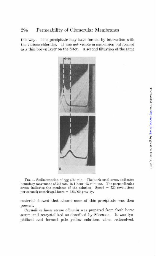
fusion; the larger part of it, however, was lyophilized in small containers and kept in vucuo until used. When lyophilized albu- min was used, it was first dissolved in water and any protein \vhich did not dissolve at this pH (the isoelectric point) was filtered or centrifuged off. The pH of the solution was raised to at least 6 by the addition of sodium hydroxide before the various constitu- ents of the Ringer’s solution were added. The pH of the osygen- ated solutions was 7.4. One portion of lyophilized egg albumin, made up into perfusion fluid and aerated as usual, was used for sedimentation in the ultracentrifuge. We are indebted to Dr. Bauer and Dr. Pickels of the International Health Division of the Rockefeller Foundation for this determination which showed the protein to be homogeneous and to have a sedimentation con- stant S 200 X lOpI3 = 3.51 which compares well with that of 3.55, the latest figure from the Svedberg laboratory (7). Phohographs of the boundary movement are shown in Fig. 1.
The same methods of purification were applied to duck’s egg albumin but the crystals obtained, although pure white, were never clear cut as in the case of hen’s egg albumin.
Lactoqlobulin was prepared as described by Palmer (3) with the use of ammonium sulfate and recrystallized twice. The crystals were beautifully clear cut tabular crystals and were kept under water, preserved with toluene. They were washed with water before being dissolved in the chlorides of Ringer’s solution.
Insulin-Both amorphous and crystalline zinc insulin were supplied by Eli Lilly and Company. Since Sjogren and Svedberg (see (7)) had shown that pH 7 is the upper limit of the st,ability range for insulin, all perfusion fluids in insulin experiments were adjusted to this value instead of pH 7.4.
PPD-49609-This protein (8) supplied by Dr. Seibert shows a dry molecular weight of about 14,500 and is highly homogeneous in so far as the ultracentrifuge discloses but heterogeneous by electrophoresis. The material was supplied phosphate-butiered and lyophilized. It was dissolved in water under a partial vacuum to form a clear brown solution. In some experiments the protein was simply made up with the usual chlorides and bicarbonate of perfusion fluid, the phosphate being omitted. In other cases the protein-phosphate-chloride solution was run through a .Jena bacterial filter before use, since a fine precipitate was removed in
by guest on June 17, 2018http://w
ww
.jbc.org/D
ownloaded from
294 Permeability of Glomerular Membranes
this way. This precipitate may have formed by interaction with the various chlorides. It was not visible in suspension but formed as a thin brown layer on the filter. A second filtration of the same
FIG. 1. Sedimentation of egg albumin. The horizontal arrow indicates boundary movement of 2.5 mm. in 1 hour, 23 minutes. The perpendicular arrow indicates the meniscus of the solution. Speed = 720 revolutions per second; centrifugal force = 133,000 gravity.
material showed that almost none of this precipitate was then present.
Crystalline horse serum albumin was prepared from fresh horse serum and recrystallized as described by Sorensen. It was lyo- philized and formed pale yellow solutions when redissolved.
by guest on June 17, 2018http://w
ww
.jbc.org/D
ownloaded from

P. A. Bott and A. N. Richards 295
Salmine-“Pure protamine, salmine,” Sharp and Dohme, was used.
All the inulin used in these experiments was prepared about 6 years ago by Dr. R. F. Jackson of the Bureau of Standards. The roncentrations of protein in perfusion fluids depended to a certain extent on the availability and solubility of the material. It was necessary to make up several hundred cc. of fluid for each experi- ment. The concentrations ranged from 0.05 to 2.6 per cent. lnu- lin, when present, was in a concentration of 0.4 to 0.5 per cent.
In the indirect type of experiment the bullfrog kidney was perfused via the aorta with protein-inulin solution at a pressure usually near 24 cm. of water. In a few inst,ances plain Ringer’s solution was perfused simultaneously through the renal-portal circulation, but this did not alter the results. Urine was collected from the ureters by cannula and analyzed for protein and inulin. Perfusion fluids were likewise analyzed. Since it has been shown (1) that inulin is completely filtered through the glomeruli of Rmphibia, and on the assumption that neither protein nor inulin is secreted or reabsorbed by the tubule cells, the concentration of protein in glomerular fluid was calculated from the results of the above analyses. For example, if the inulin urine to perfusion fluid ratio is 1.2, then the prot,ein must also have been concentrated 1.2 times in passing from capsule to ureter, and the urine protein concentration is therefore divided by 1.2 to obtain the glomerular fluid protein concentration. If, by this process, the concentration of protein in glomerular fluid is found to be only one-half of that in perfusion fluid, it is concluded that the glomerular membrane is only 50 per cent permeable to the protein. For convenience this will be designated as 50 per cent filtration.
In the direct experiments glomerular fluid was collected by the method described by Wearn and Richards (12). Adult Necturi were used for this purpose, since the glomeruli are large and samples of the volume necessary for analysis could be collected fairly readily. The animals were placed in 1.5 per cent urethane solution for approximately 15 minutes, brain and spinal cord were then completely pithed, an inflow cannula inserted in the dorsal aorta above the kidney, and the outflow cannula in the postcaval vein. There are certain difficulties inherent in glomerular puncture perfusion experiments in these animals which make the collections
by guest on June 17, 2018http://w
ww
.jbc.org/D
ownloaded from

296 Permeability of Glomerular Membranes
both difficult and treacherous. It is because of these difficulties that some experiments are necessarily discarded and some must be regarded as questionable when conditions are not perfect during the perfusion.
Analytical Methods
For Experiments on Dogs-For these experiments urinary protein was determined by the gravimetric method of Folin and Denis (13) ; inulin as previously described (14).
For Indirect Experiments on Amphibia-Inulin was determined as the total reducing substance after the usual protein removal and hydrolysis (see (14)), since neither perfusion fluid nor urine contained glucose. The Somogyi reagent for small quantities of sugar (15) was used for the determination.
Protein was determined by means of the Folin and Ciocalteu (16) phenol reagent as suggested by Greenberg (17). The Evelyn photoelectric calorimeter was used for these microdeterminations, modified to fit our needs. Standardization curves were plotted with protein solutions, the protein content of which had originally been determined by Kjeldahl nitrogen analysis3 (Wong’s (18) persulfate method). For the standardization, 0.05 cc. samples of protein solutions were washed into 6 cc. volumes of water in a series of tubes. To each of these and also to a water blank was added, with swirling, 0.25 cc. of 5 N NaOH. 10 minutes after the addition of alkali to the first tube, the addition of phenol reagent was begun. 0.4 cc. was added to each tube rapidly but drop by drop and with continuous swirling. After 15 minutes had been allowed for color development, readings were taken, with the 6 cc. aperture of the calorimeter and the No. 620 filter. 0.05 cc. of 1.8 per cent egg albumin produced full scale deflections.
Perfusion fluids and urines were made up for color development as described for the standard solutions, and readings were taken, and evaluated as protein concentrations on the curve. The error of these determinations was approximately 3 per cent. When it was necessary to take samples larger or smaller than 0.05 cc., corrections were made in the volume of water used, so that the final volumes remained the same. Proteins which contain more
3 We wish to acknowledge the assistance of Miss Ethol Shiels for these and other analyses in the standardization of protein.
by guest on June 17, 2018http://w
ww
.jbc.org/D
ownloaded from

P. A. Bott and A. N. Richards 297
tyrosine or tryptophane than egg albumin gave relatively higher color development, so that for correct absolute values, the readings were evaluated from curves plotted for these proteins. Ratios of urine concentration to perfusion fluid concentration (and consequently calculated percentage filtration of proteins) were practically identical, however, whether the readings were inter- preted from the various curves mentioned or from the curve for egg albumin.
The Weber calorimetric arginine method (19) was used to determine protamine (salmine) in a few experiments. The depth of color was read photoelectrically.
For Direct Experiments on Amphibia. Ultramicro Protein L)etermination-The same method described above was used to determine protein calorimetrically, an ultramicro calorimeter being used for the readings. In this calorimeter, which was built for us by Dr. K. Hartline and Mr. J. Hervey, the potentiometer principle is utilized. The amplified photocell current is “bucked” against a current from a battery source, the null point being in- dicated by means of a milliammeter. The unique absorption cells, designed for the calorimeter by Dr. Hartline, are illustrated in Fig. 2. This type of cell met the requirements of being quickly and thoroughly cleaned. It is composed of three slides, each 1 mm. in thickness, two of which are shown (A and B) and the third of which (A’) is a mirror image of /I. Circular grooves are ground into A and A ‘, while in B there is a circular hole completely through the slide. A small drop of fluid is placed on each of the surfaces inside of the circular grooves, while the three slides are held in one hand, properly oriented so that when they are allowed to come together they will appear as is indicated in C of Fig. 2. The volume of fluid held by the cell is approximately 0.025 cc.
The range and accuracy of the calorimeter are shown by the results of readings taken when the cell is simply filled with solu- tions such as copper sulfate and indigo carmine. With concentra- tions of copper sulfate from 0.5 to 10 per cent which showed -log transmissions from 0.00358 to 0.07196, the readings ranged from 0.0 to 5.0 per cent from the theoretical, averaging 1.7 per cent. In another series of readings on indigo carmine solutions varying in concentration from 0.7 to 50 mg. per cent, the light absorption in this case varying from 4 to 90 per cent, the error
by guest on June 17, 2018http://w
ww
.jbc.org/D
ownloaded from

298 Permeability of Glomerular Membranes
was even lower. This error includes that of the instrument, of cleaning the glass cell, and filling it with solution.
Results on protein solution are not as good as those cited above, since added to these errors are those of the protein method itself (17), of measuring out the various reagents and the sample, as well as the occasional formation of bubbles. Capillary pipettes delivering by centrifugal force were used rather than washout pipettes in order to reduce the chances for bubble formation in
a
$4 i’! --COTTON
&-CEMENT
-RUBBER TUBING
“1
-CAPILLARY PIPETTE
0
B
J f C
FIG. 2. Left, apparatus for ultramicro protein determination. Right, absorption cell for micro photoelectric calorimeter. A, B, and C are slides, as described in t.he text. The entire figure is actual size.
these alkaline protein solutions. The manipulation of these pipettes will be described briefly. The protein solution (either standard or experimental) is transferred under the microscope to the capillary pipette holding about 0.5 c.mm. which is mounted in a glass holder similar to that described by Wigglesworth (20) but supplied with shoulders of rubber tubing as shown in Fig. 2. The filled pipette is placed in a sturdy test-tube into which has been measured 0.050 cc. of 0.2 N sodium hydroxide solution. The entire assembly (shown in Fig. 2) is then lowered
by guest on June 17, 2018http://w
ww
.jbc.org/D
ownloaded from

P. A. Bott and A. N. Richards 299
into a centrifuge cup and spun rapidly for a few seconds to dis- charge the sample. The pipette is removed and the fluids mixed by tapping the test-tube gently. After 10 minutes a pipette mounted similarly but made of large capillary tubing (0.6 mm. inside diameter and 11 mm. long) and holding 3 c.mm. is filled with phenol reagent which is delivered as indicated above. 15 minutes later the blue solution is drawn up in a fine tipped pipette and transferred to the absorption cell as previously described. In nineteen single determinations of protein in 0.5 c.mm. samples of 0.2 to 3.8 per cent protein solutions the average error was 4.9 and the range of error 0.0 to 14.7 per cent. In only two of the experiments was the error above 10 per cent.
Results
The results of thirty-two “direct” glomerular puncture experi- ments and over 50 “indirect” experiments are given in Tables I and II. That there are more figures for hen’s egg albumin than for any of the other proteins is due to the fact that, being most easily prepared, it was used as a basis for comparison with each of the others, and for experiments made to test technique.
The direct glomerular puncture experiments show that on the average the concentration of hen’s egg albumin in the glomerular filtrate in hrecturi is 58 per cent of that of the perfusion fluid (range, 38 to 84). The indirect experiments on frogs gave an average value of 57 per cent (39 to 77). This surprisingly close agreement is testimony to the validity of the indirect experiment. Eight different lots of the hen’s egg albumin were prepared for these experiments at different times; no significant differences ascribable to different preparations were observed. The con- centrations of the protein in the perfusion fluid in different experi- ments varied from 0.1 t,o 2.6 per cent. These differences did not affect the percentage filtered. The close agreement of the two methods can be used as evidence that neither inulin nor hen’s egg albumin is reabsorbed from the tubules. The conclusion from these experiments is that the permeability of the glomerular membrane to hen’s egg albumin is incomplete. It might be justifiable to say that only from 45 to 70 per cent of the surface of the membrane or of the pores in the membrane permits the passage of that protein.
by guest on June 17, 2018http://w
ww
.jbc.org/D
ownloaded from

300 Permeability of Glomerular Membranes
TABLE I
Protein Perfusion Experiments on Necturi, Glomerular Punctures (Direct)
T Hen’s egg albumin Duck’s egg albumin Lactoglobulin -
1 -
- I i
‘er cent 1 iltered
.-
76 38 51 60 49 83 82 54 48 48 50 43 84 50 71 57 46
‘er cent ?erfusion iltered fluid
42 53 57 96 96 58
per cent
1.00 0.74 0.58 0.53 0.44 0.40 0.22 0.17
‘er cent filtered
70 77 88
102 84
103 64 82
Pe;f;;or I Glomer- u dar fluid
per cent per cent 1.16 0.88 0.94 0.36 0.77 0.39 0.68 0.41 0.66 0.32 0.66 0.55 0.56 0.46 0.54 0.29 0.54 0.26 0.48 0.23 0.48 0.24 0.47 0.20 0.45 0.38 0.44 0.22 0.42 0.30 0.42 0.24 0.41 0.19
Glomer- 11ar fluid
per cent 0.70 0.57 0.51 0.54 0.37 0.41 0.14 0.14
?erf usion fluid
Glomer- 11ar fluid E
per cent per cent 0.65 0.27 0.53 0.28 0.51 0.29 0.49 0.47 0.47 0.45 0.41 0.24
TABLE II
Per Cent Fibration of various Proteins
3y analysis of glomerulsr By indirect method; fluid; Necturus frog
Mol. wt. Protein
70,000
35,200 class
14,500 ~-
Practically no filtrr ion
3% 84 58 50 39-77 57
42- 96 67 5 53-58 55 64-103 84 8 72-85 79
24 24
I -
1 15-20 18 1 22-42 32 6 76-95 86
Crystalline horse sewn albumin
Crystalline hen’s egg albumin
Duck’s egg albumin ‘Crystalline lacto-
I
globulin Amorphous insulin Crystalline zinc ins&r PPD from tuberculin
17
6 8
1
_--
We mere interested in trying duck’s egg albumin, since there was some evidence that this protein was more homogeneous
by guest on June 17, 2018http://w
ww
.jbc.org/D
ownloaded from

P. A. Bott and A. N. Richards 301
electrophoretically than that of the hen’s egg (21, 22). Only one component has been demonstrated for the former; the latter, which, it must be said, has been studied more completely, shows more than one. In spite of this both showed approximately the same percentage filtration through the glomeruli. It may be a coincidence that the results of indirect experiments on duck’s egg albumin were all within a narrow range (53 to 58 per cent). It happens that two of the direct experiments indicate almost complete filtration. This may mean either that the membranes in these cases were abnormal or that there was an undetected flaw in the technique used during the glomerular puncture. We are of the opinion that, could more experiments be performed, the results of these would be similar to those indicated by the other experiments; namely, 40 to 60 per cent filtered.
The membranes appear to be more permeable to Palmer’s lactoglobulin (3), a protein of the same molecular weight group (7) as egg albumin. The filtration percentage is from 72 to 85 in the bullfrog experiments and 64 to 103 in the Necturus puncture experiments. Here again we see a larger range in the latter type of experiment, but average values agree fairly well. Although there is an overlapping of the filtration ranges for egg albumin and lactoglobulin, because of the variation from animal to animal, it will be shown later that, whenever the two proteins were perfused through the same kidney, the permeability toward lactoglobulin was always greater.
Insulin is another protein of this molecular weight group. Although the homogeneity as to particle size, especially under the conditions of the experiments, cannot be considered assured, the results may be of interest.4 In three experiments with amorphous and crystalline zinc insulin (one direct and two indirect) the per- meability shown by the membrane towa.rd this protein was only 15 to 24 per cent.
The small molecule, PPD-49609 from tuberculin, showed the highest filtration, as was to be expected from its low molecular weight (14,500) (8). Because of the value of this material, we preferred to use it in several indirect experiments rather than to chance the use of approximately the same amount necessary for a
* The same is true of samples of Bence-Jones protein kindly supplied by Dr. Grace Medes of the Lankenau Hospital, Philadelphia, and by Professor D. W. Wilson; these showed filtration values of 20 to 48 per cent.
by guest on June 17, 2018http://w
ww
.jbc.org/D
ownloaded from

302 Permeability of Glomerular Membranes
single puncture experiment. Assuming that the tubules act toward this protein as they do toward the others, we must conclude that the PPD is not quite completely filtered.
Attempts to use salmine were unsuccessful, becatise the material was too toxic to the kidney even in a concentration of 0.1 per cent. When perfusion began, the membranes seemed to be nearly completely permeable to the protamine, but since permeability to horse serum albumin developed in about 5 minutes and continued even after protamine perfusion had been discontinued, it is danger- ous to consider the permeability to protamine as normal. Nor- mally the glomerular membranes were practically impermeable to horse serum albumin, which has a molecular weight almost twice that of egg albumin.
Additional Information Obtained by Indirect Method-The indirect method of determining permeability of the glomerular membranes to various proteins is advantageous in several ways: With the same amount of perfusion fluid and in the same time required for the collection of an ultramicro sample (0.5 c.mm.) of glomerular fluid, it is possible to collect several samples of urine large enough (about 0.4 to 1.0 cc.) to jpermit the analysis for inulin and protein by means of micromethods. Obviously one may perfuse first with one kind of protein, and follow with another, sometimes including as many as four different kinds of proteins in the same experiment while the kidney is still function- ing normally. It is possible to observe any alteration in permea- bility of the membrane due to the passage of protein through it and to observe as well the influence of various drugs on permea- bility. The results of five experiments of this type are given in Table III. In the first three it is demonstrated that different proteins may be perfused in the same experiment, with proper washout periods between collections, and that approximately the same result is obtained for a certain protein each time it is used. These same experiments show the difference in permeability, in the same kidney, toward the different proteins. There is no evidence here of any influence of a protein on the perrneability of the membrane except, possibly, in the case of insulin, as indicated in the fourth experiment. The results in this and another insulin experiment suggest that each time the insulin is perfused there is a slight increase in the percentage of this protein filtered, and also there seems to be an increase in permeability toward egg
by guest on June 17, 2018http://w
ww
.jbc.org/D
ownloaded from

P. A. Bott and A. N. Richards 303
albumin. We have had to use a pH of 7 throughout the insulin experiments, since that is the upper limit of the pH stability range for insulin (7). This could possibly have influenced the results, but in experiments in which we have used much lower pH in perfusing other proteins, the filtrations were decreased rather
TABLE III
Indirect Experiments on Bullfrogs
--
‘erioc NO.
1 2 3 4 1 2 3 4 1 2 3
1
2 3
4 5 6 7
1
2 3 4 5
Protein perfused
Hen’s egg albumin Lactoglobulin Hen’s egg albumin Lactoglobulin Duck’s egg albumin Hen’s I‘ “ Lactoglobulin Duck’s egg albumin Hen’s “ “ PPD Hen’s egg ailbumin Egg albumin .4morphous insulin Kgg albumin Amorphous insulin Egg albumin Amorphous insulin Egg albumin
L‘ ‘I (‘ “ + 7% urethanc ‘< s* <‘ i*
f7% 1: f 7%
‘i ‘l +7% “
Pm Pm Pm cent cent cent
1.15 0.63 0.36 0.31 1.19 0.58 0.52 0.44 1.16 0.63 0.39 0.34 1.17 0.58 0.54 0.46 1.21 0.64 0.44 0.36 1.10 0.58 0.33 0.30 1.15 0.47 0.42 0.37 1.11 0.64 0.40 0.36 1.30 0.1300.0950.07? 1.05 0.107 0.095 0.091 1.10 0.1300.0800.O7~ 1.19 0.31 0.1750.147 1.26 0.50 0.0950.07t 1.15 0.31 0.1750.152 1.11 0.50 O.lOOO.OQ( 1.03 0.31 0.1800.17f 1.10 0.50 0.1100.10c 1.05 0.31 0.2100.20E 1.3201.01 0.80 0.6Ot 0.9951.00 0.84 0.844 0.9951.00 0.94 0.94t 0.997 1.00 0.97 0.97: 0.9981.00 1.00 1.00
49 76 54 79 56 52 79 56 56 85 56 47 15 49 18 56 20 64 60 84 95 97
100
than increased. Insulin was the only protein studied (aside from the protamine, salmine, which was decidedly toxic) which appeared to affect the permeability of the glomerular membranes in these experiments.
Ethyl urethane in concentrations up to 5 per cent perfused in
by guest on June 17, 2018http://w
ww
.jbc.org/D
ownloaded from

304 Permeability of Glomerular Membranes
plain Ringer’s solution between egg albumin perfusions or added to the albumin fluid itself did not alter permeability, but in a concentration of 7 per cent increased permeability gradually (see Experiment 5 of Table III) and in a concentration of 10 per cent increased it quickly and completely. With the higher con- centrations of urethane, the membranes also became permeable to horse serum albumin, while normally, and in the presence of urethane under 5 per cent, they were practically impermeable to this protein. Whenever permeability was increased, there were also other signs of toxicity; the inulin urine to perfusion fluid ratio usually dropped to 1 or sometimes even slightly below 1.
Sodium glycocholate added to egg albumin perfusion fluid to make a concentration of 0.1 per cent resulted immediately in
G L
100
5 z
80
$ 60
z Y- 40 0
g 20 w u u2 La e HOURS AFTER INJECTION
FIG. 3. Urinary excretion of injected inulin and egg albumin by the normal dog.
almost complete permeability to egg albumin. The same con- centration of glycocholate also makes the membrane partially permeable to horse serum albumin. Concentrations of 0.02 per cent or below do not readily affect permeability. Contrary to expectations, the presence of 20 mg. per cent caffeine in an egg albumin perfusion fluid decreased the permeability slightly, as shown by the following percentage filtration: control 48, caffeine 42, control 47, caffeine 39, control 48. Cyanide in concentrations from 0.001 to 0.01 M had no marked effect on the filtration of egg albumin. Sometimes there was a slight increase and some- times a decrease, both of which were within the range of experi- mental error.
Excretion of Protein in Normal Dog-Two unanesthetized dogs,
by guest on June 17, 2018http://w
ww
.jbc.org/D
ownloaded from

P. A. Bott and A. W. Richards
injected intravenously with 0.11 and 0.14 gm. of horse serum albumin per kilo of body weight, showed only traces of protein in the urine. Several dogs, injected with the same or larger amounts of egg albumin, promptly excreted protein in high concentration. The excretion of inulin and egg albumin after injection of about 3 gm. of the former and 3.4 gm. of the latter is graphically represented in Fig. 3. The cumulative excretion curves indicate that, although the protein is eventually excreted almost as completely as the inulin, the rate at which it is excreted is slower. There may be other explanations for this result but it certainly supports the belief that the glomeruli in this animal are considerably less permeable to egg albumin than to inulin. This would, of course, be in agreement with the results obtained on Amphibia.
DISCUSSION
The finding of practically complete impermeability of the glomerular membranes toward serum albumin, only partial permeability in the case of proteins with molecular weights approximately one-half that of serum albumin, and almost complete permeability in the case of the smallest protein (14,500 mol. wt.) agrees, on the whole, with the concept set forth in the introduction, that the passages in the membrane may be of various sizes, all of them being large enough to allow inulin to pass through with water but almost none of them large enough to allow the passage of horse serum albumin. We do not find, as is the im- pression gained from the statements of Bayliss, Kerridge, and Russell (23) concerning the mammalian kidney, that egg albumin is completely filtered. In other words, there appear to be some pores large enough for the passage of inulin but not of egg albumin. The observations nevertheless do not rule out the possibility that the pore size difference may be only “apparent” and that the results are due t’o some other characteristic of the membranes.
Since the diameter of the PPD-49609 has been found (9) to be approximately 20 8., our data may be interpreted to mean that the mesh size over most of the glomerular membrane may be at least of this order. However, the difficulty which arises when we attempt to interpret the difference in filtrability between molecules supposedly of approximately the same size (especially egg albumin
by guest on June 17, 2018http://w
ww
.jbc.org/D
ownloaded from

306 Permeability of Glomerular Membranes
and lactoglobulin) should serve to caution us in drawing too rigid conclusions and to remind us of the many complicating factors in the use of such test substances. We are mindful of t,he stimulating but complicating discoveries concerning the effects of other substances, of concentration, dilution, and even environment on the association and dissociation of the protein aggregates of which not only our test substances but possibly the membranes them- selves are made (5-7). In an attempt to learn the condition of the egg albumin exactly as used experimentally, one portion of perfusion fluid containing this protein was subjected to ultra- centrifugation with the result discussed earlier in the paper; the protein proved to be homogeneous, with a sedimentation constant characteristic of the pure protein. This is important evidence in support of the presumption that this protein has not been altered in any way so as to affect molecular weight by the procedures used, before it reaches the kidney. Moreover, there was no precipitation of egg albumin in perfusion fluid when the pH was brought back to the isoelectric point by the addition of acid. This and the easy crystallisability of egg albumin in these solutions when ammonium sulfate is added indicate that no denaturation, partial or complete, has taken place. Solutions oxygenated by pressure, a technique which provides less opportunity for surface denaturation5 behave similarly and show the same filtration. It seems relatively certain, then, that the egg albumin particles as used are unaltered. Molecular weights of proteins are usually given as dry weights but the weight for the hydrated egg albumin molecule has been calculated as 61,438 by Bull (26) and as 58,000 by Adair (27) who believes the molecule approximately spherical with a minimum radius of 26.4 A. If these figures are accepted as correct, our data may be interpreted as signifying that the mesh siz: over about one-half of the glomerular membrane is at least 50 A. in diameter, since the proteins as they exist in our per- fusion fluids must be hydrated.
If egg albumin is t,aken as a standard, the filtration of lactoglob- ulin seems too high on the basis of weight or size. The wet molec- ular weight of lactoglobulin as determined from x-ray data by Crowfoot and Riley (see (6)) is 67,000 and the molecular volume
5 Comparatively enormous pressures are required to denature proteins as was done by Bridgman (24) and Dow and Mathews (25).
by guest on June 17, 2018http://w
ww
.jbc.org/D
ownloaded from

I?. A. Rott and A. N. Richards 307
88,000 cu. A. Although we have no confirmatory molecular weight data on our own preparations of lactoglobulin, we have indication that the protein molecules have not been altered, in that the same results were obtained whether the solutions were aerated by pressure or by bubbling. Also, the protein did not precipitate out at the isoelectric point in salt solution. We have been unable, however, to recrystallize the lactoglobulin after it has been made up into perfusion fluid. Since the crystallization of this protein is not ordinarily as readily accomplished as is the case with egg albumin, this may not necessarily indicate that there has been some change in the lactoglobulin. The possibility of a partly denatured protein cannot be entirely ruled out, al- though it seems unlikely. Artificial membranes, prepared by treating No. 300 cellophane with zinc chloride (28) so that they became approximately 50 per cent permeable to egg albumin in perfusion fluid, were also 50 per cent permeable to lactoglobulin.6 Since these membranes also allow the passage of a significant amount of serum albumin, it is probably true that they have a pore size distribution different from that of the glomerular membranes. However this may be, the suggestion remains that the glomerular membranes are in some way different from cellophane membranes which allow the passage of the same portion of egg albumin.
The sign and magnitude of the net charge of the protein molecule may be of importance in filtration through animal membranes. The work of Amberson and Klein (29), of Webster, Engel, Laug, and Amberson (30)) of Ingraham and Visscher (31)) and others has indicated that this may be a factor. The sign of the charge of the protein molecules used in our experiments must, with one ex- ception, be negative, since the pH of the perfusion fluids is far above their isoelectric points. The exception is that of s’almine, whose isoelectric point is so high that the particle bears a positive charge. The magnitude of the charges of the two proteins whose filtration differences are most puzzling t,o us (egg albumin and lactoglobulin) must be very nearly the same though perhaps not identical. Kekwick (32) has recently shown that in a mixture of
6 We were privileged to obtain preliminary information concerning t,he filtrability of proteins through such membranes from Dr. W. B. Seymour of the Department of Medicine, Western Reserve University The figures reported here are from our experiments.
by guest on June 17, 2018http://w
ww
.jbc.org/D
ownloaded from

308 Permeability of Glomerular Membranes
0.55 per cent egg albumin and 0.51 per cent lactoglobulin in phosphate buffer at pH 8 and ionic strength of 0.1, “The mobilities of these two proteins are very close, under the conditions used, and the separation achieved is not sufficient to warrant an analysis of the curve into two simple curves corresponding to each com- ponent.” It seems doubtful therefore, though not impossible, that the kidney could separate these two proteins because of differences in their net charges. Attempts were made to perfuse proteins at their isoelectric points and, although the conditions here were very unphysiological, a difference in filtration per- centages persisted.
Mixtures of the two proteins gave percentage filtration figures intermediate between those of the two proteins perfused separately, discouraging the idea that either protein caused some change in the membrane which would make it more or less permeable.
One striking difference in the characteristics of the two proteins, egg albumin and lactoglobulin, is in the magnitude of their dipole moments (see (5) and (10) for references). A high dipole moment such as that of lactoglobulin may indicate either an elongated mole- cule or an unsymmetrical charge distribution. If the first explan- ation holds for this case, it might be possible for lactoglobulin molecules oriented properly to slip through pores which would not allow the passage of more spherical egg albumin molecules of ap- proximately the same weight. If the second explanation holds, it might also be argued that a large mass of positively charged groups at one side of the particle with negatively charged grcups at the other side could also help to orient the molecules with respect to the membrane pores if these pores carry a charge.
Again using egg albumin as the standard, we find the filtration percentage for insulin comparatively low. The ultracentrifuge studies (7) on insulin show dry molecular weights of 35,100 and 40,900. More recent x-ray studies by Crowfoot (6) give a dry molecular weight of 37,400, wet molecular w$ght of 52,400, and volume per “wet” molecule of 67,000 cu. A. On the basis of size therefore, there is no reason to expect insulin to pass through the glomerular membranes less readily than egg albumin. The only dielectric dispersion data now available for insulin have been obtained in propylene glycol (5). We have, in short, no explana- tion for the comparatively low results with insulin but, as indicated earlier, our insulin experiments are open to the criticism that we
by guest on June 17, 2018http://w
ww
.jbc.org/D
ownloaded from

P. A. Bott and A. N. Richards 309
have little information concerning the dispersion of the protein in the solutions as used.
Recently several papers concerned with the excretion of hemo- globin have appeared in the literature (33, 34). Hemoglobin is excreted in the urine even though its molecular weight is regarded as near that of serum albumin. We have not used hemoglobin in any of our experiments as yet and do not believe that our results are necessarily in disagreement with these findings. Possibly the fact that this protein seems to be dissociated and associated rather readily (7) has not been given sufficient consideration. Although the concentrations of protein solutions and the conditions of our experiments may not be such that “athrocytosis” (34, 35) could be detected, we have not been able to demonstrate “re- absorption” due to this cause as is suggested by Monke and Yuile (34) for hemoglobin.
SUMMARY
1. A large number of direct (glomerular puncture) and indirect (protein-inulin) experiments have been performed on Amphibia to determine the extent of permeability of the glomerular mem- branes to small protein molecules.
2. Agreement between the results of the two methods is suf- ficiently good to warrant the belief that results obtained by the indirect method are not influenced by reabsorption of protein.
3. Although the glomerular membranes were found to be par- tially permeable to each of the proteins of the 35,200 molecular weight group, differences existed between the extents of this permeability toward the various proteins.
4. The purified protein derivative (mol. wt. 14,500) is filtered more completely than proteins of the group mentioned above.
5. In the light of present knowledge of the size of protein mole- cules it appears that most of the mesh surface of the glomerular membranes is coarse enough to permit the passage of particles about 20 A. in diameter and that only one-half of it allows particles of about 50 8. to go through.
6. Permeability of the membranes to proteins appears to be grossly according to size of particles, but there is a possibility that other factors influence the relative permeability toward particles of approximately the same size.
7. The indirect method provides an approach to measurement
by guest on June 17, 2018http://w
ww
.jbc.org/D
ownloaded from

310 Permeability of Glomerular Membranes
of effects on glomerular permeability. ,4 few experiments il- lustrating this have been made with drugs and poisons.
BIBLIOGRAPHY
1. Hendrix, J. P., Westfall, B. B., and Richards, A. r\T,, J. Riol. Chem., 116, 735 (1936).
2. Barkan, G., Broemser, P., and Hahn, A., 2. Biol., 74, 1 (1921). 3. Pa.lmer, A. H., J. Biol. Chem., 104, 359 (1934). 4. Vickery, H. B., Ann. New York Acad. SC., 41, 87 (1941). 5. On&y, J. L., Snn. New York Acad. Se., 41, 121 (1941). 6. Fankuchen, I., Ann. New York Acad. Xc., 41, 157 (1941). 7. Svedberg, T., and Pedersen, K. O., The ultracentrifuge, Oxford (1940). 8. Seibert,, F. B., and Glenn, J. T., Am. Rev. Tuberc., 44, 9 (1941). 9. Spiegel-Adolf, M., Seibert, F. B., and Henny, G. C., J. Riol. Chem.,
137, 503 (1941). 10. Cohn, E. J., in Cold Spring Harbor symposia on quantitative biology,
Cold Spring Harbor, 6, 8 (1938). 11. Kekwick, R. A., and Cannan, R. K., Biochem. J., 30, 227 (1936). 12. Wearn, J. T., and Richards, A. N., Am. J. Physiol., 71, 209 (1924). 13. Folin, O., and Denis, W., J. Biol. Chem., 18, 273 (1914). 14. Richards, A. T\i., Westfall, B. B., and Bot,t, P. z4., J. Biol. Chem., 116,
749 (1936). 15. Somogyi, M., J. Biol. Chem., 11’7, 771 (1937). 16. Folin, O., and Ciocalteu, V., J. Biol. Chem., 73, 627 (1927). 17. Greenberg, D. M., J. Biol. Chem., 82, 545 (1929). 18. Wong, S. Y., J. RioZ. Chem., 66,427 (1923). 19. Weber, C. J., J. BioZ. Chem., 86,217 (1930). 20. Wigglesworth, V. B., Biochem. .I., 31, 1719 (1937). 21. Tiselius, A., and Eriksson-Quensel, I., Biochem. J., 33, 1752 (1939). 22. Longsworth, L. G., Cannan, R. K., and MacInnes, D. A., J. Am. Chem.
Sot., 62, 2580 (1940). 23. Bayliss, L. E., Kerridge, P. M. T., and Russell, D. S., 1. Physiol.. 77,
386 (1933). 24. Bridgman, P. ‘VV., J. BioZ. Chem., 19,511 (1914). 25. Dow, R. B., and Mathews, J. IX., Phil. Mug., 27, 637 (1939). 26. Bull, H. B., .I. BioZ. Chem., 137, 143 (1941). 27. Adair, G. S., Proc. Roy. Sot. London, Series B, 127, 18 (1939). 28. McBain, J. W., and Kistler, S. S., J. Gen. Physiol., 12, 187 (1928). 29. Amberson, W. R., and Klein, H., J. Gen. Physiol., 11, 823 (1928). 30. Webster, M. D., Engel, F. L., Laug, E. P., and Amberson, W. R., J.
Cell. and Comp. Physiol., 6, 399 (1934). 31. Ingraham, R. C., and Visscher, M. B., J. Gen. Physiol., 18, 695 (1935). 32. Kekwick, R. A., Tr. Faraday Sot., 36, 47 (1940). 33. Ottenberg, R., and Fox, C. L., Am. J. Physiol., 123, 516 (1938). 34. Monks, J. V., and Yuile, C. L., J. Exp. Med., 72, 149 (1940). 35. GBrard, P., J. Anat., 70, 354 (1936).
by guest on June 17, 2018http://w
ww
.jbc.org/D
ownloaded from

P. A. Bott and A. N. RichardsGLOMERULAR MEMBRANESMOLECULES THROUGH THE THE PASSAGE OF PROTEIN
1941, 141:291-310.J. Biol. Chem.
http://www.jbc.org/content/141/1/291.citation
Access the most updated version of this article at
Alerts:
When a correction for this article is posted•
When this article is cited•
alerts to choose from all of JBC's e-mailClick here
tml#ref-list-1
http://www.jbc.org/content/141/1/291.citation.full.haccessed free atThis article cites 0 references, 0 of which can be
by guest on June 17, 2018http://w
ww
.jbc.org/D
ownloaded from


![Glomerular Function and Structure in Living Donors ... · glomerular filtration rate (SNGFR) and glomerular capillary hydraulic pressure (P GC)[3]. Further insights into glomerular](https://img.dokumen.tips/doc/110x75/5ed58c3d3f40d10acd516aa6/glomerular-function-and-structure-in-living-donors-glomerular-filtration-rate.jpg)