Embed Size (px)
Citation preview

This Provisional PDF corresponds to the article as it appeared upon acceptance. Fully formattedPDF and full text (HTML) versions will be made available soon.
Modifiers of notch transcriptional activity identified by genome-wide RNAi
BMC Developmental Biology 2010, 10:107 doi:10.1186/1471-213X-10-107
Philippos Mourikis ([email protected])Robert J Lake ([email protected])
Christopher B Firnhaber ([email protected])Brian S DeDecker ([email protected])
ISSN 1471-213X
Article type Research article
Submission date 30 March 2010
Acceptance date 19 October 2010
Publication date 19 October 2010
Article URL http://www.biomedcentral.com/1471-213X/10/107
Like all articles in BMC journals, this peer-reviewed article was published immediately uponacceptance. It can be downloaded, printed and distributed freely for any purposes (see copyright
notice below).
Articles in BMC journals are listed in PubMed and archived at PubMed Central.
For information about publishing your research in BMC journals or any BioMed Central journal, go to
http://www.biomedcentral.com/info/authors/
BMC Developmental Biology
© 2010 Mourikis et al. , licensee BioMed Central Ltd.This is an open access article distributed under the terms of the Creative Commons Attribution License (http://creativecommons.org/licenses/by/2.0),
which permits unrestricted use, distribution, and reproduction in any medium, provided the original work is properly cited.

1
Modifiers of notch transcriptional activity identified by genome-wide
RNAi
Running head: Genome-wide RNAi of Notch Transcriptional Response
Philippos Mourikis2, 4
, Robert J. Lake3, 4
, Christopher B. Firnhaber1 and Brian S.
DeDecker1, *
1: Department of Molecular, Cellular and Developmental Biology, University of
Colorado, Boulder, CO 80309, USA
2: Stem Cells & Development, Department of Developmental Biology, Pasteur Institute,
CNRS URA 2578, Paris, France
3: Epigenetics and Progenitor Cells Keystone Program, Fox Chase Cancer Center,
Philadelphia, PA 19111, USA
4: These authors contributed equally to this work
* Corresponding author, [email protected]

2
Abstract
Background
The Notch signaling pathway regulates a diverse array of developmental
processes, and aberrant Notch signaling can lead to diseases, including cancer. To obtain
a more comprehensive understanding of the genetic network that integrates into Notch
signaling, we performed a genome-wide RNAi screen in Drosophila cell culture to
identify genes that modify Notch-dependent transcription.
Results
Employing complementary data analyses, we found 399 putative modifiers: 189
promoting and 210 antagonizing Notch activated transcription. These modifiers included
several known Notch interactors, validating the robustness of the assay. Many novel
modifiers were also identified, covering a range of cellular localizations from the
extracellular matrix to the nucleus, as well as a large number of proteins with unknown
function. Chromatin-modifying proteins represent a major class of genes identified,
including histone deacetylase and demethylase complex components and other chromatin
modifying, remodeling and replacement factors. A protein-protein interaction map of the
Notch-dependent transcription modifiers revealed that a large number of the identified
proteins interact physically with these core chromatin components.
Conclusions
The genome-wide RNAi screen identified many genes that can modulate Notch
transcriptional output. A protein interaction map of the identified genes highlighted a
network of chromatin-modifying enzymes and remodelers that regulate Notch
transcription. Our results open new avenues to explore the mechanisms of Notch signal
regulation and the integration of this pathway into diverse cellular processes.

3
Background
The Notch (N) cell-surface receptor is the central element of one of the handful of
signaling pathways that are evolutionary conserved throughout metazoans. Notch
signaling directs the development of multicellular organisms through membrane-
anchored interactions between adjacent cells. The response to Notch signals varies
greatly and can result in diverse biological consequences, such as cell proliferation,
differentiation or apoptosis. Notch signaling is initiated by the binding of the
transmembrane Notch receptor with one of its ligands, Delta or Serrate, expressed on the
surface of a neighboring cell [1]. The receptor-ligand interaction induces a series of
proteolytic events that releases the Notch intracellular domain (Nicd), which then
translocates to the nucleus and complexes with transcription factors and co-activators to
regulate target gene expression. Nicd, together with Suppressor of Hairless [Su(H)], a
DNA binding protein in the CSL (CBF1/Su(H)/Lag2) family, and mastermind (mam)
proteins form the core transcriptional complex of the signaling pathway, with the
Enhancer of Split [E(spl)] locus being the most thoroughly characterized downstream
transcriptional target. The Notch signaling pathway is modulated at many levels: Notch
protein abundance, trafficking, and post-translational processing, as well as the regulated
formation of repressive and promoting complexes on the DNA. The final cell fate
outcome is a complex interplay between all the cellular factors that regulate Notch
activity.
We designed a genome-wide RNA interference (RNAi) screen using a Drosophila
cell culture-based system aimed to identify novel proteins that directly influence the
signaling capacity of the core Notch pathway. This genome-wide RNAi screen was
performed on Drosophila Kc167 cell cultures that were transfected with a construct that
expresses a constitutively active, membrane-tethered form of the Notch receptor, N∆ecn
[2]. Notch pathway activity was monitored by measuring the transcriptional response of
a luciferase-reporter gene cassette (m3-luc) containing the native promoter element of the
E(spl)m3 gene [3], the most Notch responsive E(spl) target in cell culture [4].
The results of our study reveal the identity of proteins that influence the signaling
output of the core Notch pathway. Employing complementary data analyses, we found
399 putative modifiers – 189 promoting and 210 antagonizing Notch signaling. These

4
included several known Notch interactors, validating the robustness of the assay and our
experimental approach. Molecules residing in the extracellular matrix (2%), the plasma
membrane (3%), the cytosol (16%), and the nucleus (26%), as well as a large number of
proteins with unknown function and localization (53%), were also recovered (Table 1).
To further analyze and categorize our dataset, the Notch signaling modifiers
identified in the study were combined with physical interaction data from public
databases. The interaction map that was generated revealed classes of interacting Notch
modifiers such as mRNA processing and ribosomal proteins. The network analysis also
highlighted a central core of chromatin regulating genes, including chromatin modifying
enzymes and remodelers that interact directly with the Su(H) DNA binding complex.

5
Results and Discussion
Development of a robust assay to measure changes in Notch transcriptional activity
A reporter assay was developed to measure Notch activity in a high-throughput
Drosophila cell-based approach. The assay consists of three components: 1) a Notch
activity reporter construct with two, tandem copies of the E(spl)m3 promoter positioned
upstream of the firefly luciferase gene (m3-luc) [3]; 2) the constitutively active,
membrane-tethered form of the Notch receptor with the extracellular domain removed
(N∆ecn), driven by the viral OpIE2 promoter; 3) a control construct that constitutively
expresses firefly luciferase, also driven by the viral OpIE2 promoter (con-luc). Con-luc
was used to normalize signal intensity relative to transfection efficiency, cell density and
viability, and general effects on OpIE2-mediated transcription.
To test the sensitivity and specificity of the Notch activity assay, a series of
experiments were performed in cells treated with interfering RNA targeting known
components of the Notch signaling pathway. Cells were incubated with dsRNA against
mastermind (mam), Hairless (H), and the major downstream co-transcription factor
Suppressor of Hairless (Su(H)) and then split and transfected for three assays. N-induced
(N∆ecn > m3-luc) luciferase expression levels were measured relative to either con-luc
(Figure 1A) or uninduced E(spl)m3 promoter (m3-luc) (Figure 1B). Uninduced promoter
levels were also tested by normalizing m3-luc measurements with corresponding con-luc
signals (Figure 1C).
As predicted, we found that targeting Su(H) and mam with RNAi in cells
expressing activated Notch resulted in a sharp reduction of the reporter luciferase activity
(Figure 1A and B). Conversely, knock-down of Su(H) increased the basal activity of the
m3-luc reporter in the absence of N∆ecn (Figure 1C). These opposing effects of Su(H)
RNAi on E(spl)m3 expression are consistent with the dual roles of Su(H) as a
transcriptional repressor in the absence of Notch activation, as well as a transcriptional
activator when complexed with processed Nicd in the nucleus [4]. In contrast, RNAi
against Hairless resulted in a marked decrease in the ratio of induced:uninduced signal of
m3-luc (Figure 1B). This decrease is expected due to the specific de-repression of the
uninduced Notch target promoter when H is knocked down (Figure 1C) and shows that
there is robust Su(H)/H complex repressor activity in the uninduced Kc167 cells. The

6
different ratios for H RNAi treatment obtained by the two different normalization
methods (Figures 1A and 1B) highlights the additional mechanistic information that can
be deduced when normalizing by the uninduced E(spl)m3 promoter activity. Hairless
acts as a repressor in the uninduced cells, but has no apparent role in Notch activated
cells.
Splitting the cells into three different assays also allows the uninduced Notch
target promoter measurement to be used as an alternative and specific control for Notch
induced activity. This additional control flags dsRNA treatments that may specifically
affect transcription of the viral OpIE2 promoter. RNAi treatment may modulate either
the signal of interest and/or the control signal and the resulting ratios may be altered
indistinguishably between these possibilities. Whereas this second control will sort a
subset of these dsRNAs as definitively altering Notch target transcription (positive by
both normalization methods).
The Notch activity assay responded in a predictable and specific manner to RNAi
of known Notch signaling components, and these data establish our experimental set-up
(robotic cell transfer, normalization methods, incubation time) as optimal for detecting
changes in Notch transcriptional activities.
Genome-wide RNAi screen and data analysis
The RNAi screen was performed using a dsRNA library from the Drosophila
RNAi Screening Center (DRSC), containing a total of 23,560 dsRNAs, targeting known
and predicted gene products. After four days of RNAi treatment, cells were uniformly
dispensed by robotic liquid handling into microplates containing the different transfection
mixes (con-luc, m3-luc and N∆ecn > m3-luc). Each assay was performed in duplicate,
and firefly luciferase activity was measured 24 h after transfection.
For data analysis, we eliminated all wells containing dsRNA with more than one
off-target, as predicted by the Drosophila RNAi Screening Center (DRSC). Of the
dsRNA in the final hit lists, 12% contained a single possible predicted off-target and are
noted in the data tables. Data from the screen were analyzed by the two complementary
methods described above (see Figures 1A and 1B). Prospective hits were selected as
dsRNAs that significantly perturbed the Notch induced signal (N∆ecn > m3-luc),

7
normalized by the control promoter (con-luc), resulting in 153 hits with significantly low
and 130 with significantly high signals respectively (Figure 2A and Additional file 1A
and B). A complementary set of hits were selected with signals from Notch induced
reporter (N∆ecn > m3-luc), normalized by the uninduced promoter (m3-luc), resulting in
74 hits with significantly low and 75 hits with significantly high signals (Figure 2B and
Additional file 2A and B). Analyzing the data by these two methods provided a full
spectrum of Notch signaling effectors that could be further categorized by their respective
activities. Hits that scored in both normalization methods represent the subset of genes
that either affect Notch induced transcription specifically or have opposing effects
between induced and uninduced transcription, such as Su(H) (Figure 2C, area a.). Hits
that scored only for Notch induced signal (N∆ecn > m3-luc) normalized by the viral
promoter (con-luc) primarily selected for genes that affect the Notch induced and
uninduced transcription by the same percentage (Figure 2C, area b.). The histone
deacetylase, Rpd3 and the Brahma complex subunit, Bap55 fell into this category
(Additional file 1A). Hits that scored only for Notch induced signal (N∆ecn > m3-luc)
normalized by the uninduced E(spl)m3 promoter (m3-luc) represent genes that primarily
affect uninduced reporter transcription, such as the repressor complex component
Hairless and the Brahma complex chromatin remodeling factor moira (Figure 2C, area
c.).
Classification of the identified proteins
Classification of modifiers identified in the screen was based upon gene
ontologies (GO terms) as reported by Flybase [5]. These classes are shown as a
percentage of genes with that GO term and median z-scores of that class (Additional file
3). Certain classes showed particularly significant z-scores. For instance, activators of
Notch induced transcription as normalized by the control reporter (con-luc) contained 10
chromatin-associated factors, 6.5% of the hits, and 16 transcription factors, representing
10.5%. Both these classes have a median z-score of -2.9, placing these groups in the top
0.2% of the calculated genome-wide distribution (Additional file 3C). Of the identified
genes, 90 have predicted and known human orthologs associated with human genetic
disorders (Additional file 4) [6].
8
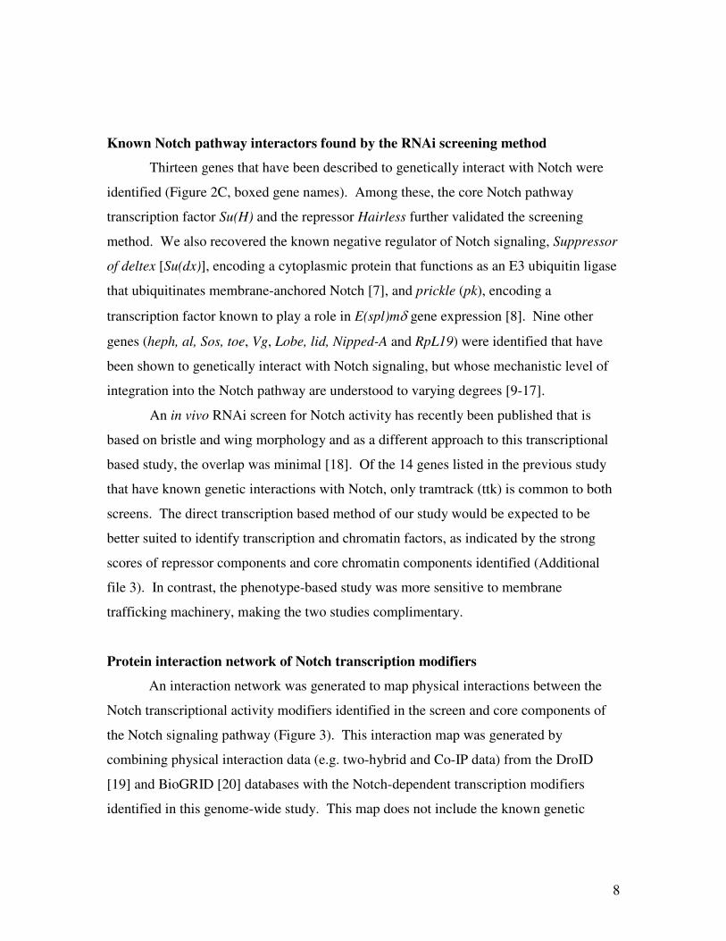
Known Notch pathway interactors found by the RNAi screening method
Thirteen genes that have been described to genetically interact with Notch were
identified (Figure 2C, boxed gene names). Among these, the core Notch pathway
transcription factor Su(H) and the repressor Hairless further validated the screening
method. We also recovered the known negative regulator of Notch signaling, Suppressor
of deltex [Su(dx)], encoding a cytoplasmic protein that functions as an E3 ubiquitin ligase
that ubiquitinates membrane-anchored Notch [7], and prickle (pk), encoding a
transcription factor known to play a role in E(spl)mδ gene expression [8]. Nine other
genes (heph, al, Sos, toe, Vg, Lobe, lid, Nipped-A and RpL19) were identified that have
been shown to genetically interact with Notch signaling, but whose mechanistic level of
integration into the Notch pathway are understood to varying degrees [9-17].
An in vivo RNAi screen for Notch activity has recently been published that is
based on bristle and wing morphology and as a different approach to this transcriptional
based study, the overlap was minimal [18]. Of the 14 genes listed in the previous study
that have known genetic interactions with Notch, only tramtrack (ttk) is common to both
screens. The direct transcription based method of our study would be expected to be
better suited to identify transcription and chromatin factors, as indicated by the strong
scores of repressor components and core chromatin components identified (Additional
file 3). In contrast, the phenotype-based study was more sensitive to membrane
trafficking machinery, making the two studies complimentary.
Protein interaction network of Notch transcription modifiers
An interaction network was generated to map physical interactions between the
Notch transcriptional activity modifiers identified in the screen and core components of
the Notch signaling pathway (Figure 3). This interaction map was generated by
combining physical interaction data (e.g. two-hybrid and Co-IP data) from the DroID
[19] and BioGRID [20] databases with the Notch-dependent transcription modifiers
identified in this genome-wide study. This map does not include the known genetic

9
interactions identified between the candidate genes and caution should be noted as to the
presence of possible false positives in the protein interaction data.
The importance of chromatin in Notch regulation has recently become apparent
and this transcription-based screen was suited to uncover this class of regulators. On
average, chromatin-modifying genes scored relatively high in the data analysis
(Additional file 3). The interaction map reveals a central core of chromatin modifying
components that have multiple physical connections to the nuclear elements of the Notch
pathway such as Su(H) and H (Figure 3B). Many of these chromatin components are
known to interact genetically and physically with the Notch pathway [14, 21-23].
The protein interaction network also shows a number of protein classes that have
no known mechanistic link to Notch transcriptional regulation. For these classes of
molecules (mRNA processing proteins and ribosomal factors, discussed later), the
network suggests that they may be affecting Notch signaling through direct interactions
with these core chromatin components (Figure 3D and E).
Epistatic analysis of candidate genes
The subset of candidate Notch modifiers that overlapped between the two
normalization methods (28 genes) was retested with redesigned dsRNAs (Additional file
5). Luciferase reporter activity was assessed in cells in which Notch had been activated
by either the membrane tethered N∆ecn or the downstream intracellular Nicd, aiming to
discriminate between factors that regulate Notch processing at the plasma membrane
versus factors that affect Notch signaling downstream in the nucleus (Figures 4 and 5).
Of the re-designed dsRNA, 79% retested by either normalization method, 67% re-tested
the m3-luc normalized signal and 64% the con-luc normalized signal. Three genes were
identified that exclusively promote the activity of the membrane bound Notch and may
function to inhibit the intramembrane proteolysis of the receptor. This class includes Patj
and two genes of unknown function, CG7099 and CG17189 (Figure 4B, Class IV and
Figure 5C, Class IV). The soluble protein Patj has not been shown to modulate Notch
activity directly, but is known to associate with the transmembrane protein Crumbs that,
in turn, is known to repress Notch activity [24]. Crumbs is a central regulator of
epithelial apical–basal polarity in Drosophila and has been shown to down regulate γ-

10
secretase activity and the membrane proteolysis of Notch [24]. Our observation in Kc167
cell culture, a non-polarized cell line, suggests that Patj may be modifying Notch
signaling not via influencing the localization of the receptor, but instead by acting in the
Crumbs-based complex to down regulate membrane proteolysis of Notch. In contrast,
RNAi against nuclear factors such as Su(H), His3.3A & B, Nipped-A, ttk and Sin3A
(Figures 4 and 5), had similar effects on N∆ecn and Nicd induced transcription,
indicating interactions with Notch downstream of the proteolytic processing events.
These results demonstrate that the screening method identified components of Notch
signaling that modulate activities that take place on the plasma membrane as well as
nuclear and chromatin-based regulation.
Factors involved in chromatin modification
The transcription-based screening method using an endogenous E(spl)m3
promoter sequence was particularly useful for identifying chromatin components. We
identified several chromatin factors previously shown to affect Notch-dependent
transcription. A component of the SAGA histone acetyltransferase complex, Nipped-A,
was identified. Nipped-A, the Drosophila homologue of yeast Tra1 and mammalian
TRAP proteins, is a key factor of the SAGA complex. It has been shown previously that
reduced Nipped-A dosage enhances the wing notching phenotype of both mastermind
and Notch mutants [14, 25]. The RNAi treated cell culture data demonstrates that
Nipped-A promotes transcription at the E(spl)m3 promoter both in the presence and
absence of activated Notch (Figures 4 and 5). This shows that the result of Nipped-A
function is independent of whether active Nicd is localized on the target promoter.
We also identified several homologues of components of the Rpd3 histone
deacetylase co-repressor complex, including Sin3a, Sds3 (CG14220), a putative ortholog
of SAP130 (Sin3A Associated Protein 130, CG11006), and Rpd3 itself (Figures 4, 5 and
Additional file 1B). When these factors were targeted by RNAi, there was an increase in
Notch-induced reporter transcription, consistent with the role of the Rpd3 complex and
histone deacetylation as a transcriptional inhibitor [26]. Conversely, knocking down
Sin3a had the opposite effect on the uninduced baseline activity of the E(spl)m3 promoter
(Figure 5C). Thus, unlike the histone acetylation complex (SAGA), the activity of the

11
deacetylation complex (Sin3A) on the E(spl)m3 promoter is dependent on the presence of
activated Notch.
The screen identified several components of the chromatin remodeling complex
Brahma: Brm Associated Protein 55 (Bap55), Brm Associated Protein 170 (Bap170),
polybromo, and moira (mor). A previous Drosophila phenotype based screen has found
a genetic interaction between the Notch ligand Delta and another component of the
Brahma complex, brahma (brm) [21]. Loss of function brm alleles were found to
enhance Delta mutant phenotypes in eye and bristle development [21]. The various
Brahma components identified in this study show a complex array of effects on the
transcription of the E(spl)m3 promoter, some consistent with previously described loss of
function brm alleles while others opposing. RNAi directed against Bap55 and polybromo
demonstrated a specific reduction in Notch induced transcription (Figure 2C, area b) that
is consistent with the previously observed role of brm in Notch signaling during
Drosophila development [21]. Unexpected are the Brahma subunits identified that
modulate transcription from the uninduced E(spl)m3 promoter: Bap170 and mor. The
screen reveals that both of these components specifically mediate transcription from the
uninduced E(spl)m3 promoter, while Bap170 activates and mor represses (Figure 2C,
area c.).
In addition to chromatin modifying complexes, a new interaction between the
histone variant H3.3 and Notch signaling is seen. RNAi treatment of either genomic
copy of the H3.3 histone variant (H3.3A and H3.3B) shows a dramatic decrease in Notch
activated transcription (Figures 4 and 5). The histone variant H3.3 has been shown to be
incorporated into the promoters of actively transcribed genes in a replication independent
process to maintain transcription and its influence on Notch targeted transcription
remains to be explored [27].
A major question that arises from these data is, how specific can the identified
chromatin factors be to regulating Notch transcription? It has recently been noted that
chromatin components are more selective in function than was previously thought.
Surprisingly, there are now a handful of examples where modulating the expression of a
single target gene can rescue the phenotype associated with a null mutation in a
chromatin remodeling complex component [28]. By immunoprecipitation and mass-spec

12
analysis, it has recently been shown that the Notch repressor complex contains a host of
chromatin modifying components [22]. These identified components include Sin3A,
Rpd3, lid, Bap55 and moira, factors that were also uncovered in this screen as modifiers
of Notch target transcription. This repressor complex has been shown to be recruited to
Notch target promoters by Su(H) and this interaction may provide a mechanism for
targeting the activity of these chromatin factors to Notch signaling [22, 23]. This is
consistent with the observation that the genetic interactions demonstrated between this
repressor complex and Notch were not seen when tested against a host of other signaling
pathways [22, 23]. Control reporter transcription levels in this study indicated that
targeting these chromatin genes by dsRNA did not significantly reduce cell viability and
growth over the course of the five-day RNAi incubation in culture. The screen data
shows that Notch signaling may be particularly sensitive to the levels of these chromatin
components in the cell, while the protein interaction network confirms that many of these
chromatin factors physically interact with Su(H) and Hairless suggesting a mechanism to
explain this observation.
Regulation of histone position and modification are known factors that determine
the "context dependent" nature of Notch signaling during development. These factors
differentially interpret the signals received from the cell surface by recording an
epigenetic history on the target promoter. This transcription-based screen revealed new
chromatin factors that can be further studied for their role in Notch-mediated
development.
mRNA processing factors
The genome-wide transcription assay revealed two other classes of proteins not
conventionally associated with transcriptional regulation. A number of ribosomal
components and proteins associated with mRNA processing were found to regulate
transcription of the activated Notch target gene (Figures 2 and 3). What is unexpected
about these interactions is their relative specificity, as was for the chromatin components.
Again, any RNAi treatments in the genome screen that significantly effected cell viability
or general transcription were excluded from the analysis. In addition, all Notch induced
target transcription signals were subsequently normalized to either the control signal or

13
the uninduced Notch target promoter. This analysis demonstrated that knocking down
these components of the ribosome and splicing machinery did not significantly affect
general cell viability and had a relatively specific effect on Notch target transcription.
A number of mRNA splicing and processing components were found to interact
with Notch-activated transcription (Figure 2A and 2C). As expected, these proteins
demonstrated extensive physical interactions with each other (Figure 3D). Unexpectedly,
these mRNA modifying proteins show physical interactions with the core chromatin
components identified in this transcription based screen (Figure 3D). The polypyrimidine
tract binding proteins Sex lethal (Sxl) and hephaestus (heph) were found to repress and
activate Notch promoter activity, respectively, in our cell culture assay. Heph was
previously found to interact genetically with Notch signaling during wing development
[12]. Other mRNA processing components, such as the non-sense mediated decay
factors Upf1, Upf2 and Smg1, were found to modulate Notch activated transcription in
the analysis (Figures 2 and 3D). These mRNA components may be interacting indirectly
with Notch transcription through their mRNA processing functions – for instance, by
specifically controlling the mRNA processing of transcripts for an essential Notch
signaling factor such as Su(H). The network suggests a possible alternate mechanism to
explain the interaction between the identified mRNA processing factors and Notch
transcription, one that is mediated though the chromatin machinery (Figure 3D).
In plants, components of the nuclear cap-binding complex (including Cbp20)
functionally interact with microRNA (miRNA) processing components, such as Ars2,
giving these proteins dual roles in splicing and miRNA processing [29]. The role of
Cbp20 in miRNA processing was also confirmed in Drosophila and mammalian systems
[30, 31]. The nuclear cap-binding complex component Cbp20 was found to mediate
Notch transcription in this study (Figure 2) and demonstrates physical interactions with
the chromatin remodeling component Ssrp (Figure 3D). The interaction network
suggests that the miRNA processing activity of Cbp20 may be targeted to Notch
signaling through interactions with the chromatin remodeling machinery.
Ribosomal factors and the classical Minute mutations
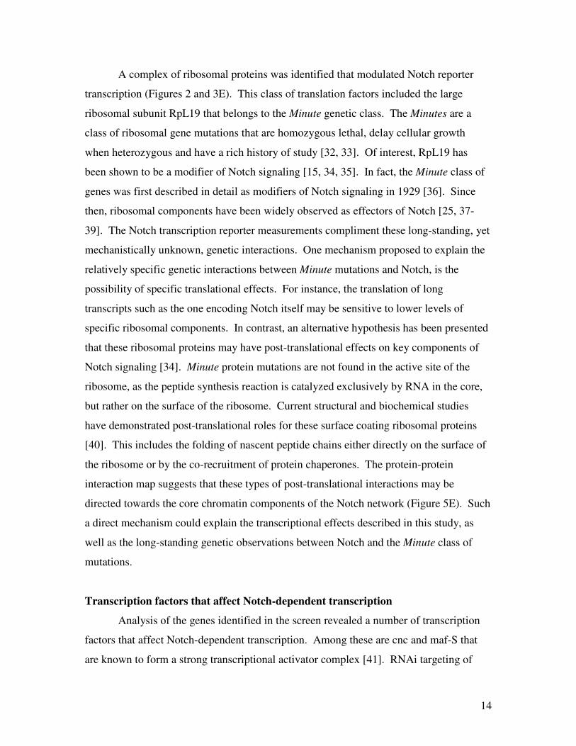
14
A complex of ribosomal proteins was identified that modulated Notch reporter
transcription (Figures 2 and 3E). This class of translation factors included the large
ribosomal subunit RpL19 that belongs to the Minute genetic class. The Minutes are a
class of ribosomal gene mutations that are homozygous lethal, delay cellular growth
when heterozygous and have a rich history of study [32, 33]. Of interest, RpL19 has
been shown to be a modifier of Notch signaling [15, 34, 35]. In fact, the Minute class of
genes was first described in detail as modifiers of Notch signaling in 1929 [36]. Since
then, ribosomal components have been widely observed as effectors of Notch [25, 37-
39]. The Notch transcription reporter measurements compliment these long-standing, yet
mechanistically unknown, genetic interactions. One mechanism proposed to explain the
relatively specific genetic interactions between Minute mutations and Notch, is the
possibility of specific translational effects. For instance, the translation of long
transcripts such as the one encoding Notch itself may be sensitive to lower levels of
specific ribosomal components. In contrast, an alternative hypothesis has been presented
that these ribosomal proteins may have post-translational effects on key components of
Notch signaling [34]. Minute protein mutations are not found in the active site of the
ribosome, as the peptide synthesis reaction is catalyzed exclusively by RNA in the core,
but rather on the surface of the ribosome. Current structural and biochemical studies
have demonstrated post-translational roles for these surface coating ribosomal proteins
[40]. This includes the folding of nascent peptide chains either directly on the surface of
the ribosome or by the co-recruitment of protein chaperones. The protein-protein
interaction map suggests that these types of post-translational interactions may be
directed towards the core chromatin components of the Notch network (Figure 5E). Such
a direct mechanism could explain the transcriptional effects described in this study, as
well as the long-standing genetic observations between Notch and the Minute class of
mutations.
Transcription factors that affect Notch-dependent transcription
Analysis of the genes identified in the screen revealed a number of transcription
factors that affect Notch-dependent transcription. Among these are cnc and maf-S that
are known to form a strong transcriptional activator complex [41]. RNAi targeting of

15
either of these two genes strongly suppressed both the Notch-induced as well as non-
induced E(spl)m3 reporter activity (Additional file 1A). Also, among the 15 transcription
factors that promote Notch activity, we found the DNA binding protein Deaf-1. Cnc,
maf-S, and Deaf-1 are reported to interact with the Hox protein Deformed (Dfd) to
regulate segmentation, but their roles in other developmental events are not known [42].
Our results provide a possible role of these proteins in Drosophila development by
promoting Notch signaling.
Another transcription factor that we found to play an agonistic role in Notch
signaling is the homeobox containing protein Aristaless (al) (Figure 5B). Al has been
tentatively linked to Notch signaling, as it cell autonomously represses the Notch ligand
Delta in the pretarsus during leg morphogenesis [10]. It is possible that al is involved in a
Notch-mediated lateral inhibition mechanism, where al expressing cells remain
undifferentiated by favoring active Notch signaling whereas their neighboring cells are
free to express Delta and differentiate. It has also been shown that Notch mutant clones
in the developing leg disk show diminished al levels, suggesting that al is a Notch target
gene. This would be the predicted relationship in a lateral inhibition system, where a
Notch/al positive feedback loop would amplify the Notch activity differences between
neighboring cells.
Two additional transcription factors that have been previously shown to be
involved in leg morphogenesis were found to promote Notch signaling: Bonus (bon), a
homologue of the vertebrate TIF1beta transcriptional cofactor [43], and crooked legs
(croI), a zinc finger protein [44]. Notch signaling is known to play an important role in
Drosophila leg development, and the recovery of these two transcription factors as
modifiers of Notch-induced E(spl)m3 expression suggests that bon and croI may function
to modulate Notch target gene output in the developing leg [45].
We also identified the Drosophila orthologues of two mammalian proto-
oncogenes kayak (c-fos), and c-Myb, as positive regulators of Notch-signaling. Although
a direct functional link between these proteins and Notch signaling has not been
described, kayak has been shown to interact genetically with Hairless [46] and c-Myb
genetically interacts with bon, a novel Notch modifier described above [47]. In addition,
our data reveals a synergistic relationship between the positive regulator of Ras signaling,

16
14-3-3ε, and Notch. Once again, the protein interaction network shows extensive
contacts between 14-3-3 ε and the chromatin machinery, suggesting a mechanism for
modulating Notch target transcription through Su(H) mediated chromatin modifications.
Interactions between Notch and oncogenic pathways are of particular interest, as the
involvement of Notch in cancer biology and stem cell maintenance is becoming
increasingly apparent.
An unexpected Notch target transcription modifier identified in the screen is the
Notch target gene Tramtrack (ttk). We found that targeting of ttk with dsRNA resulted in
reduced Notch activity (Figures 4 and 5). In contrast, ttk expression itself has been
shown to increase in response to ectopic Notch activity [48]. The RNAi treatment data
suggest that ttk may function in a positive feedback mechanism to promote Notch
activated transcription and the network analysis suggests that this interaction may be
mediated by a direct contact with Notch itself (Figure 3C).
Conclusions
A complementary, genome-wide RNAi approach has revealed a subset of factors
that modulate Notch target transcription that may not have been found by traditional
genetic approaches. For instance, pleiotropic effects combined with non-saturating
mutagenesis may have obscured the detection of some components in traditional genetic
screens. Several novel modifiers were identified in this RNAi transcription-based screen,
and these factors will be further investigated for their precise roles in the regulation of
Notch signaling during development. In addition, the interaction network of these factors
suggests that many may work through contacts with chromatin machinery components
that are in turn directed to Notch target promoters by the transcription factor Su(H).

17
Methods
DNA constructs
Constitutively active Notch constructs were made with cDNA encoding either
membrane-tethered, Drosophila Notch (N∆ecn), constitutively activated by the removal
of the extracellular domain, or the soluble intracellular domain (Nicd) [2]. These
truncated Notch constructs were cloned into the pIZ-V5/His expression vector
(Invitrogen) producing non-tagged proteins. The control expression plasmid (con-luc)
was constructed by cloning firefly luciferase into the same pIZ-V5/His expression vector.
The luciferase reporter construct (m3-luc) contains a 1.4 kb tandem duplication of
E(spl)m3 upstream regulatory sequences, cloned into a pGL2-Basic vector (Promega), as
described [3].
Genome-wide RNAi method
A total of 23,560 dsRNAs, made available from the Drosophila RNAi Screening
Center (DRSC) at Harvard Medical School, were screened by the following method:
Kc167 cells were washed three times and resuspended in serum-free Sang’s M3 medium
(Sigma) at a concentration of 5x10⁵ cells/ml. Using a robotic liquid handler, 10
4 cells (20
µl) were uniformly dispensed into the wells of 384-well polypropylene plates containing
dsRNA and incubated for 45 min at room temperature. An equal volume of M3 medium
containing 10% fetal bovine serum was added and incubated for four days. On day four,
the RNAi-treated cells were diluted with 100 µl of medium, mixed and 20 µl were
dispensed into the wells of six new 384-well plates, pre-aliquoted with 20 µl of
transfection mix. The six plates contained the three different transfection mixes, each in
duplicate. Transfection mixes were prepared with Effectene Transfection Reagent
(Qiagen), following the manufacturer’s guidelines. Luciferase activity was measured 24
h post transfection using the Steady-Glo Luciferase Assay System (Promega).
This method requires only two plasmids to be transfected at one time and gave
acceptable signal to noise ratios for high throughput screening in 384-well plate format
(Figure 1). Whereas, the conventional renilla dual-glo assay was not robust enough to
scale to 384 well format with this Notch reporter system using an endogenous target. In
contrast to pathways with soluble ligands, the reporter and constitutively active Notch

18
constructs are required to transfect the same cell to activate transcription. With the
renilla dual-glo system, adding the control construct required the co-transfection of three
individual plasmids and this reduced the signal to noise ratio to insufficient levels.
Data analysis
Duplicate measurements for each of the three signals were averaged; Notch
specific E(spl)m3 promoter in the presence of activated Notch (N∆ecn > m3-luc), the
E(spl)m3 promoter alone (m3-luc) and the unrelated viral promoter OplE2 (con-luc). The
N∆ecn > m3-luc signal was normalized two different ways, by either the m3-luc or con-
luc signals. The z-scores of the log2 ratios were calculated by using the standard
deviation and mean of the measurements that corresponded to the 96-wells of the original
dsRNA stock plates [49]. To remove ratios that contained data that did not sufficiently
replicate in the original duplicate measurements, a distribution was calculated for the
individual errors (estimated from the duplicate data sets) and ratios with an associated
error z-score above 3 were removed from further analysis (0.9% of the total data).
To remove data associated with dsRNA that greatly reduced general transcription
or cell viability, a distribution of the signals from the control promoter (con-luc) was
calculated, and data with z-scores below -2 were removed (4% of the total wells). All
calculations were done by in house software written in JAVA.
Hits were chosen as those log2 ratios with a z-score above 2 or below -2 for
N∆ecn > m3-luc normalized by the viral promoter OplE2 (con-luc). For the data set
normalized by the E(spl)m3 promoter alone (m3-luc), a z-score above 1.8 or below -1.8
was used. The m3-luc normalized distribution had more defined outliers indicating a
better data set. As a consequence, m3-luc normalized data distribution had higher
kurtosis as seen by a slightly sharper peak in Figure 2. This does not change the rank
order or relative differences in the hits of that data set, but to make the cut-offs more
equivalent between the two normalization methods, the different cut-off values were
used.
RNAi retest procedure

19
Genes were chosen for retesting that were selected as positive by both
normalization methods (Figure 2c, area a.). This second set of 28 dsRNAs were
independently redesigned by the method of Arziman et al. with no predicted off targets
and are listed in Additional file 5 [50].
DNA templates for T7 reactions were generated by PCR from Kc167 cell
genomic DNA and dsRNA was produced using the MEGAscript RNAi kit (Ambion).
Per well, 25 ml of Kc167 cells at a concentration of 8 x 10⁵ cells/ml were incubated with
1.25 µg of dsRNA (0.5 mg/ml stock) for 1 h in serum-free M3 medium. M3 medium
with 10% FBS (75 µl) was then added and incubated for 4 days. On the fourth day, 125
µl of medium was added, and treated cells were split into 4 wells with 50 µl per well,
each containing 50 µl of the following transfection mixes, prepared as above: a. con-luc,
b. m3-luc, c. m3-luc & pIZ-N∆ecn, d. m3-luc & pIZ-Nicd. Luciferase levels were
measured after 25 h, as above. Retests were done in quadruplicate for each dsRNA, and
the results are given in Additional file 5 for the 22 positive retests that have p-values <
0.05 (compared with control dsRNA treated cells).
Notch interaction network construction
The Notch interaction network was generated by combining physical interaction
data (e.g. two-hybrid data) from the DroID database [19] (that contained interactions
from the BioGRID database [20]) with Notch transcription modifiers identified in the
genome-wide study. Genetic interactions were not used for the network map. The
resulting network was drawn using Cytoscape [51].
Orthology prediction
All orthology predictions for candidate genes were made using InParanoid [52].

20
Authors' contributions
PM, RL and BD designed and performed experiments, analyzed data and wrote
the manuscript. CF performed experiments and analyzed data. All authors read and
approved the final manuscript.
Acknowledgments
We thank S. Artavanis-Tsakonas for generous support of this work. For providing
the RNAi library, we thank N. Perrimon and the Drosophila RNAi Screening Center
(DRSC). Screening resources and expertise were provided by C. Shamu and the ICCB –
Longwood screening facility.
This work was supported by NIH grants HG003616 and NS26084 under S.
Artavanis-Tsakonas. The Drosophila RNAi Screening Center (DRSC) is supported by
grant 2R01GM067761-05 from the NIGMS division of NIH.

21
Figure legends
Figure 1. Validation of the Notch activity reporter and application for high
throughput RNAi.
Validation of the m3-luc reporter by RNAi targeting of known Notch pathway
components. Notch induced E(spl)m3 signal normalized against A. the control viral
promoter or B. uninduced E(spl)m3 promoter transcription. C. Uninduced E(spl)m3
promoter expression normalized by control promoter. For each dsRNA, 32 independent
wells were measured in a 384-well plate format. Each box encloses 50% of the data with
the median value displayed. The error bars mark the full range excluding the shown
outliers. D. Schematic of the automated high throughput screen in 384-well plates.
Drosophila Kc167 cells were incubated with a unique dsRNA per well. After a four day
incubation, the cells were split into three different transfection mixes in duplicate. Firefly
luciferase signals were read 24 h after transfection.
Figure 2. RNAi data analysis overview.
Histograms of targeted genes binned by standard deviations from the mean (z-scores). A.
Histogram of z-scores for Notch-induced E(spl)m3 reporter (N∆ecn > m3-luc)
normalized by the signal from the control reporter (con-luc) (Additional file 1A and B).
B. Signals of Notch-induced E(spl)m3 reporter (N∆ecn > m3-luc) normalized by the
signal from the non-induced E(spl)m3 reporter (Additional file 2A and B). Cutoffs for
genes selected are highlighted in grey for both A and B. C. Plot of the z-scores from
histogram 2A on the x-axis and histogram 2B on the y-axis. Regions outside the red box
are listed as potential hits and the overlaps between the two normalization methods are
shaded in blue (area a). Area a. represents the subset of genes that either affect Notch
induced transcription specifically or have opposing effects on induced and non-induced
reporter transcription (e.g. Su(H)). RNAi for this overlapping set was redesigned and
retested (Additional file 5). Area b. represents genes that affect both Notch induced and
non-induced transcription by similar percent amounts (e.g. heph and Bap55). Area c.
represents genes that primarily affect non-induced reporter transcription specifically (e.g.
H, RpL19 and Bap170). Red and/or boxed genes have known genetic interactions with

22
Notch. Blue are chromatin components, Yellow are mRNA processing factors, and
Green are ribosomal components (Minute class).
Figure 3. Protein-protein interaction map of Notch transcription modifiers.
The Notch interaction network was generated by connecting the Notch transcription
modifiers identified in the genome-wide study with protein-protein interaction links (e.g.
two-hybrid and Co-IP data from the DroID database [19]). This resulting network
included 126 genes (nodes) with 237 physical interactions (edges). Genetic interactions
were not used for the network and the resulting map was drawn using Cytoscape [51]. A.
These physical links are shown in relation to components of the activated Notch pathway
(N and Su(H)) and the Notch repressor complex (Su(H), H, CtBP and gro), shown in red.
B. Expanded view of the chromatin factors identified in this study that form the central
core of the interaction network (blue). C. Ttk is a known downstream target of Notch
signaling. The transcriptional and physical interaction data suggests that this factor may
have a positive feedback role in Notch induced transcription. D. Factors with roles in
mRNA processing (yellow). The interaction network suggests that these proteins may be
working though the chromatin machinery to modulate Notch transcription. E. The
interaction network suggests the possibility of a similar chromatin based mechanism for
the class of ribosomal proteins known as Minute. The network file is included with the
supplemental data (Additional file 6) and can be viewed in detail using the open source
Cytoscape viewer (www.cytoscape.org).
Figure 4. Analysis of retested genes.
A. Retested genes (from selected set, Figure 2C area a) that show significantly reduced
signaling when down regulated by RNAi. Signals are shown as (+/-) percent deviation
from the control RNAi signal. Three general classes are shown. All three classes down
regulate both soluble (Nicd) and membrane bound (N∆ecn) Notch-induced signal, yet
have different effects on the E(spl)m3 promoter in the absence of active Notch. Class I
genes have positive, Class II neutral and Class III negative effect on the uninduced signal.
B. Selected set of retested genes that show significantly enhanced signaling when down
regulated by RNAi. Two classes of hits are noted. Class IV is only effective on the

23
membrane bound form of Notch (N∆ecn), while class V is effective on both membrane
bound and soluble forms (Nicd). All deviations are calculated to be significant by two-
tailed t-test with p-values < 0.05 from control RNAi treatment (Additional file 5 for full
statistics).
Figure 5. Modulation of Notch transcription for subset of retested genes.
A. The two constitutively active Notch constructs used to determine epistatic
relationships in the pathway, N∆ecn and Nicd. N∆ecn is a truncated form of N missing
the extracellular domain that is initially membrane bound. N∆ecn undergoes constitutive
cleavage to form the soluble Nicd that is transported to the nucleus to activate
transcription. Su(H) is the canonical Notch pathway transcription factor that represses
transcription in the absence of Nicd and is essential for the Nicd activated transcription of
targets such as E(spl)m3. B. Transcriptional response to RNAi treatment of selected
retested genes that promote Notch signaling. The E(spl)m3 reporter was induced with
either N∆ecn or Nicd or left in the uninduced repressed state. All three classes down
regulate both soluble (Nicd) and membrane bound (N∆ecn) Notch-induced signal, yet
have different effects on the E(spl)m3 promoter in the absence of active Notch. Class I
genes have positive, Class II neutral and Class III negative effect on the repressed signal.
C. Transcriptional response to RNAi treatment of selected retested genes that repress
Notch signaling. Two classes of hits are noted. Class IV is only effective on the
membrane bound form of Notch (N∆ecn), while class V is effective on both membrane
bound and soluble forms (Nicd). Error bars represent the standard error of the mean
(SEM). *Significant deviation from control RNAi treatment, calculated by two-tailed t-
test with a p-value < 0.05 (Additional file 5 for full statistics).

24
Tables
Table 1. Cellular distribution of Notch modifiers selected by the complimentary
analysis methods.
Analysis Method Total Extracellular Membrane Cytosolic Nuclear Unknown
N∆ecn > m3-luc / con-luc
(Activators) 153 8 (5.2%) 2 (2.6%) 28 (18.3%) 40 (26.1%) 73 (47.7%)
N∆ecn > m3-luc / con-luc
(Repressors) 130 0 5 (3.8%) 17 (13.1%) 27 (20.8%) 81 (62.3%)
N∆ecn > m3-luc / m3-luc
(Activators) 75 0 1 (1.3%) 12 (16.0%) 28 (37.3%) 34 (45.3%)
N∆ecn > m3-luc / m3-luc
(Repressors) 74 0 2 (2.7%) 13 (17.6%) 5 (20.3%) 44 (59.5%)
Total (discounting
duplicates) 399 2% 3% 16% 26% 53%
The majority of the proteins are of unknown cellular localization and/or function.
Of the annotated proteins, most are predicted to reside in the nucleus, followed by
cytosolic proteins with a small percentage found in the plasma membrane and
extracellular matrix.

25
Additional files
Additional file 1
Title: A. Excel table of screening data for Notch induced transcription normalized by the
unrelated control promoter (N∆ecn > m3-luc / con-luc).
Description: Hits listed from initial screen with potential agonists of Notch induced
transcription listed in A. and potential antagonists listed in B.
Additional file 2
Title: Excel table of screening data for Notch induced transcription normalized by the
uninduced E(spl)m3 promoter (N∆ecn > m3-luc / m3-luc).
Description: Hits listed from initial screen with potential agonists of Notch induced
transcription or antagonists of uninduced E(spl)m3 transcription listed in A. and vice
versa in B.
Additional file 3
Title: Figure representing the distribution of Notch modifiers by gene ontology classes.
Description: Pie chart distributions for percentage of genes represented in major gene
ontology classes and corresponding box plots of median z-scores for the various classes.
Box plots are graphed as in Figure 1a. A. Distribution of genes that enhance the Notch
induced signal as normalized by the uninduced E(spl)m3 promoter. B. Distribution of
genes that suppress the Notch induced signal as normalized by the uninduced E(spl)m3
promoter. C. Distribution of genes that enhance the Notch induced signal as normalized
by the unrelated control promoter (con-luc). D. Distribution of genes that suppress the
Notch induced signal as normalized by con-luc.
Additional file 4
Title: Excel table of orthology predictions
Description: Human orthologs of the Drosophila genes identified in the screen were
predicted using InParanoid [52]. Human diseases associated with the predicted genetic
counterparts are also listed.

26
Additional file 5
Title: Excel table of re-test data for the redesigned dsRNA
Description: Genes were chosen for retesting that were selected as positive by both
normalization methods (Figure 2c, area a.). This second set of 28 dsRNAs were
independently redesigned by the method of Arziman et al. with no predicted off targets
and are listed in Additional file 5 [50]. Retests were done in quadruplicate for each
dsRNA, and the results are given for the 22 positive retests that have p-values < 0.05
(compared with control dsRNA treated cells).
Additional file 6
Title: Notch interaction network file.
Description: A network file that can be viewed in detail using the open source
Cytoscape viewer (www.cytoscape.org) [51]. The Notch interaction network was
generated by using Notch transcription modifiers identified in the genome-wide study as
nodes and physical interactions (e.g. two-hybrid data) for edges. Genetic interactions
were not used for the network map.

27
References
1. S Artavanis-Tsakonas, M Rand, R Lake: Notch signaling: cell fate control and
signal integration in development. Science 1999, 284:770-6.
2. I Rebay, RG Fehon, S Artavanis-Tsakonas: Specific truncations of Drosophila
Notch define dominant activated and dominant negative forms of the
receptor. Cell 1993, 74:319-29.
3. A Mukherjee, A Veraksa, A Bauer, C Rosse, J Camonis, S Artavanis-Tsakonas:
Regulation of Notch signalling by non-visual beta-arrestin. Nat Cell Biol
2005, 7:1191-201.
4. A Krejcí, S Bray: Notch activation stimulates transient and selective binding
of Su(H)/CSL to target enhancers. Genes & Development 2007, 21:1322-7.
5. R Wilson, J Goodman, V Strelets: FlyBase: integration and improvements to
query tools. Nucleic Acids Res 2008, 36:D588-93.
6. L Reiter, L Potocki, S Chien, M Gribskov, E Bier: A systematic analysis of
human disease-associated gene sequences in Drosophila melanogaster.
Genome Res 2001, 11:1114-25.
7. M Fostier, D Evans, S Artavanis-Tsakonas, M Baron: Genetic characterization
of the Drosophila melanogaster Suppressor of deltex gene: A regulator of
notch signaling. Genetics 1998, 150:1477-85.
8. M Cooper, S Bray: Frizzled regulation of Notch signalling polarizes cell fate in
the Drosophila eye. Nature 1999, 397:526-30.
9. R Abu-Issa, S Cavicchi: Genetic interactions among vestigial, hairy, and
Notch suggest a role of vestigial in the differentiation of epidermal and
neural cells of the wing and halter of Drosophila melanogaster. J Neurogenet
1996, 10:239-46.
10. G Campbell: Regulation of gene expression in the distal region of the
Drosophila leg by the Hox11 homolog, C15. Dev Biol 2005, 278:607-18.
11. J Chern, K Choi: Lobe mediates Notch signaling to control domain-specific
growth in the Drosophila eye disc. Development 2002, 129:4005-13.
12. D Dansereau, M Lunke, A Finkielsztein, M Russell, W Brook: Hephaestus
encodes a polypyrimidine tract binding protein that regulates Notch
signalling during wing development in Drosophila melanogaster.
Development 2002, 129:5553-66.
13. M Dominguez, D Ferres-Marco, F Gutierrez-Avino, S Speicher, M Beneyto:
Growth and specification of the eye are controlled independently by Eyegone
and Eyeless in Drosophila melanogaster. Nat Genet 2004, 36:31-9.
14. M Gause, JC Eissenberg, AF Macrae, M Dorsett, Z Misulovin, D Dorsett:
Nipped-A, the Tra1/TRRAP subunit of the Drosophila SAGA and Tip60

28
complexes, has multiple roles in Notch signaling during wing development.
Mol Cell Biol 2006, 26:2347-59.
15. K Hart, T Klein, M Wilcox: A Minute encoding a ribosomal protein enhances
wing morphogenesis mutants. Mech Dev 1993, 43:101-10.
16. E Verheyen, K Purcell, M Fortini, S Artavanis-Tsakonas: Analysis of dominant
enhancers and suppressors of activated Notch in Drosophila. Genetics 1996,
144:1127-41.
17. R Liefke, F Oswald, C Alvarado, D Ferres-Marco, G Mittler, P Rodriguez, M
Dominguez, T Borggrefe: Histone demethylase KDM5A is an integral part of
the core Notch-RBP-J repressor complex. Genes & Development 2010, 24:590-
601.
18. JL Mummery-Widmer, M Yamazaki, T Stoeger, M Novatchkova, S Bhalerao, D
Chen, G Dietzl, BJ Dickson, JA Knoblich: Genome-wide analysis of Notch
signalling in Drosophila by transgenic RNAi. Nature 2009, 458:987-92.
19. J Yu, S Pacifico, G Liu, RL Finley: DroID: the Drosophila Interactions
Database, a comprehensive resource for annotated gene and protein
interactions. BMC Genomics 2008, 9:461.
20. B-J Breitkreutz, C Stark, T Reguly, L Boucher, A Breitkreutz, M Livstone, R
Oughtred, DH Lackner, J Bähler, V Wood, et al: The BioGRID Interaction
Database: 2008 update. Nucleic Acids Res 2008, 36:D637-40.
21. JA Armstrong, AS Sperling, R Deuring, L Manning, SL Moseley, O Papoulas, CI
Piatek, CQ Doe, JW Tamkun: Genetic screens for enhancers of brahma reveal
functional interactions between the BRM chromatin-remodeling complex
and the delta-notch signal transduction pathway in Drosophila. Genetics
2005, 170:1761-74.
22. YM Moshkin, TW Kan, H Goodfellow, K Bezstarosti, RK Maeda, M Pilyugin, F
Karch, SJ Bray, JAA Demmers, CP Verrijzer: Histone chaperones ASF1 and
NAP1 differentially modulate removal of active histone marks by LID-RPD3
complexes during NOTCH silencing. Mol Cell 2009, 35:782-93.
23. H Goodfellow, A Krejcí, Y Moshkin, CP Verrijzer, F Karch, SJ Bray: Gene-
specific targeting of the histone chaperone asf1 to mediate silencing.
Developmental Cell 2007, 13:593-600.
24. H Herranz, E Stamataki, F Feiguin, M Milan: Self-refinement of Notch activity
through the transmembrane protein Crumbs: modulation of gamma-
secretase activity. EMBO Rep 2006, 7:297-302.
25. R Rollins, P Morcillo, D Dorsett: Nipped-B, a Drosophila homologue of
chromosomal adherins, participates in activation by remote enhancers in the
cut and Ultrabithorax genes. Genetics 1999, 152:577-93.
26. A Wolffe: Histone deacetylase: a regulator of transcription. Science 1996,
272:371-2.
27. K Ahmad, S Henikoff: The histone variant H3.3 marks active chromatin by
replication-independent nucleosome assembly. Mol Cell 2002, 9:1191-200.
28. L Ho, GR Crabtree: Chromatin remodelling during development. Nature 2010,
463:474-84.
29. S Laubinger, T Sachsenberg, G Zeller, W Busch, JU Lohmann, G Rätsch, D
Weigel: Dual roles of the nuclear cap-binding complex and SERRATE in

29
pre-mRNA splicing and microRNA processing in Arabidopsis thaliana. Proc
Natl Acad Sci USA 2008, 105:8795-800.
30. JJ Gruber, DS Zatechka, LR Sabin, J Yong, JJ Lum, M Kong, W-X Zong, Z
Zhang, C-K Lau, J Rawlings, et al: Ars2 links the nuclear cap-binding complex
to RNA interference and cell proliferation. Cell 2009, 138:328-39.
31. LR Sabin, R Zhou, JJ Gruber, N Lukinova, S Bambina, A Berman, C-K Lau, CB
Thompson, S Cherry: Ars2 regulates both miRNA- and siRNA- dependent
silencing and suppresses RNA virus infection in Drosophila. Cell 2009,
138:340-51.
32. G Morata, P Ripoll: Minutes: mutants of drosophila autonomously affecting
cell division rate. Dev Biol 1975, 42:211-21.
33. A Lambertsson: The minute genes in Drosophila and their molecular
functions. Adv Genet 1998, 38:69-134.
34. LE Hall, SJ Alexander, M Chang, NS Woodling, B Yedvobnick: An EP
overexpression screen for genetic modifiers of Notch pathway function in
Drosophila melanogaster. Genet Res 2004, 83:71-82.
35. T Klein, JA Campos-Ortega: Second-site modifiers of the Delta wing
phenotype in Drosophila melanogaster. Roux's Archives of Developmental
Biology 1992, 202:49-60.
36. J Schultz: The Minute Reaction in the Development of DROSOPHILA
MELANOGASTER. Genetics 1929, 14:366-419.
37. U Dietrich, JA Campos-Ortega: The expression of neurogenic loci in imaginal
epidermal cells of Drosophila melanogaster. J Neurogenet 1984, 1:315-32.
38. G Röttgen, T Wagner, U Hinz: A genetic screen for elements of the network
that regulates neurogenesis in Drosophila. Mol Gen Genet 1998, 257:442-51.
39. A Schmidt, M Hollmann, U Schäfer: A newly identified Minute locus,
M(2)32D, encodes the ribosomal protein L9 in Drosophila melanogaster. Mol
Gen Genet 1996, 251:381-7.
40. LD Cabrita, CM Dobson, J Christodoulou: Protein folding on the ribosome.
Current Opinion in Structural Biology 2010, 20:33-45.
41. A Veraksa, N McGinnis, X Li, J Mohler, W McGinnis: Cap 'n' collar B
cooperates with a small Maf subunit to specify pharyngeal development and
suppress deformed homeotic function in the Drosophila head. Development
2000, 127:4023-37.
42. A Veraksa, J Kennison, W McGinnis: DEAF-1 function is essential for the
early embryonic development of Drosophila. genesis 2002, 33:67-76.
43. R Beckstead, J Ortiz, C Sanchez, S Prokopenko, P Chambon, R Losson, H Bellen:
Bonus, a Drosophila homolog of TIF1 proteins, interacts with nuclear
receptors and can inhibit betaFTZ-F1-dependent transcription. Mol Cell
2001, 7:753-65.
44. P D'Avino, C Thummel: crooked legs encodes a family of zinc finger proteins
required for leg morphogenesis and ecdysone-regulated gene expression
during Drosophila metamorphosis. Development 1998, 125:1733-45.
45. J de Celis, D Tyler, J de Celis, S Bray: Notch signalling mediates segmentation
of the Drosophila leg. Development 1998, 125:4617-26.

30
46. D Muller, S Kugler, A Preiss, D Maier, A Nagel: Genetic modifier screens on
Hairless gain-of-function phenotypes reveal genes involved in cell
differentiation, cell growth and apoptosis in Drosophila melanogaster.
Genetics 2005, 171:1137-52.
47. T Nomura, J Tanikawa, H Akimaru, C Kanei-Ishii, E Ichikawa-Iwata, M Khan, H
Ito, S Ishii: Oncogenic activation of c-Myb correlates with a loss of negative
regulation by TIF1beta and Ski. J Biol Chem 2004, 279:16715-26.
48. M Guo, L Jan, Y Jan: Control of daughter cell fates during asymmetric
division: interaction of Numb and Notch. Neuron 1996, 17:27-41.
49. J Quackenbush: Microarray data normalization and transformation. Nat
Genet 2002, 32 Suppl:496-501.
50. Z Arziman, T Horn, M Boutros: E-RNAi: a web application to design
optimized RNAi constructs. Nucleic Acids Res 2005, 33:W582-8.
51. P Shannon, A Markiel, O Ozier, NS Baliga, JT Wang, D Ramage, N Amin, B
Schwikowski, T Ideker: Cytoscape: a software environment for integrated
models of biomolecular interaction networks. Genome Res 2003, 13:2498-504.
52. A-C Berglund, E Sjölund, G Ostlund, ELL Sonnhammer: InParanoid 6:
eukaryotic ortholog clusters with inparalogs. Nucleic Acids Res 2008,
36:D263-6.

4 d a y s D r o s o p h i l aK c 1 6 7 c e l l s+ 2 3 , 5 6 0 d s R N A1 d a yN ∆
ecn> m3 %l uc/ con %l uc
m3 %l uc/ con %l uc 0 . 10 . 20 . 3 0 . 40 . 60 . 8c o n t r o l S u ( H ) m a m H
c o n t r o l S u ( H ) m a m H m e a s u r e l u c i f e r a s e s i g n a l sc o n A l u c m 3 A l u c N∆
e c n > m 3 A l u c
2 01 051 5c o n t r o l S u ( H ) m a m HN ∆
ecn> m3 %l uc/ m3 %l uc
Figure 1

0 1000 2000count0 2 4 6� 2� 4� 6N
∆
e c n > m 3 � l u c / m 3 � l u c ( z � s c o r e )S u ( H )H
04008001200count
0 2 4 6� 2� 4� 6N∆
e c n > m 3 � l u c / c o n � l u c ( z � s c o r e )S u ( H )
S u ( H )� 8� 6� 4� 2 0 246 8
� 8 � 6 � 4 � 2 0 2 4 6 8N ∆
ecn> m3 2l uc/ m3 2l uc( z 2score)
N∆
e c n > m 3 � l u c / c o n � l u c ( z � s c o r e )s e l e c t e d &r e t e s t e d H S u ( d x )
s e l e c t e d
s e l e c t e d s e l e c t e d &r e t e s t e d
Lp k
R p L 6S o st o ev g
a la
ab b
c
cB a p 5 5
B a p 1 7 0m o r
p o l y b r o m oU p f 1 S m g 1h e p h C b p 2 0 S x l
N i p p e d � AR p S 1 7R p L 1 9
R S 1 0 bR S 9T r i p 1R p L 1 9
h e p hN i p p e d i A
S i n 3 AH 3 .3 AU p f 2t t k
T i p 6 0R p d 3
S i n 3 A
H i s 3 . 3 A H i s 3 . 3 B
l i dH c f
S s r pE ( P c )D M A P 1H 3 .3 A
N i p p e d i A l i dS i n 3 AC b p 2 0
Figure 2

A B
C D E
p d m 2H S u ( H )N
d e ib i p 2 t t k
R p d 3 C G 1 4 2 2 0S i n 3 AN i p p e d � AH i s 3 . 3 AH i s 3 . 3 B
H c f S s r pm o rB a p 1 7 0p o l y b r o m oD M A P 1B a p 5 5C t B Pg r o
M E D 1 7C d k 8S u ( H )N
E ( P c )l i dT i p 6 0
S m g 1 e c aU p f 2C G 3 1 9 1 7
M y b1 4 � 3 � 3
ε
S x l R p L 6C d k 9 h e p hR p I 1U p f 1 T r i p 1C b p 2 0 R p S 1 7H S u ( H )C t B Pg r o R p d 3S i n 3 AH i s 3 . 3 AH i s 3 . 3 B T i p 6 0R p d 3 S s r pm o r R a t 1 K l p 6 7 An c dA t g 1a l dC G 9 2 9 3
R p S 1 0 bR p S 9R p L 1 9
H S u ( H )NC t B Pg r o C h r o m a t i n f a c t o r s
R i b o s o m a l f a c t o r sm R N A p r o c e s s i n g
Figure 3

> 6 0> 4 0> 2 0n o t s i g n i f i c a n t< � 2 0I VVH i s 3 . 3 AH i s 3 . 3 BS u ( H )t t kl ( 2 ) N C 1 3 6C G 7 5 3 0N i p p e d � Ai a la lt l kn o r dC G 1 8 3 0 4
II II I I > 3 0> 1 5n o t s i g n i f i c a n t< � 1 5< � 3 0< � 4 5< � 6 0C G 1 7 1 8 9P a t jC G 7 0 9 9S i n 3 AC G 1 4 2 2 0C G 6 0 3 8C G 5 1 0 7C G 1 0 3 3 3C G 1 8 1 4C G 3 7 0 2N
∆ e c n > m 3 2l u c / m 3 2l u cN∆ e c n > m 3 2l u c / c o n 2l u cN i c d > m 3 2l u c / c o n 2l u cm 3 2l u c / c o n 2l u c N
∆ e c n > m 3 2l u c / m 3 2l u cN∆ e c n > m 3 2l u c / c o n 2l u cN i c d > m 3 2l u c / c o n 2l u cm 3 2l u c / c o n 2l u c
% c h a n g e % c h a n g eFigure 4

I .I I . I I I .
I V . V .
n u c l e u sN
∆
e c nN i c dND e l t a
S u ( H )E ( s p l ) m 30 . 80 . 60 . 40 . 2 0
N∆
e c n > m 3 ' l u cc o n ' l u c N i c d > m 3 ' l u cc o n ' l u c m 3 ' l u cc o n ' l u cN
∆
e c n > m 3 ' l u cc o n ' l u c N i c d > m 3 ' l u cc o n ' l u c m 3 ' l u cc o n ' l u cN
∆
e c n > m 3 ' l u cc o n ' l u c N i c d > m 3 ' l u cc o n ' l u c m 3 ' l u cc o n ' l u c
N∆
e c n > m 3 ' l u cc o n ' l u c N i c d > m 3 ' l u cc o n ' l u c m 3 ' l u cc o n ' l u cN
∆
e c n > m 3 ' l u cc o n ' l u c N i c d > m 3 ' l u cc o n ' l u c m 3 ' l u cc o n ' l u c
c o n t r o lH i s 3 . 3 A H i s 3 . 3 B S u ( H ) c o n t r o lH i s 3 . 3 AH i s 3 . 3 BS u ( H ) c o n t r o lH i s 3 . 3 AH i s 3 . 3 BS u ( H )1 . 20 . 80 . 40
0 . 40 . 20 . 30 . 100 . 80 . 60 . 40 . 20
1 . 20 . 80 . 400 . 20 . 10c o n t r o lt t k l ( 2 ) N C 1 3 6C G 7 5 3 0 c o n t r o lt t k l ( 2 ) N C 1 3 6C G 7 5 3 0 c o n t r o l t t k l ( 2 ) N C 1 3 6C G 7 5 3 0
0 . 80 . 60 . 40 . 201 . 20 . 80 . 40
0 . 20 . 10c o n t r o lN i p p e d MAi a l a l c o n t r o lN i p p e d MAi a l a l c o n t r o lN i p p e d MAi a l a l1 . 41 . 00 . 60 . 2 1 . 52 . 01 . 00 . 50c o n t r o lC G 1 7 1 8 9P a tj C G 7 0 9 9 c o n t r o lC G 1 7 1 8 9P a tj C G 7 0 9 9 c o n t r o lC G 1 7 1 8 9P a tj C G 7 0 9 9
0 . 1 40 . 10 . 0 60 . 0 21 . 41 . 00 . 60 . 2 1 . 52 . 01 . 00 . 50
0 . 1 40 . 1 00 . 0 60 . 0 2c o n t r o lS i n 3 A C G 1 4 2 2 0C G 6 0 3 8 c o n t r o l S i n 3 A C G 1 4 2 2 0C G 6 0 3 8 c o n t r o lS i n 3 A C G 1 4 2 2 0C G 6 0 3 8
N i c d * * * * * * * * ** * * * * * * * * * * * * * *
* * * * * * * * * * *
l u c i f e r a s e
Figure 5

Additional files provided with this submission:
Additional file 1: Additional file 1.xls, 248Khttp://www.biomedcentral.com/imedia/2029388094646297/supp1.xlsAdditional file 2: Additional file 2.xls, 143Khttp://www.biomedcentral.com/imedia/1625488371464629/supp2.xlsAdditional file 3: Additional file 3.pdf, 364Khttp://www.biomedcentral.com/imedia/2136765303468223/supp3.pdfAdditional file 4: Additional file 4.xls, 29Khttp://www.biomedcentral.com/imedia/3802846024646298/supp4.xlsAdditional file 5: Additional file 5.xls, 59Khttp://www.biomedcentral.com/imedia/1495932605464629/supp5.xlsAdditional file 6: Additional file 6.cys, 2091Khttp://www.biomedcentral.com/imedia/1902662821464629/supp6.zip


![Drosophila: RNAi and Non-RNAi - biblio.ugent.be · subsequently in Drosophila [3] and Caenorhabditis elegans [4]. In Drosophila, the major RNAi pathway involved in antiviral immunity](https://img.dokumen.tips/doc/110x75/5ccc3ff188c99335448bc823/drosophila-rnai-and-non-rnai-subsequently-in-drosophila-3-and-caenorhabditis.jpg)