Embed Size (px)
Citation preview

The Paediatric EEG: Value and Abuses
Associate Professor Annie Bye

How can EEG help in epilepsy?
Diagnosis of epilepsy
– First Seizure
– Distinction between focal and generalized seizure disorder
– Identification of syndrome
– Recognition of photic sensitivity
Management of epilepsy
– Assessing risk of recurrence after unprovoked seizure
– Selection of medication
– Likelihood of relapse if medication withdrawn-limited
– Investigation of cognitive decline
– Detection of nonconvulsive status
– Identification of epileptogenic region in surgical candidates

Overview
Value: – Sensitivity and Specificity
– Syndromal Diagnosis
– First Seizure
– Comments on chronic epilepsy and drug withdrawal
– Other: PICU, Encephalopathy, Regression, Arrest, NICU
Potential Abuse:
– Behavioural
– Autism
– Learning
– Febrile Convulsion
– Interpretation and access

THE EEG
EEG most important test in diagnosis
and management of epilepsies and
first seizure
In the patient with a seizure disorder
what does one look for in the EEG?

Interictal epileptiform discharges are
associated with epilepsy
Pattern Recognition


THE EEG
Sensitivity. Relatively low 25-56%

The Request
Please do an EEG to exclude
epilepsy.

• In a patient with a seizure disorder a routine EEG may be normal in 50%
of recordings
• This information must be carefully explained to a carer
• The diagnosis of epilepsy is based predominantly on clinical features.
• EEG adds collateral information.
10
FIRST UNPROVOKED SEIZURE
ELECTROENCEPHALOGRAPHY (EEG)

Prevalence of Interictal Epileptiform Discharges in
Epileptic Patients
Problem of varying methodologies in studies
EEG protocol
Spectrum of seizure burden in series
Adult epilepsy centre: serial eegs (4),
IEDs: 80-90%
sleep (most epileptiform activity (IED) in first 20 mins of sleep or
immediately before waking, less prominent in deep sleep) J Clin Neurophysiol 1984,1,83
sleep deprivation
repeat EEG: 20% second,< 10% after fourth Epilepsia 1987;28, 331
hyperventilation
photic stimulation
timing (within 24 hours of seizure IED 51%
compared 34% later) Lancet 1998;352:1007-11
increased coverage
co-medication
prolonged sampling
The yield of IEDs can be increased by activation methods and
other factors:

The Value of Partial Sleep Deprivation as a
Routine Measure in Pediatric
Electroencephalography.
(J Child Neurol 2000; 15:26-29).

Quantifying Effect of Sleep Deprivation
Sleep deprivation increases yield, weighted decision Pediatrics : DeRoos 2009
– Randomized blinded comparison of routine EEGs vs sleep-deprived EEGs in 206
children, 0-18 years
– > 1 seizure or first seizure (83%) or unclear spell (17%)
– Primary outcome: proportion of normal EEGs
– Sleep deprivation, not sleep gave modest increase in yield. To identify one additional
child with epileptiform activity (EA) – 11 sleep deprived EEGS.
– Highest yield >3years with seizures.
The diagnostic yield of a second EEG after Partial sleep Deprivation: Prospective
Study in Children with Newly Diagnosed Seizures. Epilepsia 38(5):595-599, 1997.
– Standard EEGs showed EA in 309 (56%) and Repeat EEGs with partial age
dependent sleep deprivation added 61 (11%). In about half EA only in sleep.
– Note EEG methodology – second EEG was longer recording.

Optimizing Electroencephalographic Studies for Epilepsy Diagnosis in
Children With New-Onset Seizures (Arch Neurol 2010;67(11):345-49)
● Cohort of 92 children, 2-16 years presenting to ED with new onset
seizures. All had early and subsequent sleep deprived EEG
● Early EEG (24 hours) and sleep-deprived EEG (48 hours to 4 weeks)
studies have a similar yield of epileptiform abnormalities
● Background abnormalities are more frequent in early EEGs
● The EEG and clinical picture led to electroclinical syndrome
diagnosis in 50%

•Juvenile Absence Epilepsy
•Epilepsy with Grand Mal on Awakening
•Juvenile Myoclonic Epilepsy
•Eyelid Myoclonia with Absences
•Severe Myoclonic Epilepsy of Infancy
Photic Stimulation Induces IEDs in 10% of Epileptic Patients

Sampling: limitations
Scalp coverage: basal and mesial hemispheres
not covered
Why Do Some Patients Have No Interictal Epileptiform
Discharges?

THE EEG
Specificity.
78-98%

• The EEG is not 100% specific for epilepsy
• Interictal epileptiform discharges (focal spikes or sharp waves)
occur in 1.9% non-epileptic children [Eeg-Olofsson et al., 1971]
Other authors up to 3.5%.
– Incidence increases substantially in cerebral pathologies. (10-30%)
• Findings must be interpreted in the light of the clinical picture.
Treat the patient not the EEG
19
FIRST UNPROVOKED SEIZURE
ELECTROENCEPHALOGRAPHY (EEG)

The common interictal patterns (illustrated in the following EEGs) seen in
non-epileptic children are:
• Centrotemporal (1.9 - 3.5% normal children) [Cavazutti et al., 1980; Eeg-
Olofsson et al., 1971]
• Generalised spike and wave particularly if strong family history
• Photoparoxysmal discharges - 63% of IEDs in subjects without epilepsy
Only a subset of children with centrotemporal spikes have the syndrome of
Benign Rolandic Epilepsy (8.8%) [Luders et al.,1987] 20
Interictal Epileptiform Discharges in Nonepileptic Subjects

Morphology: Centrotemporal Spikes
F4-C4
C4-P4
F8-T4
T4

Generalised spike and slow wave
Spike
Slow wave
Generalised

Photic Paroxysmal Response at 18 Hz
Photic
Stimulation

EEG in Syndromal Diagnosis

The EEG
Syndromal Diagnosis of Epilepsy.
Role of the EEG is to establish an
accurate diagnosis.
This is valuable!

Juvenile Myoclonic Epilepsy
“Childhood Absence Epilepsy”
Epileptic spasms
Lennox Gastaut Syndrome
Benign Rolandic Epilepsy
Benign Occipital Epilepsy
Partial epilepsies
The Interictal EEG Provides Information In Syndromal Diagnosis:

EEG assists in syndromal diagnosis
A child of 12 years presents with tonic clonic seizure on
waking.
History reveals myoclonic seizures early morning.
Latter are aggravated by sleep deprivation.

Early morning - awake


Intractable partial epilepsy.
Aged 9 years.
18 months: prolonged febrile seizure.
4 years: onset of seizures with epigastric aura, staring,
a dazed appearance and lip smacking.


Intractable Complex partial seizures in boy of 12 years
Visual distortion
Altered awareness
Eyes to right

Interictal-active L occipital focus

Right eye Left eye


EEG IN FIRST SEIZURE
Valuable


EEG studies after first unprovoked seizure Shinnar et al: Pediatrics 1990,85,6;1076-1085)
EEG, 30 min recording, following partial sleep deprivation, activation studies – HV, PS
EEG most important predictor of recurrence : genetic
Epileptiform 62/81)and nonepileptiform 22/81, slowing predominantly)
26 % 23 % 15% Normal
56 % 54 % 41 % Abnormal
3 years 2 years 1 year EEG

Practice Parameters: Evaluation of
First Non-Febrile Seizure in Childhood
(Neurology 2000;55(5):616-23)
● Routine EEG recommended (Class I evidence) to predict
recurrence, classify seizure and syndrome
This is critical
● Inclusion of both an awake and sleep tracing, hyperventilation and
photic stimulation are recommended by American EEG society
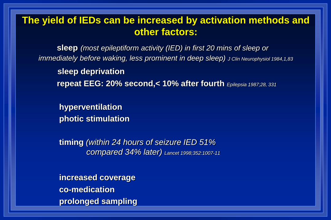
Electroencephalogram after first unprovoked seizure in
children: routine, unnecessary or case specific J Pediatr Neurosci: 2013 Jan-April; 8(1):1-4
Royal College of Pediatrics and Child Health asserted: “ There is no need for an
EEG following a first simple afebrile seizure” (Arch Dis Child 2004;89: 278-280)
Arguments against based on: level of prediction of recurrence, reticence to treat
after first seizure, quality of EEG interpretation and access
Arguments for based on: potential of syndromic diagnosis and patient/family
entitlement to specific diagnosis and impact on counselling.
Recommendation: diagnosis of seizure, type, syndrome should be primarily
based on clinical grounds and EEG not routinely requested without purpose. If
requested should answer specific question, to aid diagnosis of epilepsy. Quality
of report be considered, in doubt ask expert.
NICE Guidelines 2012: emphasis on clinical evaluation. Discusses EEG.

Chronic Epilepsy
Limited value
– “Not every 6 months””
– Weak association between amount of IED and seizure frequency and
antiepileptic medication has variable effect on IEDs.
Define the question
– Wrong diagnosis
– Review of syndrome
– New symptoms unexplained eg worsening seizures/cognition

Prognostic Role of EEG After Withdrawal of Anti Epileptic
Drugs
Conflicting data. Abnormal EEG associated with increased risk of
relapse but should not be used as sole basis in decision of
withdrawal. (Neurology 1994;44:601-8; Epilepsia 1992;33(4) 681-6)
Evolution of EEG in drug withdrawal correlates with final outcome
especially IGE. (Seizure 1993; 2: 213-220)
Syndromal diagnosis and Aetiology maybe critical determinants.

Other valuable uses of the EEG
Encephalopathy
Regression
Nonconvulsive status : confusion
Status Epilepticus management
Arrest
Neonatal Asphyxia


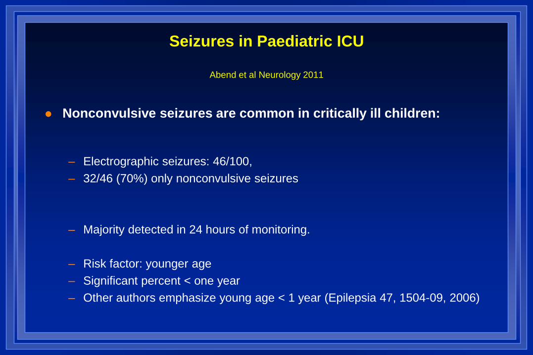
Seizures in Paediatric ICU
Abend et al Neurology 2011
Nonconvulsive seizures are common in critically ill children:
– Electrographic seizures: 46/100,
– 32/46 (70%) only nonconvulsive seizures
– Majority detected in 24 hours of monitoring.
– Risk factor: younger age
– Significant percent < one year
– Other authors emphasize young age < 1 year (Epilepsia 47, 1504-09, 2006)


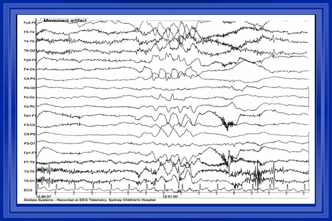
14 month infant who fails to wake after prolonged seizure with fever and minor illness

Following 36 hours aggressive anticonvulsant treatment

DWI
Acute Encephalopathy with Subcortical white matter Diffusion
changes. Influenza A and HHV6 identified. Excitatory insult.

EEG Background Patterns/Prognostication of Neonatal
Encephalopathy
Journal of Clinical Neurophysiology volume 30, 2, 122-125, 2014
Neonatal EEG valuable in predicting outcome in neonates with hypoxic ischemic
encephalopathy.
– 89-100% of those with normal or mildly abnormal EEG at 1-7 days have a normal
outcome.
– Inactive background (<5µV) or low-voltage plus theta(<15µV): outcome is death or
significant neurodevelopmental disability in 89% to 100%
– Burst suppression pattern : 80-100% die or have severe neurodevelopmental disability
but variation in definition
– Normal sleep-wake cycle in first 6-24 hours associated with good outcome.


Abuses
When not to order an EEG?

Utility of Electroencephalography in the Evaluation of
Common Neurological Conditions in Children (J Child Neurol 2003)
534 children
– Clinical seizure
– Epilepsy
– ADHD
– Headache
– Syncope
– Learning Disabilities
– TIC Disorders
– Sleep Disorders
Epileptiform activity rarely found in patients without epilepsy.
– Interictal EEG overused in nonepileptic conditions.
– Decrease waiting times.

Uses and abuses of paediatric electroencephalography Hong Kong Med J vol 18, 25-29, 2012
Misconceptions about the diagnostic capability of standard EEG in
paediatrics are common.
44% (total:109) standard EEG requests were inappropriate eg febrile
convulsions, funny turns
Appropriate requests were highly correlated with EEG results that
were contributory to clinical management
– Definite, probable epilepsy/seizure
– New diagnosis
– Status epilepsy
– Established epilepsy-subclinical EEG changes leading to symptoms
– Non-epilepsy: encephalopathy, neurodegeneration, organic brain disturbances

Can sodium valproate improve learning in children with
epileptiform bursts but without clinical seizures? Devel Med Child Neurol 2000, 42, 751-55. Ronen et al
Randomized double-blind, single-crossover trial carried out with VPA or
placebo on 8 participants with learning/ behavioural problems. Exclusion
criteria – known epileptic encephalopathies. No clinical seizures.
Abundant generalized or focal bursts of spike/slow wave on repeated
EEGs.
Clinically none of the children improved. No consistent EEG change
Children on valproate were more distractible on formal testing, increased
delay in response time, showed lower memory scores.
Data did not support use of valproate.

Autism, EEG and Regression.
Treatment
Seizure occurrence: 5-39%
Epileptiform abnormalities:6-60% with the higher incidence with epilepsy,
lower IQ and with regression
No data to support routine EEG screening in Autism. (J Child Neurol,
2005). Look at clinical phenotype-age, language regression or global,
fluctuation, seizures.
No evidence epilepsy/epileptiform abnormalities cause autistic
regression or that treatment of seizures or interictal abnormalities impact
language or social deficits

Artefact
Reporting : Who reports- eg benign variant
Access to reading by paediatric neurologist
Payment and availability of good technicians
Abuses: Skill Base
There are Problems with EEG Interpretation
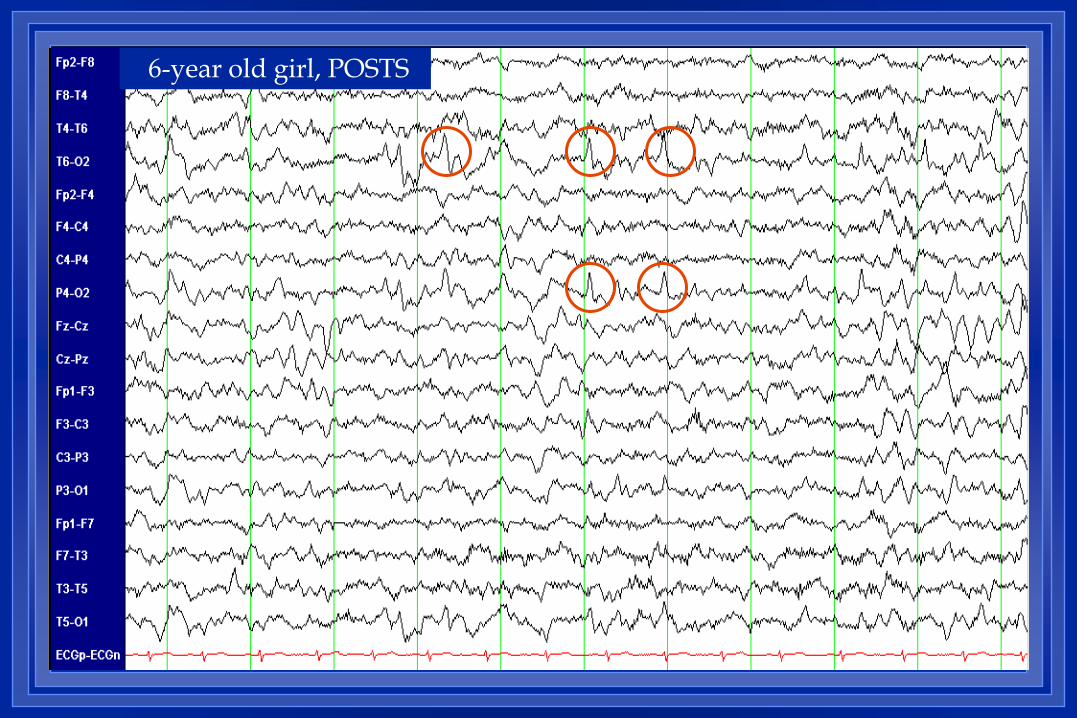
6-year old girl, POSTS

6-Hz SPIKE AND WAVE

Inter-rater reliability of the EEG in patients with childhood
idiopathic epilepsy Epilepsy Research 66 (2005) 195-198
To access level of agreement by experienced readers.
3 trained electroencephalographers examined 21 EEG records each (21
wake, 6 sleep)
Moderate agreement on majority of features of wake and sleep EEG.
– Unsatisfactory for background activity.
– Ictal discharges, distribution and location more easily identified.
– Interictal discharges suboptimal agreement (distribution and location)

Conclusions
EEG most important test in diagnosis and management of epilepsies
provided:
– Experienced technician and reporter AND interpreted in clinical
setting
– Access
– The EEG is a valuable test for an appropriate question.
Interictal epileptiform discharges are highly correlated with epileptic
seizures. It has limitations. Sensitivity and Specificity.
The EEG has in general no role in headache, syncope, learning and
behavioural disorders

Should be interpreted in clinical context.

Landau-Kleffner Syndrome
Verbal Auditory Agnosia
Focal epileptiform activity, sleep activation
Seizure disorder common but not always
Onset: 18 months to 13 years
Previously normal development

Prognosis Role of EEG After a First
Epileptic Seizure
Majority of series have shown EEG
abnormalities are associated with an increased
risk of seizure recurrence

Interictal Epileptiform Discharges in Non-Epileptic
Patients
Children (2.2-3.5%)
Adults (0.2-0.5%)
Patterns seen: (majority)
Centrotemporal
Generalised
Photoparoxysmal
TREAT THE PATIENT NOT EEG

Spike Frequency:Activation and Reactivity Awake Drowsy
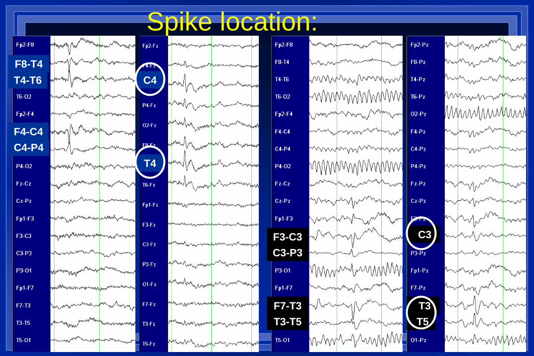
Spike location:
F8-T4
T4-T6
F4-C4
C4-P4
T4
C4
T3
T5
C3 F3-C3
C3-P3
F7-T3
T3-T5
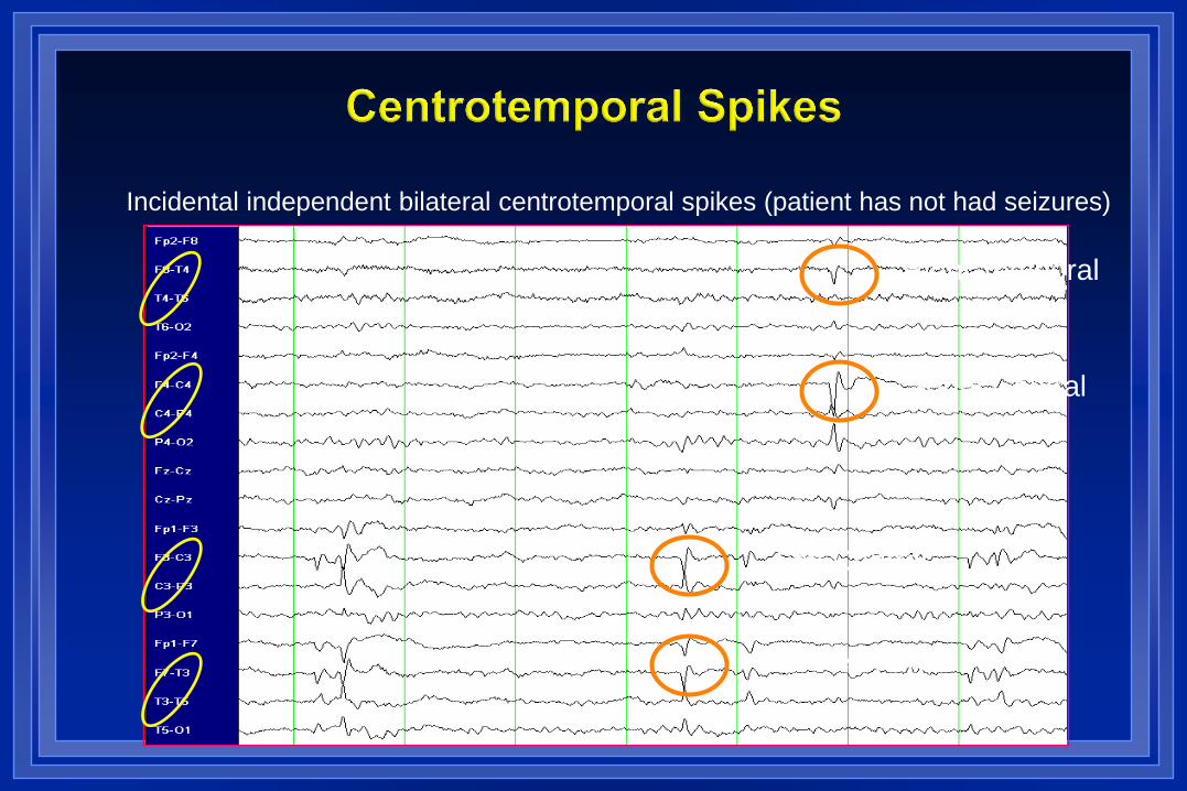
Incidental independent bilateral centrotemporal spikes (patient has not had seizures)
Right Central
Right Temporal
Left Central
Left Temporal

4:10:37 PM - Hyperventilation

4:11:58 PM- Hyperventilation

Role of EEG in Assessing Cognitive Decline
Dementia or language regression
Non convulsive status presenting as confusion, subtle
motor events
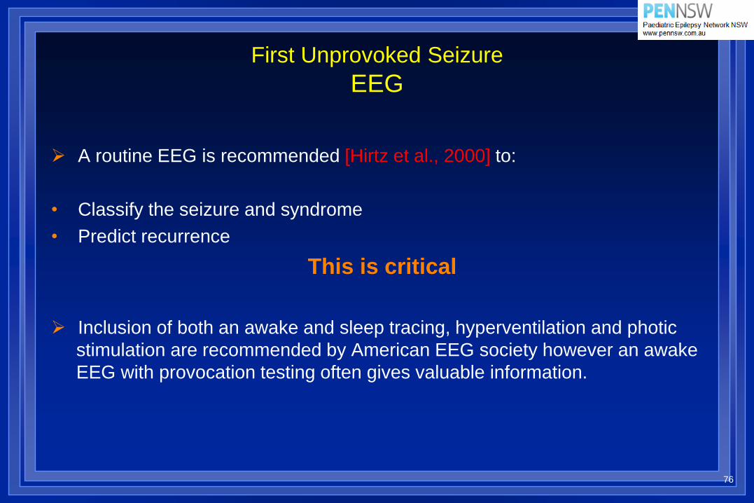
A routine EEG is recommended [Hirtz et al., 2000] to:
• Classify the seizure and syndrome
• Predict recurrence
This is critical
Inclusion of both an awake and sleep tracing, hyperventilation and photic
stimulation are recommended by American EEG society however an awake
EEG with provocation testing often gives valuable information.
76
First Unprovoked Seizure
EEG

THE EEG
In a patient with a seizure disorder
a routine EEG maybe normal in 50%
of recordings.

The EEG
Localisation Tool in
Epilepsy Surgery

EEG Abnormalities
Background abnormalities: significant asymmetries and/or degree of slowing inappropriate for clinical state or age
Interictal abnormalities associated with seizures and epilepsy
– Spikes
– Sharp waves
– Spike-wave complexes
May be focal, lateralized, generalized C-
Slid
e 79
Return to index
American Epilepsy Society 2015

EEG: Hypsarrhythmia
American Epilepsy Society 2015 PC Slide-80

EEG: Slow Spike and Wave
American Epilepsy Society 2015 PC Slide-81

EEG: Paroxysmal Fast Activity
American Epilepsy Society 2015 PC Slide-82

POSTS


8-year old child – trains in wakefulness, drowsiness and sleep