Embed Size (px)
Citation preview

The Origin and Pathogenesis of Epithelial Ovarian Cancer:A Proposed Unifying Theory
Robert J. Kurman, MD and Ie-Ming Shih, MD, PhD
Abstract: Ovarian cancer is the most lethal gynecologic
malignancy. Efforts at early detection and new therapeuticapproaches to reduce mortality have been largely unsuccessful,because the origin and pathogenesis of epithelial ovarian cancer
are poorly understood. Despite numerous studies that havecarefully scrutinized the ovaries for precursor lesions, none havebeen found. This has led to the proposal that ovarian cancerdevelops de novo. Studies have shown that epithelial ovarian
cancer is not a single disease but is composed of a diverse groupof tumors that can be classified based on distinctive morphologicand molecular genetic features. One group of tumors, designated
type I, is composed of low-grade serous, low-grade endo-metrioid, clear cell, mucinous and transitional (Brenner) carci-nomas. These tumors generally behave in an indolent fashion,
are confined to the ovary at presentation and, as a group, arerelatively genetically stable. They lack mutations of TP53, buteach histologic type exhibits a distinctive molecular genetic
profile. Moreover, the carcinomas exhibit a shared lineage withthe corresponding benign cystic neoplasm, often through anintermediate (borderline tumor) step, supporting the morpho-logic continuum of tumor progression. In contrast, another
group of tumors, designated type II, is highly aggressive, evolvesrapidly and almost always presents in advanced stage. Type IItumors include conventional high-grade serous carcinoma,
undifferentiated carcinoma, and malignant mixed mesodermaltumors (carcinosarcoma). They displayTP53 mutations in over80% of cases and rarely harbor the mutations that are found
in the type I tumors. Recent studies have also provided cogentevidence that what have been traditionally thought to beprimary ovarian tumors actually originate in other pelvic organsand involve the ovary secondarily. Thus, it has been proposed
that serous tumors arise from the implantation of epithelium(benign or malignant) from the fallopian tube. Endometrioidand clear cell tumors have been associated with endometriosis
that is regarded as the precursor of these tumors. As it isgenerally accepted that endometriosis develops from endome-trial tissue by retrograde menstruation, it is reasonable to
assume that the endometrium is the source of these ovarianneoplasms. Finally, preliminary data suggest that mucinous andtransitional (Brenner) tumors arise from transitional-typeepithelial nests at the tubal-mesothelial junction by a process
of metaplasia. Appreciation of these new concepts will allowfor a more rationale approach to screening, treatment, andprevention that potentially can have a significant impact on
reducing the mortality of this devastating disease.
Key Words: ovarian carcinogenesis, type I and type II tumors,
p53 mutation, KRAS, BRAF, PTEN, PIK3CA mutations
(Am J Surg Pathol 2010;34:433–443)
‘‘Nothing will come from nothing’’
King Lear, Act I
The origin and pathogenesis of epithelial ovariancancer has perplexed investigators for decades. Despitenumerous studies that have carefully scrutinized theovaries for precursor lesions, none have been found. Thishas led to the proposal that ovarian cancer developsde novo.2 ‘‘Nothing will come from nothing,’’ but eachyear in the United States, approximately 21,550 womendevelop ovarian cancer ‘‘de novo,’’ and 14,600 women diefrom this disease.18 Ovarian cancer is, in fact, the mostlethal gynecologic malignancy. It is clear that de novoreflects our ignorance about the early events of ovariancarcinogenesis rather than our insight into its perplexingorigin. The time-honored concepts that have forged ourviews of ovarian carcinogenesis can be summarized asfollows: (1) although it is recognized that there areprofound differences among the various histologic types,the vast majority of ovarian carcinomas are high-gradeserous carcinomas and therefore ovarian cancer isregarded as a single disease; (2) ovarian cancer originatesfrom the ovarian surface epithelium (mesothelium) thatinvaginates into the underlying stroma resulting ininclusion cysts that eventually undergo malignant trans-formation; (3) ovarian cancer spreads from the ovary tothe pelvis, abdomen, and distant sites. On the basis of theseviews of ovarian carcinogenesis, efforts at improving thesurvival have focused on early detection of ovarian can-cer, when it is still confined to the ovary, and on the devel-opment of new chemotherapeutic drugs and routes ofdelivery irrespective of the histologic type. Unfortunately,Copyright r 2010 by Lippincott Williams & Wilkins
From the Departments of Pathology, Gynecology and Obstetrics andOncology, The Johns Hopkins University School of Medicine,Baltimore, Maryland.
Supported by the grants: DOD-OCRP 080469, RO1CA116184,RO1CA129080, RO1CA103937.
Correspondence: Robert J. Kurman, MD, Departments of Pathology,Gynecology and Obstetrics, and Oncology, The Johns HopkinsUniversity School of Medicine, Baltimore, Maryland (e-mail:[email protected]).
SPECIAL ARTICLE
Am J Surg Pathol � Volume 34, Number 3, March 2010 www.ajsp.com | 433
these efforts have not been successful as evidenced by thefact that the overall survival for women with ovariancancer has not changed over the last 50 years. The reasonsfor this are that the concepts of histogenesis, on whichthese approaches are based, are flawed.
Recent morphologic and molecular genetic studieshave illuminated our understanding of ovarian carcino-genesis in ways that have been quite unexpected and havechallenged the conventional wisdom regarding theirorigin and development. Indeed, they have resulted in aparadigm shift that has important implications for researchand for radically changing our approaches to early detec-tion, prevention, and treatment.
THE MORPHOLOGIC AND MOLECULARHETEROGENEITY OF EPITHELIAL
OVARIAN CANCEROne of the major problems in elucidating the patho-
genesis of ovarian cancer is that it is a heterogeneousdisease composed of different types of tumors with widelydiffering clinicopathologic features and behavior. On thebasis of a series of morphologic and molecular geneticstudies, we have proposed a dualistic model that categorizesvarious types of ovarian cancer into two groups desig-nated type I and type II.43 Type I tumors are clinicallyindolent and usually present at a low stage. They exhibit ashared lineage between benign cystic neoplasms and thecorresponding carcinomas, often through an intermediate(borderline tumor) step, supporting the morphologiccontinuum of tumor progression in these neoplasms. Thisstepwise sequence of events parallels the adenoma-carcinomasequence that occurs in colorectal carcinoma. Type I tu-mors include low-grade serous, low-grade endometrioid,clear cell and mucinous carcinomas. In contrast to theclear-cut and distinctive morphologic differences amongtype I tumors, the morphologic differences among thetype II tumors are more subtle and, as a result, there isconsiderable overlap in the diagnosis of these tumors bydifferent pathologists. Type II tumors exhibit papillary,glandular, and solid patterns and are diagnosed as high-grade serous, high-grade endometrioid. and undifferen-tiated carcinomas depending on the dominant pattern.Generally, most pathologists classify them as high-gradeserous carcinomas even though they bear little resem-blance to tubal-type epithelium (the basis for typing atumor as serous); arguably many of those lacking distinc-tive serous or endometrioid features could be classified as‘‘high-grade adenocarcinoma’’. In addition to these neo-plasms, malignant mixed mesodermal tumors (carcino-sarcomas) are included in the type II category, becausethey have epithelial components identical to the pure typeII carcinomas. Type II tumors are highly aggressive andalmost always present in advanced stage. As they accountfor approximately 75% of all epithelial ovarian carcino-mas and have relatively similar morphologic features anda uniformly poor outcome, ovarian cancer has beenerroneously regarded as a single disease. The morphologicdifferences between type I and type II tumors are
mirrored by marked differences in their molecular geneticfeatures.7 As a group, type I tumors are genetically morestable than type II tumors and display specific mutationsin the different histologic cell types.21 Thus, KRAS,BRAF, and ERBB2 mutations occur in approximatelytwo thirds of low-grade serous carcinomas, whereas TP53mutations are rare in these tumors. Low-grade endome-trioid carcinomas have aberrations in the Wnt signalingpathway involving somatic mutations of CTNNB1(encoding b-catenin), PTEN and PIK3CA.7 Mucinouscarcinomas have KRAS mutations in more than 50% ofspecimens.1,28 Clear cell carcinoma is unique in that it hasa high percentage of PIK3CA activating mutations whenpurified tumor samples and cell lines are analyzed.22
There is little available molecular genetic data ontransitional cell (Brenner) tumors. High-grade serouscarcinoma, the prototypic type II tumor is characterizedby very frequent TP53 mutations (>80% of cases) andCCNE1 (endcoding cyclin E1) amplification but rarelymutations that characterize most type I tumors, such asKRAS, BRAF, ERBB2, PTEN, CTNNB1, and PIK3-CA.7 Although only a small number of malignant mixedmesodermal tumors have been analyzed molecularly, thefew that have been analyzed, display a similar moleculargenetic profile. In summary, type I tumors as a group, aregenetically more stable than type II tumors and display adistinctive pattern of mutations that occur in specific celltypes (low-grade serous, low-grade endometrioid, clearcell and mucinous). In contrast, the type II tumors (high-grade serous, high-grade endometrioid, malignant mixedmesodermal tumors, and undifferentiated carcinomas)show greater morphologic and molecular homogeneity,are genetically unstable, and have a very high frequencyof TP53 mutations. These findings suggest that differenttypes of ovarian carcinomas develop along differentmolecular pathways.
THE CELL OF ORIGIN OF MOST EPITHELIALOVARIAN CANCER IS NOT OVARIANThe cell of origin of ovarian cancer and the
mechanisms by which cancer develops have been longdebated. The traditional view of ovarian carcinogenesishas been that the various tumors are all derived from theovarian surface epithelium (mesothelium), and thatsubsequent metaplastic changes lead to the developmentof the different cell types [serous, endometrioid, clear cell,mucinous, and transitional cell (Brenner)], which mor-phologically resemble the epithelia of the fallopian tube,endometrium, gastrointestinal tract, or endocervix andurinary bladder, respectively. The normal ovary, how-ever, has no constituents that resemble these tumors.Moreover, the cervix, endometrium, and fallopian tubesare derived from the mullerian ducts, whereas the ovariesdevelop from mesodermal epithelium on the urogenitalridge separate from the mullerian ducts. Therefore, analternate theory proposes that tumors with a mullerianphenotype (serous, endometrioid, and clear cell) are derivedfrom mullerian-type tissue and not from mesothelium.11
Kurman and Shih Am J Surg Pathol � Volume 34, Number 3, March 2010
434 | www.ajsp.com r 2010 Lippincott Williams & Wilkins

This mullerian-type tissue (columnar epithelium, oftenciliated) lines cysts located in paratubal and paraovarianlocations that have been referred to collectively as the‘‘secondary mullerian system’’.23 According to thistheory, ovarian tumors develop from these cysts. As thetumor enlarges, it compresses and eventually obliteratesovarian tissue resulting in an adenxal tumor that seems tohave arisen in the ovary. More recently, another theoryhas been advanced, which argues that the majority ofovarian carcinomas that are high-grade serous carcino-mas, arise from high-grade intraepithelial serous carcino-mas in the fallopian tube, which then spread to the ovary.These conflicting views led us to undertake a review of theliterature in an effort to determine which of the theories isbest able to explain the various aspects of ovariancarcinogenesis.
Evaluating these theories is problematic, because itis difficult to construct experimental systems to test theirvalidity. Accordingly, our evaluation is based on criticalanalysis of these studies in the light of observations wehave made in the course of pathologic examination ofovarian tumors. The discussion that follows is an attemptto distill the most plausible components from the varioustheories of cellular origin and integrate them with theclinicopathologic and molecular genetic data from thedualistic model to construct a unifying theory of ovariancarcinogenesis.
The theory of origin from ovarian surface epithe-lium (mesothelium) has a number of limitations. Histo-logically, the single layer of generally attenuated mesotheliumoverlying the ovaries bears no resemblance to serous,endometrioid, mucinous, clear cell, or transitional (Bren-ner) carcinomas. As noted above to account for this apparentcontradiction, it was proposed that the mesothelium over-lying the ovary invaginates into the underlying stroma toform so-called ‘‘cortical inclusion cysts’’. These cystsunder the influence of local factors, possibly steroid hor-mones, undergo a metaplastic change, which results in themesothelium being converted to mullerian-type epithe-lium. These inclusion cysts, with their newly acquiredmullerian phenotype, can then undergo malignant trans-formation resulting in carcinomas corresponding to thedifferent cell types (serous, endometrioid and clear cellcarcinomas).6 Although cortical inclusion cysts lined byciliated (mullerian-type epithelium) are frequently obser-ved in the ovarian cortex, well-documented examplesof what can be interpreted as a transition from these cyststo carcinoma have not been reported. Moreover, corticalinclusion cysts lined by intestinal-type epithelium toaccount for the development of mucinous carcinomasare distinctly rare. The same can be said for the absence oftransitional-type epithelium lining cortical inclusion cyststo account for the development of Brenner tumors.
The limitations of the secondary mullerian systemtheory are that precursor lesions resembling serous, endomet-rioid, and clear cell carcinomas have rarely, if ever, beenreported in paratubal and paraovarian cysts. Moreover,the vast majority of mucinous tumors display intestinalrather than endocervical-type mucinous differentiation
and therefore do not qualify as mullerian-type tumors. Asimilar problem exists for transitional cell (Brenner)tumors resembling urothelium that is not mullerian inorigin.
The most compelling evidence suggests that the vastmajority of what seems to be primary ovarian cancers,namely serous, endometrioid, and clear cell carcinomas,are derived from the fallopian tube and endometrium andnot directly from the ovary. Sporadic reports of tubalcarcinoma and ‘‘dysplasia’’ had been reported in thepast,15 but in 2001, a group of Dutch investigatorsdescribed these lesions that closely resemble high-gradeovarian serous carcinoma in women with a geneticpredisposition to ovarian cancer.33 This was a surprisingfinding, as numerous studies over the past two decades thatcarefully examined the ovaries of women with a geneticpredisposition to ovarian cancer never reported similarlesions. In addition, other studies of normal appearingovaries contralateral to sporadic (nonhereditary) unilat-eral ovarian carcinomas had never identified a convincingprecursor lesion. These latter studies reported a numberof morphologic changes in grossly normal appear-ing ovaries, such as an increased number of surfacepapillae and cortical inclusion cysts, including some dis-playing minor degrees of atypia. The data, however, havebeen conflicting, some studies reporting a significantdifference of these changes in cases versus controls, andother studies reporting no difference. In any event, noneof these changes even remotely resemble high-grade serouscarcinoma. It was precisely because of a lack of convinc-ing precursor lesions that the de novo hypothesis wasinvoked.
In hindsight, because it was assumed that precursorsof ovarian carcinoma would logically be in the ovaries,the fallopian tubes were not carefully examined.10,42
Subsequent studies, in which fallopian tubes were morecarefully examined, confirmed that in situ and small, earlyinvasive tubal carcinomas occurred in women with agenetic predisposition for the development of ovariancancer.4,5,8,12,27,29,41 This led to fallopian tube carcinomabeing included as part of the cancer spectrum associatedwith inherited BRCA mutations. It was subsequentlyproposed that a proportion of ovarian carcinomas mightdevelop as a result of implantation of malignant cellsfrom the tubal carcinoma to the ovary.34,35 The nextimportant step linking what had been termed ‘‘tubalintraepithelial carcinoma’’ and, subsequently, ‘‘serous tubalintraepithelial carcinoma’’ (STIC) with ovarian carcinomawas the observation that over 70% of sporadic (non-hereditary) ovarian and peritoneal high-grade serouscarcinomas showed mucosal tubal involvement includingSTICs.19 This observation gave substantial support to theproposal that STICs, which almost always are detected inthe fimbria, may be the source of ovarian high-gradeserous carcinoma in both women with hereditary muta-tions in BRCA and women who did not have a knowngenetic predisposition for ovarian cancer. Although it canbe argued that mucosal tubal involvement could representsecondary spread from an ovarian carcinoma present in
Am J Surg Pathol � Volume 34, Number 3, March 2010 Ovarian Carcinogenesis
r 2010 Lippincott Williams & Wilkins www.ajsp.com | 435

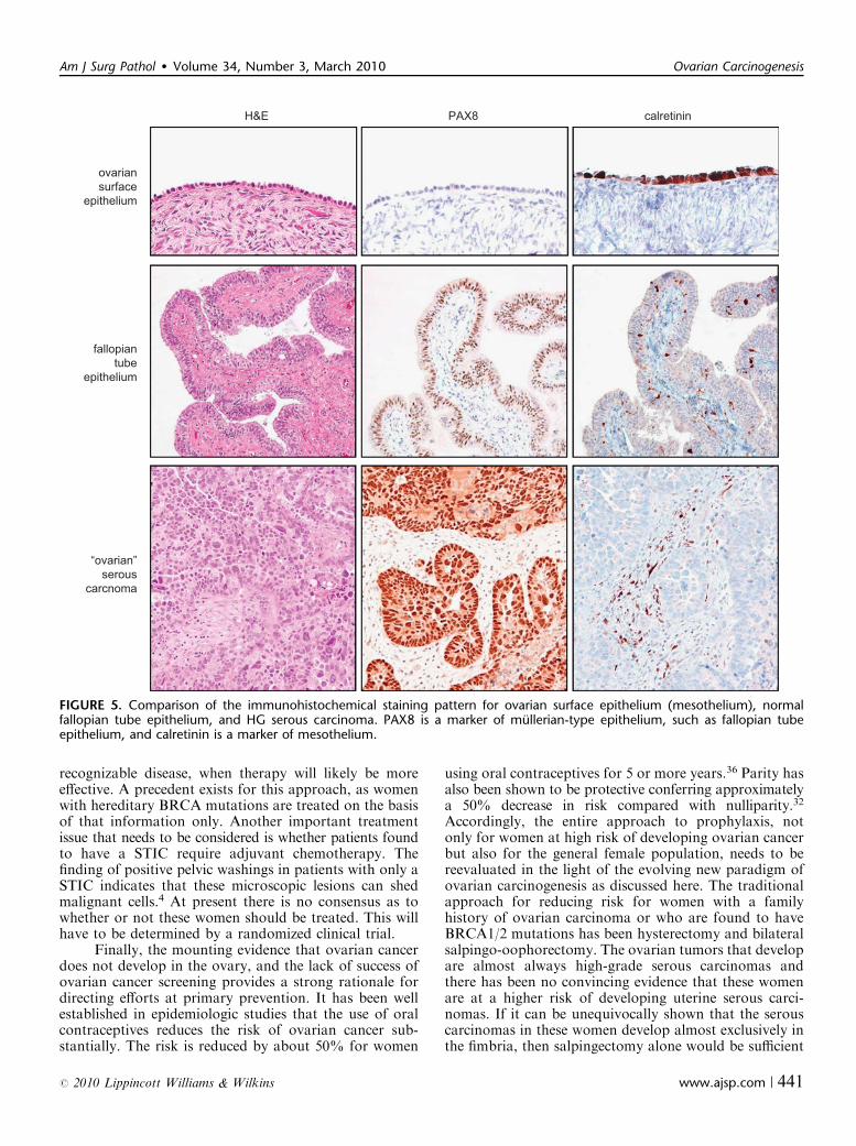
the same specimen, the presence of focal noncontiguousintraepithelial lesions (STICs) would be an unusual mani-festation of metastasis. Furthermore, the identification ofSTICs in prophylactic specimens from women with ahereditary predisposition to ovarian cancer, in whichcomplete microscopic evaluation of the fallopian tubesand ovaries failed to identify invasive carcinoma in theseorgans, lends additional support to the concept that theserous neoplastic process may well begin in the fallopiantube rather than in the ovary. Further support for thisargument is the finding that nearly all STICs overexpressp53, similar to high-grade serous carcinoma (Fig. 1).Laser capture microdissection studies of these lesions haveshown that they harbor mutated TP53.19 In addition,STICs associated with a concomitant ovarian carcinomashare not only morphologic features but also identicalTP53 mutations indicating a clonal relationship betweenthem. Adnexal malignant mixed mesodermal tumors(another type II tumor) have also been associated withSTICs supporting the existence of a common precursorlesion for type II tumors.14 Further evidence implicatingthe fallopian tube rather than ovarian surface epitheliumas the site of origin of serous neoplasms comes from agene profiling study showing that the gene expressionprofile of high-grade serous carcinoma is more closelyrelated to the fallopian tube than to the ovarian surfaceepithelium.25 In addition, high-grade serous carcinomasexpress PAX8, a mullerian marker, but not calretinin, amesothelial marker (Shih, Unpublished data).
A recent finding has been the identification ofbenign tubal epithelium, specifically secretory as opposedto ciliated cells, that express p53 and in which lasercapture microdissection studies have reported TP53mutations in 57% of cases.24 These lesions termed ‘‘p53signatures’’ are found in association with STICs and innormal appearing fallopian tubes of women withoutSTICs or carcinoma; they have been observed in approxi-mately one third of women with and without BRCA
mutations.13,17,41 Like STICs, p53 signatures express g-H2AX that localizes to areas of DNA damage in nuclei.24
When associated with STICs and ovarian carcinoma, thep53 signature has had the identical TP53 mutation as theSTIC and the carcinoma in some cases but not in others.Based on these findings, a sequence of pathogeneticevents has been proposed beginning with genotoxicDNA damage, followed by TP53 mutation and progres-sive loss of cell cycle control, which then eventuate in thedevelopment of carcinoma.24 There are a number ofquestions that must be resolved, however, before thishypothesis can be completely accepted. First, as noted insome instances, TP53 mutations, when present in the p53signature, are not always identical with the mutations inthe STICs and carcinomas in the same specimen. Second,women at high risk have the same frequency of p53signatures as women who are not at high risk. Third, thehigh prevalence of p53 signatures (a third of all women)compared with the low prevalence of high-grade serousovarian carcinoma suggests that either a small minority ofp53 signatures progress or that they are not related tocarcinoma. It is conceivable that p53 signatures reflectan appropriate and physiologic upregulation of p53 inresponse to DNA damage based on the observation thatTP53 mutations are absent in nearly half of p53 signa-tures. Although the proposal that the p53 signature is aprecursor lesion is intriguing, its role in the genesis ofovarian high-grade serous carcinoma is far from clear atthis time. As fallopian tubes are more carefully examinedand these lesions studied, the nature of p53 signatures andtheir relationship to STICs will become better defined.
Generally, before a carcinoma acquires the abilityto metastasize, it must first invade and gain access toblood vessels or lymphatics. We have observed that thefimbria contain a rich angiolymphatic vasculature. More-over, they are in almost direct contact with the basementmembrane of the tubal epithelium, and therefore a tubalcarcinoma may not need to attain a very large size toinvade this highly accessible angiolymphatic network. Inaddition, invasion in the case of a STIC may not be anecessary prerequisite for dissemination. Tubal intra-epithelial carcinomas are morphologically and immuno-histochemically similar to endometrial intraepithelialcarcinomas that are regarded as precursors or early formsof uterine serous carcinoma. These lesions have also beentermed ‘‘uterine surface serous carcinomas’’. They havebeen shown to disseminate throughout the peritonealcavity presumably by the passage of malignant cellsthrough the fallopian tube without requisite myometrialinvasion.45 The cells that comprise both endometrial andtubal intraepithelial carcinomas are highly anaplastic andidentical morphologically to high-grade serous carci-noma. The lesions form papillary tufts and the constitu-ent cells are loosely cohesive. Presumably, these cells canshed and implant on the surface of the ovary and theperitoneum in the absence of invasive growth in thefallopian tube. Evidence supporting this possibility arethe reports of positive pelvic washings in women whoseonly lesion was a STIC.4
A B
FIGURE 1. Serous tubal intraepithelial carcinoma (STIC). A,High magnification. Hematoxylin and eosin stain. B, Immuno-histochemical stain for p53. Arrows point to STIC and anasterisk defines the boundary of the lesion.
Kurman and Shih Am J Surg Pathol � Volume 34, Number 3, March 2010
436 | www.ajsp.com r 2010 Lippincott Williams & Wilkins

As earlier noted, in studies of ovarian and primaryperitoneal high-grade serous carcinomas in which theentire fallopian tubes were carefully sectioned, mucosalinvolvement of the tube, including STICs, was identifiedin approximately 70% of cases.19 The question arises asto the source of the remaining ovarian carcinomas thatlack evidence of tubal involvement. There are a numberof possible explanations. First, despite thorough section-ing, a small STIC could have been missed (unpublisheddata). Second, occasionally high-grade serous carcinomasare intimately associated with serous borderline tumorsand low-grade serous carcinomas. In these cases, thehigh-grade tumors have had KRAS mutations identicalto those in the serous borderline tumors and lacked TP53mutations.9 This finding suggests that some high-gradeserous carcinomas arise from low-grade serous tumors
and not by the usual (type II) pathway that begins with aTP53 mutation. Third, clear-cut mucosal tubal involve-ment could have been obscured by overgrowth of thepelvic carcinoma. Fourth, the fimbria of the fallopiantube normally is in intimate contact with the ovariansurface at the time of ovulation. It is conceivable thatwhen the ovarian surface epithelium is disrupted at thetime of ovulation, normal tubal epithelial cells from thefimbria may be dislodged and implant in the ovary toform an inclusion cyst (Fig. 2) from which a high-gradeserous carcinoma could develop (see below). Evidence tosupport this notion is the observation that fallopian tubeepithelial cells are easily obtained for culture by flushingthe fallopian tube (Shih, Unpublished data).34 Thismechanism could also explain the development of endo-salpingiosis, a lesion composed of glands and papillary
FIGURE 2. Transfer of normal tubal epithelium to the ovary. A, Anatomical relationship of fallopian tube with the ovary at thetime of ovulation. The fimbria envelops the ovary. B, Ovulation. The ovarian surface ruptures with expulsion and transfer ofthe oocyte to the fimbria. The fimbria is in intimate contact with the ovary at the site of rupture. C, Tubal epithelial cells from thefimbria are dislodged and implant on the denuded surface of the ovary resulting in the formation of an inclusion cyst.
Am J Surg Pathol � Volume 34, Number 3, March 2010 Ovarian Carcinogenesis
r 2010 Lippincott Williams & Wilkins www.ajsp.com | 437
structures lined by tubal-type epithelium that is found onperitoneal surfaces in the pelvis, omentum, and beneaththe capsule of pelvic and para-aortic lymph nodes. Endo-salpingiosis is frequently found in association with low-grade serous tumors and has been viewed as a possibleprecursor of these tumors. Finally, the possibility that somehigh-grade serous carcinomas arise in cortical inclusioncysts as a metaplastic process from the ovarian surfaceepithelium rather than from implantation of normalfallopian tube epithelium cannot be entirely dismissed.
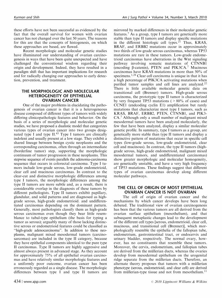
Direct implantation of tubal epithelium into theovary to form an inclusion cyst, which in turn is the siteof origin of ovarian serous carcinoma, although notyet shown, is an attractive alternative theory to that ofmetaplasia from the surface epithelium (mesothelium).Implantation of fallopian tube epithelium from thefimbria at the time of ovulation when the surface epithe-lium is disrupted can explain the derivation of low-gradeand high-grade serous carcinomas. In the case of a low-grade serous carcinoma, the process develops slowly froma serous cystadenoma and then a serous borderline tumorafter a KRAS or BRAF mutation, whereas in the case ofa high-grade serous carcinoma, the process evolves rapidly,presumably from a cortical inclusion cyst after a TP53mutation with the development of an intraepithelial
carcinoma as an intermediate step. According to thisview, both low-grade and high-grade serous carcinomasare ultimately of tubal (mullerian) origin, and, in a sense,the ovary is involved secondarily (Fig. 3).
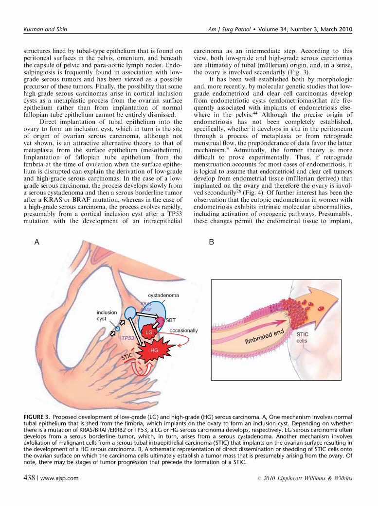
It has been well established both by morphologicand, more recently, by molecular genetic studies that low-grade endometrioid and clear cell carcinomas developfrom endometriotic cysts (endometriomas)that are fre-quently associated with implants of endometriosis else-where in the pelvis.44 Although the precise origin ofendometriosis has not been completely established,specifically, whether it develops in situ in the peritoneumthrough a process of metaplasia or from retrogrademenstrual flow, the preponderance of data favor the lattermechanism.3 Admittedly, the former theory is moredifficult to prove experimentally. Thus, if retrogrademenstruation accounts for most cases of endometriosis, itis logical to assume that endometrioid and clear cell tumorsdevelop from endometrial tissue (mullerian derived) thatimplanted on the ovary and therefore the ovary is invol-ved secondarily26 (Fig. 4). Of further interest has been theobservation that the eutopic endometrium in women withendometriosis exhibits intrinsic molecular abnormalities,including activation of oncogenic pathways. Presumably,these changes permit the endometrial tissue to implant,
A B
STICcells
cystadenoma
inclusioncyst
HG
SBT
TP53occasionallyLG
HG
FIGURE 3. Proposed development of low-grade (LG) and high-grade (HG) serous carcinoma. A, One mechanism involves normaltubal epithelium that is shed from the fimbria, which implants on the ovary to form an inclusion cyst. Depending on whetherthere is a mutation of KRAS/BRAF/ERRB2 or TP53, a LG or HG serous carcinoma develops, respectively. LG serous carcinoma oftendevelops from a serous borderline tumor, which, in turn, arises from a serous cystadenoma. Another mechanism involvesexfoliation of malignant cells from a serous tubal intraepithelial carcinoma (STIC) that implants on the ovarian surface resulting inthe development of a HG serous carcinoma. B, A schematic representation of direct dissemination or shedding of STIC cells ontothe ovarian surface on which the carcinoma cells ultimately establish a tumor mass that is presumably arising from the ovary. Ofnote, there may be stages of tumor progression that precede the formation of a STIC.
Kurman and Shih Am J Surg Pathol � Volume 34, Number 3, March 2010
438 | www.ajsp.com r 2010 Lippincott Williams & Wilkins

survive, and invade on ovarian and peritoneal surfaces.3
This hypothesis, by which endometrioid and clear cellcarcinoma develop from endometrial tissue implantedon the ovary, is supported by epidemiologic evidence showingthat a protective effect for tubal ligation was seen only forendometrioid and clear cell carcinoma of the ovary.37
Finally, the derivation of mucinous tumors ofgastrointestinal type and transitional cell (Brenner) tumorsmay also not involve the ovaries directly. The origin ofthese tumors is puzzling, as unlike serous, endometrioid,and clear cell tumors, they do not display a mullerianphenotype. Although it has been argued that these muci-nous tumors bear some relationship with the endocervix,the mucinous epithelium that characterizes these neo-plasms more closely resembles gastrointestinal mucosa. Itseems most unlikely that they develop from corticalinclusion cysts. as mucinous metaplasia involving corticalinclusion cysts is a very rare finding. On the other hand,the association of Brenner tumors and mucinous tumorshas been recognized for many years. In a provocativestudy of mucinous cystadenomas and Brenner tumors, itwas reported that after extensive sectioning, mucinouscystadenomas contained foci of Brenner tumor in 18% ofcases.40 Interestingly, mucinous tumors were frequentlyassociated with Walthard cell nests that are composed ofbenign transitional-type epithelium, frequently found inparaovarian and paratubal locations. This raises thepossibility that mucinous tumors and Brenner tumorshave the same histogenesis arising from these microscopictransitional cell nests at the tubal-mesothelial junction inkeeping with their nonmullerian appearance. The study
reported that Brenner tumors are small (median size0.5 cm, range 0.02 to 20 cm), whereas mucinous cystade-nomas are large (median size 9 cm, range 1 to 30 cm). Theinvestigators speculated that as a small Brenner tumorgrows, the mucinous component becomes dominantresulting in the development of a mucinous cystadenomathat, as it enlarges, compresses and eventually obliteratesthe adjacent ovary giving the appearance that it arose inthe ovary. The findings in this study are intriguing butmust be regarded as preliminary. Additional morphologicand molecular genetic studies are necessary to determinewhether this concept is valid.
In summary, none of the existing theories adequatelyreconciles all aspects of ovarian carcinogenesis. All ofthem have something to offer in explaining the develop-ment of ovarian carcinomas, but none are all inclusive. Itdoes seem that the vast majority of what have beenthought to be primary epithelial ovarian and primaryperitoneal carcinomas are, in fact, secondary. Thus, themost persuasive data support the view that serous tumorsdevelop from the fimbriated portion of the fallopian tube,endometrioid, and clear cell tumors from endometrialtissue passing through the fallopian tube resulting inendometriosis and mucinous, and Brenner tumors fromtransitional-type epithelium located at the tubal-mesothe-lial junction where the fimbria makes contact with theperitoneum. The concept that the majority of epithelialovarian carcinomas originates outside the ovary andinvolves it secondarily has emerged only recently, becausein the past, the default diagnosis of carcinomas involvingthe pelvis and abdomen was that they were ovarian. Acarcinoma was classified as tubal in origin only when thebulk of the tumor involved the fallopian tube rather thanthe ovary, and there was evidence of an intraepithelial (insitu) tubal carcinoma.39 A diagnosis of primary peritonealcarcinoma is even more restrictive. Even with extensivetumor involving the peritoneum, omentum, and otherabdominal organs, a carcinoma is classified as primaryovarian, if there is as little as 5mm of tumor involving theovaries. Thus, there has been an inherent bias inclassifying pelvic tumors as being ovarian in origin.
Although the data suggesting that epithelial ovariancarcinoma arises in extraovarian sites and involves theovaries secondarily are compelling, serous neoplasms (low-and high-grade) involve the ovaries and other pelvic andabdominal organs, such as the omentum and mesentery,much more extensively than the fallopian tubes. Similarly,although endometrioid carcinomas develop from endome-triosis that frequently involves multiple sites in the pelvis,these neoplasms are almost always confined to the ovaries.It is likely that the propensity for growth in the ovary ismulifactorial, but the precise reasons for this are unknown.
IMPLICATIONS FOR RESEARCH, SCREENING,PREVENTION, AND TREATMENT
The implications of this new paradigm of ovariancarcinogenesis for investigators, clinicians, and womenare significant. For researchers, the implication of tubal
CCC
EM/CCborderline tumor
endometriosis
FIGURE 4. Proposed development of LG endometrioid andclear cell carcinoma. Endometrial tissue, by a process ofretrograde menstruation, implants on the ovarian surface toform an endometriotic cyst from which a LG endometrioidor clear cell carcinoma can develop. CCC indicates clear cellcarcinoma of the ovary; EMC, LG endometrioid carcinoma ofthe ovary.
Am J Surg Pathol � Volume 34, Number 3, March 2010 Ovarian Carcinogenesis
r 2010 Lippincott Williams & Wilkins www.ajsp.com | 439

origin of ovarian serous carcinoma challenges many of theearlier reports showing ‘‘overexpressed’’ ovarian cancer-associated genes in which their expression levels in carci-noma are almost always compared with their ‘‘normal’’counterpart, ovarian surface epithelium. As the geneexpression profiles in ovarian surface epithelium that is ofmesothelial origin, are distinct from fallopian tube epithe-lium which is of mullerian origin, experiments in whichovarian surface epithelium (mesothelium) has been usedas a control may not be valid. Whether the overexpressedgenes that have been reported are indeed upregulated,when they are compared with the more likely source ofovarian serous carcinoma, that is, fallopian tube epithe-lium, needs to be revisited. In fact, a recent moleculargenetic study showed that the different histologic types ofovarian cancer do indeed display distinct expressionprofiles that are concordant with the normal tissues theyresemble and show little similarity to ovarian surfaceepithelium (mesothelium). Thus, the genes expressedin serous carcinoma were similar to those expressed innormal fallopian tube, whereas the expression profiles ofendometrioid and clear cell carcinomas resembled endome-trial epithelium. Interestingly, the expression profile ofmucinous tumors resembled normal colonic epithelium.25
We have also observed (unpublished data) that PAX8, amarker of mullerian-type epithelium, is expressed in ovarianserous carcinoma but not in ovarian surface epithelium(mesothelium), whereas calretinin, a mesothelial marker,reacts with ovarian surface epithelium and mesotheliomabut not with tubal epithelium or ovarian serous carci-noma (Fig. 5). In the future, the analysis of overexpressedgenes in ovarian cancer should take into account thehistologic type of the tumors being studied and the datacompared with the appropriate normal tissue.
From a clinical perspective, the implications of thisnew paradigm are even more far reaching. For the lasttwo decades, numerous studies, including large clinicaltrials, have been conducted in an effort to develop screeningtests for ovarian cancer. The goal of these studies is todetect tumors, when they are still confined to the ovaries,thereby increasing the likelihood of cure and reducing themortality of the disease. The modalities that are currentlybeing used to screen women are pelvic examination,transvaginal ultrasound, and measurement of serum CA125. An awareness of the dualistic model, which high-lights the heterogeneity of ovarian carcinoma, clearlyindicates that one screening test will not be effective indetecting all the different types of ovarian carcinomas.Type I tumors (low-grade serous, low-grade endome-trioid, clear cell, and mucinous) are slow growing andattain a large size while still confined to the ovary. Theyare easily detected by pelvic examination and/or transva-ginal ultrasound. They constitute, however, only 25% ofovarian cancers and account for approximately 10% ofovarian cancer deaths.16 Therefore, it can be argued thatthe development of a biomarker screening test is noturgently needed for type I tumors. More importantly,the recognition that the majority of type II tumors[high-grade serous and undifferentiated carcinomas, and
malignant mixed mesodermal tumors (carcinosarcomas)]originate outside the ovary illustrates the underlying flawsin screening approaches designed to detect these tumorswhile confined to the ovary. Moreover, type II tumorsrepresent approximately 75% of all ovarian carcinomasand are responsible for 90% of ovarian cancer deaths.16 Itis the type II tumors that should be targeted for screening,but unfortunately these tumors are rarely confined to theovary, even at their inception. In a study of nearly 400patients who were carefully staged from the WashingtonCenter Hospital in Washington DC, which is largely aprimary care hospital, less than 1.25% of high-gradeserous carcinomas were confined to the ovary (Seidman etal, unpublished data). Similarly, the British ColumbiaTumor Registry reported that only 0.5% of high-gradeserous carcinomas were limited to the ovary at diag-nosis.38 The futility of detecting early-stage ovariancancer was recently underscored in a large multiinstitu-tional prospective study [Prostate, Lung, Colorectal, andOvarian Cancer Screening Trial] in which, despite intensiveannual screening of nearly 35,000 women with CA 125and transvaginal ultrasound, 70% of the women presentedwith advanced stage disease. This was no different fromunscreened populations.31 For the type II tumors, thegoal in screening should be the detection of low volume,not low stage disease. This can only be accomplished bydeveloping a panel of sensitive and specific biomarkersthat are expressed early in ovarian carcinogenesis.
As with early detection, the treatment of type Iand type II tumors must be individualized. Type I tumorsare generally low-grade, slow growing and localized tothe ovary at diagnosis spreading late in their evolution.Accordingly, when confined to the ovary, salpingo-oophorectomy may suffice. On the other hand, when theyhave spread beyond the ovary, chemotherapeutic agentsthat are effective against the more rapidly proliferating typeII tumors are not as effective for type I tumors, because thelatter are slow growing. Therefore, new approaches foradvanced- stage type I tumors are needed. Deregulation ofprotein kinase activity as a result of somatic mutation inthese genes occurs in many type I tumors. Mutations inthese genes constitutively activate the signaling pathwaysthey control, and tumor cells with mutations becomedependent on those mutations for progression. Therefore,these genes could provide potential targets for therapeuticintervention. For example, in many type I carcinomas,there is constitutive activation of the MAPK signalingpathway because of mutations in ERBB2, KRAS orBRAF, the upstream regulators of MAPK. It is thereforeconceivable that BRAF inhibitors and other MAPK kinaseinhibitors could prolong disease-free interval and improveoverall survival in patients with these types of advancedstage type I tumors, when combined with conventionaltherapeutic modalities.
The approach to the treatment of type II tumorsshould be completely different from that of the type Itumors. Treatment for type II tumors should be initiatedon the basis of detection of sensitive and specificbiomarkers before the appearance of morphologically
Kurman and Shih Am J Surg Pathol � Volume 34, Number 3, March 2010
440 | www.ajsp.com r 2010 Lippincott Williams & Wilkins
recognizable disease, when therapy will likely be moreeffective. A precedent exists for this approach, as womenwith hereditary BRCA mutations are treated on the basisof that information only. Another important treatmentissue that needs to be considered is whether patients foundto have a STIC require adjuvant chemotherapy. Thefinding of positive pelvic washings in patients with only aSTIC indicates that these microscopic lesions can shedmalignant cells.4 At present there is no consensus as towhether or not these women should be treated. This willhave to be determined by a randomized clinical trial.
Finally, the mounting evidence that ovarian cancerdoes not develop in the ovary, and the lack of success ofovarian cancer screening provides a strong rationale fordirecting efforts at primary prevention. It has been wellestablished in epidemiologic studies that the use of oralcontraceptives reduces the risk of ovarian cancer sub-stantially. The risk is reduced by about 50% for women
using oral contraceptives for 5 or more years.36 Parity hasalso been shown to be protective conferring approximatelya 50% decrease in risk compared with nulliparity.32
Accordingly, the entire approach to prophylaxis, notonly for women at high risk of developing ovarian cancerbut also for the general female population, needs to bereevaluated in the light of the evolving new paradigm ofovarian carcinogenesis as discussed here. The traditionalapproach for reducing risk for women with a familyhistory of ovarian carcinoma or who are found to haveBRCA1/2 mutations has been hysterectomy and bilateralsalpingo-oophorectomy. The ovarian tumors that developare almost always high-grade serous carcinomas andthere has been no convincing evidence that these womenare at a higher risk of developing uterine serous carci-nomas. If it can be unequivocally shown that the serouscarcinomas in these women develop almost exclusively inthe fimbria, then salpingectomy alone would be sufficient
H&E PAX8 calretinin
ovariansurface
epithelium
fallopiantube
epithelium
“ovarian”serous
carcnoma
FIGURE 5. Comparison of the immunohistochemical staining pattern for ovarian surface epithelium (mesothelium), normalfallopian tube epithelium, and HG serous carcinoma. PAX8 is a marker of mullerian-type epithelium, such as fallopian tubeepithelium, and calretinin is a marker of mesothelium.
Am J Surg Pathol � Volume 34, Number 3, March 2010 Ovarian Carcinogenesis
r 2010 Lippincott Williams & Wilkins www.ajsp.com | 441
to reduce the risk of ovarian cancer. This approach wouldhave to be evaluated in a randomized clinical trial compar-ing it to the standard treatment of bilateral salpingo-oophorectomy. For women who are not considered to beat high risk but who undergo a hysterectomy for benignuterine disease, many gynecologists have argued that bi-lateral oophorectomy should be carried out to reduce therisk of developing ovarian cancer. In a recent prospectivestudy of nearly 30,000 women in the Nurses’ Health Study,it was shown that compared with ovarian conservation,bilateral oophorectomy at the time of hysterectomy wasassociated with an increased risk of all-cause mortality,fatal and nonfatal coronary heart disease, and lung cancer.30
Accordingly, for women undergoing a hysterectomy forbenign uterine disease, removal of only the fallopian tubeswith sparing of the ovaries would improve quality of lifeand overall survival while still reducing the risk of ovariancarcinoma. Such an approach has important public healthimplications, as approximately 300,000 women in theUnited States undergo elective oophorectomy each year.
CONCLUSIONSA new paradigm for the pathogenesis of ovarian
cancer based on a dualistic model and the recognition thatthe majority of ‘‘ovarian’’ carcinomas originate outside theovary assist in organizing this complex group of neoplasmsand facilitates the development of new and novel approa-ches to prevention, screening, and treatment. One groupof tumors (type I) is generally indolent, presents in stage I(tumor confined to the ovary) and develops from well-established precursors, so-called borderline tumors. Thesetumors are characterized by specific mutations, includingKRAS, BRAF, ERBB2, CTNNB1, PTEN and PIK3CA,but rarely TP53. They are relatively genetically stable.The other group (type II) is composed of tumors that areaggressive, present in advanced stage, and develop fromintraepithelial carcinomas in the fallopian tube. Theyhave a very high frequency of TP53 mutations but rarelyharbor the mutations detected in type I tumors. They aregenetically highly unstable.
This proposed model is intended to serve as aframework for studying ovarian cancer. It is not completeand does not resolve all issues. For example, clear cellcarcinoma is classified as a type I tumor based on havinga characteristic PIK3CA mutation, relative geneticstability, frequent presentation in stage I, and associationwith endometriosis, a well-established precursor lesion.But unlike other type I tumors, clear cell carcinoma ishigh-grade at presentation. The inability to reconcile allof the many issues relating to ovarian pathogenesis doesnot invalidate or negate the utility of the paradigm.Thomas Kuhn, who introduced the concept of paradigmsas a way of explaining how science progresses, pointedout: ‘‘To be accepted as a paradigm, a theory must seembetter than its competitors, but it need not, and in factnever does, explain all the facts with which it can beconfronted.’’20
Recent studies on the origin of ovarian cancer havedirected attention to a putative precursor lesion in thefallopian tube that morphologically and molecularlyresembles high-grade ovarian serous carcinoma, and thathas been designated ‘‘serous intraepithelial tubal carcino-ma (STIC)’’. Thus, rather than developing de novo fromthe ovary, as earlier proposed, the majority of type IItumors seem to arise from a STIC in the fimbriated endof the fallopian tube that spreads to the ovary. Anotherpossible mechanism for the development of ‘‘ovarian’’carcinoma is dislodgement of normal tubal epitheliumfrom the fimbria, which implants on the site of rupturewhere ovulation occurred resulting in the formation ofan inclusion cyst that may then undergo malignanttransformation. Thus, serous tumors may develop frominclusion cysts, as has been thought, but by a process ofimplantation of tubal (mullerian-type) tissue rather thanby a process of metaplasia from ovarian surface epithelium(mesothelial). Endometrioid and clear cell carcinomasmay also originate from nonovarian, mullerian-type tissue, asit is widely accepted that these tumors develop fromendometriosis that is thought to develop as a result ofretrograde menstruation. The origin of mucinous andtransitional cell (Brenner) tumors is still not well established,although recent data suggest a possible origin fromtransitional epithelial nests located in paraovarian loca-tions. Thus, there is mounting evidence that type I andtype II ovarian tumors develop independently along diffe-rent molecular pathways, and that both types developoutside the ovary and involve it secondarily. This explainswhy current screening strategies designed to detect ovariancancer, when it is confined to the ovary, are ineffective inaccomplishing this goal.
Given the obstacles in early detection (screening)and the significant but relatively limited success in treatment,attention should be directed to primary prevention. Thistakes on particular relevance with the recognition that themajority of ovarian carcinomas are derived from cells inthe fallopian tube or from passage of the endometrialtissue through the fallopian tubes and the important roleof ovulation in ovarian carcinogenesis. Salpingectomyalone may be sufficient to accomplish this, as removal ofthe fallopian tubes would reduce the risk of ovariancancer while preserving ovarian function. Ovarian conser-vation seems to be particularly important for a woman’shealth, as it has been shown that oophorectomy isassociated with increased overall mortality and a higherfrequency of nonfatal coronary heart disease. Otherapproaches should also be explored, for example the useof oral contraceptives that presumably by preventingovulation reduces the risk of ovarian cancer by as muchas 50%. In any case, new diagnostic, prevention andtherapeutic approaches must be developed on the basis ofour evolving understanding of ovarian carcinogenesis.
ACKNOWLEDGMENTSThe authors thank Drs Kathleen Cho, Lora Hedrick
Ellenson, Brigitte M. Ronnett, Jeffrey, D. Seidman, and
Kurman and Shih Am J Surg Pathol � Volume 34, Number 3, March 2010
442 | www.ajsp.com r 2010 Lippincott Williams & Wilkins
Russell Vang for their critical review of the manuscript andhelpful suggestions.
REFERENCES1. Auner V, Kriegshauser G, Tong D, et al. KRAS mutation analysis
in ovarian samples using a high sensitivity biochip assay. BMCCancer. 2009;9:111.
2. Bell DA, Scully RE. Early de novo ovarian carcinoma. A study offourteen cases. Cancer. 1994;73:1859–1864.
3. Bulun SE. Endometriosis. N Engl J Med. 2009;360:268–279.4. Callahan MJ, Crum CP, Medeiros F, et al. Primary fallopian
tube malignancies in BRCA-positive women undergoing surgeryfor ovarian cancer risk reduction. J Clin Oncol. 2007;25:3985–3990.
5. Carcangiu ML, Radice P, Manoukian S, et al. Atypical epithelialproliferation in fallopian tubes in prophylactic salpingo-oophor-ectomy specimens from BRCA1 and BRCA2 germline mutationcarriers. Int J Gynecol Pathol. 2004;23:35–40.
6. Cheng W, Liu J, Yoshida H, et al. Lineage infidelity of epithelialovarian cancers is controlled by HOX genes that specify regionalidentity in the reproductive tract. Nat Med. 2005;11:531–537.
7. Cho KR, Shih I. Ovarian cancer. Annu Rev Pathol. 2009;4:287–313.8. Colgan TJ, Murphy J, Cole DE, et al. Occult carcinoma in
prophylactic oophorectomy specimens: prevalence and associationwith BRCA germline mutation status. Am J Surg Pathol. 2001;25:1283–1289.
9. Dehari R, Kurman RJ, Logani S, et al. The development of high-grade serous carcinoma from atypical proliferative (borderline)serous tumors and low-grade micropapillary serous carcinoma:a morphologic and molecular genetic analysis. Am J Surg Pathol.2007;31:1007–1012.
10. Deligdisch L, Gil J, Kerner H, et al. Ovarian dysplasia inprophylactic oophorectomy specimens: cytogenetic and morpho-metric correlations. Cancer. 1999;86:1544–1550.
11. Dubeau L. The cell of origin of ovarian epithelial tumours. LancetOncol. 2008;9:1191–1197.
12. Finch A, Shaw P, Rosen B, et al. Clinical and pathologic findings ofprophylactic salpingo-oophorectomies in 159 BRCA1 and BRCA2carriers. Gynecol Oncol. 2006;100:58–64.
13. Folkins AK, Jarboe EA, Roh MH, et al. Precursors to pelvic serouscarcinoma and their clinical implications. Gynecol Oncol. 2009;113:391–396.
14. Gagner JP, Mittal K. Malignant mixed Mullerian tumor of thefimbriated end of the fallopian tube: origin as an intraepithelialcarcinoma. Gynecol Oncol. 2005;97:219–222.
15. Gordts S, Campo R, Rombauts L, et al. Endoscopic visualization ofthe process of fimbrial ovum retrieval in the human. Hum Reprod.1998;13:1425–1428.
16. Guth U, Huang DJ, Bauer G, et al. Metastatic patterns at autopsy inpatients with ovarian carcinoma. Cancer. 2007;110:1272–1280.
17. Jarboe E, Folkins A, Nucci MR, et al. Serous carcinogenesis in thefallopian tube: a descriptive classification. Int J Gynecol Pathol.2008;27:1–9.
18. Jemal A, Siegel R, Ward E, et al. Cancer statistics, 2009. CA CancerJ Clin. 2009;59:225–249.
19. Kindelberger DW, Lee Y, Miron A, et al. Intraepithelial carcinomaof the fimbria and pelvic serous carcinoma: evidence for a causalrelationship. Am J Surg Pathol. 2007;31:161–169.
20. Kuhn TS. The Structure of Scientific Revolutions. Chicago:University of Chicago Press; 1996.
21. Kuo KT, Guan B, Feng Y, et al. Analysis of DNA copy numberalterations in ovarian serous tumors identifies new molecular geneticchanges in low-grade and high-grade carcinomas. Cancer Res.2009;69:4036–4042.
22. Kuo KT, Mao TL, Jones S, et al. Frequent activating mutations ofPIK3CA in ovarian clear cell carcinoma. Am J Pathol. 2009;174:1597–1601.
23. Lauchlan SC. The secondary Mullerian system. Obstet GynecolSurv. 1972;27:133–146.
24. Lee Y, Miron A, Drapkin R, et al. A candidate precursor to serouscarcinoma that originates in the distal fallopian tube. J Pathol.2007;211:26–35.
25. Marquez RT, Baggerly KA, Patterson AP, et al. Patterns of geneexpression in different histotypes of epithelial ovarian cancercorrelate with those in normal fallopian tube, endometrium, andcolon. Clin Cancer Res. 2005;11:6116–6126.
26. Martin DC. Cancer and endometriosis: do we need to be concerned?Semin Reprod Endocrinol. 1997;15:319–324.
27. Medeiros F, Muto MG, Lee Y, et al. The tubal fimbria is a preferredsite for early adenocarcinoma in women with familial ovarian cancersyndrome. Am J Surg Pathol. 2006;30:230–236.
28. Mok SC, Bell DA, Knapp RC, et al. Mutation of K-rasprotooncogene in human ovarian epithelial tumors of borderlinemalignancy. Cancer Res. 1993;53:1489–1492.
29. Paley PJ, Swisher EM, Garcia RL, et al. Occult cancer of thefallopian tube in BRCA-1 germline mutation carriers at prophy-lactic oophorectomy: a case for recommending hysterectomy atsurgical prophylaxis. Gynecol Oncol. 2001;80:176–180.
30. Parker WH, Broder MS, Chang E, et al. Ovarian conservation at thetime of hysterectomy and long-term health outcomes in the nurses’health study. Obstet Gynecol. 2009;113:1027–1037.
31. Partridge E, Kreimer AR, Greenlee RT, et al. Results from fourrounds of ovarian cancer screening in a randomized trial. ObstetGynecol. 2009;113:775–782.
32. Permuth-Wey J, Sellers TA. Epidemiology of ovarian cancer.Methods Mol Biol. 2009;472:413–437.
33. Piek JM, van Diest PJ, Zweemer RP, et al. Dysplastic changes inprophylactically removed Fallopian tubes of women predisposed todeveloping ovarian cancer. J Pathol. 2001;195:451–456.
34. Piek JM, van Diest PJ, Zweemer RP, et al. Tubal ligation and risk ofovarian cancer. Lancet. 2001;358:844.
35. Piek JM, Verheijen RH, Kenemans P, et al. BRCA1/2-relatedovarian cancers are of tubal origin: a hypothesis. Gynecol Oncol.2003;90:491.
36. Risch HA, Weiss NS, Lyon JL, et al. Events of reproductive life andthe incidence of epithelial ovarian cancer. Am J Epidemiol. 1983;117:128–139.
37. Rosenblatt KA, Thomas DB. Reduced risk of ovarian cancer inwomen with a tubal ligation or hysterectomy. The World HealthOrganization Collaborative Study of Neoplasia and Steroid Contra-ceptives. Cancer Epidemiol Biomarkers Prev. 1996;5:933–935.
38. Salvador S, Gilks B, Kobel M, et al. The fallopian tube: primary siteof most pelvic high-grade serous carcinomas. Int J Gynecol Cancer.2009;19:58–64.
39. Sedlis A. Primary carcinoma of the fallopian tube. Obstet GynecolSurv. 1961;16:209–226.
40. Seidman JD, Khedmati F. Exploring the histogenesis of ovarianmucinous and transitional cell (Brenner) neoplasms and theirrelationship with Walthard cell nests: a study of 120 tumors. ArchPathol Lab Med. 2008;132:1753–1760.
41. Shaw PA, Rouzbahman M, Pizer ES, et al. Candidate serous cancerprecursors in fallopian tube epithelium of BRCA1/2 mutationcarriers. Mod Pathol. 2009;22:1133–1138.
42. Sherman ME, Lee JS, Burks RT, et al. Histopathologic features ofovaries at increased risk for carcinoma. A case-control analysis. Int JGynecol Pathol. 1999;18:151–157.
43. Shih I, Kurman RJ. Ovarian tumorigenesis: a proposed model basedon morphological and molecular genetic analysis. Am J Pathol.2004;164:1511–1518.
44. Veras E, Mao TL, Ayhan A, et al. Cystic and adenofibromatousclear cell carcinomas of the ovary: distinctive tumors that differ intheir pathogenesis and behavior: a clinicopathologic analysis of 122cases. Am J Surg Pathol. 2009;33:844–853.
45. Wheeler DT, Bell KA, Kurman RJ, et al. Minimal uterine serouscarcinoma: diagnosis and clinicopathologic correlation. Am J SurgPathol. 2000;24:797–806.
Am J Surg Pathol � Volume 34, Number 3, March 2010 Ovarian Carcinogenesis
r 2010 Lippincott Williams & Wilkins www.ajsp.com | 443