Embed Size (px)
Citation preview

Österr. Z. Pilzk. 14 (2005)
The nivicolous myxomycetes described by MARIANNE MEYER, MICHELPOULAIN and JEAN BOZONNET 1
G. MORENOH. SINGERC. ILLANADepartamento Biologia VegetalUniversidad de AlcaläE- 28871 Alcalä de Henares, Madrid, SpainEmail: [email protected]
Received 22. 12.2004
Key words: Myxomycota, Diacheopsis, Dianema, Lepidoderma. - Nivicolous myxomycetes,taxonomy.
Abstract: Materials from M. MEYER, M. POULAIN, and J. BOZONNET of the following speciesdescribed as new by them are studied: Diacheopsis kowalskii, D. pauxilla, D. reticulospora, Dianemainconspicuum, Lepidoderma alpeslroides and L. perforatum. Their validity is confirmed. SEMmicrographs of spores and important characters are provided.
Zusammenfassung: Material von M. MEYER, M. POULAIN und J. BOZONNET der folgenden von ihnenals neu beschriebenen Arten wird untersucht: Diacheopsis kowalskii, D. pauxilla, D. reticulospora,Dianema inconspicuum, Lepidoderma alpeslroides und L. perforatum. Ihre Gültigkeit wird bestätigt.SEM Mikrofotografien der Sporen und wichtiger Merkmale werden gegeben.
The nivicolous species of myxomycetes are organisms that belong to different generasharing the same environmental conditions in their life cycle. These conditions can befound in the mountains, where, on the highest summits, the snow cover remains intactduring the coldest months. In spring, with the increase of temperature, when thaw startsslowly, the sudden change from cold to warm favours the germination of the spores thathave remained covered under the snow. Subsequently, the formation of plasmodia andfructifications over the vegetation that had remained under the snow occurs.
These extreme environmental conditions inducing the fructification of the nivi-colous species can lead to the development of aberrant sporophores that may causeerroneous determinations. This is the case in some species that have been describedbased on sporocarps and plasmodiocarps with malformations of peridium, capillitiumand spores, such as: Diacheopsis spinosifila M. L. FARR & R. L. CRITCHF., Didermanigrum KOWALSKI, Lepidoderma didermoides KOWALSKI, and Trichia synsporaKOWALSKI (MORENO & al. 2003 a, b, 2004; SINGER & al. 2003). The study of the typematerial and the comparison of the material determined with collections of other speciescan avoid mistakes when it comes to propose new species. In this way, we will be able toachieve a good diagnosis both of the macroscopic and the microscopic characters. Thelight microscope is in many cases insufficient for the study of the spore ornamentation.Therefore we consider the electron microscope an indispensable tool for correctdeterminations.
©Österreichische Mykologische Gesellschaft, Austria, download unter www.biologiezentrum.at

2 G. MORENO & al.: Nivicolous myxomycetes described by M. MEYER, M. POULAIN and J. BOZONNET
MARIANNE MEYER, MICHEL POULAIN, and JEAN BOZONNET have studied the nivi-colous species of myxomycetes of the French Alps, mainly in the department of Savoie,in the east of France. They proposed 12 new species and one new variety, belonging tothe genera Diacheopsis, Dianema, Lepidoderma, and Lamproderma: Diacheopsiskowalskii MEYER & POULAIN, D. pauxilla MEYER & POULAIN, D. reticulosporaMEYER & POULAIN, Dianema inconspicuum POULAIN, MEYER & BOZONNET, Lam-proderma aeneum MEYER & POULAIN, L. cacographicum BOZONNET, MEYER &POULAIN, L. pseudomaculatum MEYER & POULAIN, L. pulveratum MEYER & POULAIN,L. spinulosporum MEYER, NOWOTNY & POULAIN, L. zonatum MEYER & POULAIN,Lepidoderma alpestroides MEYER & POULAIN, L. perforatum MEYER & POULAIN, andLamproderma maculatum var. macrosporum MEYER & POULAIN (BOZONNET & al.1991, 1995, 1997; MEYER & al. 1994; MEYER & POULAIN 1990, 1998; POULAIN & al.
2000, 2002 a, b).They provide keys to all the species known in the genera Diacheopsis, Dianema,
and Lepidoderma (MEYER & POULAIN 1998; POULAIN & al. 2000, 2002 a) and a key tothe nivicolous species of Lamproderma that have a spotted peridium (BOZONNET & al.1995).
In the present paper we present a revision of all the species proposed by the MEYERteam. We excluded the genus Lamproderma in this review to which we will dedicate aseparate study.
Materials and methods
The material collected was studied with a binocular microscope and, after mounting in Hoyer's medium,with a Nikon (Optiphot) microscope (LM). Spore measurements were made under the oil immersionobjective and include surface structures such as spines or warts.
Scanning electron microscopy (SEM) micrographs were taken in the University of Alcalä deHenares using a Zeiss DSM-950. In order to examine the spore ornamentation the critical point dryingtechnique was applied to the specimens according to the protocol cited in SINGER & al. (2005).
List of taxa
Diacheopsis kowalskii M E Y E R & POULAIN, Bull. Fed. Mycol. Dauphine-Savoie38(150): 29. 1998 (Figs. 1,2, 15)
Specimen examined: France: Les Arcs. Savoie, 1900 m s. m., 24. 5. 1996, MM 16593, paratype, dupli-cate in AH 31777.
Latin diagnosis: Sporocysti gregarii, pulviniformes, brunnei obscuri; peridiumiridescens, parvulis aciculis subalbis conspersum; capillitium constitutum ex planis filis,aspectu bicolori, brunneis et hyalinis alternis vicibus, et laxarum macularum reticulurnpraebens. Sporae tenuibus et uniformiter distributis spinulis ornatae, (15-)16-17(-18,5)Urn diam. Plasmodium ignotum.
The specimen examined consists of gregarious to clustered, sessile fructifications.Sporotheca subglobose-pulvinate, 1.2-2.5 mm in diam., more often elongate-pulvinate,2-2.5 x 0.9-1.3 mm, sometimes forming small plasmodiocarps up to 3.5 mm in length,dark brown, with iridescent silver reflections. Hypothallus little developed, reddishbrown. Peridium simple, persistent, membranous, rugose, iridescent silver, light brown
©Österreichische Mykologische Gesellschaft, Austria, download unter www.biologiezentrum.at

Österr. Z. Pilzk. 14(2005) 3
by LM, covered by scattered filiform and white crystals; dehiscence irregular. Colu-mella absent. Capillitium brownish white by magnifying glass, bicoloured by LM, withlight brown zones alternating with hyaline zones; threads 1 -5 |im in diam., formed by amore or less dense net of irregular and lax meshes with triangular nodes. Spores blackishbrown in mass, violaceous brown with a clearer zone by LM, 15-17 urn in diam., crestedto crested-subreticulate; by SEM spore ornamentation formed by low fusing crestsforming a more or less distinct subreticulum.
Observations: The material studied of Diacheopsis kowalskii is characterized by acapillitium with light brown zones alternating with hyaline areas and triangular nodes,and by spores 15-17 urn in diam. crested to crested-subreticulate.
A close species is Diacheopsis metallica MEYL., which can be distinguished by thepresence of a uniformly coloured capillitium, dark brown to brown, progressively palertowards the extremities, without triangular nodes, and smaller spores (12-)13-14(-15)um in diam., strongly spinose by LM.
Diacheopsis pauxilla MEYER & POULAIN, Bull. Fed. Mycol. Dauphine-Savoie38(150): 30. 1998. (Figs. 3, 4, 16, 17)
Specimen examined: France: La Bathie, Savoie, on Vaccinium myrtillus and Luzula spec, 1800 m s.m., 26. 5. 1997, MM 17460, isotype, duplicate in AH 31774.
Latin diagnosis: Sporocysti pulviniformes, 1-2 mm longi, conjuncti in laxas turmas;peridium maxime iridescens, praebens varietates colons caeruleas, virides, violaceas etaureas. Capillitium brunneum, erectum, anastomosans. Sporae densis et uniformiterdistributis spinulis ornatae, 13,5-17 (im diam. Plasmodium ignotum.
The isotype studied has solitary to scattered, sessile sporocarps. Sporothecasubglobose-pulvinate to elongate-pulvinate, 0.6-1.2 x 0.6-0.8 mm, dark brown, irides-cent with bluish and greenish reflections. Hypothallus inconspicuous, reddish brown.Peridium simple, persistent, membranous, rugose, iridescent with bluish and greenishreflections, light brown by LM; dehiscence irregular. Capillitium brown by magnifyingglass, dark brown by LM; threads 2-3 (im in diam., very sinuous, forming a lax net withmore or less triangular nodes. Spores blackish brown in mass, violaceous brown with aclearer zone by LM, 14-15 um in diam., spinulose, by SEM spore ornamentation formedby irregularly distributed baculae.
Observations: Diacheopsis pauxilla is very close to D. kowalskii and D. metallica.Diacheopsis pauxilla differs by dispersed growth habit, small fructifications, darkbrown capillitium net formed by very sinuous filaments and spores 14-15 um in diam.,with a spore ornamentation formed by baculae by SEM.
Diacheopsis reticulospora M E Y E R & POULAIN, Beitr. Kenntnis Pilze Mitteleur. 6: 35.1990. (Figs. 5,6, 18)
Specimen examined: France: Esserts-Blay, Savoie, on Vaccinium myrtillus, 1700 m s. m., 19. 5.2000, MM 21082, duplicate in AH 31778.
©Österreichische Mykologische Gesellschaft, Austria, download unter www.biologiezentrum.at

4 G. MORENO & al.: Nivicolous myxomycetes described by M. MEYER, M. POULAIN and J. BOZONNET
F:gs. 1 -6. Spores by SEM. - Figs. 1,2. Diacheopsis kowalskii (paratype). I. Spore, bar: 2 |im. 2. Detailof spore ornamentation, bar: 1 urn. - Figs. 3, 4. Diacheopsis pauxilla (isotype). 3. Spore, bar: 2 ^m.4 Detail of spore ornamentation, bar: 1 |im. - Figs. 5, 6. Diacheopsis reticulospora (MM 21082).5 Spore, bar: 2 |im. 6. Detail of spore ornamentation, bar: I urn.
©Österreichische Mykologische Gesellschaft, Austria, download unter www.biologiezentrum.at
Österr. Z. Pilzk. 14(2005)
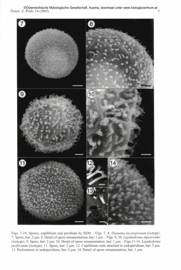
Figs. 7-14. Spores, capillitium and peridium by SEM. - Figs. 7, 8. Dianema inconspicuum (isotype).7. Spore, bar: 2 \im. 8. Detail of spore ornamentation, bar: 1 um. - Figs. 9, 10. Lepidoderma alpestroides(isotype). 9. Spore, bar: 2 (im. 10. Detail of spore ornamentation, bar: 1 (am. - Figs. 11-14. Lepidodermaperforatum (isotype). 11. Spore, bar: 2 urn. 12. Capillitium ends attached to endoperidium, bar: 5 um.13. Perforations in endoperidium, bar: 5 (im. 14. Detail of spore ornamentation, bar: 1 urn.
©Österreichische Mykologische Gesellschaft, Austria, download unter www.biologiezentrum.at

6 G. MORENO & al.: Nivicolous myxomycetes described by M. MEYER, M. POULAIN and J. BOZONNET
Latin diagnosis: Fructificationibus sessilibus, praecipue in forma plasmodiocar-porum, (extendentibus usque ad 5 cm), ab 0,5 ad 1 mm latis, atrobrunneis, nitentibuscum reflexibus aureis; hypothallo atro; peridio membranaceo, persistenti, simplici, laevi;columella nulla, capillitio atro, vix ad apices clariore, densissimum et cohaerens etpaulum elasticum reticulum efficiente, a toto peridio facile soluto; sporis globosis,tenuiter et dense reticulatis, brunneis, 12,5-15 urn diam; plasmodio ignoto.
The specimen studied has vermiform plasmodiocarps, 0.6-1 mm broad and up to 1.2cm long, sometimes effuse, extensive and forming a small reticulum, shiny dark brown.Hypothallus little developed, blackish. Peridium simple, persisting, membranous,iridescent silver, brownish by LM; with irregular dehiscence. Capillitium very abundant,dark by magnifying glass, blackish brown by LM, with clearer ends; threads 1-2 urn indiam., filiform, forming a very dense net with meshes of very variable sizes. Spores darkbrown in mass, dark brown with a clearer zone by LM, 12-14 urn in diam., verru-cous-reticulate, by SEM spore ornamentation formed by a reticulum of irregular andtight meshes.
Observations: Diacheopsis reticulospora can easily be recognized by plasmo-diocarpous fructifications, capillitium forming a very dense network with meshes ofvery variable sizes and spores 12-14 urn in diam., completely reticulate. Othernivicolous species of the genus Diacheopsis that form plasmodiocarps are D. effusaKOWALSKI and D. serpula KOWALSKI. However, D. reticulospora differs clearly by thespore ornamentation which is unique in the genus.
Dianema inconspkuum POLLAIN, MEYER & BOZONNET, Stapfia 73: 86. 2000. (Figs.7,8, 19,20)
Specimens examined: France: La Bathie, Savoie, on Vaccinium myrtillus, 1800 m s. m., 15. 7. 1999,MM 20461, isotype, duplicate in AH 31775; - - 3. 7. 1999, MM 20415, duplicate in AH 31779.
Latin diagnosis: Sporocysti saepissime solitarii, sessiles, pulviniformes-complanati, 0,8-1,1 (-4) x 0,7-0,8(-l,5) mm, et circa 0,3 mm alti. Peridium membrana-ceum, persistens, spadiceum, lucens, laeve vel plus minus plicatum. Columella nulla.Capillitium copiosum filamentis tenuibus, erectis, flexuosis, raras ramificationes etanastomosas praebentibus. Sporae globosae, hyalinae flavidae, tenuibus spinulosisornatae, (9,5-) 10-11(-12) um diam. Plasmodium ignotum.
In the isotype studied, solitary sessile sporocarps can be observed. Sporothecaepulvinate to flattened, 0.4-1 mm in diam., ochraceous yellowish. Hypothallusinconspicuous. Peridium simple, persisting, membranous, ochraceous yellowish bymagnifying glass and LM; with irregular dehiscence. Capillitium light brown bymagnifying glass, pale yellowish by LM; threads 2-4 um in diam., hardly branched andanastomosed, strongly united with the peridium. Spores yellowish ochraceous in mass,yellowish hyaline by LM, 10-12 um in diam., spinulose, by SEM spore ornamentationformed by small lax baculae.
Observations: Dianema inconspkuum is characterized by solitary rather flattenedfructifications, ochraceous yellowish, hardly branched and anastomosed capillitium,
©Österreichische Mykologische Gesellschaft, Austria, download unter www.biologiezentrum.at

Österr. Z. Pilzk. 14 (2005) 7
yellowish by LM, and spores 10-12 um in diam. As the name indicates, it is a tiny orinconspicuous species that due to the small fructifications may have been overlookeduntil recently.
Lepidoderma alpestroides MEYER & POULAIN in POULAIN, MEYER & BOZONNET,Bull. Fed. Mycol. Dauphine-Savoie 42(165): 9. 2002. (Figs. 9, 10, 21, 22)
Specimens examined: France: Bourg-St. Maurice, Les Arcs, Savoie, on Vaccinium myrtillus, 1900 m s.m., 24. 5. 1996, MM 16595, isotype; - - on living shrub, mainly Rhododendron spec, 28. 5. 1997, MM17476, duplicate in AH 31776.
Latin diagnosis: Plasmodiocarpi albi vel cremei, saepe robiginis maculis notati.Peridium duplex, pars exterior exilibus squamulis imbricatis constituta, fere levemcrustam opacam efficientibus; pars interior hyalina flavida luce transmissa. Capillitiumbrunneum obscurum, rigidum. Sporae (12,5-)14-15(-17) um. Plasmodium ignotum.Species nivalis.
The isotype examined has vermiform plasmodiocarps, 4-8 x 1.4-2.5 mm, sinuousand confluent. Hypothallus little developed, yellowish. Peridium double; exoperidiumformed by a calcareous layer, thick, granulöse, creamy white to yellowish creamy,somewhat shiny, rarely with some reddish spots; endoperidium membranous, hyalineby magnifying glass, yellowish hyaline by LM, closely united with the exoperidiumfrom which it is hardly separable; dehiscence irregular. Columella in the form of a crestspreading out all along the plasmodiocarp, with a broad base occupying almost thewhole base of the fructification. Capillitium abundant, blackish brown by magnifyingglass, dark brown by LM, with a greyish apex; threads 1-2 urn in diam., rigid, straight,hardly ramified and anastomosed, except at the apices, with scanty nodes. Spores black-ish brown in mass, violaceous brown by LM, 14-15 (im in diam., spinose, by SEM thespore ornamentation formed by large baculae with coralloid apices.
Observations: Lepidoderma alpestroides is characterized by creamy colouredfructifications in the form of plasmodiocarps, resembling those of Physarum alpeslreMlTCHEL, S. W. CHAPM. & M. L. FARR, with an exoperidium formed by a layer of verydense, not distinguishable calcareous scales, abundant dark brown capillitium formedby parallel and straight filaments and spores 14-15 urn in diam., with large baculae withcoralloid apices by SEM.
Lepidoderma carestianum (RABENH.) ROSTAF. and L. granuliferum (W. PHILLIPS)R. E. FR. are two very close nivicolous species that share with L. alpestroides the plas-modiocarpic growth. Lepidoderma granuliferum differs principally by the presence ofcalcareous nodes in the capillitium and L. carestianum by the absence of a columella, alight capillitium, hyaline to light brown, branched and anastomosed, and sporeornamentation formed by large spines with pointed and not coralloid apices by SEM.
Lepidoderma perforatum M E Y E R & POULAIN in POLLAIN, MEYER & BOZONNET,Bull. Fed. Mycol. Dauphine-Savoie 42(165): 6. 2002. (Figs.l 1-14, 23)
Specimen examined: France: La Bathie, Savoie, on Vaccinium myrtillus, 1300 tn s. m., 22. 4. 1987,MM 2696, isotype, duplicate in AH 31773.
©Österreichische Mykologische Gesellschaft, Austria, download unter www.biologiezentrum.at

8 G. MORENO & al.: Nivicolous myxomycetes described by M. MEYER, M. POULAIN and J. BOZONNET
Latin diagnosis: Plasmodiocarpi ochraceolutei. Peridium duplex, pars exteriorexilibus squamulis tecta; pars interior subcartilaginosa, brunneolutea luce transmissa, etperforata ubi capillitii fila haerent. Capillitium fuscum satis rigidum. Sporae 14,5-16,5um, gracilibus spinulis ornatae. Plasmodium ignotum. Species nivalis.
The isotype studied has flattened plasmodiocarps, 2-5 mm broad and up to 3 cmlong. Hypothallus little developed, reddish brown. Peridium double; exoperidium thick,honey yellowish, covered by large whitish, calcareous, separated, more or lessisodiametric scales; endoperidium membranous, dark brown by magnifying glass, lightbrown and perforated by LM, closely attached to the exoperidium; dehiscence irregular,by SEM perforations of the internal peridium originating by the rupture of it in the areasof union with the capillitium. Columella absent. Capillitium abundant, blackish brownby magnifying glass, dark brown by LM, with hyaline ends; threads 2-3 urn in diam.,rigid and parallel between them, strongly attached to the endoperidium, hardly branchedand anastomosed. Spores blackish brown in mass, dark brown with a clearer zone byLM, 15-16 urn in diam., spinulous, by SEM spore ornamentation formed by dense bacu-lae with an irregular surface.
Observations: According to POULAIN & al. (2002 a) Lepidoderma perforatum isclose to L. carestianum, with which it has in common the plasmodiocarpous fructifica-tions, double peridium and verrucose to finely spinulous spores. However, L. perfora-tum differs by an internal peridium having perforations, resulting by the separation orrupture of the capillitium united to the endoperidium, by robust dark brown capillitiumthreads and by honey yellowish fructifications. Whereas L. carestianum has anon-perforated endoperidium, a capillitium formed by thin hyaline to light brownthreads and greyish brown fructifications.
Discussion
After revision of material of the six taxa described by M. MEYER, M. POULAIN, and J.BOZONNET, we consider the following species to have remarkable characters that makepossible their distinction from other close taxa: Diacheopsis reticulospora, Dianemainconspicuum, Lepidoderma alpestroides and L. perforatum.
Diacheopsis reticulospora has spores 12-14 urn in diam., reticulate, especiallyvisible by SEM. This ornamentation is unique and unmistakable within the genus.Dianema inconspicuum can easily be distinguished by solitary sporocarps, of such smallsize that makes it difficult to find it in the field, and spores with little-developedornamentation. Lepidoderma alpestroides is a very characteristic species due to itsmorphological resemblance to Physarum alpestre. However, the layer composed ofcalcareous scales covering the endoperidium distinguishes it clearly, as well as thedifferent capillitium. Lepidoderma perforatum is easily distinguishable by microscopiccharacters, having an endoperidium with typical perforations, and furthermore it pre-sents a spore ornamentation formed by tight baculae of irregular surface.
On the other hand, Diacheopsis kowalskii and D. pauxilla are very close to D. metal-lica. The latter species differs by the morphology of the capillitium and sporeornamentation. D. pauxilla furthermore presents a dispersed growth habit and smallfructifications.
©Österreichische Mykologische Gesellschaft, Austria, download unter www.biologiezentrum.at
Östcrr. /.. Pil/.k. 14(2005)
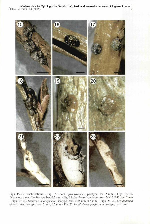
Figs. 15-23. Fructifications. - Fig. 15. Diacheopsis kowalskii. paratype. bar: 2 mm. - Figs. 16. 17.Diacheopsis pauxilla. isotype, bar: 0.5 mm. - Fig. 18. Diacheopsis reticiilospora. MM 2I082. bar: 2 mm.- Figs. 19. 20. Dianema inconspicuum. isotype, bars: 0.25 mm. 0.5 mm. - Figs. 21. 22. Lepidodermaalpestroides, isotype, bars: 2 mm. 0.5 mm. - Fig. 23. Lepidoderma perforatum, isotype, bar: 1 um.
©Österreichische Mykologische Gesellschaft, Austria, download unter www.biologiezentrum.at

] 0 G. MORENO & al.: Nivicolous myxomycetes described by M. MEYER, M. POULAIN and J. BOZONNET
This investigation has been partly financed by the Research Project of the Ministry of Science andTechnology, National Plan of Scientific Investigation, Technological Development and Innovation,REN2OO2-O1965. We are especially grateful to Mrs M. MEYER for her collaboration and the shipment ofspecimens and want to express our gratitude to Mr D. W. MITCHELL for the revision of the manuscript.We wish to thank Mr J. A. PEREZ and Mr A. PRIEGO of the Electron Microscopy Service of theUniversity of Alcalä for their invaluable help with the SEM. We wish to thank Dr J. REJOS, curator ofthe herbarium AH. Mr H. SINGER thanks the National Program of the Professorship Formation, Ministryof Education and Culture of Spain, the conceded scholarship for the realisation of his doctoral thesis inthe University of Alcalä.
References
BOZONNET, J., MEYER, M., POULAIN, M., 1991: Especes nivales de Myxomycetes. - Soc. Hist. Nat.Pays Montbeliard: 51-72.
1995: Les especes nivales du genre Lamproderma (Myxomycetes) ä peridium macule. - Doc.Mycol. 24(96): 1-8.
1997: Lamproderma cacographicum une nouvelle espece nivale de Myxomycetes. - Bull. Fed,Mycol. Dauphine-Savoie 37(144): 117-121.
MEYER, M., POULAIN, M., 1990: Une nouvelle espece nivale du genre Diacheopsis. - Beitr. KenntnisPilze Mitteleur. 6: 35-38.
1998: Diacheopsis kowalskii et Diacheopsis pauxiila deux nouvelles especes de myxomycetes. -Bull. Fed. Mycol. Dauphine-Savoie 38(150): 27-37.
— NOWOTNY, W., POULAIN, M., 1994: Une espece nouvelle du genre Lamproderma ROST.(Myxomycetes). - Bull. Fed. Mycol. Dauphine-Savoie 33(132): 34-38.
MORENO, G., SINGER, H., ILLANA, C , 2003 a: Diacheopsis spinosifila, a synonym of Lepidodermadidermoides. - Mycotaxon 88: 123-128.
LlZARRAGA, M., 2003 b: Diderma nigrum, a synonym of Diderma asteroides (myxomycetes).- Österr. Z. Pilzk. 12:23-29.
2004: A taxonomic review on the nivicolous myxomycete species described by KOWALSKI. II.Physarales and Trichiales. -Österr. Z. Pilzk. 13: 61-74.
POULAIN, M., MEYER, M., BOZONNET, J., 2000: Dianema inconspicuum POULAIN, MEYER &BOZONNET, espece nouvelle de myxomycota, et les especes nivales du genre Dianema. - Stapfia 73:85-92.
2002 a: Deux especes nouvelles de myxomycetes: Lepidoderma alpestroides et Lepidodermaperforaium. - Bull. Fed. Mycol. Dauphine-Savoie 42(165): 5-18.
2002 b: Lamproderma caresliae (CES. & DE NOT.) MEYL. et Lamproderma aeneum sp. nov. -Soc. Hist. Nat. Pays Montbeliard: 47-54.
SINGER, H., MORENO, G., ILLANA, C , LlZARRAGA, M., 2003: Trichia synspora, a synonym of Trichiavaria. - Mycotaxon 87: 243-248.
— — — 2005: Mountainous and nivicolous myxomycetes described by CHARLES MEYLAN. ASEM-study. - Österr. Z. Pilzk. 14: 11-29.
©Österreichische Mykologische Gesellschaft, Austria, download unter www.biologiezentrum.at