Embed Size (px)
Citation preview

lable at ScienceDirect
Brain Stimulation xxx (xxxx) xxx
Contents lists avai
Brain Stimulation
journal homepage: http : / /www.journals .elsevier .com/brain-st imulat ion
The neuromodulatory and hormonal effects of transcutaneous vagusnerve stimulation as evidenced by salivary alpha amylase, salivarycortisol, pupil diameter, and the P3 event-related potential
C.M. Warren a, b, *, 1, K.D. Tona a, b, L. Ouwerkerk a, J. van Paridon a, c, F. Poletiek a, c,Henk van Steenbergen a, b, J.A. Bosch d, e, S. Nieuwenhuis a, b
a Institute of Psychology, Leiden University, Leiden, 2333, AK, Netherlandsb Leiden Institute for Brain and Cognition, Leiden University, Leiden, 2300, RC, Netherlandsc Max Planck Institute of Psycholinguistics, Nijmegen, 6525, XD, Netherlandsd Department of Clinical Psychology, University of Amsterdam, Amsterdam, 1018, XA, Netherlandse Mannheim Institute of Public Health, Heidelberg University, Mannheim, 68167, Germany
a r t i c l e i n f o
Article history:Received 17 September 2018Received in revised form7 December 2018Accepted 12 December 2018Available online xxx
Keywords:Transcutaneous vagus nerve stimulationNorepinephrineAlpha amylaseCortisolEEGP3
* Corresponding author. Institute of Psychology, LeAK, the Netherlands.
E-mail address: [email protected] (C.M. Warre1 Present Address: Utah State University, Departm
84322.
https://doi.org/10.1016/j.brs.2018.12.2241935-861X/© 2018 Elsevier Inc. All rights reserved.
Please cite this article as:Warren CM et al., Tby salivary alpha amylase, salivary cortisolj.brs.2018.12.224
a b s t r a c t
Background: Transcutaneous vagus nerve stimulation (tVNS) is a new, non-invasive technique beinginvestigated as an intervention for a variety of clinical disorders, including epilepsy and depression. It isthought to exert its therapeutic effect by increasing central norepinephrine (NE) activity, but the evi-dence supporting this notion is limited.Objective: In order to test for an impact of tVNS on psychophysiological and hormonal indices ofnoradrenergic function, we applied tVNS in concert with assessment of salivary alpha amylase (SAA) andcortisol, pupil size, and electroencephalograph (EEG) recordings.Methods: Across three experiments, we applied real and sham tVNS to 61 healthy participants while theyperformed a set of simple stimulus-discrimination tasks. Before and after the task, as well as during onebreak, participants provided saliva samples and had their pupil size recorded. EEG was recordedthroughout the task. The target for tVNS was the cymba conchae, which is heavily innervated by theauricular branch of the vagus nerve. Sham stimulation was applied to the ear lobe.Results: P3 amplitude was not affected by tVNS (Experiment 1A: N¼ 24; Experiment 1B: N¼ 20; Bayesfactor supporting null model¼ 4.53), nor was pupil size (Experiment 2: N¼ 16; interaction of treatmentand time: p¼ .79). However, tVNS increased SAA (Experiments 1A and 2: N¼ 25) and attenuated thedecline of salivary cortisol compared to sham (Experiment 2: N¼ 17), as indicated by significant in-teractions involving treatment and time (p¼ .023 and p¼ .040, respectively).Conclusion: These findings suggest that tVNS modulates hormonal indices but not psychophysiologicalindices of noradrenergic function.
© 2018 Elsevier Inc. All rights reserved.
Introduction
Invasive vagus nerve stimulation (VNS) is a somewhat promisingtreatment for depression [1e3] and epilepsy [4,5] that likely exertspart of its therapeutic effect by increasing norepinephrine (NE)release from the locus coeruleus (LC). The vagus nerve projects to
iden University Leiden, 2333
n).ent of Psychology, Logan, UT
he neuromodulatory and horm, pupil diameter, and the P3
the nucleus tractus solitarius, which projects both directly andindirectly to the LC [1e3]. Transcutaneous VNS can be achieved bydelivering electrical impulses to the cervical or the auricularbranches of the vagus nerve, which are situated close to the surfaceof the skin of the neck and outer ear respectively [4]. Functionalmagnetic resonance imaging (fMRI) studies in healthy humansdemonstrate that the more commonly applied transcutaneousauricular VNS (taVNS) elicits widespread changes in cortical andbrainstem activity [5e8]. In light of the clinical potential of taVNS, itwould be valuable to establish if taVNS, like invasive VNS, affects NE,using relatively inexpensive and easy-to-use biomarkers of NE. Herewe evaluated the effect of taVNS on NE levels using three accepted
onal effects of transcutaneous vagus nerve stimulation as evidencedevent-related potential, Brain Stimulation, https://doi.org/10.1016/
C.M. Warren et al. / Brain Stimulation xxx (xxxx) xxx2
biomarkers and one putative biomarker of central NE activity:salivary alpha amylase (SAA), salivary cortisol, pupil size, and the P3component of the event-related brain potential (ERP), respectively.
SAA is a digestive enzyme that is released by the saliva glands inresponse to local sympathetic nervous system activity [9]. SAAsecretion is increased during stress and correlates with bloodplasma NE during exercise [10,11]. SAA is a proxy marker ofsympathetic-adreno-medullary activation [9,12], which is driven bycentral NE, leading to the assumption that SAA marks central NEactivity [13e16]. One preliminary study [17] has reported sugges-tive evidence that taVNS increases SAA relative to sham stim-ulationdreason to be optimistic that a larger study with a moretargetedmethodologymight reveal a robust effect of taVNS on SAA.
Salivary cortisol is a glucocorticoid stress hormone that corre-lates with hypothalamo-pituitary-adrenal axis activation [12,18].Salivary cortisol may likewise be a reliable index of central NE ac-tivity, mediated in part by noradrenergic inputs to the hypothala-mus [12,18,19]. Salivary cortisol is sensitive to pharmacologicallyinduced changes in central NE activity [16,20].
Pupil size is correlated with activity of NE-releasing neurons inthe LC [21e24]. This relationshipmay bemediated by activity in therostral ventrolateral medulla, which projects to the LC and alsoinnervates the peripheral sympathetic ganglia regulating the pupil[25]. Studies of primates and rodents show that LC activity corre-lates with baseline pupil diameter [21,24] and the magnitude oftask-evoked pupil dilations [21,23]. In human participants, BOLDactivity in the LC covaries with pupil size at rest and during simpledecision-making tasks [22,26]. In rats, direct stimulation of thecentral stump of the vagus nerve provokes pupil dilation [27], butresults in humans have been mixed [28,29].
Phasic changes in cortical NE levels are associated with thescalp-recorded P3 component [30e37]. Events that lead toincreased phasic firing of the LC also lead to increased P3 amplitude[30]. Noradrenergic drugs influence P3 amplitude in both animals[38] and humans [39e41], and lesion of the LC eliminates the P3 inmonkeys [42]. Of interest here, the amplitude of the P3 is increasedby invasive VNS [29,31,35].
Although LC-NE activity is associated with changes in SAA,salivary cortisol, pupil size and the P3, these psychophysiologicalmeasures are not exclusively diagnostic of changes in LC-NE ac-tivity. For example, fluctuations in pupil size have been shown totrack activity in a number of neuromodulatory brainstem centers,including the LC, the dopaminergic ventral tegmental area, and thecholinergic basal forebrain [24,26]. Also, P3 amplitude can bemodulated by dopaminergic and cholinergic pharmacological ma-nipulations, suggesting a role for those systems in P3 generation[30]. Thus, although our study is well-equipped to pick upconverging evidence for taVNS effects on the noradrenergic system,it does not allow us to fully discriminate between noradrenergicand other neuromodulatory and hormonal effects of taVNS.
To explore the claim that taVNS increases central NE, we assayedSAA, salivary cortisol, pupil size and P3 amplitude across threeexperiments. In Experiments 1A and 2, we collected saliva samplesand analyzed these samples all together. In Experiment 2, we alsorecorded pupil size. Experiment 1B was a partial replication of 1A,wherein we recorded EEG data during a classic oddball task, theseminal task for eliciting a P3 [43].
Experiment 1A
Method
ParticipantsTwenty-four students at Leiden University (6 male, mean age
22.6) participated in return for V30. We used the following
Please cite this article as:Warren CM et al., The neuromodulatory and hormby salivary alpha amylase, salivary cortisol, pupil diameter, and the P3j.brs.2018.12.224
exclusion criteria: history of psychiatric or neurological disorders,head trauma, migraine, current use of psychoactive drugs, preg-nancy, active implants (cochlear implant, pacemaker) and a per-manent ear-piercing.
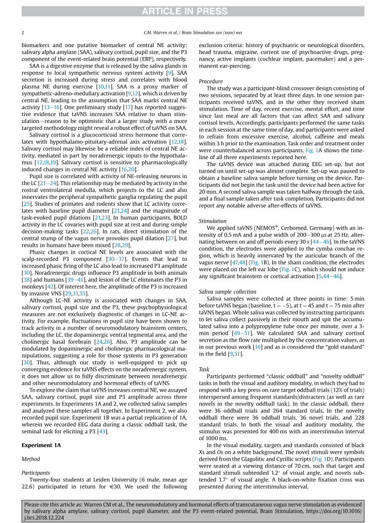
ProcedureThe study was a participant-blind crossover design consisting of
two sessions, separated by at least three days. In one session par-ticipants received taVNS, and in the other they received shamstimulation. Time of day, recent exercise, mental effort, and timesince last meal are all factors that can affect SAA and salivarycortisol levels. Accordingly, participants performed the same tasksin each session at the same time of day, and participants were askedto refrain from excessive exercise, alcohol, caffeine and mealswithin 3 h prior to the examination. Task order and treatment orderwere counterbalanced across participants. Fig. 1A shows the time-line of all three experiments reported here.
The taVNS device was attached during EEG set-up, but notturned on until set-up was almost complete. Set-up was paused toobtain a baseline saliva sample before turning on the device. Par-ticipants did not begin the task until the device had been active for20min. A second saliva samplewas taken halfway through the task,and a final sample taken after task completion. Participants did notreport any notable adverse after-effects of taVNS.
StimulationWe applied taVNS (NEMOS®, Cerbomed, Germany) with an in-
tensity of 0.5mA and a pulse width of 200e300 ms at 25 Hz, alter-nating between on and off periods every 30 s [44e46]. In the taVNScondition, the electrodes were applied to the cymba conchae re-gion, which is heavily innervated by the auricular branch of thevagus nerve [47,48] (Fig. 1B). In the sham condition, the electrodeswere placed on the left ear lobe (Fig. 1C), which should not induceany significant brainstem or cortical activation [5,44e46].
Saliva sample collectionSaliva samples were collected at three points in time: 5min
before taVNS began (baseline, t¼�5), at t¼ 45 and t¼ 75min aftertaVNS began.Whole salivawas collected by instructing participantsto let saliva collect passively in their mouth and spit the accumu-lated saliva into a polypropylene tube once per minute, over a 3-min period [49e51]. We calculated SAA and salivary cortisolsecretion as the flow rate multiplied by the concentration values, asin our previous work [16] and as is considered the “gold standard”in the field [9,51].
TaskParticipants performed “classic oddball” and “novelty oddball”
tasks in both the visual and auditory modality, inwhich they had torespond with a key press on rare target oddball trials (12% of trials)interspersed among frequent standards/distracters (as well as rarenovels in the novelty oddball task). In the classic oddball, therewere 36 oddball trials and 264 standard trials. In the noveltyoddball there were 36 oddball trials, 36 novel trials, and 228standard trials. In both the visual and auditory modality, thestimulus was presented for 400ms with an interstimulus intervalof 1000ms.
In the visual modality, targets and standards consisted of blackXs and Os on a white background. The novel stimuli were symbolsderived from the Glagolitic and Cyrillic scripts (Fig.1D). Participantswere seated at a viewing distance of 70 cm, such that target andstandard stimuli subtended 1.2� of visual angle, and novels sub-tended 1.7� of visual angle. A black-on-white fixation cross waspresented during the interstimulus interval.
onal effects of transcutaneous vagus nerve stimulation as evidencedevent-related potential, Brain Stimulation, https://doi.org/10.1016/
Fig. 1. A) Depiction of the timing of each experiment. For questionnaires see [52e54]. B) Position of the taVNS device that gives targeted stimulation to auricular branch of the vagusnerve. C) Position of the taVNS device for “sham” stimulation that putatively does not impact activity in the vagus nerve. D) Examples of the visual novelty stimuli used in thenovelty oddball P3 task in Experiment 1A.
C.M. Warren et al. / Brain Stimulation xxx (xxxx) xxx 3
In the auditory modality, the fixation cross remained on-screenconstantly. The auditory stimuli were tones of high (500Hz) or lowpitch (350 HZ), presented at 70 dB through speakers. Target fre-quency was counterbalanced across participants. The novel stimuliconsisted of unusual noises pulled from the set of Fabiani andFriedman [55].
EEG collection and processingEEGwas recorded from 64 channels (ActiveTwo system, Biosemi
B.V., Netherlands) in the standard 10e20 configuration. Data werepre-amplified at the electrode site and recorded with a samplingrate of 512 Hz with reference to a common mode sense. Imped-ances were kept below 32 kU. EEG recordings were processed usingBrain Vision Analyzer 2 (Brain Products GmbH, Germany). Datawere re-referenced offline to the right mastoid, and band-passfiltered (0.1 Hz-20.0 Hz). Ocular artifacts were removed [56].Epochs were extracted from the EEG from �200ms to 800msrelative to stimulus onset, using the first 200ms for baselinecorrection. Trials in which the change in voltage at any channelexceeded 35 mV per sampling point were removed as were trialswith slow drifts (>300 mV/200ms) and lowactivity (<.5mV/100ms).
We created difference waves to simplify figures and analyses, toisolate the topography of the P3, and to distinguish the P3 tooddballs (“oddball P3”) and the P3 to novel stimuli (“novelty P3”).To isolate the oddball P3 we subtracted the standard ERP from theoddball ERP, and to isolate the novelty P3 we subtracted the stan-dard ERP from the novelty ERP. In each case P3 amplitude wasquantified as the most positive mean amplitude from a 200-mssliding window across the entire difference wave.
The oddball P3 was analyzed using an ANOVA including thefactors treatment (taVNS vs sham), modality (visual vs auditory),task (classic oddball vs novelty oddball) and electrode (Fz, Cz, Pz).The novelty P3 obtained in the novelty oddball task was analyzed
Please cite this article as:Warren CM et al., The neuromodulatory and hormby salivary alpha amylase, salivary cortisol, pupil diameter, and the P3j.brs.2018.12.224
using an ANOVA including the factors treatment (taVNS vs sham),modality (visual vs auditory) and electrode (Fz, Cz, Pz). In addition,treatment order was added as a between-subjects variable-of-no-interest, to account for additional error variance.
Results
Oddball P3
The amplitude of the oddball P3 showed significant main effectsof electrode, indicating a typical parietal distribution (Fig. 2), F(2,44)¼ 60.82, p< .001, task (classic oddball: 7.7 mV; noveltyoddball: 7.0 mV; Fig. 3), F (1,22)¼ 13.36, p¼ .001, and modality(visual: 7.7 mV; auditory: 6.4 mV), F (1,22)¼ 28.65, p< .001. Inaddition, electrode interacted with task, F (2,44)¼ 5.15, p¼ .010,and exhibited a three-way interaction with task and modality, F(2,44)¼ 3.56, p¼ .037. Treatment did not significantly affectoddball P3 amplitude (taVNS: 7.1 mV; sham: 7.0 mV), F(1,22)¼ 0.020, p¼ .89. Treatment did not interact with electrode,task, or modality.
Novelty P3As expected, the novelty P3 had a more frontal distribution than
the oddball P3, with largest amplitude at electrode Cz, F(2,44)¼ 14.05, p< .001 (Fig. 2). The novelty P3 was larger in theauditory modality (10.5 mV), than in the visual modality (2.8 mV), F(1,22)¼ 82.36, p< .001 (Fig. 3). In addition, the effect of modality onnovelty P3 amplitude was larger at Fz and Cz than at Pz, F(2,44)¼ 34.17, p< .001. Treatment did not significantly affect nov-elty P3 amplitude (taVNS: 6.4 mV; sham: 6.8 mV), F (1,22)¼ 0.65,p¼ .43.
onal effects of transcutaneous vagus nerve stimulation as evidencedevent-related potential, Brain Stimulation, https://doi.org/10.1016/

Fig. 2. Scalp distribution of the oddball P3 and novelty P3 for taVNS (top row) and sham stimulation (bottom row), for the novelty and classic oddball tasks in Exp. 1A, and theclassic oddball task in Exp. 1B. For Exp. 1A, scalp distributions are collapsed across the visual and auditory modalities, and for the oddball P3 they are collapsed across tasks as well.
C.M. Warren et al. / Brain Stimulation xxx (xxxx) xxx4
Experiment 1B
Our task in Experiment 1A yielded a typical oddball P3 andnovelty P3, exhibiting characteristic effects of electrode, task andmodality. There were no effects of treatment on the oddball P3 ornovelty P3. Given that P3 amplitude was shown to be increased by
Fig. 3. ERP waveforms from Experiments 1A and 1B for electrodes Cz (top) and Pz (bottom)oddball task, in both the visual and auditory modality. Experiment 1B included only a clas
Please cite this article as:Warren CM et al., The neuromodulatory and hormby salivary alpha amylase, salivary cortisol, pupil diameter, and the P3j.brs.2018.12.224
invasive VNS, we considered the possibility that our null effect oftreatment on P3 amplitude was a type-2 error, and followed upwith a simplified experiment on new participants, composed of asingle classic oddball task with a greater number of trials, and withthe exact same stimulation protocol.
were not affected by taVNS. Exp. 1A included both a novelty oddball task and a classicsic oddball task in the visual modality.
onal effects of transcutaneous vagus nerve stimulation as evidencedevent-related potential, Brain Stimulation, https://doi.org/10.1016/

C.M. Warren et al. / Brain Stimulation xxx (xxxx) xxx 5
Method
ParticipantsTwenty Leiden University students (11 males, mean age 23.6)
participated in return for V25. Exclusion criteria were the same asExperiment 1A.
Task and procedureThe task was an abbreviated version of the task used in Exper-
iment 1A. Experiment 1B included only the visual version of theclassic oddball paradigm. We increased the number of trials from300 to 400 (352 standards, 48 targets). The materials (Xs and Os)were the same, as were all the timing parameters.
The exact same stimulation protocol was used as in Experiment1A. The first 55min of each session in Experiment 1Bwere identicalto Experiment 1A, except that participants did not fill out anyquestionnaires, and participants did not provide saliva samples(Fig. 1A).
EEG collection and processingEEG collection and processing methods as well as statistical
analysis were identical as described for Experiment 1A, except asfollows. Recordings for five participants had noisy signals at oneelectrode. For these participants the bad electrode was removedbefore ocular correction, and its signal interpolated from theremaining electrodes using a spline-based method.
Results
The oddball P3 in Experiment 1B exhibited a parietal distribu-tion (Fig. 2), with largest amplitude at Pz, F (2,34)¼ 32.54, p< .001.The oddball P3 was larger in the taVNS condition (7.6 mV) than inthe sham condition (6.5 mV). This difference was not significant, F(1,17)¼ 2.32, p¼ .15 (Fig. 3).
Bayesian analyses
In Experiment 1A taVNS did not affect the size of the oddball P3or novelty P3. We ran Experiment 1B to perform a second exami-nation of this potential effect, and found no effect (p¼ .15). Thecombined results suggest that taVNS does not affect P3 amplitude,but such a conclusion cannot be firm under the rules of null hy-pothesis statistical testing. In order to quantify evidence for the nullhypothesis, we ran a Bayesian-evidence synthesis analysis [57] onthe visual oddball P3 data from Experiments 1A and 1B.
Method
Data were analyzed using JASP (JASP Team, 2016), which yieldsBayes factors that give the relative probability of competing modelsof the data. We ran a Bayesian repeated-measures ANOVA withtreatment as the repeated factor, and two between-subjects fac-tors: treatment order and experiment.
Results
Bayes factor comparisons favored the null model over a modelincluding an effect of treatment (BF01¼4.53). This quantity meansno effect of treatment is 4.53 times more probable, given the data,than an effect of treatment, which constitutes “substantial evi-dence” for the null hypothesis [58]. The null model was also su-perior to a model including an interaction of treatment withexperiment (BF01¼13.48). This Bayes factor constitutes “strongevidence” in favor of the null model [58]. Taken together with the
Please cite this article as:Warren CM et al., The neuromodulatory and hormby salivary alpha amylase, salivary cortisol, pupil diameter, and the P3j.brs.2018.12.224
frequentist methods reported above, the appropriate conclusion isthat taVNS does not affect the (visual) oddball P3.
Experiment 2
Method
ParticipantsSeventeen young adults (all males, mean age 22.1) participated
in Experiment 2. One participant gave saliva samples but did nothave his pupil data recorded. Exclusion criteriawere the same as forExperiment 1A.
ProcedureThe timeline of Experiment 2 is displayed in Fig. 1A. Pupil data
were recorded at three time points during each session. Salivasamples were collected with the exact same methodology andtiming relative to taVNS as described in Experiment 1A. taVNS wasapplied with the exact same timing, positioning and parameters asin Experiments 1A and 1B. Participants performed a cued task-switching task [62] and other tests from which data will be re-ported elsewhere.
Pupil size recording and analysisParticipants sat in a dimly lit room with head held steady in a
chin rest, and fixated on a luminance-controlled, salmon-coloredfixation cross on a slate blue computer screen. Pupil diameter wasrecorded for 96 s at 60 Hz using a Tobii T120 eye tracker (TobiiTechnology, Stockholm, Sweden). Recordings were made roughly10min before stimulation began (t¼�10), between practice andcritical task-switching trials (t¼ 25) and upon completion of thetask (t¼ 70). Pupil data were analyzed using custom-made macrosin BrainVision Analyzer 2.0 (Brain Products GmbH). Linear inter-polation was applied to artifacts such as blinks and missing data.
Results
We analyzed mean pupil size across the 96-s epoch at each timepoint. Treatment did not significantly affect pupil size (p¼ .37), norinteract with time (p¼ .79, Greenhouse-Geisser corrected). Therewas an effect of time on pupil size whereby pupil size increasedfrom baseline during task performance, F (1,15)¼ 9.78, p¼ .007(Greenhouse-Geisser corrected) (Fig. 4).
Analysis of saliva data
Method
ParticipantsExperiments 1a and 2 were run simultaneously with identical
taVNS protocol and identical saliva collection, processing andanalysis protocol. Saliva samples were collected from twenty par-ticipants in Experiment 1a, and seventeen participants in Experi-ment 2. Twenty-five participants provided SAA samples withconcentrations within the sensitivity range of our assay in all sixcells of the design (3 samples x 2 treatment conditions). In thefollow-up analyses of the effect of time in each treatment condition,thirty-three participants provided utilizable samples in all 3 cells ofthe taVNS condition, and twenty-seven participants provided uti-lizable samples in all 3 cells of the sham condition. Salivary cortisolwas determined only for the saliva samples from Experiment 2. Allseventeen participants provided sufficient samples to includecortisol secretion data in every cell of the design.
onal effects of transcutaneous vagus nerve stimulation as evidencedevent-related potential, Brain Stimulation, https://doi.org/10.1016/
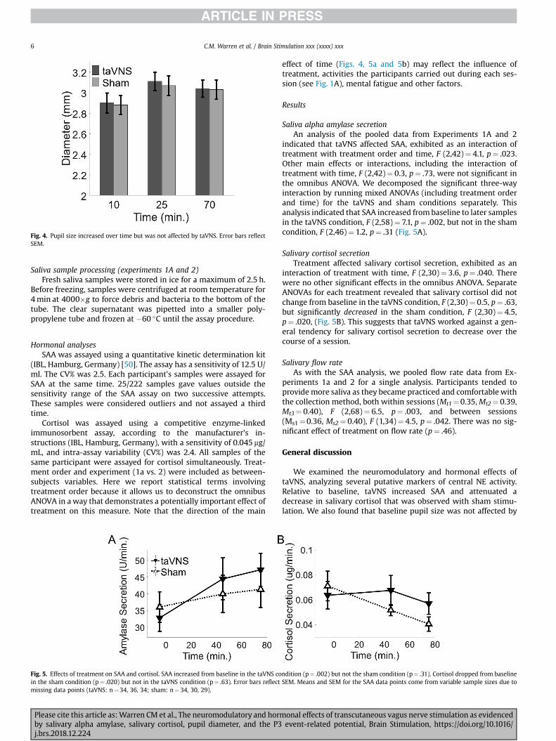
Fig. 4. Pupil size increased over time but was not affected by taVNS. Error bars reflectSEM.
C.M. Warren et al. / Brain Stimulation xxx (xxxx) xxx6
Saliva sample processing (experiments 1A and 2)Fresh saliva samples were stored in ice for a maximum of 2.5 h.
Before freezing, samples were centrifuged at room temperature for4min at 4000�g to force debris and bacteria to the bottom of thetube. The clear supernatant was pipetted into a smaller poly-propylene tube and frozen at �60 �C until the assay procedure.
Hormonal analysesSAA was assayed using a quantitative kinetic determination kit
(IBL, Hamburg, Germany) [50]. The assay has a sensitivity of 12.5 U/ml. The CV% was 2.5. Each participant's samples were assayed forSAA at the same time. 25/222 samples gave values outside thesensitivity range of the SAA assay on two successive attempts.These samples were considered outliers and not assayed a thirdtime.
Cortisol was assayed using a competitive enzyme-linkedimmunosorbent assay, according to the manufacturer's in-structions (IBL, Hamburg, Germany), with a sensitivity of 0.045 mg/mL, and intra-assay variability (CV%) was 2.4. All samples of thesame participant were assayed for cortisol simultaneously. Treat-ment order and experiment (1a vs. 2) were included as between-subjects variables. Here we report statistical terms involvingtreatment order because it allows us to deconstruct the omnibusANOVA in away that demonstrates a potentially important effect oftreatment on this measure. Note that the direction of the main
Fig. 5. Effects of treatment on SAA and cortisol. SAA increased from baseline in the taVNS coin the sham condition (p¼ .020) but not in the taVNS condition (p¼ .63). Error bars reflectmissing data points (taVNS: n¼ 34, 36, 34; sham: n¼ 34, 30, 29).
Please cite this article as:Warren CM et al., The neuromodulatory and hormby salivary alpha amylase, salivary cortisol, pupil diameter, and the P3j.brs.2018.12.224
effect of time (Figs. 4, 5a and 5b) may reflect the influence oftreatment, activities the participants carried out during each ses-sion (see Fig. 1A), mental fatigue and other factors.
Results
Saliva alpha amylase secretionAn analysis of the pooled data from Experiments 1A and 2
indicated that taVNS affected SAA, exhibited as an interaction oftreatment with treatment order and time, F (2,42)¼ 4.1, p¼ .023.Other main effects or interactions, including the interaction oftreatment with time, F (2,42)¼ 0.3, p¼ .73, were not significant inthe omnibus ANOVA. We decomposed the significant three-wayinteraction by running mixed ANOVAs (including treatment orderand time) for the taVNS and sham conditions separately. Thisanalysis indicated that SAA increased frombaseline to later samplesin the taVNS condition, F (2,58)¼ 7.1, p¼ .002, but not in the shamcondition, F (2,46)¼ 1.2, p¼ .31 (Fig. 5A).
Salivary cortisol secretionTreatment affected salivary cortisol secretion, exhibited as an
interaction of treatment with time, F (2,30)¼ 3.6, p¼ .040. Therewere no other significant effects in the omnibus ANOVA. SeparateANOVAs for each treatment revealed that salivary cortisol did notchange from baseline in the taVNS condition, F (2,30)¼ 0.5, p¼ .63,but significantly decreased in the sham condition, F (2,30)¼ 4.5,p¼ .020, (Fig. 5B). This suggests that taVNS worked against a gen-eral tendency for salivary cortisol secretion to decrease over thecourse of a session.
Salivary flow rateAs with the SAA analysis, we pooled flow rate data from Ex-
periments 1a and 2 for a single analysis. Participants tended toprovidemore saliva as they became practiced and comfortablewiththe collection method, both within sessions (Mt1¼0.35,Mt2¼ 0.39,Mt3¼ 0.40), F (2,68)¼ 6.5, p¼ .003, and between sessions(Ms1¼0.36, Ms2¼ 0.40), F (1,34)¼ 4.5, p¼ .042. There was no sig-nificant effect of treatment on flow rate (p¼ .46).
General discussion
We examined the neuromodulatory and hormonal effects oftaVNS, analyzing several putative markers of central NE activity.Relative to baseline, taVNS increased SAA and attenuated adecrease in salivary cortisol that was observed with sham stimu-lation. We also found that baseline pupil size was not affected by
ndition (p¼ .002) but not the sham condition (p¼ .31). Cortisol dropped from baselineSEM. Means and SEM for the SAA data points come from variable sample sizes due to
onal effects of transcutaneous vagus nerve stimulation as evidencedevent-related potential, Brain Stimulation, https://doi.org/10.1016/

C.M. Warren et al. / Brain Stimulation xxx (xxxx) xxx 7
taVNS, and that taVNS did not affect P3 amplitude. Thus, two of ourfour physiological markers responded sensitively to taVNS,consistent with increases in central NE.
Our results compliment work by Ventura-Bort and colleagues[17], who reported preliminary evidence that taVNS increases SAA.We demonstrate amore robust effect, using a larger sample size (25vs. 18 participants), more post-stimulation saliva samples (2 vs. 1),and a superior method of saliva collection (whole saliva method vs.absorbent cotton sponges) [9,51]. In addition, we report the firstevidence that taVNS influences salivary cortisol. Together, thesehormonal analyses add to an accumulating pharmacological liter-ature suggesting that SAA and salivary cortisol might be effectivemarkers of central NE activity [13e16,20].
The relationship between pupil size and NE activity has beensupported by direct recordings from the LC [21,23], direct stimu-lation of the LC [24], and by fMRI data from human participants[22,26]. We found no effect of taVNS on pupil size, suggesting thattaVNS might not increase NE. An alternative possibility is that ourpupil experiment was underpowered in terms of sample size orrecording duration, or otherwise not sensitive enough to the taVNSmanipulation. Our null result resonates with Schevernels and col-leagues [29], who found no effect on pupil size of invasive VNS.Although two other invasive VNS studies found significant pupileffects, we know of no other taVNS study that has measured pupildata. Thus, our work serves as a first exploration that should berevisited with methods adjusted accordingly.
The LC-P3 hypothesis proposes that the P3 reflects the change inneural gain produced by a phasic burst of NE release [30]. Only oneresearch group has reported an effect of taVNS on P3 amplitude[17,59]. In one study [59] these researchers analyzed data from aSimon task and report that taVNS increased both conflict adapta-tion and N2 amplitude on incompatible trials, but not P3 amplitude.In a separate study they found taVNS increased P3 amplitude [17].This study was exploratory, reporting significant simple effects inspecific cells of their design, without justification from interactionsin the omnibus ANOVA. In light of their other null effect, and ourBayesian evidence in favor of no effect, we must acknowledge thatthe evidence that the P3 is directly affected by invasive or trans-cutaneous VNS is mixed at best.
We note some limitations to this work. Saliva data was pooledacross two experiments. In Experiment 1a participants were beingset up with EEG between the baseline sample and subsequentsamples, whereas in Experiment 2 participants were practicing thetask-switching experiment. The stimulation protocols and collec-tion methods were identical, and no statistical tests were inter-preted between experiments, but the difference likely introducedsome variability to the data. This could have contributed to therelatively weak statistical support for the effect of taVNS on SAA.That is, the two-way interaction of treatment with time was notsignificant so we relied on the three-way interaction of treatmentwith time and treatment order to justify decomposing the omnibusANOVA. The salivary cortisol results gave more straightforwardevidence of an effect of taVNS, but the sample size was smallish(n¼ 17), though still within an appropriate range of sample sizesfor investigating phasic changes in salivary cortisol (for a review seeRef. [51]). An additional limitation of this work concerns the generalparameters and targets used for taVNS. We used a stimulation in-tensity of 0.5mA for all participants. In contrast, some studiestitrate stimulus intensity to the participant's perceptual threshold(e.g. Ref. [60] titrated to an intensity of 3.14mA). In addition, ifstimulation in the sham condition was painful, it could haveincreased central NE, creating a ceiling effect that minimized ourability to detect a further increase due to taVNS. Our stimulationintensity was low, but pre-treating with taVNS or sham first andthen running the oddball task with no simultaneous stimulation
Please cite this article as:Warren CM et al., The neuromodulatory and hormby salivary alpha amylase, salivary cortisol, pupil diameter, and the P3j.brs.2018.12.224
could address this concern. Finally, there is an unresolved questionas to whether the cymba conchae is the best target for taVNS,though both sites yield significant changes in cortical activity[47,61,62]. It is possible that some of our null effects were due to ouruse of a weak current, a ceiling effect, or a sub-optimal target.Nevertheless, our entire protocol was based on previous work[44e46] and use of this protocol lead to significant effects onsalivary markers of NE activity. Keeping the discussed null effectsand limitations in mind, our results provide support for the clinicaland experimental use of taVNS.
Declarations of interest
None.
Funding
This work was supported by a Starting Grant of the EuropeanResearch Council (#283314, 2011).
References
[1] Vonck K, Raedt R, Naulaerts J, De Vogelaere F, Thiery E, Van Roost D, et al.Vagus nerve stimulation. 25 years later! what do we know about the effectson cognition? Neurosci Biobehav Rev 2014;45:63e71. https://doi.org/10.1016/j.neubiorev.2014.05.005.
[2] Nemeroff CB, Mayberg HS, Krahl SE, McNamara J, Frazer A, Henry TR, et al.VNS therapy in treatment-resistant depression: clinical evidence and putativeneurobiological mechanisms. Neuropsychopharmacology 2006;31:1345e55.https://doi.org/10.1038/sj.npp.1301082.
[3] George MS, Aston-Jones G. Noninvasive techniques for probing neurocircuitryand treating illness: vagus nerve stimulation (VNS), transcranial magneticstimulation (TMS) and transcranial direct current stimulation (tDCS). Neuro-psychopharmacology 2010;35:301e16. https://doi.org/10.1038/npp.2009.87.
[4] Ellrich J. Transcutaneous vagus nerve stimulation. Eur Neurol Rev 2011;6:254e6. https://doi.org/10.17925/ENR.2011.06.04.254.
[5] Kraus T, Kiess O, H€osl K, Terekhin P, Kornhuber J, Forster C. CNS BOLD fMRIeffects of sham-controlled transcutaneous electrical nerve stimulation in theleft outer auditory canal - a pilot study. Brain Stimul 2013;6:798e804. https://doi.org/10.1016/j.brs.2013.01.011.
[6] Kraus T, H€osl K, Kiess O, Schanze A, Kornhuber J, Forster C. BOLD fMRI deac-tivation of limbic and temporal brain structures and mood enhancing effect bytranscutaneous vagus nerve stimulation. J Neural Transm 2007;114:1485e93.https://doi.org/10.1007/s00702-007-0755-z.
[7] Frangos E, Ellrich J, Komisaruk BR. Non-invasive access to the vagus nervecentral projections via electrical stimulation of the external ear: FMRI evi-dence in humans. Brain Stimul 2015;8:624e36. https://doi.org/10.1016/j.brs.2014.11.018.
[8] Yakunina N, Kim SS, Nam E-C. Optimization of transcutaneous vagus nervestimulation using functional MRI. Neuromodulation Technol Neural Interface2017;20:290e300. https://doi.org/10.1111/ner.12541.
[9] Bosch JA, Veerman ECI, de Geus EJ, Proctor GB. А-amylase as a reliable andconvenient measure of sympathetic activity: don't start salivating just yet!Psychoneuroendocrinology 2011;36:449e53. https://doi.org/10.1016/j.psyneuen.2010.12.019.
[10] Chatterton RT, Vogelsong KM, Lu Y, Ellman AB, Hudgens GA. Salivary a-amylase as a measure of endogenous adrenergic activity. Clin Physiol1996;16:433e48. https://doi.org/10.1111/j.1475-097X.1996.tb00731.x.
[11] Bosch JA, Brand HS, Ligtenberg TJM, Bermond B, Hoogstraten J,Amerongen AVN. Psychological stress as a determinant of protein levels andsalivary-induced aggregation of Streptococcus gordonii in human wholesaliva. Psychosom Med 1996;58.
[12] Bosch JA, de Geus EJC, Carroll D, Goedhart AD, Anane LA, van Zanten JJV, et al.A general enhancement of autonomic and cortisol responses during socialevaluative threat. Psychosom Med 2009;71:877e85. https://doi.org/10.1097/PSY.0b013e3181baef05.
[13] Speirs RL, Herring J, Cooper WD, Hardy CC, Hind CRK. The influence of sym-pathetic activity and isoprenaline on the secretion of amylase from the humanparotid gland. Arch Oral Biol 1974;19:747e52. https://doi.org/10.1016/0003-9969(74)90161-7.
[14] Ehlert U, Erni K, Hebisch G, Nater U. Salivary a-amylase levels after yohimbinechallenge in healthy men. J Clin Endocrinol Metab 2006;91:5130e3. https://doi.org/10.1210/jc.2006-0461.
[15] Van Stegeren A, Rohleder N, Everaerd W, Wolf OT. Salivary alpha amylase asmarker for adrenergic activity during stress: effect of betablockade. Psycho-neuroendocrinology 2006;31:137e41. https://doi.org/10.1016/j.psyneuen.2005.05.012.
onal effects of transcutaneous vagus nerve stimulation as evidencedevent-related potential, Brain Stimulation, https://doi.org/10.1016/
C.M. Warren et al. / Brain Stimulation xxx (xxxx) xxx8
[16] Warren CM, van den Brink RL, Nieuwenhuis S, Bosch JA. Norepinephrinetransporter blocker atomoxetine increases salivary alpha amylase. Psycho-neuroendocrinology 2017;78. https://doi.org/10.1016/j.psyneuen.2017.01.029.
[17] Ventura-Bort C, Wirkner J, Genheimer H, Wendt J, Hamm AO, Weymar M.Effects of transcutaneous vagus nerve stimulation (tVNS) on the P300 andalpha-amylase level: a pilot study. Front Hum Neurosci 2018;12:1e12.https://doi.org/10.3389/fnhum.2018.00202.
[18] Hill SA, Taylor MJ, Harmer CJ, Cowen PJ. Acute reboxetine administrationincreases plasma and salivary cortisol. J Psychopharmacol 2003;17:273e5.https://doi.org/10.1177/02698811030173008.
[19] Dunn AJ, Swiergiel AH, Palamarchouk V. Brain circuits involved incorticotropin-releasing factor-norepinephrine interactions during stress. AnnN Y Acad Sci 2004;1018:25e34. https://doi.org/10.1196/annals.1296.003.
[20] Chamberlain SR, Müller U, Cleary S, Robbins TW, Sahakian BJ. Atomoxetineincreases salivary cortisol in healthy volunteers. J Psychopharmacol 2007;21:545e9. https://doi.org/10.1177/0269881106075274.
[21] Joshi S, Li Y, Kalwani RM, Gold JI. Relationships between pupil diameter andneuronal activity in the locus coeruleus, colliculi, and cingulate cortex. Neuron2016;89:221e34. https://doi.org/10.1016/j.neuron.2015.11.028.
[22] Murphy PR, O'Connell RG, O'Sullivan M, Robertson IH, Balsters JH. Pupildiameter covaries with BOLD activity in human locus coeruleus. Hum BrainMapp 2014;35:4140e54. https://doi.org/10.1002/hbm.22466.
[23] Varazzani C, San-Galli A, Gilardeau S, Bouret S. Noradrenaline and dopamineneurons in the reward/effort trade-off: a direct electrophysiological compar-ison in behaving monkeys. J Neurosci 2015;35:7866e77. https://doi.org/10.1523/JNEUROSCI.0454-15.2015.
[24] Reimer J, McGinley MJ, Liu Y, Rodenkirch C, Wang Q, McCormick DA, et al.Pupil fluctuations track rapid changes in adrenergic and cholinergic activity incortex. Nat Commun 2016;7:1e7. https://doi.org/10.1038/ncomms13289.
[25] Nieuwenhuis S, De Geus EJ, Aston-Jones G. The anatomical and functionalrelationship between the P3 and autonomic components of the orientingresponse. Psychophysiology 2011;48:162e75. https://doi.org/10.1111/j.1469-8986.2010.01057.x.
[26] de Gee JW, Colizoli O, Kloosterman NA, Knapen T, Nieuwenhuis S, Donner TH.Dynamic modulation of decision biases by brainstem arousal systems. Elife2017;6:1e36. https://doi.org/10.7554/eLife.23232.
[27] Bianca R, Komisaruk BR. Pupil dilatation in response to vagal afferent elec-trical stimulation is mediated by inhibition of parasympathetic outflow in therat. Brain Res 2007;1177:29e36. https://doi.org/10.1016/j.brainres.2007.06.104.
[28] Desbeaumes Jodoin V, Lesp�erance P, Nguyen DK, Fournier-Gosselin MP,Richer F. Effects of vagus nerve stimulation on pupillary function. Int J Psy-chophysiol 2015;98:455e9. https://doi.org/10.1016/j.ijpsycho.2015.10.001.
[29] Schevernels H, van Bochove ME, De Taeye L, Bombeke K, Vonck K, VanRoost D, et al. The effect of vagus nerve stimulation on response inhibition.Epilepsy Behav 2016;64:171e9. https://doi.org/10.1016/j.yebeh.2016.09.014.
[30] Nieuwenhuis S, Aston-Jones G, Cohen JD. Decision making, the P3, and thelocus coeruleus–norepinephrine system. Psychol Bull 2005;131:510.
[31] Neuhaus AH, Luborzewski A, Rentzsch J, Brakemeier EL, Opgen-Rhein C,Gallinat J, et al. P300 is enhanced in responders to vagus nerve stimulation fortreatment of major depressive disorder. J Affect Disord 2007;100:123e8.https://doi.org/10.1016/j.jad.2006.10.005.
[32] Murphy PR, Robertson IH, Balsters JH, O’connell RG. Pupillometry and P3 in-dex the locus coeruleusenoradrenergic arousal function in humans. Psycho-physiology 2011;48:1532e43.
[33] Warren CM, Tanaka JW, Holroyd CB. What can topology changes in theoddball N2 reveal about underlying processes? Neuroreport 2011;22. https://doi.org/10.1097/WNR.0b013e32834bbe1f.
[34] Warren CM, Holroyd CB. The impact of deliberative strategy dissociates ERPcomponents related to conflict processing vs. Reinforcement learning. FrontNeurosci 2012. https://doi.org/10.3389/fnins.2012.00043.
[35] De Taeye L, Vonck K, van Bochove M, Boon P, Van Roost D, Mollet L, et al. TheP3 event-related potential is a biomarker for the efficacy of vagus nervestimulation in patients with epilepsy. Neurotherapeutics 2014;11:612e22.https://doi.org/10.1007/s13311-014-0272-3.
[36] Wolff N, Mückschel M, Ziemssen T, Beste C. The role of phasic norepinephrinemodulations during task switching: evidence for specific effects in parietalareas. Brain Struct Funct 2018;223:925e40.
[37] Chmielewski WX, Mückschel M, Ziemssen T, Beste C. The norepinephrinesystem affects specific neurophysiological subprocesses in the modulation ofinhibitory control by working memory demands. Hum Brain Mapp 2017;38:68e81.
[38] Swick D, Pineda JA, Foote SL. Effects of systemic clonidine on auditory event-related potentials in squirrel monkeys. Brain Res Bull 1994;33:79e86.
[39] Brown SBRE, Slagter HA, van Noorden MS, Giltay EJ, van der Wee NJA,Nieuwenhuis S. Effects of clonidine and scopolamine on multiple target
Please cite this article as:Warren CM et al., The neuromodulatory and hormby salivary alpha amylase, salivary cortisol, pupil diameter, and the P3j.brs.2018.12.224
detection in rapid serial visual presentation. Psychopharmacology (Berlin)2016;233:341e50.
[40] Brown SBRE, Van der Wee NJA, Van Noorden MS, Giltay EJ, Nieuwenhuis S.Noradrenergic and cholinergic modulation of late ERP responses to deviantstimuli. Psychophysiology 2015;52:1620e31.
[41] De Rover M, Brown SBRE, Band GP, Giltay EJ, Van Noorden MS, Van DerWee NJA, et al. Beta receptor-mediated modulation of the oddball P3 but noterror-related ERP components in humans. Psychopharmacology (Berlin)2015;232:3161e72. https://doi.org/10.1007/s00213-015-3966-2.
[42] Pineda JA, Foote SL, Neville HJ. The effects of locus coeruleus lesions on asquirrel monkey late positive component: a preliminary study. Electro-encephalogr Clin Neurophysiol Suppl 1987;40. 481d486.
[43] Sutton S, Braren M, Zubin J, John ER. Evoked-potential correlates of stimulusuncertainty. Science 1965;150:1187e8. 80-.
[44] Beste C, Steenbergen L, Sellaro R, Grigoriadou S, Zhang R, Chmielewski W, et al.Effects of concomitant stimulation of the GABAergic and norepinephrine sys-tem on inhibitory control e a study using transcutaneous vagus nerve stimu-lation. Brain Stimul 2016;9:811e8. https://doi.org/10.1016/J.BRS.2016.07.004.
[45] Steenbergen L, Sellaro R, Stock AK, Verkuil B, Beste C, Colzato LS. Trans-cutaneous vagus nerve stimulation (tVNS) enhances response selection duringaction cascading processes. Eur Neuropsychopharmacol 2015;25:773e8.https://doi.org/10.1016/j.euroneuro.2015.03.015.
[46] Sellaro R, van Leusden JWR, Tona K-D, Verkuil B, Nieuwenhuis S, Colzato LS.Transcutaneous vagus nerve stimulation enhances post-error slowing.J Cognit Neurosci 2015;27:2126e32. https://doi.org/10.1162/jocn_a_00851.
[47] Badran BW, Brown JC, Dowdle LT, Mithoefer OJ, LaBate NT, Coatsworth J, et al.Tragus or cymba conchae? Investigating the anatomical foundation of trans-cutaneous auricular vagus nerve stimulation (taVNS). Brain Stimul 2018;11:947e8. https://doi.org/10.1016/j.brs.2018.06.003.
[48] Peuker ET, Filler TJ. The nerve supply of the human auricle. Clin Anat 2002;15:35e7. https://doi.org/10.1002/ca.1089.
[49] Beltzer EK, Fortunato CK, Guaderrama MM, Peckins MK, Garramone BM,Granger DA. Salivary flow and alpha-amylase: collection technique, duration,and oral fluid type. Physiol Behav 2010;101:289e96. https://doi.org/10.1016/j.physbeh.2010.05.016.
[50] Nagy T, van Lien R, Willemsen G, Proctor G, Efting M, Fül€op M, et al. A fluidresponse: alpha-amylase reactions to acute laboratory stress are related tosample timing and saliva flow rate. Biol Psychol 2015;109:111e9. https://doi.org/10.1016/j.biopsycho.2015.04.012.
[51] Strahler J, Skoluda N, Kappert MB, Nater UM. Simultaneous measurement ofsalivary cortisol and alpha-amylase: application and recommendations.Neurosci Biobehav Rev 2017;83:657e77. https://doi.org/10.1016/j.neubiorev.2017.08.015.
[52] Spielberger CD, Gorsuch RL, Lushene RE. State-trait anxiety inventory. PaloAlto. 1970.
[53] Russell JA, Weiss A, Mendelsohn GA. Affect grid: a single-item scale of plea-sure and arousal. J Pers Soc Psychol 1989;57:493e502. https://doi.org/10.1037/0022-3514.57.3.493.
[54] Benedict RHB, Schretlen D, Groninger L, Brandt J. Hopkins verbal learningTest ? Revised: normative data and analysis of inter-form and test-retestreliability. Clin Neuropsychol 1998;12:43e55. https://doi.org/10.1076/clin.12.1.43.1726.
[55] Fabiana M, Friedman D. Changes in brain activity patterns in aging: thenovelty oddball. Psychophysiology 1995;32:579e94. https://doi.org/10.1111/j.1469-8986.1995.tb01234.x.
[56] Gratton G, Coles MGH, Donchin E. A newmethod for off-line removal of ocularartifact. Electroencephalogr Clin Neurophysiol 1983;55:468e84.
[57] Scheibehenne B, Jamil T, Wagenmakers EJ. Bayesian evidence synthesis canreconcile seemingly inconsistent results: the case of hotel towel reuse. Psy-chol Sci 2016;27:1043e6. https://doi.org/10.1177/0956797616644081.
[58] Jeffreys H. Theory of Probability 1961. https://doi.org/10.1038/163464a0.[59] Fischer R, Ventura-Bort C, Hamm A, Weymar M. Transcutaneous vagus nerve
stimulation (tVNS) enhances conflict-triggered adjustment of cognitive con-trol. Cognit Affect Behav Neurosci 2018:1e14. https://doi.org/10.3758/s13415-018-0596-2.
[60] Badran BW, Dowdle LT, Mithoefer OJ, LaBate NT, Coatsworth J, Brown JC, et al.Neurophysiologic effects of transcutaneous auricular vagus nerve stimulation(taVNS) via electrical stimulation of the tragus: a concurrent taVNS/fMRIstudy and review. Brain Stimul 2018;11:492e500. https://doi.org/10.1016/j.brs.2017.12.009.
[61] Fallgatter AJ, Neuhauser B, Herrmann MJ, Ehlis A-C, Wagener A,Scheuerpflug P, et al. Far field potentials from the brain stem after trans-cutaneous vagus nerve stimulation. J Neural Transm 2003;110:1437e43.https://doi.org/10.1007/s00702-003-0087-6.
[62] Burger AM, Verkuil B. Transcutaneous nerve stimulation via the tragus: are wereally stimulating the vagus nerve? Brain Stimul 2018;11:945e6. https://doi.org/10.1016/j.brs.2018.03.018.
onal effects of transcutaneous vagus nerve stimulation as evidencedevent-related potential, Brain Stimulation, https://doi.org/10.1016/


![Transcutaneous electrical nerve stimulation for acute painuir.ulster.ac.uk/22796/1/TENS_acute_pain_cochrane_review.pdf · [Intervention Review] Transcutaneous electrical nerve stimulation](https://img.dokumen.tips/doc/110x75/5b5932df7f8b9a4e1b8cebc7/transcutaneous-electrical-nerve-stimulation-for-acute-intervention-review.jpg)