Embed Size (px)
Citation preview

The The Nervous Nervous SystemSystem
Protection of the Central Protection of the Central Nervous SystemNervous System
1.1. Scalp and skinScalp and skin
2.2. Skull and vertebral columnSkull and vertebral column
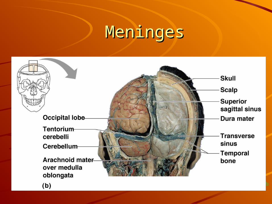
3.3. Meninges- 3 connective tissue Meninges- 3 connective tissue layerslayers
4.4. Cerebrospinal fluid (CSF)Cerebrospinal fluid (CSF)
5.5. Blood-brain barrierBlood-brain barrier
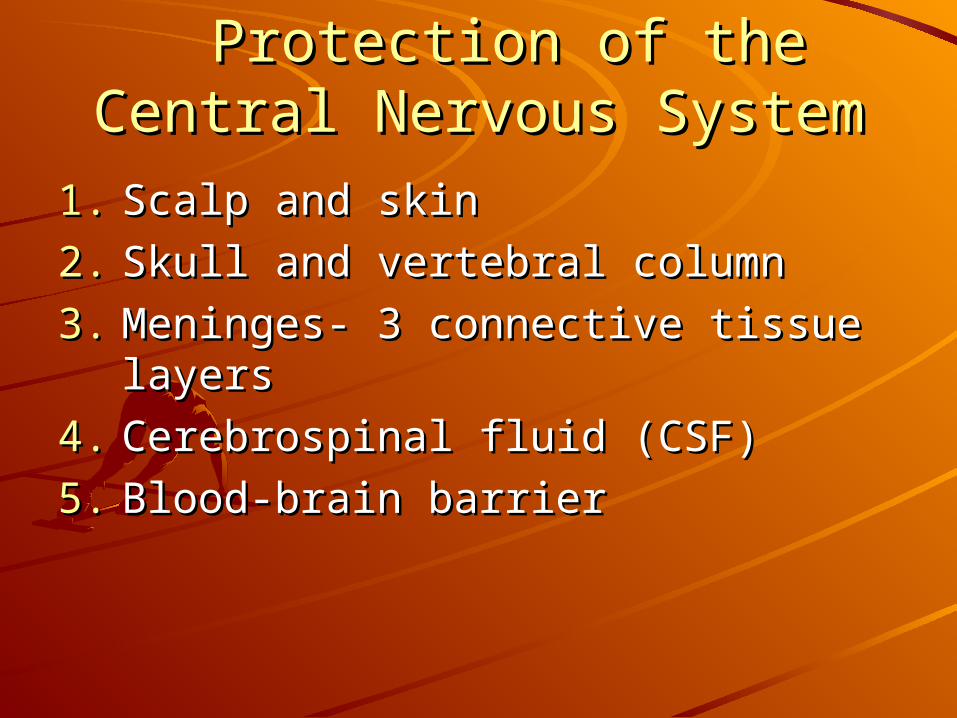
Meninges: 3 layers of connective Meninges: 3 layers of connective tissuetissue
1. Dura mater- “tough mother”1. Dura mater- “tough mother”– Double-layered covering that surrounds the brainDouble-layered covering that surrounds the brain
Periosteal layer (periosteum)—attached to inner surface of Periosteal layer (periosteum)—attached to inner surface of the skullthe skull
Meningeal layer—outer covering of the brainMeningeal layer—outer covering of the brain
– Folds inward in several areasFolds inward in several areas
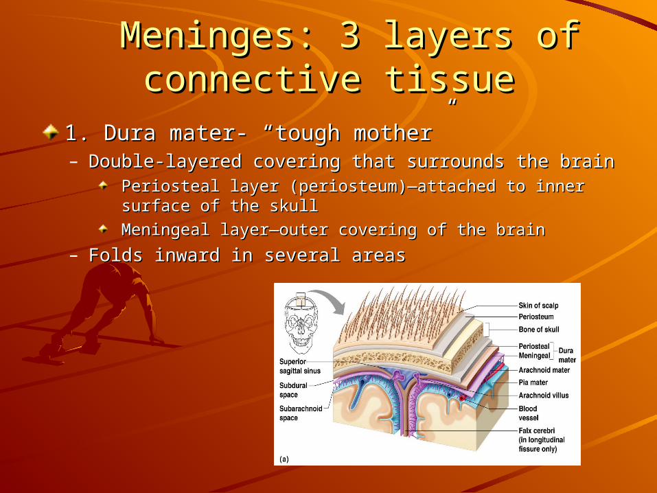
MeningesMeninges2. Arachnoid layer- “spider” layer2. Arachnoid layer- “spider” layer– Middle layer; Web-likeMiddle layer; Web-like– Attaches to the pia materAttaches to the pia mater
3. Pia mater- “gentle mother”3. Pia mater- “gentle mother”– Internal layerInternal layer– Clings to the surface of the brain & spinal cordClings to the surface of the brain & spinal cord
MeningesMeninges
Figure 7.17b
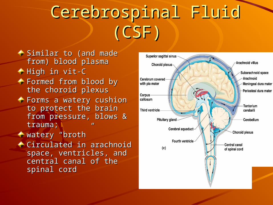
Cerebrospinal Fluid (CSF)Cerebrospinal Fluid (CSF)Similar to (and made from) Similar to (and made from) blood plasmablood plasmaHigh in vit-CHigh in vit-CFormed from blood by the Formed from blood by the choroid plexuschoroid plexusForms a watery cushion to Forms a watery cushion to protect the brain from protect the brain from pressure, blows & trauma;pressure, blows & trauma;watery “broth” watery “broth” Circulated in arachnoid Circulated in arachnoid space, ventricles, and space, ventricles, and central canal of the spinal central canal of the spinal cordcord
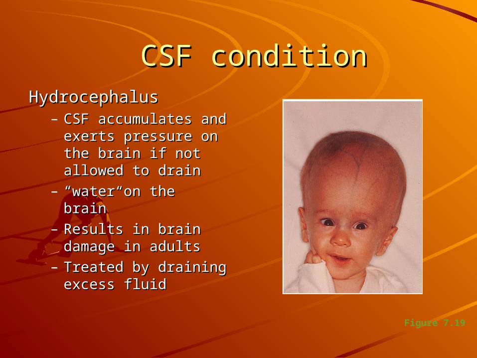
CSF conditionCSF conditionHydrocephalusHydrocephalus
– CSF accumulates CSF accumulates and exerts pressure and exerts pressure on the brain if not on the brain if not allowed to drainallowed to drain
– ““water on the water on the brain”brain”
– Results in brain Results in brain damage in adultsdamage in adults
– Treated by draining Treated by draining excess fluidexcess fluid
Figure 7.19
Blood-Brain BarrierBlood-Brain BarrierIncludes the least permeable capillaries of the Includes the least permeable capillaries of the body (doesn’t allow much to pass thru)body (doesn’t allow much to pass thru)Excludes many potentially harmful substancesExcludes many potentially harmful substancesUseless as a barrier against some substances. Useless as a barrier against some substances. – Respiratory gasesRespiratory gases– Fats and fat soluble moleculesFats and fat soluble molecules
AlcoholAlcoholNicotineNicotineAnesthesiaAnesthesia
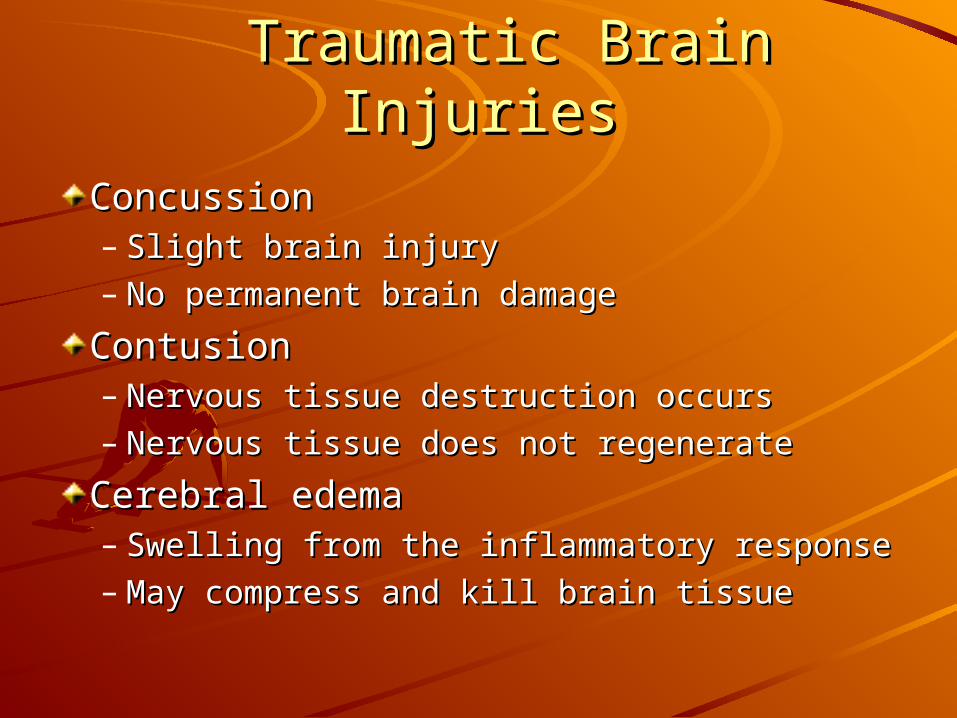
Traumatic Brain InjuriesTraumatic Brain Injuries
ConcussionConcussion– Slight brain injurySlight brain injury– No permanent brain damageNo permanent brain damage
ContusionContusion– Nervous tissue destruction occursNervous tissue destruction occurs– Nervous tissue does not regenerateNervous tissue does not regenerate
Cerebral edemaCerebral edema– Swelling from the inflammatory responseSwelling from the inflammatory response– May compress and kill brain tissueMay compress and kill brain tissue

Cerebrovascular Accident Cerebrovascular Accident (CVA)(CVA)
Commonly called a strokeCommonly called a stroke
The result of a ruptured blood vessel The result of a ruptured blood vessel supplying a region of the brainsupplying a region of the brain
Brain tissue supplied with oxygen Brain tissue supplied with oxygen from that blood source diesfrom that blood source dies
Loss of some functions or death may Loss of some functions or death may resultresult

Alzheimer’s DiseaseAlzheimer’s DiseaseProgressive degenerative brain diseaseProgressive degenerative brain disease
Mostly seen in the elderly, but may begin Mostly seen in the elderly, but may begin in middle agein middle age
Structural changes in the brain include Structural changes in the brain include abnormal protein deposits and twisted abnormal protein deposits and twisted fibers within neuronsfibers within neurons
Victims experience memory loss, Victims experience memory loss, irritability, confusion, and ultimately, irritability, confusion, and ultimately, hallucinations and deathhallucinations and death
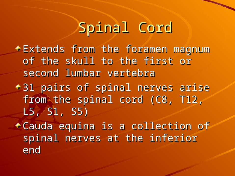
Spinal CordSpinal Cord
Extends from the foramen magnum Extends from the foramen magnum of the skull to the first or second of the skull to the first or second lumbar vertebralumbar vertebra
31 pairs of spinal nerves arise from 31 pairs of spinal nerves arise from the spinal cord (C8, T12, L5, S1, S5)the spinal cord (C8, T12, L5, S1, S5)
Cauda equina is a collection of spinal Cauda equina is a collection of spinal nerves at the inferior endnerves at the inferior end
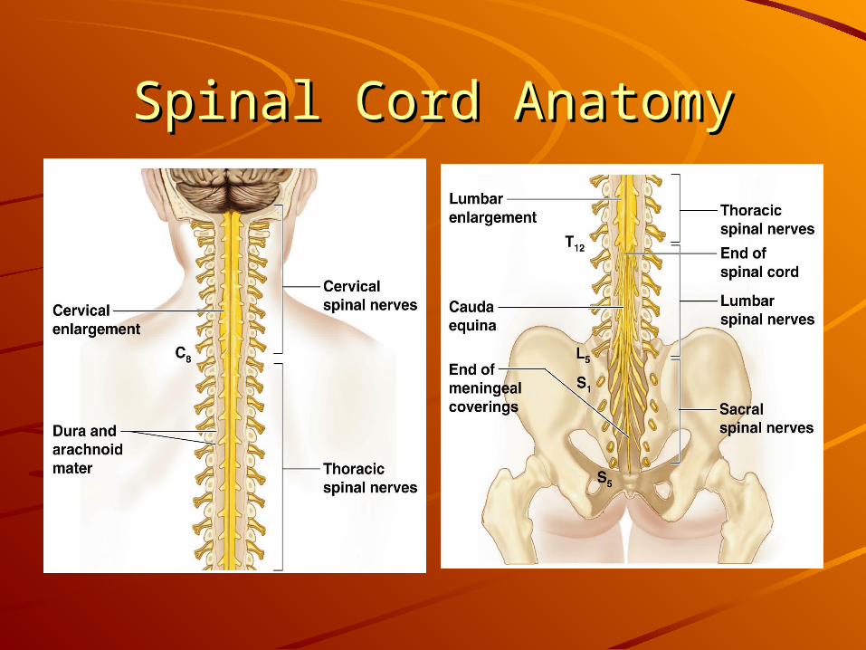
Spinal Cord AnatomySpinal Cord Anatomy
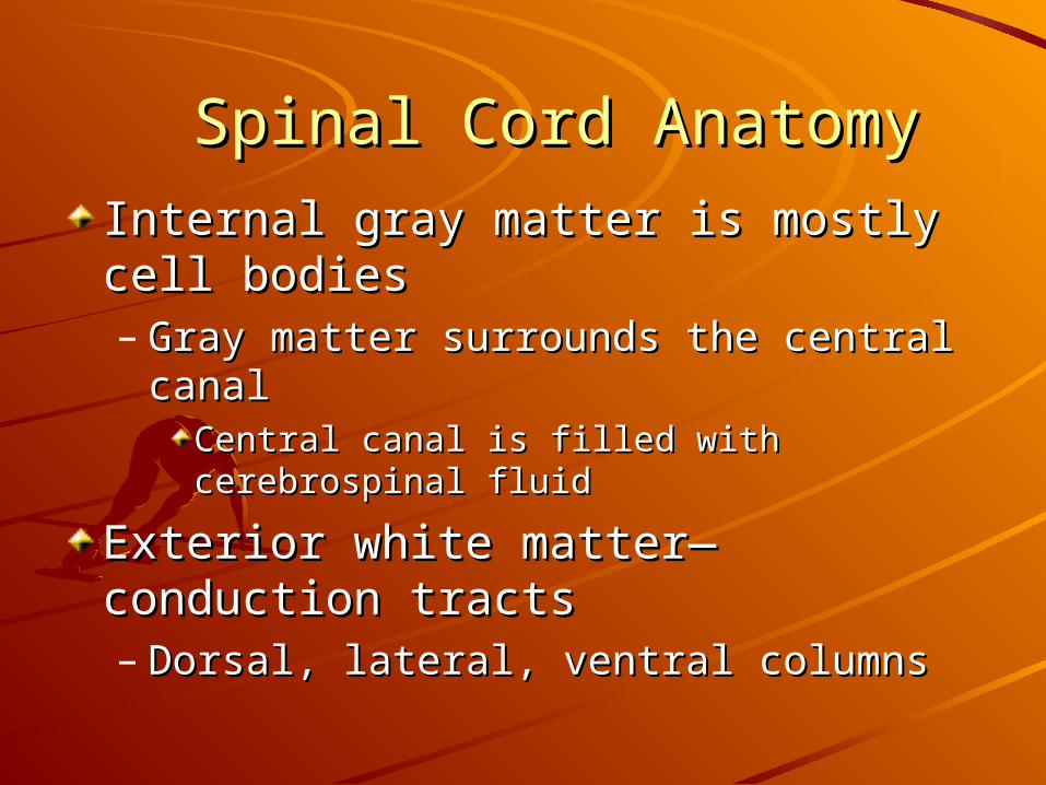
Spinal Cord AnatomySpinal Cord Anatomy
Internal gray matter is mostly cell Internal gray matter is mostly cell bodiesbodies– Gray matter surrounds the central canalGray matter surrounds the central canal
Central canal is filled with cerebrospinal fluidCentral canal is filled with cerebrospinal fluid
Exterior white matter—conduction Exterior white matter—conduction tractstracts– Dorsal, lateral, ventral columnsDorsal, lateral, ventral columns

Spinal Cord AnatomySpinal Cord Anatomy
Figure 7.21
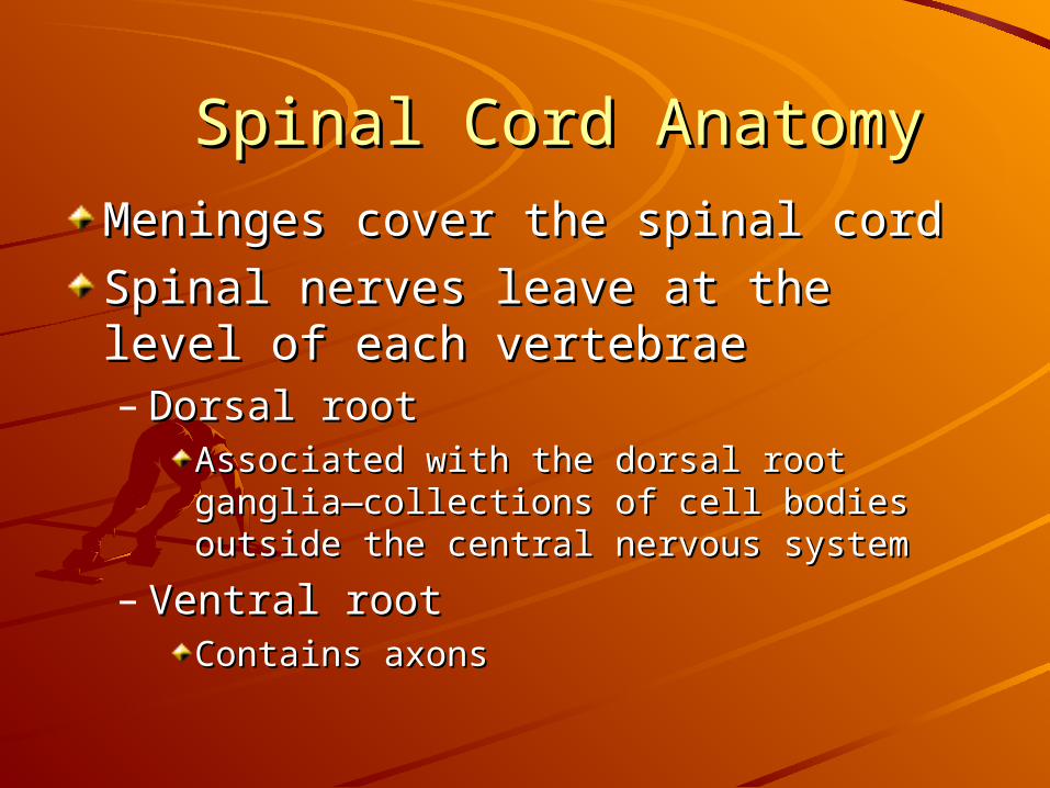
Spinal Cord AnatomySpinal Cord Anatomy
Meninges cover the spinal cordMeninges cover the spinal cord
Spinal nerves leave at the level of Spinal nerves leave at the level of each vertebraeeach vertebrae– Dorsal rootDorsal root
Associated with the dorsal root ganglia—Associated with the dorsal root ganglia—collections of cell bodies outside the central collections of cell bodies outside the central nervous systemnervous system
– Ventral rootVentral rootContains axonsContains axons
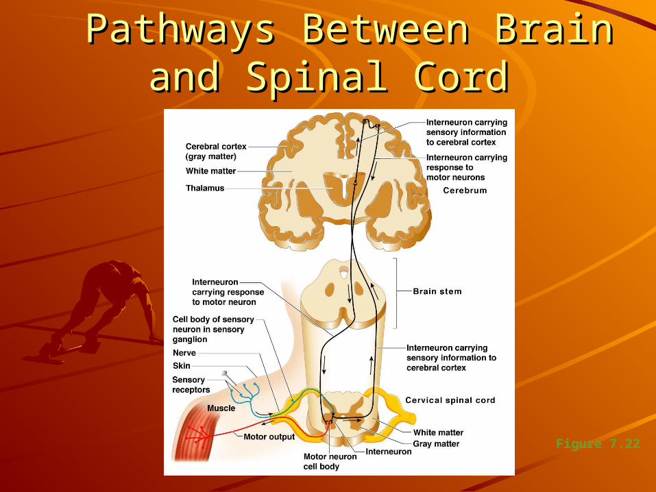
Pathways Between Brain and Pathways Between Brain and Spinal CordSpinal Cord
Figure 7.22
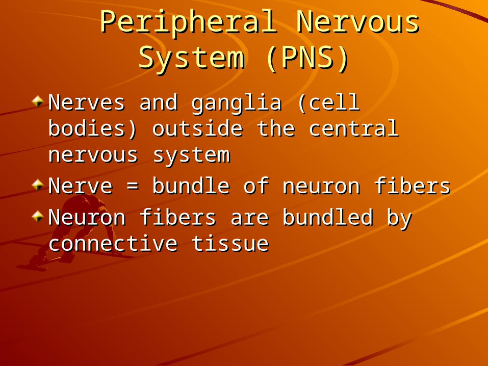
Peripheral Nervous System Peripheral Nervous System (PNS)(PNS)
Nerves and ganglia (cell bodies) Nerves and ganglia (cell bodies) outside the central nervous systemoutside the central nervous system
Nerve = bundle of neuron fibersNerve = bundle of neuron fibers
Neuron fibers are bundled by Neuron fibers are bundled by connective tissueconnective tissue
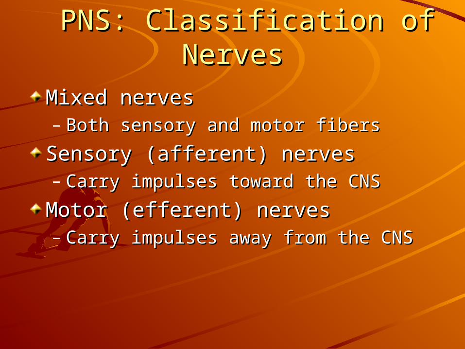
PNS: Classification of NervesPNS: Classification of Nerves
Mixed nervesMixed nerves– Both sensory and motor fibersBoth sensory and motor fibers
Sensory (afferent) nervesSensory (afferent) nerves– Carry impulses toward the CNSCarry impulses toward the CNS
Motor (efferent) nervesMotor (efferent) nerves– Carry impulses away from the CNSCarry impulses away from the CNS
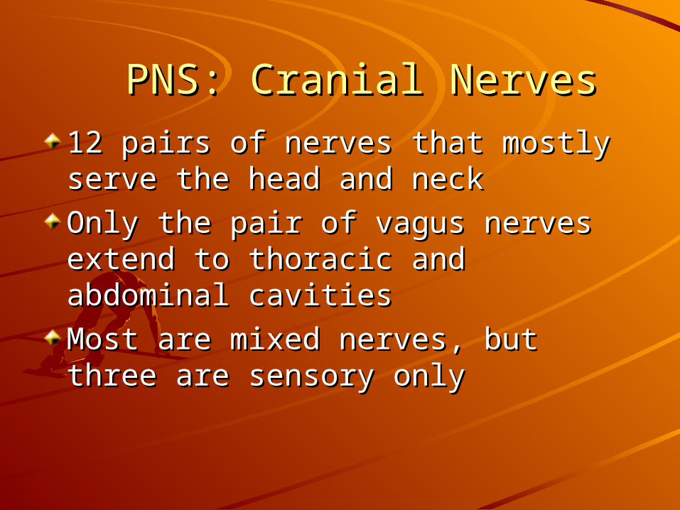
PNS: Cranial NervesPNS: Cranial Nerves
12 pairs of nerves that mostly serve 12 pairs of nerves that mostly serve the head and neckthe head and neck
Only the pair of vagus nerves extend Only the pair of vagus nerves extend to thoracic and abdominal cavitiesto thoracic and abdominal cavities
Most are mixed nerves, but three are Most are mixed nerves, but three are sensory onlysensory only
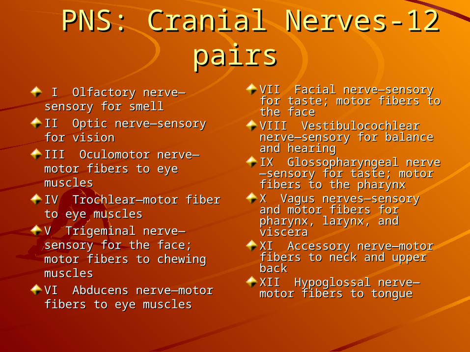
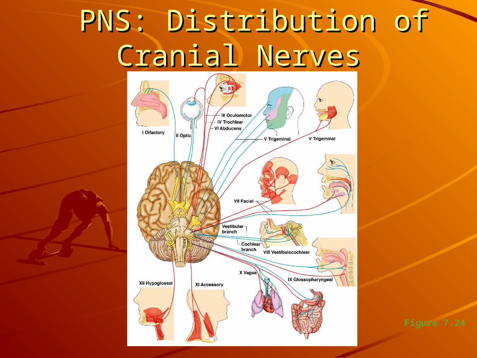
PNS: Cranial Nerves-12 pairsPNS: Cranial Nerves-12 pairs I Olfactory nerve—I Olfactory nerve—sensory for smellsensory for smell
II Optic nerve—sensory for II Optic nerve—sensory for visionvision
III Oculomotor nerve—III Oculomotor nerve—motor fibers to eye motor fibers to eye musclesmuscles
IV Trochlear—motor fiber IV Trochlear—motor fiber to eye musclesto eye muscles
V Trigeminal nerve—V Trigeminal nerve—sensory for the face; motor sensory for the face; motor fibers to chewing musclesfibers to chewing muscles
VI Abducens nerve—motor VI Abducens nerve—motor fibers to eye musclesfibers to eye muscles
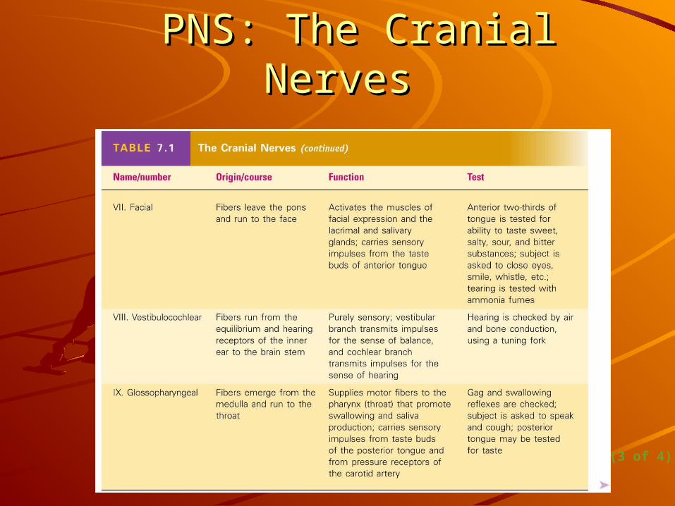
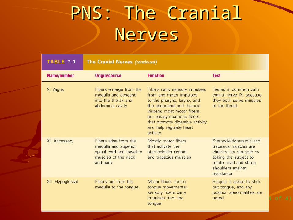
VII Facial nerve—sensory VII Facial nerve—sensory for taste; motor fibers to for taste; motor fibers to the facethe faceVIII Vestibulocochlear VIII Vestibulocochlear nerve—sensory for balance nerve—sensory for balance and hearingand hearingIX Glossopharyngeal nerveIX Glossopharyngeal nerve—sensory for taste; motor —sensory for taste; motor fibers to the pharynxfibers to the pharynxX Vagus nerves—sensory X Vagus nerves—sensory and motor fibers for and motor fibers for pharynx, larynx, and pharynx, larynx, and visceravisceraXI Accessory nerve—motor XI Accessory nerve—motor fibers to neck and upper fibers to neck and upper backbackXII Hypoglossal nerve—XII Hypoglossal nerve—motor fibers to tonguemotor fibers to tongue
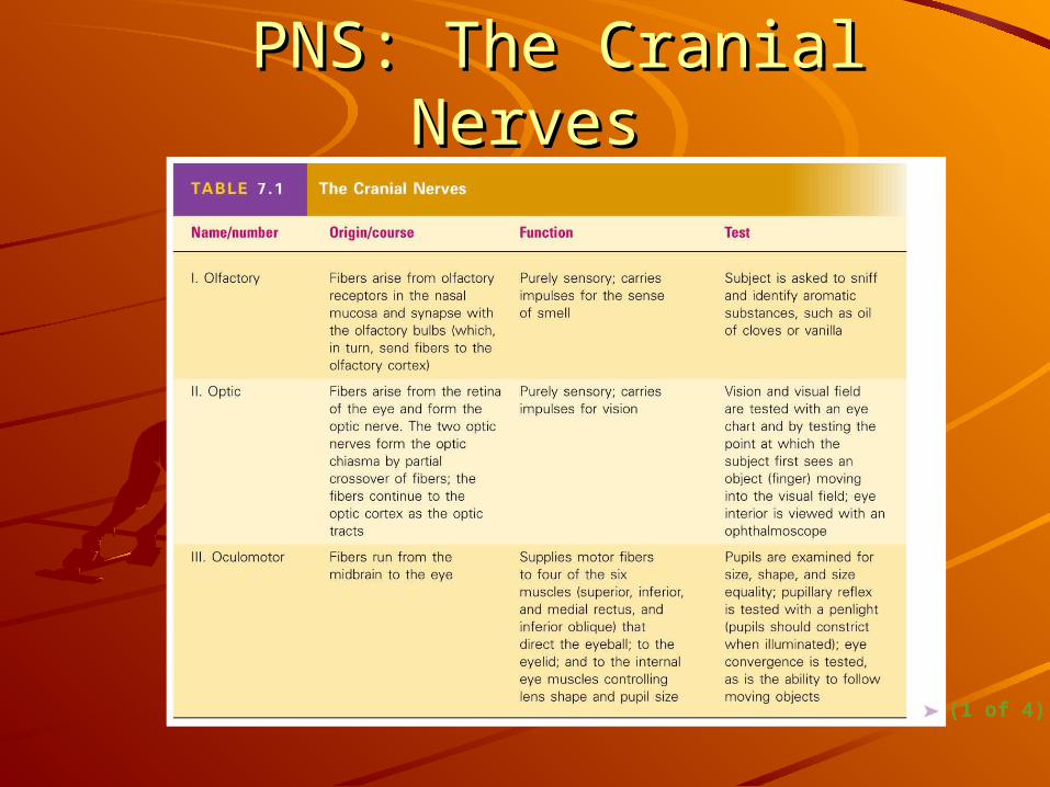
PNS: The Cranial NervesPNS: The Cranial Nerves
Table 7.1 (1 of 4)
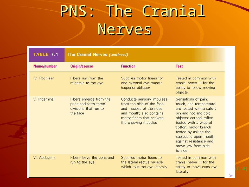
PNS: The Cranial NervesPNS: The Cranial Nerves
Table 7.1 (2 of 4)
PNS: The Cranial NervesPNS: The Cranial Nerves
Table 7.1 (3 of 4)
PNS: The Cranial NervesPNS: The Cranial Nerves
Table 7.1 (4 of 4)
PNS: Distribution of Cranial PNS: Distribution of Cranial NervesNerves
Figure 7.24