Embed Size (px)
DESCRIPTION
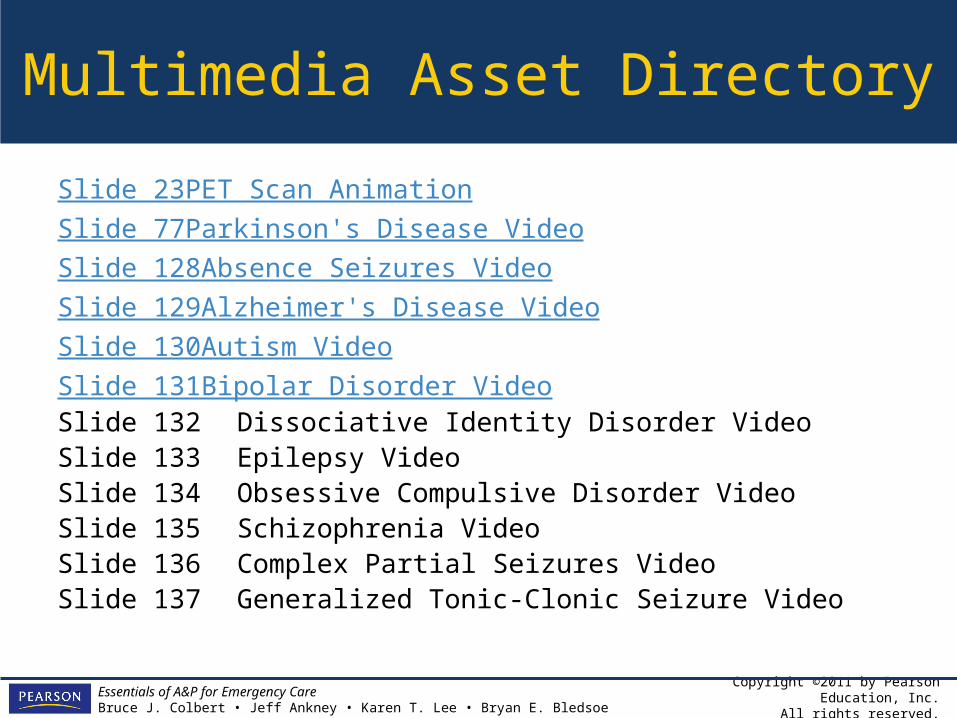
10. The Nervous System: Part Two: The Traffic Control Center. Multimedia Asset Directory. Slide 23PET Scan Animation Slide 77Parkinson's Disease Video Slide 128Absence Seizures Video Slide 129Alzheimer's Disease Video Slide 130Autism Video Slide 131Bipolar Disorder Video - PowerPoint PPT Presentation
Citation preview
CHAPTER
ESSENTIALS OF A&PFOR EMERGENCY CARE
Copyright ©2011 by Pearson Education, Inc.All rights reserved.
Essentials of A&P for Emergency CareBruce J. Colbert • Jeff Ankney • Karen T. Lee • Bryan E. Bledsoe
The Nervous System: Part Two: The Traffic Control Center
10
Copyright ©2011 by Pearson Education, Inc.All rights reserved.
Essentials of A&P for Emergency CareBruce J. Colbert • Jeff Ankney • Karen T. Lee • Bryan E. Bledsoe
Multimedia Asset Directory
Slide 23PET Scan AnimationSlide 77Parkinson's Disease VideoSlide 128Absence Seizures VideoSlide 129Alzheimer's Disease VideoSlide 130Autism VideoSlide 131Bipolar Disorder VideoSlide 132 Dissociative Identity Disorder VideoSlide 133 Epilepsy VideoSlide 134 Obsessive Compulsive Disorder VideoSlide 135 Schizophrenia VideoSlide 136 Complex Partial Seizures VideoSlide 137 Generalized Tonic-Clonic Seizure Video
Copyright ©2011 by Pearson Education, Inc.All rights reserved.
Essentials of A&P for Emergency CareBruce J. Colbert • Jeff Ankney • Karen T. Lee • Bryan E. Bledsoe
Multimedia Asset Directory
Slide 138 Panic Attacks VideoSlide 139 Delirium AnimationSlide 140 Stroke AnimationSlide 141 Shock AnimationSlide 142 Pharmacy Video
Copyright ©2011 by Pearson Education, Inc.All rights reserved.
Essentials of A&P for Emergency CareBruce J. Colbert • Jeff Ankney • Karen T. Lee • Bryan E. Bledsoe
Introduction
• In this chapter we will focus on the main control of the nervous system – the brain.
• Then, we will put the whole system together to show the big picture of how the nervous system functions.
Copyright ©2011 by Pearson Education, Inc.All rights reserved.
Essentials of A&P for Emergency CareBruce J. Colbert • Jeff Ankney • Karen T. Lee • Bryan E. Bledsoe
Learning Objectives
• Organize the hierarchy of the nervous system.
• Locate and define the internal and external structures and their corresponding functions of the brain.
• List and describe the cranial nerves and their functions.
Copyright ©2011 by Pearson Education, Inc.All rights reserved.
Essentials of A&P for Emergency CareBruce J. Colbert • Jeff Ankney • Karen T. Lee • Bryan E. Bledsoe
Learning Objectives
• Describe the sensory and motor functions of the brain with related structures.
• Contrast the parasympathetic and sympathetic branches of the autonomic nervous system.
• Discuss some representative diseases of the nervous system.

Copyright ©2011 by Pearson Education, Inc.All rights reserved.
Essentials of A&P for Emergency CareBruce J. Colbert • Jeff Ankney • Karen T. Lee • Bryan E. Bledsoe
Pronunciation GuideClick on the megaphone icon before each item to hear the pronunciation.
anterior commissure (an TEE ree or KAHM ih shoorz)
basal nuclei (BAY sal noo KLEE eye)cerebellum (ser eh BELL um)cerebrum (ser EE brum)corpus callosum (KOR pus kah LOH sum)diencephalon (DYE in SEFF ah lon)fornix (FOR niks)gyri (JIE rie)hypothalamus (high poh THAL ah mus)

Copyright ©2011 by Pearson Education, Inc.All rights reserved.
Essentials of A&P for Emergency CareBruce J. Colbert • Jeff Ankney • Karen T. Lee • Bryan E. Bledsoe
Pronunciation GuideClick on the megaphone icon before each item to hear the pronunciation.
limbic system (LIM bick)medulla oblongata (meh DULL ah OB long GA ta)occipital lobe (awk SIP eh tal)parietal lobe (pah RYE eh tal)pineal body (PIN ee al)subarachnoid space (sub ah RACK noyd)sulcus (SULL cus)thalamus (THAL ah mus)

Copyright ©2011 by Pearson Education, Inc.All rights reserved.
Essentials of A&P for Emergency CareBruce J. Colbert • Jeff Ankney • Karen T. Lee • Bryan E. Bledsoe
The Brain and Cranial Nerves
• The brain and cranial nerves represent the major controls of the nervous system.
• The brain acts more as the main processor and director of the entire system.
• The cranial nerves leave the brain and go mainly to the head where they receive information and send it back to the brain (sensory) and the brain sends back instructions to move (motor).

Copyright ©2011 by Pearson Education, Inc.All rights reserved.
Essentials of A&P for Emergency CareBruce J. Colbert • Jeff Ankney • Karen T. Lee • Bryan E. Bledsoe
The Brain
• At the top of the spinal cord, beginning at the level of the foramen magnum and filling the skull, is the brain.
• The brain can be divided into several anatomical and functional sections.
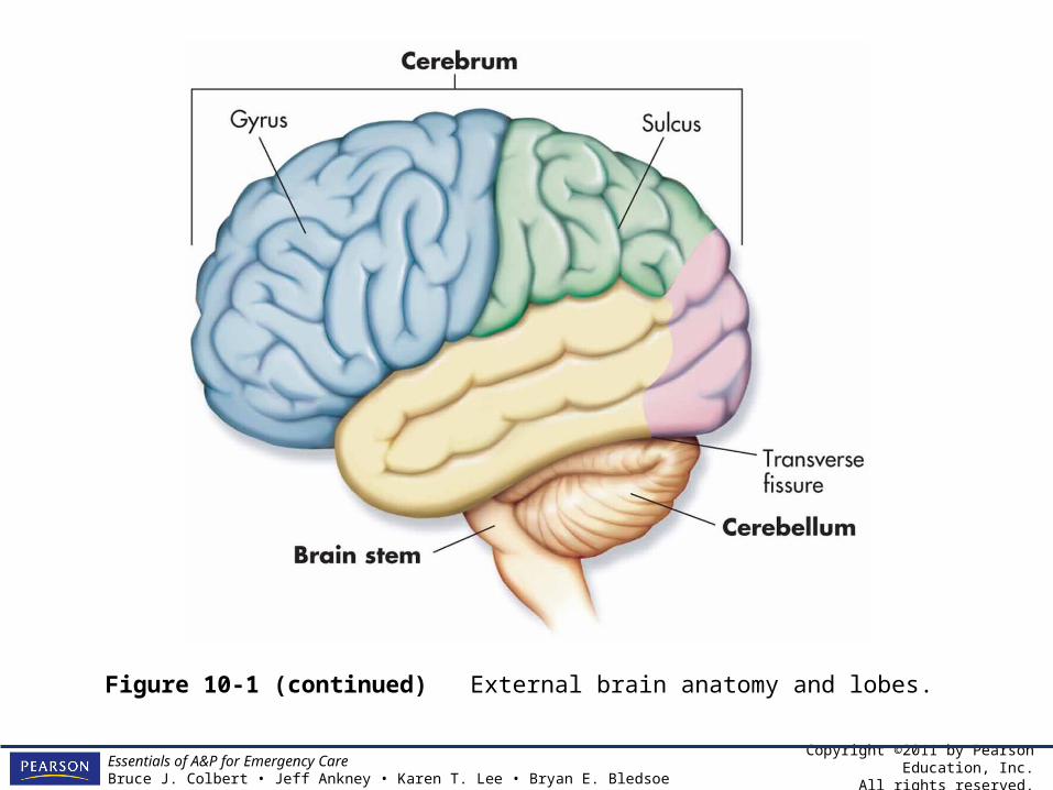
• External anatomy– Cerebrum – Cerebellum– Brain stem

Copyright ©2011 by Pearson Education, Inc.All rights reserved.
Essentials of A&P for Emergency CareBruce J. Colbert • Jeff Ankney • Karen T. Lee • Bryan E. Bledsoe
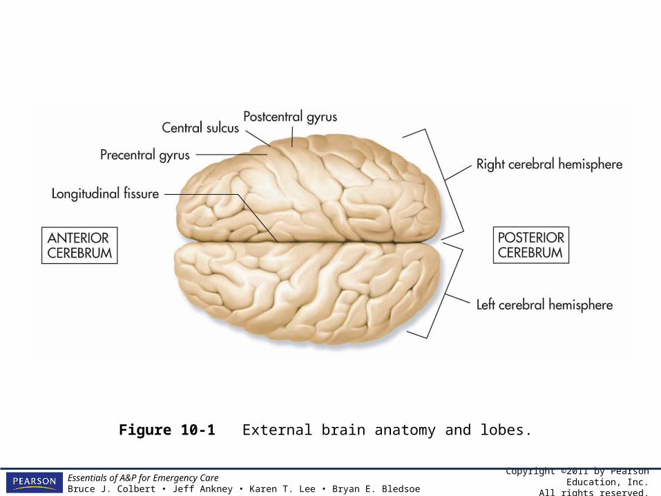
Cerebrum
• The cerebrum is the largest part of the brain.
• It is divided into the right and left hemisphere by the longitudinal fissure and divided from the cerebellum by the transverse fissure.
• The surface of the cerebrum is not smooth, but broken by ridges (gyri) and grooves (sulci) collectively known as convolutions.

Copyright ©2011 by Pearson Education, Inc.All rights reserved.
Essentials of A&P for Emergency CareBruce J. Colbert • Jeff Ankney • Karen T. Lee • Bryan E. Bledsoe
Cerebrum
• These convolutions serve a very important purpose by increasing the surface area of the brain, so you can pack more brain in a smaller space.
• Most of the sulci are extremely variable in their locations among humans, but a few are in basically the same place in every brain. These divide the brain into lobes.
Copyright ©2011 by Pearson Education, Inc.All rights reserved.
Essentials of A&P for Emergency CareBruce J. Colbert • Jeff Ankney • Karen T. Lee • Bryan E. Bledsoe
Lobes of the Brain
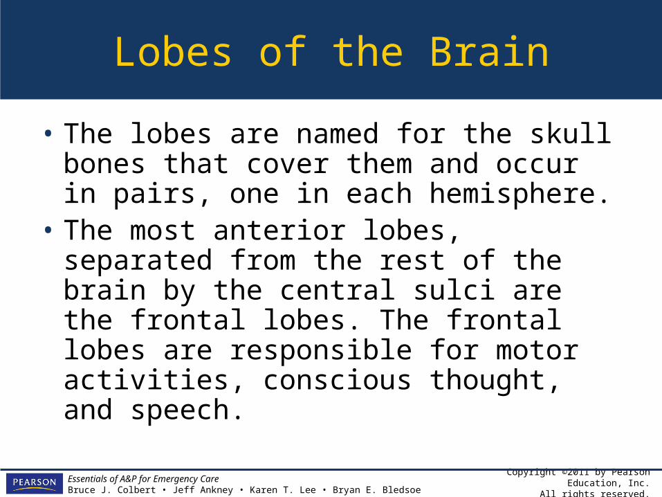
• The lobes are named for the skull bones that cover them and occur in pairs, one in each hemisphere.
• The most anterior lobes, separated from the rest of the brain by the central sulci are the frontal lobes. The frontal lobes are responsible for motor activities, conscious thought, and speech.

Copyright ©2011 by Pearson Education, Inc.All rights reserved.
Essentials of A&P for Emergency CareBruce J. Colbert • Jeff Ankney • Karen T. Lee • Bryan E. Bledsoe
Lobes of the Brain
• Posterior to the frontal lobe are the parietal lobes. The parietal lobes are involved with body sense perception and language comprehension.
• Posterior to the parietal lobes are the occipital lobes, which are responsible for vision.
• The most inferior lobes, separated by the lateral sulci, are the temporal lobes, which are involved in hearing and integration of emotions.
Copyright ©2011 by Pearson Education, Inc.All rights reserved.
Essentials of A&P for Emergency CareBruce J. Colbert • Jeff Ankney • Karen T. Lee • Bryan E. Bledsoe
Lobes of the Brain
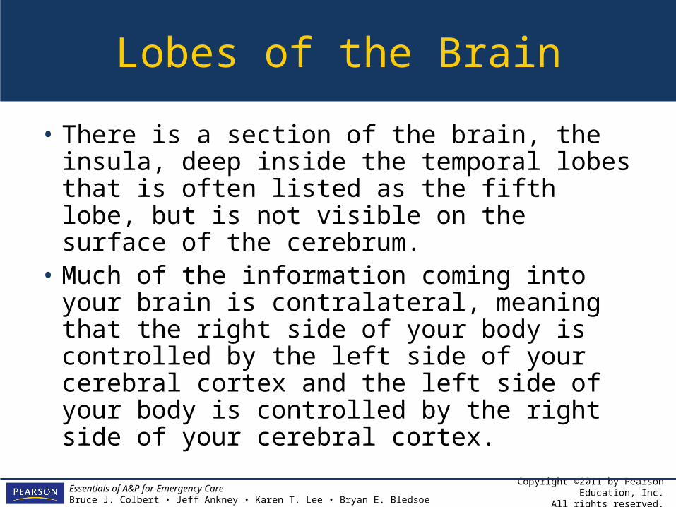
• There is a section of the brain, the insula, deep inside the temporal lobes that is often listed as the fifth lobe, but is not visible on the surface of the cerebrum.
• Much of the information coming into your brain is contralateral, meaning that the right side of your body is controlled by the left side of your cerebral cortex and the left side of your body is controlled by the right side of your cerebral cortex.
Copyright ©2011 by Pearson Education, Inc.All rights reserved.
Essentials of A&P for Emergency CareBruce J. Colbert • Jeff Ankney • Karen T. Lee • Bryan E. Bledsoe
Specific Regions of Cerebrum
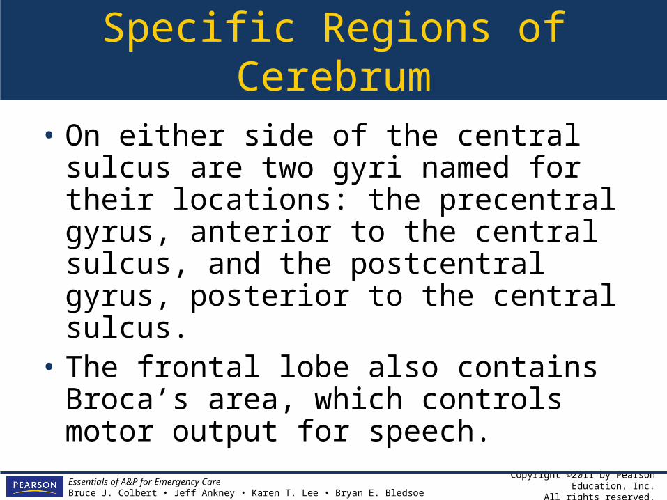
• On either side of the central sulcus are two gyri named for their locations: the precentral gyrus, anterior to the central sulcus, and the postcentral gyrus, posterior to the central sulcus.
• The frontal lobe also contains Broca’s area, which controls motor output for speech.
Copyright ©2011 by Pearson Education, Inc.All rights reserved.
Essentials of A&P for Emergency CareBruce J. Colbert • Jeff Ankney • Karen T. Lee • Bryan E. Bledsoe
Specific Regions of Cerebrum
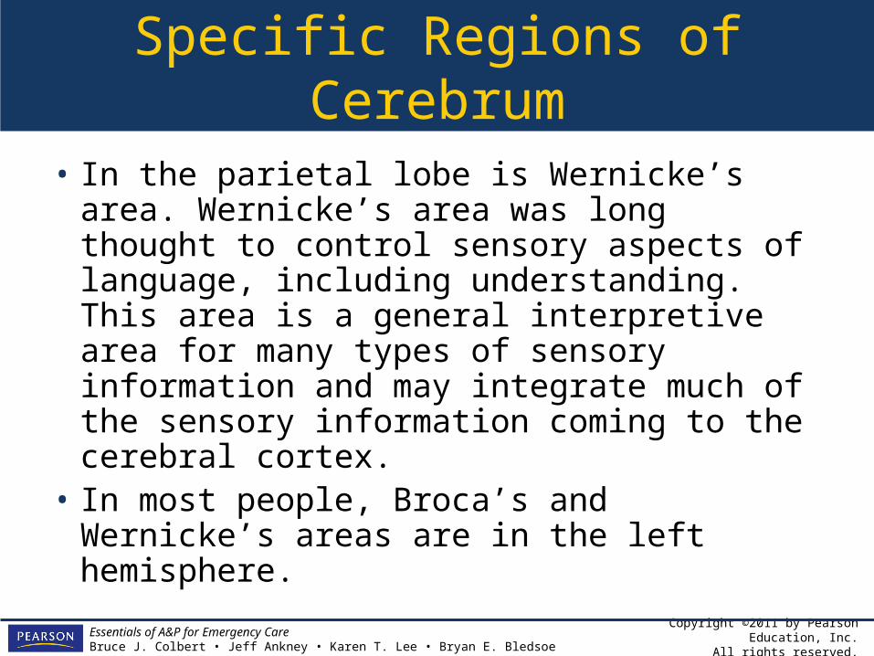
• In the parietal lobe is Wernicke’s area. Wernicke’s area was long thought to control sensory aspects of language, including understanding. This area is a general interpretive area for many types of sensory information and may integrate much of the sensory information coming to the cerebral cortex.
• In most people, Broca’s and Wernicke’s areas are in the left hemisphere.

Copyright ©2011 by Pearson Education, Inc.All rights reserved.
Essentials of A&P for Emergency CareBruce J. Colbert • Jeff Ankney • Karen T. Lee • Bryan E. Bledsoe
The Cerebellum
• The cerebellum is posterior to the cerebrum.• It too is divided into hemispheres by a raised
ridge called the vermis.• The surface is convoluted like that of the
cerebrum.• From its external appearance it is easy to see
why the cerebellum is called the little brain.• The cerebellum is involved in sensory and
motor coordination and balance.
Copyright ©2011 by Pearson Education, Inc.All rights reserved.
Essentials of A&P for Emergency CareBruce J. Colbert • Jeff Ankney • Karen T. Lee • Bryan E. Bledsoe
Figure 10-1 External brain anatomy and lobes.
Copyright ©2011 by Pearson Education, Inc.All rights reserved.
Essentials of A&P for Emergency CareBruce J. Colbert • Jeff Ankney • Karen T. Lee • Bryan E. Bledsoe
Figure 10-1 (continued) External brain anatomy and lobes.

Copyright ©2011 by Pearson Education, Inc.All rights reserved.
Essentials of A&P for Emergency CareBruce J. Colbert • Jeff Ankney • Karen T. Lee • Bryan E. Bledsoe
Figure 10-1 (continued) External brain anatomy and lobes.
Copyright ©2011 by Pearson Education, Inc.All rights reserved.
Essentials of A&P for Emergency CareBruce J. Colbert • Jeff Ankney • Karen T. Lee • Bryan E. Bledsoe
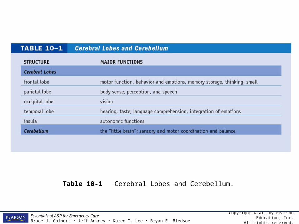
Table 10-1 Cerebral Lobes and Cerebellum.
Copyright ©2011 by Pearson Education, Inc.All rights reserved.
Essentials of A&P for Emergency CareBruce J. Colbert • Jeff Ankney • Karen T. Lee • Bryan E. Bledsoe
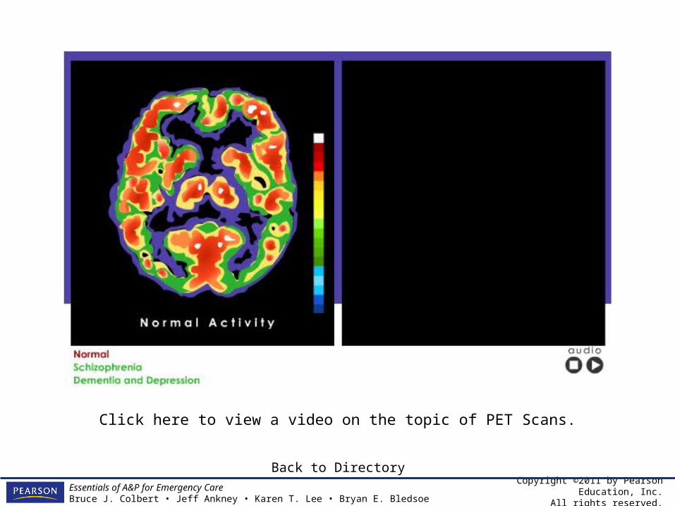
Click here to view a video on the topic of PET Scans.
Back to Directory

Copyright ©2011 by Pearson Education, Inc.All rights reserved.
Essentials of A&P for Emergency CareBruce J. Colbert • Jeff Ankney • Karen T. Lee • Bryan E. Bledsoe
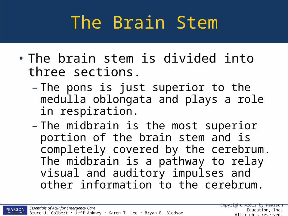
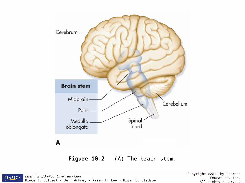
The Brain Stem
• The brain stem is a stalk-like structure inferior to, and partially covered by, the cerebrum.
• The brain stem is divided into three sections.– The medulla oblongata is continuous with the
spinal cord. Responsible for control of heartbeat, respiration, and blood vessel diameter.
Copyright ©2011 by Pearson Education, Inc.All rights reserved.
Essentials of A&P for Emergency CareBruce J. Colbert • Jeff Ankney • Karen T. Lee • Bryan E. Bledsoe
The Brain Stem
• The brain stem is divided into three sections.– The pons is just superior to the medulla
oblongata and plays a role in respiration.– The midbrain is the most superior portion of
the brain stem and is completely covered by the cerebrum. The midbrain is a pathway to relay visual and auditory impulses and other information to the cerebrum.

Copyright ©2011 by Pearson Education, Inc.All rights reserved.
Essentials of A&P for Emergency CareBruce J. Colbert • Jeff Ankney • Karen T. Lee • Bryan E. Bledsoe
The Brain Stem
• The brain stem receives sensory information and contains control systems for vital processes such as blood pressure, heart rate, and ventilation.
Copyright ©2011 by Pearson Education, Inc.All rights reserved.
Essentials of A&P for Emergency CareBruce J. Colbert • Jeff Ankney • Karen T. Lee • Bryan E. Bledsoe
Figure 10-2 (A) The brain stem.
Copyright ©2011 by Pearson Education, Inc.All rights reserved.
Essentials of A&P for Emergency CareBruce J. Colbert • Jeff Ankney • Karen T. Lee • Bryan E. Bledsoe
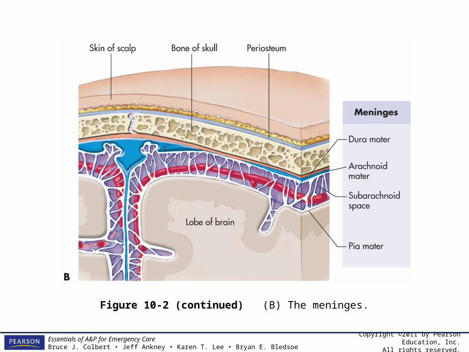
Figure 10-2 (continued) (B) The meninges.

Copyright ©2011 by Pearson Education, Inc.All rights reserved.
Essentials of A&P for Emergency CareBruce J. Colbert • Jeff Ankney • Karen T. Lee • Bryan E. Bledsoe
Table 10-2 The Brain Stem.
Copyright ©2011 by Pearson Education, Inc.All rights reserved.
Essentials of A&P for Emergency CareBruce J. Colbert • Jeff Ankney • Karen T. Lee • Bryan E. Bledsoe
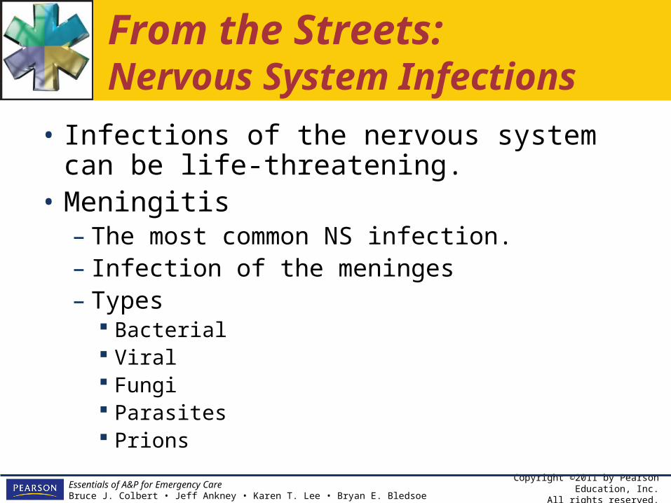
From the Streets:Nervous System Infections
• Infections of the nervous system can be life-threatening.
• Meningitis– The most common NS infection.– Infection of the meninges– Types
Bacterial Viral Fungi Parasites Prions

Copyright ©2011 by Pearson Education, Inc.All rights reserved.
Essentials of A&P for Emergency CareBruce J. Colbert • Jeff Ankney • Karen T. Lee • Bryan E. Bledsoe
From the Streets:Nervous System Infections
• Meningitis– Causes– Signs and symptoms– Brudinski’s sign– Kernig’s sign– Diagnostic tests– Treatment
Copyright ©2011 by Pearson Education, Inc.All rights reserved.
Essentials of A&P for Emergency CareBruce J. Colbert • Jeff Ankney • Karen T. Lee • Bryan E. Bledsoe
Internal Anatomy of the Brain
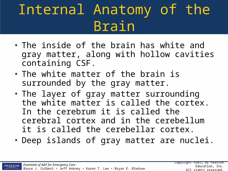
• The inside of the brain has white and gray matter, along with hollow cavities containing CSF.
• The white matter of the brain is surrounded by the gray matter.
• The layer of gray matter surrounding the white matter is called the cortex. In the cerebrum it is called the cerebral cortex and in the cerebellum it is called the cerebellar cortex.
• Deep islands of gray matter are nuclei.
Copyright ©2011 by Pearson Education, Inc.All rights reserved.
Essentials of A&P for Emergency CareBruce J. Colbert • Jeff Ankney • Karen T. Lee • Bryan E. Bledsoe
Internal Anatomy of the Brain
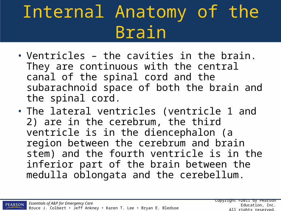
• Ventricles – the cavities in the brain. They are continuous with the central canal of the spinal cord and the subarachnoid space of both the brain and the spinal cord.
• The lateral ventricles (ventricle 1 and 2) are in the cerebrum, the third ventricle is in the diencephalon (a region between the cerebrum and brain stem) and the fourth ventricle is in the inferior part of the brain between the medulla oblongata and the cerebellum.
Copyright ©2011 by Pearson Education, Inc.All rights reserved.
Essentials of A&P for Emergency CareBruce J. Colbert • Jeff Ankney • Karen T. Lee • Bryan E. Bledsoe
From the Streets:Burr Holes
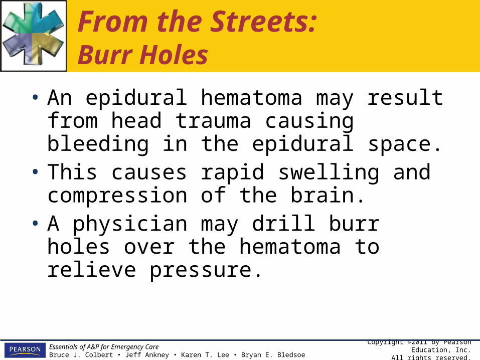
• An epidural hematoma may result from head trauma causing bleeding in the epidural space.
• This causes rapid swelling and compression of the brain.
• A physician may drill burr holes over the hematoma to relieve pressure.

Copyright ©2011 by Pearson Education, Inc.All rights reserved.
Essentials of A&P for Emergency CareBruce J. Colbert • Jeff Ankney • Karen T. Lee • Bryan E. Bledsoe
Figure 10-3 (A) Superior sectional view of the brain.

Copyright ©2011 by Pearson Education, Inc.All rights reserved.
Essentials of A&P for Emergency CareBruce J. Colbert • Jeff Ankney • Karen T. Lee • Bryan E. Bledsoe
Figure 10-3 (continued) (B) Sagittal sectional view of the brain.
Copyright ©2011 by Pearson Education, Inc.All rights reserved.
Essentials of A&P for Emergency CareBruce J. Colbert • Jeff Ankney • Karen T. Lee • Bryan E. Bledsoe
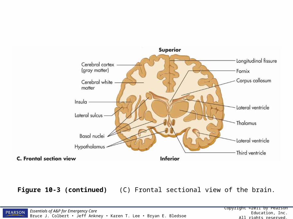
Figure 10-3 (continued) (C) Frontal sectional view of the brain.
Copyright ©2011 by Pearson Education, Inc.All rights reserved.
Essentials of A&P for Emergency CareBruce J. Colbert • Jeff Ankney • Karen T. Lee • Bryan E. Bledsoe
Clinical Application: CSF Circulation and Hydrocephalus
• The ventricles of the brain, the central canal of the spinal cord, and the subarachnoid space surrounding both the brain and spinal cord are filled with CSF. The CSF is filtered from blood in the ventricles by tissue called choroid plexus.

Copyright ©2011 by Pearson Education, Inc.All rights reserved.
Essentials of A&P for Emergency CareBruce J. Colbert • Jeff Ankney • Karen T. Lee • Bryan E. Bledsoe
Clinical Application: CSF Circulation and Hydrocephalus
• CSF made in the lateral ventricles flows through a tiny opening into the third ventricle and then through another opening into the fourth ventricle. CSF flows into the central canal of the spinal cord and the subarachnoid space. CSF is returned to the blood via special “ports” between subarachnoid space and blood spaces in the dura mater.

Copyright ©2011 by Pearson Education, Inc.All rights reserved.
Essentials of A&P for Emergency CareBruce J. Colbert • Jeff Ankney • Karen T. Lee • Bryan E. Bledsoe
Clinical Application: CSF Circulation and Hydrocephalus
• The balance of CSF made and CSF reabsorbed by the blood is very important. The brain is a very delicate organ captured between the liquid CSF and the bones of the skull. If there is too much CSF, pressure inside the skull will rise and eventually crush brain tissue.
Copyright ©2011 by Pearson Education, Inc.All rights reserved.
Essentials of A&P for Emergency CareBruce J. Colbert • Jeff Ankney • Karen T. Lee • Bryan E. Bledsoe
Clinical Application: CSF Circulation and Hydrocephalus
• This condition, in which there is too much CSF, is called hydrocephalus (water on the brain). Hydrocephalus can be caused by blockage of the narrow passages due to trauma, a birth defect, tumor, or decreased reabsorption of CSF. It can be treated by medication or, more commonly, a shunt is surgically placed to drain fluid to the heart or abdominal cavity.
Copyright ©2011 by Pearson Education, Inc.All rights reserved.
Essentials of A&P for Emergency CareBruce J. Colbert • Jeff Ankney • Karen T. Lee • Bryan E. Bledsoe
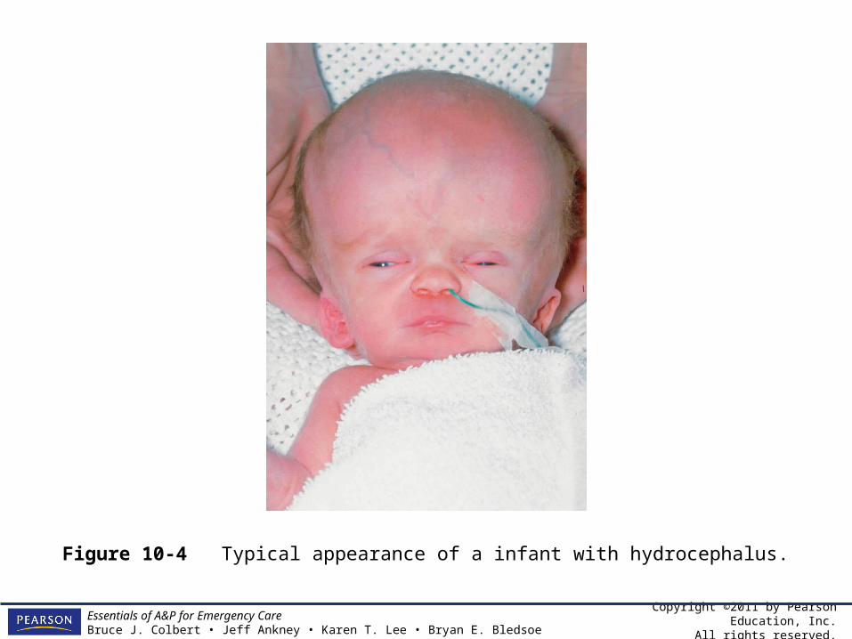
Figure 10-4 Typical appearance of a infant with hydrocephalus.

Copyright ©2011 by Pearson Education, Inc.All rights reserved.
Essentials of A&P for Emergency CareBruce J. Colbert • Jeff Ankney • Karen T. Lee • Bryan E. Bledsoe
The Cerebrum
• The inside of the cerebrum reflects the external anatomy. The lobes (frontal, parietal, temporal, and occipital) are clearly visible.
• The right and left hemispheres are connected by several white matter pathways surrounding the lateral ventricles called the corpus callosum, the fornix, and the anterior commissure.

Copyright ©2011 by Pearson Education, Inc.All rights reserved.
Essentials of A&P for Emergency CareBruce J. Colbert • Jeff Ankney • Karen T. Lee • Bryan E. Bledsoe
The Cerebrum
• Several prominent cerebral nuclei are also involved in motor coordination, sensation, and motor control.

Copyright ©2011 by Pearson Education, Inc.All rights reserved.
Essentials of A&P for Emergency CareBruce J. Colbert • Jeff Ankney • Karen T. Lee • Bryan E. Bledsoe
The Diencephalon
• Inferior to the cerebrum is a section of the brain, that is not visible from the exterior, called the diencephalon.
• Consists of several parts including the thalamus, hypothalamus, pineal body, and the pituitary gland which interface with the endocrine system.

Copyright ©2011 by Pearson Education, Inc.All rights reserved.
Essentials of A&P for Emergency CareBruce J. Colbert • Jeff Ankney • Karen T. Lee • Bryan E. Bledsoe
The Diencephalon
• The diencephalon contains the third ventricle and a number of nuclei. – Basal nuclei and limbic system – Nuclei responsible for controlling hormone
levels, hunger and thirst, body temperature, sleep-wake cycles, and for coordination of the flow of information around the brain

Copyright ©2011 by Pearson Education, Inc.All rights reserved.
Essentials of A&P for Emergency CareBruce J. Colbert • Jeff Ankney • Karen T. Lee • Bryan E. Bledsoe
Table 10-3 Diencephalon.

Copyright ©2011 by Pearson Education, Inc.All rights reserved.
Essentials of A&P for Emergency CareBruce J. Colbert • Jeff Ankney • Karen T. Lee • Bryan E. Bledsoe
The Cerebellum
• The external similarities between the cerebellum and cerebrum are also obvious internally.
• The cerebellum has a gray matter cortex and a white matter center, known as the arbor vitae (tree of life).
• The cerebellum also has nuclei that coordinate motor and sensory activity.
• Essentially, the cerebellum fine tunes voluntary skeletal muscle activity and helps in the maintenance of balance.
Copyright ©2011 by Pearson Education, Inc.All rights reserved.
Essentials of A&P for Emergency CareBruce J. Colbert • Jeff Ankney • Karen T. Lee • Bryan E. Bledsoe
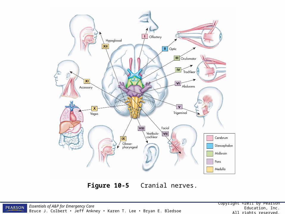
Cranial Nerves
• In order for the CNS to function, it must be connected to the outside world via nerves of the PNS.
• Like the spinal cord has spinal nerves the brain has nerves called cranial nerves.
• Cranial nerves are like spinal nerves in that they are the input and output pathways for the brain.
• There are only 12 pairs of cranial nerves, all but two of which arise from the brain stem.
Copyright ©2011 by Pearson Education, Inc.All rights reserved.
Essentials of A&P for Emergency CareBruce J. Colbert • Jeff Ankney • Karen T. Lee • Bryan E. Bledsoe
Cranial Nerves
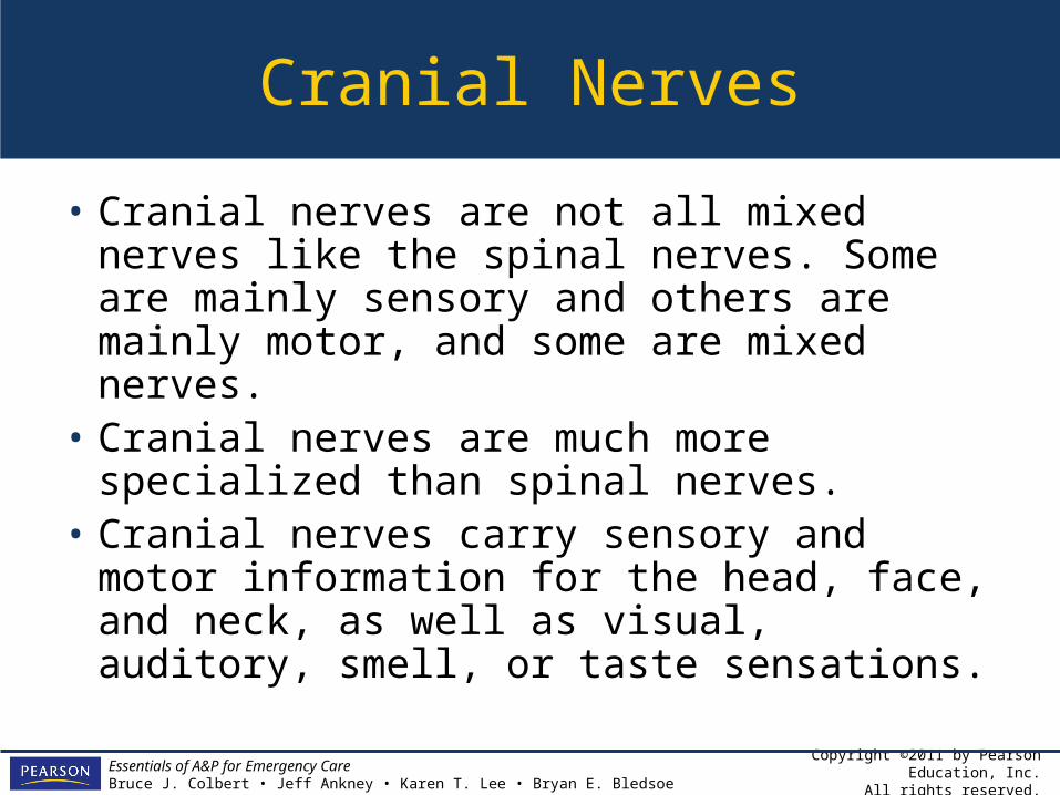
• Cranial nerves are not all mixed nerves like the spinal nerves. Some are mainly sensory and others are mainly motor, and some are mixed nerves.
• Cranial nerves are much more specialized than spinal nerves.
• Cranial nerves carry sensory and motor information for the head, face, and neck, as well as visual, auditory, smell, or taste sensations.
Copyright ©2011 by Pearson Education, Inc.All rights reserved.
Essentials of A&P for Emergency CareBruce J. Colbert • Jeff Ankney • Karen T. Lee • Bryan E. Bledsoe
Figure 10-5 Cranial nerves.

Copyright ©2011 by Pearson Education, Inc.All rights reserved.
Essentials of A&P for Emergency CareBruce J. Colbert • Jeff Ankney • Karen T. Lee • Bryan E. Bledsoe
Table 10-4 Cranial Nerves and Functions.

Copyright ©2011 by Pearson Education, Inc.All rights reserved.
Essentials of A&P for Emergency CareBruce J. Colbert • Jeff Ankney • Karen T. Lee • Bryan E. Bledsoe
The Somatic Sensory System
• The somatic sensory system provides sensory input for your nervous system and allows you to feel the world around you.
• Somatic sensation includes fine touch, crude touch, vibration, pain, temperature, and body position.
• Information for somatic sensation comes into both the brain and the spinal cord.
• To attach meaning to the sensation, it must get to the brain for interpretation.

Copyright ©2011 by Pearson Education, Inc.All rights reserved.
Essentials of A&P for Emergency CareBruce J. Colbert • Jeff Ankney • Karen T. Lee • Bryan E. Bledsoe
Figure 10-6 Nervous system flowchart highlighting our current location.
Copyright ©2011 by Pearson Education, Inc.All rights reserved.
Essentials of A&P for Emergency CareBruce J. Colbert • Jeff Ankney • Karen T. Lee • Bryan E. Bledsoe
The Somatic Sensory System
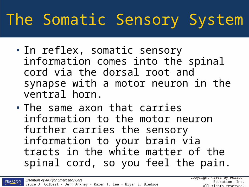
• In reflex, somatic sensory information comes into the spinal cord via the dorsal root and synapse with a motor neuron in the ventral horn.
• The same axon that carries information to the motor neuron further carries the sensory information to your brain via tracts in the white matter of the spinal cord, so you feel the pain.

Copyright ©2011 by Pearson Education, Inc.All rights reserved.
Essentials of A&P for Emergency CareBruce J. Colbert • Jeff Ankney • Karen T. Lee • Bryan E. Bledsoe
Somatic Sensory Pathways
• Three pathways carry somatic sensory information to your spinal cord and then to your brain.– The dorsal column tract carries fine touch and
vibration information to the cerebral cortex.– The spinothalmic tract carries temperature,
pain, and crude touch information to the cerebral cortex.
– The spinocerebellar tract carries information about posture and position to the cerebellum.

Copyright ©2011 by Pearson Education, Inc.All rights reserved.
Essentials of A&P for Emergency CareBruce J. Colbert • Jeff Ankney • Karen T. Lee • Bryan E. Bledsoe
Table 10-5 Spinal Cord Pathways for Sensory Information.
Copyright ©2011 by Pearson Education, Inc.All rights reserved.
Essentials of A&P for Emergency CareBruce J. Colbert • Jeff Ankney • Karen T. Lee • Bryan E. Bledsoe
Primary Somatic Sensory Cortex
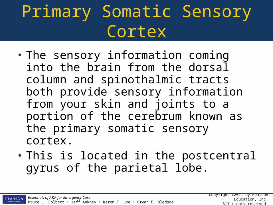
• The sensory information coming into the brain from the dorsal column and spinothalmic tracts both provide sensory information from your skin and joints to a portion of the cerebrum known as the primary somatic sensory cortex.
• This is located in the postcentral gyrus of the parietal lobe.
Copyright ©2011 by Pearson Education, Inc.All rights reserved.
Essentials of A&P for Emergency CareBruce J. Colbert • Jeff Ankney • Karen T. Lee • Bryan E. Bledsoe
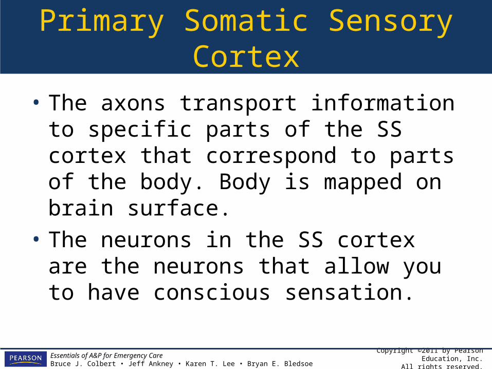
Primary Somatic Sensory Cortex
• The axons transport information to specific parts of the SS cortex that correspond to parts of the body. Body is mapped on brain surface.
• The neurons in the SS cortex are the neurons that allow you to have conscious sensation.

Copyright ©2011 by Pearson Education, Inc.All rights reserved.
Essentials of A&P for Emergency CareBruce J. Colbert • Jeff Ankney • Karen T. Lee • Bryan E. Bledsoe
Primary Somatic Sensory Cortex
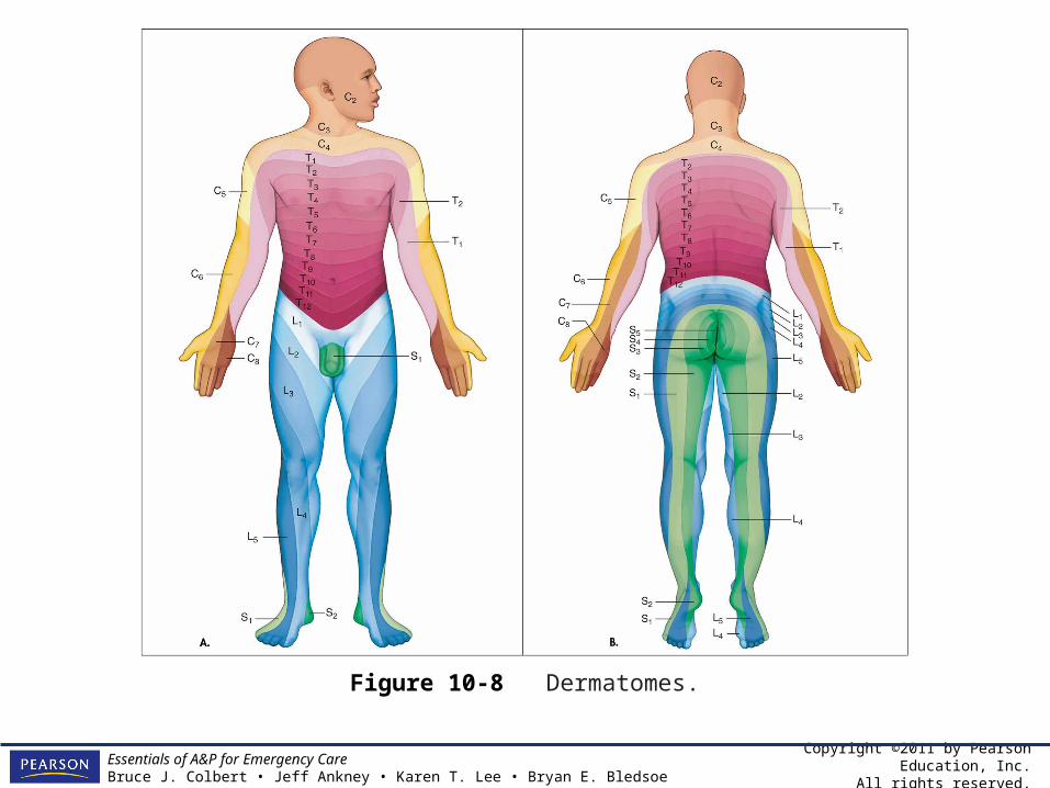
• The map is also evident on your body surface. The map of the body surfaces innervated by each spinal nerve consists of a band or region of skin supplied by a single sensory nerve. Each band is called a dermatome.
Copyright ©2011 by Pearson Education, Inc.All rights reserved.
Essentials of A&P for Emergency CareBruce J. Colbert • Jeff Ankney • Karen T. Lee • Bryan E. Bledsoe
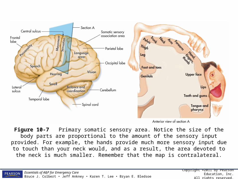
Figure 10-7 Primary somatic sensory area. Notice the size of the body parts are proportional to the amount of the sensory input provided. For example, the hands provide much more
sensory input due to touch than your neck would, and as a result, the area devoted to the neck is much smaller. Remember that the map is contralateral.
Copyright ©2011 by Pearson Education, Inc.All rights reserved.
Essentials of A&P for Emergency CareBruce J. Colbert • Jeff Ankney • Karen T. Lee • Bryan E. Bledsoe
Figure 10-8 Dermatomes.
Copyright ©2011 by Pearson Education, Inc.All rights reserved.
Essentials of A&P for Emergency CareBruce J. Colbert • Jeff Ankney • Karen T. Lee • Bryan E. Bledsoe
Understanding Somatic Sensation
• Another area of the cerebral cortex that allows understanding and interpretation of somatic sensory information is located just posterior to the SS cortex in the parietal lobe and is known as the somatic sensory association area.
Copyright ©2011 by Pearson Education, Inc.All rights reserved.
Essentials of A&P for Emergency CareBruce J. Colbert • Jeff Ankney • Karen T. Lee • Bryan E. Bledsoe
Understanding Somatic Sensation
• The somatic sensory system works on a kind of hierarchy with the sensory neurons in the spinal cord and brain stem, collecting information and passing it to the areas in the thalamus, cerebellum, and cerebral cortex for processing.
• Understanding of complex sensory input happens only after the information is passed to the SS cortex and the SS association area.

Copyright ©2011 by Pearson Education, Inc.All rights reserved.
Essentials of A&P for Emergency CareBruce J. Colbert • Jeff Ankney • Karen T. Lee • Bryan E. Bledsoe
From the Streets:Seizure
• A seizure is an episode of abnormal neurological function caused by an abnormal electrical discharge of the brain neurons.
• Causes– Epilepsy-clinical syndrome of recurrent
seizures.– Primary / idiopathic– Secondary
Copyright ©2011 by Pearson Education, Inc.All rights reserved.
Essentials of A&P for Emergency CareBruce J. Colbert • Jeff Ankney • Karen T. Lee • Bryan E. Bledsoe
From the Streets:Seizure
• Categories:– Generalized tonic-clonic
Grand mal seizures
– Partial Absence seizures Focal seizures
• Signs and symptoms
Copyright ©2011 by Pearson Education, Inc.All rights reserved.
Essentials of A&P for Emergency CareBruce J. Colbert • Jeff Ankney • Karen T. Lee • Bryan E. Bledsoe
From the Streets:Seizure
• Assessment– Postictal phase
• Diagnostic tests• Treatments

Copyright ©2011 by Pearson Education, Inc.All rights reserved.
Essentials of A&P for Emergency CareBruce J. Colbert • Jeff Ankney • Karen T. Lee • Bryan E. Bledsoe
The Motor System
• The motor system is also a hierarchy, working in parallel with the SS system, with two obvious differences.– Information moves in the opposite direction
from brain to spinal cord.– Second, the motor system has two divisions,
the somatic motor system and the autonomic nervous system.

Copyright ©2011 by Pearson Education, Inc.All rights reserved.
Essentials of A&P for Emergency CareBruce J. Colbert • Jeff Ankney • Karen T. Lee • Bryan E. Bledsoe
Figure 10-9 Our current position on the nervous system flowchart.

Copyright ©2011 by Pearson Education, Inc.All rights reserved.
Essentials of A&P for Emergency CareBruce J. Colbert • Jeff Ankney • Karen T. Lee • Bryan E. Bledsoe
Somatic Motor System
• The somatic motor system controls voluntary movements under orders from the cerebral cortex.
• In the frontal lobe are the premotor and prefrontal areas which plan movements.
• The plan from these two areas is sent to the primary motor cortex.

Copyright ©2011 by Pearson Education, Inc.All rights reserved.
Essentials of A&P for Emergency CareBruce J. Colbert • Jeff Ankney • Karen T. Lee • Bryan E. Bledsoe
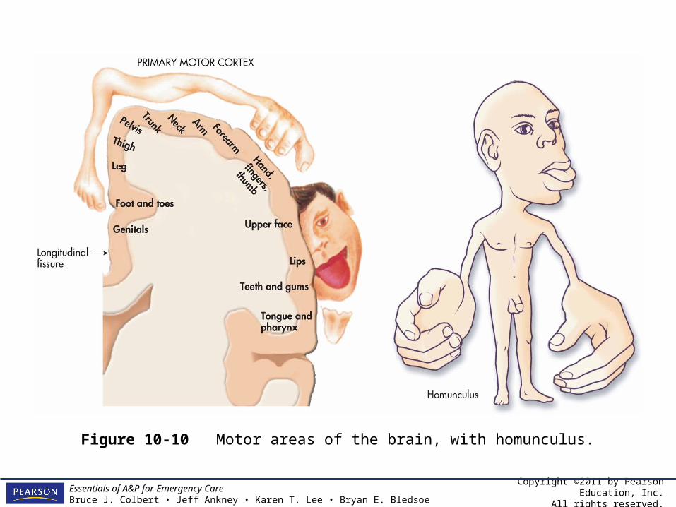
Somatic Motor System
• The primary motor cortex is located in the precentral gyrus in the frontal lobe, just anterior to the SS cortex.
• The primary motor cortex also has a map of the body and is contralateral. The size of the map is proportional to the amount of movement control. Thus the hands and the tongue have bigger maps than the trunk or the forearms.

Copyright ©2011 by Pearson Education, Inc.All rights reserved.
Essentials of A&P for Emergency CareBruce J. Colbert • Jeff Ankney • Karen T. Lee • Bryan E. Bledsoe
Somatic Motor System
• Like the somatic sensory cortex, the primary motor cortex is contralateral.
Copyright ©2011 by Pearson Education, Inc.All rights reserved.
Essentials of A&P for Emergency CareBruce J. Colbert • Jeff Ankney • Karen T. Lee • Bryan E. Bledsoe
Figure 10-10 Motor areas of the brain, with homunculus.

Copyright ©2011 by Pearson Education, Inc.All rights reserved.
Essentials of A&P for Emergency CareBruce J. Colbert • Jeff Ankney • Karen T. Lee • Bryan E. Bledsoe
Subcortical Structures
• Nuclei– Deep in the cerebrum are several areas of
gray matter. Areas of gray matter found within the cerebrum and surrounded by white matter are known as nuclei.
– The nuclei can be part of the basal nuclei, a motor coordination system, or part of the limbic system, which controls emotion and mood.

Copyright ©2011 by Pearson Education, Inc.All rights reserved.
Essentials of A&P for Emergency CareBruce J. Colbert • Jeff Ankney • Karen T. Lee • Bryan E. Bledsoe
Subcortical Structures
• Nuclei, Thalamus, Cerebellum– The plan for movement leaves the motor
cortex and connects with neurons in the thalamus, located in the diencephalon.
– The thalamus, basal nuclei, and cerebellum are part of a complicated motor coordination loop.

Copyright ©2011 by Pearson Education, Inc.All rights reserved.
Essentials of A&P for Emergency CareBruce J. Colbert • Jeff Ankney • Karen T. Lee • Bryan E. Bledsoe
Subcortical Structures
• Nuclei, Thalamus, Cerebellum– Here, the movement must be fine-tuned,
posture and limb positions are taken into account, other movements are turned off, and movement and senses are integrated.
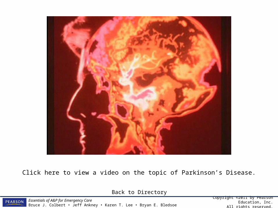
– Patients with Parkinson’s have a disorder in the basal nuclei, making them unable to start some movements or turn other movements off. They have difficulty walking, swallowing, and have tremors when sitting still.
Copyright ©2011 by Pearson Education, Inc.All rights reserved.
Essentials of A&P for Emergency CareBruce J. Colbert • Jeff Ankney • Karen T. Lee • Bryan E. Bledsoe
Click here to view a video on the topic of Parkinson’s Disease.
Back to Directory

Copyright ©2011 by Pearson Education, Inc.All rights reserved.
Essentials of A&P for Emergency CareBruce J. Colbert • Jeff Ankney • Karen T. Lee • Bryan E. Bledsoe
Spinal Cord Pathways
• After the movement information is processed by the thalamus and basal nuclei, it moves to the spinal cord and brain stem via ascending spinal cord tracts.
• The corticospinal and corticobulbar tracts from your motor cortex are direct pathways, whereas others coming from subcortical structures are considered indirect pathways.
Copyright ©2011 by Pearson Education, Inc.All rights reserved.
Essentials of A&P for Emergency CareBruce J. Colbert • Jeff Ankney • Karen T. Lee • Bryan E. Bledsoe
Spinal Cord Pathways
• The axons synapse on motor neurons in the ventral horn. These motor neurons project to skeletal muscles, via the cranial nerves or the ventral roots and spinal nerves, sending orders to the skeletal muscles to carry out the planned movement and to coordinate it.
Copyright ©2011 by Pearson Education, Inc.All rights reserved.
Essentials of A&P for Emergency CareBruce J. Colbert • Jeff Ankney • Karen T. Lee • Bryan E. Bledsoe
Spinal Cord Pathways
• A second function of the motor tracts is fine tuning of reflexes. These tracts inhibit reflexes, making them softer than they would be if they had no influence from the brain.

Copyright ©2011 by Pearson Education, Inc.All rights reserved.
Essentials of A&P for Emergency CareBruce J. Colbert • Jeff Ankney • Karen T. Lee • Bryan E. Bledsoe
Table 10-6 Spinal Cord Pathways for Motor Information

Copyright ©2011 by Pearson Education, Inc.All rights reserved.
Essentials of A&P for Emergency CareBruce J. Colbert • Jeff Ankney • Karen T. Lee • Bryan E. Bledsoe
The Role of the Cerebellum
• The cerebellum has both motor and sensory inputs and outputs from the cerebral cortex, the thalamus, basal nuclei, and the spinal cord.
Copyright ©2011 by Pearson Education, Inc.All rights reserved.
Essentials of A&P for Emergency CareBruce J. Colbert • Jeff Ankney • Karen T. Lee • Bryan E. Bledsoe
The Role of the Cerebellum
• The cerebellum gets information about the planned movement and the actual movement and compares the plan to the actual. If the plan and the actual don’t match, the cerebellum can adjust the actual movement to fit the plan.
Copyright ©2011 by Pearson Education, Inc.All rights reserved.
Essentials of A&P for Emergency CareBruce J. Colbert • Jeff Ankney • Karen T. Lee • Bryan E. Bledsoe
The Role of the Cerebellum
• The function of the cerebellum is subtle and still a bit of a mystery, but without the cerebellum movements would be inaccurate at best.
Copyright ©2011 by Pearson Education, Inc.All rights reserved.
Essentials of A&P for Emergency CareBruce J. Colbert • Jeff Ankney • Karen T. Lee • Bryan E. Bledsoe
From the Streets:Headache
• Headache is a common complaint and usually a benign symptom.
• However, sometimes it can be associated with a serious disease.
• Types– Migraine– Cluster– Tension

Copyright ©2011 by Pearson Education, Inc.All rights reserved.
Essentials of A&P for Emergency CareBruce J. Colbert • Jeff Ankney • Karen T. Lee • Bryan E. Bledsoe
From the Streets:Headache
• Migraine– Often caused by a trigger– Pathophysiology
• Cluster: characterized by very severe, unilateral pain in the orbit, forehead, or temple.
• Tension: characterized with nonpulsating pain on both sides of the head.

Copyright ©2011 by Pearson Education, Inc.All rights reserved.
Essentials of A&P for Emergency CareBruce J. Colbert • Jeff Ankney • Karen T. Lee • Bryan E. Bledsoe
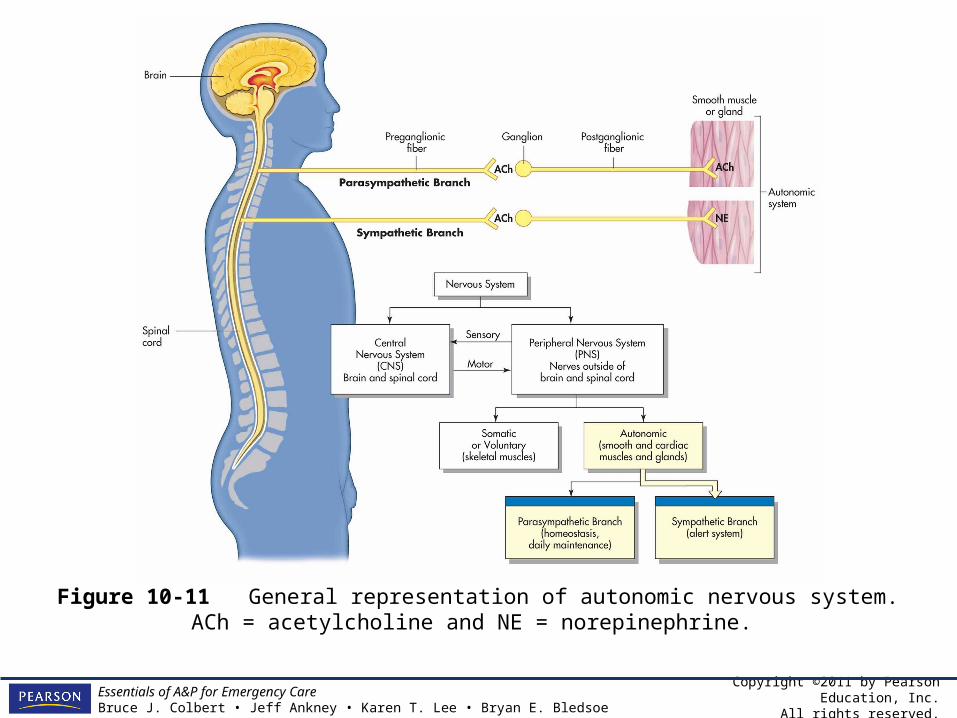
Autonomic Nervous System
• The motor system is divided into two systems: – The somatic system controls skeletal muscles.– The autonomic system controls physiological
characteristics such as blood pressure, heart rate, respiratory rate, digestion, and sweating.
• The neurons for the autonomic system, like the somatic motor neurons, are located in the spinal cord and brain stem, and release the neurotransmitter acetylcholine. This is where similarities end.
Copyright ©2011 by Pearson Education, Inc.All rights reserved.
Essentials of A&P for Emergency CareBruce J. Colbert • Jeff Ankney • Karen T. Lee • Bryan E. Bledsoe
Autonomic Neurons
• The autonomic motor neurons are all located in the lateral horn rather than the ventral horns and unlike the somatic motor neurons, autonomic neurons do not project directly to muscles.
• They make a synapse in a ganglion outside the CNS. These are preganglionic neurons.
• Then a second motor neuron, called a postganglionic neuron, projects to the muscle.
• There are no autonomic neurons in the cervical spinal cord.

Copyright ©2011 by Pearson Education, Inc.All rights reserved.
Essentials of A&P for Emergency CareBruce J. Colbert • Jeff Ankney • Karen T. Lee • Bryan E. Bledsoe
Autonomic Nervous System
• The autonomic nervous system is divided into two subdivisions:– The sympathetic division– The parasympathetic division
Copyright ©2011 by Pearson Education, Inc.All rights reserved.
Essentials of A&P for Emergency CareBruce J. Colbert • Jeff Ankney • Karen T. Lee • Bryan E. Bledsoe
Figure 10-11 General representation of autonomic nervous system. ACh = acetylcholine and NE = norepinephrine.

Copyright ©2011 by Pearson Education, Inc.All rights reserved.
Essentials of A&P for Emergency CareBruce J. Colbert • Jeff Ankney • Karen T. Lee • Bryan E. Bledsoe
The Sympathetic Branch
• The sympathetic division controls the “flight or fight” response; it is charged with responding to emergencies.
• Sympathetic effects increase heart rate, BP, and sweating, also causing a dry mouth – symptoms of an adrenaline rush.

Copyright ©2011 by Pearson Education, Inc.All rights reserved.
Essentials of A&P for Emergency CareBruce J. Colbert • Jeff Ankney • Karen T. Lee • Bryan E. Bledsoe
The Sympathetic Branch
• The preganglionic neurons are located in the thoracic and first two lumbar segments of the spinal cord. The preganglionic neurons, which secrete acetylcholine, synapse with the postganglionic neurons in the sympathetic ganglia.
Copyright ©2011 by Pearson Education, Inc.All rights reserved.
Essentials of A&P for Emergency CareBruce J. Colbert • Jeff Ankney • Karen T. Lee • Bryan E. Bledsoe
The Sympathetic Branch
• The ganglia form a pair of chain-like structures that run parallel to the spinal cord (paravertebral ganglia), where neurons in the ganglia release the neurotransmitter norepinephrine.
• Most importantly, the sympathetic system stimulates the adrenal glands to release the hormone epinephrine that causes the adrenaline rush.
Copyright ©2011 by Pearson Education, Inc.All rights reserved.
Essentials of A&P for Emergency CareBruce J. Colbert • Jeff Ankney • Karen T. Lee • Bryan E. Bledsoe
The Parasympathetic Branch
• The parasympathetic division is often called “resting and digesting” as it has the opposite effect of the sympathetic division.
• The parasympathetic system is responsible for everyday activities, as well as reversing the sympathetic effects.
Copyright ©2011 by Pearson Education, Inc.All rights reserved.
Essentials of A&P for Emergency CareBruce J. Colbert • Jeff Ankney • Karen T. Lee • Bryan E. Bledsoe
The Parasympathetic Branch
• Parasympathetic effects include decreased heart rate, respiration, and blood pressure, and increased digestive activity including salivation and stomach activity.
• The neurons of the parasympathetic system are in the brain stem and the sacral spinal cord. The neurotransmitter acetylcholine is released by postganglionic neurons.

Copyright ©2011 by Pearson Education, Inc.All rights reserved.
Essentials of A&P for Emergency CareBruce J. Colbert • Jeff Ankney • Karen T. Lee • Bryan E. Bledsoe
From the Streets:Sympathetic Nervous System
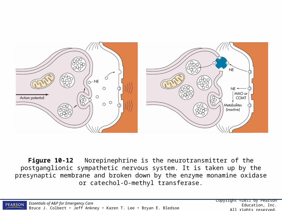
• Sympathetic nervous system results in the release of norepinephrine from postganglionic nerves.
• Two enzymes break down norepinephrine with the synapse:– monamine oxidase (MAO)– catechol-O-methyl transferase (COMT)
Copyright ©2011 by Pearson Education, Inc.All rights reserved.
Essentials of A&P for Emergency CareBruce J. Colbert • Jeff Ankney • Karen T. Lee • Bryan E. Bledsoe
Figure 10-12 Norepinephrine is the neurotransmitter of the postganglionic sympathetic nervous system. It is taken up by the presynaptic membrane and broken down by the enzyme
monamine oxidase or catechol-O-methyl transferase.
Copyright ©2011 by Pearson Education, Inc.All rights reserved.
Essentials of A&P for Emergency CareBruce J. Colbert • Jeff Ankney • Karen T. Lee • Bryan E. Bledsoe
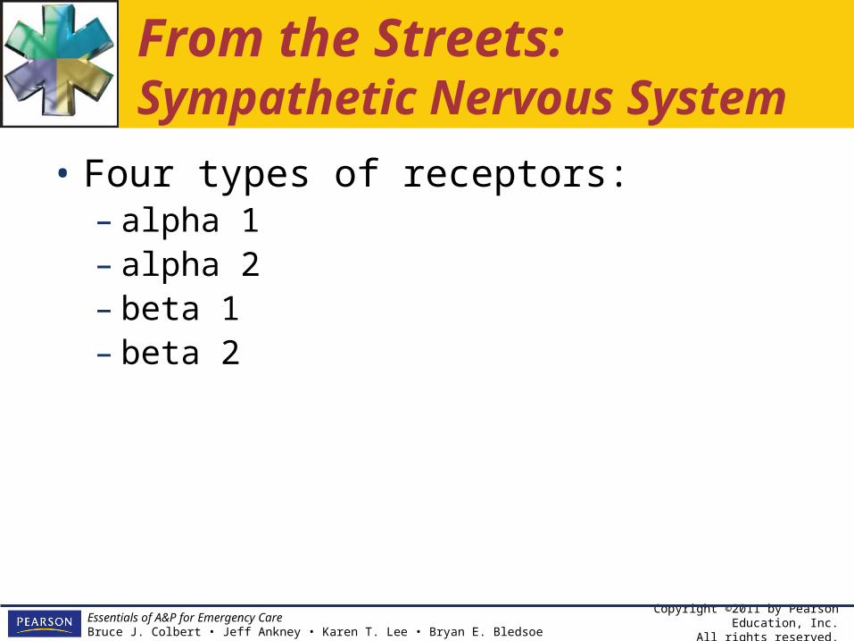
From the Streets:Sympathetic Nervous System
• Four types of receptors:– alpha 1– alpha 2– beta 1– beta 2
Copyright ©2011 by Pearson Education, Inc.All rights reserved.
Essentials of A&P for Emergency CareBruce J. Colbert • Jeff Ankney • Karen T. Lee • Bryan E. Bledsoe
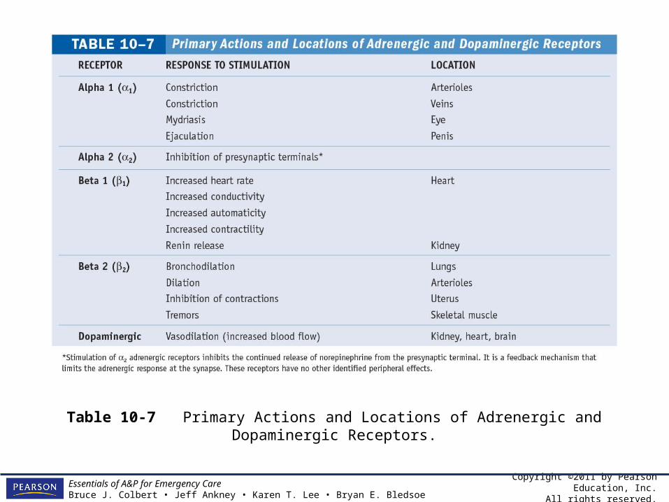
Table 10-7 Primary Actions and Locations of Adrenergic and Dopaminergic Receptors.
Copyright ©2011 by Pearson Education, Inc.All rights reserved.
Essentials of A&P for Emergency CareBruce J. Colbert • Jeff Ankney • Karen T. Lee • Bryan E. Bledsoe
From the Streets:Parasympathetic Nervous System
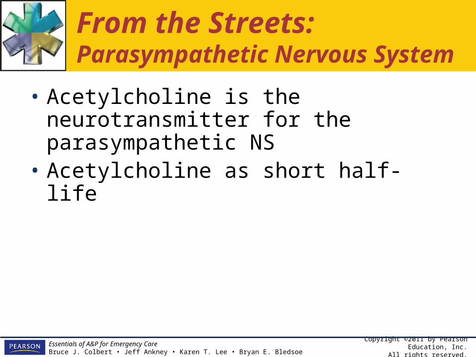
• Acetylcholine is the neurotransmitter for the parasympathetic NS
• Acetylcholine as short half-life

Copyright ©2011 by Pearson Education, Inc.All rights reserved.
Essentials of A&P for Emergency CareBruce J. Colbert • Jeff Ankney • Karen T. Lee • Bryan E. Bledsoe
From the Streets:Parasympathetic Nervous System
• Parasympathetic NS has two types of acetylcholine receptors:– nicotinic– muscarinic
Copyright ©2011 by Pearson Education, Inc.All rights reserved.
Essentials of A&P for Emergency CareBruce J. Colbert • Jeff Ankney • Karen T. Lee • Bryan E. Bledsoe
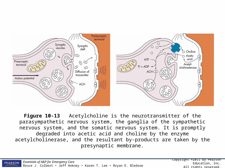
Figure 10-13 Acetylcholine is the neurotransmitter of the parasympathetic nervous system, the ganglia of the sympathetic nervous system, and the somatic nervous system. It is promptly degraded into acetic acid and choline by the enzyme acetylcholinerase, and the resultant by-
products are taken by the presynaptic membrane.

Copyright ©2011 by Pearson Education, Inc.All rights reserved.
Essentials of A&P for Emergency CareBruce J. Colbert • Jeff Ankney • Karen T. Lee • Bryan E. Bledsoe
Table 10-8 Location and Effect of Muscarinic Receptors.

Copyright ©2011 by Pearson Education, Inc.All rights reserved.
Essentials of A&P for Emergency CareBruce J. Colbert • Jeff Ankney • Karen T. Lee • Bryan E. Bledsoe
From the Streets:Organophosphate Poisoning
• Organophosphates are a class of insecticides used widely in agriculture.
• The principle action of organophosphates is deactivation of the enzymes acetylcholinerase (cholinesterase) in the nervous system.

Copyright ©2011 by Pearson Education, Inc.All rights reserved.
Essentials of A&P for Emergency CareBruce J. Colbert • Jeff Ankney • Karen T. Lee • Bryan E. Bledsoe
From the Streets:Organophosphate Poisoning
• Organophosphates bind to cholinesterase, thus inactivating the enzyme.
• Signs and symptoms• Treatment
Copyright ©2011 by Pearson Education, Inc.All rights reserved.
Essentials of A&P for Emergency CareBruce J. Colbert • Jeff Ankney • Karen T. Lee • Bryan E. Bledsoe
The Limbic System
• The limbic system is a series of nuclei in the cerebrum, diencephalon, and superior brain stem.
• The limbic system is involved in mood, emotion, and memory.
• One nucleus helps attach emotion to movement, another coordinates emotion and your sense of smell. Still another is responsible for storing and retrieving information.

Copyright ©2011 by Pearson Education, Inc.All rights reserved.
Essentials of A&P for Emergency CareBruce J. Colbert • Jeff Ankney • Karen T. Lee • Bryan E. Bledsoe
The Reticular System
• The reticular system is a diffuse network of nuclei in the brain stem that is responsible for “waking up” your cerebral cortex.
• The reticular system activity is vital for the maintenance of conscious awareness of your surroundings.
• General anesthesia inhibits the reticular system, rendering surgery patients unconscious.
Copyright ©2011 by Pearson Education, Inc.All rights reserved.
Essentials of A&P for Emergency CareBruce J. Colbert • Jeff Ankney • Karen T. Lee • Bryan E. Bledsoe
The Reticular System
• Injury due to ischemia, trauma, or drugs can damage the reticular system, leading to coma.

Copyright ©2011 by Pearson Education, Inc.All rights reserved.
Essentials of A&P for Emergency CareBruce J. Colbert • Jeff Ankney • Karen T. Lee • Bryan E. Bledsoe
Common Disorders of the Nervous System: Part II
• Spastic paralysis• Flaccid paralysis• Cerebral palsy• CVA• Subdural hematoma• Huntington’s disease

Copyright ©2011 by Pearson Education, Inc.All rights reserved.
Essentials of A&P for Emergency CareBruce J. Colbert • Jeff Ankney • Karen T. Lee • Bryan E. Bledsoe
Spastic Paralysis
• Paralysis is the inability to control voluntary movements.
• Paralysis can be spastic or flaccid.• Spastic paralysis is characterized by
muscle rigidity or increased muscle tone (hypertonia) and overactive reflexes (hyperreflexia).

Copyright ©2011 by Pearson Education, Inc.All rights reserved.
Essentials of A&P for Emergency CareBruce J. Colbert • Jeff Ankney • Karen T. Lee • Bryan E. Bledsoe
Spastic Paralysis
• In spastic paralysis the muscles are rigid and the reflexes overreactive due to decreased communication from the brain to the ventral horn of the motor neurons in the spinal cord.
• Muscles contract randomly and reflexes do not have any control signal from the brain.
• Strokes, head injuries, and spinal cord injury can cause spastic paralysis.

Copyright ©2011 by Pearson Education, Inc.All rights reserved.
Essentials of A&P for Emergency CareBruce J. Colbert • Jeff Ankney • Karen T. Lee • Bryan E. Bledsoe
Flaccid Paralysis
• Flaccid paralysis is characterized by floppy muscles (hypotonia) and decreased reflexes (hyporeflexia).
• It is caused by damage to the spinal nerves, so impulses cannot get to the muscles from the motor neuron.
• Flaccid paralysis occurs with peripheral injury or disorders like polio or Guillain-Barré syndrome.
Copyright ©2011 by Pearson Education, Inc.All rights reserved.
Essentials of A&P for Emergency CareBruce J. Colbert • Jeff Ankney • Karen T. Lee • Bryan E. Bledsoe
Cerebral Palsy
• Cerebral palsy (CP) is a collection of movement disorders that are not progressive and occur in young children.
• Signs of classic spastic paralysis are seen.
• CP is caused by improper development or damage to the motor system of the brain.

Copyright ©2011 by Pearson Education, Inc.All rights reserved.
Essentials of A&P for Emergency CareBruce J. Colbert • Jeff Ankney • Karen T. Lee • Bryan E. Bledsoe
Cerebral Palsy
• Symptoms range from minor motor loss to significant motor deficits, including the inability to walk or speak.
• Intelligence may or may not be affected, depending on the cause of CP.

Copyright ©2011 by Pearson Education, Inc.All rights reserved.
Essentials of A&P for Emergency CareBruce J. Colbert • Jeff Ankney • Karen T. Lee • Bryan E. Bledsoe
Cerebral Vascular Accident
• A cerebral vascular accident (CVA), or a stroke, is caused by interruption of blood flow to a portion of the brain due to hemorrhage or a blood clot.
• If oxygen supply is disrupted long enough, the brain cells will die.
Copyright ©2011 by Pearson Education, Inc.All rights reserved.
Essentials of A&P for Emergency CareBruce J. Colbert • Jeff Ankney • Karen T. Lee • Bryan E. Bledsoe
Cerebral Vascular Accident
• Symptoms vary depending on area affected and can include paralysis, inability to speak, blindness, loss of memory, or lack of sensation.
• Symptoms appear suddenly or can be a series of small minor strokes. These mini-strokes are called transient ischemic accidents (TIAs).

Copyright ©2011 by Pearson Education, Inc.All rights reserved.
Essentials of A&P for Emergency CareBruce J. Colbert • Jeff Ankney • Karen T. Lee • Bryan E. Bledsoe
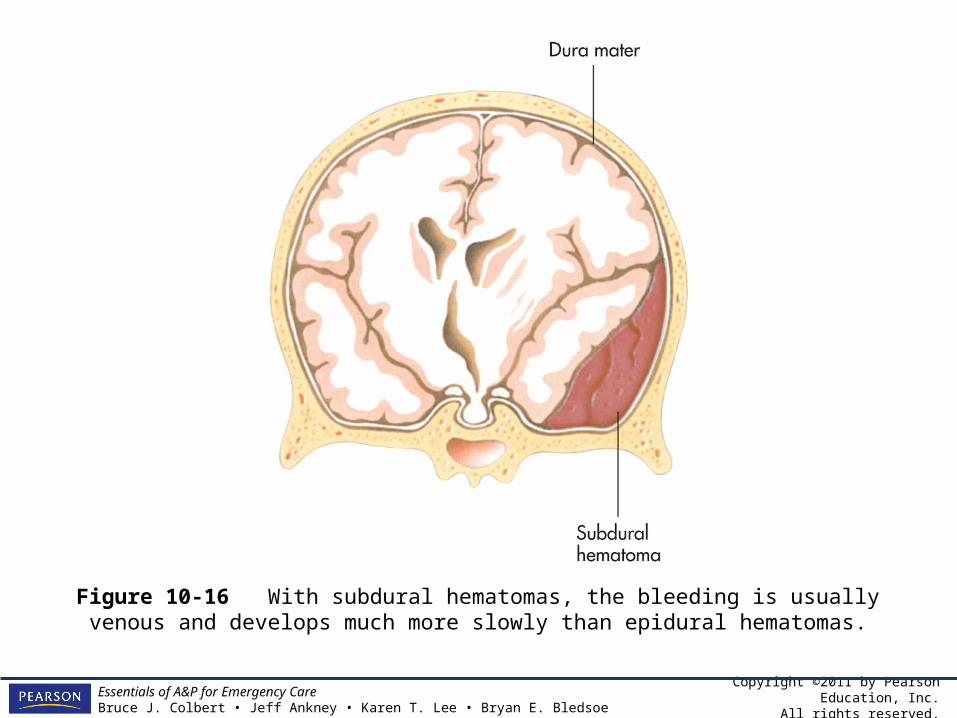
Subdural Hematoma
• A subdural hematoma is a pool of blood between the dura mater and arachnoid mater, in the subdural space.
• Subdural hematomas are caused by head injuries rupturing tiny blood vessels.
• A large, or growing, hematoma can cause brain damage by increasing pressure in the skull.
• Some resolve themselves (small ones) while others can require surgery to relieve the pressure.

Copyright ©2011 by Pearson Education, Inc.All rights reserved.
Essentials of A&P for Emergency CareBruce J. Colbert • Jeff Ankney • Karen T. Lee • Bryan E. Bledsoe
Huntington’s Disease
• Huntington’s disease is a progressive, genetic disorder causing deterioration of neurons in the basal nuclei and, eventually, the cerebral cortex.
• The disease begins with wide mood swings, memory disturbances, writhing movements of the hands or face, or clumsiness. Eventually, difficulty swallowing, speaking, and walking as well as memory loss, psychosis, and loss of cognitive function will occur.

Copyright ©2011 by Pearson Education, Inc.All rights reserved.
Essentials of A&P for Emergency CareBruce J. Colbert • Jeff Ankney • Karen T. Lee • Bryan E. Bledsoe
Huntington’s Disease
• There is no cure and most patients die from injuries, infections, or other complications.
• Genetic testing can identify those at risk.

Copyright ©2011 by Pearson Education, Inc.All rights reserved.
Essentials of A&P for Emergency CareBruce J. Colbert • Jeff Ankney • Karen T. Lee • Bryan E. Bledsoe
From the Streets:Extrapyramidal Motor Syndromes
• Several drugs used in emergency medicine can cause side effects that involve the extrapyramidal system (EPS).
• EPS side effects are similar to the effects of Parkinson’s disease, but are reversible.
• EPS signs and symptoms include muscle spasms of the neck, face, tongue, and back (dystonia).
• Dystonia and akathisia

Copyright ©2011 by Pearson Education, Inc.All rights reserved.
Essentials of A&P for Emergency CareBruce J. Colbert • Jeff Ankney • Karen T. Lee • Bryan E. Bledsoe
From the Streets:Neurologic Trauma
• Traumatic brain injury (TBI) can be devastating and cause permanent disability.
• Blunt trauma– Primary injury– Secondary injury– Focal injuries

Copyright ©2011 by Pearson Education, Inc.All rights reserved.
Essentials of A&P for Emergency CareBruce J. Colbert • Jeff Ankney • Karen T. Lee • Bryan E. Bledsoe
Figure 10-15 In an epidural hematoma, the bleeding usually results from arterial bleeding and can develop rapidly.
Copyright ©2011 by Pearson Education, Inc.All rights reserved.
Essentials of A&P for Emergency CareBruce J. Colbert • Jeff Ankney • Karen T. Lee • Bryan E. Bledsoe
Figure 10-16 With subdural hematomas, the bleeding is usually venous and develops much more slowly than epidural hematomas.
Copyright ©2011 by Pearson Education, Inc.All rights reserved.
Essentials of A&P for Emergency CareBruce J. Colbert • Jeff Ankney • Karen T. Lee • Bryan E. Bledsoe
From the Streets:Neurologic Trauma
• Diffuse injuries• Coup and Contrecoup injuries
Copyright ©2011 by Pearson Education, Inc.All rights reserved.
Essentials of A&P for Emergency CareBruce J. Colbert • Jeff Ankney • Karen T. Lee • Bryan E. Bledsoe
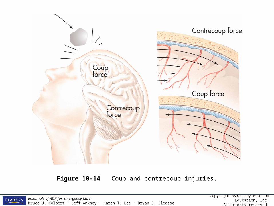
Figure 10-14 Coup and contrecoup injuries.
Copyright ©2011 by Pearson Education, Inc.All rights reserved.
Essentials of A&P for Emergency CareBruce J. Colbert • Jeff Ankney • Karen T. Lee • Bryan E. Bledsoe
From the Streets:Neurologic Trauma
• Penetrating trauma
Copyright ©2011 by Pearson Education, Inc.All rights reserved.
Essentials of A&P for Emergency CareBruce J. Colbert • Jeff Ankney • Karen T. Lee • Bryan E. Bledsoe
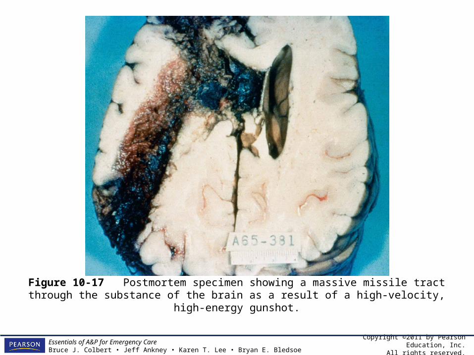
Figure 10-17 Postmortem specimen showing a massive missile tract through the substance of the brain as a result of a high-velocity, high-energy gunshot.
Copyright ©2011 by Pearson Education, Inc.All rights reserved.
Essentials of A&P for Emergency CareBruce J. Colbert • Jeff Ankney • Karen T. Lee • Bryan E. Bledsoe
Click here to view a video on the topic of Absence Seizures.
Back to Directory

Copyright ©2011 by Pearson Education, Inc.All rights reserved.
Essentials of A&P for Emergency CareBruce J. Colbert • Jeff Ankney • Karen T. Lee • Bryan E. Bledsoe
Click here to view a video on the topic of Alzheimer’s Disease.
Back to Directory

Copyright ©2011 by Pearson Education, Inc.All rights reserved.
Essentials of A&P for Emergency CareBruce J. Colbert • Jeff Ankney • Karen T. Lee • Bryan E. Bledsoe
Click here to view a video on the topic of Autism.
Back to Directory
Copyright ©2011 by Pearson Education, Inc.All rights reserved.
Essentials of A&P for Emergency CareBruce J. Colbert • Jeff Ankney • Karen T. Lee • Bryan E. Bledsoe
Click here to view a video on the topic of Bipolar Disorder.
Back to Directory

Copyright ©2011 by Pearson Education, Inc.All rights reserved.
Essentials of A&P for Emergency CareBruce J. Colbert • Jeff Ankney • Karen T. Lee • Bryan E. Bledsoe
Click here to view a video on the topic of Dissociative Identity Disorder.
Back to Directory
Copyright ©2011 by Pearson Education, Inc.All rights reserved.
Essentials of A&P for Emergency CareBruce J. Colbert • Jeff Ankney • Karen T. Lee • Bryan E. Bledsoe
Click here to view a video on the topic of Epilepsy.
Back to Directory
Copyright ©2011 by Pearson Education, Inc.All rights reserved.
Essentials of A&P for Emergency CareBruce J. Colbert • Jeff Ankney • Karen T. Lee • Bryan E. Bledsoe
Click here to view a video on the topic of Obsessive Compulsive Disorder.
Back to Directory
Copyright ©2011 by Pearson Education, Inc.All rights reserved.
Essentials of A&P for Emergency CareBruce J. Colbert • Jeff Ankney • Karen T. Lee • Bryan E. Bledsoe
Click here to view a video on the topic of Schizophrenia.
Back to Directory

Copyright ©2011 by Pearson Education, Inc.All rights reserved.
Essentials of A&P for Emergency CareBruce J. Colbert • Jeff Ankney • Karen T. Lee • Bryan E. Bledsoe
Click here to view a video on the topic of Complex Partial Seizures.
Back to Directory
Copyright ©2011 by Pearson Education, Inc.All rights reserved.
Essentials of A&P for Emergency CareBruce J. Colbert • Jeff Ankney • Karen T. Lee • Bryan E. Bledsoe
Click here to view a video on the topic of Generalized Toni-clonic Seizures.
Back to Directory

Copyright ©2011 by Pearson Education, Inc.All rights reserved.
Essentials of A&P for Emergency CareBruce J. Colbert • Jeff Ankney • Karen T. Lee • Bryan E. Bledsoe
Click here to view a video on the topic of Panic Attacks.
Back to Directory

Copyright ©2011 by Pearson Education, Inc.All rights reserved.
Essentials of A&P for Emergency CareBruce J. Colbert • Jeff Ankney • Karen T. Lee • Bryan E. Bledsoe
Click here to view an animation on the topic of Delirium.
Back to Directory

Copyright ©2011 by Pearson Education, Inc.All rights reserved.
Essentials of A&P for Emergency CareBruce J. Colbert • Jeff Ankney • Karen T. Lee • Bryan E. Bledsoe
Click here to view an animation on the topic of Stroke.
Back to Directory

Copyright ©2011 by Pearson Education, Inc.All rights reserved.
Essentials of A&P for Emergency CareBruce J. Colbert • Jeff Ankney • Karen T. Lee • Bryan E. Bledsoe
Click here to view an animation on the topic of Shock.
Back to Directory

Copyright ©2011 by Pearson Education, Inc.All rights reserved.
Essentials of A&P for Emergency CareBruce J. Colbert • Jeff Ankney • Karen T. Lee • Bryan E. Bledsoe
Click here to view a video on the topic of the Pharmacy.
Back to Directory

Copyright ©2011 by Pearson Education, Inc.All rights reserved.
Essentials of A&P for Emergency CareBruce J. Colbert • Jeff Ankney • Karen T. Lee • Bryan E. Bledsoe
Snapshots from the Journey
• The nervous system is your body’s computer system. Without it you could not sample your environment, make decisions, or respond to stimuli. Your CNS handles millions of pieces of information every minute, regulates other body systems, and corrects problems that occur.

Copyright ©2011 by Pearson Education, Inc.All rights reserved.
Essentials of A&P for Emergency CareBruce J. Colbert • Jeff Ankney • Karen T. Lee • Bryan E. Bledsoe
Snapshots from the Journey
• The brain is a hierarchical organ, divided into compartments (lobes) with specific functions. There are twelve cranial nerves attached to it that can be sensory, motor, or mixed.

Copyright ©2011 by Pearson Education, Inc.All rights reserved.
Essentials of A&P for Emergency CareBruce J. Colbert • Jeff Ankney • Karen T. Lee • Bryan E. Bledsoe
Snapshots from the Journey
• The cerebrum controls your conscious movement and sensation. Beneath the cerebrum are the diencephalon, brain stem, and cerebellum. Each part plays important roles in coordinating sensory and motor information for the cerebrum.
Copyright ©2011 by Pearson Education, Inc.All rights reserved.
Essentials of A&P for Emergency CareBruce J. Colbert • Jeff Ankney • Karen T. Lee • Bryan E. Bledsoe
Snapshots from the Journey
• Other parts of the brain, called association areas, allow you to make connections between different types of sensory information and to compare current experience to memories.

Copyright ©2011 by Pearson Education, Inc.All rights reserved.
Essentials of A&P for Emergency CareBruce J. Colbert • Jeff Ankney • Karen T. Lee • Bryan E. Bledsoe
Snapshots from the Journey
• There are motor and sensory maps of the body in the cerebral cortex. Orders for voluntary movements originate in the primary motor cortex in the precentral gyrus of the frontal lobe and travel down the spinal cord via direct spinal cord tracts. Subcortical structures coordinate this information via indirect tracts. The somatic sensory cortex is in the postcentral gyrus of the parietal lobe.
Copyright ©2011 by Pearson Education, Inc.All rights reserved.
Essentials of A&P for Emergency CareBruce J. Colbert • Jeff Ankney • Karen T. Lee • Bryan E. Bledsoe
Snapshots from the Journey
• Sensory information from the spinal cord tracts eventually ends up in this part of the cortex. When the information arrives there, you become aware of your sense of touch.
Copyright ©2011 by Pearson Education, Inc.All rights reserved.
Essentials of A&P for Emergency CareBruce J. Colbert • Jeff Ankney • Karen T. Lee • Bryan E. Bledsoe
Snapshots from the Journey
• The nervous system also controls smooth muscle, cardiac muscle, and endocrine glands via a part of the system known as the autonomic nervous system. The autonomic nervous system has two branches. The sympathetic division controls the flight-or-fight response, and the parasympathetic division controls day-to-day activities.
Copyright ©2011 by Pearson Education, Inc.All rights reserved.
Essentials of A&P for Emergency CareBruce J. Colbert • Jeff Ankney • Karen T. Lee • Bryan E. Bledsoe
Case Study
• A young woman finds her elderly father lying at the bottom of the basement stairs. He is paralyzed on his right side, but he seems to be able to feel that side of his body. At the hospital he is diagnosed with a stroke.

Copyright ©2011 by Pearson Education, Inc.All rights reserved.
Essentials of A&P for Emergency CareBruce J. Colbert • Jeff Ankney • Karen T. Lee • Bryan E. Bledsoe
Case Study Questions
• What part of his brain is damaged? • How can you tell? • What would you expect to happen to his
ability to speak and understand language?

Copyright ©2011 by Pearson Education, Inc.All rights reserved.
Essentials of A&P for Emergency CareBruce J. Colbert • Jeff Ankney • Karen T. Lee • Bryan E. Bledsoe
From the Streets
You are called to the scene of a 3 year-old male, who parents state, “has been shaking all over and now won’t wake up”. You find your patient slow to respond with hot, dry, & moist skin. You note that he has a recent history of a viral illness and has a rectal temperature of 105ºF.

Copyright ©2011 by Pearson Education, Inc.All rights reserved.
Essentials of A&P for Emergency CareBruce J. Colbert • Jeff Ankney • Karen T. Lee • Bryan E. Bledsoe
From the Streets Questions
• What is the term used to describe the “shaking all over” that the parents observed?
• What is his most likely diagnosis?• What is the most likely cause of the
condition?• What is his prognosis?
Copyright ©2011 by Pearson Education, Inc.All rights reserved.
Essentials of A&P for Emergency CareBruce J. Colbert • Jeff Ankney • Karen T. Lee • Bryan E. Bledsoe
From the Streets Questions
• What is the term used to describe the “shaking all over” that the parents observed? Convulsions (or clonic-tonic activity)
• What is his most likely diagnosis? Seizure• What is the most likely cause of the
condition? A rapid developing and high fever is the most common cause of pediatric seizures.
Copyright ©2011 by Pearson Education, Inc.All rights reserved.
Essentials of A&P for Emergency CareBruce J. Colbert • Jeff Ankney • Karen T. Lee • Bryan E. Bledsoe
From the Streets Questions
• What is his prognosis? He is currently post-ictal, but will return to normal shortly. Provide safety, oxygen, and suction if needed.

Copyright ©2011 by Pearson Education, Inc.All rights reserved.
Essentials of A&P for Emergency CareBruce J. Colbert • Jeff Ankney • Karen T. Lee • Bryan E. Bledsoe
End of ChapterReview Questions
1. One of the following brain parts is a cortical structure. Which one?a. Hypothalamusb. Medulla oblongatac. Precentral gyrusd. Pineal body
Copyright ©2011 by Pearson Education, Inc.All rights reserved.
Essentials of A&P for Emergency CareBruce J. Colbert • Jeff Ankney • Karen T. Lee • Bryan E. Bledsoe
End of ChapterReview Questions
2. This cranial nerve controls the abdominal visceral:a. Olfactory (I)b. Trigeminal (V)c. Vestibulocochlear (VIII)d. Vagus (X)
Copyright ©2011 by Pearson Education, Inc.All rights reserved.
Essentials of A&P for Emergency CareBruce J. Colbert • Jeff Ankney • Karen T. Lee • Bryan E. Bledsoe
End of ChapterReview Questions
3. The size of the map of each body part in the postcentral gyrus is determined by the:a. Sensitivity of the body partb. Size of the body partc. Importance of the body partd. Fine-motor control of the body part
Copyright ©2011 by Pearson Education, Inc.All rights reserved.
Essentials of A&P for Emergency CareBruce J. Colbert • Jeff Ankney • Karen T. Lee • Bryan E. Bledsoe
End of ChapterReview Questions
4. The sympathetic nervous system:a. Causes decreased heart rateb. Has ganglia near the organsc. Has ganglia near the spinal cordd. All of the above

Copyright ©2011 by Pearson Education, Inc.All rights reserved.
Essentials of A&P for Emergency CareBruce J. Colbert • Jeff Ankney • Karen T. Lee • Bryan E. Bledsoe
End of ChapterReview Questions
5. This part of the brain contains the body’s set points and controls most of its physiology, including blood pressure and hunger level.a. Thalamusb. Hypothalamus c. Amygdalad. Hippocampus

Copyright ©2011 by Pearson Education, Inc.All rights reserved.
Essentials of A&P for Emergency CareBruce J. Colbert • Jeff Ankney • Karen T. Lee • Bryan E. Bledsoe
End of ChapterReview Questions
6. After a severe blow to the head, Jill has uncontrollable hunger and thirst, her body temperature varies wildly and she keeps passing out because her blood pressure is not well controlled. Neurological tests detect a hemorrhage.

Copyright ©2011 by Pearson Education, Inc.All rights reserved.
Essentials of A&P for Emergency CareBruce J. Colbert • Jeff Ankney • Karen T. Lee • Bryan E. Bledsoe
End of ChapterReview Questions
6. Where is the bleed located?a. Frontal lobe
b. Hypothalamus
c. Basal Nuclei
d. Spinal cord
Copyright ©2011 by Pearson Education, Inc.All rights reserved.
Essentials of A&P for Emergency CareBruce J. Colbert • Jeff Ankney • Karen T. Lee • Bryan E. Bledsoe
End of ChapterReview Questions
7. After having meningitis, Billy loses his sight. What part of his nervous system may be damaged.a. Frontal lobeb. Cranial Nerve c. Spinal cordd. Broca’s area

Copyright ©2011 by Pearson Education, Inc.All rights reserved.
Essentials of A&P for Emergency CareBruce J. Colbert • Jeff Ankney • Karen T. Lee • Bryan E. Bledsoe
End of ChapterReview Questions
8. During a party, Jim begins to feel just awful. His heart is racing, he can’t catch his breath and he is sweating and beginning to panic. He suspects somebody has spiked his drink. What part of his nervous system is stimulated?a. Hypothalamusb. Parasympathetic c. Frontal lobed. None of the above
Copyright ©2011 by Pearson Education, Inc.All rights reserved.
Essentials of A&P for Emergency CareBruce J. Colbert • Jeff Ankney • Karen T. Lee • Bryan E. Bledsoe
End of Chapter Review Questions
1. The ______ are nuclei that coordinate motor output.
2. The occipital lobe is responsible for this sensation: ___.
3. The white matter of the spinal cord contains ______ tracts, which are motor, and _____ tracts, which are sensory.
4. Emotion, mood, and memory are controlled by this collection of nuclei: ___.
Copyright ©2011 by Pearson Education, Inc.All rights reserved.
Essentials of A&P for Emergency CareBruce J. Colbert • Jeff Ankney • Karen T. Lee • Bryan E. Bledsoe
End of Chapter Review Questions
5. This portion of the brainstem has vital nuclei for respiration and the cardiovascular system: ___.
6. Bea has become very uncoordinated lately, her movements are slow and stilted. She can move, but not easily. A neurologist determines that she has had a small stroke that has damaged this part of the motor system. ____________
Copyright ©2011 by Pearson Education, Inc.All rights reserved.
Essentials of A&P for Emergency CareBruce J. Colbert • Jeff Ankney • Karen T. Lee • Bryan E. Bledsoe
End of ChapterReview Questions
1. List the differences between cranial and spinal nerves.
2. List the differences between the sympathetic and parasympathetic nervous systems.
3. Explain how the cerebral cortex and subcortical structures interact to produce motor output.
Copyright ©2011 by Pearson Education, Inc.All rights reserved.
Essentials of A&P for Emergency CareBruce J. Colbert • Jeff Ankney • Karen T. Lee • Bryan E. Bledsoe
End of ChapterReview Questions
4. Explain how somatic sensory information travels from skin sensation to understanding.
5. Explain the overall hierarchy of the nervous system that exists between the cerebral cortex, subcortical structures, and the spinal cord.