Embed Size (px)
Citation preview

The natural course of idiopathic ketotic
hypoglycemia A retrospective cohort-study and systematic literature search
Rixt van der Ende
1964119
Research supervisor:
Dr. T.G.J. Derks
Research department:
Section of Metabolic Diseases, Beatrix Children’s Hospital, and Laboratory of Metabolic
Diseases, Department of Laboratory Medicine, and Center for Liver, Digestive and Metabolic
diseases; GUIDE. Hanzeplein 1, 9700 RB, Groningen. Tel: +31-50-3614147. E-mail:
2
TABLE OF CONTENTS
Keywords 3
Abbreviations 3
Abstract 4
Introduction 6
Methods 10
Results 11
Discussion 17
Conclusion 20
References 21
Appendices 24

3
Keywords:
Hypoglycemia – fasting – idiopathic ketotic hypoglycemia.
Abbreviations:
ADHD, attention deficit hyperactivity disorder; ALTE, apparent life threatening events; EEG,
electroencephalogram; EGP, endogenous glucose production; FFA, free fatty acid; FI, fasting
intolerance; GH, growth hormone; GGL, glycogenolysis; GNG, gluconeogenesis; GSD,
glycogen storage disease; IEM, inborn error of metabolism; IKH, idiopathic ketotic
hypoglycemia; KB, ketone body; PDD–NOS, pervasive developmental disorder – not
otherwise specified.

4
ABSTRACT
Idiopathic ketotic hypoglycemia (IKH) is the most common cause of fasting
intolerance and defined as recurrent neurohypoglycemic symptoms with ketosis in children.
IKH is a diagnosis per exclusion, of which the diagnostic tests do not indicate a traditional
monogenetic inborn error of metabolism (IEM). The etiology and pathophysiology of IKH are
incompletely understood and long-term follow-up data of IKH patients have not been reported
before. Therefore we performed this retrospective study on the natural course of IKH in a
well-selected cohort. In addition, we performed a systematic literature search.
Mean age at onset was 51.1 (± 5.9) months. More boys were diagnosed with IKH (41
out of 68 patients). Children with IKH were in general smaller than other children, median z-
score for height was -1.5 (range -3.2 - 3.7), and they were more vulnerable to diseases, like
infections (37%). We found clinical manifestations that have not been described before.
Common clinical manifestations in our cohort of IKH patients during follow-up were
convulsions (25%), apparent life threatening events (ALTE) (3%), developmental retardation
(13%), electroencephalogram (EEG) abnormalities (3%), attention deficit hyperactivity
disorder (ADHD) (9%), pervasive developmental disorder – not otherwise specified (PDD-
NOS) (9%), behavior disorders (13%), problems with eating (9%), asthma (13%) and eczema
(10%). Based on the systematic literature study, most common manifestations described, were
convulsions, cataracts, EEG abnormalities and developmental retardation.
This study increases the knowledge on the natural course of IKH. This has
implications for monitoring of IKH patients in the future. We recommend focusing on the
common clinical manifestations described in this study during the follow-up, to optimize the
development of patients.
SAMENVATTING
Idiopathische ketotische hypoglycemie (IKH) is de meest voorkomende oorzaak van
vastenintolerantie (FI) en gedefinieerd als terugkerende ketotische hypoglcyemieën bij
kinderen. De diagnose IKH wordt per exclusionem gesteld, als de diagnostische testen geen
andere traditionele monogenetische aangeboren fout in het metabolisme (IEM) kunnen
aantonen. De etiologie en pathofysiologie van IKH worden niet volledig begrepen en lange
termijn follow-up data zijn niet eerder beschreven. Daarom hebben wij een onderzoek naar
het natuurlijk beloop van IKH uitgevoerd, gebaseerd op een retrospectieve studie van een
goed geselecteerd cohort en een systematisch literatuuronderzoek.
De gemiddelde leeftijd waarop IKH zich voor het eerst presenteerde was 51.1 (± 5.9)
maanden. Jongens werden vaker gediagnosticeerd met IKH (41 van de 68 patiënten).
Kinderen met IKH waren kleiner dan andere kinderen, de mediaan z-score voor lengte was -
1.5 (range -3.2 - 3.7), en gevoeliger voor het krijgen van ziekten, zoals infecties (37%). In ons
cohort van patiënten hebben wij niet eerder beschreven klinische manifestaties gevonden.
Veel voorkomende klinische manifestaties waren convulsies (25%), apparent life threatening
events (ALTE) (3%), ontwikkelingsachterstand (13%), elektro-encefalografie (EEG)
afwijkingen (3%), attention deficit hyperactivity disorder (ADHD) (9%), pervasive
developmental disorder – not otherwise specified (PDD-NOS) (9%), gedragsproblemen
(13%), problemen met eten (9%), astma (13%) en eczeem (10%). In de literatuur waren de
meest beschreven klinische manifestaties convulsies, cataracts, EEG-afwijkingen en
ontwikkelingsachterstand.
Deze studie vergroot de kennis over het natuurlijk beloop van IKH. Dit heeft betekenis
voor het volgen van IKH-patiënten in de toekomst. Wij raden aan om te focussen op de veel

5
voorkomende klinische manifestaties, beschreven in dit onderzoek, om zo de ontwikkeling
van patiënten te optimaliseren.

6
INTRODUCTION
Glucose homeostasis in infants and children
In the post absorption fasting state, blood glucose concentrations are determined by both
endogenous glucose production (EGP) and peripheral glucose clearance/utilization. Adults are
able to maintain a normal blood glucose concentration for weeks [1]. In contrast, healthy
infants and children are not capable to maintain normal plasma glucose concentrations that
long. Even after a relatively short fast (24-36 hours), glucose concentration decreases [2, 3].
Fasting intolerance (FI) is intimately associated with perturbed glucose homeostasis.
Hormones and other factors, like cytokines, regulate the storage, mobilization and utilization
of glucose. After a meal, the plasma glucose concentration increases in normal individuals.
The glucose transporter 2 provides the transport of glucose into the pancreatic beta-cell,
where glucose is phosphorylated by glucokinase and metabolized via the glycolytic pathway.
An increase in the ATP/ADP ratio is the result, which causes insulin secretion. Conversely, a
reduction of insulin secretion is the result of a decrease in the ATP/ADP ratio, when blood
glucose concentrations are low [4].
When glucose concentration decreases, glycogenolysis (GGL) is stimulated by epinephrine
and glucagon. After a certain time, when the glycogen stores are not able to sustain GGL
anymore, gluconeogenesis (GNG), stimulated by growth hormone (GH) and cortisol, ensues.
These counter regulatory hormone responses are bigger in children than in adults during
controlled insulin-induced hypoglycemia [5, 6]. Substrates for gluconeogenesis (alanine,
lactate and glycerol) are derived from skeletal muscle and adipose tissue. Besides, the
mobilization and oxidation of fatty acids play a crucial role in the maintenance of glucose
homeostasis in infants and children. Free fatty acids (FFAs) and ketone bodies (KBs),
produced of fatty acids by the liver, can be used by different body tissues and provide energy
when blood sugar is low/during prolonged fasting. The brain cannot use FFAs, because they
are not transported across the blood-brain barrier. Therefore the brain is dependent on KBs as
an alternative energy source in a hypoglycemic condition [1].
The glucose utilization and EGP rate are higher in (young) children compared to adults [7]. In
adults the rate of glucose clearance/utilization in the overnight post absorptive state (14-hour
fast) is approximately 11 to 13 µmol/kg/min and 9.8 µmol/kg/min by 30 hours of fasting.
These rates are in infants and children nearly three times higher, 25 µmol/kg/min in the
overnight absorptive state and this value decreases to 23 µmol/kg/min after a 30- to 40-hour
fast [7, 8]. Recently, Huidekoper et al. constructed a regression model for EGP as a function
of age, and compared this with glucose supplementation using commonly used dextrose-based
saline solutions at fluid maintenance rate in children [9]. They found that with standard
dextrose-based saline solutions infused at fluid maintenance rate, only approximately 50% or
less of EGP is provided. With prolonged infusion of these solutions, the deficit between
exogenous glucose supplementation and EGP may induce a catabolic state and may ultimately
lead to hypoglycemia, especially in younger children.
More than 90% of the glucose is utilized by the brain in prematures and term infants. This
value decreases to approximately 40% in adults [4]. Infants and children are more susceptible
to hypoglycemia, because the proportion of brain mass to body size is relatively high

7
compared to adults and the rates of glucose turnover per kilogram of body weight in infants
and children is higher [7, 8].
Hypoglycemia in infants and children
Hypoglycemia is the most common metabolic emergency in childhood [10]. Severe,
prolonged or repeated episodes of hypoglycemia in infants and children can cause irreversible
brain damage. Therefore, early identification, management and identification of the cause are
extremely important.
Although the precise definition of hypoglycemia is a matter of debate, it has been defined as a
plasma glucose concentration of <2.6 mmol/L regardless of age [11]. For the interpretation of
plasma glucose value it is important to know the conditions preceding the collection of blood,
this because the plasma glucose concentration will be different following for example a meal
or an overnight fast and depends on the child’s age, gestation and/or weight. In children and
adults signs of hypoglycemia can be divided into two categories, signs caused by the
autonomic response to hypoglycemia and signs caused by neuroglycopenia. The autonomic
response includes sweating, weakness, tachycardia, tremor and feelings of nervousness and/or
hunger. These manifestations occur at a blood glucose concentration between 2.2 and 3.9
mmol/L. Signs of neuroglycopenia occur at a blood glucose concentration of approximately
0.6 to 2.8 mmol/L, with prolonged hypoglycemia, and include lethargy, irritability, confusion,
uncharacteristic behavior, hypothermia and in extreme low blood sugar concentrations,
seizure and coma [12]. The signs of hypoglycemia in infants are often nonspecific, but may
include the signs of neuroglycopenia. Examples of nonspecific symptoms during
hypoglycemia in infants are jitteriness, irritability, feeding problems, lethargy, cyanosis,
tachypnea and hypothermia.
Diagnostic process of hypoglycemia in infants and children
For infants and children with FI, the diagnostic follow-up can be very complex. Many
inherited hormonal and metabolic disorders are associated with FI, in which glucose
homeostasis is often perturbed. To clarify the pathogenesis/etiology of the various
hypoglycemic disorders, a systematic approach is important. First, a critical plasma sample is
obtained, following the anamnesis (age at onset, dietary factors, family history), physical
examination and a blood test that measures i.e. transaminases, lipids, lactate and
acylcarnitines. Results guide further testing. A possible further test is the elective fast. The
elective fast or fasting tolerance test is an invasive and potentially dangerous test and
therefore should only be performed under controlled conditions. Plasma concentrations of
glucose, KBs, lactate, alanine and insulin are measured and compared with normal values.
GH and cortisol plasma concentrations should be measured at the time of hypoglycemia. The
duration of the fast depends upon the child’s age and normal feeding pattern. Until a decade
ago, the elective fast or fasting tolerance test had been applied as an informative in vivo test.
Since the introduction of new laboratory techniques, like acylcarnitine profiling since the
1990ies and the more recent genetic developments, it is considered to be obsolete.
Idiopathic Ketotic Hypoglycemia
IKH is the most common cause of FI and defined as recurrent neurohypoglycemic symptoms
with ketosis in children [13]. Typical biochemical findings during a fasting tolerance test in
the hypoglycemic episode, that are consistent with IKH, are decreased insulin levels, normal

8
lactate and pyruvate concentrations and elevated GH, cortisol, FFAs and KBs. IKH is a
diagnosis per exclusion, of which the diagnostic tests do not indicate a traditional
monogenetic inborn error of metabolism (IEM). IKH typically presents in children between
the ages of eighteen months and five years, when the child encounters a catabolic stress such
as infection or periods of caloric restriction, and spontaneously remits by higher age [14].
Known is that there are more boys diagnosed with IKH.
The etiology and pathophysiology of IKH are incompletely understood. Different studies have
demonstrated that a normal glycemic response is evoked by glucagon in patients [15, 16].
This indicates a normal activity of glycogenolytic enzymes and the presence of hepatic
glycogen. Activities of fructose-1,6-diphosphatase and glucose-6-phosphatase, enzymes that
are responsible for steps in converting fructose and glycerol into glucose, are normal [17, 18].
Plasma glycerol levels are not different from those of control children, so this substrate is not
rate-limiting [18]. Besides responses to infusions of beta-hydroxybutyrate, a KB, are not
different in normal children [19]. At time of hypoglycemia, plasma insulin concentrations are
low [16, 17, 20] and glucagon and cortisol concentrations are increased [18]. Children with
IKH have normal venous lactate and pyruvate concentrations, but have hypoalaninaemia prior
to and during fasting [15, 18]. Glucocorticoid insufficiency can be excluded, because plasma
cortisol concentrations are higher in children with IKH and tests of the pituitary adrenal axis
are normal [15, 17]. Plasma glucagon and growth hormone do not differ compared to those of
normal children. Alanine, a gluconeogenic precursor, infusion in children with IKH, leads to a
rise in plasma glucose without prominent changes in blood lactate, pyruvate or insulin [16,
18]. This indicates that the entire gluconeogenic pathway is intact and so a gluconeogenic
enzyme defect can be excluded.
It could be hypothesized that IKH patients represent the lower tail of the Gaussian distribution
of fasting tolerance in children [21]. It is described that children with IKH frequently are
smaller than age-matched control subjects [15, 16, 20]. These smaller children could be more
vulnerable to hypoglycemia, because the proportion of brain mass to body size is relatively
high compared to other children. An imbalance in the suppression of glucose utilization,
which is the result of increasing concentrations of FFAs and
KBs in the blood, and the rate of hepatic glucose production, could also be the cause of IKH.
The hepatic glycogen stores could be depleted before the blood concentration of FFAs and
KBs has reached the certain level that is responsible for suppression of glucose utilization
[22]. Another possibility is an insufficient increase in renal gluconeogenesis. Proof for this
possibility is the spontaneous remission at a time when the proportion of brain mass to body
size is decreasing and endogenous substrate availability is increasing. Marcus et al. studied a
pair of homozygote twin boys, one of them had severe IKH from the age of fourteen months
and the other boy was apparently healthy [23]. In this study the boys were investigated at the
age of six. During a fasting tolerance test, the boy with IKH showed hypoglycemia after
eighteen hours. Three hours before he had ten times higher beta-hydroxybutyrate levels than
his brother, who showed no signs of hypoglycemia at that moment. The glucose production
rates and lipolysis rates were normal and similar. In the twin with IKH the plasma level of
beta-hydroxybutyrate increased five to ten times more during repeated 60-min infusions of
beta-hydroxybutyrate. This indicates a disturbed clearance or metabolism of beta-
hydroxybutyrate, in contrary to the results of earlier research described before [16]. They
concluded that, because the boys are homozygotic twins and only one of them is affected,
IKH is most likely caused by an altered imprinting of genes involved in regulating metabolic
pathways. Huidekoper et al. determined glucose kinetics during fasting in patients with IKH
in order to study the pathophysiology of hypoglycemia in IKH [14]. They found, during a

9
fasting tolerance test, a significant lower rate of EGP and lower mean GGL and GNG in the
five youngest subjects (age 2.5-3.9 years), who became hypoglycemic, compared to the older,
normoglycemic subjects. Also, plasma alanine levels were significantly lower at the end of
the test in the hypoglycemic subjects. They concluded that hypoglycemia in IKH is caused by
the inability to sustain an adequate EGP during fasting in view of the higher glucose
requirement in young children. The decrease in GGL is not accompanied by a significant
increase in GNG, possibly because of a limitation in the supply of alanine. Their results
support the hypothesis that IKH represents the lower tail of the Gaussian distribution of
fasting tolerance in children.
This study
The natural course of IKH is incompletely understood, because knowledge is mainly derived
from historical cross-sectional cohort studies. This is a retrospective longitudinal multicenter
cohort study performed in a well-selected cohort of IKH patients. The aims of this study are:
1. To describe the natural course of these 68 IKH patients and
2. To perform a systematic literature review on the natural course of IKH.
We hypothesize that children with IKH are more vulnerable to other diseases, like infections,
and children with IKH grow slower than other children. Children with IKH are unable to
sustain normal blood glucose levels in periods of higher glucose requirement, for example
moments of catabolic stress (infections) and growth. Children with IKH are slower in their
development than other children. Repeated episodes of hypoglycemia in infants and children
can irreversibly damage the central nervous system of these children.

10
METHODS
Retrospective cohort-study
Patients and methods
The section of Metabolic Diseases, Beatrix Children's Hospital, University Medical Center
Groningen is a tertiary center receiving referrals from the northeastern part of the
Netherlands. In this department in the period 1993-2012, 539 clinical fasting tolerance tests
have been performed in 476 patients. In an earlier JSM pilot project, from this cohort 68 IKH
patients have been identified, based upon the following criteria [24]:
1. Increased concentrations of KBs in plasma and urine.
2. Exclusion of a recognized hormonal disorder or monogenetic IEM, genetic disease
and/or known chronic disease.
3. Hypoglycemia as defined by the so-called Whipple’s triad [11]:
a. Clinical symptoms and signs likely to be attributed to hypoglycemia
b. Blood glucose concentrations below 2.6 mmol/L during attacks
c. Resolution of symptoms and signs after administration of glucose
Subsequently, a retrospective descriptive analysis of the clinical and biochemical
characteristics of these 68 IKH patients during the fasting tolerance test has been performed.
This is a retrospective longitudinal multicenter cohort study performed in these 68 patients to
describe the natural course of IKH. Long-term follow-up data was anonymously collected in
peripheral, referring hospitals by using a case record form (appendix). According to the
Medical Treatment Contracts Act, permission of patients or parents was not necessary.
Information recorded for each subject included gestational age, birth weight, gender, age at
onset, age at performing the fasting test, symptoms of presentation, growth characteristics,
severity of hypoglycemia, hospitalization, other diagnoses and relevant family-history.
Statistics
Data-analysis was performed by using SPSS Statistics. The Kolmogorov-Smirnov test was
used to determine whether the data was normally distributed or not. The mean and the SD of
the mean were presented when the data was normally distributed. The median and the range
were presented when the data was not normally distributed. Differences in variables between
subgroups (boys/girls) of the 68 IKH patients were analysed by using unpaired independent t-
tests and Wilcoxon-Mann-Whitney tests. A P value <0.05 was considered to be statistically
significant.
Systematic literature search
The objective of the systematic literature search was to describe the characteristics of IKH
patients in other studies. An electronic search was conducted. All EBSCO databases, the
Cochrane Library and PubMed were used for this search. Studies that describe more than 1
case were included. Excluded were articles written in other languages than English. The
Medical Subject Headings (MeSH) ‘hypoglycemia’ and ‘child’ and additionally the term
‘ketotic’ were used for the search.

11
RESULTS
Retrospective cohort-study
The group of IKH patients consisted of 41 boys and 27 girls from 67 families. Two girls were
sisters. According to our results the pregnancy period was shorter in boys diagnosed with IKH
than in girls. Mean age at onset was 51.1 (± 5.9) months. Children with IKH were in general
smaller than other children. Common clinical manifestations in our cohort of IKH patients
during follow-up were convulsions (25%), infections (37%), Apparent Life Threatening
Events (ALTE) (3%), developmental retardation (13%), electroencephalogram (EEG)
abnormalities (3%), Attention Deficit Hyperactivity Disorder (ADHD) (9%), Pervasive
Developmental Disorder – Not Otherwise Specified (PDD-NOS) (9%), behavior disorders
(13%), problems with eating (9%), asthma (13%), eczema (10%). None of the patients died
during follow-up. One patient developed a posttraumatic stress disorder after hospitalizations
for IKH. The sibling, diagnosed with arthrogryposis multiplex congenita, of one IKH patient
died in the period after a vaccination of fever. Six patients had a relative diagnosed with
epilepsy. Seven patients had relatives with a history of Diabetes Mellitus. Long-term follow-
up data are presented in the tables and figure below.
Table 1. Demographics of all IKH patients
Mean SE Median Range
Pregnancy period (weeks) 39 30-44
Birth weight (g) 3151 124
Age at onset (months) 51.1 5.9
Age at performing fasting tolerance test
(months)
51.8 13-188
Z-score height -1.5 -3.2-3.7
Z-score height during fasting tolerance test -0.3 -3.7-2.1
Z-score weight -0.2 0.2
Z-score weight during fasting tolerance test -0.1 -5.1-2.1

12
Z-score BMI during fasting tolerance test
-0.1 1.1
Target height boys during fasting tolerance
test (cm)
182.2 1.1
Target height girls during fasting tolerance
test (cm)
176.4 1.7
Lowest recorded glucose concentration
(mmol/L)*
3.9 0.2
Hospitalizations for IKH 2** 0-14
*Glucose concentrations during the fasting tests are not included.
**A total of 135 hospitalizations in 41 patients.
Figure 1. Clinical manifestations in IKH patients during follow-up
Table 2. Differences between boys and girls in demographics of the long-term follow-up
Mean male
(SE)
Median
male
[range]
Mean
female
(SE)
Median
female
[range]
P-value
Pregnancy period
(weeks)
37 (1) 40 [32-43] 0.043*
0 20 40 60 80 100
Convulsions
Infection
ALTE
Developmental retardation
EEG abnormalities
ADHD
PDD-NOS
Behavior disorder
Problems with eating
Eczema
Asthma
Clinical manifestations
Percentage

13
Birth weight (g) 3205 (202) 2976 (276) 0.77
Age at onset (months) 39 (8) 36 (10) 0.96
Age at performing fasting
tolerance test (months)
53 [17-
201]
70 (22) 0.26
Z-score height (cm) -1.0 [-2.7-
2.0]
-2.0 [-3.2-
3.7]
0.055
Z-score height during
fasting tolerance test (cm)
-0.3 [-3.7-
1.2]
-0.9 (0.3) 0.073
Z-score weight (kg) 0.0 [-3.3-
2.5]
-0.2 (0.3) 0.19
Z-score weight during
fasting tolerance test (cm)
-0.7 (0.4) -1.0 (0.3) 0.33
Z-score BMI during
fasting tolerance test
(kg/m3)
-0.7 (0.4) -1.0 (0.3) 0.87
Target height during
fasting tolerance test (cm)
182.2 (1.1) 176.4 (1.7) 0.003*
Lowest recorded glucose
concentration (mmol/L)*
3.7 (0.5) 3.8 (0.8) 0.12
Hospitalizations for IKH
(number)
4.1 (1.1) 3.0 (0.7) 0.75
*Statistically significant difference
Systematic literature search
The search generated a total of 165 abstracts in all EBSCO databases, 0 articles in the
Cochrane Library and 95 articles in PubMed. In addition, a hand search was performed in
reference lists of identified studies for relevant literature. 237 articles were excluded after
reviewing titles and abstracts. 23 articles described more than 1 case and were suitable for this
systematic literature review. Table 3 shows an overview of IKH in the literature. In literature,
more boys than girls with IKH were described and age at onset varied from 7 to 72 months.
Most common manifestations described were convulsions, cataracts, EEG abnormalities and
developmental retardation.

14
Table 3. IKH in literature, based on papers with > 1 case.
First author Year Number
of IKH
patients
Gender
(F-M)
Age at onset
(months;
range)
Clinical data
Colle [15] 1964 8 3-5 18-60 7/8 morning convulsions
2/8 born prematurely
1x case bilateral cataracts
Senior [21] 1969 8 0-8 8/8 seizures
Kogut [25] 1696 13 4-9 9-48* 13/13 morning seizures
7/13 low birth weight
6/13 distant relatives with
a history of Diabetes
Mellitus
2x affected siblings
1x affected mother
6/13 intelligence quotients
or developmental
quotients below 90
5x EEG abnormalities
1x severe convulsive
disorder
1x coronal synostosis with
sever spasticity
1x bilateral cataract
1x severe visual
impairment secondary to
optic atrophy and
congenital colobomata
Wilson [26] 1969 14 4-10 9-36 Birth weight: 1500-4500 g
3/14 mentally retarded
5/14 convulsions
1x EEG abnormalities
1x affected siblings, 3rd
brother died in coma
1x affected mother
1x poor vision
2x cataract
3/14 born prematurely
Grunt [20] 1970 8 3-5 8-31 Mean birth weight: 2480 g
Mean maternal age: 31
years
5/8 pregnancies:
preeclampsia, toxaemia or
other significant illness
2x cataract

15
4x EEG abnormalities
Loridan [19] 1970 4
Habbick [27] 1971 20 6-14 17/20 convulsions
6 girls and 14 boys below
the 10th
percentile of
weight for maturity
2/20 mentally retarded
2x siblings with a history
of convulsions
4 boys were the smaller
and lighter of non-
identical twins
Pagliara [16] 1972 8 3-5 7/8 seizures
1x EEG abnormalities
2/8 presented with coma
1x mental retardation
Rosenbloom
[28]
1972 2 1-1 7-48 1 patient treated by
glucocorticoids
Chaussain
[29]
1973 10 4-6 Each case had at least one
seizure
Sizonenko
[30]
1973 5 0-5 18-27 5/5 convulsions
2x twins with an
unaffected sibling
1x mental retardation
3/5 heights and weights in
the lower percentiles
Chaussain
[31]
1974 6 1-5
Haymond
[18]
1974 10 3-7 13-63 (mean
34±5)
Lowest spontaneous blood
glucose 12-43 mg/100 ml
(mean 27 ± 3)
Birth weight: 1620-3780 g
(mean 2937 ± 287)
Mean height at the 26th
percentile (± 8) and mean
weight at the 35th
(± 8)
Murphy [32] 1975 5 4-1 12-31 Birth weight: 2.7-4.5 kg
1/5 convulsions
1x EEG abnormalities
Falorni [33] 1978 5 1-4 Birth weight: 2.65-4.20 kg
1 patient presented with
neonatal hypoglycemia
3/5 family history of
hypoglycemia
2x EEG abnormalities
Dahlquist
[34]
1979 6 2-4 18-72 3/6 small for gestational
age
3/6 neonatal symptomatic

16
hypoglycemia
4/6 convulsions
Wolfsdorf
[35]
1982 18 5-13
Saudubray
[36]
1982 6
Wets [37] 1982 40 Mean age of
presentation:
20 months
15/40 (9 male, 6 female)
developed cataracts
Birth weight of these 15
patients: 1.298-4.605 g
(mean: 2060 g)
Convulsions and EEG
abnormalities in more than
half of these patients
Age at onset in these 15
patients: 5-47 months
Pershad [38] 1998 18 6-12 Weight of 5/18 below the
25th
percentile
Daly [39] 2003 24 9-15 Mean age of
presentation:
30.8 months
Weight of 68% below the
25th
percentile
30% presented with
seizures
5% presented with coma
Birth weight: 2.1-4.0 kg
Gestational age: 28-40
weeks
No patient was found to
have a hypoglycemic
episode after 7 years of
age
Matsubara
[40]
2003 18 4-14 Convulsions
Bodamer [22] 2006 9 2-7
Huidekoper
[14]
2008 12 4-8 SD for height varied from
-2.0 to 2.0 and SD for
weight from -0.7 to 3.0 kg
Nessa [41]
2012 50 24-26 12-72 Mean birth weight: 3243 g
(1235-4720 g)
3/50 were born
prematurely (28-36
weeks)
3/50 born presenting
intrauterine growth
retardation
Total 327 93-184 7-72
* 1 case at 0 months.

17
DISCUSSION
This is a retrospective, longitudinal study on the natural course of 68 IKH patients. Mean age
at onset was 51.1 (± 5.85) months. More boys are diagnosed with IKH (41 out of 68 patients).
Children with IKH are in general smaller than other children and they are more vulnerable to
diseases, like infections (37%). We found clinical manifestations that have not been described
in literature before. Common clinical manifestations in our cohort of IKH patients during
follow-up were convulsions (25%), ALTE (3%), developmental retardation (13%), EEG
abnormalities (3%), ADHD (9%), PDD-NOS (9%), behavior disorders (13%), problems with
eating (9%), asthma (13%) and eczema (10%). In addition, based on the systematic literature
study, most common manifestations described, were convulsions, cataracts, EEG
abnormalities and developmental retardation (table 3).
Long-term follow-up data of IKH patients have not been reported before. In our cohort of
IKH patients we found clinical manifestations that have not been described in literature
before. Monitoring of all manifestations should be done to optimize the development of IKH
patients.
A limit of our study was incomplete long-term follow-up data of patients. Data recorded for
each subject (gestational age, birth weight, gender, age at onset, age at performing the fasting
test, symptoms of presentation, growth characteristics, severity of hypoglycemia,
hospitalization, other diagnoses and relevant family-history) in peripheral, referring hospitals
was for most patients not complete, because in both paper and electronic medical files
information was missing.
After establishment of the diagnosis, hospital admissions for IKH were required in 41 patients
and varied from 1 to 14 times during the follow-up (a total of 135 hospital admissions).
Intravenous administering of glucose was provided during most of these admissions. Hospital
admissions were equally dived between different peripheral, referring hospitals.
The median z-score for height in our patients was -1.5 and varied from -3.2 to 3.7. It can be
argued that children with IKH do have a short stature. This supports the hypothesis that IKH
represents the lower tail of the Gaussian distribution. These smaller children could be more
vulnerable for hypoglycemia, because the proportion of brain mass to body size is relatively
high compared to other children [21]. On the other hand, long-term follow-up growth data
was not available of all IKH patients. It could be the case that only the growth data of children
with growth retardation is recorded in the follow-up, so that the mean z-score is not
representative for all IKH patients.
The median z-score for height during the fasting tolerance test was -0.3 and varied from -3.7
to 2.1. This value differs from the z-score for height in general. It could be the case that late
evening feeding is prescribed in children with fasting intolerance and that this intervention
has had a positive influence on the growth of children with IKH.
The mean lowest recorded glucose concentration upon follow-up was 3.9 (± 0.2) mmol/L.
This mean value is higher than the for hypoglycemia defined blood glucose concentration of
<2.6 mmol/L [11]. This can be explained by several factors. First, long-term follow-up data of
most patients was not complete. For example in periods of hypoglycemia, laboratory values,
like blood glucose concentrations, were not recorded in both paper and electronic medical

18
files. Second, most blood glucose concentrations were measured in periods of
normoglycemia. Last, it could be the case that measurements have been done in older
patients, in which IKH spontaneously had remitted.
In our group of IKH patients, mean age at onset was 51.1 (± 5.9) months. The age of onset
varied from 1 to 183 months. This is partially in agreement with findings in previous studies
(table 3), age at onset in literature varied from 7 to 72 months. The variation in age is bigger
in our study. This bigger variation can be explained by the fact that in our study a large group
of patients (n=68) is characterized. This large group of patients consisted of all patients
diagnosed with IKH in the University Medical Center Groningen, Beatrix Children's Hospital,
section of metabolic diseases in the period 1993-2012. In previous studies, fewer cases were
described, in most of these previous studies a selection of patients had been made.
Only target height during the fasting test and pregnancy period differ significantly between
boys and girls. Differences between boys and girls with IKH have not been described before
and this study confirms that IKH presents similar in boys and girls.
Obviously our group of 68 patients consisted of far more male than female patients.
Theoretically, this could mean ‘under diagnosing’ of X-linked diseases in which patients
present with hypoglycemia, like glycogen storage disease (GSD) type IX. It was recently
reported that especially GSD IX is an unappreciated cause of IKH [42]. GSD IX should
therefore be considered in boys with IKH. To exclude GSD IX in all male IKH patients,
DNA-analysis should be done.
In previous studies (table 3) cataracts have been described in IKH patients. Cataract is
diagnosed in none of our 68 IKH patients. Possible under diagnosing of cataracts in our IKH
patients could be an explanation for this. Eyes of IKH patients in our metabolic center are not
regularly checked. Because cataracts in IKH patients are a common finding in other studies,
we suggest including eye examination in the follow-up of our IKH patients.
This study increases the knowledge on the natural course of IKH. This has implications for
monitoring of IKH patients in the future. We recommend focusing on the common clinical
manifestations described in this study during the follow-up, to optimize the development of
patients.
Future perspectives
Although the etiology of IKH is still not known, there are several hypotheses. One may argue
that these patients simply represent the lower tail of the Gaussian distribution of fasting
tolerance. Furthermore, it cannot be excluded that the cohort of IKH-patients includes IEMs
that are difficult to diagnose in plasma and/or urine. This was recently described for GSD IX
[42] and for mutations in the MCT1 transporter, the transporter that catalyzes the transport of
monocarboxylates across the plasma membrane and is pivotal for import of ketones in extra
hepatic tissues [43]. Mutations in genes involved in glycogen synthesis and degradation were
commonly found in children with idiopathic ketotic hypoglycemia. Van Hasselt et al. showed
that contrary to the current concept of freely diffusing ketone bodies, facilitated transport of
ketones by MCT1 is essential to allow adequate ketone utilization and maintain acid-base
balance. IKH could also be caused by synergistic heterozygosity and hence, considered as an
inborn variation of metabolism instead of a monogenetic disorder. The concept of synergistic
heterozygosity has been described as concurrent partial defects in more than one pathway or

19
at multiple steps in one pathway in patients with an IEM. Vockley et al. hypothesized that
patients diagnosed with an IEM exhibit clinically significant reduction in energy metabolism
related tot the compound effects of more partial defects. Based on the frequencies of known
disorders of energy metabolism, they propose that synergistic heterozygosity may represent a
relatively common mechanism of disease [44]. Schuler et al. have used mice to test the
concept of synergistic heterozygosity. They found that physiologic reduction of the beta-
oxidation pathway, characterized as cold intolerance, occurred in mice with double or triple
heterozygosity and these results substantiate the concept of synergistic heterozygosity and
illustrate the potential complexity involved in diagnosis and characterization of IEM in
humans [45]. We propose exome sequencing as a next step to unravel the etiology and to
generate a genetic platform of IKH.
During the fasting tests of the 68 patients in this study, time series of blood and urine samples
have been obtained at regular intervals for analysis of kinetic metabolic parameters. Kinetic
metabolic parameters include glucose, lactate, pyruvate, blood gass analysis, hormones
(insulin, cortisol, glucagon, growth hormone), free fatty acids, 3-hydroxybutyrate,
acetoacetate, 21 acylcarnitines and 23 amino acids. Besides, timed urine samples have been
collected for organic acid analysis. These parameters have been analysed and will, together
with the long-term follow-up data of this study, lead to the selection of a homogenous group
of 20 IKH patients for a subsequent exome sequencing project.
Hypoglycemia is the most common metabolic emergency in childhood [10]. Severe,
prolonged or repeated episodes of hypoglycemia in infants and children can cause irreversible
brain damage. Therefore, early identification, management and identification of the cause are
extremely important. Analysing a critical plasma sample is the gold standard in the diagnostic
process, following the anamnesis and physical examination. A possible further test is the
elective fast. The elective fast of fasting tolerance test is an invasive and potentially dangerous
test and therefore should only be performed under controlled conditions. An alternative for
the elective fast could be exome sequencing. Exome sequencing could importantly improve
the diagnostic process for patients after only drawing one blood sample. This test would be
fast, relatively cheap, non-invasive and safe.
Besides a subsequent exome sequencing project to unravel the etiology and to generate a
genetic platform of IKH, we will perform a study on the contribution of GSD in our cohort of
IKH patients by exome sequencing. The ketotic GSD types 0, III, VI, IX and XI are
associated with fasting ketotic hypoglycemia. These types are considered relatively mild
compared to GSD type I and can be misinterpreted as IKH or ketolysis defects. We
hypothesize that GSD types 0, III, VI, IX and XI can be identified in our cohort of IKH
patients. For exome sequencing we will first apply in-solution hybrid capturing methods to
target all exons, in total comprising ~37 Mb (Agilent Technologies). The sequences of the
captured DNA fragments will be determined by massive parallel sequencing using the
Illumina Genome Bioanalyzer II. Following, we will focus on class IV and V mutations in
genes involved in glycogen synthesis and degradation to study the contribution of GSD.

20
CONCLUSION
IKH is associated with significant co-morbidity after establishment of the diagnosis. Common
clinical manifestations in IKH patients were convulsions, ALTE, developmental retardation,
EEG abnormalities, ADHD, PDD-NOS, behavior disorders, problems with eating, asthma,
eczema and cataracts. Children with IKH were in general smaller than other children and they
were more vulnerable to diseases, like infections. Mean age at onset was 51.1 (± 5.85)
months. More boys were diagnosed with IKH. We recommend focusing on the common
clinical manifestations described in this study during the follow-up, to optimize the
development of patients.
21
REFERENCES
[1] Cahill GF, Herrera MG, Morgan AP, Soeldner JS, Steinke J, Levy PL, et al. Hormone-fuel
interrelationships during fasting. J Clin Invest 1966 Nov;45(11):1751-1769.
[2] Chaussain JL, Georges P, Calzada L, Job JC. Glycemic response to 24-hour fast in normal
children: III. Influence of age. J Pediatr 1977 Nov;91(5):711-714.
[3] Haymond MW, Karl IE, Clarke WL, Pagliara AS, Santiago JV. Differences in circulating
gluconeogenic substrates during short-term fasting in men, women, and children. Metabolism
1982 Jan;31(1):33-42.
[4] Haymond MW, Sunehag A. Controlling the sugar bowl. Regulation of glucose
homeostasis in children. Endocrinol Metab Clin North Am 1999 Dec;28(4):663-694.
[5] Huopio H, Shyng SL, Otonkoski T, Nichols CG. K(ATP) channels and insulin secretion
disorders. Am J Physiol Endocrinol Metab 2002 Aug;283(2):E207-16.
[6] Cornblath M, Schwartz R. Disorders of Carbohydrate Metabolism in Infancy. Blackwell
Publications, Cambridge, MA 1991.
[7] Haymond MW, Howard C, Ben-Galim E, DeVivo DC. Effects of ketosis on glucose flux
in children and adults. Am J Physiol 1983 Oct;245(4):E373-8.
[8] Bier DM, Leake RD, Haymond MW, Arnold KJ, Gruenke LD, Sperling MA, et al.
Measurement of "true" glucose production rates in infancy and childhood with 6,6-
dideuteroglucose. Diabetes 1977 Nov;26(11):1016-1023.
[9] Huidekoper HH, Ackermans MT, Ruiter AF, Sauerwein HP, Wijburg FA. Endogenous
glucose production from infancy to adulthood: a non-linear regression model. Arch Dis Child
2014 Jul 4.
[10] Aynsley-Green A. Hypoglycemia in infants and children. Clin Endocrinol Metab (1982),
11 (1): 159-94.
[11] Koh, T.H. Aynsley-Green, A. Tarbit, M. Eyre, J. A. Neural dysfunction during
hypoglycaemia. Arch Dis Child (1988), 63:1353-1358.
[12] Cryer PE. Banting Lecture. Hypoglycemia: the limiting factor in the management of
IDDM. Diabetes 1994 Nov;43(11):1378-1389.
[13] Thomas M, Jacob JJ, Alexander P. Idiopathic ketotic hypoglycemia presenting as febrile
seizures. Indian J Pediatr 2010 Dec;77(12):1448-1449.
[14] Huidekoper HH, Duran M, Turkenburg M, Ackermans MT, Sauerwein HP, Wijburg FA.
Fasting adaptation in idiopathic ketotic hypoglycemia: a mismatch between glucose
production and demand. Eur J Pediatr 2008 Aug;167(8):859-865.
[15] Colle E, Ulstrom RA. Ketotic Hypoglycemia. J Pediatr 1964 May;64:632-651.
22
[16] Pagliara AS, Kari IE, De Vivo DC, Feigin RD, Kipnis DM. Hypoalaninemia: a
concomitant of ketotic hypoglycemia. J Clin Invest 1972 Jun;51(6):1440-1449.
[17] Senior B, Loridan L. Gluconeogenesis and insulin in the ketotic variety of childhood
hypoglycemia and in control children. J Pediatr 1969 Apr;74(4):529-539.
[18] Haymond MW, Karl IE, Pagliara AS. Ketotic hypoglycemia: an amino acid substrate
limited disorder. J Clin Endocrinol Metab 1974 Apr;38(4):521-530.
[19] Loridan L, Senior B. Effects of infusion of ketones in children with ketotic
hypoglycemia. J Pediatr 1970 Jan;76(1):69-74.
[20] Grunt JA, McGarry ME, McCollum AT, Gould JB. Studies of children with ketotic
hypoglycemia. Yale J Biol Med 1970 Jun;42(6):420-438.
[21] Senior B. Ketotic hypoglycemia. A tale (tail) of Gauss? J Pediatr 1973 Mar;82(3):555-
556.
[22] Bodamer OA, Hussein K, Morris AA, Langhans CD, Rating D, Mayatepek E, et al.
Glucose and leucine kinetics in idiopathic ketotic hypoglycaemia. Arch Dis Child 2006
Jun;91(6):483-486.
[23] Marcus C, Alken J, Eriksson J, Blom L, Gustafsson J. Insufficient ketone body use is the
cause of ketotic hypoglycemia in one of a pair of homozygotic twins. J Clin Endocrinol
Metab
[24] Alkén J. Glucose and Ketone Body Metabolism: With Emphasis on Ketotic
Hypoglycemia (thesis). Stockholm, Sweden: Karolinska Institute, 2008. Availabele at:
http://diss.ki.b.ki.se/2008/978-91-7357-502-7/thesis/pdf
[25] Kogut MD, Blaskovics M, Donnell GN. Idiopathic hypoglycemia: a study of twenty-six
children. J Pediatr 1969 Jun;74(6):853-871.
[26] Wilson WA. Ocular findings in ketotic hypoglycemia. Trans Am Ophthalmol Soc
1969;67:355-368.
[27] Habbick BF, McNeish AS, Stephenson JB. Diagnosis of ketotic hypoglycaemia of
childhood. Arch Dis Child 1971 Jun;46(247):295-300.
[28] Rosenbloom AL, Tiwary CM. Ketotic (idiopathic glucagon unresponsive)
hypoglycaemia. Catecholamine excretion and effects of ephedrine therapy. Arch Dis Child
1972 Dec;47(256):924-926.
[29] Chaussain JL. Glycemic response to 24 hour fast in normal children and children with
ketotic hypoglycemia. J Pediatr 1973 Mar;82(3):438-443.
[30] Sizonenko PC, Paunier L, Vallotton B, Cuendet GS, Zahnd G, Marliss EB. Response to
2-deoxy-D-glucose and to glucagon in "ketotic hypoglycemia" of childhood: evidence for
epinephrine deficiency and altered alanine availability. Pediatr Res 1973 Dec;7(12):983-993.

23
[31] Chaussain JL, Georges P, Olive G, Job JC. Glycemic response to 24-hour fast in normal
children and children with ketotic hypoglycemia: II. Hormonal and metabolic changes. J
Pediatr 1974 Dec;85(6):776-781.
[32] Murphy A, Raftery J. Ketotic hypoglycaemia. Ir Med J 1975 Dec 27;68(23):586-588.
[33] Falorni A, Massi-Benedetti F, Sposito M, Barboni G, Lato M. Insulin and glucagon
secretion in the ketotic (idiopathic glucagon unresponsive) hypoglycemia of childhood. J
Endocrinol Invest 1979 Jan-Mar;2(1):51-57.
[34] Dahlquist G, Gentz J, Hagenfeldt L, Larsson A, Low H, Persson B, et al. Ketotic
hypoglycemia of childhood--a clinical trial of several unifying etiological hypotheses. Acta
Paediatr Scand 1979 Sep;68(5):649-656.
[35] Wolfsdorf JI, Sadeghi-Nejad A, Senior B. Hypoalaninemia and ketotic hypoglycemia:
cause or consequence? Eur J Pediatr 1982 Feb;138(1):28-31.
[36] Saudubray JM, Marsac C, Limal JM, Dumurgier E, Charpentier C, Ogier H, et al.
Variation in plasma ketone bodies during a 24-hour fast in normal and in hypoglycemic
children: relationship to age. J Pediatr 1981 Jun;98(6):904-908.
[37] Wets B, Milot JA, Polomeno RC, Letarte J. Cataracts and ketotic hypoglycemia.
Ophthalmology 1982 Sep;89(9):999-1002.
[38] Pershad J, Monroe K, Atchison J. Childhood hypoglycemia in an urban emergency
department: epidemiology and a diagnostic approach to the problem. Pediatr Emerg Care
1998 Aug;14(4):268-271.
[39] Daly LP, Osterhoudt KC, Weinzimer SA. Presenting features of idiopathic ketotic
hypoglycemia. J Emerg Med 2003 Jul;25(1):39-43.
[40] Matsubara T, Yoshitomi T, Kaneko M, Ichiyama T, Furukawa S. Ketotic hypoglycemia
in patients with allergic diseases. Pediatr Int 2003 Dec;45(6):653-655.
[41] Nessa A, Kumaran A, Kirk R, Dalton A, Ismail D, Hussain K. Mutation al analysis of
the GYS2 gene in patients diagnosed with ketotic hypoglycaemia. J Pediatr Endocrinol
Metab. 2012;25(9-10):963-967.
[42] Brown LM, Corrado MM, van der Ende RM, Derks TG, Chen MA, Siegel S, et al.
Evaluation of glycogen storage disease as a cause of ketotic hypoglycemia in children. J
Inherit Metab Dis 2014 Jul 29.
[43] Van Hasselt PM, Ferdinandusse S, Monroe GR, Ruiter, JPN, Turkenburg M, Geerlings
MJ, et al. Monocarboxylate transporter type 1 deficiency impairs ketone utilization and causes
profound ketoacidosis upon catabolic stress [abstract]. SSIEM annual symposium 2014.
[44] Vockley J, Rinaldo P, Bennett MJ, Matern D, Vladutiu GD. Synergistic heterozygosity:
disease resulting from multiple partial defects in one or more metabolic pathways. Mol Genet
Metab 2000 Sep-Oct;71(1-2):10-18.

24
[45] Schuler AM, Gower BA, Matern D, Rinaldo P, Vockley J, Wood PA. Synergistic
heterozygosity in mice with inherited enzyme deficiencies of mitochondrial fatty acid beta-
oxidation. Mol Genet Metab 2005 May;85(1):7-11.

25
APPENDICES
Appendix 1. Case Record Form
CASE RECORD FORM LONG-TERM FOLLOW-UP IKH PATIENTS
Patient initials:
Sex:
Date of birth:
Birth weight:
Date of fasting test:
Date of recording:
Medical history:
Family-history (i.a. consanguinity, older/younger brothers/sisters?):
Outpatient (0) and hospital admissions (1) before fasting test:
Date —/—/—
Date —/—/—
Date —/—/—
Date —/—/—
Reason 0/1
Anamnesis (y/n)
Hypoglycaemia
Vomiting
Drinking
Hepatomegaly
Hypotonia
Convulsions

26
Muscle pain
Exercise intoleration
Syncope
Temperature (ºC)
Growth parameters
Height (cm)
Height (SD)
Weight (kg)
Weight (SD)
Weight for height (SD)
BMI (kg/m2)
BMI (SD)
Head circumference
(cm)
Head circumference
(SD)
Physical examination
Temperature (ºC)
Blood pressure (mm
Hg)
Neurological status
(GCS, coma,
convulsions)
Muscle tone
(hypertonic, hypotonic,
normal)
Sweating
Liver size (cm below
rib cage)
Heart palpitations
Labaratory values
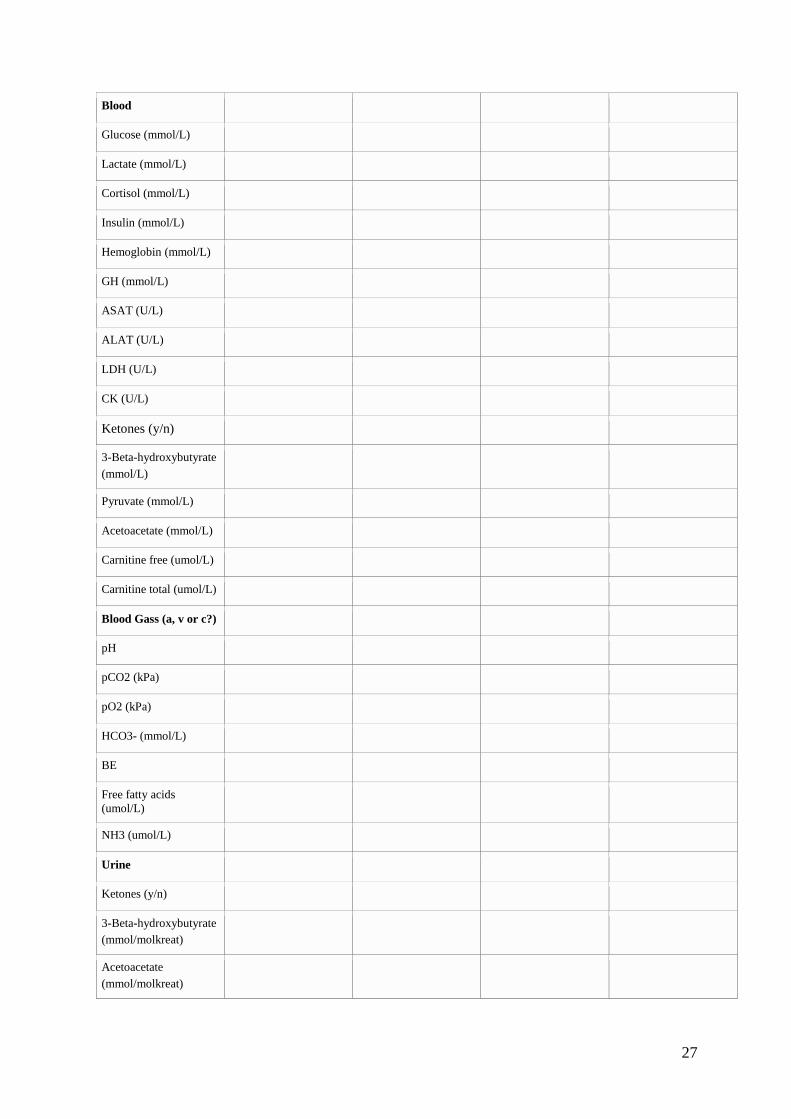
27
Blood
Glucose (mmol/L)
Lactate (mmol/L)
Cortisol (mmol/L)
Insulin (mmol/L)
Hemoglobin (mmol/L)
GH (mmol/L)
ASAT (U/L)
ALAT (U/L)
LDH (U/L)
CK (U/L)
Ketones (y/n)
3-Beta-hydroxybutyrate
(mmol/L)
Pyruvate (mmol/L)
Acetoacetate (mmol/L)
Carnitine free (umol/L)
Carnitine total (umol/L)
Blood Gass (a, v or c?)
pH
pCO2 (kPa)
pO2 (kPa)
HCO3- (mmol/L)
BE
Free fatty acids
(umol/L)
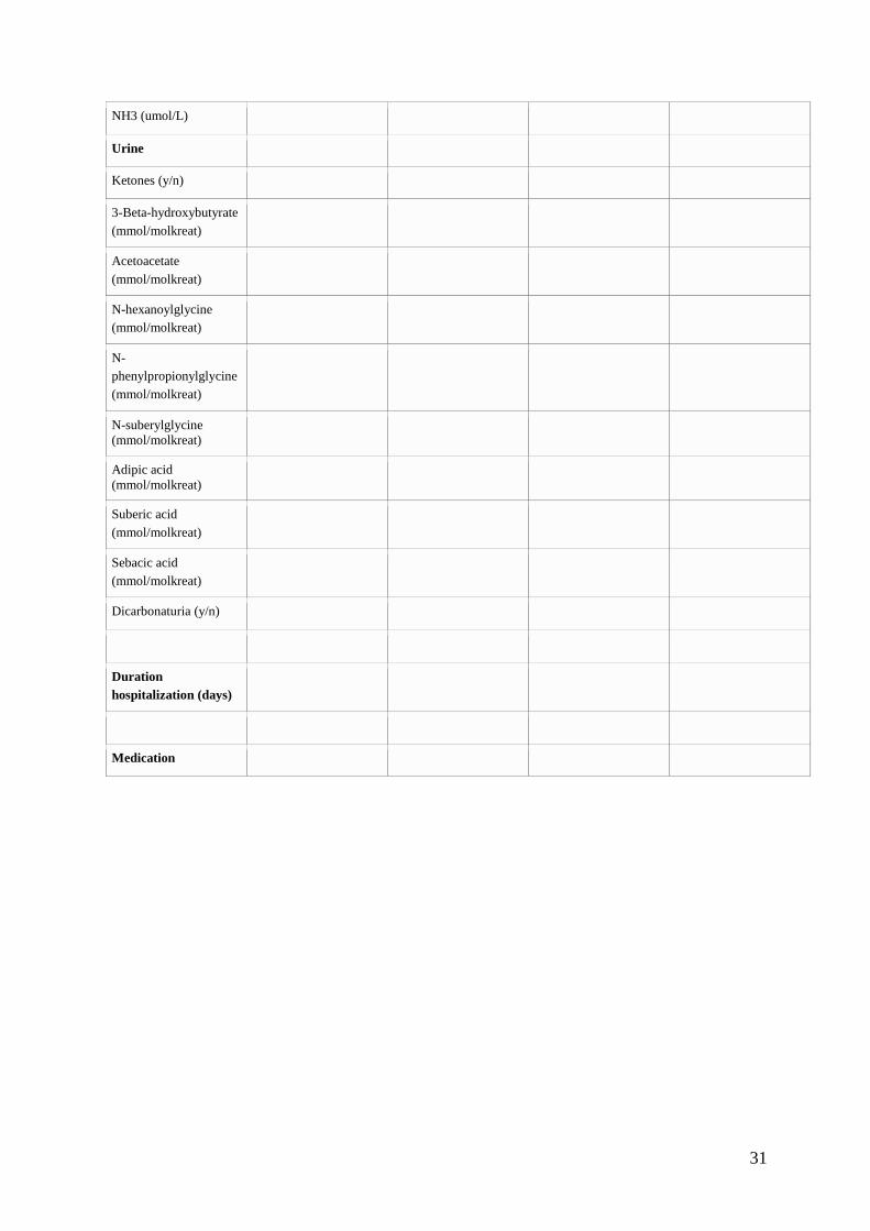
NH3 (umol/L)
Urine
Ketones (y/n)
3-Beta-hydroxybutyrate
(mmol/molkreat)
Acetoacetate
(mmol/molkreat)

28
N-hexanoylglycine
(mmol/molkreat)
N-
phenylpropionylglycine
(mmol/molkreat)
N-suberylglycine
(mmol/molkreat)
Adipic acid
(mmol/molkreat)
Suberic acid
(mmol/molkreat)
Sebacic acid
(mmol/molkreat)
Dicarbonaturia (y/n)
Duration
hospitalization (days)
Medication
Outpatient (0) and hospital admissions (1) after fasting test:
Date —/—/—
Date —/—/—
Date —/—/—
Date —/—/—
Reason 0/1
Anamnesis (y/n)
Hypoglycaemia
Vomiting
Drinking
Hepatomegaly
Hypotonia
Convulsions
Muscle pain
Exercise intoleration
Syncope

29
Temperature (ºC)
Growth parameters
Height (cm)
Height (SD)
Weight (kg)
Weight (SD)
Weight for height (SD)
BMI (kg/m2)
BMI (SD)
Head circumference
(cm)
Head circumference
(SD)
Physical examination
Temperature (ºC)
Neurological status
(GCS, coma,
convulsions)
Muscle tone
(hypertonic, hypotonic,
normal)
Sweating
Liver size (cm below
rib cage)
Heart palpitations
Neurological
abnormalities
Living situation
Education (special
education, IQ-tests)
Assistance
(physiotherapist,
psychologist)

30
Particulars
development
(motoric or
psychologic or
social)
EEG
ECG
Labaratory values
Blood
Glucose (mmol/L)
Lactate (mmol/L)
Cortisol (mmol/L)
Insulin (mmol/L)
GH (mmol/L)
ASAT (U/L)
ALAT (U/L)
LDH (U/L)
CK (U/L)
Ketones (y/n)
3-Beta-hydroxybutyrate
(mmol/L)
Pyruvate (mmol/L)
Acetoacetate (mmol/L)
Carnitine free (umol/L)
Carnitine total (umol/L)
Blood Gass (a, v or c?)
pH
pCO2 (kPa)
pO2 (kPa)
HCO3- (mmol/L)
BE
Free fatty acids
(umol/L)
31
NH3 (umol/L)
Urine
Ketones (y/n)
3-Beta-hydroxybutyrate
(mmol/molkreat)
Acetoacetate
(mmol/molkreat)
N-hexanoylglycine
(mmol/molkreat)
N-
phenylpropionylglycine
(mmol/molkreat)
N-suberylglycine
(mmol/molkreat)
Adipic acid
(mmol/molkreat)
Suberic acid
(mmol/molkreat)
Sebacic acid
(mmol/molkreat)
Dicarbonaturia (y/n)
Duration
hospitalization (days)
Medication


![Hyperosmolar Non Ketotic Dm [Autosaved]](https://img.dokumen.tips/doc/110x75/54b967564a7959637e8b4629/hyperosmolar-non-ketotic-dm-autosaved.jpg)