Embed Size (px)
Citation preview

The Most Economical CT of its Class.SOMATOM Perspective
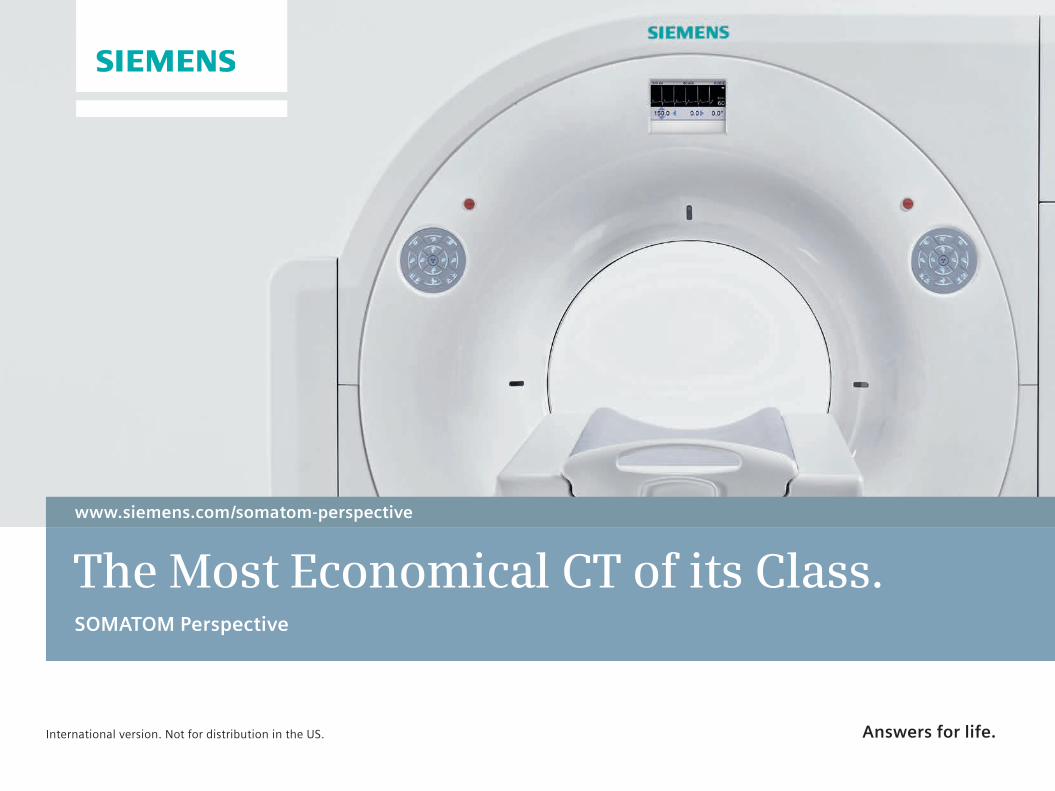
www.siemens.com/somatom-perspective
Global Siemens Headquarters Siemens AG Wittelsbacherplatz 2 80333 Muenchen Germany
Legal Manufaturer Siemens AG Wittelsbacherplatz 2 DE-80333 Muenchen Germany
Global Siemens Healthcare Headquarters Siemens AG Healthcare Sector Henkestrasse 127 91052 Erlangen Phone: +49 9131 84 0 Germany
On account of certain regional limitations of sales rights and service availability, we cannot guarantee that all products included in this brochure are available through the Siemens sales organization worldwide. Availability and packaging may vary by country and is subject to change without prior notice. Some/all of the features and products described herein may not be available in the United States.
The information in this document contains general technical descriptions of specifications and options as well as standard and optional features which do not always have to be present in individual cases.
Siemens reserves the right to modify the design, packaging, specifications, and options described herein without prior notice. Please contact your local Siemens sales representative for the most current information.
Order No. A91CT-03018-74C1-7600 | Printed in Germany | CC CT 1109 02132. | © 02.2013, Siemens AG
Global Business Unit Siemens AG Medical Solutions Computed Tomography & Radiation Oncology Siemensstr. 1 DE-91301 Forchheim Germany Phone: +49 9191 18 0 Fax: +49 9191 18 9998
Answers for life.www.siemens.com/healthcare
www.siemens.com/somatom-perspective
The Most Economical CT of its Class. SOMATOM Perspective
Note: Any technical data contained in this document may vary within defined tolerances. Original images always lose a certain amount of detail when reproduced.
International version. Not for distribution in the US.

www.siemens.com/somatom-perspective

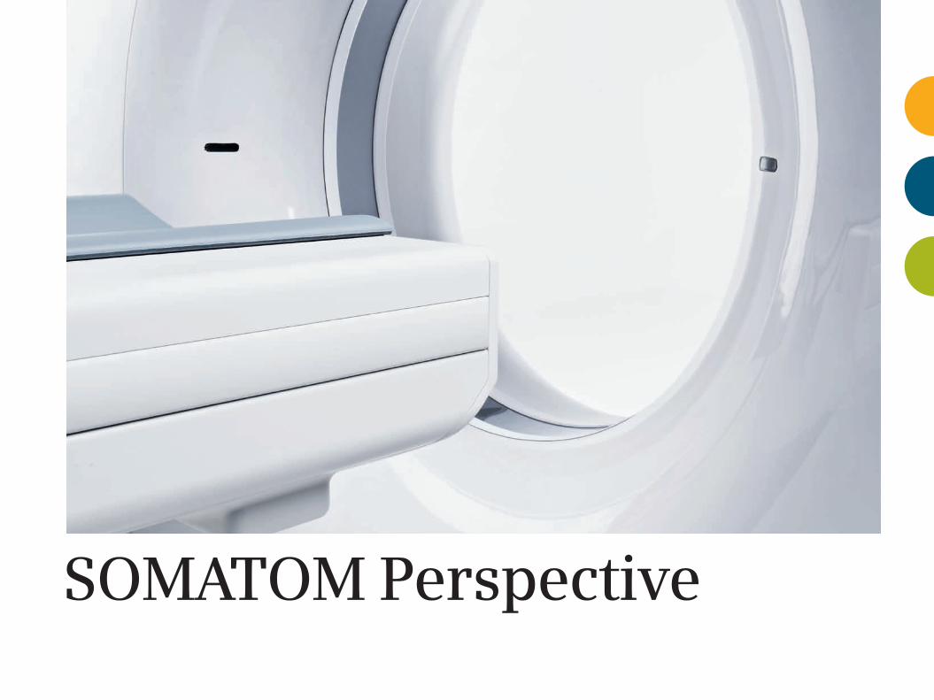
SOMATOM Perspective


33
Product Benefits 07
Manage your financial performance 08
Widen your clinical portfolio 10
Ease your working day 12
Added benefits with syngo.via 14
Clinical Images 17
Core Technologies 27
SAFIRE 28
iTRIM 30
eMode 32
Customer Services 34
1109_CT_SOMATOM_Perspective_Inhalt_final.indd 3 11.02.13 10:30
4
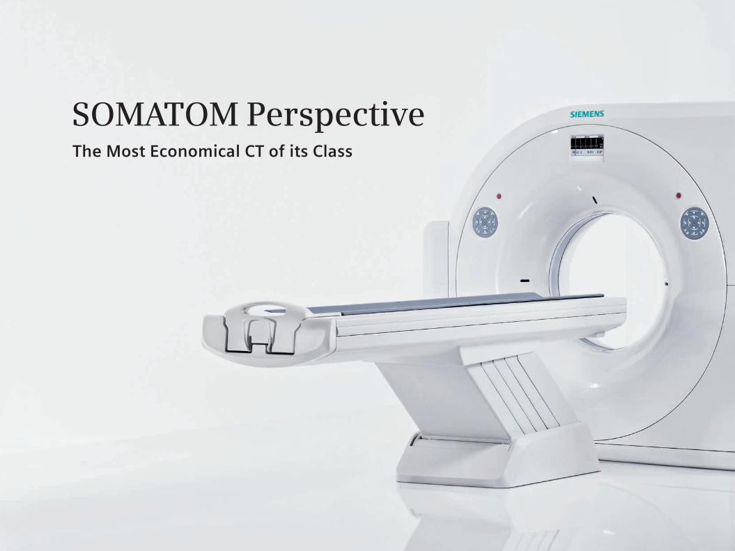
The Most Economical CT of its Class
SOMATOM Perspective
5
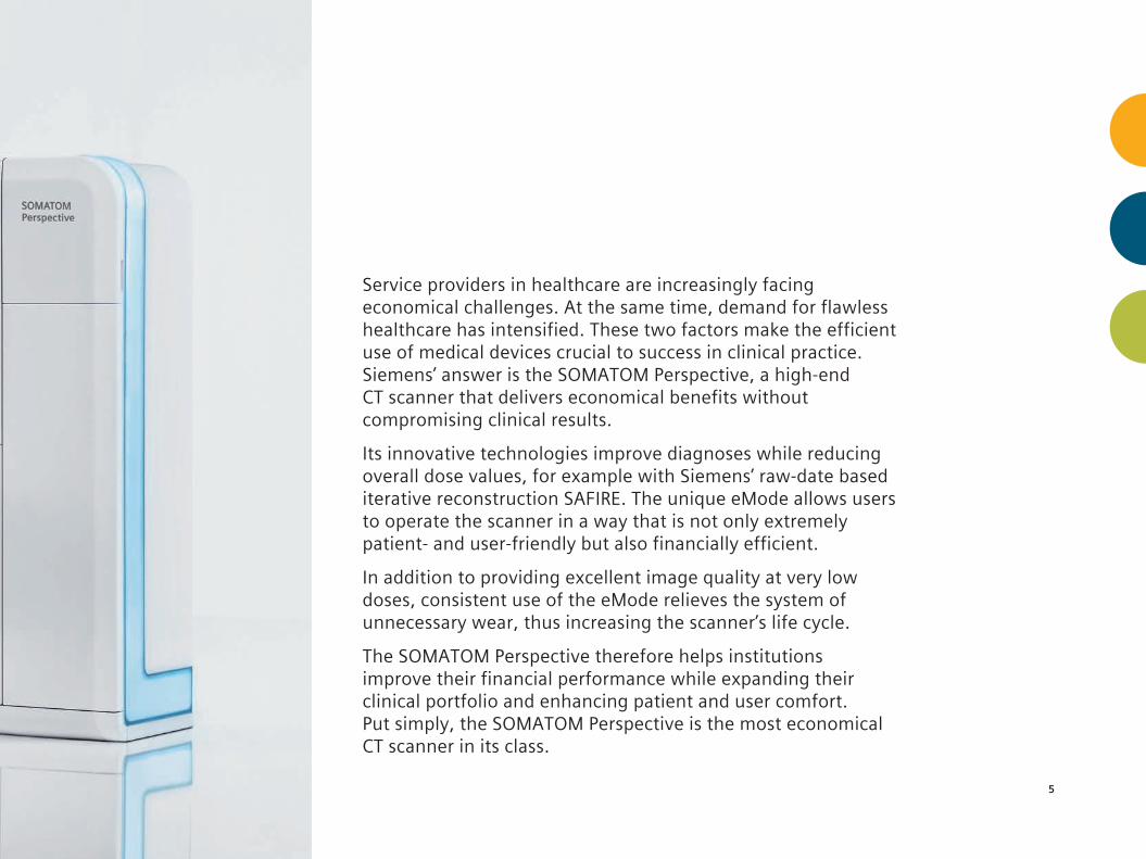
Service providers in healthcare are increasingly facing economical challenges. At the same time, demand for flawless healthcare has intensified. These two factors make the efficient use of medical devices crucial to success in clinical practice. Siemens’ answer is the SOMATOM Perspective, a high-end CT scanner that delivers economical benefits without compromising clinical results.
Its innovative technologies improve diagnoses while reducing overall dose values, for example with Siemens’ raw-date based iterative reconstruction SAFIRE. The unique eMode allows users to operate the scanner in a way that is not only extremely patient- and user-friendly but also financially efficient.
In addition to providing excellent image quality at very low doses, consistent use of the eMode relieves the system of unnecessary wear, thus increasing the scanner’s life cycle.
The SOMATOM Perspective therefore helps institutions improve their financial performance while expanding their clinical portfolio and enhancing patient and user comfort. Put simply, the SOMATOM Perspective is the most economical CT scanner in its class.


Product Benefits
8
Efficient scanner usage with eMode The unique eMode reduces wear and increases the scanner's life cycle. With a single click, the scanner can be operated in a highly patient-friendly and financially efficient way. The potential for a longer life cycle is just one of the many economical advantages the SOMATOM Perspective offers. The eMode service contract, for example, offers additional economic benefits.
Manage your financial performance
Excellent image quality The successful diagnosis and treatment of patients is largely determined by image quality. Medical staff need the best possible information to provide effective and efficient care. In this area, the SOMATOM Perspective sets a new standard. It provides the answers clinicians need while opti- mizing total cost of ownership – from the initial investment and runtime costs to innovative service solutions and improved workflow efficiency.
The SOMATOM Perspective is designed to reduce costs, thus allowing more medical institutions to afford enhanced patient care.
9
Optimized total cost of ownership The SOMATOM Perspective's sleek design and thin gantry only takes up 18 m2 (194 sq.ft.) of floor space. Its installation is uncomplicated and often takes less than two days. As the SOMATOM Perspective requires less operating power, it also reduces the scanner's heat dissipation. Therefore it also requires less cooling, consequently reducing the overall power consumption: 71 kVA of electricity for only 7 kW of heat dissipation.
Economical scanner concept Choosing SOMATOM Perspective also means choosing Siemens as a trusted partner. CT integrates reliable performance with innovations allowing it to be adjusted to users’ demands. The scanner can be fully upgraded onsite, growing with customers' demands from 64 - to 128 - slice configuration.
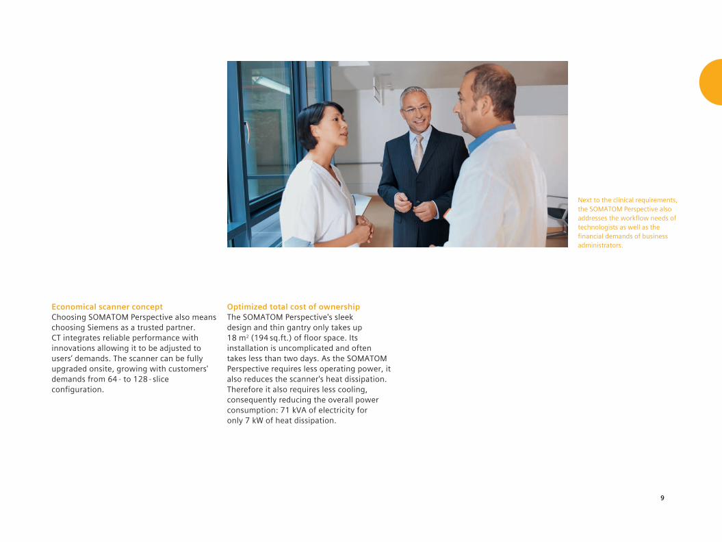
Next to the clinical requirements, the SOMATOM Perspective also addresses the workflow needs of technologists as well as the financial demands of business administrators.
10
Routine high-speed imaging Detection of small fractures or bleeding in an acute care scenario is a challenge in CT imaging. Thanks to the combination of the unique Siemens SureView concept and a 38.4 mm detector width, trade-offs between scan speed and image quality are no longer necessary – and long scan ranges at sub-millimeter collimation are no longer difficult. Thus, SureView's spiral image recon- struction algorithm facilitates challenging and long scans in clinical routine.
Dedication to low dose In addition to excellent image quality, the SOMATOM Perspective offers an impressive spectrum of dose reduction features. The UFC Detector, for example, lowers radiation dose thanks to its short afterglow and fast decay behavior. CARE Dose4D™ adapts tube current to patient size in real-time for dose-efficient scanning. The SOMATOM Perspective also comes with CARE Dose4D pediatric modulation curves for scanning children.
Widen your clinical portfolioThe SOMATOM Perspective offers a wide range of features that help make even challenging exams clinical routine.
11
Powerful cardio package For the complex procedures of cardiac imaging, the SOMATOM Perspective offers a variety of solutions. iTrim (Iterative Temporal Resolution Improvement Method), for example, makes possible a temporal resolution as low as 195 ms. In addition, there is the FAST Cardio Wizard, which provides step-by-step guidance towards higher reliability and reproducibility. The most suited arterial phases are automatically identified by ECG Check and Cardio BestPhase.
Latest generation iterative reconstruction The SOMATOM Perspective has been designed to the oustanding diagnostic detail. It uses information from up to 128 slices with its unique Interleaved Volume Reconstruction (IVR). Adding the raw- data-based iterative reconstruction method SAFIRE (Sinogram Affirmed Iterative Reconstruction) delivers up to 60 % dose* reduction and even superior image quality. It is easily incorporated into daily routine thanks to an excellent reconstruction speed of up to 15 images per second.
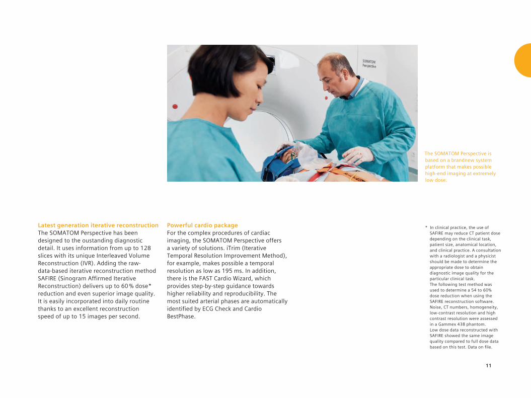
The SOMATOM Perspective is based on a brandnew system platform that makes possible high-end imaging at extremely low dose.
* In clinical practice, the use of SAFIRE may reduce CT patient dose depending on the clinical task, patient size, anatomical location, and clinical practice. A consultation with a radiologist and a physicist should be made to determine the appropriate dose to obtain diagnostic image quality for the particular clinical task. The following test method was used to determine a 54 to 60% dose reduction when using the SAFIRE reconstruction software. Noise, CT numbers, homogeneity, low-contrast resolution and high contrast resolution were assessed in a Gammex 438 phantom. Low dose data reconstructed with SAFIRE showed the same image quality compared to full dose data based on this test. Data on file.

12
Streamlined workflow To avoid unnecessary interruptions in the daily routine, the SOMATOM Perspective provides support during the entire scanning procedure. Patient positioning, often one of the most time-consuming steps in the exam process, is simplified with innovative features like the integrated storage box and the predefined buttons on the gantry control panel. The intuitive syngo® user interface further aligns procedures with the demands placed on staff.
Fast procedures in daily routine Saving time, both at the onset of an exam and during reconstruction, enables staff to spend more time with patients and allows more scans per day. The SOMATOM Perspective includes Fully Assisting Scanner Technologies (FAST) that automate time-consuming procedures. Adiditonally, WorkStream4D, virtually eliminates manual reconstruction steps. Oblique and double-oblique reconstructions with up to 20 images per second are immediatly available, thus making examinations faster.
Ease your working dayThe SOMATOM Perspective offers a streamlined workflow to ease the examination procedures for both staff and patients.

13
Illumination Moodlight The most striking design element of the SOMATOM Perspective is the Illumination Moodlight LED panel. It provides a more comfortable scanning environment and can be easily adjusted to the operator’s or the patient’s personal preference. The Illumination Moodlight changes color dynamically throughout the day or may be set to a single color that harmonizes best with the environment.
Integrated display panel* The gantry front display shows current scan parameters such as kV, mA, scan time, table position, gantry tilt, and ECG trace – a great advantage during cardio exams. Storage box The convenient storage box holds all the basic CT positioning accessories. With accessories close at hand, patient positioning is faster and easier.
Slim design The slim gantry design increases patient comfort while offering easy access for interventional procedures.
A streamlined workflow is a lot more than just throughput. It implies that both staff and the CT system are set for maximum efficiency in every case.
* Optional

14
Added Benefits with syngo.via
Image networking syngo.via speeds up the way users connect and share information with clinical partners and patients – even on the go.* syngo.via’s client-server-based based nature supports a smooth, teamwork-like sharing of tasks, just as it is required in 3D labs and larger radiology departments. Images can be shared among multiple users at once, providing a sound basis for joint preprocedural planning.
syngo®.via for sustainable care As the number of chronic disease patients rises, the demand for high-quality and efficient care is increasing. syngo.via, Siemens' state-of-the-art imaging software, creates an exciting experience in efficiency and ease of use. syngo.via helps foster sustainable care by equipping physicians with workflows and applications for evaluating images from multiple modalities. In cardiovascular CT, for example, it makes possible a rule-out of coronary artery disease in less than a minute.
Automatic Case Preparation syngo.via assists in the analysis of individual cases, prepares images, suggests an opti- mized workflow, and offers guidance when needed. For example, when a cardiac case is opened, the Automated Case Preparation has already pre-processed the images and displays them in the appropriate layout together with the right evaluation tools. Evaluation of the coronary vessels, the functional parameters, and the prepared calcium score can start immediately.
Regardless of volume or disease, syngo.via helps prepare cases, eases interdisciplinary collaboration, and helps facilitating a faster, more reliable diagnosis.
15
* Prerequisites include: internet connection to clinical network, DICOM compliance, meeting of minimum hardware requirements, and adherence to local data security regulations.
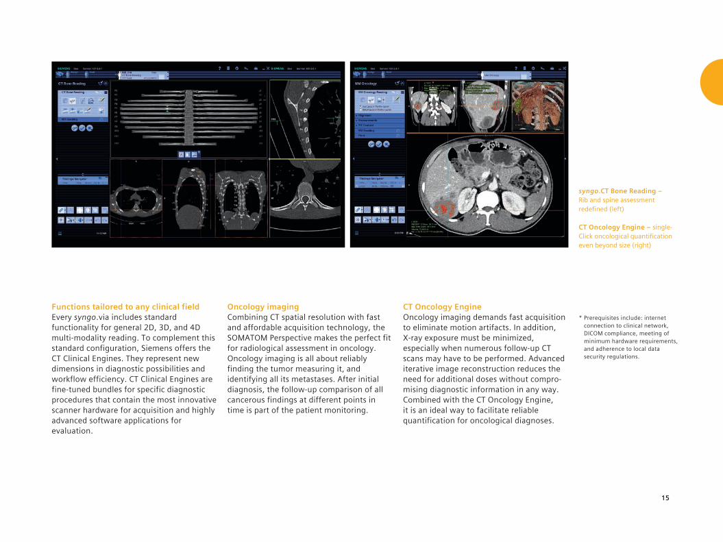
Functions tailored to any clinical field Every syngo.via includes standard functionality for general 2D, 3D, and 4D multi-modality reading. To complement this standard configuration, Siemens offers the CT Clinical Engines. They represent new dimensions in diagnostic possibilities and workflow efficiency. CT Clinical Engines are fine-tuned bundles for specific diagnostic procedures that contain the most innovative scanner hardware for acquisition and highly advanced software applications for evaluation.
Oncology imaging Combining CT spatial resolution with fast and affordable acquisition technology, the SOMATOM Perspective makes the perfect fit for radiological assessment in oncology. Oncology imaging is all about reliably finding the tumor measuring it, and identifying all its metastases. After initial diagnosis, the follow-up comparison of all cancerous findings at different points in time is part of the patient monitoring.
CT Oncology Engine Oncology imaging demands fast acquisition to eliminate motion artifacts. In addition, X-ray exposure must be minimized, especially when numerous follow-up CT scans may have to be performed. Advanced iterative image reconstruction reduces the need for additional doses without compro-mising diagnostic information in any way. Combined with the CT Oncology Engine, it is an ideal way to facilitate reliable quantification for oncological diagnoses.
syngo.CT Bone Reading – Rib and spine assessment redefined (left)
CT Oncology Engine – single-Click oncological quantification even beyond size (right)

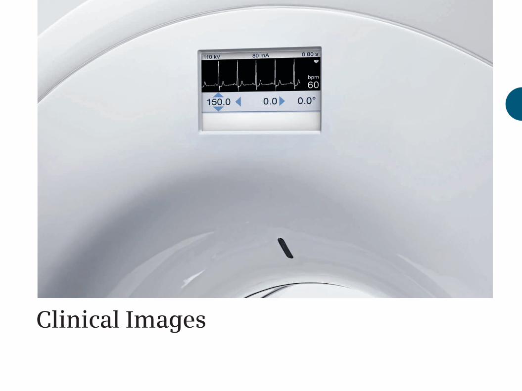
Clinical Images

18
Neurology – non-contrast cerebral spiral scan.
Axial images demonstrate excellent soft tissue contrast
resolution. No clinical findings.
Radiology Department of Israelitisches Krankenhaus, Hamburg, Germany
collimation: 32 x 0.6 mm
scan time: 20 s
scan length: 147 mm
rotation time: 1.5 s
tube settings: 130 kV, 302 mAs
CTDIvol: 73.35 mGy
DLP: 1379.15 mGy cm
eff. dose: 2.89 mSv
19
Diagnosezentrum Favoriten, Vienna, Austria
Acute Care – pulmonary spiral scan with contrast media. Axial, MPR, and VRT images reveal detailed information to the sub-segmental pulmonary embolisms in great details.
collimation: 64 x 0.6 mm
scan time: 4 s
scan length: 305 mm
rotation time: 0.6 s
tube settings: 110 kV, 75 mAs
CTDIvol: 5.59 mGy
DLP: 241.46 mGy cm
eff. dose: 3.38 mSv

20
Institut Sainte Marie, Paris, France
Oncology – high resolution pulmonary scans.
Axial and MPR images show excellent spatial resolution for pulmonary structures. A solid,
irregular nodule is clearly depicted in the right upper lobe
suggesting a lung tumor. Fibrosis and ground glass
opacifications are also visualized in the left lung.
collimation: 64 x 0.6 mm
scan time: 4 s
scan length: 367 mm
rotation time: 0.6 s
tube settings: 110 kV, 66 mAs
CTDIvol: 4.93 mGy
DLP: 249.74 mGy cm
eff. dose: 3.5 mSv
21
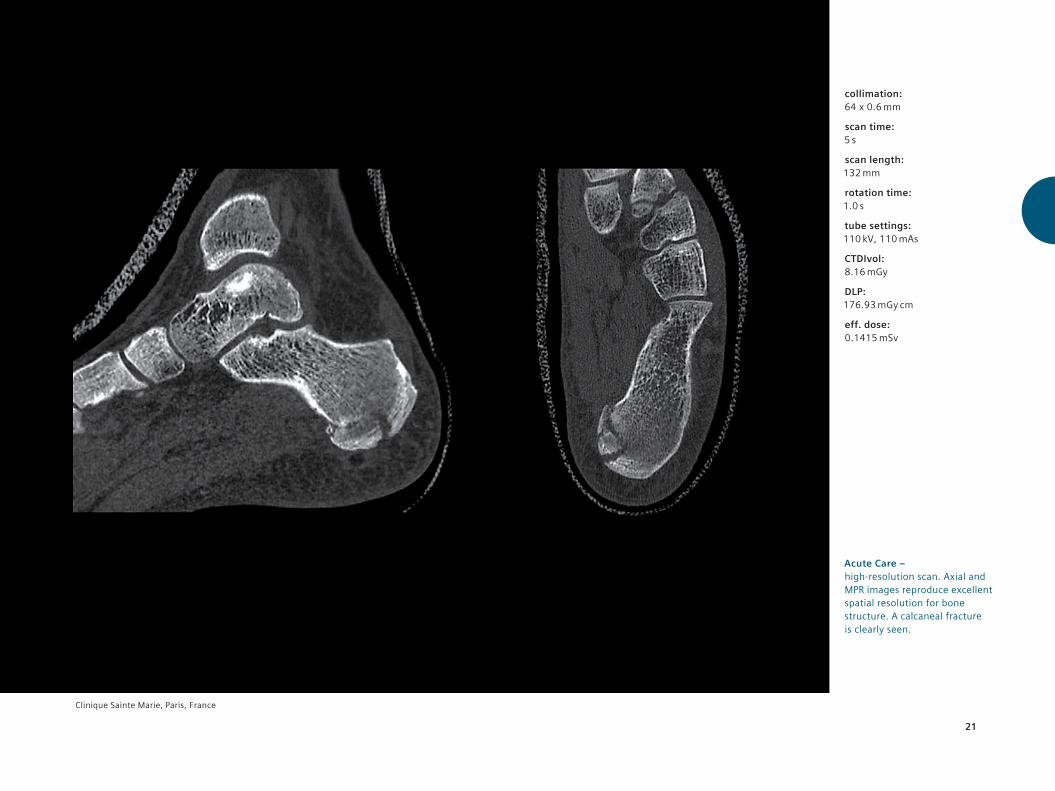
Clinique Sainte Marie, Paris, France
Acute Care – high-resolution scan. Axial and MPR images reproduce excellent spatial resolution for bone structure. A calcaneal fracture is clearly seen.
collimation: 64 x 0.6 mm
scan time: 5 s
scan length: 132 mm
rotation time: 1.0 s
tube settings: 110 kV, 110 mAs
CTDIvol: 8.16 mGy
DLP: 176.93 mGy cm
eff. dose: 0.1415 mSv
22
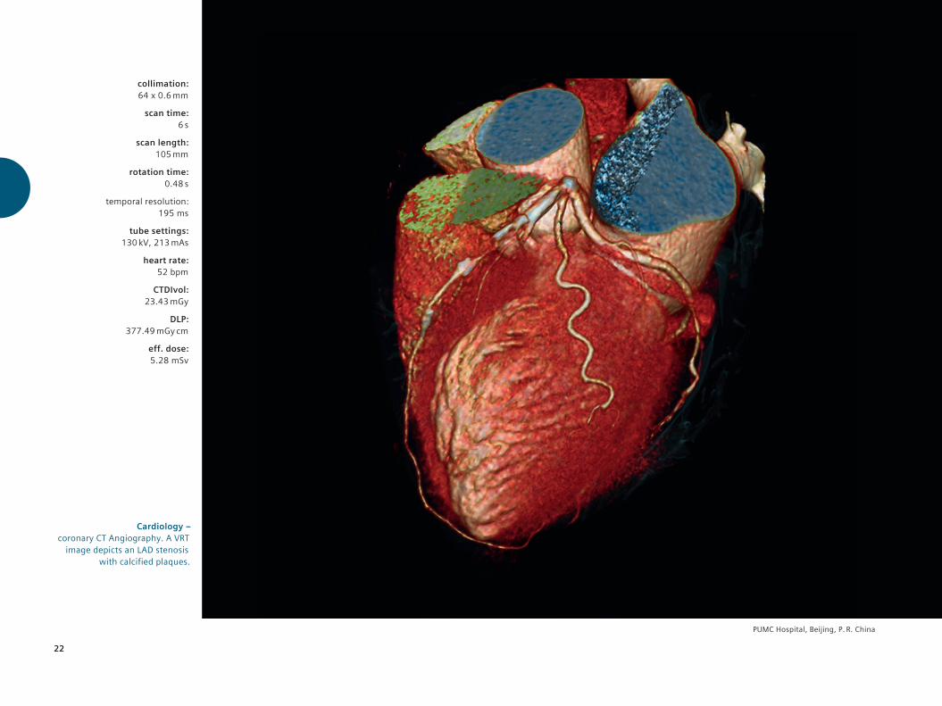
PUMC Hospital, Beijing, P. R. China
Cardiology – coronary CT Angiography. A VRT
image depicts an LAD stenosis with calcified plaques.
collimation: 64 x 0.6 mm
scan time: 6 s
scan length: 105 mm
rotation time: 0.48 s
temporal resolution: 195 ms
tube settings: 130 kV, 213 mAs
heart rate: 52 bpm
CTDIvol: 23.43 mGy
DLP: 377.49 mGy cm
eff. dose: 5.28 mSv
23
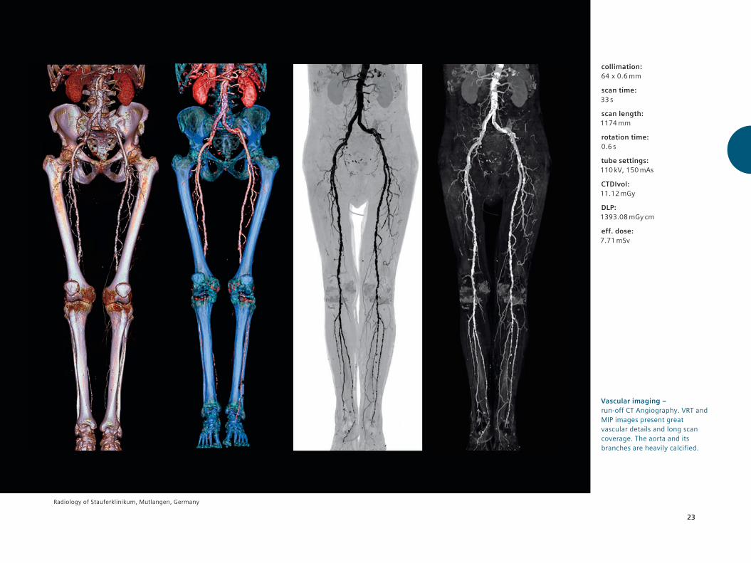
Vascular imaging – run-off CT Angiography. VRT and MIP images present great vascular details and long scan coverage. The aorta and its branches are heavily calcified.
Radiology of Stauferklinikum, Mutlangen, Germany
collimation: 64 x 0.6 mm
scan time: 33 s
scan length: 1174 mm
rotation time: 0.6 s
tube settings: 110 kV, 150 mAs
CTDIvol: 11.12 mGy
DLP: 1393.08 mGy cm
eff. dose: 7.71 mSv

24
PUMC Hospital, Beijing, P. R. China
3D Interventional imaging –interventional procedures, such as lung nodule biopsies can be
navigated in all three dimensions.
collimation: 64 x 0.6 mm
scan time: 0.92 s
scan length: 60 mm
rotation time: 0.6 s
tube settings: 130 kV, 70 mAs
CTDIvol: 7.87 mGy
DLP: 147.67 mGy cm
eff. dose: 2 mSv

25
Bariatric imaging – an abdominal scan of an obese patient. Axial and MPR images clearly demonstrate multiple hepatic lesions, despite the presence of ascites. VRT image shows the vascular structures of the abdominal aorta.
Diagnosezentrum Favoriten, Vienna, Austria
collimation: 64 x 0.6 mm
scan time: 6 s
scan length: 239 mm
rotation time: 0.6 s
tube settings: 110 kV, 166 mAs
CTDIvol: 12.33 mGy
DLP: 390.46 mGy cm
eff. dose: 5.8 mSv


Core Technologies

28
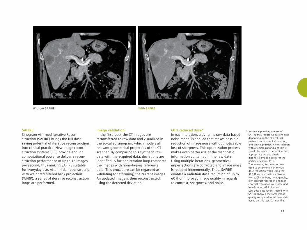
SAFIRESAFIRE delivers excellent clinical results with up to 60 % dose reduction or superior image quality at a reconstruction speed suitable for daily routine.
Standard Filtered Back Projection
Ultra-fast reconstruction without iterations
Well-established image impression
Limited dose reduction
SAFIRE
More powerful dose reduction than image-based methods
Well-established image impression
Superior image quality
Fast reconstruction in image and raw-data space
Improved workflow with variable settings
Image data rRaw data
reconRaw data
reconImage data
reconImage
correction
29
SAFIRE Sinogram Affirmed Iterative Recon- struction (SAFIRE) brings the full dose-saving potential of iterative reconstruction into clinical practice. New image recon- struction systems (IRS) provide enough computational power to deliver a recon- struction performance of up to 15 images per second, thus making SAFIRE suitable for everyday use. After initial reconstruction with weighted filtered back projection (WFBP), a series of iterative reconstruction loops are performed.
Image validation In the first loop, the CT images are retransferred to raw data and visualized in the so-called sinogram, which models all relevant geometrical properties of the CT scanner. By comparing this synthetic raw-data with the acquired data, deviations are identified. A further iteration loop compares the images with homologous reference data. This procedure can be regarded as validating (or affirming) the current images. An updated image is then reconstructed, using the detected deviation.
60 % reduced dose* In each iteration, a dynamic raw-data-based noise model is applied that makes possible reduction of image noise without noticeable loss of sharpness. This optimization process makes even better use of the diagnostic information contained in the raw data. Using multiple iterations, geometrical imperfections are corrected and image noise is reduced incrementally. Thus, SAFIRE enables a radiation dose reduction of up to 60 % or improved image quality in regards to contrast, sharpness, and noise.
Without SAFIRE With SAFIRE
* In clinical practice, the use of SAFIRE may reduce CT patient dose depending on the clinical task, patient size, anatomical location, and clinical practice. A consultation with a radiologist and a physicist should be made to determine the appropriate dose to obtain diagnostic image quality for the particular clinical task. The following test method was used to determine a 54 to 60% dose reduction when using the SAFIRE reconstruction software. Noise, CT numbers, homogeneity, low-contrast resolution and high contrast resolution were assessed in a Gammex 438 phantom. Low dose data reconstructed with SAFIRE showed the same image quality compared to full dose data based on this test. Data on file.
30
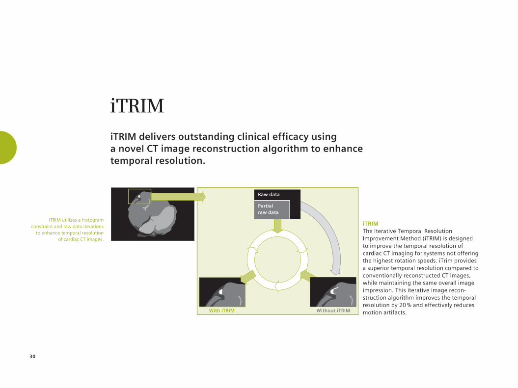
iTRIM The Iterative Temporal Resolution Improvement Method (iTRIM) is designed to improve the temporal resolution of cardiac CT imaging for systems not offering the highest rotation speeds. iTrim provides a superior temporal resolution compared to conventionally reconstructed CT images, while maintaining the same overall image impression. This iterative image recon-struction algorithm improves the temporal resolution by 20 % and effectively reduces motion artifacts.
iTRIMiTRIM delivers outstanding clinical efficacy using a novel CT image reconstruction algorithm to enhance temporal resolution.
iTRIM utilizes a histogram constraint and raw data iterations
to enhance temporal resolution of cardiac CT images.
Without iTRIMWith iTRIM
Raw data
Partial raw data
31
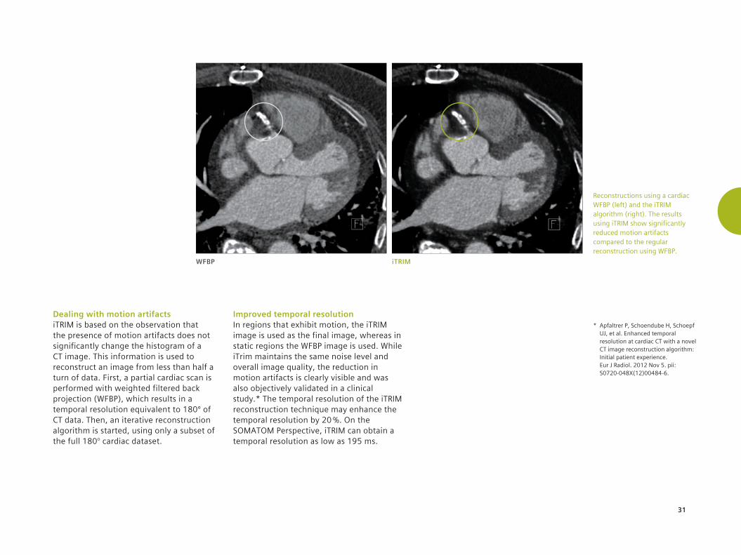
Dealing with motion artifacts iTRIM is based on the observation that the presence of motion artifacts does not significantly change the histogram of a CT image. This information is used to reconstruct an image from less than half a turn of data. First, a partial cardiac scan is performed with weighted filtered back projection (WFBP), which results in a temporal resolution equivalent to 180º of CT data. Then, an iterative reconstruction algorithm is started, using only a subset of the full 180° cardiac dataset.
Improved temporal resolution In regions that exhibit motion, the iTRIM image is used as the final image, whereas in static regions the WFBP image is used. While iTrim maintains the same noise level and overall image quality, the reduction in motion artifacts is clearly visible and was also objectively validated in a clinical study.* The temporal resolution of the iTRIM reconstruction technique may enhance the temporal resolution by 20 %. On the SOMATOM Perspective, iTRIM can obtain a temporal resolution as low as 195 ms.
WFBP iTRIM
Reconstructions using a cardiac WFBP (left) and the iTRIM algorithm (right). The results using iTRIM show significantly reduced motion artifacts compared to the regular reconstruction using WFBP.
* Apfaltrer P, Schoendube H, Schoepf UJ, et al. Enhanced temporal resolution at cardiac CT with a novel CT image reconstruction algorithm: Initial patient experience. Eur J Radiol. 2012 Nov 5. pii: S0720-048X(12)00484-6.
32
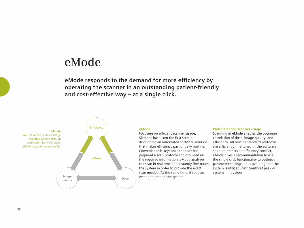
eMode Focusing on efficient scanner usage, Siemens has taken the first step in developing an automated software solution that makes efficiency part of daily routine. Convenience is key: once the user has prepared a scan protocol and provided all the required information, eMode analyzes the scan in real time and instantly fine-tunes the system in order to provide the exact scan needed. At the same time, it reduces wear and tear on the system.
Well-balanced scanner usage Scanning in eMode enables the optimum correlation of dose, image quality, and efficiency. All routine standard protocols are efficiently fine-tuned. If the software solution detects an efficiency conflict, eMode gives a recommendation to use the single click functionality to optimize parameter settings, thus avoiding that the system is utilized inefficiently at peak or system limit values.
eMode
eMode Well-balanced scanner usage
depends on an optimum correlation between dose,
efficiency, and image quality.
Dose
Efficiency
Image quality
eMode
eMode responds to the demand for more efficiency by operating the scanner in an outstanding patient-friendly and cost-effective way – at a single click.

33
Siemens service offerings Siemens UPTIME Services focuses on real-time remote monitoring and preventive maintenance, enabling increased system availability, optimized performance, and workflow efficiency. A service contract, including Utilization Management (UM) and tube coverage for the SOMATOM Perspective, allows users to take full advantage of eMode benefits.
Detailed utilization information The SOMATOM Perspective usage will be monitored 24/7 via Siemens Remote Service (SRS) and every scan will be tracked and summarized in the UM reports. UM will provide exact information detailing the CT performance indicators, including how much eMode was utilized.
Service Benefits*
1. syngo Remote Trainer
2. Preventive maintenance out of prime working time
3. syngo Remote Assist
4. eMode service contract adjustments
+Service
contractGood outlook for service contract holders with eMode annual eMode
scans
≥ 80 %
* Individual service benefit availability is subject to country-specific offerings.
www.siemens.com/perspective-emode
Scan to learn more about SOMATOM Perspective’s unique eMode.
34
eMode service benefits* The system will be analyzed every 12 months in order to determine the eMode usage: if eMode was used for at least 80 % of the scans, customers are entitled to select one of the valuable eMode service benefits. Users can significantly profit from these benefits and leverage the efficiency potential of their scanners.
1. syngo Remote Trainer Users may gain expertise in new clinical areas or become more familiar with any application chosen.
2. Preventive maintenance eMode usage benefit may be applied to preserve uninterrupted workflows by opting for overnight maintenance services.
3. syngo Remote Assist One-on-one remote assistance exactly when needed for any difficulty encountered
4. eMode service contract adjustments The service contract price may be actively optimized for each coming year, thus reflecting the advantages of consistent eMode usage.
Customer ServicesA range of innovative service solutions that raise quality and productivity in healthcare.

35
Siemens Performance Plans Service and maintenance are highly important to prevent unscheduled downtimes. Siemens Performance Plans are designed to help run operations smoothly and thus to improve workflow – with predictable costs, lower risks, and higher efficiency. Modules can be combined with a Performance Plan Pro, Plus, or Top and an individual solution with substantial benefits. Siemens Virus Protection, for example, offers top-level defense against viruses.
Education Know-how is the key to success. With the extensive Siemens portfolio of education and training programs, healthcare practitioners can deepen their knowledge and clinical expertise. The portfolio offers a wide range of choices:
• Individual on-site training • Classroom training • Web-based training • Fellowships • Remote assistance
* Individual service benefit availability is subject to country-specific offerings.
Siemens Remote Service Siemens offers innovative service solutions based on SRS technology. SRS establishes a highly efficient, dependable and secure VPN remote connection. Our unique Guardian ProgramTM further reduces costs through automatic monitoring of the CT system to detect potential problems before they occur.


www.siemens.com/somatom-perspective
Global Siemens Headquarters Siemens AG Wittelsbacherplatz 2 80333 Muenchen Germany
Legal Manufaturer Siemens AG Wittelsbacherplatz 2 DE-80333 Muenchen Germany
Global Siemens Healthcare Headquarters Siemens AG Healthcare Sector Henkestrasse 127 91052 Erlangen Phone: +49 9131 84 0 Germany
On account of certain regional limitations of sales rights and service availability, we cannot guarantee that all products included in this brochure are available through the Siemens sales organization worldwide. Availability and packaging may vary by country and is subject to change without prior notice. Some/all of the features and products described herein may not be available in the United States.
The information in this document contains general technical descriptions of specifications and options as well as standard and optional features which do not always have to be present in individual cases.
Siemens reserves the right to modify the design, packaging, specifications, and options described herein without prior notice. Please contact your local Siemens sales representative for the most current information.
Order No. A91CT-03018-74C1-7600 | Printed in Germany | CC CT 1109 02132. | © 02.2013, Siemens AG
Global Business Unit Siemens AG Medical Solutions Computed Tomography & Radiation Oncology Siemensstr. 1 DE-91301 Forchheim Germany Phone: +49 9191 18 0 Fax: +49 9191 18 9998
Answers for life.www.siemens.com/healthcare
www.siemens.com/somatom-perspective
The Most Economical CT of its Class. SOMATOM Perspective
Note: Any technical data contained in this document may vary within defined tolerances. Original images always lose a certain amount of detail when reproduced.
International version. Not for distribution in the US.
www.siemens.com/healthcare
Global Siemens Headquarters Siemens AG Wittelsbacherplatz 2 80333 Muenchen Germany
Global Siemens Healthcare Headquarters Siemens AG Healthcare Sector Henkestrasse 127 91052 Erlangen Phone: +49 9131 84 0 Germany
On account of certain regional limitations of sales rights and service availability, we cannot guarantee that all products included in this brochure are available through the Siemens sales organization worldwide. Availability and packaging may vary by country and is subject to change without prior notice. Some/all of the features and products described herein may not be available in the United States.
The information in this document contains general technical descriptions of specifications and options as well as standard and optional features which do not always have to be present in individual cases.
Siemens reserves the right to modify the design, packaging, specifications, and options described herein without prior notice. Please contact your local Siemens sales representative for the most current information.
Note: Any technical data contained in this document may vary within defined tolerances. Original images always lose a certain amount of detail when reproduced.
Global Business Unit Siemens AG Medical Solutions Computed Tomography & Radiation Oncology Siemensstr. 1 DE-91301 Forchheim Germany Phone: +49 9191 18 0 Fax: +49 9191 18 9998
Order No. A91CT-03018-74C1-7600 | Printed in Germany | CC CT 1109 02132. | © 02.2013, Siemens AG


![Welcome []Address Student Service Center of Faculty of Engineering Erwin-Rommel-Str. 60 91058 Erlangen Telephone +49 9131 85 27850 Email tf-stib@fau.de Internet](https://img.dokumen.tips/doc/110x75/60847fd5557d1f0bd946aaae/welcome-address-student-service-center-of-faculty-of-engineering-erwin-rommel-str.jpg)


