Embed Size (px)
Citation preview
The Molecular Cloning and Characterization of Potential Chick DM-GRASP Homologs in Zebrafish and Mouse
John P. Kanki,'.*,+ Susannah Chang,* and John Y. Kuwada'
' Department of Biology, University of Michigan, Ann Arbor, Michigan 481 09-1 048; and 'Department of Anatomy, University of Pennsylvania School of Medicine, Philadelphia, Pennsylvania 191 04
SUMMARY
A full-length zebrafish cDNA clone and a partial mouse cDNA clone similar to chick DM-GRASP were isolated and analyzed. The nucleotide sequence of the full-length zebrafish clone shares 54% identity, and predicts 39% amino acid identity, with chick DM-GRASP. The partial mouse clone shares 76% nucleotide identity, and predicts 76% amino acid identity, with chick DM-GRASP. The predicted proteins encoded by both of these clones ex- hibit conserved structural domains that are characteris- tic of the chick protein. These features may identify them as a distinct subfamily within the immunoglobulin super- family of cell adhesion molecules. Expression of the ze- brafish DM-GRASP protein is similar to chick DM- GRASP and is principally restricted to a small subset of
developing sensory and motor neurons during axonogen- esis. Zebrafish DM-GRASP expression was temporally regulated and limited to specific axon domains. This re- gional expression correlated with fasciculated axon do- mains. These results suggest that the zebrafish and mouse cDNA clones represent the respective fish and mammalian homologs of chick DM-GRASP. The highly selective expression of zebrafish DM-GRASP suggests that it is involved in the selective fasciculation and guid- ance of axons along their normal pathways. 0 1994 John
Keywords: zebrafish, DM-GRASP, Ig superfamily, axon guidance, fasciculation.
Wiley & Sons, Inc.
INTRODUCTION
The nervous system develops and functions nor- mally because neural growth cones find and estab- lish appropriate connections with specific targets. Specific connections are formed early during em- bryogenesis by axons following stereotyped path- ways to their synaptic targets. Growth cones follow stereotyped patterns of axonal outgrowth by recog- nizing cues that distinguish their proper pathways from inappropriate ones. Growth cones respond to a variety of attractive and inhibitory signals in a
Received January 1 1, 1994; accepted February 16, 1994 Journal of Neurobiology, Vol. 25, No. 7, pp. 83 1-845 (1994) 0 1994 John Wiley & Sons, Inc. CCC 0022-3034/94/07083 1- 15
* To whom correspondence should be addressed. Present address: Department of Molecular Biology, Mof-
fett Laboratory, Princeton University, Princeton, NJ 08544.
complex and changing environment (e.g., Bentley and Caudy, 1983; Raper et al., 1983; Tosney and Landmesser, 1985; Bastiani et al., 1986; Kapf- hammer and Raper, 1987; Kuwada, 1986; Hams et al., 1987; Dodd and Jessell, 1988 ). These signals may include cell surface proteins (Rathjen et al., 1987), extracellular matrix proteins (Reichardt and Tomaselli, 199 1 ), and diffusable factors ( Lumsden and Davies, 199 1 ; Tessier-Lavigne and Placzek, 199 1 ). Molecules whose expression is re- stricted to specific substrates or axons are of partic- ular interest since they represent potential cues for guiding growth cones along their pathways.
Cell surface molecules that are expressed by subsets of developing axons have been identified in a variety of embryos. Many of these molecules are members of the immunoglobulin (1s) superfamily of adhesion proteins (Williams and Barclay, 1988). These proteins mediate specific cell adhe-
831
832 Kanki et al.
sion interactions that, in the case of neurons, may influence selective adhesion to substrates along a specific axon pathway. Some of the fasciclins are well-characterized members of the Ig superfamily which are expressed on specific axonal bundles in insects. Genetic and antibody perturbation experi- ments suggest that fasciclin I1 is necessary for growth cones to fasciculate and extend upon spe- cific axon bundles during development ( Harrelson and Goodman, 1988; Grenningloh et al., 1990). In vertebrates a number of putative axon guidance molecules have also been characterized ( Rathjen and Jessell, 199 1 ). Some of these molecules are expressed selectively on subsets of axons. One in- triguing molecule is DM-GRASP (also called SC 1 and BEN) which is a member of the Ig superfamily and is principally expressed on developing sensory and motor axons in the chick (Tanaka and Obata, 1984; Pourquie et al., 1990; Burns et al., 199 1; El- Deeb et al., 1992 ). It exhibits homophilic binding and promotes neurite extension in vitvo suggesting that it participates in selective axon fasciculation and axon guidance (Burns et al., 199 1 ; Tanaka et al., 1991; Pourquie et al., 1992).
In addition to being expressed on selective sub- sets of neurons, these molecules may also be re- gionally expressed on specific axon domains. For example, TAG-1 and Ll are expressed in a do- main-specific pattern on the axons of commissural neurons in the rat spinal cord (Dodd et al., 1988). Their expression is spatially regulated such that TAG- 1 is expressed on the ipsilateral, circumferen- tial portion of the axons as they run towards the floorplate at the ventral midline. Once the com- missural axons pass the floorplate to the contralat- era1 side they turn, run anteriorly, and express L1. The switch from TAG-1 to L1 expression suggests that these molecules may be responsible for guid- ing the growth cones along different regions oftheir pathway.
We have cloned a zebrafish cDNA similar to chick DM-GRASP in order to examine the expres- sion of DM-GRASP in the well-characterized ner- vous system of the zebrafish embryo. The zebrafish is an attractive vertebrate system for in vivo analy- sis of early development (Eisen, 199 1 ; Weinberg, 1992 ) . During early embryonic stages it is possible to examine the development of individual neurons as they project their axons and establish highly ste- reotyped pathways (e.g., IOmmel et al., 1982; Myers, 1985; Eisen et al., 1986; Kuwada and Bern- hardt, 1990). The well-characterized development of these identified neurons makes it possible to dis- tinguish regional differences in the expression of
molecules on their axonal domains. Thus the ze- brafish embryo is an attractive system for the de- tailed examination of the expression of putative axon guidance cues such as DM-GRASP. The de- tailed analysis of the developmental expression of such molecules on these neurons may elucidate their potential roles in directing axonal outgrowth in vivo.
We have cloned potential homologs of DM- GRASP in the mouse and the zebrafish and have examined DM-GRASP expression on identified neurons in the early zebrafish embryo. Our results indicate that this molecule is selectively expressed on a small subset of axon tracts, and that it is re- gionally expressed on specific axon domains. The selectivity of DM-GRASP expression and its dif- ferential expression on specific axon domains may regulate their ability to fasciculate with each other and thereby determine the pattern of their axonal trajectories.
METHODS
Sequencing and Analysis
A hgt 1 1 cDNA library was constructed from total RNA isolated from 32- to 48-h zebrafish embryos (K. Zinn). Approximately 6 X lo5 plaques were screened using established procedures for low- stringency hybridization screening ( Sambrook et al., 1989). Duplicate filter lifts were probed with a 32P-random-primed fragment ( 1.6 kb) of the chick DM-GRASP clone. Three hybridizing plaques were isolated and their cDNA inserts were sub- cloned into Bluescript SK- (Stratagene) and se- quenced using the dideoxy chain termination method (Sanger et al., 1977) with Sequenase (USB). Sequence alignments to Genbank using the FASTA program (ktup = 6, Pearson and Lip- man, 1988) were performed and further nucleotide and amino acid analysis was done using the Mac- Vector (JBI ) and Geneworks ( IntelliGenetics) pro- grams. Protein alignments were performed using a PAM-250 scoring matrix. Two additional clones were isolated under conditions of high stringency from a XZAP library (Stratagene) made from 20- to 28-h zebrafish embryos (generously supplied by D. J. Grunwald ), using a 32P-random-primed frag- ment ( 1.6 kb) of the zebrafish DM-GRASP clone as probe. These duplicate clones were isolated from the XZAP phage and sequenced as described above.
To obtain the mouse cDNA clone, lo7 plaques
DM-GRASP Homologs in Zebrafish and Mouse 833
from an adult mouse brain cDNA library (Clon- tech ) were screened using a 32P-random-primed fragment ( 1.5 kb) of the chick DM-GRASP clone. Seven hybridizing plaques were isolated using stan- dard conditions for low-stringency hybridization. After the cDNA inserts were subcloned, one was sequenced and further analyzed as described above.
Generation of Glutathione S-Transferase Fusion Proteins and Polyclonal Antisera
The polymerase chain reaction (PCR) (Saiki et al., 1988) was used to amplify an 812-bp sequence from the 5' end of the zebrafish DM-GRASP clone with oligonucleotides containing Nut1 ( 5 ' ) and Smal (3') linkers. The amplified fragment corre- sponded to zebrafish DM-GRASP nucleotides 160-972 encoding amino acids 1-271. The PCR fragment was isolated using TA cloning (Invitro- gen), and then digested with Nut 1 and Swia 1. This fragment was inserted into pGEX-KN (Hakes and Dixon, 1992) which had been digested with EcoRl, end-filled with the Klenow fragment of DNA polymerase I, and digested with Null. This placed the zebrafish DM-GRASP amino acid se- quence in-frame behind the glutathione S-transfer- ase sequence. The fusion protein was expressed in Escherichia Coli, and purified from the bacterial extracts by affinity chromatography with glutathi- one-sepharose ( Pharmacia) (Smith and Johnson, 1988). The purified zebrafish DM-GRASP fusion protein was used for the production of antisera. Then, 50 pg of protein was injected at 4-week inter- vals into three rabbits (UM5, UM27, and UM28) (Cocalico Biologicals). Preimmune serum was ob- tained from each rabbit prior to immunization with the antigen. After immunization, antiserum was collected every 2 weeks. The sera were tested for reactivity by immunoblot analysis using bacte- rial lysates containing the fusion protein. Anti- serum from UM5 and pooled antisera from UM27 and UM28 were affinity purified using established methods (Harlow and Lane 1988) on a column containing the zebrafish fusion protein covalently coupled to CNBr-activated sepharose ( Phar- macia).
lmmunoblot Analysis and lmmunocytochemistry
Zebrafish embryos were collected from a labora- tory breeding colony, maintained at 28S"C, and
developmentally staged according to procedures described in Myers et al. ( 1986).
For immunoblot analysis embryos were col- lected at various stages, deyolked if necessary, and lysed ( 2 4 lysis buffer/embryo) in a 20-mMPhos- phate buffer (pH 7.5) containing 100 m M NaCI, 10 m M EDTA, 1% sucrose, 1% NP40, 1 m M PMSF, 1 pg/ml aprotinin and leupeptin. The ly- sates were centrifuged briefly, and an equal volume of 3% SDS reducing gel sample buffer was added to the supernatant. Lysates containing the bacterial fusion protein were prepared by adding sample buffer directly to bacterial pellets. The lysates were fractionated by SDS-PAGE on 8% or 10% gels. Proteins were either stained with Coomassie blue or transferred to nitrocellulose filters. The filters were blocked in Tris-buffered saline containing 1 % BSA and probed with antiserum at 1 :250- 1000 di- lution, followed by 1251-Protein A ( Amersham) . Blots were exposed to Kodak X-OMAT film with intensifying screens at -70°C.
For immunocytochemical analysis, embryos were collected between 12 h and 5 days postfertil- ization, fixed in 4% paraformaldehyde in 0.1 A4 phosphate buffer (pH 7.4) for 6-1 8 h at 4°C and subsequently processed as whole-mount prepara- tions or cryostat frozen sections as described in the Zebrafish Book (ed. Westerfield, 1993). For frozen sections embryos were embedded in 1.5% agar/ 5% sucrose, cryoprotected in 30% sucrose, and frozen in OCT compound. Sections (20 pm) were cut on a Reichert-Jung 2800 cryostat. Whole-mounted em- bryos and sections were incubated with primary antisera (dilutions ranged from 1 : 10- 1000) for 16-24 h at 4°C. Slides with frozen sections were maintained in humidified chambers. Primary anti- bodies were detected with a diaminobenzidine (DAB) reaction product after incubating with a biotinylated anti-rabbit secondary antibody fol- lowed by a biotin! avidin peroxidase complex ( Vectastain ABC, Vector Laboratories). Whole- mounted embryos and sections were viewed and photographed with a Leitz Orthoplan2 microscope using differential interference contrast optics.
In Vitro Transcription and Translation
PCR was used to amplify the zebrafish DM- GRASP cDNA insert directly from the XGTl1 clone using phage X primers. The amplified prod- uct was isolated using TA cloning (Invitrogen). Zebrafish DM-GRASP mRNA was transcribed in vilro using SP6 polymerase, and was isolated by phenol/chloroform extraction followed by ethanol
834 Kunki et ul.
precipitation (Sambrook et al., 1989). The mRNA was then translated in vitro using a rabbit reticulo- cyte lysate ( Promega) in the presence of 35S-labeled methionine ( Amersham). Translations were per- formed in the presence and absence of canine pan- creatic microsomal membranes (Promega). The translation products were analyzed by SDS-PAGE on 8% gels followed by fluorography with Enlight- ening (Dupont). Flourographed gels were exposed to Kodak X-OMAT film at -70°C.
RESULTS
Isolation and Analysis of a Zebrafish DM-GRASP cDNA Clone
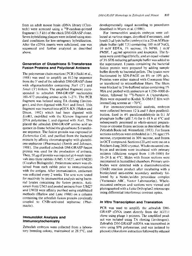
A 1.6-kb fragment of chick DM-GRASP was used to screen an oligo(dT)-primed zebrafish cDNA li- brary made from 32- to 48-h embryos. Three posi- tive plaques were isolated and subcloned, the larg- est of these contained an insert of 2070 base pairs. Sequence analysis revealed an open reading frame of 1692 bp with a 159 bp leader and 2 18 bp of 3' untranslated sequence (Fig. 1 ). The open reading frame predicts a precursor protein of 564 amino acids including a putative 22 amino acid signal se- quence (von Heijne, 1986) that directs protein translation to the endoplasmic reticulum (ER). A developmental Northern blot of total RNA ex- tracted from zebrafish embryos between 24 and 74 h was probed with the zebrafish cDNA clone and revealed a single 3.6 kb mRNA transcript (data not shown). This differs from the chick where two ap- parent mRNA transcripts encoding DM-GRASP have been described (Tanaka et al., 199 1 ).
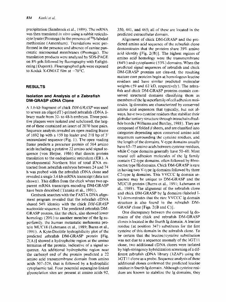
Genbank searches with the FASTA DNA align- ment program revealed that the zebrafish cDNA shared 54% identity with the chick DM-GRASP nucleotide sequence. The predicted zebrafish DM- GRASP protein, like the chick, also showed lower homology (20%) to another member of the Ig su- perfamily, the human metastatic melanoma pro- tein MUC 18 (Lehmann et al., 1989; Burns et al., 199 1 ). A Kyte-Doolitle hydrophilicity plot of the predicted zebrafish DM-GRASP protein [Fig. 2(A)] showed a hydrophobic region at the amino terminus of the protein, indicative of a signal se- quence. An additional hydrophobic region near the carboxyl end of the protein predicted a 22 amino acid transmembrane domain from amino acids 507-529, that is followed by a hydrophilic cytoplasmic tail. Four potential asparagine-linked glycosylation sites are present at amino acids 92,
350, 44 1, and 465; all of these are located in the predicted extracellular domain.
Alignment of chick DM-GRASP and the pre- dicted amino acid sequence of the zebrafish clone demonstrates that the proteins share 39% amino acid identity [Fig. 2(B)]. The highest regions of amino acid homology were the transmembrane (64%) and cytoplasmic ( 53%) domains. When the predicted signal sequences of zebrafish and chick DM-GRASP proteins are cleaved, the resulting mature core proteins begin at homologous leucine residues and have similar predicted molecular weights (59 and 62 kD, respectively). The zebra- fish and chick DM-GRASP proteins contain con- served structural domains classifying them as members of the Ig superfamily of cell adhesion mol- ecules. Ig domains are characterized by conserved amino acid sequences that typically, but not al- ways, have two cysteine residues that stabilize their globular tertiary structure through intrachain disul- fide bonds (Williams and Barclay, 1988 ). They are composed of folded 0 sheets, and are classified into categories depending upon conserved amino acid sequences surrounding the cysteine residues, and the length of the domains. V-type domains usually have 65-75 amino acids between cysteine residues, while C-type domains generally have 50-60. Most neural cell adhesion molecules of the Ig family contain C2-type domains, often followed by fibro- nectin type I11 domains. Chick DM-GRASP is rare in having two V-type Ig domains followed by three C2-type Ig domains. This VVCCC Ig domain se- quence may be unique to DM-GRASP and the MUC18 protein (Burns et al., 1991; Lehmann et al., 1989). The alignment of the zebrafish clone and chick DM-GRASP by Ig domains (labeled I- V) demonstrates that the rare VVCCC Ig domain structure is also found in the zebrafish DM- GRASP clone [Figs. 2( B and C ) ] .
One discrepancy between the conserved Ig do- mains of the chick and zebrafish DM-GRASP clones is located in the fourth Ig domain. A leucine residue (at position 347) substitutes for the first cysteine of this domain in the zebrafish clone. To be certain that the leucine/cysteine substitution was not due to a sequence anomaly of the XGTl 1 clone, two additional cDNA clones were isolated by high-stringency hybridization screening of a dif- ferent zebrafish cDNA library (XZAP) using the XGTl 1 clone as a probe. Sequence analysis of these additional clones confirmed the predicted leucine residue in fourth Ig domain. Although cysteine resi- dues are known to stabilize the Ig domains, they
DM-GRASP Homologs in Zebrafish and Mouse 835
CCG TCT AAG C T C CAG ACT CCA CCT GCG C T C T G T AGA GCA ACC CGT CTG ATC T T T CAG CGC A C T CAA C T C TCA ATA C I A TCT AAC AAC 'TTC 9 0
At iA CC'T ACA TCr CTC TCT ATT GAG ACT GTC GCC GGA CLG TA'T AAA GGA GAA CCG GGG T l T T C 1 PTA A G G AlG C A ! LCC S I T A:C 1 t i C CII l8C Met H i s Sor V a l I l e Cys Lou 7
T T C GGT GCC T T C ATA GCA GCC GCT TTG T T T GCT CCA GGG AGC TGC CTG CCG ACG GTT ATA GGT CTG TAC GGT GAG ACC ATC GAA GTG CCA 2 7 0 Phe cly A h Phr X l r A.la A.la Ala Leu Pha Ala Pro Gly ser cur L e u P r o T h r V a l l i e G l y L e u T y r G l y t i i u 1 n L l i e GLU V d L P r o 3 7
TGC AAC AAT GGA AAT AAC AAG CCA GAT GGC C T T A T T T T C ACC AAA TGG AAA TAT GCA AAA GAT GAC GGC TCC CCT GGC GAT CTA CTA ATA 360 cys A s n A s n G l y A s n A s n L ~ S p r o ASP G l y L e u I l e Ptie T h r LYS T r p LYS T y r A l a L ~ S ASP ASP G l y Ser p ro ~ l y ASP L e u L e u I l e 6 7
* AAG CAG GCA CAG AAA GAT GAT CCG ACT GTG TCT GCT ATG GAC GGC TAC AAA ACT AGA GTT AGC ATC GCT GCT AAT TCC AGC TTA CTG ATT 4 5 0 L y s G l n A l a G l n L y s A s p A s p P r o T h r V a l Ser A l a M e t A s p G l y T y r L y s T h r Ar9 V a l Ser I le A l a A l a A s n ser s e r L e u L e u I l e 9 1
GCC CAG GGC T C T TTG ACT GAC CAA AGA GTC T T C ACC TGC ATG GTG GTG T C T TCA ACT AAT CTG GAG GAA T T C TCT GTG GAA GTT AAA GTT 5 4 0 A i a G l n G l y ser L e u T h r A s p G l n A r g V a l P h e T h r C y s M e t V a l V a l ser Ser T h r A s n L e u G l u G l u P h e Ser V a l G l u V a l L y s V a l 1 2 7
CAC AAA AAA CCA TCA GCC CCT GTA ATC AAA AAC AAA G'PG AAA GAR CTG GAA AAT GGC AAA CTG ACG CAG TTG GGG GAA TGT GTG GTG GAG 630 H i s L y s L y s P r o Ser A l a P r o V a l I l e L y s A s n L y s V a l L y s G l u L e u G l u A s n G l y L y s L e u T h r G l n L e u G l y Glu C y s V a l V a l G l u 157
AGC GCC AAC CCA GCA GCA GAT C T C A T C TGG ATG AAG AAC AAC CAG GCT CTG GTG GAT GAT GGC AAG ACG ATC AT'l ATC ACA TCA GAT GTC 7 2 0
ser A l a A s n P r o A l a A l a A s p L e u I l e T r p M e t L y s A s n A s n G l n A l a L e u V a l A s p A s p G l y L y s T h r I l e I l e I l e T h r Ser A s p V a l 187
ACC AAG GAC CCA GTC ACC GGT CTG T C C AGC ACC T C T TCC AGA CTG CAG TAC ACA GCA AGG AAA GAG GAT GTG GCA TCA CAG T T C ACC TGC 8 1 0 T h r L y s A s p P r o V a l T h r G l y L e u Ser Ser T h r Ser ser A r g L e u G l n T y r T h r A l a A r g L y s G l u A s p V a l A l a S e r G l n P h e T h r C y s 2 1 1
GTT GCA AAG CAC GTG ACG GGA CCC AAC CAG GTT TCA ACA CCC GAT ACC T T C CAA A T T CGC T A T CCC ACT GAG AAG GTG AGT CTA CAG GTT 9 0 0 V a l A l a L y s H i s V a l T h r G l y P r o A s n G l n V a l Ser T h r P r o A s p T h r P h e G l n I l e A r g T y r P r o T h r G l u L y s V a l Ser L e u G l n V a l 2 4 1
GTC TCT CAG AGC CCC A T T AGG GAA GGT GAT GAT GTG ACT CTG AAA TGC CAG GCG GAC GGA AAC CCT CCT CCT ACT AGC T T C AAC T T T ARC 990 V a l Ser G l n ser P r o I l e A r g G l u G l y A s p A s p V a l T h r L e u L y s C y s G l n A l a A s p G l y A s n P r o P r o P r o T h r Ser P h e A S n P h e A s n 2 7 7
ATT AAG GGA AAG AAG GTC ACG GTG ACG GAC AAG GAT GTC TAC ACA CTG ACC GGC GTC ACC CGA GCC GAC AGC GGT GTG TAC AAG TGC TCT 1080 I l e ~ y s G l y L y s L y s V a l T h r V a l T h r A s p L y s A s p V a l T y r T h r L e u T h r G l y V a l T h r A r g A l a A s p Ser G l y V a l T y r L y s C y s Ser 3 0 1
CTG CTT GAC AAC GAT GTG ATG GAG TCC ACT CAG ATC GTC ACA GTG AGC T T T CTG GAT GCA AGC CTC ACT CCT ACA GGC AAG GTG TTA AAA l l - l O ieu ~ e u A s p ~ s n ASP V a l M e t G i u Ser T h r G l n I l e V a l T h r V a l s e r P h e L e u A s p la Ser L e u T h r P L O TtlK G l y L y s V a l L e u L y s 3 3 1
AAG CTC GGG GAA AAC TTG GTA GTG TCT TTG GAG AAG AAT GCC TCT T C T GAA GTA AAA GTG ACG TGG ACT AAG GAT AAC CGT AAA CTG GAC 1 2 6 0 L ~ S L e u G l y G l u A s n L e u V a l V a l Ser L e u G l u L y s A s n A l a S e c Ser G l u V a l L y s V a l T h e T r p T h r Lys A s p A s n A r g L y s L e u A s p 367
AAA CTG CCT GAT T T C TCC CAG TTG AGA TAC AGC GAC GCG GGC TTA TAC GTG TGT GAT GTG TCC A T T GAA GGA ATC AAA CAC AGC T T T TCC 1350 L y s L e u P r o A s p P h e Ser G l n L e u A r g T y r ser A s p A l a G l y L e u T y f V a l C y s A s p V a l Ser I l e Glu G l y Ile L y s H i s Ser P h e Ser 391
TTC GAG C T T ACT GTG GAA GGT GGT CCA AGA A T T ACC GGC CTG ACA AAG CAT CGC AGC AAT GAC GGA AAA CAC AAA GTG TTG ACG TGC GAG 1 4 4 0 P h e G l u L e u T h r V a l G 1 u G l y G l y P r o A r g I l e T h r G l y L e u T h r Lys His A r g Ser A m A s p G l y L y s His L y s V a l L e u T h r C y s G l u 4 2 1
GCA GAA GGT TCA CCT AAA CCT GAA GTG CAG TGG AGT GTC AAC GGA ACC GAC GAT GAA ACA TCG TAT GTC AAC GGA AAA GCC ACA TAC AAA 1 5 3 0 A l a G l u G l y Ser P r o L y s P r o G l u V a l G l n T r p Ser V a l A s n G l y T h r A s p A s p G l u T h r Ser T y r V a l A s n G l y L y s A l a T h r T y r L y s 4 5 7
t
t
CTG ACG GTG GTC CCG AGT AAG AAC CTC ACC GTC AGC TGC CTC GTG ACC AAT AAA CTC GGT T T C GAC ACA AAG GAC ATC AGC GTG T T T TCC 1 6 2 0 L e u T h r V a l V a l P r o Ser Lys A s n L e u T h r V a l Ser C y s L e u V a l T h r A s n L y s L e u G l y P h e A s p T h r L y s A s p rle Ser V a l P h e ser 4 8 1
CTA T T T GAG GAG GAC AAG CCC AAA CCA GGA AAA AAT GAA GAT GGC GCA GAC CAA GCC AAA GTG ATT GTG GGT GTC GTG GTT GGA CTG T T T 1 1 1 0 L e u P h e G l U G l U A S P LYS P I 0 LYS P r o G l y LYS A s n G l U A s p G l y A l a A s p G l n A l a L y S V a l I l e V a l G l y V a l V a l V a l G l y L e u P h e 5 1 7
CTA GCC GCT GCC CTG GTG GGA CTC ATC TAC TGG TTG TAT ATC AAG AAA ACA AGA CAA GGC AGC TGG AAG ACC GtiA GAG AAG GAG ACT GGC 1800 L e u A l a A l a A l a L e u V a l G l y L e u I l e T y T T T p L e u T y f I l e L y s L y s T h r A r g G l n G l y ser T r p L y s T h r G l y G l U L y s G l u T h r G l y 5 4 7
ACT TCA GAG GAG AGT AAG APA CTG GAG GAG AAC AAT CAT AAA GCA GAT GTC TAA GAG TGG AGA GAG CCG TCA AAC GGA ACG AAG RAG GTT 1 8 9 0
T h r Ser G l u G l u Ser L y s L y s L e u G l u G1u A s n A s n His L y s A l a A s p V a l 5 6 4
TGA ACG AGG AAA GAG GCC TTG TCA CAC TGA ATG TGC AGT ACT ACA CAC TTC GAT C T T C T T A T T CCA GAC ATC TGA TTG GAC GGC GAC GCC 1 9 8 0 TCC T T C TGG CTG C T C GTG CGC ATA CCG CAT CGC AGA ATG T T T CCA AAA TAT T T C CTA C T C CAA ACA AAC AAT T T C CTA AAA AAA AAA RAG 2 0 1 0
Figure 1 Nucleotide and predicted amino acid sequence ofzebrafish DM-GRASP. The puta- tive signal sequence is in bold italics, and the transmembrane domain is underscored. Asterisks denote potential asparagine-linked glycosylation sites. The zebrafish sequence has been regis- tered with Genbank and given the following accession number: ZEFDMGRASP, L25273.
are not mandatory. A number of other molecules in the Ig superfamily are known to replace the cys- teine residue with other hydrophobic residues, such as feucine, isoleucine, valine, tyrosine, or me- thionine (Williams and Barclay, 1988). Such sub- stitutions are thought to be suitable for the mainte- nance and stabilization of the intrachain fold.
Isolation and Analysis of a Mouse DM-GRASP cDNA Clone
A 1.5-kb fragment of chick DM-GRASP was used to screen a cDNA library from adult mouse brain (Clontech) . Seven plaques were isolated and sub- cloned; one of these contained a 1288 bp cDNA
836 Kanki rt ul.
A
B I
II
I l l
IV
V
C
Hydrophilicity Window Size = 7 Scale = Kyte-Doolittle 5.00
c 3.00
!.= 1.00 0.00 - -2.00 I” -3.00 -4.00 -5.00
3. 4.00
.L G 2.00
e -1.00 c
- A.A. 100 200 300 400 500
Zebrafish DM-GRASP *------ HSVI*-- - - *FGAFI rAALF ~~~~ ~
Chick DM-GRASP MEPPSXRRPASCRRRPLLCLLL ASLCM Mouse DM-GRASP
A*GSC*P**IGL**E**EV**NNGNNK****I’T**~*AKDD***GDLLIKQ~Q*D~PTVS~DG’*T*VSIAA*SS*L*AQGSLTDQRV*T~*V*SSTNLE*FSVE*‘*H*K‘*A PPALOLYTVNAVYGDTITMPCR--LEVPDGLMFGKWKYEMPNGSPVFIAFRSSTKKNVQYDDVPDYKDRLSLSENYTLSIKNARIRHEKRFVCMLVTEDDVSEEPTWKVFKQPSQ
Figure 2 Zebrafish and mouse DM-GRASP are putative transmembrane glycoproteins and are members ofthe Immunoglobulin superfamily of cell adhesion proteins. (A) Kyte-Doolittle hydrophilicity plot of coding region of zebrafish DM-GRASP (window = 7 amino acids). (B) Protein alignment (Geneworks) of zebrafish DM-GRASP and mouse DM-GRASP with chick DM-GRASP. Putative signal sequences are in bold italics, and transmembrane domains are underscored. The entire amino acid sequence of chick DM-GRASP is shown, while in the zebrafish and mouse sequences, only nonidentical amino acids are shown. Dashes in the zebrafish and mouse sequences represent identity with the aligned amino acids of the chick sequence. Boxed residues indicate amino acid identity between all three proteins. The se- quences are aligned such that corresponding Ig domains (I-V) are identified. Each domain begins 20 amino acids before the first of two cysteine residues (bold and underscored) that define them (Williams and Barclay, 1988). Blank spaces indicate gaps inserted by the align- ment program. The asterisk indicates the leucine residue substituted for the first cysteine in the fourth Ig domain of the zebrafish DM-GRASP sequence (see text). The mouse sequence has been registered with Genbank and given the following accession number: MUSDMGRASP, L25274. (C) Diagram illustrating the predicted protein structure of zebrafish DM-GRASP. V-type and C-type Ig domains are indicated along with potential N-linked glycosylation sites. Transmembrane (TMD) and cytoplasmic (CD) domains are indicated.
insert containing a 1071 bp open reading frame followed by 2 1 5 bp of 3‘ untranslated sequence. The sequence of this partial cDNA clone was aligned to the 3‘portion ofchick DM-GRASP( resi-
dues 795-2,036) and was found to share 76% nu- cleotide identity (data not shown). The open read- ing frame of the mouse clone predicts a 357 amino acid protein which shares 76% identity with the
DM-GRASP Homologs in Zebrafish and Mouse 837
carboxyl portion of the chick protein [Fig. 2( B)] . Comparison of protein subsequences between the mouse and chick clones indicates that over the ex- tent of their partial alignment the mouse protein contains nearly all of the predicted structural fea- tures characteristic of chick DM-GRASP. These include a portion of the second V type Ig domain, three C2-type Ig domains, identical sites for poten- tial asparagine-linked glycosylation, and a particu- larly high degree of homology in the predicted transmembrane domain (95%) and cytoplas- mic tail (85%). The three-way alignment of the mouse clone with the carboxyl portions of both chick DM-GRASP and the zebrafish clone, indi- cates that they share 37% amino acid identity [Fig. 2(B)].
Characterization of the Zebrafish DM-GRASP Protein
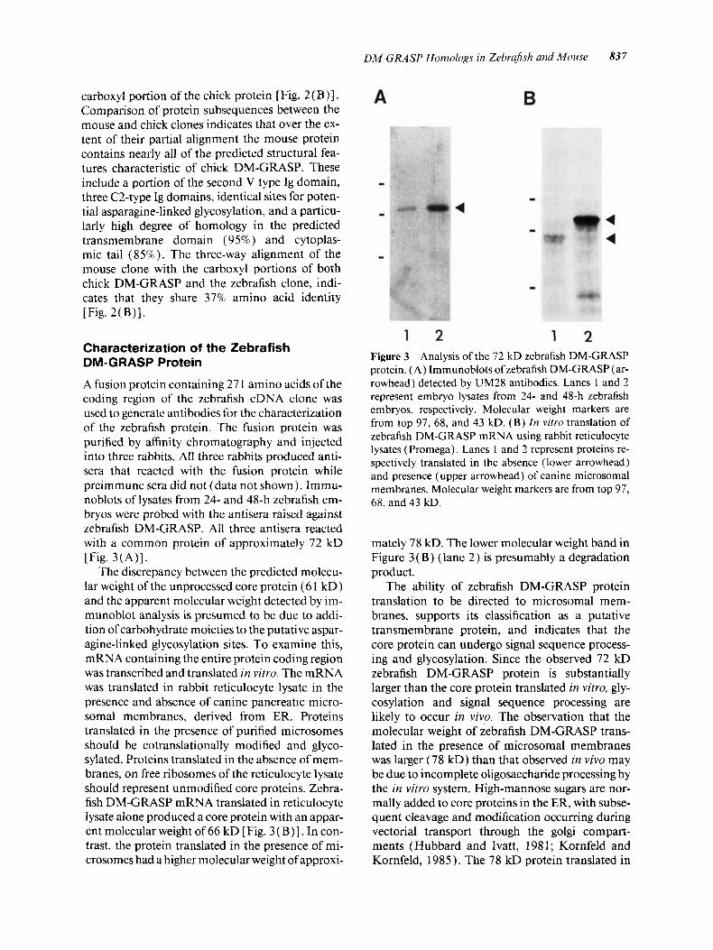
A fusion protein containing 27 1 amino acids of the coding region of the zebrafish cDNA clone was used to generate antibodies for the characterization of the zebrafish protein. The fusion protein was purified by affinity chromatography and injected into three rabbits. All three rabbits produced anti- sera that reacted with the fusion protein while preimmune sera did not (data not shown). Immu- noblots of lysates from 24- and 48-h zebrafish em- bryos were probed with the antisera raised against zebrafish DM-GRASP. All three antisera reacted with a common protein of approximately 72 kD [Fig. 3(A)].
The discrepancy between the predicted molecu- lar weight of the unprocessed core protein (6 1 kD) and the apparent molecular weight detected by im- munoblot analysis is presumed to be due to addi- tion of carbohydrate moieties to the putative aspar- agine-linked glycosylation sites. To examine this, mRNA containing the entire protein coding region was transcribed and translated in vitro. The mRNA was translated in rabbit reticulocyte lysate in the presence and absence of canine pancreatic micro- soma1 membranes, derived from ER. Proteins translated in the presence of purified microsomes should be cotranslationally modified and glyco- sylated. Proteins translated in the absence of mem- branes, on free ribosomes of the reticulocyte lysate should represent unmodified core proteins. Zebra- fish DM-GRASP mRNA translated in reticulocyte lysate alone produced a core protein with an appar- ent molecular weight of 66 kD [Fig. 3 (B)] . In con- trast, the protein translated in the presence of mi- crosomes had a higher molecular weight of approxi-
A B
1 2 1 2 Figure 3 Analysis of the 72 kD zebrafish DM-GRASP protein. (A) Irnmunoblots of zebrafish DM-GRASP (ar- rowhead) detected by UM28 antibodies. Lanes 1 and 2 represent embryo lysates from 24- and 48-h zebrafish embryos, respectively. Molecular weight markers are from top 97, 68, and 43 kD. (B) In vitro translation of zebrafish DM-GRASP mRNA using rabbit reticulocyte lysates ( Promega). Lanes 1 and 2 represent proteins re- spectively translated in the absence (lower arrowhead) and presence (upper arrowhead) of canine microsomal membranes. Molecular weight markers are from top 97, 68. and 43 kD.
mately 78 kD. The lower molecular weight band in Figure 3 (B) (lane 2) is presumably a degradation product.
The ability of zebrafish DM-GRASP protein translation to be directed to microsomal mem- branes, supports its classification as a putative transmembrane protein, and indicates that the core protein can undergo signal sequence process- ing and glycosylation. Since the observed 72 kD zebrafish DM-GRASP protein is substantially larger than the core protein translated in vitro, gly- cosylation and signal sequence processing are likely to occur in vivo. The observation that the molecular weight of zebrafish DM-GRASP trans- lated in the presence of microsomal membranes was larger (78 kD) than that observed in vivo may be due to incomplete oligosaccharide processing by the in vitro system. High-mannose sugars are nor- mally added to core proteins in the ER, with subse- quent cleavage and modification occumng during vectorial transport through the golgi compart- ments (Hubbard and Ivatt, 1981; Kornfeld and Kornfeld, 1985). The 78 kD protein translated in
838 Kunki et ul.
the presence of ER microsomes may represent a high-mannose precursor form of the mature 72 kD zebrafish DM-GRASP glycoprotein expressed in vivo.
Developmental Expression Pattern of Zebrafish DM-GRASP
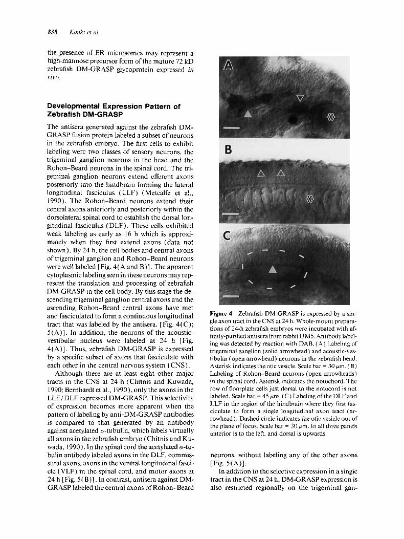
The antisera generated against the zebrafish DM- GRASP fusion protein labeled a subset of neurons in the zebrafish embryo. The first cells to exhibit labeling were two classes of sensory neurons, the trigeminal ganglion neurons in the head and the Rohon-Beard neurons in the spinal cord. The tri- geminal ganglion neurons extend efferent axons posteriorly into the hindbrain forming the lateral longitudinal fasciculus (LLF) (Metcalfe et al., 1990). The Rohon-Beard neurons extend their central axons anteriorly and posteriorly within the dorsolateral spinal cord to establish the dorsal lon- gitudinal fasciculus (DLF). These cells exhibited weak labeling as early as 16 h which is approxi- mately when they first extend axons (data not shown). By 24 h, the cell bodies and central axons of trigeminal ganglion and Rohon-Beard neurons were well labeled [Fig. 4( A and B)] . The apparent cytoplasmic labeling seen in these neurons may rep- resent the translation and processing of zebrafish DM-GRASP in the cell body. By this stage the de- scending trigeminal ganglion central axons and the ascending Rohon-Beard central axons have met and fasciculated to form a continuous longitudinal tract that was labeled by the antisera. [Fig. 4(C) ; 5(A)]. In addition, the neurons of the acoustic- vestibular nucleus were labeled at 24 h [Fig. 4( A)]. Thus, zebrafish DM-GRASP is expressed by a specific subset of axons that fasciculate with each other in the central nervous system (CNS).
Although there are at least eight other major tracts in the CNS at 24 h (Chitnis and Kuwada, 1990; Bernhardt et al., 1990 1, only the axons in the LLF/DLF expressed DM-GRASP. This selectivity of expression becomes more apparent when the pattern of labeling by anti-DM-GRASP antibodies is compared to that generated by an antibody against acetylated 0-tubulin, which labels virtually all axons in the zebrafish embryo (Chitnis and Ku- wada, 1990). In the spinal cord the acetylated a-tu- bulin antibody labeled axons in the DLF, commis- sural axons, axons in the ventral longitudinal fasci- cle (VLF) in the spinal cord, and motor axons at 24 h [Fig. 5 ( B ) 1. In contrast, antisera against DM- GRASP labeled the central axons of Rohon-Beard
Figure 4 Zebrafish DM-GRASP is expressed by a sin- gle axon tract in the CNS at 24 h. Whole-mount prepara- tions of 24-h zebrafish embryos were incubated with af- finity-purified antisera from rabbit UM5. Antibody label- ing was detected by reaction with DAB. (A) Labeling of trigeminal ganglion (solid arrowhead) and acoustic-ves- tibular (open arrowhead) neurons in the zebrafish head. Asterisk indicates the otic vesicle. Scale bar = 30 pm. ( B ) Labeling of Rohon-Beard neurons (open arrowheads) in the spinal cord. Asterisk indicates the notochord. The row of floorplate cells just dorsal to the notocord is not labeled. Scale bar = 45 pm. (C) Labeling of the DLF and LLF in the region of the hindbrain where they first fas- ciculate to form a single longitudinal axon tract (ar- rowhead). Dashed circle indicates the otic vesicle out of the plane of focus. Scale bar = 30 pm. In all three panels anterior is to the left, and dorsal is upwards.
neurons, without labeling any of the other axons [Fig. 5(A) ] .
In addition to the selective expression in a single tract in the CNS at 24 h, DM-GRASP expression is also restricted regionally on the trigeminal gan-
DM-GRASP Homologs in Zebrafish and Mouse 839
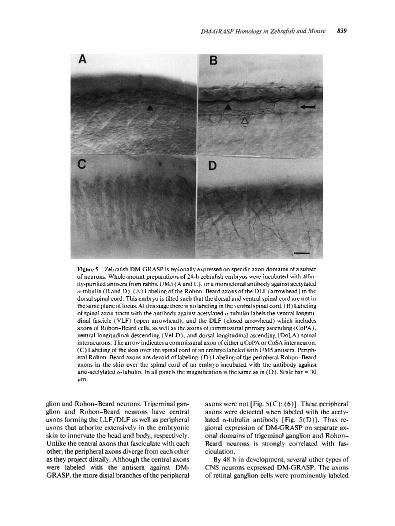
Figure 5 Zebrafish DM-GRASP is regionally expressed on specific axon domains of a subset of neurons. Whole-mount preparations of 24-h zebrafish embryos were incubated with affin- ity-purified antisera from rabbit UM5 ( A and C), or a monoclonal antibody against acetylated a-tubulin (B and D ) . (A) Labeling of the Rohon-Beard axons of the DLF (arrowhead) in the dorsal spinal cord. This embryo is tilted such that the dorsal and ventral spinal cord are not in the same plane of focus. At this stage there is no labeling in the ventral spinal cord. ( B ) Labeling of spinal axon tracts with the antibody against acetylated a-tubulin labels the ventral longitu- dinal fascicle (VLF) (open arrowhead), and the DLF (closed arrowhead) which includes axons of Rohon-Beard cells, as well as the axons of commissural primary ascending (CoPA), ventral longitudinal descending ( VeLD), and dorsal longitudinal ascending (DoLA) spinal interneurons. The arrow indicates a commissural axon of either a CoPA or CoSA interneuron. ( C ) Labeling of the skin over the spinal cord of an embryo labeled with UM5 antisera. Periph- eral Rohon-Beard axons are devoid of labeling. (D) Labeling of the peripheral Rohon-Beard axons in the skin over the spinal cord of an embryo incubated with the antibody against anti-acetylated a-tubulin. In all panels the magnification is the same as in (D). Scale bar = 30 Pm.
glion and Rohon-Beard neurons. Trigeminal gan- glion and Rohon-Beard neurons have central axons forming the LLF/DLF as well as peripheral axons that arborize extensively in the embryonic skin to innervate the head and body, respectively. Unlike the central axons that fasciculate with each other, the peripheral axons diverge from each other as they project distally. Although the central axons were labeled with the antisera against DM- GRASP, the more distal branches of the peripheral
axons were not [Fig. 5 ( C); ( 6 ) ] . These peripheral axons were detected when labeled with the acety- lated a-tubulin antibody [Fig. 5(D)]. Thus re- gional expression of DM-GRASP on separate ax- onal domains of trigeminal ganglion and Rohon- Beard neurons is strongly correlated with fas- ciculation.
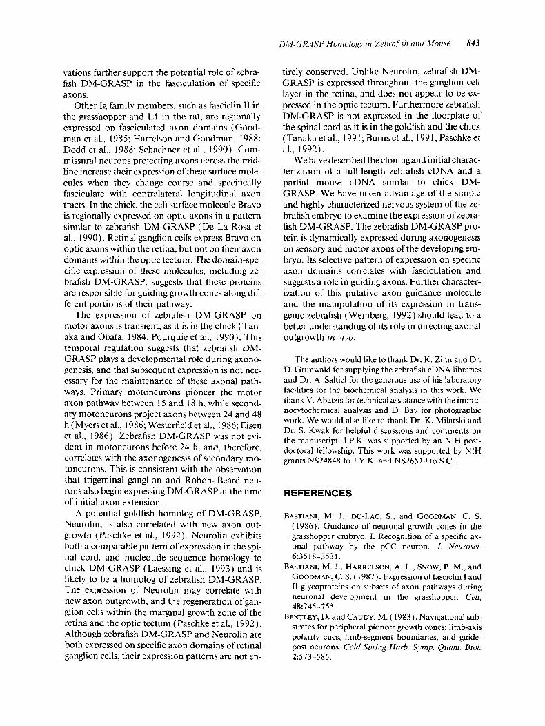
By 48 h in development, several other types of CNS neurons expressed DM-GRASP. The axons of retinal ganglion cells were prominently labeled
840 Kunki l>t ul.
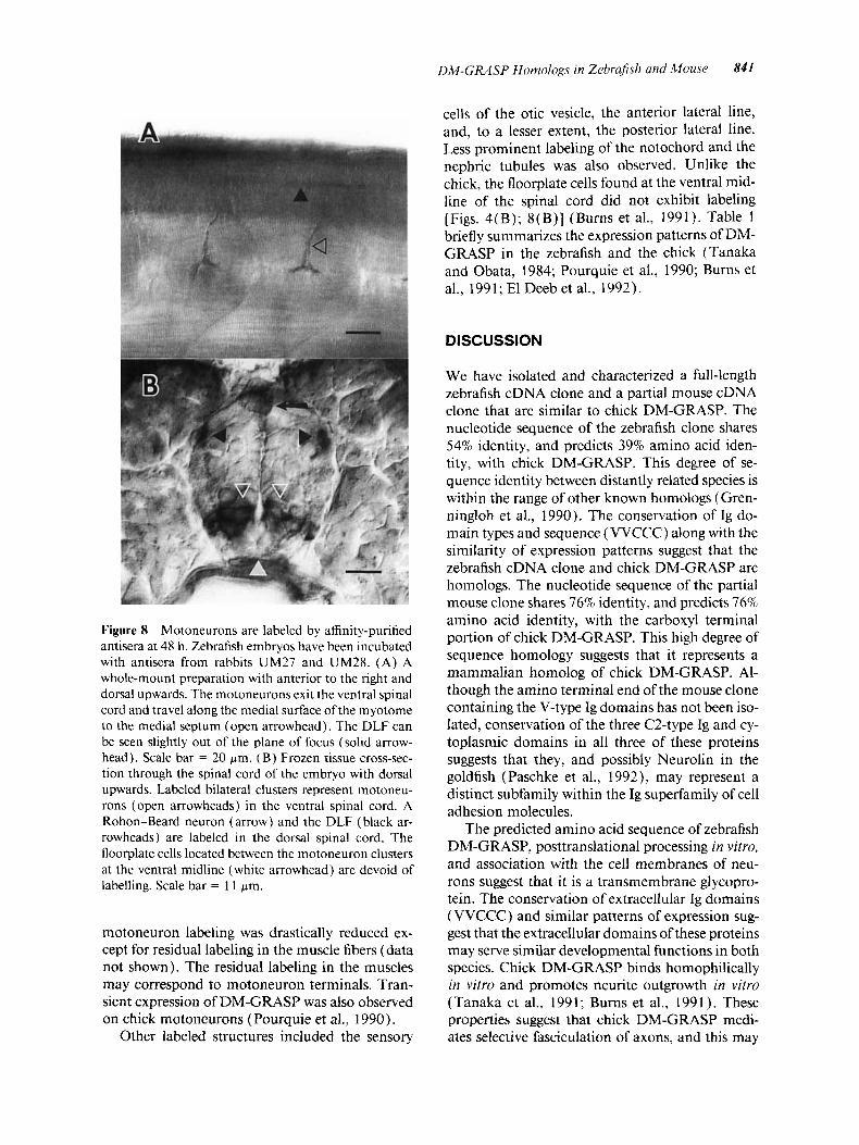
Although the axons and cell bodies of motoneu- rons in the spinal cord did not express zebrafish DM-GRASP at 24 h, labeling was detected by 48 h (Fig. 8 ) . The expression of zebrafish DM-GRASP on motoneuron axons was transient and by 5 days
Figure 6 A diagram illustrating the relative positions and morphology of trigeminal ganglion neurons (open arrowhead) and Rohon-Beard neurons (closed arrow- head). Trigeminal ganglion neurons project central axons posteriorly forming the LLF while Rohon-Beard neurons project central axons bidirectionally forming the DLF. These central projections express Zebrafish DM-GRASP and fasciculate to form a single longitu- dinal axon tract. The trigeminal ganglion and Rohon- Beard neurons also project peripheral axons which ar- borize extensively in the skin. These peripheral axons do not express DM-GRASP.
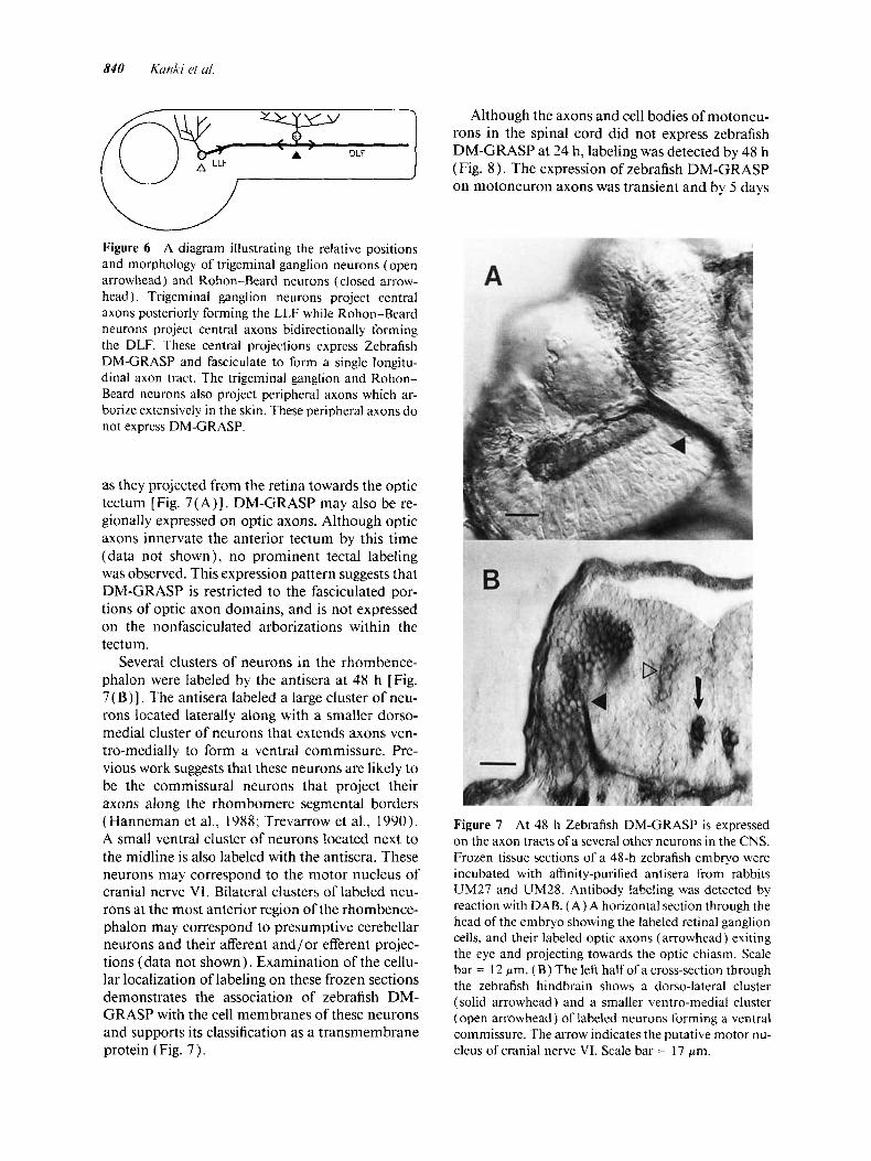
as they projected from the retina towards the optic tectum [Fig. 7( A)]. DM-GRASP may also be re- gionally expressed on optic axons. Although optic axons innervate the anterior tectum by this time (data not shown), no prominent tectal labeling was observed. This expression pattern suggests that DM-GRASP is restricted to the fasciculated por- tions of optic axon domains, and is not expressed on the nonfasciculated arborizations within the tectum.
Several clusters of neurons in the rhombence- phalon were labeled by the antisera at 48 h [Fig. 7( B)] . The antisera labeled a large cluster of neu- rons located laterally along with a smaller dorso- medial cluster of neurons that extends axons ven- tro-medially to form a ventral commissure. Pre- vious work suggests that these neurons are likely to be the commissural neurons that project their axons along the rhombomere segmental borders (Hanneman et al., 1988; Trevarrow et al., 1990). A small ventral cluster of neurons located next to the midline is also labeled with the antisera. These neurons may correspond to the motor nucleus of cranial nerve VI. Bilateral clusters of labeled neu- rons at the most anterior region of the rhombence- phalon may correspond to presumptive cerebellar neurons and their afferent and/or efferent projec- tions (data not shown), Examination of the cellu- lar localization of labeling on these frozen sections demonstrates the association of zebrafish DM- GRASP with the cell membranes of these neurons and supports its classification as a transmembrane protein (Fig. 7 ) .
Figure 7 At 48 h Zebrafish DM-GRASP is expressed on the axon tracts of a several other neurons in the CNS. Frozen tissue sections of a 48-h zebrafish embryo were incubated with affinity-purified antisera from rabbits UM27 and UM28. Antibody labeling was detected by reaction with DAB. (A) A horizontal section through the head of the embryo showing the labeled retinal ganglion cells, and their labeled optic axons (arrowhead) exiting the eye and projecting towards the optic chiasm. Scale bar = 12 pm. ( B ) The left half of a cross-section through the zebrafish hindbrain shows a dorso-lateral cluster (solid arrowhead) and a smaller ventro-medial cluster (open arrowhead) of labeled neurons forming a ventral commissure. The arrow indicates the putative motor nu- cleus of cranial nerve VI. Scale bar = 17 pm.
DM-GRASP Homologs in Zehrajsh und Mouse 841
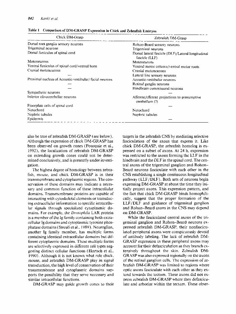
cells of the otic vesicle, the anterior lateral line, and, to a lesser extent, the posterior lateral line. Less prominent labeling of the notochord and the nephric tubules was also observed. Unlike the chick, the floorplate cells found at the ventral mid- line of the spinal cord did not exhibit labeling [Figs. 4(B); 8(B)] (Burns et al., 1991). Table 1 briefly summarizes the expression patterns of DM- GRASP in the zebrafish and the chick (Tanaka and Obata, 1984; Pourquie et al., 1990; Burns et al., 1991; El Deeb et al., 1992).
DISCUSSION
We have isolated and characterized a full-length zebrafish cDNA clone and a partial mouse cDNA clone that are similar to chick DM-GRASP. The nucleotide sequence of the zebrafish clone shares 54% identity, and predicts 39% amino acid iden- tity, with chick DM-GRASP. This degree of se- quence identity between distantly related species is within the range of other known homologs (Gren- ningloh et al., 1990). The conservation of Ig do- main types and sequence (VVCCC) along with the similarity of expression patterns suggest that the zebrafish cDNA clone and chick DM-GRASP are homologs. The nucleotide sequence of the partial mouse clone shares 76% identity, and predicts 76% amino acid identity, with the carboxyl terminal portion of chick DM-GRASP. This high degree of sequence homology suggests that it represents a mammalian homolog of chick DM-GRASP. Al- though the amino terminal end Of the containing the V-tYPe Ig domains has not been iS0-
Figure 8 Motoneurons are labeled by affinity-purified antisera at 48 h. Zebrafish embryos have been incubated with antisera from rabbits UM27 and UM28. ( A ) A whole-mount preparation with anterior to the right and dorsal upwards. The motoneurons exit the ventral spinal cord and travel along the medial surface of the myotome to the medial septum (open arrowhead). The DLF can be seen slightly out of the plane of focus (solid arrow- head). Scale bar = 20 pm. (B) Frozen tissue cross-sec- tion through the spinal cord of the embryo with dorsal upwards. Labeled bilateral clusters represent motoneu- rons (open arrowheads) in the ventral spinal cord. A Rohon-Beard neuron (arrow) and the DLF ( black ar- rowheads) are labeled in the dorsal spinal cord. The floorplate cells located between the motoneuron clusters at the ventral midline (white arrowhead) are devoid of labelling. Scale bar = 1 1 pm.
motoneuron labeling was drastically reduced ex- cept for residual labeling in the muscle fibers (data not shown). The residual labeling in the muscles may correspond to motoneuron terminals. Tran- sient expression of DM-GRASP was also observed on chick motoneurons (Pourquie et al., 1990).
Other labeled structures included the sensory
lated, conservation of the three C2-type Ig and cy- toplasmic domains in all three of these proteins suggests that they, and possibly Neurolin in the goldfish (Paschke et al., 1992), may represent a distinct subfamily within the Ig superfamily of cell adhesion molecules.
The predicted amino acid sequence of zebrafish DM-GRASP, posttranslational processing in vitro, and association with the cell membranes of neu- rons suggest that it is a transmembrane glycopro- tein. The conservation of extracellular Ig domains (VVCCC) and similar patterns of expression sug- gest that the extracellular domains of these proteins may serve similar developmental functions in both species. Chick DM-GRASP binds homophilically in vitro and promotes neurite outgrowth in vitro (Tanaka et al., 1991; Burns et al., 1991). These properties suggest that chick DM-GRASP medi- ates selective fasciculation of axons, and this may
842 Kanki et a/.
Table 1 Comparison of DM-GRASP Expression in Chick and Zebrafish Embryos
Chick DM-Grasp Zebrafish DM-Grasp Dorsal root ganglia sensory neurons Trigeminal neurons Trigeminal neurons Dorsal funiculus of spinal cord
Motoneurons Motoneurons Ventral funiculus of spinal cord/ventral horn Cranial motoneurons Cranial motoneurons
Proximal nucleus of Acoustic-vestibular/ facial neurons
Rohon-Beard sensory neurons
Dorsal lateral fascicle (DLF)/Lateral longitudinal fascicle (LLF)
Ventral motor column/ventral motor roots
- Lateral line sensory neurons Acoustic-vestibular neurons
- Retinal ganglia neurons -
Sympathetic neurons Inferior olivocerebellar neurons
Floorplate cells of spinal cord Notochord Nephric tubules Epidermis
Hindbrain commissural neurons
Afferent/efferent projections to presumptive -
cerebellum (?) -
Notochord Nephric tubules
-
also be true of zebrafish DM-GRASP (see below). Although the expression of chick DM-GRASP has been observed on growth cones (Pourquie et al., 1992), the localization of zebrafish DM-GRASP on extending growth cones could not be deter- mined conclusively, and is presently under investi- gation.
The highest degree of homology between zebra- fish, mouse, and chick DM-GRASP is in their transmembrane and cytoplasmic regions. The con- servation of these domains may indicate a neces- sary and common function of these intracellular domains. Transmembrane proteins are capable of interacting with cytoskeletal elements or transduc- ing extracellular information to specific intracellu- lar signals through specialized cytoplasmic do- mains. For example, the Drumphila LAR protein is a member of the Ig family containing both extra- cellular Ig domains and cytoplasmic tyrosine phos- phatase domains (Streuli et al., 1989). Neuroglian, another Ig family member, has multiple forms containing identical extracellular domains but dif- ferent cytoplasmic domains. These multiple forms are selectively expressed in different cell types sug- gesting distinct cellular functions (Hortsch et al., 1990). Although it is not known what role chick, mouse, and zebrafish DM-GRASP play in signal transduction, the high level of conservation of their transmembrane and cytoplasmic domains sup- ports the possibility that they serve necessary and similar intracellular functions.
DM-GRASP may guide growth cones to their
targets in the zebrafish CNS by mediating selective fasciculation of the axons that express it. Like chick DM-GRASP, the zebrafish homolog is ex- pressed on a subset of axons. At 24 h, expression was restricted to the axons forming the LLF in the hindbrain and the DLF in the spinal cord. The cen- tral axons of the trigeminal ganglion and Rohon- Beard neurons fasciculate with each other in the CNS establishing a single continuous longitudinal pathway (LLF/DLF) . Both sets of neurons begin expressing DM-GRASP at about the time they ini- tially project axons. This expression pattern, and the fact that chick DM-GRASP binds homophili- cally, suggest that the proper formation of the LLF/ DLF and guidance of trigeminal ganglion and Rohon-Beard axons in the CNS may depend on DM-GRASP.
While the fasciculated central axons of the tri- geminal ganglion and Rohon-Beard neurons ex- pressed zebrafish DM-GRASP, their nonfascicu- lated peripheral axons were conspicuously devoid of antibody labeling. The lack of zebrafish DM- GRASP expression in these peripheral axons may account for their defasciculation as they branch ex- tensively throughout the skin. Zebrafish DM- GRASP was also expressed regionally on the axons of the retinal ganglion cells. The expression of ze- brafish DM-GRASP was limited to regions where optic axons fasciculate with each other as they ex- tend towards the tectum. These axons did not ex- press zebrafish DM-GRASP where they defascicu- late and arborize within the tectum. These obser-
DM-GRASP Homologs in Zebrafish and Mouse 843
vations further support the potential role of zebra- fish DM-GRASP in the fasciculation of specific axons.
Other Ig family members, such as fasciclin I1 in the grasshopper and Ll in the rat, are regionally expressed on fasciculated axon domains (Good- man et al., 1985; Harrelson and Goodman, 1988; Dodd et al., 1988; Schachner et al., 1990). Com- missural neurons projecting axons across the mid- line increase their expression of these surface mole- cules when they change course and specifically fasciculate with contralateral longitudinal axon tracts. In the chick, the cell surface molecule Bravo is regionally expressed on optic axons in a pattern similar to zebrafish DM-GRASP (De La Rosa et al., 1990). Retinal ganglion cells express Bravo on optic axons within the retina, but not on their axon domains within the optic tectum. The domain-spe- cific expression of these molecules, including ze- brafish DM-GRASP, suggests that these proteins are responsible for guiding growth cones along dif- ferent portions of their pathway.
The expression of zebrafish DM-GRASP on motor axons is transient, as it is in the chick (Tan- aka and Obata, 1984; Pourquie et al., 1990). This temporal regulation suggests that zebrafish DM- GRASP plays a developmental role during axono- genesis, and that subsequent expression is not nec- essary for the maintenance of these axonal path- ways. Primary motoneurons pioneer the motor axon pathway between 15 and 18 h, while second- ary motoneurons project axons between 24 and 48 h (Myers et al., 1986; Westerfield et al., 1986; Eisen et al., 1986). Zebrafish DM-GRASP was not evi- dent in motoneurons before 24 h, and, therefore, correlates with the axonogenesis of secondary mo- toneurons. This is consistent with the observation that trigeminal ganglion and Rohon-Beard neu- rons also begin expressing DM-GRASP at the time of initial axon extension.
A potential goldfish homolog of DM-GRASP, Neurolin, is also correlated with new axon out- growth (Paschke et al., 1992). Neurolin exhibits both a comparable pattern of expression in the spi- nal cord, and nucleotide sequence homology to chick DM-GRASP (Laessing et al., 1993) and is likely to be a homolog of zebrafish DM-GRASP. The expression of Neurolin may correlate with new axon outgrowth, and the regeneration of gan- glion cells within the marginal growth zone of the retina and the optic tectum (Paschke et al., 1992). Although zebrafish DM-GRASP and Neurolin are both expressed on specific axon domains of retinal ganglion cells, their expression patterns are not en-
tirely conserved. Unlike Neurolin, zebrafish DM- GRASP is expressed throughout the ganglion cell layer in the retina, and does not appear to be ex- pressed in the optic tectum. Furthermore zebrafish DM-GRASP is not expressed in the floorplate of the spinal cord as it is in the goldfish and the chick (Tanakaet al., 1991; Burnset al., 1991; Paschkeet al., 1992).
We have described the cloningand initial charac- terization of a full-length zebrafish cDNA and a partial mouse cDNA similar to chick DM- GRASP. We have taken advantage of the simple and highly characterized nervous system of the ze- brafish embryo to examine the expression of zebra- fish DM-GRASP. The zebrafish DM-GRASP pro- tein is dynamically expressed during axonogenesis on sensory and motor axons of the developing em- bryo. Its selective pattern of expression on specific axon domains correlates with fasciculation and suggests a role in guiding axons. Further character- ization of this putative axon guidance molecule and the manipulation of its expression in trans- genic zebrafish (Weinberg, 1992) should lead to a better understanding of its role in directing axonal outgrowth in vivo.
The authors would like to thank Dr. K. Zinn and Dr. D. Grunwald for supplying the zebrafish cDNA libraries and Dr. A. Saltiel for the generous use of his laboratory facilities for the biochemical analysis in this work. We thank V. Abatzis for technical assistance with the immu- nocytochemical analysis and D. Bay for photographic work. We would also like to thank Dr. K. Milarski and Dr. S. Kwak for helpful discussions and comments on the manuscript. J.P.K. was supported by an NIH post- doctoral fellowship. This work was supported by NIH grants NS24848 to J.Y.K. and NS26519 to S.C.
REFERENCES
BASTIANI, M. J., DU-LAC, S., and GOODMAN, C. S. (1986). Guidance of neuronal growth cones in the grasshopper embryo. I. Recognition of a specific ax- onal pathway by the pCC neuron. J . Neurosci.
BASTIANI, M. J., HARRELSON, A. L., SNOW, P. M., and GOODMAN, C. S. ( 1987). Expression of fasciclin I and I1 glycoproteins on subsets of axon pathways during neuronal development in the grasshopper. Cell,
BENTLEY, D. and CAUDY, M. ( 1983). Navigational sub- strates for peripheral pioneer growth cones: limb-axis polarity cues, limb-segment boundaries, and guide- post neurons. Cold Spring Hurb. Symp. Quant. Biol.
6~3518-3531.
48~745-755.
2:513-585.
844 Kanki et al.
BERNHARDT, R. R., CHITNIS, A. B., LINDAMER, L., and KUWADA, J. Y. ( 1990). Identification of spinal neu- rons in the embryonic and larval zebrafish. J. Comp. Neurol. 302:603-6 16.
BURNS, F. R., VON-KANNEN, S., GUY, L., RAPER, J. A,, KAMHOLZ, J., and CHANG, S. ( I99 1 ) . DM-GRASP, a novel immunoglobulin superfamily axonal surface protein that supports neurite extension. Neuron
CHITNIS, A. B. and KUWADA, J. Y. ( 1990). Axonogene- sis in the brain of zebrafish embryos. J. Neurosci.
DE LA ROSA, E. J., KAWEM, J. F., ROMAN, J . M., STIER- HOF, Y . D., DREYER, W. J. , and SCHWARZ, U. ( 1990). Topologically restricted appearance in the developing chick retinotectal system of Bravo, a neural surface protein: experimental modulation by environmental cues. J. Cell Biol. 111:3087-3096.
DODD, J. and JESSELL, T. M. (1988). Axon guidance and the patterning of neuronal projections in verte- brates. Science 242~692-699.
DODD, J . , MORTON, S. B., KARAGOGEOS, D., YAMA- MOTO, M., and JESSELL, T. M. (1988). Spatial regula- tion of axonal glycoprotein expression on subsets of embryonic spinal neurons. Neuron 1:105-116.
EISEN, J. S. ( 199 1 ). Developmental neurobiology of the zebrafish. J. Neurosci. 11:3 1 1-3 17.
EISEN, J . S., MYERS, P. Z., and WESTERFIELD, M. (1986). Pathway selection by growth cones of identi- fied motoneurones in live zebra fish embryos. Nature
EL DEEB, S., THOMPSON, S. C., and COVAULT, J. ( 1992). Characterization of a cell surface adhesion molecule expressed by a subset of developing chick neurons. Dev. Bid . 149:2 13-227.
GOODMAN, C. S., BASTIANI, M. J. , DOE, C. Q., and Du- LAC, S. ( 1985). Growth cone guidance and cell recog- nition in insect embryos. In: Developmenid Biology: A Comprehensive Synthesis, vol. 3. M. S. Steinberg, Ed. Plenum, New York, pp. 283-300.
GRENNINGLOH, G. , BIEBER, A. J . , REHM, E. J., SNOW, P. M.. TRAQUINA, Z. R., HORTSCH, M., PATEL, N. H., and GOODMAN, C. S. ( 1990). Molecular genet- ics of neuronal recognition in Drosophila: evolution and function of immunoglobulin superfamily cell ad- hesion molecules. Cold Spring Harb. Symp. Quani. Bid. 55327-340.
HAKES, D. J . and DIXON, J. E. ( 1992). New vectors for high level expression of recombinant proteins in bacte- ria. Anal. Biochem. 202:293-298.
HANNEMAN, E., TREVARROW, B., METCALFE, W. K., KIMMEL, C. B., and WESTERFIELD, M. (1988). Seg- mental pattern of development of the hindbrain and spinal cord of the zebrafish embryo. Development 103:49-58.
HARLOW, E. and LANE, D. ( 1988). Antibodies: A Labo- ratory Manual. Cold Spring Harbor Laboratory: Cold Spring Harbor Laboratory, New York.
7~209-220.
lO(6): 1892-1905.
320~269-27 1.
HARRELSON, A. L. and GOODMAN, C. S. (1988). Growth cone guidance in insects: fasciclin I1 is a member of the immunoglobulin superfamily. Science
HARRIS, W. A., HOLT, C. E., and BONHOEFFER, F. ( 1987). Retinal axons with and without their somata, growing to and arborizing in the tectum of Xenopus embryos: a time-lapse video study of single fibres in vivo. Development 101:123-133.
HORTSCH, M., BIEBER, A. J., PATEL, N. H., andGooD- MAN, c. s. (1990). Differential splicing generates a nervous system-specific form of Drosophila neuro- glian. Neuron 4:697-709.
HUBBARD, S. C. and IVATT, R. J . ( 198 1 ). Synthesis and processing of asparagine-linked oligosaccharides. Ann. Rev. Biochem. 50555-583.
KAPFHAMMER, J . P. and RAPER, J. A. ( 1987). Collapse of growth cone structure on contact with specific neu- rites in culture. J. Neurosci. 7:20 1-2 12.
KIMMEL, C. B., POWELL, S. L., and METCALFE, W. K. (1982). Brain neurons which project to the spinal cord in young larvae of the zebrafish. J. Comp. Neurol.
KORNFELD, R. and KORNFELD, S. ( 1985). Assembly of asparagine-lhked oligosaccharides. Ann. Rev. Bio- chem. 54:63 1-664.
KUWADA. J. Y. (1986). Cell recognition by neuronal growth cones in a simple vertebrate embryo. Science
KUWADA, J. Y . and BERNHARDT, R. R. ( 1990). Axonal outgrowth by identified neurons in the spinal cord of zebrafish embryos. Exp. Ncurol. 109:29-34.
LAESSING, U., GIORDANO, S., LOTTSPEICH, F., and STUERMER, C. A. 0. ( 1993). Molecular cloning of Neurolin and its expression in goldfish and embryonic zebrafish CNS. SOC. Neurosci. Abstr 19:1090.
LEHMANN, J. M., RIETHMULLER, G. , and JOHNSON, J . P. ( 1989). MUC18, a marker of tumor progression in human melanoma, shows sequence similarity to the neural cell adhesion molecules of the immunoglobu- lin superfamily. Proc. Nail Acad. Sci. U.S.A.
METCALFE, W. K., MYERS, P. Z., TREVARROW, B., BASS, M. B., and KIMMEL, C. B. ( 1990). Primary neu- rons that express the L2 /HNK- 1 carbohydrate during early development in the zebrafish. Developmenf
MYERS, P. Z. ( 1985). Spinal motoneurons of the larval zebrafish. J . Comp. Neurol. 236555-561.
MYERS, P. Z., EISEN, J. S., and WESTERFIELD, M. ( 1986). Development and axonal outgrowth ofidenti- fied motoneurons in the zebrafish. J. Neurosci. 6:2278-2289.
PASCHKE, K. A., LOTTSPEICH, F., and STUERMER, C. A. ( 1992). Neurolin, a cell surface glycoprotein on grow- ing retinal axons in the goldfish visual system, is reex- pressed during retinal axonal regeneration. J. Cell Biol. 1172363-875.
PEARSON, W. R. and LIPMAN, D. J. (1988). Improved
242~700-707.
205~112-127.
233~740-746.
86~989 1-9895.
110~49 1-504.
DM-GRASP Homologs in Zebrafish and Mouse 845
tools for biological sequence comparison. Proc. Natl. Acad. Sci. U.S.A. 85:2444-2448.
POURQUIE, O., COLTEY, M., THOMAS, J. L., and LE- DOUARIN, N. M. ( 1990). A widely distributed antigen developmentally regulated in the nervous system. De- velopment 109:743-752.
POURQUIE, O., CORBEL, C., LE-CAER, J. P., ROSSIER, J., and LE-DOUARIN, N. M. ( 1992). BEN, a surface gly- coprotein of the immunoglobulin superfamily, is ex- pressed in a variety of developing systems. Pmc. Natl. Acad. Sci. U.S.A. 89526 1-5265.
RAPER, J . A., BASTIANI, M., and GOODMAN, C. S. (1983). Pathfinding by neuronal growth cones in grasshopper embryos. 11. Selective fasciculation onto specific axonal pathways. J. Neurosci. 3:3 1 --4 I .
RATHJEN, F. G. and JESSELL, T. M. ( 199 1 ). Glycopro- teins that regulate the growth and guidance of verte- brate axons: domains and dynamics of the immuno- globulin/fibronectin type 111 subfamily. In: Sem. in Neurosci. Harcourt Brace Jovanovich, London, En- gland, pp. 297-307.
RATHJEN, F. G., WOLFF, J. M., FRANK, R., BON-
brane glycoproteins involved in neurite fasciculation. J. Cell Biol. 104:343-353.
REICHARDT, L. F. and TOMASELLI, K. J. ( 199 1 ) . Extra- cellular matrix molecules and their receptors: func- tions in neural development. Annu. Rev. Neurosci.
SAIKI, R. K., GELFAND, D. H., STOFFEL, S., SCHARF, S. J., HIGUCHI, R., HORN, G. T., MULLIS, K. B., and ERLICH, H. A. ( 1988). Primer-directed enzymatic amplification of DNA with a thermostable DNA poly- merase. Science 239:487-49 1.
SAMBROOK, J., FRITSCH, E. F., and MANIATIS, T. ( 1989). Molecular Cloning: A Laboratory Manual. Cold Spring Harbor Laboratory, Cold Spring Harbor, New York.
SANGER, F., NICKLEN, S., and COULSON, A. R. ( 1977). DNA sequencing with chain-terminating inhibitors. Proc. Natl. Acad. Sci. U.S.A. 74:5463-5467.
SCHACHNER, M., ANTONICEK, H., FAHRIG, T., FAISSNER, A., FISCHER, G., KUNEMUND, V., MAR- TINI, R., MEYER, A., PERSOHN, E., POLLEKBERG, E., PROBSTMEIER, R., SADOUL, K., SADOUL, R., SEIL- HEIMER, B., and THOR, G. ( 1990). Families of neural cell adhesion molecules. In: Morphoregulatory Mole-
HOEFFER, F., and RUTISHAUSER, U. (1987). Mem-
14~531-570.
cules. G. M. Edelman, B. A. Cunningham, and J. P. Thierry, Eds. John Wiley and Sons, New York, pp.
SMITH, D. B. and JOHNSON, K. S. (1988). Single-step purification of polypeptides expressed in Escherichia coli as fusions with glutathione S-transferase. Gene
STREULI, M., KRUEGER, N. X., TSAI, A. Y., and SAITO, H. ( 1989). A family of receptor-linked protein tyro- sine phosphatases in humans and Drosophila. Proc. Natl. Acad. Sci. U.S.A. 86:8698-8702.
TANAKA, H., MATSUI, T., AGATA, A., TOMURA, M., KUBOTA, I., MCFARLAND, K. C., KOHR, B., LEE, A., PHILLIPS, H. S., and SHELTON, D. L. ( 199 1 ). Molecu- lar cloning and expression of a novel adhesion mole- cule, SCl. Neuron 7535-545.
TANAKA, H. and OBATA, K. (1984). Developmental changes in unique cell surface antigens of chick em- bryo spinal motoneurons and ganglion cells. Dev. Biol. 106:26-37.
TESSIER-LAVIGNE, M. and PLACZEK, M. ( 1991 ). Target attraction: are developing axons guided by chemotro- pism? Trends Neurosci. 14:303-3 10.
TOSNEY, K. W. and LANDMESSER, L. T. ( 1985). Growth cone morphology and trajectory in the lumbosacral region of the chick embryo. J. Neurosci. 523452358.
TREVARROW, B., MARKS, D. L., and KIMMEL, C. B. ( 1990). Organization ofhindbrain segments in the ze- brafish embryo. Neuron 4:669-679.
VON HEIJNE, G. (1986). A new method for predicting signal sequence cleavage sites. Nucleic Acids Res.
WEINBERG, E. S. ( 1992). Analysis of early development in the zebrafish embryo. Results Probl. Cell Differ. 18:91-150.
WESTERFIELD, M. ( 1993). The Zebrajish Book. Univer- sity of Oregon Press, Eugene, OR.
WESTERFIELD, M., MCMURRAY, J. V., and EISEN, J. S. ( 1986). Identified motoneurons and their innervation of axial muscles in the zebrafish. J. Neurosci. 6:2267- 2277.
WILLIAMS, A. F. and BARCLAY, A. N. ( 1988). The im- munoglobulin superfamily-domains for cell surface recognition. Annu. Rev. Immunol. 6:38 1-405.
WILSON, S. W. and EASTER, S., JR. ( 199 1 ). Stereotyped pathway selection by growth cones of early epiphysial neurons in the embryonic zebrafish. Development
443-468.
67:3 1-40.
14:4683-4690.
112~723-746.