Embed Size (px)
Citation preview

Brit. J. Pharmacol. (1965), 24, 89-97.
THE MODE OF ACTION OF DRUGS BLOCKINGGANGLIONIC TRANSMISSION IN THE RAT
BY
D. G. SHAND
From the Department of Pharmacology, Medical College of St. Bartholomew's Hospital, London, E. C. 1
(Received May 18, 1964)
Quilliam & Shand (1964) found that the ganglion-blocking action of drugs in the rat canbe either highly selective, moderately selective or nonselective. As it appeared that the modeof block produced by drugs within the same selectivity group was not always precisely thesame, the present work was carried out to investigate these differences.
METHODSThe isolated superior cervical ganglion preparation of the rat was used in the manner described by
Quilliam & Shand (1964). The ganglion, together with the pre- and postganglionic nerves, was excised, theconnective tissue sheath removed and the preparation mounted horizontally in a bath of Krebs solutionat 300 C equilibrated with a mixture of 95% oxygen and 5% carbon dioxide. The preganglionic nerve couldbe stimulated through a pair of platinum electrodes at 6 shocks/min and the ganglionic action potentialrecorded from a pair of nonpolarizable silver-silver chloride-agar saline electrodes, one on the ganglionand the other on the cut end of the postganglionic nerve. The recording electrodes were connected to aDC amplifier. The potentials were displayed on a cathode-ray oscilloscope. To record, the preparationwas raised above the fluid level, giving moist chamber conditions, and the action potential in response tothe third stimulus was photographed.
In some experiments the depolarizing action of acetylcholine was measured using the moving fluid elec-trode technique of Pascoe (1956), by which the potential at any point along the ganglion can be measuredrelative to the cut end of the postganglionic nerve, using a space base in the oscilloscope.The effects of the following drugs were observed: acetylcholine chloride (Roche), hexamethonium
chloride (Geigy), tetraethylammonium bromide (B.D.H.), tubocurarine chloride and bretylium tosylate(Burroughs Wellcome), atropine sulphate (Samoore), amylobarbitone sodium (Boots), methylpentynol andmethylpentynol carbamate (British Schering), mephenesin (B.D.H.), paraldehyde (B.D.H.), benactyzinehydrochloride (Levanol, I.C.I.), procaine hydrochloride (Samoore), tetramethylammonium bromide(B.D.H.) and nicotine hydrogen tartrate (Samoore).
Unless otherwise stated the doses are given as the salts.
RESULTS
Effects on the form of the ganglionic action potentialThroughout drugs were added to the bath in a cumulative fashion, each dose being in
contact with the preparation for 5 min. This allowed rapid comparison and the exposuretime was considered sufficient for equilibration (Quilliam & Shand, 1964). While all thedrugs used blocked the spike component of the ganglionic action potential, increasing con-centrations produced different effects on the ganglionic afterpotentials. The depolarizing

D. G. SHAND
agents, nicotine, tetramethylammonium, carbachol and acetylcholine, had qualitatively thesame action as that described by Eccles (1956) on the isolated superior cervical ganglion ofthe rabbit and by Paton & Perry (1953) for acetylcholine, tetramethylammonium and nico-tine on the intact ganglion of the cat. The effect of nicotine was typical and is shown inFig. 1. As the amplifier used was DC, all upward shifts of the baseline indicated a depolar-ization of the ganglion relative to the cut end of the postganglionic nerve. With a low
C I xIO-6 2x1o-6 4x10⁻⁶
Nicotine
Fig. 1. Effects of nicotine on the ganglionic action potential. The potentials recorded were in response tosingle maximal stimuli to the preganglionic nerve. C indicates control response. Cumulative dosesof nicotine were added to the bath and records were taken after 5-min exposure. Drug concentrationsare shown above each response. An upward shift of the baseline relative to the lower trace (referencebeam) indicates a depolarization of the ganglion. Calibration, 25 cycles/sec with an amplitudeequivalent to 2.5 mV. The changes in the shape of the ganglionic action potential are typical not onlyof the action of nicotine but also of the other depolarizing drugs, tetramethylammonium, carbacholand acetylcholine. Initially (1 x 10⁶) the N wave was decreased and the P wave augmented, withlittle depression of the spike. Then (2 x 10⁻⁶) the P wave was shortened and both the N wave and thespike were further depressed. Finally (4 x 10⁻⁶) all the potentials became markedly reduced as fullblock of transmission was approached.
concentration (1 x 10⁻⁶), the negative afterpotential (N wave) was reduced in size and thepositive afterpotential (P wave) was augmented with little or no reduction in the height ofthe spike potential. With greater concentrations, the spike potential was decreased and theN wave was abolished. The P wave was first shortened in duration, returning more rapidlyto isopotential than with low doses, before being gradually abolished by high drugconcentrations (4 x 10⁻⁶).
Tubocurarine (Fig. 2a) reduced all the components of the ganglionic action potentialequally. At full block, a synaptic potential was obtained which was about 10 to 20% ofthe initial spike height or 30 to 50% of the initial size of the N wave. This is not illustrated,but the synaptic potential with tubocurarine was the same in form as that produced bytetraethylammonium in Fig. 2d. Depolarization was never observed.The action of tetraethylammonium and bretylium appeared to be midway between those
of tubocurarine and nicotine as regards their effects on the shape of the ganglionic potential(Fig. 2c and d). Initially, as with small doses of the depolarizing agents, the N wave wasdecreased and the P wave increased with only a small decrease in the spike potential.Increase in dosage decreased the P wave and finally produced a synaptic potential very likethat seen after tubocurarine. In keeping with their effects on the ganglionic potential, bothtetraethylammonium and bretylium caused a modest depolarization of the ganglion.Hexamethonium (Fig. 2b) had much the same action as tubocurarine, but with low dosestended to depress the N wave and cause slight depolarization, though not to the same extentas did tetraethylammonium. There was a graduated increase in the stimulatory effects, as
90

MODES OF GANGLIONIC BLOCK
C 4x 10⁻⁵ 8x 10⁻⁵ 1.6x 10⁻⁴
(a) Tubocurarine
C 2x 10⁻⁵ 4x 10⁻⁵ 8x 10⁻⁵
(b) Hexamethonium
C I x 10-4 2x10⁻⁴ 4x10-'
(c):Bretylium
C 4x10⁻⁵ 8x10⁻⁵ 3.2x 10⁻⁴
(d) Tetraethylammonium
Fig. 2. Effects of (a) tubocurarine, (b) hexamethonium, (c) bretylium and (d) tetraethylammonium on theganglionic action potential. Records as in Fig. 1. Cumulative doses of the drugs (concentrationsgiven above traces) added after obtaining control records (C). Calibrations, 25 cycles/sec and 3 mV.Hexamethonium, bretylium and tetraethylammonium produced some depolarization. Low doses(second column of traces) depressed the N wave and augmented the P wave with little reduction ofspike height as do low doses of the depolarizing drugs (Fig. 1). These stimulatory properties becomemore marked as one passes from tubocurarine, through hexamethonium to bretylium and tetraethyl-ammonium but were always less marked than those observed with the depolarizing drugs, Puresynaptic potentials are illustrated for tetraethylammonium and bretylium at complete block oftransmission, but not for tubocurarine or for hexamethonium, for which block was incomplete.
judged by the depolarization and effect on the shape of the action potential complex, asone passed from tubocurarine, through hexamethonium to bretylium and tetraethyl-ammonium. Amylobarbitone (Fig. 3c), methylpentynol and methylpentynol carbamatehad the same effect on the ganglionic potential as tubocurarine, but the time-course of thepotential was increased slightly, probably due to a slowing of conduction in the pre- andpostganglionic nerve fibres, since these drugs produce some neuronal depression in blockingconcentrations (Quilliam & Shand, 1964).
Increasing doses ofprocaine (Fig. 3a) abolished the spike potential and the after-positivity,leaving the N wave unaltered in amplitude, but increased in duration. The longer time-course of the N wave was probably due to the decrease in the P wave, for these two after-potentials appear to sum algebraically (Eccles, 1935; Paton & Perry, 1953). Furtherincrease in dose gradually diminished and finally abolished the N wave. Paraldehyde andmephenesin produced the same change in shape of the ganglionic action potential as didprocaine.
91

D. G. SHAND
C 2x 10⁻⁵ 4x 10⁻⁵ 8XI0-5
(a) Procaine
C 2X 10⁻⁵ 4x 10⁻⁵ 8x 10⁻⁵
(b) Atropine
C 8x10- 1.6x 10⁻⁴ 3.2x 10⁻⁴
(c) Amylobarbitone
C I X 10-5 2x10⁻⁵ 4x 10⁻⁵
(d) Benactyzine
Fig. 3. Effects of procaine and benactyzine on the ganglionic action potential. Records as for Fig. 1.Control records are indicated by C and cumulative drug concentrations are given above tracings.Calibrations, 25 cycles/sec and 3 mV. Increasing concentrations of procaine gradually reduced andabolished the spike and P wave, leaving the N wave. Benactyzine in low concentration reduced theN wave with little change in the spike or P wave but further increase in concentration reduced all thepotentials. Atropine reduced the spike, N wave and P wave leaving a synaptic potential, which hada longer time-course than that seen with curarization. The effects of amylobarbitone wereindistinguishable from those of tubocurarine (compare Fig. 1, first row).
With atropine (Fig. 3b), the spike was reduced and then abolished, leaving a synapticpotential similar to that after tubocurarine, but with a more prolonged time-course. Thisincreased duration may be related to the reduction of the P wave which occurred with lowconcentrations of atropine, resulting in an increased duration of the N wave.
Benactyzine (Fig. 3d) was unique in its effects, for in low dosage it depressed the N wavewith little or no effect on the P wave. Increase in dose gradually blocked all components ofthe ganglion action potential.
Relationship between depolarization and blockWith DC amplification, depolarization and block of transmission produced by graded
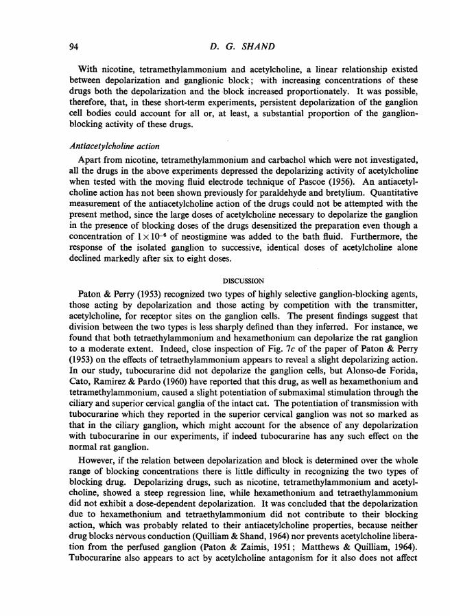
concentrations of drugs can be measured at the same time. Nicotine, tetramethyl-ammonium, acetylcholine, carbachol, tetraethylammonium, bretylium and hexamethoniumproduced depolarization of the ganglion but the magnitude of the ganglionic block was notalways directly related to the amount of depolarization produced. This is illustrated inFig. 4 in which depolarization, expressed as a percentage of the voltage of the control spike
92

MODES OF GANGLIONIC BLOCK
100
C._
0 800
8
C00 60
°-40C0C
*.20 0
0~~~~
4 0O-0~~~~~~~
0~~~~~~~~~
0 20 40 60 80 100Ganglion-block (%)
Fig. 4. Relation between depolarization and ganglion-block for nicotine, tetramethylammonium, acetyl-choline, tetraethylammonium, bretylium and hexamethonium. Action potentials were recorded as forFig. 1. One experiment was performed with each drug and the collective results have been plotted.Ordinates: depolarization (measured from shift of baseline) and expressed as a percentage of the heightof the control action potential so that different experiments may be compared. Abscissae: percentageblock of ganglionic transmission. Filled circles, nicotine; filled triangles, tetramethylammonium;filled squares, acetylcholine; empty circles, tetraethylammonium; empty triangles, hexamethonium;empty squares, bretylium; crosses, tubocurarine (as control). The solid line was drawn by eye as thebest fit for the solid symbols and the dotted line as the best fit for the open symbols.
Note: Two groups of drugs can be distinguished: (1) Nicotine, tetramethylammonium andacetylcholine gave values around the steeper solid regression line and depolarization was dose-dependent. (2) Tetraethylammonium, hexamethonium and bretylium gave values around thehorizontal dotted regression line and depolarization was not dose-dependent.
potential, has been plotted against the percentage block of ganglionic transmission. Pointsfor tubocurarine (crosses), which did not depolarize, have been included as a control. Thedrugs could be divided into two groups; on the one hand those shown as filled symbols inFig. 4, namely nicotine, tetramethylammonium, acetylcholine and carbachol (the last-named not being included in Fig. 4) and on the other those represented by empty symbols,tetraethylammonium, hexamethonium and bretylium. While tetraethylammonium, brety-lium and hexamethonium all caused some depolarization of the ganglion, it was invariablysmaller than that produced by the other drugs and, furthermore, increasing the dose of thedrug gave very little, if any, increase in the depolarization. The absolute magnitudes of theslight depolarizations produced by these three drugs tended to vary somewhat from experi-ment to experiment. Although the differences were small, usually the depolarizationproduced by hexamethonium was least, that by tetraethylammonium was greatest and thatby bretylium intermediate. It was concluded, therefore, that the depolarizing activity ofthese drugs provided no important contribution to their blocking action.
93
D. G. SHAND
With nicotine, tetramethylammonium and acetylcholine, a linear relationship existedbetween depolarization and ganglionic block; with increasing concentrations of thesedrugs both the depolarization and the block increased proportionately. It was possible,therefore, that, in these short-term experiments, persistent depolarization of the ganglioncell bodies could account for all or, at least, a substantial proportion of the ganglion-blocking activity of these drugs.
Antiacetylcholine actionApart from nicotine, tetramethylammonium and carbachol which were not investigated,
all the drugs in the above experiments depressed the depolarizing activity of acetylcholinewhen tested with the moving fluid electrode technique of Pascoe (1956). An antiacetyl-choline action has not been shown previously for paraldehyde and bretylium. Quantitativemeasurement of the antiacetylcholine action of the drugs could not be attempted with thepresent method, since the large doses of acetylcholine necessary to depolarize the ganglionin the presence of blocking doses of the drugs desensitized the preparation even though aconcentration of 1 x 10⁻⁶ of neostigmine was added to the bath fluid. Furthermore, theresponse of the isolated ganglion to successive, identical doses of acetylcholine alonedeclined markedly after six to eight doses.
DISCUSSION
Paton & Perry (1953) recognized two types of highly selective ganglion-blocking agents,those acting by depolarization and those acting by competition with the transmitter,acetylcholine, for receptor sites on the ganglion cells. The present findings suggest thatdivision between the two types is less sharply defined than they inferred. For instance, wefound that both tetraethylammonium and hexamethonium can depolarize the rat ganglionto a moderate extent. Indeed, close inspection of Fig. 7c of the paper of Paton & Perry(1953) on the effects of tetraethylammonium appears to reveal a slight depolarizing action.In our study, tubocurarine did not depolarize the ganglion cells, but Alonso-de Forida,Cato, Ramirez & Pardo (1960) have reported that this drug, as well as hexamethonium andtetramethylammonium, caused a slight potentiation of submaximal stimulation through theciliary and superior cervical ganglia of the intact cat. The potentiation of transmission withtubocurarine which they reported in the superior cervical ganglion was not so marked asthat in the ciliary ganglion, which might account for the absence of any depolarizationwith tubocurarine in our experiments, if indeed tubocurarine has any such effect on thenormal rat ganglion.
However, if the relation between depolarization and block is determined over the wholerange of blocking concentrations there is little difficulty in recognizing the two types ofblocking drug. Depolarizing drugs, such as nicotine, tetramethylammonium and acetyl-choline, showed a steep regression line, while hexamethonium and tetraethylammoniumdid not exhibit a dose-dependent depolarization. It was concluded that the depolarizationdue to hexamethonium and tetraethylammonium did not contribute to their blockingaction, which was probably related to their antiacetylcholine properties, because neitherdrug blocks nervous conduction (Quilliam & Shand, 1964) nor prevents acetylcholine libera-tion from the perfused ganglion (Paton & Zaimis, 1951; Matthews & Quilliam, 1964).Tubocurarine also appears to act by acetylcholine antagonism for it also does not affect
94

MODES OF GANGLIONIC BLOCK
nervous conduction (Quilliam & Shand, 1964) nor affect acetylcholine liberation (Brown &Feldberg, 1936). Whether such antiacetylcholine action is truly competitive in the bio-chemical sense has yet to be determined, but it would seem that the prevention of thetransmitter action by occupation of receptors by antagonist molecules provides the mostplausible explanation of the action of the competitive ganglion-blocking agents.
The weak depolarizing action of the competitive drugs, hexamethonium and tetraethyl-ammonium, may also be explained in terms of the receptor theory of drug action, for if adrug possesses the molecular configuration required for a good receptor fit so as to denyaccess to acetylcholine molecules, then it might also be expected to show some weak stimu-lant (depolarizing) properties. This explanation holds for both the classical " occupation "concept of drug action and for the " rate " hypothesis of Paton (1961) in which the drugaction is related to the rate at which a drug molecule combines with the receptor, a naturalcorollary being that all drugs which combine with the receptor may stimulate (that is,depolarize) at least to some extent.
Tetraethylammonium has an additional stimulant property, namely that of increasingthe amplitude of the preganglionic action potential in blocking doses (Quilliam & Shand,1964). This is probably related to the increased transmitter output following preganglionicnerve stimulation with tetraethylammonium observed by Douglas & Lywood (1961) andMatthews & Quilliam (1964). Collier & Exley (1963) also found that tetraethylammoniumincreased the acetylcholine output from the rat phrenic nerve-diaphragm preparation.While an increase in the output of acetylcholine following preganglionic nerve stimulationin the presence of tetraethylammonium could conceivably account for a potentiation oftransmission, the depolarization of the ganglion in the resting state is compatible with adirect action on the ganglion cells. In this regard it is interesting to note that tetraethyl-ammonium increased the antidromic spike potential (Quilliam & Shand, 1964), but thiseffect was only marked with doses larger than those causing the depolarization describedhere.
The slight depolarizing action of bretylium in low doses also does not account for itsblocking action which has been attributed to a depression of the conduction in the post-ganglionic nerve (Quilliam & Shand, 1964), in keeping with its action as an adrenergicneurone blocking agent (Green, 1961). This postganglionic depression could also accountfor the reduction of depolarization in the higher dosage (4 x 10⁻⁴) depicted in Fig. 2c. Itsslight stimulant (depolarizing) action is probably related to its quaternary ammoniumstructure.
The finding that the ganglionic blocking action of nicotine, tetramethylammonium andacetylcholine was directly related to their depolarizing effect accords with the classificationof these agents by Paton & Perry (1953) as depolarizing ganglion-blocking drugs. Carbacholwould appear to act in the same manner. These drugs also produced similar changes inthe shape of the ganglionic action potential in the rat as did electrical depolarization of thefrog sympathetic ganglion cells in the elegant experiments of Nishi & Koketsu (1960)which provides further support for the contention that these drugs act by depolarization.However, the work of Paton & Perry (1953), Lundberg & Thesleff (1953) and Trendelenberg(1957) in the cat suggests that these drugs may have a dual mode of action and this problemawaits study in the rat.
D
95

D. G. SHAND
Quilliam & Shand (1964) found that the blocking action of procaine, mephenesin,benactyzine, methylpentynol and methylpentynol carbamate was nonselective and attributedthis to a general depression of nervous conduction in the pre- and postganglionic com-ponents of the isolated rat superior cervical ganglion preparation. These drugs did notalways affect the shape of the action potential in the same way, which implies that theirprecise modes of action differ. Procaine and mephenesin in low doses abolished the spikepotential with only a small reduction of the N wave. It may be that these drugs prevent thesynaptic potential from generating a spike, for in the concentrations giving an isolated Nwave, nervous conduction would be only slightly impaired (Quilliam & Shand, 1964).These effects and the gradual abolition of the N wave and nervous conduction with higherdoses seem most likely due to membrane stabilization which has been suggested as themode of action of the local anaesthetic agents by Shanes (1958). Methylpentynol and itscarbamate ester reduced all components of the action potential equally and their mode ofaction is one of general neuronal depression. The stimulant action on the cat ganglion ofmethylpentynol carbamate reported by Marley & Paton (1959) and confirmed by Brown &Quilliam (personal communication) on the cat ganglion was not observed in the presentstudy on the rat. Benactyzine reduced the Nwave considerably before blocking transmissionof the spike potential. Although the reason for this is obscure, it may indicate that thesynaptic potential is capable of considerable reduction without impairing the generationof the spike, that is, there is a substantial margin of safety for transmission. The anti-acetylcholine properties of benactyzine have been suggested as a basis for its central action(Jacobsen, 1958). This does not appear true of the ganglionic synapse.The moderately selective drugs amylobarbitone and atropine produced much the same
changes in shape of the action potential as did tubocurarine and appear to act by acetyl-choline antagonism, although with large doses some neuronal depression is added. Theobservation that paraldehyde produced the same changes in the action potential as thenonselective drugs procaine and mephenesin may be related to the finding of Quilliam &Shand (1964) that it had the lowest selectivity ratio of their moderately selective drugs.
SUMMARY
1. The mode of ganglionic block produced by several drugs was investigated by measuringtheir effects on potentials recorded externally from the isolated superior cervical ganglionof the rat.
2. Nicotine, tetramethylammonium, carbachol and acetylcholine depolarized theganglion. With 10-min exposures, this depolarization was directly proportional to thedegree of ganglionic block.
3. Hexamethonium, tetraethylammonium and bretylium caused a slight depolarizationwhich remained constant despite increasing concentrations which increased the degree ofblock.
4. The blocking action of hexamethonium and tetraethylammonium was attributed toantiacetylcholine activity and that ofbretylium to depression ofthe postganglionic neurones.
5. Tubocurarine did not depolarize the ganglion.6. The moderately selective drugs, atropine, amylobarbitone and paraldehyde, and the
nonselective drugs, procaine, mephenesin, benactyzine, methylpentynol and methylpentynol
96

MODES OF GANGLIONIC BLOCK
carbamate, produced various changes in the shape of the ganglionic action potential,demonstrating that drugs in the same selectivity group did not necessarily have preciselythe same mode of action.
This work was undertaken in partial fulfilment ofthe requirements for the degree ofPh.D. in the Universityof London. I should like to thank Professor J. P. Quilliam for his advice and encouragement throughout,and Mr P. M. G. Bell and Mr K. A. H. Didcock for their kind help. This work was supported by a grant(No. AF 61 (052)-25) from the United States Air Force Office of Scientific Research (OAR).
REFERENCESALONSO-DE FORIDA, F., CATO, J., RAmIEz, L. & PARDO, E. G. (1960). Effects of several blocking agents
on the sympathetic and parasympathetic postganglionic action potentials. J. Pharmacol. exp. Ther.,129, 433-437.
BRowN, G. L.& FELDBERG, W. (1936). Differential paralysis of the superior cervical ganglion. J. Physiol.(Lond.), 86, 10-11P.
COLLR, B. & ExLEY, K. A. (1963). Increased output of acetylcholine from motor nerve endings in thepresence of tetraethylammonium. J. Physiol. (Lond.), 169, 119P.
DouGLAs, W. W. & LYWOOD, D. A. (1961). The stimulant effect ofTEA on acetylcholine output from thesuperior cervical ganglion: comparison with barium. Fed. Proc., 20, 324.
ECCLES, J. C. (1935). Slow potential waves in the superior cervical ganglion. J. Physiol. (Lond.), 85,464-501.
EccLES, R. M. (1956). The effect of nicotine on synaptic transmission in the sympathetic ganglion.J. Pharmacol. exp. Ther., 118, 26-38.
GREEN, A. F. (1961). In Adrenergic Mechanisms, ed. VANE, J. R., WoLSTENHOLME, G. E. W. & O'CoNNoR,M., p. 143. London: Churchill.
JACOBSEN, E. (1958). The pharmacological classification of central nervous depressants. J. Pharm.Pharmacol., 10, 273-294.
LUNDBERG, A. & THssLEFF, S. (1953). Dual action of nicotine on sympathetic ganglion of cat. Actaphysiol. scand., 28, 218-223.
MARLEY, E. & PATON, W. D. M. (1959). The effect of methylpentynol and methylpentynol carbamate onthe perfused superior cervical ganglion of the cat. Brit. J. Pharmacol., 14, 303-306.
MATTHEWS, E. K. & QUILLIAM, J. P. (1964). Effects of central depressant drugs upon acetylcholine release.Brit. J. Pharmacol., 22, 415-440.
Nissu, S. & KOKETSU, K. (1960). Electrical properties and activities of single sympathetic neurons in frogs.J. cell. comp. Physiol., 55, 15-30.
PASCOE, J. E. (1956). The effects of acetylcholine and other drugs on the isolated superior cervical ganglion.J. Physiol. (Lond.), 132,242-255.
PATON, W. D. M. (1961). A theory of drug action based on the rate of drug-receptor combination. Proc.roy. Soc. B, 154, 21-69.
PATON, W. D. M. & PERRY, W. L. M. (1953). The relationship between depolarizing action and block inthe cat's superior cervical ganglion. J. Physiol. (Lond.), 119, 43-57.
PATON, W. D. M. & ZAmns, E. J. (1951). Paralysis of autonomic ganglia by methonium salts. Brit. J.Pharmacol., 6, 155-168.
QUILLIAM, J. P. & SHAND, D. G. (1964). The selectivity of drugs blocking ganglionic transmission in therat. Brit. J. Pharmacol., 23, 273-284.
SHANES, A. M. (1958). Electrochemical aspects of physiological and pharmacological action in excitablecells. Pharmacol. Rev., 10, 59-273.
TRENDELENBERG, U. (1957). Reaktion sympathischer Ganglien wahrend der Ganglienblockade durchNicotin. Naunyn-Schmiedeberg's Arch. exp. Path. Pharmak., 230, 448-456.
97