Embed Size (px)
Citation preview

The Midwestern Association of Plastic Surgeons
In Conjunction with Trinity Iowa Health Services
Presents
51th
Annual Educational Meeting
Saturday – April 21, 2012
Sunday – April 22, 2012
Northwestern Memorial Hospital
Feinberg Pavilion – 3rd Floor Conference Center
251 East Huron Street
Chicago, Illinois

2
MIDWESTERN ASSOCIATION OF PLASTIC SURGEONS
51th
Annual Educational Meeting
Educational Purpose and Intended Audience
Incorporate a new approach to migraine headache treatment
using injection and surgical techniques
Understand and utilize the critical points in executing a
rhinoplasty
Discuss current challenges in patient safety and implement
changes in practice to minimize complications
Establishment of best practice model to ensure patient safety in
plastic surgery
Acquire the latest research in both academic and clinical setting
to apply to improve patient outcomes
Presentations from invited experts in the field on expertise for
specific difficult patient conditions including lectures and panel
discussions.
Experience with different approaches to clinical problems in plastic
surgery will be shared between practicing plastic surgeons in both private
practice and university-based practice. The meeting will present different
approaches to these problems to address gaps in knowledge among
meeting attendees and recognize best practice models for patient safety
and favorable outcomes. Presentations in basic science and clinical
research will demonstrate how these projects translate to plastic surgeons'
individual practices and how they impact patient care.

3
Registration Fees:
The registration fee is $125.00 for members; $75.00 for fellows, residents
& medical students. A late fee will be added after March 15, 2011 making
the registration fee as follows: $175.00 for members; $100.00 for fellows,
residents & medical students. These fees cover registration, instruction,
educational material, Saturday lunch, Welcome reception and Awarding of
CME credits.
Due to the small staff and very reasonable fees, Refunds are not available
for the MAPS Conference except under special or unusual circumstances
or hardship.
Officers of the Association
President Karol Gutowski, MD
President-Elect Benjamin Van Raalte, MD
Vice President / Program Chair Reuben A. Bueno, MD
Treasurer Raphael Lee, MD, ScD
Secretary John Hijjawi, MD
Members at Large Robert Whitfield, MD
Iliana Sweis,MD
Anuja Antony, MD
Board of Trustees
Michael Bentz, MD
Joseph Daw, MD
Michael W. Neumeister, MD
Rajendra R. Shah, MD
Steven D. Williams, MD

4
Past Presidents of the Association
1961 Hugh A. Johnson – Founder* 1986 Harold D. Harvey
1962 John K. Grotting* 1987 Elvin G. Zook
1963 William C. Huffman* 1988 Bryan D. Hubble
1964 James F. Dowd* 1989 Andrew E. Stafani
1965 Christopher R. Dix* 1990 Robert W. Parsons
1966 Adrian E. Flatt 1991 William B. Webber
1967 James K. Masson 1992 Curtis Jahala
1968 Clarence W. Monroe 1993 Mimis Cohen
1969 Richard C. Ye* 1994 Reid Hansen
1970 William H. Frackelton 1995 Albert E. Cram
1971 Orion H. Stuteville 1996 Arien D. Denny
1972 Robert J. Richardson* 1997 Henry Onken
1973 G. William LeWorthy 1998 John M Heibert
1974 Morrison D. Beers* 1999 John M. Heibert
1975 Frederick Weissman* 2000 Ian T. Jackson
1976 A. Bart Lissner* 2001 Victor L. Lewis, Jr
1977 Harold J. Hoops, Jr. 2002 Bruce S. Bauer
1978 Gerald D. Nelson 2003 Phyllis Chang
1979 Richard C. Schultz 2004 Richard Tholen
1980 Bums G. Newby* 2005 William Dzwierznski
1981 John P. Docktor 2006 Julia Corcoran
1982 High A. Johnson* 2007 Michael Bentz
1983 George S. Pap 2008 Joseph Daw
1984 Stuart J.F. Landa* 2009 Michael W. Neumeister
1985 Tommy E. Kendall 2010 Rajendra R. Shah
*Deceased 2011 Steven D. Williams

5
ACCREDITATION STATEMENT
This activity has been planned and implemented in accordance with the
Essentials and Standards of the Accreditation Council for Continuing
Medical Education through the joint sponsorship of Trinity Medical Center
and Midwestern Association of Plastic Surgeons. Trinity Medical Center
is accredited by the Illinois State Medical Society to provide continuing
medical education for physicians.
Trinity Medical Center designates this live activity for a maximum of 11.5
AMA PRA Category 1 Credit(s)™. Physicians should claim only the
credit commensurate with the extent of their participation in the activity.
The Midwestern Association of Plastic Surgeons acknowledges exhibitor
sponsorship from for this activity.
The following MAPS CME planning members involved with this activity
have no significant financial relationships to disclose:
Karol Gutowski, MD Reuben A. Bueno, MD
Benjamin Van Raalte, MD John Hijjawi, MD
Robert Whitfield, MD Anuja Antony, MD
Iliana Sweiss, MD

6
We gratefully acknowledge our exhibitors:

7
SATURDAY April 21, 2012
06:30 REGISTRATION OPEN
07:20 – 07:30 WELCOME REMARKS
Karol Gutowski, MD - President
SESSION I WOUND HEALING, BURNS, BASIC SCIENCE, PATIENT CARE
Time Keepers: Raphael Lee, MD, ScD and John Hijjawi, MD
Block A: Wounds and Burns
07:30 – 07:35 Cocaine Induced Full Thickness Tissue Necrosis
Ashley Amalfi, MD, Michael W. Neumeister, MD,
Reuben A. Bueno, MD, Nicole Z. Sommer, MD, Nada N. Berry, MD
Southern Illinois University School of Medicine
07:36 – 07:41 Efficacy of Serum Nutritional Measurements as a Predictor of Wound
Healing Complications in Bariatric Patients Undergoing
Panniculectomy: A Multi-Institutional Study
John R. Barbour, MD, 1 Patrick J. O’Neill, MD,
2 Thomas H. Tung, MD
1
Washington University School of Medicine1
Medical University of South Carolina2
07:42 – 07:47 Multidrug-Resistant Acinetobacter in a Burn Intensive Care Unit:
Epidemiology, Progression, and Containment Protocol
Megan Henderson, MD, Theresa Hegge, MD, Alyssa Moore, PA,
Reuben A. Bueno, MD, Michael W. Neumeister, MD
Southern Illinois University School of Medicine
07:47 – 07:52 Questions and Discussions
Block B: Wounds and Burns
07:53 – 07:58 Human Bites Resulting in Hand Infections: Is Eikenella a Bug of the
Past?
Victor J. Hassid, MD1,3
, Simple Banipal, MD2, Benjamin Liliav, MD
1,3,
Mansour V. Makhlouf, MD3, Orhan Kaymakcalan, MD
3
1 University of Illinois at Chicago, Chicago, IL; Department of Surgery
2 Mount Sinai Hospital, Chicago, IL
3 Mount Sinai Hospital, Chicago, IL
07:59 – 08:04 Indirect Diagnosis of Osteomyelitis: A Dilemma
Ashesha Mechineni, Victor L. Lewis Jr, MD
Kamineni Institute of Medical Science

8
08:05 – 08:10 Retrospective Review of Octogenarian Burn Patient Mortality
Cedar Helen Malone, MD, Michael W Neumeister MD,
Reuben A. Bueno, MD, Nichole Z. Sommer, MD, SJ Markwell,
Jennifer L. Koechle
Southern Illinois University School of Medicine – Springfield, IL
08:10 – 08:15 Questions & Discussion
Block C: Basic Science
08:16– 08:21 How Many Bacteria Does it Take? The Host Immune Response Fails to
Prevent Even Low Numbers of Bacteria from Rapidly Establishing
Biofilm Infection in Wounds
Anandev Gurjala, MD, Matt Geringer, Robert Galiano,MD
Thomas Mustoe, MD, Kai Leung
Northwestern University Feinberg School of Medicine – Chicago, IL
08:22 – 08:27 Immunoregulatory and regenerative properties of adipose stem cell –
hydrogel constructs
Summer E. Hanson, MD, PhD; Suzanne King, MA; Susan L. Thibeault, PhD,
Michael L. Bentz, MD, Peiman Hematti, MD
University of Wisconsin School of Medicine and Public Health
08:28 – 08:33 Adipose-derived stem cell to skin stem cell transdifferentiation: a
mechanism to improve understanding of fat grafts’ skin regenerative
potential
Brian M. Derby, MD, Hui Dai MD, PhD, Joel Reichensperger,
Carrie Harrison, Lisa Cox, Reuben A. Bueno, MD,
Michael W. Neumeister MD
Southern Illinois University School of Medicine
08:34 – 08:39 Evaluating the Effects of Subclinical, Cyclic Ischemia-Reperfusion
Injury on Wound Healing Using a Novel Device in the Rabbit Ear
Jordan P. Steinberg, MD, Ph.D., Anandev N. Gurjala, MD, MS,
Shengxian Jia, MD, PhD, Seok Jong Hong, Ph.D, Robert D. Galiano, MD,
Thomas A. Mustoe, MD
Northwestern University Feinberg School of Medicine
08:39 – 08:44 Questions and Discussions
Block D Patient Care
08:45 – 08:50 Bactericidal surgical drain development
Andrew D. Navarrete, MD, Samuel O. Poore, MD, PhD
University of Wisconsin Hospital and Clinics

9
08:51 – 08:56 Postoperative Nausea and Vomiting: Guidelines for the Plastic Surgeon
Sara Yegiyants, MD, Iliana Sweis, MD, Mimis Cohen, MD
University of Illinois Chicago
08:57 – 09:02 On the Shoulders of Giants: Plastic Surgery History and Our
Connection to It
Donald W. Buck, II MD
Northwestern University, Feinberg School of Medicine
09:02 – 09:07 Questions & Discussions
09:07 – 09:30 Break
09:30 Keynote Address
Rhinoplasty: Cardinal Points
Bahman Guyuron, MD
10:30 Keynote Address
Botox Injection & Surgical Treatment of Migraine Headaches
Bahman Guyuron, MD
11:30 – 12:15 Lunch
Residents & Medical Students:
Q & A Luncheon with Leroy Young, MD
Location: Conference Room F
SESSION II Selected Topics in Plastic Surgery
Time Keeper: Reuben A. Bueno, MD
Block A
12:15 – 12:20 Single Stage Nipple-Areolar Complex Reconstruction:
Outcomes and Patient Satisfaction
Benjamin Liliav, MD, Jennifer Loeb, MS, Anuja K. Antony, MD, MPH
University of Illinois Chicago
12:21 – 12:26 Herpes Simplex Virus Type 1 Infection in Burn Patients
Kelli N. Webb, MD, Alyssa N. Moore, PA-C, Nicole Z. Sommer, MD,
Reuben A. Bueno, MD, Michael W. Neumeister, MD, FRSCS, FACS.
Southern Illinois University School of Medicine
12:27 – 12:32 A Comparison of Outcomes between Alloplastic and Utologous Breast
Reconstruction: 2005-2010 Data from the National Surgical Quality
Improvement Program (NSQIP) Sahil Kapur, MD, Victor Rajamanickam, MD, Samuel Poore, MD, PhD
University of Wisconsin
10
12:33 – 12:38 Patient Safety in the Operating Room: A Review of Perioperative
Risks and Methods to Minimize Risks
Nyama M. Sillah,1 MD, Samuel O. Poore.
1 MD, PhD,
Ashish Y. Mahajan,1
MD, Karol A. Gutowski,2 MD
1University of Wisconsin Hospital and Clinics
2Northshore University Health System
12:38 – 12:43 Questions & Discussions
12:44 – 01:30 Surgeons in Practice Panel I
Breast Augmentation Panel: Selecting the Right Implant
Moderator: Karol Gutowski, MD
Panelists: Peter Geldner, MD, Otto Placik, MD, Clark Shierele, MD
01:30 – 02:30 Keynote Address
Update on Patient Safety in Plastic Surgery
Leroy Young, MD
ASAPS Traveling Professor
02:30 – 02:50 Break
02:50 – 03:50 Surgeons in Practice Panel II
Breast Reconstruction Panel: Perforator Flap Breast Reconstruction
Moderator: Mark Sisco, MD
Panelists: Anuja Antony, MD, MPH, John Hijjawi, MD, David Song, MD,
MBA
SESSION III BREAST AND COSMETIC
Time Keepers: Anuja Antony, MD, MPH, Iliana Sweiss, MD
Block A Breast
03:50 – 03:55 Technique of Inframammary Fold Reconstruction (IMFR) in Two-Stage
Tissue Expander/Implant (TE/I) Breast Reconstruction (BR)
Karina Laura Paulius Quinn, MD, Anuja Antony, MD , MPH
University of Illinois
03:56 – 04:01 Deep Inferior Epigastric Perforator Flap for Breast Reconstruction after
Abdominoplasty Jonathan Bank, MD*, Lucio A. Pavone, MD+, Michelle C. Roughton, MD+,
Loren S. Schechte, MD+
*University of Chicago Medical Center, +
University Plastic Surgery
04:02 – 04:07 Patient Satisfaction with 3D Imaging Technology in Cosmetic Breast
Augmentation: A Pilot Study
Cedar Helen Malone, MD, Ashley Amalfi, MD, Nichole Z. Sommer, MD,

11
Michael W. Neumeister, MD
Southern Illinois University School of Medicine
04:07 – 04:12 Discussion & Questions
04:13 – 04:18 Comparison of delayed and immediate tissue expander breast
reconstruction in the setting of post-mastectomy radiation therapy
Hayley R. Silver, BS, Akhil K. Seth, MD, Elliot M. Hirsch, MD,
Neil A. Fine, MD
Northwestern University
04:19 – 04:24 Breat Aesthetics: The Golden Ratio
Ramasamy Kalimuthu, MD, Sara Yegiyants, MD, Barbara Krueger, MD
University of Illinois Chicago
04:25 – 04:30 Oncoplastic Breast Reduction: A Safe and Aesthically Acceptable
Option for Breast Conserving Therapy in the Treatment of Breast
Cancer in large Breasted Women Laura Bonneau, MD
University of Wisconsin - Madison
04:30 – 04:35 Discussions and Questions
Block B Cosmetic
04:36 – 04:41 Revisitng Browpexy As An Adjunct to Blepharoplasty: Introduction of
the Lateral Brow Retaining Apparatus
Trang Q. Nguyen, MD, Julius W. Few, MD
University of Chicago Medical Center
04:42 – 04:47 Near-fatal Complication Following Injection of poly-L-lactic acid
(Sculptra) Sonya P. Agnew, MD, Victor Lewis, MD
Northwestern University, Division of Plastic and Reconstructive Surgery
04:48 – 04:53 The Lateral Brow Lift under Local Anesthesia: A Simplified Technique
Jennifer Cheesborough, MD , Thomas A. Mustoe, MD
Northwestern Memorial Hospital
04:53 – 04:58 Discussions & Questions
04:59 – 05:30 Surgeons in Practice Panel III
Family Feud: Management of the Mangled Hand
Moderator: Reuben A. Bueno, MD
Panelists: Jeffrey Weinzweig, MD, Norman Weinzweig, MD

12
05:30 – 06:00 Surgeons in Practice Panel IV
Hand Surgery Update
Different Approaches to Thumb CMC Arthritis
Reuben A. Bueno, MD
Current Therapy and Concepts for the Dysvascular Hand
Ginard Henry, MD
To Follow Program Welcome Reception
Location: Krumlovsky Atrium

13
SUNDAY April 22, 2012
06:30 am Registration Opens
07:30 – 08:00 MAPS Business Meeting (members only)
Session IV CRANIOFACIAL, NERVE, GENERAL RECONSTRUCTION
Time Keeper: Michael Bentz, MD, Reuben Bueno, MD, Anuja Antony, MD, MPH
Block A Craniofacial
08:00 – 08:05 CAD/CAM Designed Surgical Positioning Guides: The Link Between
Virtual and Actual Surgery
Troy Pittman, MD, Christina Tragos, MD, John W. Polley, MD,
Alvaro Figueroa, DDS
Rush University Medicine Center
08:06 – 08:11 Critical Age: Objective, patient-specific timing of helmet therapy in
treatment of positional plagiocephaly and brachiocephaly
Roshni Rawlani, Vinay Rawlani, MD, Caitlin Connor, BS,
Frank Vicari, MD.
Northwestern University
08:12 – 08:17 Endoscopically assisted release of sagittal craniosynostosis:
a meta-analysis Pravin Patel, MD, Mazen S. Harake, MD, MS
University of Illinois
08:18 – 08:23 Predicting airway compromise in the infant with Pierre Robin Sequence
Carolyn R. Rogers, MD, Delora L. Mount, MD,
University of Wisconsin
08:24 – 08:29 Questions & Discussion
Block B Nerve
08:30 – 08:35 Cortical remodeling observed on BOLD fMRI over a 10 month period
in a rat survival model of brachial plexus avulsion and cross C7 nerve
transfer
Nicholas Flugstad, MD, J.B. Stephenson, R. Li , J.G. Yan, J.S. Hyde,
H.S. Matloub, MD
Medical College of Wisconsin

14
08:36 – 08:41 Management of Peripheral Nerve Pathology in the Lower Extremity: A
Review of the Literature and Case Series
Michael S. Gart, MD, Donald W. Buck II, MD, Gregory A. Dumanian, MD
Northwestern Memorial Hospital
08:42 – 08:47 The Effects of Folic Acid on Peripheral Nerve Recovery
in a Rat Sciatic Nerve Model
Emily C. Hartmann, MD, MS, Samuel O. Poore, MD, PhD,
Bermans J. Iskandar ,MD
University of Wisconsin
08:48 – 08:53 A Side-to-Side Nerve Bridge Preserves Muscle Viability Following
Peripheral Nerve Injury
Shaun D. Mendenhalla, MD, Jared W. Garlick
b, BS; Jill Shea, PhD
b;
Linh A. Moran, BSb, Mohamed E. Salama, MD
b, Jayant P. Agarwal, MD
b
a) Southern Illinois University School of Medicine.
b) University of Utah School of Medicine
08:54 – 08:59 Discussion & Questions
Block C General Reconstruction
09:00 – 09:05 Technical Considerations for the Free Fibula in Mandible
Reconstruction: Virtual Surgical Planning Systems and Osteointegrated
Implants
Suhair Maqusi, MD, Kolokythas A, Cohen, MN, Anuja Antony, MD, MPH
University of Illinois – Chicago
09:06 – 09:11 Synthetic vs Bioprosthetic Utilization in Chest Wall Reconstruction
35 year Meta-Analysis
Christopher Surek, DO, Richard Korentager, MD
University of Kansas Medical Center
09:12 – 09:17 Assessing surgical skill in plastic surgery residency training: Developing
and implementing a low-cost online video assessment system to
document proficiency
Nathan Wetter, BS, Kelli Webb, MD, Reuben Bueno, MD,
Michael Neumeister, MD
Southern Illinois University School of Medicine
09:17 – 09: 22 Discussions & Questions
09:23 – 09: 28 Modification of Pectoralis Myocutaneous Advancement Flap for Sternal
Wound Reconstruction Sara Yegiyants, MD, Ramasamy Kalimuthu, MD
University of Illinois - Chicago

15
09:29 – 09:34 Outcomes of secondary sternal fixation using rigid plates for infected
and dehisced sternotomy wounds
Eugene Park
Northwestern University Feinberg School of Medicine
09:35 – 09:40 Twenty Year experience with Microsurgical Reconstruction of
Hemifacial Atrophy and Linear Sclerodema
Daniel Schmid, MD
University of Wisconsin Madison
09:40 – 09:45 Discussions & Questions
09:45 – 10:15 Break
10:15 – 11:15 Surgeons in Practice Panel V
Management of Cleft Lip & Palate Patient Moderator: Michael Bentz, MD
Panelists: Timothy King, MD, PhD, Russell Reid, MD, PhD,
Pravin Patel, MD
11:15 – 12:30 Surgeons in Practice Panel VI
New Concepts in Plastic Surgery
Update on Cutaneous Melanoma
William Dzwierznski, MD
The Versatile Thoracodorsal Flap: A Valuable Tool in Breast
Reconstruction
Thomas Lawrence, MD
Aesthetic Surgery of the Chin Joseph Daw, MD, DDS
Brachioplasty Victor Makhlouf, MD
Anterolateral Thight Flap Reconstruction after
Laryngopharyngectomy Wei Chen, MD
12:30 – 12:45 Awards and Adjournment

16
ABSTRACTS

17
SESSION I
WOUND HEALING, BURNS, BASIC SCIENCE, PATIENT CARE
Block A & B
Wounds & Burns

18
Cocaine Induced Full Thickness Tissue Necrosis
Ashley Amalfi, MD Michael W. Neumeister, MD,
Reuben A. Bueno, MD, Nicole Z. Sommer, MD, Nada N. Berry, MD
SIU School of Medicine
Introduction: We report a series of cocaine-induced full-thickness tissue necrosis of 10-20% total
body surface area. Wound management included serial, aggressive surgical debridement and split-
thickness skin grafting.
Methods: A retrospective review was performed of patients presenting with cocaine- induced skin
necrosis. IRB approval was obtained to review patient demographics, past medical history and
social history. Skin histology, serum immunology, and the patient’s clinical course were compared.
Laboratory evaluation was performed on the drug paraphernalia from one patient to help identify
the various contaminants in the cocaine.
Results: Four patients with cocaine-induced skin necrosis presented to our Regional Burn Center
between December 2010 and February 2011. All patients had positive toxicology for cocaine use.
Each presented with constinutional symptoms and a prodrome of purpura that progressed to full
thickness necrosis within 4-6 weeks. Three of four patients had a known autoimmune disease and
all demonstrated circulating autoimmune antibodies. All patients required aggressive surgical
treatment of their wounds including serial excisional debridements, temporary xenografting, and
definitive split-thickness skin grafting. Facial involvement was allowed to heal by secondary intent.
One patient spontaneously eviscerating through her necrosis and required emergent surgical
correction and hernia repair.
Conclusions: An adulterant is something that is added to cocaine to increase the bulk and weight
of the cocaine, and to potentiate its effects. The adulterant levamsiole has been implicated in the
literature to cause purpuritic lesions that mimic a systemic vasculitis. These lesions reportedly
dissipate with abstinence from cocaine. Our series represents the first cases that evolved into full-
thickness necrosis necessitating surgical and reconstructive management.
Levamisole is now detected in over 82% of cocaine transported into the US according to the DEA.
Historically used to treat nephrotic syndrome in children, levamisole was removed from the market
due to side effects including agranulocytosis and purpura of the auricular helices. It is now used
solely in veterinary medicine as an anti-helminth to deworm livestock.
We were able to successfully identify levamisole on the drug paraphernalia of our patient. With its
historic side effect profile, and the abundance of similar precursor lesions in the literature, we have
evidence to suggest levamisole in cocaine as the most-likely causative agent. As reconstructive
surgeons, we must have a heightened awareness of this emerging entity both to aid in prompt
diagnosis and surgical management of cocaine-induced full thickness tissue necrosis.

19
Efficacy of Serum Nutritional Measurements as a Predictor of Wound Healing Complications
in Bariatric Patients Undergoing Panniculectomy: A Multi-Institutional Study
John R. Barbour, MD 1 Patrick J. O’Neill, MD
2 Thomas H. Tung, MD
1
Division of Plastic & Reconstructive Surgery,
Washington University School of Medicine1
Medical University of South Carolina2
Introduction: Hypo-proteinemia and nutritional deficiencies and are common following bariatric
surgery, and while massive weight loss patients experience increased wound complication rates, the
association has not been causatively determined. Structured follow-up and multi-disciplinary
emphasis on supplementation in bariatric patients has shown improvement in traditional nutritional
markers. This study investigated the relationship between pre-operative nutritional parameters and
wound complications in massive weight loss patients (post-bariatric and diet-controlled) undergoing
panniculectomy at two academic institutions.
Methods: One-hundred sixty-one consecutive patients undergoing elective panniculectomy, either
following bariatric surgery or diet-controlled weight loss, were identified. Patient demographics,
total and percentage weight loss, time from surgery, and nutritional measures (serum protein,
albumin, and micro-nutrient levels) were analyzed. Complications including wound separation,
infection, and subsequent debridements were compared between groups. Post-hoc comparisons
tested for correlation between complications and nutritional markers.
Results: Post-bariatric patients lost an average of 151 pounds and presented at an average of 32
months following gastric bypass. Diet-controlled weight loss patients lost an average of 124
pounds, and all patients were weight-stable prior to surgery. Despite massive weight loss, albumin
levels were higher in the bariatric group (3.8 gm/dL vs 3.4 gm/dL, p<0.05). Conversely, bariatric
patients experienced increased wound complications (27% vs 14%; p<0.05). Factors which were
found to correlate to increased risk of wound dehiscence and infection were elevated BMI at time of
panniculectomy and amount of tissue removed. Multi-variate analysis did not show serum albumin
nor percent weight loss to independently predict complications.
Conclusions: Bariatric patients presenting for elective operations are at risk for protein and micro-
nutritient deficiency. Despite aggressive replacement and normalization of nutritional markers,
bariatric patients experience increased wound complications when compared to non-bariatric
patients. Traditional measures of nutritional evaluation for surgery may be insufficient in bariatric
patients, and additional studies investigating the cellular etiology of wound complications are
warranted.

20
Multidrug-Resistant Acinetobacter in a Burn Intensive Care Unit: Epidemiology,
Progression, and Containment Protocol
Megan Henderson MD, Theresa Hegge MD, Alyssa Moore PA,
Rebuen Bueno MD, Michael Neumeister MD
SIU School of Medicine
Introduction: Recent rising rates of Multidrug-resistant Acinetobacter (MDRAB) infections have
presented particular challenges to intensive care units due to lack of treatment protocols and
immune compromised status of patients. The purpose of this study was to retrospectively evaluate a
recent outbreak of this bacteria strain in the Memorial Medical Center burn unit to help provide
guidance for future containment efforts.
Methods: A retrospective chart review done by MMC Infection Control examining a 2010 outbreak
of Acinetobacter involving eighteen patients.
Results: Mean days from admit to infection was 13.5 days. Common positive culture sites included:
sputum( 61 %), blood( 38%), wound( 27%), and urine( 27%). Seventy-two percent of patients were
on mechanical ventilator during hospitalization. Investigations included cultures of all ventilators,
sinks, and beds. If identified, patients were placed on strict contact isolation, and room
decontamination was completed using both a bleach solution and a UV light.
Discussion:The overall prevalence of MDRAB has increased from only 1.3%-2.4% in 2004 to as
high as 25% in some settings. Multidrug-resistant Acinetobacter infections are difficult and costly
to eradicate. Aggressive infection control practices, antimicrobial sensitivity testing, and
appropriate antibiotic treatment are keys to preventing and managing MDRAB associated
infections. First line antimicrobial treatment includes imipenem, meropenem, tigecycline or
polymyxins.
Acinetobacter’s ability to survive harsh conditions for lengthy periods of time precludes traditional
decontamination methods, necessitating the use of newer, more effective techniques such as UV
light. At the Memorial Burn Unit, the use of UV light, traditional decontamination, multidrug
therapy, cohort isolation and strict contact precautions have become mainstay practices. In
addition, we find it necessary to examine patient factors, infection types, bacterial characteristics,
transmission routes and treatment outcomes to further delineate causative factors that can be
addressed in future prevention plans.

21
Human Bites Resulting in Hand Infections: Is Eikenella a Bug of the Past?
Victor J. Hassid, MD1,3
, Simple Banipal, MD2, Benjamin Liliav, MD
1,3, Mansour V. Makhlouf,
MD3, Orhan Kaymakcalan, MD
3
1 University of Illinois at Chicago, Chicago, IL; Department of Surgery – Division of Plastic,
Reconstructive and Cosmetic Surgery 2 Mount Sinai Hospital, Chicago, IL; Department of Surgery
3 Mount Sinai Hospital, Chicago, IL; Department of Surgery – Division of Hand Surgery
Background: Human bites and other wounds contaminated with oral flora are common and result
in frequent visits to emergency rooms. Successful management depends on timely diagnosis,
appropriate cultures, early administration of broad spectrum empiric antibiotics and tailoring based
on culture results, thorough emergent surgical debridement and irrigation, and close follow-up.
Traditionally, hand infections as a result of clenched-fist injury have been associated with Eikenella
corrodens. The purpose of the current study is to identify the incidence of cultured microorganisms
as a result of human bites, which would contribute to the initiation of a more accurate antimicrobial
empirical therapy.
Methods: A retrospective chart review of patients who were evaluated by the Hand Surgery service
at Mount Sinai Hospital as a result of human bite to the hand during the time period between April,
2007 and October 2011 was performed. Patients without culture results were not included in the
study population. In order for these patients to be identified the ICD-9 codes E928.3 and E928.7
were used, which represent "human bite" and "accidental, environmental causes", respectively.
Results: A total of 46 patients were identified who met the inclusion criteria. Of those, 40 hand
infections were the result of clenched-fist injury, 4 of human bite, and 2 of nail biting. The most
frequently isolated micro-organisms were Gram-positive aerobes (58%), of which 32.4% belonged
to Streptococcus species. More than half (57.1%) of Staphylococcus aureus isolated was resistant to
methicillin (MRSA). Eikenella corrodens was isolated in 6.7% of specimens.
Conclusions: Timely surgical debridement and accurate broad spectrum antibiotic therapy
initiation are of significant importance in the treatment of hand infections resulting from human
bites. Gram-positive aerobes are the most frequently isolated micro-organisms from such wounds,
followed by Gram-negative anaerobes. Eikenella corrodens remains an important micro-organism
related to human bites. The empiric antibiotic regimen chosen should be effective against both these
groups of pathogens, including MRSA, and tailored appropriately based on final culture results.

22
Indirect Diagnosis of Osteomyelitis: A Dilemma
Ashesha Mechineni – medical student Kamineni Institue of Medical Science
Victor L. Lewis Jr., M.D
Standard infectious disease practice bases the decision to treat bony changes about the pelvis
radiographically consistent with chronic osteomyelitis as diagnosed by the findings observed on the
MRI. Laboratory results such as sedimentation rate, white blood cell count, and alkaline
phosphatase generally do not need to be abnormal for the institution of a two to six week course of
antibiotics. Subsequently, then the reconstructive surgeon must decide whether chronic
osteomyelitis was ever present, is still present, and whether the wound can be safely closed.
Clinical Case: In September 2010, a 43 year-old man requested our evaluation for a
recommendation for right hemipelvictomy for chronic osteomyelitis. His long complex history
began at age 18 with right hip trauma from a motor vehicle accident. His problem progressed to hip
arthritis, hip joint replacement, infection and removal of the prosthesis, long-term Girdlestone
defect, and eventual chronic open anterior thigh wound, ischial pressure sore, and non-ambulatory
status.
Following successful ischial wound closure in 2008, the patient received multiple courses of
parenteral antibiotics for the chronic anterior thigh wound with a radiologic diagnosis of chronic
osteomyelitis of the pelvis. In all the records we have reviewed, no fever or elevated white blood
cell count was documented. Bone biopsy in September 2011 was negative for osteomyelitis. Repeat
multiple biopsies of all areas of the wound in January 2012 were negative for chronic osteomyelitis.
Bone cultures by standard techniques grew nothing.
Discontinuation of all antibiotics in January 2012 resulted in no fever, elevated sedimentation rate,
or leukocytes. Wound appearance did not change. There were factors in the wound which could
contribute to poor healing including scar, heterotopic calcification, and muscle atrophy. The last
imaging study in 2011 still showed the changes interpreted as chronic osteomyelitis. We estimate
antibiotic costs at over $100,000, but do not know if anything was ever treated or if anything such
as biofilm is present now. We estimate the financial cost of the last four years of therapy. We
propose an algorithm to limit empiric antibiotic therapy, and perhaps direct effective treatment.

23
Retrospective Review of Octogenarian Burn Patient Mortality
Cedar Helen Malone, MD, MW Neumeister, RA Bueno, NZ Sommer, SJ Markwell, JL Koechle
Southern Illinois University School of Medicine
Introduction: Historically poor outcomes in the elderly burn population have encouraged a bias
toward comfort measures only treatment. Advances in burn management over the past 50 years
have improved mortality rates for patients with burn injuries. There are only a few studies of the
octogenarian burn population, and most studies of this age group institute comfort measures only
care in the majority of these patients.
Methods: We performed a retrospective review of 72 patients admitted to the Memorial Medical
Center burn unit from 1997 to 2011. Data collection included patient age, percentage total body
surface area (%TBSA) burn, percentage total body surface area full thickness (%FTSA) burn,
presence of inhalation injury, disposition, and comfort measures only care. We examined our
results both including and excluding comfort measures only patients. Univariate and independent
predictors of death were identified, and the optimal cut-off for %TBSA burn to predict patient death
was determined.
Results: Our study population had a mean age of 84.7 years of age. The mean %TBSA burn and
%FTSA burn was 22.1% and 15.4%, respectively. Comfort measures only care was instituted in
15.3% of the study population. The overall mortality rate was 36.1% and 26.2% when patients with
comfort measures only care were included and excluded, respectively. The only independent
predictor of death was %TBSA burn. Patients with greater than a 26% TBSA burn were
considerably more likely to expire, OR=59.71 (95% CI 11.34-314.38), p=0.0001.
Conclusion: The mortality rates in our study compared favorably with current literature reports,
and we employed comfort measures only care less often. Treatment of burn injury in the
octogenarian burn patient should no longer be viewed as a futile endeavor, and comfort measures
care should be used sparingly. Aggressive resuscitation and early surgical treatment of elderly burn
patients appear to yield better outcomes especially in patients with less than 26% TBSA burns.

24
SESSION I
WOUND HEALING, BURNS, BASIC SCIENCE, PATIENT CARE
Block C
Basic Science

25
How Many Bacteria Does it Take? The Host Immune Response Fails to Prevent Even Low
Numbers of Bacteria from Rapidly Establishing Biofilm Infection in Wounds
Anandev Gurjala, Matt Geringer, Robert Galiano, Thomas Mustoe, Kai Leung
Northwestern University Feinberg School of Medicine
The establishment of bacterial biofilm in wounds is a key event: once biofilm is present, it is
extremely difficult to eliminate, and proven to impair wound healing leading to the development of
chronic wounds. The objective of this study was to determine how many bacteria it takes to
establish biofilm in wounds. Full thickness dermal punch wounds were created in the ears of New
Zealand white rabbits, and inoculated with increasing concentrations of planktonic Klebsiella
pneumonia bacteria, ranging from 10^2 through 10^7 bacteria per wound. It was hypothesized that
lower bacterial concentrations would be eliminated by host defenses, and that higher bacterial
concentrations would overcome the host immune response. Imaging of the wound surface by
electron microscopy, quantification of bacterial number by viability counts and qPCR, and analysis
of neutrophil counts, however, yielded opposite than expected results. By 96 hours post
inoculation, even the lowest 10^2 concentration of bacteria had proliferated to a level of 10^7
CFU/ml per wound, forming fully mature biofilm. Higher inocula, however, proliferated only
minimally, peaking at between 10^7 and 10^8 CFU/ml. Neutrophil response was minimal to lower
inocula, increasing in accord with bacterial proliferation, but ultimately proving inadequate to
eliminate the infection. Neutrophil response to higher inocula on the other hand was more rapid,
and appeared to limit proliferation to a ceiling of 10^8. Repetition of these experiments in a
compromised ischemic rabbit model yielded a higher bacterial ceiling of 10^9 CFU/ml per wound.
These results provide new insight into the interaction between host and bacteria, demonstrating the
surprising ease with which even extremely low numbers of bacteria can rapidly establish in wounds,
and that although unable to eliminate biofilm formation, the host response is able to contain it.
These findings challenge the conventional 10^5 paradigm, and further support the clinical
significance of biofilms in the pathogenesis of chronic wounds.

26
Immunoregulatory and regenerative properties of adipose stem cell - hydrogel constructs
Summer E. Hanson, MD, PhD; Suzanne King, MA; Susan L. Thibeault, PhD;
Michael L. Bentz, MD; Peiman Hematti, MD
Division of Plastic and Reconstructive Surgery,
University of Wisconsin School of Medicine and Public Health
There is increasing interest in regenerative medicine to combine mesenchymal stromal cells
(MSCs) with biomaterial scaffolds for tissue engineering or repair of injured tissues. Macrophages
play a pivotal role in the initiation, progression, and resolution of tissue injuries, as well as in the
host defense against biomaterial scaffolds or implants. Given the known immunomodulatory
effects of MSCs in vitro, combining these cells with biomaterials may provide not only a tissue
construct but an approach for suppressing macrophage-induced foreign body response against such
constructs. The objective of the current investigation was to analyze the in vitro immunoregulatory
and regenerative properties of adipose MSCs (AMSCs) in a hyaluronic acid (HA) hydrogel co-
cultured with macrophages during a seven day culture period. Cytokines and growth factors were
measured using a Bio-plex assay from the cell culture supernatants. Extracellular matrix (ECM)
gene expression was analyzed using real-time PCR. Overall, we found higher concentrations of
inflammatory cytokines IL-1β, MIP-1α, TNF-α, IFN-γ, IL-10, and IL-12 in the AMSC-constructs
compared to hydrogel only conditions which plateaued over time. Additionally, significant
increases in expression of ECM proteins (pro-collagen, collagen-I and -III, and MMP-9) were found
compared to hydrogel alone. As proof of concept, constructs implanted on the backs of nude mice
demonstrated neoadipogenesis at 4 weeks. Our results indicate both immunomodulatory and
regenerative roles of AMSC-biomaterial constructs important in cell-based tissue engineering.
27
Adipose-derived stem cell to skin stem cell transdifferentiation: a mechanism to improve
understanding of fat grafts’ skin regenerative potential
Brian M. Derby MD, Hui Dai MD, PhD, Joel Reichensperger, Carrie Harrison, Lisa Cox,
Reuben A. Bueno MD, Michael W. Neumeister MD, FRCSC, FACS
SIU School of Medicine
Goals/Purpose: Facial soft tissue augmentation, using autologous fat, also reportedly affects texture,
porosity and appearance of overlying skin. Investigators suggest that adipose-derived stem cells (ADSCs)
are key contributors to this process through growth factor production. We sought to explore an alternative
mechanism of ADSC influence on overlying skin – cellular transdifferentiation. In vivo evidence of ADSC
transdifferentiation into epithelial cells is sparse, and focuses on colocalization of ADSCs with epithelial cell
surface markers. But, with stratified epithelial cells in a constant state of turnover, we question the
sustainability of engrafted ADSCs’ impact on overlying skin if they transdifferentiate solely into simple
parenchymal skin cells. Identifying ADSC transdifferentiation into skin-derived stem cells, through
colocalization of engrafted ADSCs with the epithelial stem cell marker p63, may offer insight into how
ADSC transdifferentiation contributes to lasting skin improvements after fat grafting. Such findings may
help refine techniques for ADSC application to aesthetic and reconstructive skin tissue engineering. We
aim to provide in vivo evidence of ADSC dermal-epidermal migration, and transdifferentiation into
epithelial stem cell lineages after fat harvest, refinement, and subdermal fat grafting.
Methods/Technique: Twelve male, GFP (green fluorescent protein) producing mice served as adipose
tissue donors. Twenty-four nude mice served as recipients. Recipients were subdivided into four arms (6
mice/each arm). Experimental arms included nude mice that received whole inguinal adipose specimen
(unrefined fat + ADSCs) (Group 1), ADSCs alone (Group 2), 1ml of refined adipose specimen + ADSCs
(Group 3), or 1ml of refined adipose specimen without ADSCs (Group 4) engrafted, respectively, into the
left parascapular subdermal plane. The right parascapular subdermal plane was subjected to one of two
control parameters (1ml of phosphate buffered saline or sham surgery). Tissue was harvested at 8 weeks,
sectioned, and subjected to confocal microscopy for identification of GFP producing ADSC migration within
overlying dermal-epidermal layers. We anticipated co-localization of GFP with p63, an epidermal cell
marker used to demonstrate ADSC differentiation towards epidermal cell lineages. Real time polymerase
chain reaction (RT-PCR) was used for quantification of p63 expression for each experimental group (n=6
each group). The statistical significance of the difference between group mean values was evaluated using
the Student’s t-test. *p<0.05; ** p<0.01
Results/Complications: At tissue harvest, whole fat tissue specimens (Group 1) were noted to have
subjectively increased blood vessel formation overlying engrafted specimens, suggestively supporting the
known contribution ADSCs make towards neovascularization (Figure 1). Confocal microscopy of Group 1
(Figure 2) and Group 2 (Figure 3a) sections demonstrated ADSC cell migration into overlying dermal
architecture. P63 co-localized to the GFP producing donor cells seen migrating through the dermis of
recipient skin specimens (Figure 3b). Statistical analysis of RT-PCR for p63 demonstrated significantly
increased levels of p63 expression in the refined fat + ADSC experimental group (Group 3), when compared
to groups 1 and 4 (Figure 4). Group 2 was not included in this analysis as its ADSC cell population had been
expanded in vitro, prior to implantation, which would have confounded the comparison.
Conclusion: We offer direct evidence of ADSC migration into overlying skin architecture after fat grafting.
Previous studies demonstrated ADSC transdifferentiation into cells possessing one of the many epithelial
cell surface markers. To the best of our knowledge, we offer the first account of ADSC colocalization with
p63 (at 8 weeks after fat grafting in our model.) This find suggests ADSCs’ potential for in vivo
transdifferentiation into epithelial stem cells after fat grafting. P63 is considered essential for normal
stratified development of epithelium, and as a marker for epithelial stem cells. As cell and tissue-engineered
regenerative therapies for skin injury and aesthetic medicine are developed, these findings may help direct
such research efforts.

28
Deep Inferior Epigastric Perforator Flap for Breast Reconstruction after Abdominoplasty
Jonathan Bank*, Lucio A. Pavone+, Michelle C. Roughton
+, Loren S. Schechter
+
*University of Chicago Medical Center, Chicago, IL +University Plastic Surgery, Morton Grove, IL
Case Description A 49-year-old female with a T1N0M0 invasive ductal carcinoma of the left
breast presented seeking immediate autologous reconstruction. Her surgical history included an
abdominal hysterectomy through a Pfannensteil approach as well as an abdominoplasty. Physical
examination revealed large breasts with grade 2 ptosis, a low transverse abdominal scar extending
between the anterior superior iliac spines, and a periumbilical scar. A moderate abdominal pannus
was noted, sufficient for a unilateral reconstruction.
Methods A preoperative CT angiogram identified a single suitable medial row perforator on both
sides of the hemi-abdomen and confirmed patency of the deep inferior epigastric systems
bilaterally. The patient underwent uncomplicated skin-sparing mastectomy and sentinel lymph node
biopsy simultaneous with flap elevation. The location of the radiographically-identified perforators
was confirmed via Doppler auscultation and the flap was centered at this location, placing the
inferior incision at the previous lower abdominal scar. The perforators were isolated and laser-
assisted indocyanine green imaging (SPY) confirmed adequate flap perfusion based on the single
left-sided perforator alone.
Results The flap was harvested based on this perforator and anastomoses to the internal mammary
system were performed in the usual fashion. The early postoperative course was complicated by a
kink at the venous anastomosis, which required revision. The remainder of the patient’s
postoperative course was unremarkable.
Conclusion In this case, preoperative CT angiography confirmed: 1) presence of perforators 2)
perforator communication with the deep inferior epigastric system and 3) perforator location
acceptable for flap design. Additionally, laser-assisted indocyanine green angiography: 1)
facilitated perforator selection and 2) provided intra-operative assessment of flap perfusion.
Utilization of these modalities allowed safe completion of an operation considered to be
contraindicated by conventional algorithms and highlights their role in complex perforator flap
reconstruction.

29
SESSION I
WOUND HEALING, BURNS, BASIC SCIENCE, PATIENT CARE
Block D
Patient Care

30
Bactericidal surgical drain development
Andrew D Navarrete, MD Samuel O Poore, MD
University of Wisconsin Hospital and Clinics
Background: Surgical site infections continue to plague surgeons and patients alike following a
vast array of operations. In breast reconstruction with tissue expander placement, reported infection
rates vary from 5.3% to greater than 15.6%. Surgical drains are one purported entry site for
bacteria causing these infections, and prophylactic antibiotics are given by many surgeons while
these drains are in place. Cental venous catheter-related bloodstream infections have been
significantly reduced utilizing chlorhexidine dressings and various antibiotic and silver coatings.
Utilizing such a strategy in surgical drains may lead to a similar reduction in expander and surgical
site infections.
Methods: A Blake-type silicone surgical drain was designed in conjunction with Biomedical
Engineering colleagues, incorporating both silver coating and a chlorhexidine-impregnated sponge
at the skin level. Initial microbiologic testing of various foam types and densities was performed
against five bacteria commonly associated with expander infections. Staphylococcus aureus
(MSSA), methicillin-resistant S. aureus, S. epidermidis, Streptococcus pyogenes, and Pseudomonas
aeruginosa were plated using standard methods. Both chlorhexidine-treated and control sponges
were placed on these plates, and zones of inhibition were measured at 24 hours. The sponges were
then moved to new inoculated plates. This was repeated daily for 14 days to assess for continued
release of chlorhexidine.
Results: After 6 foam types were tested for activity against MSSA, three foam types with the
greatest duration of activity and largest zones of inhibition were selected for further testing. Of
these, all had activity for the full 14 days against all bacteria, with the exception of P. aeruginosa,
against which activity ceased at 7 days.
Conclusions: We have developed a prototype for a surgical drain which has bactericidal properties,
utilizing a silver coating and a chlorhexidine-treated sponge. Initial microbiologic testing of
chlorhexidine-treated sponges reveal that these properties are retained at 14 days, with the
exception of P. aeruginosa, against which silver has previously been shown to be active. Further
studies are needed to evaluate the effect of silver drain coating, as well as prototype testing in an
animal model.

31
Postoperative Nausea and Vomiting: Guidelines for the Plastic Surgeon
Sara Yegiyants MD, Iliana Sweis MD, Mimis Cohen MD
The peri-operative course of surgical patients can have a tremendous impact on the surgical
outcome and patient satisfaction. One significant issue is postoperative nausea and vomiting
(PONV) which despite being a common side effect of general anesthesia has received very little
attention in the plastic surgery literature.
Incidence and potential consequences of PONV are frequently underestimated and consequently the
need for prophylaxis is often overlooked. There are significant consequences to this seemingly
minor morbidity that extend beyond patient discomfort and dissatisfaction. These include
hematoma formation from mechanical forces involved in retching and vomiting, disruption of skin
grafts, and muscular strain, which can be problematic in delicate cases such as rhytidectomy or
blepharoplasty. In addition to being considered a significant undesirable outcome by patients severe
cases of PONV may necessitate unplanned hospital admissions following outpatient procedures.
In this presentation we overview etiology, pathophysiology, risk factors for PONV and provide a
comprehensive algorithmic approach to the management of PONV.

32
On the Shoulders of Giants: Plastic Surgery History and Our Connection to It
Donald W. Buck II M.D.
Division of Plastic and Reconstructive Surgery, Northwestern University,
Feinberg School of Medicine, Chicago, Illinois, USA
Purpose: Much has been written about the history of Plastic and Reconstructive Surgery. Rich in
creativity and innovation, our specialty was borne out of general surgery and propelled forward by a
few incredible individuals. Thanks to their mentorship and willingness to pass the torch to those
that followed, these founding fathers fostered the development of many more pioneers who were
responsible for further advancement in our field. As Morain rightly stated, the “future of Plastic
Surgery remains deeply tied to its origins.” While not related by blood, instead by creativity and a
fondness for human form and function, I have often wondered what connections I have with the
pioneers of our field. With this thought in mind, I embarked on a journey through our history to
uncover the roots to my Plastic Surgery family tree.
Methods: Beginning two of the faculty members most responsible for my plastic surgery education,
I traced backward mentor to mentor, or chairman to chairman, to find the earliest influences to my
surgical training. Through personal discussion and the assistance of technological advances
including Google®, PubMed®, electronic journal archives, and electronic historical libraries, I was
able to trace my lineage and develop a Plastic Surgery Family Tree.
Results: Much to my surprise and excitement, I uncovered many direct connections to our founding
fathers and other important pioneers in our history – some closer than I expected. Likewise, I
discovered how truly small and connected the plastic surgery community is, as there were many
shared branches to my tree.
Conclusion: Through this discovery, I have realized that I was handed an invaluable torch that I
will pass on to others, with the hope they will carry this flame with pride, before passing it on to
those who follow them. Although I was not born into Plastic Surgery, my surgical roots are deep,
reaching back to the founding fathers. And as I continue in my career, I too, will carry these
spiritual genes to the next generation of Plastic Surgeons.

33
SESSION II
SELECTED TOPICS IN PLASTIC SURGERY
Block A

34
Single Stage Nipple-Areolar Complex Reconstruction:
Technique, Outcomes and Patient Satisfaction
Benjamin Liliav MD, Jennifer Loeb MS, Anuja K Antony MD, MPH
University of Illinois Chicago
Background Nipple-areolar reconstruction (NAR) is usually the final phase of the breast
reconstruction process and is associated with increased patient satisfaction. NAR is typically done
in two stages, with tattooing performed before or after nipple construction. Two-stage NAR
lengthens the time to completion of the reconstructive process, increases the time burden
experienced by the patient and can result in irregular tattoo uptake (decreased uptake along suture
lines and difficulty tattooing the 3D construct). To alleviate these issues, we recently introduced a
novel method of single stage NAR, which combines creation of a local flap and medical tattooing in
one session with predictable outcomes and high patient satisfaction.
Methods /technique A retrospective chart review of a prospectively-maintained database was
conducted to identify patients who underwent single stage NAR at our institution between
September of 2010 and November of 2011. Nipple reconstruction was carried out in one stage with
tattooing of the outlined CVV flap, followed by elevation and construction of the nipple, and then
remarking and tattooing of the areola. Patient demographics, complications, and outcomes were
reviewed. Additional parameters assessed included necessity for additional procedures such as
touch-up tattooing and patient satisfaction with their new NAC in terms of size, shape, nipple
projection and color.
Results Seventeen single stage nipple-areola reconstructions were carried out in eleven patients;
five patients underwent unilateral NAR and 6 patients underwent bilateral NAR. Mean age was
44_ years old (range 34-60); 2 were Caucasian, 4 were AA, 2 were Hispanic, and 3 were Other
Ethnic background. No major complications (dehiscence, infection, nipple loss or implant
exposure) were identified. Only one patient underwent repeat tattooing to touch up the areola.
Nipple projection was good to excellent, comparable to reported results of CVV flap for nipple
reconstruction. Tattoo color uptake was good (n=4) to excellent (n=13) with improvement of
uptake noted with a change from #7 to #5 (Permark) needle. Patient satisfaction was high in all
patients.
Conclusion Our study demonstrates that single stage NAR is a safe procedure with good clinical
results with virtually no need for revisions. This method is cost-effective, convenient for the
patient, and shortens patient recovery time with high patient satisfaction.

35
Herpes Simplex Virus Type 1 Infection in Burn Patients
Kelli N Webb MD, Alyssa N Moore PA-C,
Nicole Z Sommer MD, Reuben A Bueno MD, Michael W Neumeister MD, FRSCS, FACS.
SIU School of Medicine – Springfield, IL
Background: Herpes simplex virus type 1 (HSV1) is common with reports that 85% of adults have
serologic evidence of exposure. Although it is standard of care to provide antiviral prophylaxis prior
to facial resurfacing procedures and to transplant patients, there is no recommendation for antiviral
prophylaxis in burn patients who are at risk for HSV1 reactivation because of both direct trauma
and immunosuppression.
Methods: A retrospective chart review was performed of adult intubated burn patients admitted to
Memorial Regional Burn Center from 2005-2010. Data collection included HSV1 presentation,
diagnosis, complications, length of hospitalization, and mortality. Data was analyzed using
independent t-test & chi-square test of independence.
Results: 134 adult intubated burn patients (103males&31females, mean age 48) were admitted
during the review period. 23 of these patients (17%, 16males&7females, mean age 49 years)
developed HSV1 infection. 19 of 23 patients had positive HSV1 viral culture, while 4 patients had
negative culture but were treated based on clinical diagnosis. Mean burn size was 29%TBSA(range
5-65%). Herpetic rash presented on average post burn day 13(range 9-27 days). Bronchial lavage
viral culture was positive in 5 patients, and presented earlier than facial lesions(day 8vs13,p=0.028).
Patients with HSV1 infection had prolonged length of hospitalization(31vs17 days,p=0.0001).
Complications of HSV1 infection included discomfort, prolonged wound healing, ophthalmic
involvement, & disseminated herpetic rash.
Conclusion: This study represents the largest review of burn patients with HSV1 infection and
found an incidence of 17%. Burn patients with HSV1 infection had a 2 week longer hospitalization,
which has profound financial implications in the intensive care setting. Based on this study, we
have developed a multicenter, prospective, randomized clinical trial to investigate whether burn
patients may benefit from antiviral prophylaxis. This could have a significant impact on both patient
care as well as cost savings if length of hospitalization is shortened.

36
A Comparison of Outcomes between Alloplastic and Autologous Breast Reconstruction: 2005-
2010 Data from the National Surgical Quality Improvement Program (NSQIP).
Sahil K. Kapur, Victoria Rajamanickam, Samuel O. Poore
Introduction: The American College of Surgeons National Surgical Quality Improvement
Program is a risk-adjusted data collection mechanism that collects and analyzes clinical outcomes
data. We analyzed this data to search for a difference in the return to operating room (OR) rate and
flap/implant failure rate between alloplastic and autologous breast reconstruction. Furthermore, we
indentified risk factors that are significant to these procedures and adjusted for them to look for a
difference in outcome.
Methods: Univariate and multivariate methods were performed on 2005-10 data to analyze the
association of alloplastic vs. autologous breast reconstruction procedures with the outcomes of
return to operating room (OR) and flap/implant failure rate.
Results: From 2005-10, 13,309 alloplastic and 3,905 autologous reconstruction procedures were
performed. There was a 9.19% return to OR rate and a 2.64% flap failure rate reported in
autologous reconstruction cases and a 6.24% return to OR rate and a 0.9% implant failure rate
reported in alloplastic reconstruction cases. The risk factors significant for both, an increase in
return to OR rate, and an increase in flap/implant failure rate included: BMI: >30, history of
smoking, history of recent operation, severe/life threatening ASA score, operative time > 4hours.
When a multivariate analysis was performed after adjusting for the above risk factors, no significant
difference in return to OR rate was found for either type of reconstruction. This analysis, however,
showed a 67% higher likelihood of flap failure in autologous reconstruction cases compared to
implant failure in alloplastic reconstruction cases (p<0.02).
Conclusion: Specific risk factors stated above significantly impact flap/implant failure. After we
adjust for these factors, we find a 67% higher rate of flap failure compared to implant failure.
Future analyses will model a one-to-one interaction between each risk factor and the type of
reconstruction to elucidate the risk of flap vs. implant failure.

37
Patient Safety in the Operating Room: A Review of Perioperative
Risks and Methods to Minimize Risks
Nyama M. Sillah1 MD ; Samuel O. Poore
1 MD, PhD; Ashish Y. Mahajan
1 MD; Karol A.
Gutowski2 MD
1University of Wisconsin Hospital and Clinics
2Northshore University Health System
Background: Beyond the controlled trauma of surgery, the operating room can be a hazardous
place for patients and healthcare workers alike. Modern plastic surgery requires a thorough
knowledge of various perioperative risks and methods to minimize these risks. As the importance
of teamwork becomes more evident, clear communication skills preoperatively, intraoperatively,
and postoperatively become equally critical. To facilitate an improvement in perioperative patient
safety, we will present aspects of communication, including crew resource management, root cause
analysis, and surgical site verification. In addition we will present other intraoperative and
postoperative risks, and techniques to decrease these risks.
Methods: The authors reviewed the literature regarding operating room safety, both primary
research and secondary reviews, via multiple PubMed queries and literature searches. Topics most
relevant to inpatient plastic surgery were included in the final analysis and summarized, as a full
review of each topic is beyond the scope of this presentation.
Results: Several preoperative, intraoperative and postoperative risks were identified, in addition to
methods designed to decrease the incidence of those risks, complications, and other adverse events
amongst plastic surgeons and their patients.
Conclusion: There are ample opportunities for the reduction of preventable adverse events in
plastic surgery. We plan to present tools to research adverse events, and a basic education in
avoiding specific preoperative, intraoperative, and postoperative events.

38
SESSION III
BREAST AND COSMETIC
Block A
Breast

39
Technique of Inframammary Fold Reconstruction (IMFR) in Two-Stage Tissue
Expander/Implant (TE/I) Breast Reconstruction (BR)
Karina Laura Paulius Quinn, Antony AK
University of Illinois - Chicago
Introduction: Implant-based breast reconstruction offers many advantages, including shorter
recovery, minimal donor site morbidity, and varying implant sizes and shapes. However, creating
an aesthetically pleasing shape with ptosis of the lower pole and definition of the inframammary
fold(IMF) remains a difficult task. We describe our preferred technique for second stage IMFR
with capsulotomy, anterior fat release/abdominal flap recruitment, and suture suspension of the
IMF.
Methods: Consecutive patients who completed the second stage of TE/IBR at the University of
Illinois at Chicago and Mt Sinai Medical Center Chicago by a single surgeon between 05/2010 and
01/2012 were identified. Medical records and photographs were then reviewed retrospectively.
The operative results were analyzed using a two-reviewer technique to assess breast shape, inferior
pole ptosis, and overall aesthetic results.
Results: During the 18-month study period, 35 second-stage TE/IBR were completed in twenty
patients. Mean age was 43yrs(range 24-60); mean BMI was 29(20-43). All of the patients were
reconstructed with silicone gel implants and 75% of the reconstructions utilized alloderm lower
pole support and coverage at the initial stage. Twelve breasts were previously radiated, of which 5
underwent latissimus flap coverage of the TE/I. In our practice, the permanent implants are
routinely upsized at the time of exchange, with mean TE volume of 586ml, while the mean
permanent implant volume was 622ml. All patients were noted to have satisfactory breast shape
with improved ptosis of the lower pole post-exchange and sharp definition of the IMF.
Conclusions: IMFR using a suture-based technique can be safely performed during second stage
TE/IBR. Notwithstanding implant upsizing, improved lower pole ptosis can be achieved with
capsulotomy, anterior fat release/abdominal flap recruitment and suture suspension of the IMF. All
patients had satisfactory outcomes with greatly improved IMF definition and lower pole ptosis,
without the need for extensive capsulectomy or bolster reinforcement.

40
Deep Inferior Epigastric Perforator Flap for Breast Reconstruction after Abdominoplasty
Jonathan Bank*, Lucio A. Pavone+, Michelle C. Roughton
+, Loren S. Schechter
+
*University of Chicago Medical Center, Chicago, IL +University Plastic Surgery, Morton Grove, IL
Case Description A 49-year-old female with a T1N0M0 invasive ductal carcinoma of the left
breast presented seeking immediate autologous reconstruction. Her surgical history included an
abdominal hysterectomy through a Pfannensteil approach as well as an abdominoplasty. Physical
examination revealed large breasts with grade 2 ptosis, a low transverse abdominal scar extending
between the anterior superior iliac spines, and a periumbilical scar. A moderate abdominal pannus
was noted, sufficient for a unilateral reconstruction.
Methods A preoperative CT angiogram identified a single suitable medial row perforator on both
sides of the hemi-abdomen and confirmed patency of the deep inferior epigastric systems
bilaterally. The patient underwent uncomplicated skin-sparing mastectomy and sentinel lymph node
biopsy simultaneous with flap elevation. The location of the radiographically-identified perforators
was confirmed via Doppler auscultation and the flap was centered at this location, placing the
inferior incision at the previous lower abdominal scar. The perforators were isolated and laser-
assisted indocyanine green imaging (SPY) confirmed adequate flap perfusion based on the single
left-sided perforator alone.
Results The flap was harvested based on this perforator and anastomoses to the internal mammary
system were performed in the usual fashion. The early postoperative course was complicated by a
kink at the venous anastomosis, which required revision. The remainder of the patient’s
postoperative course was unremarkable.
Conclusion In this case, preoperative CT angiography confirmed: 1) presence of perforators 2)
perforator communication with the deep inferior epigastric system and 3) perforator location
acceptable for flap design. Additionally, laser-assisted indocyanine green angiography: 1)
facilitated perforator selection and 2) provided intra-operative assessment of flap perfusion.
Utilization of these modalities allowed safe completion of an operation considered to be
contraindicated by conventional algorithms and highlights their role in complex perforator flap
reconstruction.

41
Patient Satisfaction with 3D Imaging Technology in Cosmetic Breast Augmentation:
A Pilot Study
Cedar Helen Malone MD, Ashley Amalfi, Nicole Sommer, Michael Neumeister
Southern Illinois University School of Medicine
Introduction: Breast implant selection is a largely subjective decision requiring significant patient
input. Traditional methods of implant size selection provide a limited ability to demonstrate
postoperative outcomes. Three-dimensional imaging technology provides a unique opportunity for
improved patient evaluation, surgical planning, and evaluation of postoperative breast stability.
Methods: A retrospective review of patients presenting to Southern Illinois University Division of
Plastic Surgery for bilateral breast augmentation from January 1, 2008 to July 1, 2011 was
performed. All patients presenting after January 1, 2010 had preoperative three-dimensional
imaging, and all patients presenting before this date did not have three-dimensional imaging.
Eligible patients received a BREAST-q questionnaire designed for postoperative evaluation of
breast augmentation patients. They also received a second survey that evaluated expected versus
actual breast outcomes, confidence in implant size selection, and surgeon-patient communication.
Results: A total of 120 surveys were mailed and 56 patients returned the survey for a response rate
of 46.7%. The group that received preoperative three-dimensional imaging had improved
BREAST-q scores regarding satisfaction with breasts, satisfaction with outcome, sexual well-being,
and satisfaction with surgeon. The group with preoperative three-dimensional imaging also had
higher size correlation scores, shape correlation scores, overall breast correlation scores, confidence
in implant size selection scores, and communication with surgeon scores. The differences between
the two groups did not reach statistical significance for either survey.
Conclusion: Three-dimensional imaging is a valuable tool in breast surgery for patient evaluation,
surgical planning, surgeon-patient communication, and patient satisfaction. Although our study
showed improvement in patient satisfaction and predicted outcome scores, our results were not
statistically significant. A larger sample size would be needed for an adequately powered study.

42
Comparison of delayed and immediate tissue expander breast reconstruction in the setting of
post-mastectomy radiation therapy
Hayley R. Silver, BS; Akhil K. Seth, MD; Elliot M. Hirsch, MD; Neil A. Fine, MD
Northwestern University – Chicago,IL
Purpose Despite the continued demand for immediate prosthetic breast reconstruction, some
suggest that delayed reconstruction may reduce complications, particularly in the setting of post-
mastectomy radiation therapy (PMRT). However, with limited comparative data available, the
extent of this benefit is unclear. This study evaluates outcomes following mastectomy and delayed
(DTER) or immediate (ITER) tissue expander reconstruction.
Methods A retrospective review of 956 consecutive patients (1276 breasts) that underwent
mastectomy with DTER or ITER at one institution from 4/1998-10/2008 was performed. Relevant
patient factors, including complication rates, were recorded. For PMRT breasts, radiation therapy
was given either before (DTER) or after (ITER) reconstruction. Complications were categorized by
type and end-outcome, including non-operative (no further surgery), operative (further surgery
except explantation), and explantation. Statistics were done using Student’s t-test and Fisher’s exact
test.
Results There were no differences in clinical factors between ITER (n=1202 breasts) and DTER
(n=74 breasts) patients. DTER breasts had lower rates of mastectomy flap necrosis (p=0.003), and
non-operative (p=0.01) and operative (p=0.001) complications relative to ITER. In ITER breasts,
PMRT increased operative complications (p=0.02) and explantation (p=0.0005), resulting in a
decrease in overall, two-stage success rate (p<0.0001). In contrast, there were no differences in
outcomes between PMRT and non-PMRT DTER breasts.
Conclusion This comparative study, the largest to date, suggests that DTER is a viable
reconstructive alternative that may improve outcomes over ITER, particularly in patients needing
PMRT. However, unlike with ITER, surgeons can evaluate patients’ potential for success with
DTER based on skin flap appearance after both mastectomy and possibly PMRT. As a result, the
benefits of DTER may be due to a careful patient selection process preoperatively. The choice of
DTER should therefore be balanced against both individual patient risk factors and the
psychological appeal of immediate reconstruction.
43
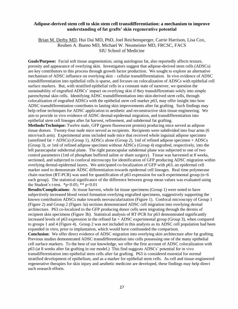
Breast Aesthetics: The Golden Ratio
Ramasamy Kalimuthu MD, FACS, Sara Yegiyants MD, Barbara Krueger MD
Objective: The purpose of this study was to assess the proportions of the aesthetic breast.
METHODS 50 breasts were analyzed in patients who were seen in consultation for breast
augmentation, breast reconstruction, ptosis correction and breast reduction. The following
measurements were collected: Sternal notch (SN) to upper pole (UP), UP to nipple (N), SN to N,
transverse diameter (TD), N to inframammary fold (IMF), and the UP slope angle off the chest wall
measured on the lateral view.
The ratios of the following relationships were calculated: SN to N/UP to N, UP to N/SN to UP.
RESULTS The following ratios were noted to be present in the aesthetic breast.
SN to Nipple/UP to Nipple 1.618
UP to Nipple/SN to UP 1.618
Upper pole slope angle Sin 38-39 degrees = 0.618
SN to UP/Nipple to IMF
1
Transverse diameter/ Nipple to UP
1
CONCLUSION Breast shape stays within the aesthetic proportions that follow the golden ratio
and the golden angle. This will aid in more precise evaluation and analysis of preoperative breast
patient to achieve a more aesthetic outcome.

44
Oncoplastic breast reduction: A safe and aesthically acceptable option for breast conserving
therapy in the treatment of breast cancer in large breasted women.
Laura Bonneau, MD
University of Wisconsin- Madison
Background The challenges of achieving aesthetically acceptable results after lumpectomy has led
some women to choose mastectomy, rather than breast conserving therapy, for the treatment of
breast cancer. For large breasted women, concurrent lumpectomy with reduction mammoplasty
(oncoplastic breast reduction) has been shown to provide a safe and aesthetically acceptable form of
breast conserving therapy.
Methods A single institution, retrospective chart review was conducted of breast cancer patients
who underwent oncoplastic breast reduction concurrently with lumpectomy. Complications and
recurrence of breast cancer were recorded. Post-operative photographs and patient remarks were
reviewed to assess patients’ satisfaction with aesthetic outcomes.
Conclusions For large breasted women who are candidates for breast conserving therapy,
oncoplastic breast reduction is a safe and aesthetically acceptable form of treatment.

45
SESSION III
BREAST AND COSMETIC
Block B
Cosmetic

46
Revisitng Browpexy As An Adjunct to Blepharoplasty:
Introduction of the Lateral Brow Retaining Apparatus
Trang Q. Nguyen, MD Julius W. Few, MD
University of Chicago Medical Center – Chicago, IL
Goals/Purpose: Increasing investigation of the surgical anatomy of the periorbital region has
resulted in increasingly varied and sophisticated surgical techniques for periorbital
rejuvenation. Since McCord and Doxanas in 1990 first described the surgical technique of
transpalpebral browpexy through the blepharoplasty incision, the importance of addressing eyebrow
position in aesthetic blepharoplasty surgery has evolved to considerations of the eyebrow and upper
eyelid as irrevocably connected, as shown by the increased use of neurotoxins. The authors
describes long term experience with a combined upper blepharoplasty and broxpexy procedure, as
well as modifications which enhance this technique for periorbital rejuvenation.
Methods/Technique: The authors retrospectively reviewed the case records of 73 patients who
underwent combined upper blepharoplasty with transpalpebral browpexy performed by the senior
author (JWF). Review of the data reveals two technical modifications which consisted of release of
the lateral brow retaining ligament for effective redraping of the periorbital tissues and lateral
wedge excision of the preorbital orbicularis oculi to efface lateral crow’s feet. Inclusion criteria
included a minimum of 24 months follow up.
Results/Complications: The average age of patients was 54.7 years, of which 84% were female
and 16% male. The average length of follow-up was 35 months. Data from the entire series
revealed absence of revisions and no cases of frontal branch nerve injury, hematoma, or post-
operative asymmetry.
Conclusion: The common goal of periorbital rejuvenation procedures is to provide a long-lasting
and natural aesthetic result. The combined upper blepharoplasty with browpexy procedure yields
excellent long-term results with minimal complications and downtime. The authors’ modifications
of browpexy in combination with blepharoplasty provides a technique which enhances the results of
upper eyelid surgery by augmenting the longevity of the blepharoplasty and addresses lateral brow
ptosis by providing a mechanism for decompression of lateral hooding.

47
Near-fatal Complication Following Injection of poly-L-lactic acid (Sculptra)
Sonya P. Agnew, MD and Lewis VL
Northwestern University, Division of Plastic and Reconstructive Surgery
Introduction: PLLA (Sculptra) is soft tissue filler widely used for the treatment of facial atrophy.
Reported side effects from injection of soft-tissue fillers for facial rejuvenation range from mild
(injection site bruising, cellulitis and nodule formation) to more severe (HSV outbreak, foreign-
body granulomatous reaction, dermal necrosis and retinal artery occlusion). We describe a patient
who suffered a massive myocardial infarction following subdermal injection of poly-L-lactic acid
(PLLA, Sculptra) in the malar region to treat HIV-lipodystrophy.
A 65 year-old man was evaluated in the senior author’s office for the ongoing management of malar
lipodystrophy related to anti-retroviral therapy for HIV. He had been treated over the past several
years with subdermal injection of Sculptra with good results. His past medical history was also
significant for hypercholesterolemia and hypertension. He did not relate that the day before
treatment he had experienced crushing chest pain, sweating and weakness, or that his partner had
tried to bring him to the emergency room. The patient underwent deep dermal injection of 6 cc
evenly divided between the bilateral malar region with the usual technique, including a dilution of
PLLA with 2 cc of 1% lidocaine with 1:100,000 epinephrine. Within the next ten minutes, the
patient collapsed and was pulseless, breathing agonally. CPR was begun immediately. The patient
was transported to the emergency room, where an ECG revealed a myocardial infarction. He was
transported to the cardiac catheterization lab within 1 hour, where a complete occlusion of his LAD
artery was diagnosed and stented successfully. After a prolonged hospital stay the patient was
discharged to rehabilitation and is alive and well today. This untoward event was filed with the
FDA.
In the post-hoc analysis, several questions were raised: Did our use of lidocaine with epinephrine as
a diluent compound the event? Was our usual history and physical pre-screening sufficient? Given
that many of these fillers are administered in a spa-type setting, should there be a standard
minimum evaluation required for all patients undergoing injections? The case should give pause to
all practitioners that even seemingly innocuous aesthetic procedures can have potentially disastrous
consequences.

48
The Lateral Brow Lift under Local Anesthesia: A Simplified Technique
Jennifer Cheesborough, MD , Thomas A. Mustoe, MD
Northwestern Memorial Hospital
Background: Several authors have promoted a limited incision lateral brow lift as an alternative to
the cumbersome endoscopic brow lift or the maximally invasive bicoronal incision. The particular
angles of tension most necessary for a rejuvenating lateral brow lift can be attained through small
temporal incisions, while the medial depressors can be reached through a complementary upper
blepharoplasty incision.
Methods: 80 consecutive patients undergoing a lateral brow lift by the senior author were
retrospectively evaluated. To allow for maximal patient convenience and safety, the senior author
(TAM) has simplified his approach to the brow lift such that it can be performed in the office with
readily available instruments under local anesthesia and minimal oral sedation. Three centimeter
bilateral temporal incisions are carried down through the temporalis fascia and the dissection
proceeds inferomedially in a subperiosteal plane along the oblique line. The forehead is also
released in the subperiosteal plane to allow for optimal redraping after the lateral lift. An upper
blepharoplasty incision allows for release of the orbital ligament and selective resection of
orbicularis oculi, corrugator supercilii, or procerus muscles.
Results: The senior author’s experience with 80 consecutive limited incision lateral brow lift
procedures between January 2008 and July 2010 has demonstrated that this is not only a technically
successful procedure, but that it is safe even in the office. Over an average of 422 days of follow up,
there were few complications and only one temporary neuropraxia of the frontal branch of the facial
nerve.
Conclusions: The limited incision lateral brow lift as described by the senior author allows for
judicious elevation of the lateral brow with a few readily available instruments. When
complemented by the upper blepharoplasty, this technique can provide excellent and natural-
appearing rejuvenation of the upper face.

49
SESSION IV
CRANIOFACIAL, NERVE, GENERAL RECONSTRUCTION
Block A
Craniofacial

50
CAD/CAM Designed Surgical Positioning Guides:
The Link Between Virtual and Actual Surgery
Troy Pittman MD, Christina Tragos MD, John W. Polley MD, Alvaro Figueroa DDS
Rush University Medicine Center – Chicago, IL
Purpose: Computer-aided design and modeling (CAD/CAM) technologies have revolutionized preoperative
planning in orthognathic and craniofacial surgery. CAD/CAM preoperative technologies have replaced the
traditional evaluative technique of 2D cephalometry and articulated model surgery. 3D virtual surgery
enables the surgeon to reposition the skeletal segments and gain an exact understanding of the bony and soft
tissue consequences before entering the operating room with the patient. Despite this advancement in pre-
operative planning, translation of this advanced technology into the operating room is lacking.
We address the gap between CAD/CAM preoperative planning and its translation into the operating room
with the introduction of occlusal-based intraoperative surgical positioning guides (SPG). SPG are
CAD/CAM designed intraoperative acrylic guides that, through initial and final guides, accurately translate
the virtual preoperative plan to the OR. SPG are designed by the surgeon during virtual surgery and are
created through stereolithographic techniques. Not only do the occlusal based guides precisely translate the
virtual plan to the OR, they also eliminate the many traditional intraoperative steps: 1. Eliminating
intraoperative intermaxillary fixation; 2. Eliminating intermediate splints in double-jaw surgeries; 3.
Eliminating manual guessing of proximal segment condylar positioning; 4. Eliminating the need for external
reference landmarks.
Methods and Materials: We present three skeletally mature patients with maxillofacial deformities that
have been treated utilizing surgical positioning guides and virtual preoperative planning for the following
cases: 1. single-piece LeFort I osteotomy; 2. LeFort I osteotomy with mandibular saggital split osteotomy; 3.
reconstruction for post-ablative enopthalmos and orbital dystopia, secondary to orbito-zygomatic trauma.
In these patients, CAD/CAM technology was used in pre-operative planning based on a 1mm cut CT scan.
The scan was obtained with the head in neutral position and the mandible in centric relation. This was
achieved by a published technique utilizing a registration bite and a gyroscope. The scan data was
manipulated with proprietary software to obtain accurate 3D digital representations of the skull. Based on
the clinical examination and record evaluation the surgical treatment plan was executed digitally in three-
dimensions. A custom single occlusal splint secured to the maxillary dentition was created through additive
stereolithographic techniques. Digital registration skeletal landmarks were created in the maxilla and
mandible or segments to be mobilized. Surgical positioning guides are digitally designed and manufactured
through stereolithography to assist the surgeon in recording the skeletal landmarks that will be used as a
reference. Based on the skeletal references and the desired skeletal movements, final positioning guides are
manufactured in a similar fashion. At surgery the guides are used to create the reference skeletal landmarks
and for precise guidance of the desired skeletal movements to their desired positions after osteotomy.
All three patients had successful treatment outcomes with their single surgical approach. Virtual planning,
virtual design and intraoperative application of SPG are illustrated through a step-by-step approach of this
technique, which will be presented and illustrated with case reports of these three patients.
Conclusion: We present three patients in which CAD/CAM preoperative planning and surgical execution of
those plans using surgical positioning guides was performed. All patients presented had successful
outcomes with their single surgery. The patients presented represent the functional and aesthetic success of
this novel evolution in orthognathic and craniofacial surgery.

51
“Critical Age:” Objective, patient-specific timing of helmet therapy in treatment
of positional plagiocephaly and brachiocephaly”
Roshni Rawlani, Vinay Rawlani MD, Caitlin Connor BS, Frank Vicari MD.
Northwestern University, Division of Plastic and Reconstructive Surgery
Bakcground: While numerous reports have demonstrated the efficacy of cranial helmet molding in
infants with severe positional plagiocephaly and brachiocephaly, there is minimal strong evidence
regarding the age at which initiate helmet therapy. Current recommendations to start helmet therapy
at 4-6 months of age appear to be based upon biased, uncontrolled, or small-scale clinical trials that
may lack reliable and validated objective outcome measures or appropriate follow up. We have
developed a method to objectively calculate the “critical age,” or age which helmet therapy must be
initiated to achieve complete correction, of infants based upon individual volumetric cranial data.
Methods: Utilizing a 3D surface scanner, the “critical age” is calculated by determining the volume
of deficiency created by deformational forces. Understanding that helmets direct all cranial growth
in the area of deficiency, one can calculate the amount of time needed to fill a volume void by
extrapolation from a modified head growth curve. If the age by which complete correction is
desired as known, the “critical age” can be determined by subtracting the time need for correction
from this age. We conducted a retrospective review of a prospectively maintained database of all
infants treated for positional plagiocephaly or brachiocephaly between 2000-2010 at Children’s
Memorial Hospital to test this concept. All enrolled infants underwent either physical therapy or
helmet therapy and were followed till cure or for a minimum of 18 months. Data was gathered
using 3D laser surface scanning and complete correction was objectively defined as a cranial ratio
of <0.85 and/or diagonal difference of 5 mm. The “critical age” was calculated for all infants as
described. The age by which complete correction was desired was set at 18 months (upper age limit
of FDA approved use for cranial helmets)
Results: In total 3,995 infants were included in the study. Of these, 1,531 infants ultimately
underwent helmet therapy. Complete correction was achieved in 98% who initiated helmet therapy
prior to their individual “critical age,” compared to a 44% complete correction rate in those who
started helmet therapy after the critical age. (p<0.005) Of the 3,995 infants, 2,998 underwent
physical therapy. 508 of these infants had moderate-severe deformation and were between 4-6
months of age; an additional 289 patients were between 6 months of age and their “critical age.”
(According to current recommendations, all these infants should require helmet therapy). With
physical therapy, 78% of the infants between 4 and 6 months and 62% of infants between 6 months
and their critical age achieved complete correction without a helmet.
Conclusion: Current recommendations to initiate helmet therapy between 4 and 6 months of age
may be overly aggressive and based upon previously published biased data. The “critical age”
allows one to objectively calculate the infant-specific age at which helmet therapy must be initiated
to achieve complete correction with a helmet. Prior to the “critical age,” physical therapy may be
highly beneficial and does not prevent complete correction if helmet therapy is ultimately required.

52
Endoscopically assisted release of sagittal craniosynostosis: a meta-analysis
Pravin Patel, MD, Mazen S. Harake, MD, MS
University of Illinois - Chicago
Background: Two decades ago the endoscopic approach to the management of sagittal synostosis
was introduced as an alternative to the conventional open approach. Studies in the literature have
shown extensive craniotomies through the conventional open approach were required to ‘normalize’
the cranial vault morphology. Is there now sufficient evidence in the literature to be able to
substantiate the initial claim that an endoscopic approach is a viable alternative? This study
investigated the morphologic outcomes including efficacy, safety, and complications utilizing the
minimally invasive endoscopic strip craniectomy technique for sagittal craniosynostosis repair.
Study Design and Methodology: A meta-analysis of the literature was conducted. Endoscopically
assisted repair of sagittal craniosynostosis studies published between 1990-2012 were identified
(Medline/PubMed), and included if they were reported in the English language and a clinical human
study. Of 462 articles identified of sagittal craniosynostosis, 8 articles were used in the meta-
analysis: a total of 480 patients of endoscopically assisted release of sagittal craniosynostosis.
Results: Data collected included nonsyndromic status, age at operation, duration of procedure, need
for perioperative blood transfusions, length of hospital stay, preoperative and postoperative head
circumference percentile and cranial index. Mean Age at endoscopic release under 4 months, length
of hospital stay 1-1.5 days, estimated blood loss less than 45 milliliters, mean operative time 45-96
minutes with the majority under 60 minutes, average perioperative transfusion rate (intra and post
operative) <9%, perioperative complications <2%, head circumference and index percentile mean
within normal limits (>75th
percentile).
Conclusion: The operative time, blood loss and hospitalization were decreased with an endoscopic
approach. Using cephalic index as a single dimensional outcome measure, the literature suggests
that endoscopically assisted release of sagittal craniosynostosis is comparable to the conventional
open approach when there is post-operative guidance with a cranial molding helmet. However,
there is insufficient data that compares the effectiveness in terms of the three-dimensional cranial
vault shape.

53
Predicting airway compromise in the infant with Pierre Robin Sequence
Carolyn R. Rogers, MD, Delora L. Mount, MD,
University of Wisconsin
A distressing feature of Pierre Robin Sequence (PRS) is the episodic airway obstruction that can
lead to feeding difficulty, cor pulmonale, and death. The protocol at our institution is to attempt
conservative management with repositioning techniques initially, and proceed with either
tracheostomy or mandibular distraction osteogenesis when conservative management
fails. Retrospective review of our experience with PRS from 2000 to 2011 is underway.
Preliminary data demonstrates high risk time points in the early post-natal course of PRS
infants. By identifying high risk time points during the PRS infants’ early post-natal course,
families and health care providers can have a heightened awareness of when to watch for impending
airway compromise and will be better be able to predict when definitive surgical intervention will
be needed.

54
SESSION IV
CRANIOFACIAL, NERVE, GENERAL RECOSNTRUCTION
Block B
Nerve

55
Cortical remodeling observed on BOLD fMRI over a 10 month period in a rat survival model
of brachial plexus avulsion and cross C7 nerve transfer
Nicholas Flugstad, MD, JB Stephenson, R Li , JG Yan, JS Hyde, HS Matloub
Medical College of Wisconsin
Total brachial plexus nerve root avulsion results in severe functional deficit of the upper extremity.
Contralateral C7 nerve root transfer is shown to improve limb function outcomes in total brachial
plexus avulsion. In this study, a rat survival model for brachial plexus avulsion injury with and
without contralateral C7 nerve transfer surgery was implemented. Blood oxygen level dependent
(BOLD) fMRI was used to detect remodeling of the primary sensory cortex of the forepaw (S1FL)
over 10 months’ time.
24 Sprague-Dawley rats were divided into three groups: control, injury, and injury & repair. The
control group underwent sham surgery with electrode placement on both median nerves. The injury
group underwent complete brachial plexus root avulsion on the right forelimb and electrode
placement on both median nerves. The injury & repair group underwent complete brachial plexus
root avulsion on the right forelimb followed by left C7 nerve root transfer to the right median nerve
via an ulnar nerve interposition graft across the chest followed by electrode placement on both
median nerves. Using a 9.4 Tesla MRI scanner, the rats then underwent BOLD fMRI imaging with
electrode stimulation at 0, 3, 5, 7, and 10 months.
Figure 1 shows BOLD fMRI images of 2 representative rats from the injury & repair group during
right median nerve stimulation. At 0 months, there is no sign of cortical activation. At 7 and 10
months, with varying speed of recovery, both rats display native, contralateral activation.
The S1FL region displays a progression of cortical plasticity as recovery takes place in each rat.
Initially, the neural pathway follows the transferred nerve and stimulation activates the ipsilateral
cortex. Over time, it appears that the cortex remodels to more closely restore the native contralateral
somatotopy. A better understanding of cortical remodeling following nerve injury and repair may
lead to improved outcomes.

56
Management of Peripheral Nerve Pathology in the Lower Extremity:
A Review of the Literature and Case Series
Michael S. Gart, MD, Donald W. Buck II, MD, Gregory A. Dumanian, MD
Northwestern Memorial Hospital, Division of Plastic Surgery
Introduction: Peripheral neuropathies in the lower extremity are commonly encountered in clinical
practice. Due to their multifactorial etiologies, they often present a diagnostic and therapeutic
challenge. To date, there are few comprehensive reports encompassing the broad spectrum of lower
extremity peripheral neuropathies. Here we present a review of the literature, with emphasis on the
relevant anatomy, pathophysiology, and management of the most common lower extremity
peripheral neuropathies. In addition, by highlighting a surgical experience with these challenging
problems, we have developed a useful algorithmic approach to the patient with lower extremity
peripheral nerve complaints.
Methods: A comprehensive electronic literature search was performed to identify original articles
in the English language relating to the diagnosis and treatment of lower extremity peripheral
neuropathies. In addition, a retrospective review 55 consecutive patients undergoing operative
treatment for lower extremity peripheral nerve complaints by a single surgeon from 2005-2010 was
performed. Objective data, including demographic information, clinical characteristics of nerve
pathology, diagnostic modalities employed, treatment modality, complications, and length of follow
up were recorded and outcomes assessed.
Results: A review of the existing literature on the relevant lower extremity peripheral nerve
pathologies is presented. Our algorithmic approach provides the surgeon with a framework for
diagnosing and treating the patient who presents with lower extremity peripheral nerve complaints.
We present a series of 55 consecutive surgical interventions by the senior author (GAD) for lower
extremity peripheral neuropathy complaints. The nerves most commonly requiring operative
intervention in our series were the peroneal nerve (20), the tibial nerve (14), the sural nerve (8), and
the common plantar nerves (7). The most common etiology of symptoms was extrinsic compression
requiring surgical release. Both operatively, and at long-term follow up, the complication rates
remained low with a significant percentage of patients experiencing complete resolution of their
symptoms.
Conclusions: Lower extremity peripheral neuropathies are commonly encountered by the
peripheral nerve surgeon. In order to optimally manage these challenging problems, a thorough
knowledge of the anatomy, pathophysiology, diagnostic and therapeutic options is imperative. In
appropriately selected patients, surgical exploration and/or decompression is safe and results in
significant improvement in symptoms.

57
The Effects of Folic Acid on Peripheral Nerve Recovery in a Rat Sciatic Nerve Model
Emily C Hartmann MD MS, Samuel O Poore MD PhD, Bermans J Iskandar MD
Background: In humans the peripheral nervous system (PNS) is capable of a remarkable degree of
healing. However, functional recovery after PNS injury is rarely fully achieved. Folic acid (FA)
has been linked to improved central nervous tissue regeneration, however there are no studies
examining its effects on peripheral nervous system.
Method: For the in vivo study 18 rats were placed into two groups. One group received FA 80
micrograms/kg for 3 days prior to the crush injury. The other group did not receive FA. The crush
injury was made to the left sciatic nerve in all rats, leaving the contralateral leg as a control. On
post-injury day 7 a fluorescent tracer was injected distal to the crush site. Bilateral dorsal root
ganglia (DRG) were harvested, cross sectioned and placed on slides. The numbers of fluorescent
cell bodies were counted. For the in vitro study, 12 rats were also placed into the same groups, one
group received FA and the other did not. Three days after the crush injury the DRGs were
harvested, cell bodies isolated, and placed in a culture medium. The axon growth was measured
using computer software at 5, 10, 13, and 17 hours following DRG harvest.
Results: The in vivo study revealed that the DRGs from non-treated rats contained 30% actively
regenerating cell bodies. The FA group is still being analyzed, however we expect and increase in
cell bodies comparatively. The in vitro study showed that neuronal cell bodies from the FA group
averaged a length of 98, 203, 410, and 498 um respectively. In the non-treated group they averaged
54, 200, 196, and 267 um respectively.
Conclusion: These findings indicate that FA may enhance peripheral nerve regeneration following
a crush injury in the rat sciatic model. Further investigation into functional recovery outcomes and
mechanism is warranted.

58
A Side-to-Side Nerve Bridge Preserves Muscle Viability Following Peripheral Nerve Injury
Shaun D. Mendenhalla, MD; Jared W. Garlick
b, BS; Jill Shea, PhD
b; Linh A. Moran, BS
b;
Mohamed E. Salama, MDb; Jayant P. Agarwal, MD
b.
a) Institute for Plastic Surgery, Southern Illinois University School of Medicine.
b) Division of Plastic Surgery, University of Utah School of Medicine
Background: High peripheral nerve injuries are a difficult clinical problem and often lead to long-
term disabilities. Despite recent advances in nerve transfer, transplantation, tendon transfer, and
tissue engineering, much improvement is still needed (1-2). This study utilizes a novel side-to-side
nerve bridge to link an injured nerve with an adjacent healthy nerve distally as a means of
preserving muscle viability following repair of a proximal nerve injury.
Methods: The sciatic, tibial and peroneal nerves were isolated in 21 Sprague-Dawley rats, which
were divided equally into 3 groups: 1) tibial nerve transection only, 2) transection + end-to-end
repair, and 3) transected + repair + collagen nerve bridge between tibial and peroneal nerves
distally. Functional gait assessment was performed every 2 weeks (3). At 90 days postop, animals
were sacrificed and gastrocnemius mass, gastrocnemius nuclear density, sciatic, tibial, and peroneal
nerve nuclear density, and axonal infiltration into the conduit were quantified.
Results: All three groups showed alteration in gate with steady improvement over time but no
significant differences between groups. Group 3 lost significantly less gastrocnemius muscle mass
and showed less histologic muscular degradation than groups 1 and 2. The tibial nerve had greater
nuclear density, both proximal and distal to the nerve repair site in group 2 compared to group 3.
Creation of the epineurial window in the peroneal nerve of group 3 showed no signs of long
standing nerve injury as evidenced by no difference in nuclear density between groups. Five of the
seven collagen conduits had evidence of neuronal sprouting within the lumen.
Conclusion: This study demonstrated better muscle preservation when a side-to-side nerve bridge
is performed distally along with traditional end-to-end repair of an injured nerve. This technique
may lead to better outcomes in high peripheral nerve injuries.
References:
1) Ray WZ, Mackinnon SE. Exp Neurol. 2010 May;223(1):77-85
2) Pfister BJ, et al. Crit Rev Biomed Eng. 2011;39(2):81-124
3) Bain JR, et al. Plast Reconstr Surg. 1989;83:129-138

59
SESSION IV
CRANIOFACIAL, NERVE, GENERAL RECOSNTRUCTION
Block C
General Reconstruction

60
Technical Considerations for the Free Fibula in Mandible Reconstruction:
Virtual Surgical Planning Systems and Osteointegrated Implants
Suhair Maqusi, MD Kolokythas A, Cohen MN, Antony AK
University of Illinois - Chicago
Purpose: Free fibula flap(FFF) reconstruction of mandibular defects has become the modern
standard of care due to superior functional and aesthetic outcomes. With introduction of
osseointegrated(OI) dental implantation, the fibula proved to provide a good denture-bearing
surface to achieve complete dental rehabilitation after mandible reconstruction. Contouring, and
accurate placement of FFF, although traditionally performed with a free-hand approach, is now
being performed with more accuracy using preoperative virtual surgical planning(VSP) systems.
The purpose of this study is to review technical aspects of mandibular planning, including VSP-
designed fibular osteotomies, positioning, and planning strategies for OI.
Methods and Materials: Consecutive patients who underwent FFF reconstruction between
October,2009 and January,2012 at a single institution were identified. Patient demographic,
reconstructive and complication data were obtained from a prospectively-maintained clinical
database. Medical records were then retrospectively reviewed to further characterize co-morbid
conditions and modifications employed to optimize results.
Results: Over the 28 month study period, 10 patients underwent FFF reconstruction. Identified
tumors included: Squamous cell carcinoma, Mucoepidermoid, Ameloblastoma, and
Osteochondrosarcoma. Osteotomy of the FFF (and mandibular resection) was typically done at the
parasymphysis, body, angle and top of ramus. Of these, 4 patients have completed OI placement. 2
patients did not require radiation postoperatively and FFF design using VSP was simulated with a
higher location on the mandible to facilitate OI placement. 2 patients requiring radiation
postoperatively, the fibula was VSP-designed lower on the mandible to minimize post-radiation
complications. For these patients, OI implants were placed on a platform to obtain adequate height
of the implants.
Conclusions: VSP is the new frontier in craniomaxillofacial surgery. Preoperative planning offers
potential for high success rates, improved operative efficiency and reduction in medical error.
Awareness of full oral restoration requires incorporating new updates in the technology with
planning of fibular design and inset, and use of osteointegrated implants.

61
Synthetic vs Bioprosthetic Utilization in Chest Wall Reconstruction
35 year Meta-Analysis
Christopher Surek D.O. 2
, Richard Korentager M.D.1,
1Chairman Department of Plastic and Reconstructive Surgery 2Resident Department of Plastic and Reconstructive Surgery
University of Kansas Medical Center
Kansas City, Kansas
Purpose: Bioprosthetics are becoming more prominent in surgical reconstruction of the breast,
abdomen and chest wall. The purpose of this study is to review the literature identifying the various
synthetic and bioprosthetic meshes that have been used for chest wall reconstruction over the past
35 years and to examine the advantages and disadvantages of each material.
Methods: A meta-analysis of the medical literature published between 1975-2010 is performed to
review all reported cases of synthetic and bioprosthetic mesh utilization in chest wall
reconstruction. Chronological and basic statistics are utilized. Advantages and disadvantages of
each material based on published literature is documented and addressed.
Results: Polytetrafluorethylene (275 cases), Polypropolene (199), Prolene (32), Marlex (44),
Mersiline (77), Silastic (10) and Vicryl (31) meshes are the synthetic materials that have been
reported in chest wall reconstruction. Bioprosthetic meshes reported for these reconstructions are
human acellular dermis-AlloDerm® (16), cross-linked porcine acellular dermis-Permacol® (1),
porcine acellular submucosal matrix-Surgisis® (28) and acellular bovine pericardium-Peri-Guard®
(9). In the year 2000, 83% of published cases used synthetic mesh and 17% used bioprosthetic
materials. In 2005, 51% of cases used synthetic mesh and 49% used bioprosthetic materials. In
2010, 43% of cases used synthetic mesh and 57% used bioprosthetic material.
Conclusions: The study indicates that over the past decade there has been a transition from
synthetic to bioprosthetic utilization in chest wall reconstruction. Synthetic meshes place significant
tension over the defect with minimal stretching, but foreign body reactions may result in infection,
erosion, and fistula formation. Polytetrafluorethylene is the most widely used synthetic mesh in
chest wall reconstruction. Early tissue in-growth, rapid revascularization and reabsorption are
among the characteristics that make bioprosthetics favorable in thoracic wall reconstruction.
AlloDerm® was among the first meshes utilized for chest wall reconstruction but its use has
decreased with the introduction of porcine derived acellular dermis which have a decreased
propensity to stretch. These materials have demonstrated success in recent literature. Due to the
increased bioprosthetic utilization in reconstruction of the chest wall, further research into the use of
these materials for this indication is warranted.

62
Assessing surgical skill in plastic surgery residency training: Developing and implementing
a low-cost online video assessment system to document proficiency
Nathan Wetter, BS; Kelli Webb, MD; Reuben Bueno, MD; Michael Neumeister, MD
SIU School of Medicine – Springfield, IL
Background: Assessment of surgical skill during training is an important component of resident
surgical education. Verification of proficiency of a surgical skill in a laboratory setting is being
used more frequently in training programs to allow residents to develop the skills necessary to
perform a surgical procedure in a non-clinical setting and demonstrate proficiency before
attempting in a clinical setting. A video recording system and an on-line evaluation tool may
facilitate faculty assessment of resident surgical skill by providing a convenient and cost-effective
method that documents resident proficiency in a specific skill.
Purpose: The purpose of this project is to develop a convenient and low-cost online system for
evaluation of video-recorded performance of surgical skill.
Methods: This evaluation system contains two components: a video recording system and an
evaluation form. To minimize costs, an in-house video server was used with a widely-available
response instrument, Survey Gizmo. Custom web programming was used to combine both
elements onto a single web page, allowing the evaluator to watch the video and complete the
evaluation form simultaneously.
Result: A convenient, easy-to-use, and low-cost system to assess and document resident surgical
performance in the laboratory setting has been developed. This system can be used by training
programs to provide valuable feedback to residents in training and allow residents to demonstrate
proficiency in a surgical skill in a non-clinical setting before moving to a clinical setting.

63
Modification of Pectoralis Myocutaneous Advancement Flap for
Sternal Wound Reconstruction
Sara Yegiyants MD, Ramasamy Kalimuthu MD, FACS
Objective: The purpose of this study was to assess the results and applicability of a modified chest
closure technique employing bilateral pectoral myocutaneous advancement flaps after sternal re-
approximation without internal fixation for postoperative sternal wound infection.
Methods: The study population is of a single surgeon's experience (n = 50). Three cases with deep
sternal wound complications underwent anatomic sternal wound reconstruction supported by the
modified corset technique of pectoralis myocutaneous advancement flaps. One patient with
macromastia underwent dermoglandular sling reinforcement to the contralateral chest wall. Follow
up was completed by outpatient record review and with telephone interviews.
Results: All three patients presented with type II sternal wounds following coronary artery bypass
surgery. Following anatomic chest wall reconstruction with the modified corset technique, all
patients had resolution of wound infection, with no wound healing complications and no mortality
at one year follow up.
Conclusions: Anatomic sternal reconstruction supported by bilateral pectoral myocutaneous
advancement flaps (corset modification ) is a good technique for recalcitrant sternal wounds to
stabilize bony framework. Additionally, medial breast dermoglandular slings to contralateral chest
wall can be utilized to further stabilize and reduce sternal retraction forces in patients with
macromastia.

64
Outcomes of secondary sternal fixation using rigid plates for infected
and dehisced sternotomy wounds
Eugene Park
Northwestern University Feinberg School of Medicine
Background: Median sternotomy is generally well-tolerated by patients undergoing cardiac
surgery, but it is not without risk of complications. Sternal wound infection and bony nonunion
occur in 0.4-5% of cases, and are associated with high mortality rates. Increased sternal stability
during healing helps minimize complications, as tight apposition of the cut edges of the sternum
facilitates bony ingrowth and healing by primary intention. Recently, fixation of the sternum using
rigid plates has been used for primary closure of the sternum after cardiac surgery with high rates of
success and minimal complications. The use of rigid plates for secondary sternal fixation in salvage
procedures for infected and dehisced sternotomy wounds has been described in the literature, but
data regarding outcomes remains scarce.
Methods: We retrospectively reviewed our experience with all patients who underwent secondary
sternal fixation at our institution using rigid titanium plates from 2008 to 2011.
Results: In all, ten patients underwent secondary sternal fixation, and we achieved bony union of
the sternum in all but one patient. The median time to discharge after sternal fixation was 6.5 days.
There were no deaths. Three patients with multiple medical comorbidities were readmitted for
sternal wound infection, and two of these patients required removal of sternal hardware. Two
patients developed seromas, both of which were successfully evacuated in the outpatient setting.
One patient developed a hematoma, which was detected two hours post-operatively and
successfully cauterized.
Conclusion: Secondary sternal fixation with rigid plates for infected and dehisced sternotomy
wounds is a safe procedure which allows for a short length of hospital stay and minimal
complications.

65
Twenty Year experience with Microsurgical Reconstruction of Hemifacial
Atrophy and Linear Sclerodema
Daniel Schmid, MD University of Wisconsin Madison
Romberg’s Disease and Linear Scleroderma (Coup de Sabre) are progressive, usually unilateral
facial atrophies of unknown etiology. The gold standard treatment for these patients has been
microsurgical reconstruction following the “burning out” of the facial atrophy and stable contour
for two years. In our experience, patients treated early in their disease course have immediate and
sustained correction of the deformity, with slowing or in most cases cessation of the disease process
upon microsurgical transfer. No disease process has recurred to date even in cases with severe
progressive disease. Equally impressive is the apparent improvement of underlying skeletal
growth seen in young children with severe disease who in previous experience would have gone on
to have much more severe skeletal deformities. We report our experience with 130 patients
involving 133 free tissue transfers with a minimum of 1 year follow up who were treated from July
of 1989 to July 2009.
All cases had severe atrophy. There were 62 males and 68 females in the series. Distribution of
disease was coup de sabre or segmental in 28 patients whereas 102 patients had a hemifacial
distribution typical of Romberg’s. 128 patients had unilateral disease and 2 patients had bilateral
atrophy without diagnosed systemic collagen vascular disease such as lupus erythematosis. The
average age of onset of disease was 12.5 years. The average duration of atrophy was 6.3 years.
Average age at operation was 29.2 years with a range from 4 to 69 years. Follow up ranged from 1
to 20 years. Complications included hematoma in 9 patients, one partial flap loss, 1 patient with
partial facial skin slough treated with local wound care, and cellulitis in 1 patient. One flap was lost
at two weeks following surgery when the patient began smoking heavily. A second free flap was
successfully preformed six months later with a stable reconstruction now four years later. Flap
revisions performed at 6 months following free tissue transfer consisting of tissue rearrangement,
debulking procedures, and resuspension when required were completed in nearly all patients and is
now a standard for optimal results. Minimal autologous fat transfers of less than 5 cc of autologous
fat were used as an adjunct in the periorbita, nasal alar rim, or minimal lip deficiencies. Recurrence
of facial atrophy was not seen.
All patients rated improvement as excellent with follow-up as long as 20 years. Microsurgical
correction of Romberg’s disease and linear scleroderma produces long-lasting, sustained results,
and may slow progression of the disease by transfer of healthy vascularized tissue to affected areas.
Stable long-term results were found even in patients with active disease at the time of microsurgical
free tissue transfer. In this article, we review the indications, choices, safety, efficacy,
complications, and technical refinements derived from our twenty year experience.

66
Notes

67
Notes