Embed Size (px)
Citation preview

RESEARCH ARTICLE Open Access
The mechanism of neurofeedback trainingfor treatment of central neuropathic pain inparaplegia: a pilot studyMuhammad Abul Hassan1,2, Matthew Fraser3, Bernard A. Conway4, David B. Allan3 and Aleksandra Vuckovic1,5*
Abstract
Background: Central neuropathic pain has a prevalence of 40 % in patients with spinal cord injury.Electroencephalography (EEG) studies showed that this type of pain has identifiable signatures, that couldpotentially be targeted by a neuromodulation therapy. The aim of the study was to investigate the putativemechanism of neurofeedback training on central neuropathic pain and its underlying brain signatures inpatients with chronic paraplegia.
Methods: Patients’ EEG activity was modulated from the sensory-motor cortex, electrode location C3/Cz/C4/P4in up to 40 training sessions Results. Six out of seven patients reported immediate reduction of pain duringneurofeedback training. Best results were achieved with suppressing Ɵ and higher β (20–30 Hz) power andreinforcing α power at C4. Four patients reported clinically significant long-term reduction of pain (>30 %)which lasted at least a month beyond the therapy. EEG during neurofeedback revealed a wide spreadmodulation of power in all three frequency bands accompanied with changes in the coherence most notablein the beta band. The standardized low resolution electromagnetic tomography analysis of EEG before and afterneurofeedback therapy showed the statistically significant reduction of power in beta frequency band in alltested patients. Areas with reduced power included the Dorsolateral Prefrontal Cortex, the Anterior CingulateCortex and the Insular Cortex.
Conclusions: Neurofeedback training produces both immediate and longer term reduction of centralneuropathic pain that is accompanied with a measurable short and long term modulation of cortical activity.Controlled trials are required to confirm the efficacy of this neurofeedback protocol on treatment of pain. Thestudy is a registered UKCRN clinical trial Nr 9824.
Keywords: Central neuropathic pain, neurofeedback, electroencephalography, paraplegia
BackgroundCentral neuropathic pain (CNP) is caused by an injuryto the somato-sensory system with a high prevalence inamputation [1], spinal cord injury [2], multiple sclerosis[3], Parkinson disease [4] and stroke [5]. CNP symp-toms do not respond well to medications and the drugsused to treat this type of pain are often associated withsignificant adverse effects [6]. This has generated interest
in nonpharmacological treatments based on neuromodula-tion and neurostimulation, such as hypnosis, meditation,neurofeedback [7], repetitive Transcranial Magnetic Stimu-lation (rTMS) and transcranial Direct Current Stimulation(tDCS) [8, 9].Neuroimagining studies have confirmed that neuromo-
dulation techniques such as hypnosis and meditation, canglobally influence pain matrix [7, 10]. Neurostimulationtechniques such as rTMS and tDCS typically targetprimary motor cortex, thereby sending inhibitory signalsdirectly to thalamus and reducing the perceived sensationof pain [11].Neurofeedback is a type of biofeeback in which patients
are provided information about their brain activity in a
* Correspondence: [email protected] Engineering and Assistive technologies, BiomedicalEngineering Research Division, University of Glasgow, Glasgow, UK5Biomedical Engineering Research Division, School of Engineering, Universityof Glasgow, James Watt building (south), G12 8QQ Glasgow, UKFull list of author information is available at the end of the article
© 2015 Hassan et al. Open Access This article is distributed under the terms of the Creative Commons Attribution 4.0International License (http://creativecommons.org/licenses/by/4.0/), which permits unrestricted use, distribution, andreproduction in any medium, provided you give appropriate credit to the original author(s) and the source, provide a link tothe Creative Commons license, and indicate if changes were made. The Creative Commons Public Domain Dedication waiver(http://creativecommons.org/publicdomain/zero/1.0/) applies to the data made available in this article, unless otherwise stated.
Hassan et al. BMC Neurology (2015) 15:200 DOI 10.1186/s12883-015-0445-7

visual or auditory form. It is believed that neurofeed-back facilitates global brain connectivity and leads toneuroplasticity [12].Neurofeedback has been used for treatment of chronic
pain, such as complex regional pain syndrome [13],fibromyalgia [14], migraine [15]. Neurofeedback studiesfor treatment of CNP are inconclusive with respect tothe optimal training protocol [16], most likely due to thesmall number of treatment sessions. Most neurofeed-back protocols for chronic pain target the temporal orcentral area of the cortex, up-regulating EEG activity inthe lower β or α band and down-regulating the activityin the Ɵ and higher β band [13–16]. Based on feedbackinformation patients can be trained to voluntarily de-crease brain activity thought to be associated with painprocessing.During neurofeedback for treatment of chronic pain,
EEG is typically measured at the training site only, pro-viding no evidences of global modulation of EEG duringtraining. Likewise, there is also the lack of evidencewhether prolonged neurofeedback treatment producedlong-term changes in cortical activity.Previous research studies have shown that CNP affects
resting state EEG causing increase in theta power andthe shift of dominant alpha frequency towards the lowerrange [17, 18]. Several fMRI studies have shown a correl-ation between CNP and reorganisation of the sensoryand motor cortex [19, 20], where, due to sensory losscaused by the injury, the affected cortical somatotopyundergoes re-mapping or reorganisation [20]. Further-more, fMRI studies showed that during motor imagerypatients with pain activate both brain areas related tocontrol of movement and to pain processing [21].In a recent study of our group [22] we showed that
CNP not only modulates the resting state EEG but alsoaffects the evoked response over the sensory-motor cor-tex during imagined movement of ‘painful’ as well asnon-painful limbs in Ɵ, α and β frequency range. Para-plegic patients with CNP had stronger EEG responsesduring imagined movement than paraplegic patientswith no pain and able-bodied people. All of these studiesindicate a close relation between the existence of CNPand ‘over activation’ of the sensory-motor cortex.We therefore hypothesize that similar to rTMS and
tDCS, neurofeedback can target the motor cortex, resultingin normalization of evoked responses and reduction ofCNP. We propose neurofeedback training for treatment ofCNP in chronic paraplegic patients setting three objectives:
(I) Testing the immediate and longer term effect ofneurofeedback training on CNP,
(II)Understanding the putative mechanism throughwhich neurofeedback induces wide-spread changesof EEG activity during neurofeedback training,
(III)Assessing the long term effect of training on allcortical structures involved in processing of pain.
MethodsPatientsSeven chronic patients with paraplegia (age 50 ± 4, 6 males,1 female) having CNP under the level of injury were re-cruited for the study from a cohort of 10 patients recruitedfor our previous study [22]. The neurological level of SpinalCord Injury (SCI) was determined using the AmericanSpinal Injury Association (ASIA) Impairment Classification[23]. Inclusion criteria were: paraplegia at level T1 or lower,at least one year post-injury, a treatment history of CNPfor at least 6 months and a report of pain level ≥ 5 on theVisual Numerical Scale (0 = no pain, 10 =worst pain im-aginable). The general exclusion criteria were: presence ofany chronic (non CNP) or acute pain at the time of the ex-periment; brain injury or other known neurological condi-tion. Patients receiving pharmacological treatment wereinstructed not to change medications during the neuro-feedback therapy. Information about patients is providedin Table 1.At the beginning and at the end of the study patients
filled out the Brief Pain Inventory [24]. Prior to the studythey filled out a 7 Point Guy/Farrar Global Impression ofChange [25] to test whether pain intensity was stable overthe past week. Ethical approval was obtained from theWest of Scotland National Health Service for the GreaterGlasgow and Clyde Ethical Committee. Informed consentfor participation and publication of the study was obtainedfrom the participants. The procedures followed were inaccordance with the Helsinki Declaration, Ethical princi-ples for medical research involving human subjects.
EEG recordingDuring neurofeedback training patient’s EEG was re-corded using 16 channel Usbamp, (Guger technologies,Austria). Sampling frequency was 256 Hz and electrodeimpedances were below 5 kΩ. The neurofeedback treat-ment was provided from one electrode at the time butup to 16 electrodes (F3, Fz, F4, T7, C3, Cz, C4, T8, Cp3,CPz, C4, P4, P3, O1, Oz and O2) were recorded simultan-eously. The ground electrode was placed on the mastoidon the training side and the reference electrode wasplaced on the opposite side.Multichannel EEG recording for off-line analysis was
performed before the first training day and after the lasttraining day, from 61 channels (Synamp2, Neuroscan,USA) with electrodes placed according to standard 10/10 locations [26] using an ear-linked reference and AFzas ground. Sampling frequency was 250 Hz and imped-ance was kept below 5 kΩ.
Hassan et al. BMC Neurology (2015) 15:200 Page 2 of 13

Real-time data acquisition and analysisReal time data acquisition and processing was performedwith g.RTanalyzer (Guger technologies, Austria) in Simu-link, Matlab (Mathworks, USA). A graphical user interfacewas developed in LabView (National Instruments, USA).To calculate EEG power in selected frequency bands,
EEG of each channel was bandpass filtered (5th order IIRButterworth) in the selected bands and was then squaredand smoothed/averaged over a half second sliding win-dow, updated after each sample, to obtain the bandpowerfeatures.
Neurofeedback protocolA daily neurofeedback session started and finished withmeasuring the baseline EEG activity for 2 min in relaxedopen eyes and in closed eyes state. One daily neurofeed-back training session consisted of 2 sub-sessions with anaudio feedback followed by 6–7 sub-sessions with avisual feedback. Each sub-session lasted 5 min.Training started with an audio neurofeedback pro-
vided from the occipital region (Oz) with patients havingtheir eyes closed, for relaxation purposes. Training pa-rameters were calculated in the lower α band (7–10 Hz)at Oz to account for lower peak frequency in patientswith CNP [17, 18, 22]. A relative power was calculatedwith respect to 2–30 Hz band. Patients were trained toincrease the α band power with a threshold set at 110 %of the baseline value. Patients listened to relaxing music,which had two levels: quieter when the α power wasabove a set threshold and louder when the α power wasunder the threshold.Following the audio training patients were provided
with a visual neurofeedback as a therapy to reduce pain.EEG power was calculated in θ (4–8 Hz), α (9–12 Hz),lower β (12–15 Hz) and higher β (20–30) Hz bands. The
higher β will be referred as β further in the text and 12–15 Hz will be called the Sensory Motor Rhythm (SMR).Relative power was calculated with respect to 2–30 Hzband.Contingencies were set such that increases in the α or
SMR and decreases in the Ɵ and β were reinforced. In-creased Ɵ band power was found positively related to thepresence of CNP [17, 18, 22, 27] and was confirmed inthis particular group of patients [22]. The β band powerwas suppressed because these oscillations are thought tobe positively associated with pain [17]. We trained pa-tients to increase either the SMR or α power becausethey showed promising results in some previous neuro-feedback studies on chronic pain including CNP [16, 28].The group of patients included in the study had thedominant α frequency on average 1 Hz lower than pa-tients with no CNP [22]. Therefore we trained patientsto reinforce the energy of EEG signal in a slightly higherα band (9–12 Hz) which does not include lowest α bandfrequencies at 8Hz. We set the training ‘threshold’ to110 % of the average power in the α/SMR band and to90 % of the average power in the Ɵ and β band. Trainingwas provided from the electrodes located over the pri-mary motor and sensory cortex C4/C3/Cz/ P4, oneelectrode at the time, order as shown in Fig 2. Theseelectrodes were also a preferred stimulation site for rTMSand tDCS [8, 29]. In addition, this group of patients hadan ‘overactive’ sensory-motor cortex during a motor im-agery task [22] so neurofeedback provided from that areacould potentially down-regulate the excessive activity.During training patients sat in front of a computer
screen that showed three bars, the size of which corre-sponded to the relative EEG power in three chosen fre-quency bands (Fig. 1). The bars had green colors whenthe power of a representative frequency band was in thereinforced range and had red color otherwise. The bar inthe middle presented a frequency band that had to berewarded (increased) and turned green when the powerwas 110 % above the baseline value. Two sidebarspresented two frequency bands that had to be inhibited(decreased) and turned green when the power was under90 % of the baseline value. Patients were instructed torelax and to ‘apply whichever mental strategy they preferto make the bars green’.We tested four protocols: Protocol 1 rewarded SMR and
inhibited Ɵ and β at Cz,. Protocol 2 rewarded α and inhib-ited Ɵ and β at P4; Protocol 3 rewarded α and inhibited Ɵand β at C3. Protocol 4 rewarded α and inhibited Ɵ and βat C4.To test for the placebo effect a) patients were shown
data from a pre-recorded neurofeedback session, b) theywere provided with a visual neurofeedback training toreinforce the α activity at Oz with eyes open; Oz is locatedat the occipital area, normally not associated with pain.
Table 1 Information about patients and outcome results of theneurofeedback therapy
Patient Level ofinjury/ASIA
Years withinjury/pain
Location ofpain
Sensationof pain
Medication
P1 T8 A 7/7 abdomen,legs,buttock, fee
pricking,stabbing
G
P2 T7 A 7/7 shanks, feet burning P
P3 T6/T7 D 9/9 legs, feet Pricking,stabbing
P
P4 T6/T7 B 25/24 Abdomen,legs,buttock,feet
Squeezing,stabbing
P
P5 T8 B 9/9 Buttock,legs, feet
Burning,stabbing
P
P6 T5/6 A 11/11 Left leg andfoot
burning G
P7 T12 B 33/4 Legs, feet Tingling p
Hassan et al. BMC Neurology (2015) 15:200 Page 3 of 13

Assessment sessionsResting state EEG was recorded with 61 channels underthe open eyes and closed eyes condition in a quiet room.The resting state EEG was recorded for 2 min for eachcondition repeated 3 times, alternating between the con-ditions. Assessment sessions were performed twice, upto a week before the first neurofeedback session and upto a week after the last neurofeedback sessions but neveron a day of neurofeedback training. Post neurofeedbackassessment was performed for patients who had 20 ormore neurofeedback sessions.
EEG off-line analysisEEG recorded during neurofeedback training was visuallyinspected and sections containing blinking, muscle activityor amplitude over 100 μV were removed, leaving a mini-mum of 3 min recording. A power spectral density (PSD)was calculated using Hamming windows over 4 s longrecording overlapped for 2 s. Logarithmic PSD was calcu-lated as 10∙log10PSD for normalization purposes. For eachfrequency, the unpaired t-test was performed to comparebetween two conditions and Holms-Bonferroni correctionwas applied to reduce the Type I error due to multiplecomparisons.To calculate changes in connectivity during a training
session compared to the baseline, coherence was calcu-lated for each channel pair for 4 s long EEG epochs.The average coherence value was calculated for a chosenfrequency band. The same statistical analysis was used asfor PSD.
Linear regression analysis Y =K1 +K2 ⋅X was performedto find the best fit curve between the pain intensity andthe number of training sessions using the parametricPearson test.For 61 channel off-line EEG analysis, data were re-
referenced to the average reference. Noise was removedas described above. The current source density wascalculated for 4 s long epochs in the Ɵ, α and β band.Localisation of the cortical three-dimensional distribu-tion of the current density of EEG was performed usingthe Standardised Low Resolution Electromagnetic Tom-ography sLORETA [30]. The sLORETA method hasbeen shown to have no localization bias [31]. The sLOR-ETA cortical map/image was computed for 6239 voxelpartitions of intracerebral volume at 5 mm spatial reso-lution. Brodmann areas are reported using the MontrealNeurological Institute (MNI) space with correction tothe Talairach Space.For a group level comparison, data of each patient were
normalised prior to averaging. A paired t-test was appliedto find a statistically significant difference between EEGbefore and after the therapy. A 5000 voxel randomizationof statistical non-parametric mapping [32] implementedin sLORETA package was used to calculate a correctedcritical thresholds and p-values.
ResultsFive out of seven patients with pain (P1-5) completedthe study. Two patients withdrew after 3 sessions, onebecause of problems with transportation, although ex-periencing reduction of pain (P 6) and the other because
Fig. 1 A graphical user interface used for neurofeedback training. Horizontal black lines show threshold values. Central bar shows power of thedominant frequency band, which had green colour when it was reinforced, I,.e. when the power was above the threshold. Side bars present theta(left) and beta (right) frequency bands that were supressed. They had green colour when the power was under the threshold, otherwise were red
Hassan et al. BMC Neurology (2015) 15:200 Page 4 of 13

of the lack of response (P7). Four patients received 40treatment sessions and the fifth patient, who stayed atthe hospital for the purpose of the study, received 20sessions. Patients who travelled from their homes re-ceived 1–3 sessions per week while the patient whostayed at the hospital received five sessions per week.None of patients reported reduction of pain during
training with Protocol 1. Patients P2 and P4 reportedthe moderate reduction of pain as a result of trainingwith Protocol 2. Both Protocols 3 and 4 resulted in asubstantial reduction of pain, going down to 0–2 on thevisual numerical scale during training. However, patientsP3-5 reported strong spasm during training with Proto-col 3, which manifested as uncontrollable movements oftheir paralyzed legs while sitting in front of the com-puter screen practicing neurofeedback. Patients weretrained with Protocol 4 on most of training days as itprovided best relief from pain with minimum side ef-fects. Fig. 2 shows the number of sessions and sequenceof training for each protocol. The order of protocol wasnot identical for each patient and depended on theirresponse (no reduction of pain, spasm, etc.).One somedays patients were trained with two protocols (3 succes-sive, 5 min long sub-sessions with each protocol). Foreach training protocol a patient was initially trained forat least two days in a raw, to allow some time to learnneurofeedback strategy for each protocol. Though Proto-col 4 seemed to achieve best reduction of pain, otherprotocols were occasionally re-tested to test whether pa-tients responded in a consistent way. Note that therewas no established successful protocol for treatment ofCNP, so having an initial hypothesis that the sensory-motor area is overactive in CNP [21, 22] we tested dif-ferent location over that area of the cortex, keepingsimilar training parameters. Initial testing of different lo-cation is a standard practice in creating a novel neuro-feedback protocol [13, 16].The effect of placebo training was tested on two days,
between 10th and 20th training day. The effect of mental
rehearsal of neurofeedback was tested within the last 5training days.
The effect of neurofeedback training on the intensity ofpainSix out of seven patients reported the immediate reduc-tion of pain already during 2nd or 3rd daily treatment.Initially however that reduction was short-term, duringneurofeedback. All five patients who received 20 ormore treatment sessions achieved a statistically significantreduction of pain, being clinically significant (>30 % of theindividual initial pain intensity as defined by a Visual nu-merical scale) in four patients (Table 2). The long-term re-duction of pain (which lasted beyond the neurofeedbacktreatment) in all patients was gradual and lasted for sev-eral weeks after termination of the therapy. Patients werecontacted about one month after the end of the treat-ment, they still had reduced intensity of pain but itincreased 1–2 grades of Visual numerical scale as com-pared to last neurofeedback session. A linear regressionanalysis showed a significant negative correlation be-tween the intensity of pain and the number of trainingsessions (P1: r = 0.74, p = 0.023, P2: r = 0.66, p = 2.5 · 10−5;P3: r = 0.61, p = 0.001; P4: r = 0.83, p = 6.85 · 10−8; P5: r =0.64, p = 0.005).Two patients who before treatment suffered from spas-
ticity/clonus (P3) and spasm/tightness (P4) reported thereduction of this symptoms on the days of training. All pa-tients who experienced the reduction of pain, includingpatients with the complete loss of sensation, reported thepleasant sensation of warmth in their legs which precededpain relief and lasted for several hours.Patients P2-5 were able to self-regulate their brain activ-
ity between treatment sessions (documented by EEG re-cordings) which helped reducing their pain. An examplefor P3 is shown in Fig 3b. However they gradually lost thatability a month after the last session due to the lack of aproper visual association.
Fig. 2 The number of sessions and sequence of training for each protocol
Hassan et al. BMC Neurology (2015) 15:200 Page 5 of 13

EEG modulation during training at the training siteFigure 3a-e show the logarithmic PSD in patients P1-5before, during and after training on one representativetraining sub-session lasting 5 min. Patients were able toselectively reinforce or suppress power in different fre-quency bands, though only P5 modulated power of allthree frequency bands simultaneously in a desired direc-tion. It can be noticed that the effect of training on EEGpower still remains in the first few minutes following thetraining (‘Post NF’). Pannel labeled with PWP1 at thebottom corresponds to PSD in patient 1. Consequtivepannels labeled with PWP2,3,4 and 5 corresponds to pa-tients 2,3,4 and 5 respectivly.Figure 4a shows PSD before training (‘Pre NF’), during
real training (‘NF’) and during placebo training (‘Placebo’)
with a pre-recorded session in P5. PSD during placebotraining was not significantly different from PSD beforetraining and no reduction of pain was reported. Duringplacebo training with a feedback provided from the elec-trode location Oz patents unsuccessfully tried to reinforcethe α band power (as the occipital alpha normally de-creases during visual attention) and did not report any re-duction of pain. It should be however mentioned thatduring a neurofeedback training with closed eyes patientssuccessfully increased their occipital α but none of the pa-tients reported the reduction of pain.Figure 4b shows PSD in P5 before training (‘Pre NF’),
during training with a visual feedback (‘NF’) and duringmental neurofeedback practice (‘Practice’) without thevisual feedback. The patient regulated EEG power inthe same direction and in the same frequency band inthe α and β range during the training with feedbackand during the mental practice. Four patients P2-5were able to modulate their PSD during mental neuro-feedback practice.
Global EEG modulation during neurofeedback trainingFigure 5 shows scalp maps of PSD over three frequencybands before and during training provided at C4 withprotocol 4. Frontal Ɵ was suppressed during training in allfive patients. This Ɵ band suppression is likely to be aconsequence of voluntary modulation, because increased
Table 2 Information about the outcome of the neurofeedbacktherapy
Patient Pain before therapy(VNS)
Pain following therapy(VNS)
Number ofsessions
P1 6 5 40
P2 7 5 40
P3 6 2 40
P4 9 6 40
P5 9 6 20
P6 8 6 3
P7 6 6 3
Fig. 3 Neurofeedback training with Protocol 4 in P1-5, EEG power as a function of frequency recorded at C4. EEG power at the baseline (‘Pre NF’),during neurofeedback training (‘NF’) and 2–3 minutes following training (‘Post NF’). Bars above graphs show three frequency bands in which trainingwas provided. Arrows up marks a band that was reinforced while arrows down mark frequency bands which were suppressed. Black bars under graphsshow statistically significant difference for ‘NF’-‘Pre NF’, while grey bars show statistically significant difference for ‘Post NF’-‘Pre NF’
Hassan et al. BMC Neurology (2015) 15:200 Page 6 of 13

concentration due to nonspecific engagement in a mentaltask should result in increased frontal Ɵ [33].In the α range, training resulted in the increase of power
over the central cortex, on both contra and ipsilateral sitein P1,2,4 5 while in P3 training resulted in the shift of amaximum α power from the occipital to the central re-gion. In general, the α rhythm characterizes the idle state,and simply focusing attention on object on a computerscreen should therefore result in decrease of α power. Awide-spread increase of α power during neurofeedbacktraining could therefore be attributed to the voluntarymodulation of brain activity rather than to the general
increase of attention. A wide spread increase in the αpower can be partially explained by the volume conduc-tion effect, spreading the α activity from the training siteat C4. However in P1 and P2 the α power increased moreon the contra than on the ipsilateral site of training and inP3 the maximum of α activity shifted from the occipital tothe central area.In four patients (P2-P5) the suppression of the frontal
β (20–30 Hz) can be noticed during training, beingstronger than suppression at the training site.Figure 6 shows the location of electrodes with statistically
significant differences in PSD between the neurofeedback
Fig. 4 The effect of (a) placebo training and (b) of mental practice with no visual feedback. Graphs present EEG power as a function of frequencyrecorded at C4 for both conditions. Fig. 4a shows EEG power in patient P5 during neurofeedback training (‘NF’), placebo training (‘Placebo’) andpre-training baseline (‘Pre NG’), Fig. 4b shows EEG power in patient P5 during neurofeedback (‘NF’), mental practice without a feedback (‘Practice’)and pre-training baseline (‘Pre NF’). Black bars under graphs a and b show statistically significant differences for ‘NF’-‘Pre NF’, while grey bars showstatistically significant difference for ‘Pre NF’-‘Placebo’ in (a) and for ‘Pre NF’-‘Practice’ in (b)
Hassan et al. BMC Neurology (2015) 15:200 Page 7 of 13
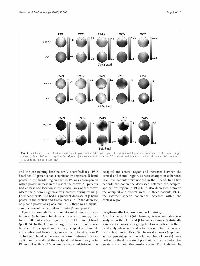
and the pre-training baseline (PSD neurofeedback- PSDbaseline). All patients had a significantly decreased Ɵ bandpower in the frontal region that in P4 was accompaniedwith a power increase in the rest of the cortex. All patientshad at least one location in the central area of the cortexwhere the α power significantly increased during training.Four patients (P2-P5) had a significant decrease of β bandpower in the central and frontal areas. In P2 the decreaseof β band power was global and in P1 there was a signifi-cant increase of the central and frontal β band power.Figure 7 shows statistically significant difference in co-
herence (coherence baseline- coherence training) be-tween different cortical regions, in the Ɵ, α and β band(p = 0.05). In the Ɵ band a large decrease in coherencebetween the occipital and central, occipital and frontal,and central and frontal regions can be noticed only in P3. In the α band, coherence increased between the oc-cipital and central and the occipital and frontal region inP1 and P4 while in P 5 coherence decreased between the
occipital and central region and increased between thecentral and frontal region. Largest changes in coherencein all five patients were noticed in the β band. In all fivepatients the coherence decreased between the occipitaland central region; in P1,2,4,5 it also decreased betweenthe occipital and frontal areas. In three patients P1,3,5the interhemispheric coherence increased within thecentral region.
Long-term effect of neurofeedback trainingA multichannel EEG (61 channles) in a relaxed state wasanalyzed in the Ɵ, α and β frequency ranges. Statisticallysignificant changes on a group level were noticed in the βband only where reduced activity was noticed in severalpain related areas (Table 3). Strongest changes (expressedas the percentage of the total number of voxels) werenoticed in the dorso-lateral prefrontal cortex, anterior cin-gulate cortex and the insular cortex. Fig. 7 shows the
Fig. 5 The influence of neurofeedback training with protocol 4 at C4 on wide spread EEG power in different frequency bands. Scalp maps duringtraining (‘NF’) and before training (‘PreNF’) in Ɵ, α and β frequency bands. Location of C4 is shown with black dots in P1 scalp maps. P1-5: patients1–5. Units on side bar graphs μV2
Hassan et al. BMC Neurology (2015) 15:200 Page 8 of 13

Fig. 6 Location of electrodes with significant difference in power (‘NF’—‘PreNF’) shown in Fig 5. Dark circles indicate power increase while greycircles indicate power decrease
Fig. 7 Scalp maps showing statistically significant changes in EEG coherence between 16 scalp sites during training compared to baseline beforetraining (neurofeedback training-baseline) for each patient in three frequency bands (a) Theta band, (b) Alpha band (c) Beta band (significancelevel p = 0.05, corrected for multiple comparison). Training provided with protocol 4 at C4, marked with a black dot in the upper left figure. Thesame EEG data used to create EEG power scalp maps in Fig. 5. Solid lines show increase in coherence and dotted lines show decrease in coherenceduring neurofeedback as compared to the baseline. The thickness of line shows strength of change in coherence (thin line: 0 to 0.1, medium line: 0.1to 0.2, thick line: 0.2 to onwards)
Hassan et al. BMC Neurology (2015) 15:200 Page 9 of 13

reduction of β activity over the surface areas of the cortex(Fig. 8a) and several deeper cortical structures (Fig. 8b-d).On the individual level, P1 had a significantly reduced
activation in the Ɵ band at the primary sensory andmotor cortices and the posterior parietal cortex and P2,P4 and P5 had significantly increased α activity over theprimary sensory and motor cortices and over all corticalstructures reported in table 3. Patient P3, on the con-trary, had a significant decrease in the α activity.
DiscussionThis paper presents the effect of neurofeedback trainingon reduction of CNP and on related neurological mea-sures. Using a visual feedback, patients learned how tomodulate their brain activity in a desired directionswhich resulted in reduction of pain.
Six out of seven tested patients achieved short-term im-mediate reduction of pain during neurofeedback training.To achieve longer lasting reduction of pain repeatedneurofeedback sessions were required. Four out of fivepatients who received a long term training achieved a clin-ically relevant reduction of pain (>30 %) lasting at least amonth following the treatment. A negative correlation be-tween the intensity of pain and the number of trainingsessions indicate that the long-term reduction of pain wasgradual and required long training. All patients experi-enced the reduction of pain while receiving a neurofeed-back training from electrodes located above the primarymotor cortex (C3/C4), which was also a preferred stimula-tion site for rTMS and tDCS [29, 34]. In Jensen’s et al.,[16]neurofeedback study for treatment of CNP, 10–15 Hzband was reinforced at C3 and C4 while α band was rein-forced at T7/T8 with a moderate (not clinically signifi-cant) reduction of pain. Better patients’ response in ourstudy might be due to different combination of frequencybands and electrode location (primary motor cortex oflegs and arms) or due to the larger number of trainingsessions, as a prolonged effect of neurofeedback graduallyincreased over the 40 sessions.Volunteers from our study that achieved the clinically
significant reduction of pain reinforced the α power andto some extent suppressed the β power during training.Although the Ɵ band power is believed to be the signatureof CNP [17], the only patient who successfully suppressedthe Ɵ rhythm experienced least reduction of pain. Thismight be related to the fact that patients received medica-tions known to increase the Ɵ band power [35].Although neurofeedback was dominantly practiced
from the right side of the central cortex at C4, patientsreported reduction of pain in their legs, confirming
Table 3 Percentage of voxels in pain related brain areasshowing the average reduced activation
Cortical Areas BAs Voxels (%) Maximumactivation
MNI coordinateswith maximum value
S1 1,2,3 NS / / / /
S2 40,43 NS / / / /
M1 4 NS / / / /
PMC 6 2 −0.97 −15 25 40
SMC 8 4 −0.96 −20 30 45
DLPC 9,46 30 −0.99 −15 40 25
APFC 10,11 4 −0.96 −20 45 30
PPC 5,7 / / / / /
ACC 24,32 20 −0.99 −15 35 20
PCC 23,31 NS / / / /
IC 13 24 −1.01 −30 15 15
-0.5
-1
1
0.5
0(a) (b)
(c) (d)
Fig. 8 Changes in activity in eyes open state in the β band before the first and after the last day of neurofeedback training (After-Before) averagedover five patients P1-5. (a) Surface maps (top, left, right and frontal), (b) BA 13 [MNI coordinate: −30 15 15, t = −1.01], (c) BA32 [MNI coordinate: −15 3520, t = −0.99], (d) BA 24 [MNI coordinate: −5 25 15, t = −0.99]. Blue colour correspond to reduced activity
Hassan et al. BMC Neurology (2015) 15:200 Page 10 of 13

observations from rTMS studies that the exact somato-topical location of stimuli is less relevant [36].Because patients were occasionally trained with dif-
ferent protocols, one cannot claim that training specif-ically at C4 produced long-term changes. However allprotocols involved sensory-motor area and were basedon decreasing theta and higher beta band and increas-ing alpha or lower beta band. In addition, training atone electrode caused wide spread changes so one canassume that all training protocols globally affectedsensory-motor cortex, though they caused to some ex-tend different physical responses. In this study, a multi-channel EEG was recorded only during training fromC4 (Protocol 4). In the future, it would be interesting tocompare global effect of training from the electrode lo-cations included in the training protocols (P1-P3).It is believed that chronic pain disrupts ‘a default
mode network’ [37]. Neurofeedback training was accom-panied by changes in coherence between occipital andcentral and occipital and frontal cortex, most notably inthe β band. Similar changes in connectivity during hyp-nosis were attributed to the disruption of pain matrix,possibly establishing a normalized default mode network[7] The higher beta band (20–30 Hz) was the only fre-quency band in which long-term changes were noticedin all participants. Largest changes occurred in the βband of the dorsolateral prefrontal cortex, the cingulatecortex and the insular cortex. The former is related tothe cognitive aspect of pain while two later are parts ofthe limbic system and deal with the emotional aspect ofpain [38]. Previous studies showed that chronic painshifts brain representation from the nociceptive to theemotional circuits [39], therefore reduction of chronicpain might be first manifested in cortical structuresregulating the emotional aspect of pain.This study tested neurofeedback protocols from the pri-
mary motor cortex which is not the part of the standardpain matrix [38]. It is possible that this neurofeedbackProtocol 4 is specific to CNP and that it would not be effi-cient for other types of chronic pain. One of the protocolsin this study involved sensory cortex (P4, Protocol 2) butpatients reported less reduction of pain than with neuro-feedback from C3 and C4. It was difficult to assess howneurofeedback practice with one protocol influenced learn-ing new protocol, though empirically we had impressionthat it helped, in particular because electrodes were chosenfrom functionally the same area of the brain. It is hard tomeasure with EEG the activity of other cortical areas in-volved in processing of chronic pain but a single daily ses-sion fMRI neurofeedback study reported reduced CNP inpatients trained to regulate the activity of the anterior cin-gulate cortex [40].A perceived sensation of pleasant warmth reported
by all patients, including these with a complete loss of
sensation, might be an indicator of indirect activationof corresponding thalamic nuclei, being in a close prox-imity to pain nuclei [41]. Alternatively, it may supportthe theory of CNP representing a thermoregulatorydysfunction [42].The ability to ‘self-administer’ therapy i.e. to modulate
their brain activity at will without a visual feedback wasachieved by 4 out of 5 patients; this ability was previ-ously reported by patients practicing mindfulness andhypnosis [43, 44]. This is a very important observationbecause CNP in this patients was caused by paralysisand as such can be more or less effectively amelioratedbut not cured. Patients ability to self-regulate brain ac-tivity on demand would be an important prerequisite forkeeping pain under control in long term.Although some patients reported spasm during train-
ing, in two patients this resulted in reduced tightnessand clonic activity following neurofeedback. It is be-lieved that sensory-motor cortex is one of the sites fromwhich it is possible to modulate monosynaptic reflexes[45] that might be related to spasm observed in two pa-tients. Previously rTMS stimulation of the primarymotor cortex was shown to reduce spasm [46] indicatingpossible similarities between neuromodulatory mechan-ism of neurofeedback from sensory-motor cortex andrTMS. To fully understand the potential of the mechan-ism of neurofeedback treatment, further randomizedcontrolled study is required.
ConclusionsThe results of the study show that prolonged neuro-feedback training may have a potential to reduce CNP.Neurofeedback training affects deeper cortical struc-tures involved in processing of chronic pain. We testedneurofeedback treatment on patients who had long-standing CNP. The effect of neurofeedback might bebetter on patients who suffered from CNP for a shorterperiod of time, as prolonged pain might cause longlasting changes in brain connectivity 19]. Larger con-trolled trial would be needed to confirm this resultsbefore it could be recommended as a neurofeedbacktraining protocol for CNP.
AbbreviationsCNP: Central Neuropathic Pain; EEG: Electroencephalography; P: Patient withpain; PSD: Power spectral density; rTMS: repetitive transcranial magneticstimulation; SCI: Spinal Cord Injury; tDCS: transcranial direct currentstimulation; α: alpha band EEG; β: band EEG; θ: band EEG.
Competing InterestsThe authors declare that they have no competing interests.
Authors’ contributionsMAH run the study, analysed results and contributed to writing; MFcontributed to creating the study and helped with patient selection; BACcontributed to analysis of results; DBA contributed to create the study;’ AVcreated the study, secured funding, analysed results and was responsible for
Hassan et al. BMC Neurology (2015) 15:200 Page 11 of 13

writing the manuscript. All authors discussed the results and commented onthe manuscript.
Authors’ InformationMAH is a lecturer in biomedical engineering at the NED University,Pakistan. He was awarded PhD from the University of Glasgow for a thesis‘Quantitative EEG and neuromodulation for the treatment of centralneuropathic pain in paraplegic patients’. AV holds PhD in BiomedicalEngineering. She is a Lecturer in Rehabilitation Engineering at theUniversity of Glasgow. Mr Matthew Fraser, FRCS is a Clinical consultant inspinal cord injury at the Queen Elizabeth National Spinal Injuries Unit. BACholds PhD in neurophysiology. He is a Professor in Biomedical Engineeringat the University of Strathclyde and a member of International SpinalResearch Trust Research Committee. DBA, FRCS is a consultant inorthopaedic surgery at the Queen Elizabeth National Spinal Injuries Unit.
AcknowledgementWe would like to thank to Professor Mark Jensen, Dr Susan Othmer, andDr Victoria Ibric, for sharing their research and clinical experience. Wethank Dr Purcell and Dr Mclean, Southern General Hospital, Glasgow, forchoosing participants of the study and to all participants for taking part.This work has been partially supported by the MRC grant G0902257/1, theGlasgow Research Partnership in Engineering and by NED University ofPakistan PhD scholarship.
Author details1Rehabilitation Engineering and Assistive technologies, BiomedicalEngineering Research Division, University of Glasgow, Glasgow, UK.2Department of Biomedical Engineering, NED University of Engineering andTechnology, Karachi, Pakistan. 3Queen Elizabeth National Spinal Injuries Unit,Southern General Hospital, Glasgow, UK. 4Department of BiomedicalEngineering, University of Strathclyde, Strathclyde, UK. 5BiomedicalEngineering Research Division, School of Engineering, University of Glasgow,James Watt building (south), G12 8QQ Glasgow, UK.
Received: 23 July 2015 Accepted: 29 September 2015
References1. Floor H. Phantom-limb pain: characteristics, causes, and treatment. Lancet
Neurol. 2002;1:182–9.2. Siddall PJ, McClelland JM, Rutkowski SB, Cousins MJ. A longitudinal study of
the prevalence and characteristics of pain in the first 5 years followingspinal cord injury. Pain. 2003;103:249–57.
3. Osterberg A, Boivie J, Thuomas KA. Central pain in multiple sclerosis-prevalence and clinical characteristics. Eur J Pain. 2005;9:531–42.
4. Beiske AG, Loge JH, Rønningen A, Svensson E. Pain in Parkinson's disease:Prevalence and characteristics. Pain. 2009;141:173–7.
5. Andersen G, Vestergaard K, Ingeman-Nielsen M, Jensen TS. Incidence ofcentral post-stroke pain. Pain. 1995;61:187–93.
6. Bakonja M, Rowbotham MC. Pharmacological therapy for neuropathic pain.In: McMahom SB, Koltzenburg M, editors. Wall and Melzack’s Textbook ofpain. Philadelphia: Elsevier; 2006. p. 1075–83.
7. Jensen MP, Day MA, Miró J. Neuromodulatory treatments for chronic pain:efficacy and mechanisms. Nat Rev Neurol. 2014;10:167–78.
8. Lefaucheur JP, Antal A, Ahdab R, Ciampi de Andrade D, Fregni F, Khedr EM,et al. The use of repetitive transcranial magnetic stimulation (rTMS) andtranscranial direct current stimulation (tDCS) to relieve pain. Brain Stimul.2008;1:337–44.
9. Leung A, Donohue M, Xu R, Lee R, Lefaucheur JP, Khedr EM, et al. rTMS forsuppressing neuropathic pain: a meta-analysis. J Pain. 2009;10:1205–16.
10. Jensen KB, Berna C, Loggia ML, Wasan AD, Edwards RR, Gollub RL. The useof functional neuroimaging to evaluate psychological and other non-pharmacological treatments for clinical pain. Neurosci Lett. 2012;520:156–64.
11. Lefaucheur JP. Neurophysiology of cortical stimulation. Int Rev Neurobiol.2012;107:57–85.
12. Niv S. Clinical efficacy and potential mechanisms of neurofeedback. Pers IndDiff. 2013;54:676–86.
13. Jensen MP, Grierson C, Tracy-Smith V, Bacigalupi SC, Othmer S.Neurofeedback Treatment for Pain Associated with Complex Regional PainSyndrome Type I. J Neurother. 2007;11:45–53.
14. Kayiran S, Dursun E, Dursun N, Ermutlu N, Karamürsel S. Neurofeedbackintervention in fibromyalgia syndrome; a randomized, controlled, ratherblind clinical trial. Appl Psychophysiol Biofeedback. 2010;35:293–302.
15. Walker JE. QEEG-guided neurofeedback for recurrent migraine headaches.Clin EEG Neurosci. 2011;42:59–61.
16. Jensen MP, Gertz KJ, Kupper AE, Braden AL, Howe JD, Hakimian S, et al.Steps toward developing an EEG biofeedback treatment for chronic pain.Appl Psychophysiol Biofeedback. 2013;38:101–8.
17. Sarnthein J, Stern J, Aufenberg C, Rousson V, Jeanmonod D. Increased EEGpower and slowed dominant frequency in patients with neurogenic pain.Brain. 2006;129:55–64.
18. Boord P, Siddall PJ, Tran Y, Herbert D, Middleton J, Craig A.Electroencephalographic slowing and reduced reactivity in neuropathicpain following spinal cord injury. Spinal Cord. 2008;46:118–23.
19. Gustin SM, Wrigley PJ, Siddall PJ, Henderson LA. Brain anatomy changesassociated with persistent neuropathic pain following spinal cord injury.Cereb Cortex. 2010;2:1409–19.
20. Wrigley PJ, Press SR, Gustin SM, Macefield VG, Gandevia SC, Cousins MJ, etal. Neuropathic pain and primary somatosensory cortexreorganizationfollowing spinal cord injury. Pain. 2009;141:52–9.
21. Gustin SM, Wrigley PJ, Henderson LA, Siddall PJ. Brain circuitry underlyingpain inresponse to imagined movement in people with spinal cord injury.Pain. 2010;148:438–45.
22. Vuckovic A, Hassan MA, Fraser M, Conway B, Nasseroleslami B, Allan DB.Dynamic oscillatory signatures of central neuropathic pain in spinal cordinjury. J Pain. 2014;15:645–55.
23. Marion RJ, Barros T, Biering-Sorensen F, Burns SP, Donovan WH, Graves DE,et al. International standards for neurological classification of spinal cordinjury. J Spinal Cord Med. 2013;26:50–6.
24. Tan G, Jensen MP, Thornby JI, Shanti BF. Validation of the brief paininventory for chronic nonmalignant pain. J Pain. 2004;5:133–7.
25. Farrar JT, Young Jr JP, LaMoreaux L, Werth JL, Poole RM. Clinical importanceof changes in chronic pain intensity measured on an 11-point numericalpain rating scale. Pain. 2001;94:149–58.
26. American Clinical Neurophysiology Society. Guideline 5 (2006) Guidelinesfor standard electrode position nomenclature. J Clin Neurophysiol.2006;23:107–10.
27. Jensen MP, Sherlin LH, Gertz KJ, Braden AL, Kupper AE, Gianas A. Brain EEGactivity correlates of chronic pain in persons with spinal cord injury: clinicalimplications. Spinal Cord. 2013;51:55–8.
28. Ibric VL, Dragomirescu LG. Neurofeedback in pain management. In:Budzynski TH, Budzynski HK, Evans JR, Abarbanel A, editors. Introduction toQEEG and neurofeedback. Burlington, MA: Elsevier; 2009. p. 417–52.
29. Bolognini N, Olgiati E, Maravita A, Ferraro F, Fregni F. Motor and parietal cortexstimulation for phantom limb pain and sensations. Pain. 2013;154:1274–80.
30. Pascual-Marqui RD. Standardized low-resolution brain electromagnetictomography (sloreta): technical details. Methods Find Exp Clin Pharmacol.2002;24:5–12.
31. Pascual-Marqui RD. Discrete, 3D distributed, linear imaging methods ofelectric neuronal activity. part 1: Exact, zero error localization. arXiv.2007;0710:3341.
32. Nichols TE, Holmes AP. Nonparametric permutation tests for functionalneuroimaging, a primer with examples. Hum Brain Mapp. 2002;15:1–25.
33. Niedermeyer E. The normal EEG of walking adult InElectroencephalography. In: Niedermeyer E, da Silva L, editors. Basicprinciples, clinical applications and related fields. Philadelphia: LippincottWilliams and Wilkins; 2005. p. 167–92.
34. Hosomi K, Shimokawa T, Ikoma K, Sugiyama K, Ugawa Y, Uozumi T, et al.Daily repetitive transcranial magnetic stimulation of primary motor cortexfor neuropathic pain: a randomized, multicenter, double-blind, crossover,sham-controlled trial. Pain. 2013;154:65–72.
35. Bauer G, Bauer R. EEG, drug effects and central neural system poisoning. In:Niedermeyer E, da Silva L, editors. Electroencephalography, basic principle,clinical applications and related fields. Philadelphia: Lippincott Williams &Wilkins; 2005. p. 701–23.
36. Lefaucheur JP, Hatem S, Nineb A, Ménard-Lefaucheur I, Wendling S, KeravelY, et al. Somatotopic organization of the analgesic effects of motor cortexrTMS in neuropathic pain. Neurology. 2006;67:1998–2004.
37. Baliki MN, Geha PY, Apkarian AV, Chialvo DR. Beyond feeling: chronic painhurts the brain, disrupting the default-mode network dynamics. J Neurosci.2008;28:1398–403.
Hassan et al. BMC Neurology (2015) 15:200 Page 12 of 13

38. Jensen MP. A neurophysiological model of pain: research and clinicalimplications. J Pain. 2010;11:2–12.
39. Hashmi JA, Baliki MN, Huang L, Baria AT, Torbey S, Hermann KM, et al.Shape shifting pain: chronification of back pain shifts brain representationfrom nociceptive to emotional circuits. Brain. 2013;136:2751–68.
40. deCharms RC, Maeda F, Glover GH, Ludlow D, Pauly JM, Soneji D, et al.Control over brain activation and pain learned by using real-time functionalMRI. Proc Natl Acad Sci U S A. 2005;102(51):18626–31.
41. Bear MF, Connors BW, Paradiso MA. The somatic sensory system. In: BearMF, Connors BW, Paradiso MA, editors. Neuroscience, exploring the brain.Baltimore: Lippincott: Williams and Wilkins; 2007. p. 287–422.
42. Craig AD. How do you feel? Interoception: the sense of the physiologicalcondition of the body. Nat Rev Neurosci. 2002;3:655–66.
43. Jensen MP, Barber J, Romano JM, Hanley MA, Raichle KA, Molton IR, et al.Effects of Self-Hypnosis Training and Emg Biofeedback Relaxation Trainingon Chronic Pain in Persons with Spinal-Cord Injury. Int J Clin Exp Hypn.2009;57:239–68.
44. Marchand WR. Mindfulness-based stress reduction, mindfulness-basedcognitive therapy, and zen meditation for depression, anxiety, pain, andpsychological distress. J Psychiatr Pract. 2012;18:233–52.
45. Thompson AK, Wolpaw JR. Operant conditioning of spinal reflexes: frombasic science to clinical therapy. Front Integr Neurosci. 2014;18:8:25.
46. Lefaucheur JP, Fénelon G, Ménard-Lefaucheur I, Wendling S, Nguyen JP.Low-frequency repetitive TMS of premotor cortex can reduce painful axialspasms in generalized secondary dystonia: a pilot study of three patients.Neurophysiol Clin. 2004;34:141–5.
Submit your next manuscript to BioMed Centraland take full advantage of:
• Convenient online submission
• Thorough peer review
• No space constraints or color figure charges
• Immediate publication on acceptance
• Inclusion in PubMed, CAS, Scopus and Google Scholar
• Research which is freely available for redistribution
Submit your manuscript at www.biomedcentral.com/submit
Hassan et al. BMC Neurology (2015) 15:200 Page 13 of 13