Embed Size (px)
Citation preview
Review
s� G
ENETO
SCREE
N
Drug Discovery Today �Volume 00, Number 00 � September 2018 REVIEWS
The leukotriene signaling pathway:a druggable target in Alzheimer’sdiseaseJohanna Michael1, Julia Marschallinger1,2 and Ludwig Aigner1
1 Institute of Molecular Regenerative Medicine, Spinal Cord Injury and Tissue Regeneration Center, Salzburg, Paracelsus Medical University, Salzburg, Austria2Department of Neurology and Neurological Sciences, Stanford University School of Medicine, Stanford, CA, USA
The underlying pathology of Alzheimer’s disease (AD) is complex and includes, besides amyloid beta
(Ab) plaque depositions and neurofibrillary tangles, brain atrophy and neurodegeneration,
neuroinflammation, impaired neurogenesis, vascular and blood–brain barrier (BBB) disruptions,
neurotransmitter disbalances, and others. Here, we hypothesize that such complex pathologies can only
be targeted efficiently through pleiotropic approaches. One interesting drug target is the leukotriene
pathway, which mediates various aspects of AD pathology. Approaching this pathway at different levels
with genetic and pharmacological tools demonstrated beneficial outcomes in several in vivo studies
using different mouse models of AD. Here, we review the current literature on the leukotriene signaling
pathway as a target for drug development in AD.
Epidemiology, etiology, and current treatment ofAlzheimer’s diseaseAD, the most prominent form of dementia, is a multifactorial and
age-related neurodegenerative disease, distinguished by progres-
sive loss of cognitive functions. Approximately 35.6 million peo-
ple lived with dementia in the year 2010 worldwide. As a
consequence of the demographic changes, the numbers are
expected to almost double every 20 years to 65.7 million in
2030 and 115.4 million in 2050 [1]. With a rapidly accelerating
worldwide prevalence, dementia has been identified as a major
global health threat by the international medical community [2].
Human genetics has classified AD into two groups. The familial
early-onset (FAD) form, which affects approximately 5% of patients
with AD and emerges between 30 and 60 years of age, is hereditary
and linked to mutations in several genes. These genes, encoding, for
example, for Ab precursor protein, presenilin 1 and/or 2, are mostly
associated with the amyloid pathway and contribute to the early
onset of symptoms [3]. The more common form of AD (95% of cases)
is the sporadic late-onset (SAD) form, which is mostly seen in people
>65-years old. The main risk factor for this form of AD is age [4]. In
addition, several genes have been identified as risk factors for SAD.
Please cite this article in press as: Michael, J. et al. The leukotriene signaling pathway: a drugdrudis.2018.09.008
Corresponding author: Aigner, L. ([email protected])
1359-6446/ã 2018 The Authors. Published by Elsevier Ltd. This is an open access article under the CC BY-Nhttps://doi.org/10.1016/j.drudis.2018.09.008
They are involved in various cellular processes, such as cholesterol
metabolism (ApoE [5], CLU [6], and ABCA7 [7]), immune response
(CR1 [8], CD33 [9], MS4A [7], and Trem2 [10,11]), and endocytosis
(PICALM [6], and EPHA1 and CD2AP [9]) [12]. Obviously, such genes
are informative for the understanding of AD pathogenesis, but have
so far not led to any therapy.
Treatment options for patients with AD remain poor and with
only moderate efficacy. The US Food and Drug Administration
(FDA) approved the acetylcholine esterase inhibitors tacrine [13],
galantamine [14], rivastigmine [15], and donepezil, which com-
pensate for the reduced levels of acetylcholine present in AD
brains. These substances show moderate efficacy in patients with
mild-to-moderate AD [3], but have adverse effects, such as nausea,
vomiting, and loss of appetite [16]. Tacrine showed severe adverse
effects, such as liver toxicity [13] and, therefore, its prescription
was discontinued in the USA [17]. A further FDA-approved option
for pharmacological treatment, used in more advanced stages of
AD, is memantine [18], a NMDA-receptor antagonist, either in
monotherapy or in combination with donepezil [19]. In mono-
therapy, memantine showed a slight cognitive benefit and reduced
symptoms, such as aggression, agitation, and psychosis in patients
with AD [20,21]. Adverse effects of memantine include headache,
constipation, confusion, and dizziness [16]. Taken together, the
gable target in Alzheimer’s disease, Drug Discov Today (2018), https://doi.org/10.1016/j.
C-ND license (http://creativecommons.org/licenses/by-nc-nd/4.0/).www.drugdiscoverytoday.com 1

REVIEWS Drug Discovery Today �Volume 00, Number 00 � September 2018
DRUDIS-2313; No of Pages 12
Reviews�G
ENETO
SCREEN
above-mentioned drugs provide short-term symptomatic im-
provement, but with adverse effects affecting the quality of life
of patients, and with no cure for AD [22].
Pathogenesis of ADThe amyloid cascade and the tau hypotheses [23–25] have been the
basis of AD research for decades and, thus, many attempts target-
ing these structures have been developed. All of these approaches
have completely failed, at least during the late clinical phases
(Table 1) and, because of these failures, the amyloid cascade
and the tau hypotheses as being causative in AD pathogenesis
are increasingly questioned [26]. Other processes and structures,
such as neuroinflammation, neurogenesis, gliosis and sclerosis,
BBB and blood vessel integrity, are increasingly in focus in AD
research and therapy development. In addition, neuronal loss as
the final denominator of all neurodegenerative diseases and caus-
ative for the functional loss remains a therapeutic aim.
Neuronal cell death in ADNeuronal cell death is a key component in AD [27]. As reviewed by
Donev et al., several pathological mechanisms in AD cause neuro-
nal death. Besides inflammation and Ab-induced neurotoxicity,
the complement system as well as reactive oxygen and nitrogen
species are discussed to promote neurodegeneration [28]. Addi-
tionally, the leukotriene system is involved in acute neuronal
damage, either directly [29], or indirectly by influencing astrocytes
and microglia, which have detrimental effects on neurons in, for
example, postischemic phases [30]. Targeting the leukotriene
system at the receptor level had protective effects regarding neu-
ron loss in mice and rats after focal cerebral ischemia [31,32].
Neuroinflammation in ADNeuroinflammation is characterized by the activity of brain-resident
glial cells, in particular microglia [33]. The relevance of microglia in
AD pathogenesis is highlighted by studies demonstrating that poly-
morphisms in CD33 and TREM2, two genes expressed by microglia
and important for phagocytosis, are associated with AD [34]. Micro-
glia are highly dynamic, mobile cells responsible for tissue surveil-
lance and phagocytosis of pathogens and debris in the brain. On a
molecular level, microglia are triggered by danger-associated molec-
ular patterns (DAMP), which are recognized via pattern recognition
receptors [35,36]. As a consequence, microglia produce more proin-
flammatory cytokines in AD [37]. They are located closely to Abplaques, where they participate in plaque clearance, although not to
a level to stop or revoke plaque formation [38]. In addition, during
the course of AD pathology, microglia become increasingly dysfunc-
tional in phagocytosis [39], and this functional decline of microglia
correlates with Ab deposition [40]. Therefore, microglia phagocyto-
sis, in addition to the secretion of pro- and anti-inflammatory
substances, is increasingly a focus of AD research.
Aside from microglia, astrocytes are involved in neuroinflamma-
tion. In healthy conditions, astrocytes regulate brain homeostasis via
maintaining extracellular K+ concentrations, they are also required
for BBB integrity, and influence epithelial cells, neurons, and micro-
glia by release of various neurotransmitters [41]. In AD, amyloid
plaque-associated astrocytes are hypertrophic, whereas non-plaque-
associated astrocytes become atrophic, resulting in dysfunctional
transmission at glutamatergic synapses, whichcouldbe a contributor
Please cite this article in press as: Michael, J. et al. The leukotriene signaling pathway: a drugdrudis.2018.09.008
2 www.drugdiscoverytoday.com
to cognitive decline [33]. Astrocytes associated with amyloid plaques
are phagocytotic, and take up and degrade Ab [42] and/or prevent
plaque formation or plaquegrowth [43]. Interestingly, astrocytesand
microglia need to be considered in the context ofeach other, because,
for example, the release of ApoE by astrocytes stimulated microglia to
clear fibrillary Ab in an animal model of AD [33]. In addition to
microglia and astrocytes, it becomes increasingly evident that pe-
ripheral immune cells of the myeloid lineage, such as macrophages,
and of the lymphoid lineage, such as T cells, invade the AD brain, and
likely shape AD pathology [44]. Therefore, neuroinflammation in AD
is a complex issue involving a variety of central nervous system
(CNS)-resident as well as peripheral immune cells.
Clearly, neuroinflammation is a major contributor to disease
progression in AD and, therefore, is considered as a therapeutic
target [45]. So far, several anti-inflammatory drugs have been
tested, some of which have shown efficacy, such as rosiglitazone,
which showed positive effects in terms of the delay of cognitive
decline in early AD and mild cognitive impairment (MCI) [46]. The
nonsteroidal anti-inflammatory drug (NSAID) indomethacin was
tested for slowing the progression of AD and showed promising
results in a double-blind, placebo-controlled clinical trial [47], but
the results of a following Phase 3 trial were inconclusive [48].
Ibuprofen was shown to be ineffective for the prevention of AD in
a Phase 2 trial [49]. Using a database search (Cochrane Dementia
and Cognitive Improvement Group’s Specialized Register) for
randomized controlled trials of anti-inflammatory drugs, a review
of 14 studies concluded that there was no evidence for the benefi-
cial use of steroidal drugs and NSAIDs in AD [50]. Nevertheless, the
extent of the beneficial effect of improved anti-inflammatory
approaches remains open and requires further investigations.
Leukotrienes are lipid mediators of neuroinflammation (reviewed
in Refs [51,52]). In the brain, leukotriene signaling acts proinflam-
matorily on microglia [53] and on astrocytes [54]. In particular,
leukotriene D4 (LTD4) activates BV2 microglia cells [55] and rat
astrocytes [54] in in vitro experiments. Microglial cells release cystei-
nyl leukotrienes (CysLTs), which can subsequently activate astro-
cytes [56]. In addition, autocrine release of CysLTs by astrocytes is
thought to be involved in reactive astrogliosis [54]. Inhibition of the
leukotriene system at the synthesizing enzyme [57,58] as well as the
receptor level [59] led to less neurotoxicity of microglia and, there-
fore, had a beneficial effect on neuroinflammation.
Neurogenesis in ADAdult neurogenesis describes the formation of new neurons from
neural stem and progenitor cells and their integration into neuronal
circuits in neurogenic niches, and is relevant for cognitive function
[60]. There is good evidence that, although repeatedly questioned,
adult hippocampal neurogenesis does exist in adult humans [61]. In
animal models of AD and in human AD brains, neurogenesis is
altered in a pathology stage-specific manner. During early and
presymptomatic phases, neurogenesis, in particular the prolifera-
tion of neuroblasts, is elevated; by contrast, neural progenitor cell
proliferation as well as the differentiation, maturation, and survival
of neurons at later stages of the disease are impaired [62]. Therefore,
neurogenesis is a target for AD therapy development.
Molecular signals that are thought to contribute to alterations
in neurogenesis during AD are PS1, soluble amyloid precursor
protein (sAPP), Notch-1 and Ab [63,64]. For example, Ab inhibits
gable target in Alzheimer’s disease, Drug Discov Today (2018), https://doi.org/10.1016/j.

Drug
Disco
very To
day
�Volume
00, Number
00�Sep
tember
2018
REV
IEWS
DRUDIS-2313;
No
of
Pages
12
Please
cite th
is article
in press
as: M
ichael,
J. et
al. The
leukotrien
e sig
nalin
g path
way:
a druggable
target
in A
lzheim
er’s disease,
Drug
Disco
v Today
(2018),
http
s://doi.o
rg/10.1016/j.
drudis.2
018.09.008
TABLE 1
Failed anti-amyloid and anti-tau clinical trialsa
Compound Company/Sponsor Target Therapy Status
AN-1792 Janssen, Pfizer Amyloid related Active immunotherapy Discontinued
Clioquinol Prana Biotechnology Limited Amyloid related Small molecule Discontinued: toxic contaminant during manufacturing process
AlzhemedTM Neurochem, Inc. Amyloid related Dietary supplement Inactive
FlurizanTM Myriad Genetics & Laboratories Amyloid related Small molecule Failed: no difference to placebo
Ibuprofen N/A Amyloid related, inflammation Small molecule Failed: no difference to placebo
Semagacestat Eli Lilly & Co. Amyloid related, y-secretase inhibitor Small molecule Discontinued: no efficacy + Increased risk for skin cancer
Avagacestat Bristol-Myers Squibb Amyloid related, y-secretase inhibitor Small molecule Failed: no effect compared to placebo + adverse effects
Bapineuzumab Janssen, Pfizer Amyloid related Passive immunotherapy Failed: no clinical benefit
CHF 5074 CereSpirTM Incorporated, ChiesiPharmaceuticals Inc.
Amyloid related, inflammation, other Small molecule Inactive
Tideglusib Zeltia Group Tau Small molecule Failed: missed primary endpoints
LY2886721 Eli Lilly & Co. Amyloid related, BACE inhibitor Small molecule Discontinued: off-target toxicity
Epothilone D Bristol-Myers Squibb Tau Small molecule Discontinued
Gammagard1 Baxter Healthcare Amyloid related, inflammation Passive immunotherapy Failed: no difference to placebo
RG7129 Roche Amyloid related, BACE inhibitor Small molecule Discontinued: no official statement (suspected reason liver toxicity)
Vanutide cridificar Janssen Amyloid related Active immunotherapy Discontinued: one serious drug related adverse event
Rember TM TauRx Therapeutics Ltd Tau Small molecule Discontinued: replaced by TRx0237
Solanezumab Eli Lilly & Co. Amyloid related Passive immunotherapy Failed (Expedition 1 + 2): no improvement of cognition or functional ability
Failed (Expedition 3): no statistical significant benefit
RG7345 Roche Tau Passive immunotherapy Discontinued
BI 1181181 Boehringer Ingelheim,Vitae Pharmaceuticals
Amyloid related, BACE inhibitor Small molecule Discontinued
GSK933776 GlaxoSmithKline (GSK) Amyloid related Passive immunotherapy Inactive
Octagam110% Octapharma Amyloid related, inflammation Passive immunotherapy Inactive
AAB-003 Janssen, Pfizer Amyloid related Passive immunotherapy Discontinued
Ponezumab Pfizer Amyloid related Passive immunotherapy Discontinued: did not reach primary endpoint
TRx0237 TauRx Therapeutics Ltd Tau Small molecule Failed
PF-06648671 Pfizer Amyloid related, y-secretase modulator Small molecule Discontinued: Pfizer discontinued neurology research in 2018
PF-06751979 Pfizer Amyloid related Small molecule Discontinued: Pfizer discontinued neurology research in 2018a From www.alzforum.org
www.drugdisco
verytoday.co
m
3
Reviews �GENE TO SCREEN

REVIEWS Drug Discovery Today �Volume 00, Number 00 � September 2018
DRUDIS-2313; No of Pages 12
Reviews�G
ENETO
SCREEN
proliferation and differentiation of progenitor cells and has adverse
effects on the survival of young neurons [65]. Boosting neurogenesis
through pharmacological interventions, for example through treat-
ment with granulocyte colony-stimulating factor (G-CSF) [66], D5
receptor agonist 027075 [67], or NDP-a-MSH [68], showed improve-
ment in cognitive performance assessed with several behavioral tests
in several animal models of AD. However, monospecific drugs acting
solely on neurogenesis are difficult to evaluate in clinical trials,
because pharmacodynamic analysis is currently impossible because
of the lack of validated neurogenesis-specific biomarkers and/or
imaging modalities for the monitoring of neurogenesis.
In the context of neurogenesis, leukotrienes are thought to
adversely affect neurogenesis, because inhibition of leukotriene
receptors boosted the proliferation of neural progenitors in cell
culture [69] and restored hippocampal neurogenesis in aged rats
[53]. However, it was also suggested that 5-lipoxygenase (5-Lox),
the rate-limiting enzyme in leukotriene biogenesis, could be nec-
essary to support proliferation, because inhibition of 5-Lox dose-
dependently reduced proliferation in neuronal precursors [70].
Moreover, leukotrienes in the context of acute inflammatory
responses are required for the initiation of brain regeneration in
zebrafish. [71] Therefore, the role of leukotrienes in the regulation
of neurogenesis is still under discussion.
Vasculature and BBB in ADThe BBB separates the brain from the blood circulation. It is formed
by endothelial cells tightly connected to each other and to the end
feet of astroglia. In addition, pericytes and smooth muscle cells
contribute to the BBB by forming a sheath around the vessels.
Selective transport systems in the BBB have an important role in
CNS homeostasis, because they selectively allow molecules and
substances to enter the brain [72,73]. In AD, widespread leakage
of the BBB occurs early during disease progression, leading to im-
paired transport and uncontrolled crossing of molecules into the
brain [74]. AD is associated with further histopathological hallmarks
related to the vasculature, such as in cerebral amyloid angiopathy
(CAA), which occurs in 80% of patients with AD [75,76]. On a
Please cite this article in press as: Michael, J. et al. The leukotriene signaling pathway: a drugdrudis.2018.09.008
Leukotrienes
Neuronal
Neuroge
Neuroinfla
Blood-brain-ba
Plaque
Tau phosph
Synaptic
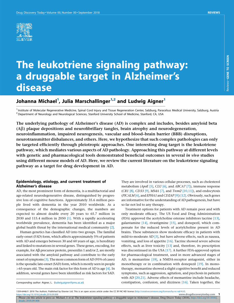
FIGURE 1
Pleiotropic effects of leukotrienes and leukotriene inhibition on the central nervousleads to enhanced synaptic integrity and to a reduction in amyloid beta (Ab) anFurthermore, several studies reported neuronal survival, increased neurogenesis, aLeukotrienes promote neuroinflammation. Inhibition of leukotrienes leads to lesssymbolize putative actions of leukotriene signaling that require further investiga
4 www.drugdiscoverytoday.com
molecular level, several mechanisms are discussed to cause BBB
leakage, although the actual cause remains elusive. Alterations
and loss of tight junction proteins [77], as well as pericyte degenera-
tion [76], might lead to BBB disruption at the capillary level. It is
likely although currently speculative, that APP and its metabolites
directly mediate toxic effects on vasculature in AD.
Leukotrienes have also been shown to increase BBB permeability
following brain damage [78,79]. Given that receptors for leuko-
trienes are expressed on endothelial cells and treatment with the
leukotriene receptor antagonist montelukast showed improved in-
tegrity of the BBB in an aging rat model [53], in a rat model of
traumatic brain injury [80], in a rat model of focal cerebral ischemia
[31], and in a mouse model of pharmacologically induced seizures
[81], it is likely that leukotrienes have a role in BBB leakage.
Complex pathologies require complex and/ormultimodal approachesAs illustrated above, the pathology of AD is complex and includes a
variety of aspects, including neuronal cell death, neuroinflamma-
tion, affected neurogenesis, disrupted BBB and vasculature system,
and others. Thus, a therapy should address as many aspects of these
pathological hallmarks as possible. For example, a combined ap-
proach targeting neurodegeneration, neuroinflammation, neuro-
genesis, and vascular homeostasis, and resealing the BBB, could be
considered. Ideally, this could be achieved with a drug that affects all
of these issues simultaneously via a pleiotropic mechanism. A pos-
sible way to achieve this might be by targeting the leukotriene
system, which mediates various aspects of AD pathology (Fig. 1).
The leukotriene systemUpstream of leukotriene synthesis, phospholipase 2 converts
phospholipids into polyunsaturated fatty acids [PUFAs; i.e., doc-
osahexaenoic acid (DHA), eicosapentaenoic acid (EPA), and ara-
chidonic acid (AA)]. DHA is further processed to anti-
inflammatory small lipid molecules, such as resolvins, maresins,
and protectins, summarized by the term ‘specialized pro-resolving
mediators’ (SPM). EPA is converted into the proinflammatory
gable target in Alzheimer’s disease, Drug Discov Today (2018), https://doi.org/10.1016/j.
Leukotrieneinhibition
survival
nesis
mmation
rrier integrity
load
orylation
integrity
Drug Discovery Today
system (CNS). Pharmacological or genetic inhibition of leukotriene signalingd tau pathology in several animal models of Alzheimer’s disease (AD).nd increased blood–brain barrier (BBB) integrity after leukotriene inhibition. neurotoxicity of microglia and also less activity of astrocytes. Broken linestions.

Drug Discovery Today �Volume 00, Number 00 � September 2018 REVIEWS
DRUDIS-2313; No of Pages 12
Review
s� G
ENETO
SCREE
N
molecules prostaglandins, thromboxanes, and prostacyclins, and
into the anti-inflammatory resolvins and protectins. AA generates
primarily proinflammatory prostanoids, such as prostaglandins,
thromboxanes, and prostacyclins, anti-inflammatory lipoxins, as
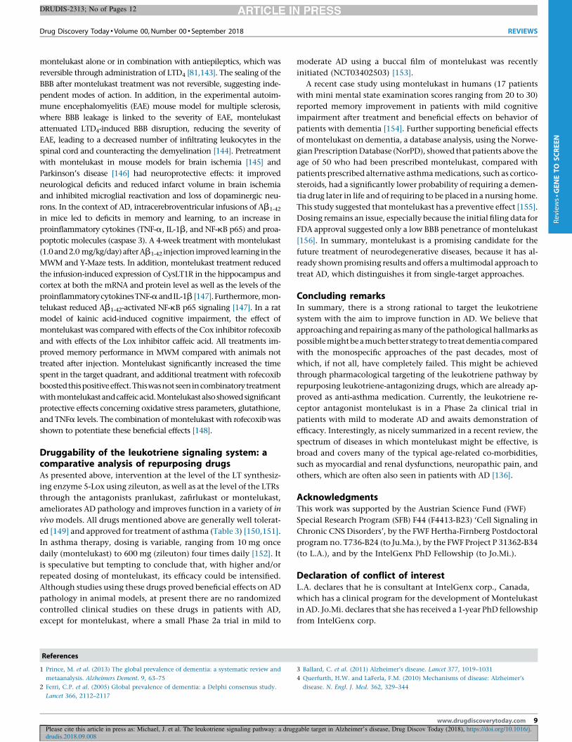
well as the proinflammatory leukotrienes. In more detail (Fig. 2),
AA is converted into 5-hydroxy-peroxy-eicosatetraenoic acid (5-
HPETE) and afterwards into leukotriene A4 (LTA4), both steps
mediated by 5-Lox [82]. Therefore, 5-Lox needs to form a complex
with 5-lipoxygenase activating protein (FLAP). LTA4 is further
processed either into leukotriene B4 (LTB4) by epoxide hydrolase
or into leukotriene C4 (LTC4) by LTC4 synthase. From LTC4, the
other two CysLTs LTD4 and LTE4 arise [83]. The CysLTs bind with
different affinities to the receptors cysteinyl leukotriene receptor 1
and 2 (CysLT1R [84] and CysLT2R [85]) and G-protein-coupled
receptor 17 (GPR17) [69,86]. For CysLT1R, LTD4 is the strongest
ligand, before LTC4 and then LTE4 [84]. LTD4 and LTC4 show
similar affinity, whereas LTE4 has a low affinity for CysLT2R [87].
The role of GPR17 in leukotriene signaling remains under debate
[88,89]. In addition, a possible binding of leukotrienes (LTE4) to
the receptor P2Y12 is has been proposed [84,90].
PUFAs and their metabolites have various functions in the
context of inflammation, in particular during the different phases
of inflammation. Whereas leukotrienes and prostanoids are proin-
Please cite this article in press as: Michael, J. et al. The leukotriene signaling pathway: a drugdrudis.2018.09.008
FLAP5-Lox
5-HPETE
CysLT1R CysLT2RGPR17
LTC4 synthase
LTA4
LTB4 LTC 4 LTD 4 LTE 4
Arachidonic acid
Zileuton,CNB-001
MK-591
Montelukast,Pranlukast,Zafirlukast
Drug Discovery Today
FIGURE 2
The leukotriene system and its inhibitors. Leukotrienes arise fromarachidonic acid, which is converted into 5-hydroxy-peroxy-eicosatetraenoicacid (5-HPETE) and afterwards into leukotriene A4, both steps mediated by5-Lox. 5-Lox needs to be activated by 5-lipoxygenase activating protein(FLAP). Leukotriene A4 is further processed either into leukotriene B4 byepoxide hydrolase or into leukotriene C4 by LTC4 synthase. From leukotrieneC4, the other two cysteinyl leukotrienes, LTD4 and LTE4, arise. The cysteinylleukotrienes bind with different affinities to the receptors CysLT1R, CysLT2R,and GPR17. Targeting the leukotriene system pharmacologically is possibleat the enzyme level, for example with zileuton or CNB-001, which inhibit 5-Lox or with MK-591, which is a FLAP inhibitor. Further downstreamleukotriene signaling can be blocked by receptor antagonists, such aspranlukast, zafirlukast or montelukast.
flammatory and involved in the initiation of inflammation, SPMs
are essential for dampening the immune system and are required
for resolving the inflammatory processes. Thereby, SPMs might
provide an opportunity for therapeutic interventions [91]. Indeed,
in patients with AD, the levels of lipoxins are lower in the cere-
brospinal fluid (CSF) and the hippocampus [92], and application of
resolvin and lipoxin reduced inflammation and Ab pathology in a
mouse model of AD [93]. Nevertheless, and despite this potential,
here we focus on leukotrienes as proinflammatory mediators in AD
and as a therapeutic target.
Leukotrienes were originally found in leukocytes, but 5-Lox
mRNA and protein are also widely expressed throughout the adult
brain [94]. 5-Lox and FLAP are highly expressed in neurons of the
hippocampus [95] and in microglia in vitro [96]. In addition,
astrocytes can generate and release CysLTs, measured by enzyme
immunoassay in cultures of rat cortical astrocytes [54]. 5-Lox also
is expressed in the cerebellum, primary olfactory cortex, superficial
neocortex, thalamus, hypothalamus, and brainstem [95]. After
traumatic brain injury, glial fibrillary acidic protein (GFAP)-posi-
tive glia cells elevate 5-Lox expression and neutrophils infiltrating
the lesioned brain, as well as endothelial cells, express 5-Lox [97].
Leukotrienes affect various cell types. For example, smooth
muscle cells increase contractility and proliferation, and epithelial
cells release more mucus following CysLT stimulation. In general,
binding of leukotrienes to leukocytes enhances inflammatory
cytokine release in the periphery [98]. LTB4 and LTD4 act chemo-
tactically on neutrophils and eosinophils [99] and on endothelial
cells [100]. CysLTs have vaso- and bronchoconstrictory effects on
cells and, therefore, they are main targets in asthma therapy [83].
In the brain, leukotrienes induce the proliferation of astrocytes
[54], and endothelial cells increase vascular permeability upon
binding of CysLTs, contributing to BBB disruption. CysLTs have a
role in several diseases of the CNS, because they influence the
activity of microglia and of astrocytes [51]. With age, 5-Lox levels
and the activity of the leukotriene pathway are elevated in the
brain, likely contributing to age-related CNS diseases [101,102].
Expression of leukotriene receptorsCysLT1R is predominantly expressed in lymphoid cells of the
spleen and in peripheral blood leukocytes [84]. It is also expressed
at lower levels in lung, colon, small intestines, kidney, liver, heart,
pancreas, and brain [103]. In primary cells derived from rodent
brain, CysLT1R mRNA expression is found mainly in microglia and
to a lower extent in endothelial cells and oligodendrocytes [96]. In
humans, CysLT1R is expressed by microvascular endothelial cells
of the healthy brain and in brain tumor (astrocytoma and gang-
lioglioma) tissue [104]. After traumatic brain injury or in brain
tumors, CysLT1R is induced in neurons and astrocytes (identifica-
tion based on location and morphology) [105]. Elevation of LTD4
leads to significantly elevated levels of CysLT1R mRNA and protein
[106]. Systemic injections of lipopolysaccharide (LPS), which in-
duce cognitive dysfunctions and neuroinflammation, led to an
upregulation of CysLT1R mRNA and protein in hippocampus
homogenates of mice [107].
CysLT2R is expressed in spleen, heart, peripheral blood leuko-
cytes, and lung [108]. Moderate expression of this receptor is also
found in the CNS, with highest expression in spinal cord and
pituitary [109]. In the healthy brain, CysLT2R is expressed by
gable target in Alzheimer’s disease, Drug Discov Today (2018), https://doi.org/10.1016/j.
www.drugdiscoverytoday.com 5

REVIEWS Drug Discovery Today �Volume 00, Number 00 � September 2018
DRUDIS-2313; No of Pages 12
Reviews�G
ENETO
SCREEN
astrocytes, but not by neurons or microglia. After middle cerebral
artery occlusion, mRNA expression of CysLT2R is found in neu-
rons in the core during the acute phase (up to 1 day after reperfu-
sion) and in the boundary zone in microglia and in hypertrophic
astrocytes in the chronic phase (up to 2 weeks after reperfusion)
[29,30]. After traumatic brain injury in humans, CysLT2R expres-
sion is induced in cerebral vascular endothelial cells [110]. In an in
vitro oxygen-glucose-deprivation model of ischemia, CysLT2R in-
duced and mediated astrocyte cell death [111]. Contrary to this,
another in vitro study culturing rat astrocytes showed no expres-
sion of CysLT2R detectable by RT-PCR [54].
GPR17 is expressed in neurons and a subset of oligodendroglial
progenitor cells in the cortex. After focal ischemia, GPR17 is
upregulated in neurons within 48 h. Co-localization of GPR17
with the microglia marker Iba1 is not found in normal brain,
but 72 h after middle cerebral artery occlusion, infiltrating micro-
glia and/or macrophages (IB4 positive) show a positive staining for
GPR17. This indicates an early and transient induction of GPR17
in neurons in the lesioned area and an induction in infiltrating
cells during the chronic phase [112]. Expression of GPR17 can also
be found in the dentate gyrus in some neuronal progenitors and
granular neurons, olig2+ cells and in some Iba1+ cells, but not in
neural stem cells or astrocytes [53].
Expression of all three leukotriene receptors is induced or
upregulated in neurons upon insult and afterwards also in micro-
glia, suggesting that the leukotriene pathway has a role during the
acute and chronic phase of inflammation.
Targeting the leukotriene pathway in AD5-Lox is elevated in the brain of AD animal models and human AD
brains at the mRNA as well as the protein level [113,114], suggest-
ing a role of the leukotriene pathway in AD. Recently, a single
nucleotide polymorphism in ALOX5AP, the gene encoding FLAP,
was identified to increase the risk for AD 1.41-fold, strengthening a
putative role of the leukotriene system in AD [115]. Here, we
summarize data on genetic as well as on pharmacological
approaches targeting leukotriene signaling in the context of AD
and amyloid pathology (summarized in Table 2).
Genetic approaches to modulate leukotriene signalingin the context of ADIn transgenic AD mice with an amyloid plaque pathology, a 5-Lox
deficiency reduced the levels of Ab and its depositions in the brain
[113]. In addition, deletion of ALOX5, the gene encoding 5-Lox, led
to memory improvement and enhanced synaptic integrity, and to a
reduction in Ab and tau pathology in AD mice [116]. Vice versa,
overexpression of 5-Lox in 3xTg mice resulted in increased plaque
formation, increased levels of g-secretase, and increased levels of
total tau and phosphorylated tau [117]. Also, dexamethasone injec-
tions, which elevate 5-Lox expression, resulted in significantly
increased levels of Ab1-40 and Ab1-42. In animals lacking ALOX5,
dexamethasone injections had no effect on Ab1-40 and Ab1-42 [118].
An influence of leukotrienes on tau pathology is substantiated by
studies in two models of tauopathy: htau mice, a transgenic model
bearing the human tau gene (MAPT), and PS19 mice, a model with a
MAPT P301S mutation. In both models, overexpression of 5-Lox led
to higher levels of activation (p25 and p35) in the cdk5 kinase
pathway, resulting in higher tau phosphorylation. Furthermore,
Please cite this article in press as: Michael, J. et al. The leukotriene signaling pathway: a drugdrudis.2018.09.008
6 www.drugdiscoverytoday.com
both models displayed a worsening of cognitive deficits in behav-
ioral testing, probably as a result of disruption in synaptic integrity
and increased neuroinflammation [119,120]. Vice versa, genetic
knockout of ALOX5 in PS19 mice had contrary effects, namely
reduced levels of tau phosphorylation, insoluble tau and p25/p35,
as well as amelioration of cognitive deficits, accompanied by pre-
served synaptic integrity and decreased neuroinflammation [121].
Effects of 5-Lox on tau phosphorylation, synaptic integrity, and
neuroinflammation were also described in a mouse model of AD-
related tau pathology (Tg2576) [122].
Pharmacological approaches to modulate leukotrienesignaling in the context of ADAs illustrated in Fig. 2, the leukotriene system can be pharmaco-
logically addressed with approved drugs at two different levels: at
the level of 5-Lox and at the level of the leukotriene receptors.
Inhibition of leukotriene synthesisIn animal models of AD, zileuton, a specific 5-Lox inhibitor,
reduced Ab levels and plaque deposition in a mouse model of
amyloidosis (Tg2576) [123], and reduced the levels of insoluble
and of hyperphosphorylated tau in a mouse model with plaques
and tangles (3xTG) [116,124]. In 12-month-old 3xTG mice, 3
months of zileuton treatment resulted in significant lower amy-
loid burdens compared with placebo treatment. In Y-maze and
fear-conditioning tests, zileuton stabilized the behavior compared
with baseline (12 month), whereas placebo-treated mice showed
significant deterioration in the behavioral tests [125]. Another
pharmacological 5-Lox blocker, CNB-001, led to significant lower
amounts of soluble Ab1-42 in treated APP/PS1 animals, improve-
ments in contextual memory (fear conditioning) and better mem-
ory in the Y-maze test [126]. Flavocoxid, a pharmacological dual
inhibitor of Cox1-2 and 5-Lox, improved learning and memory
function in 3xTg mice after 3 months of treatment compared with
vehicle-treated controls, and reduced amyloid deposition [127]. In
another study, direct pharmacological blockade of 5-Lox with the
inhibitor AA-861 as well as indirect pharmacological blockade of 5-
Lox via the inhibition of FLAP with MK-591 in a N2A cell culture
after dexamethasone challenge, counteracted the elevated levels
of Ab1-40 and Ab1-42 caused by dexamethasone, further supporting
a beneficial effect of targeting the leukotriene pathway in AD
[118]. Furthermore, targeting FLAP pharmacologically positively
influenced tau pathology in vivo in Tg2576 mice [128] and ame-
liorated pathology in 3xTg mice [129]. The mode of action of 5-Lox
inhibition on reduced plaque pathology is unclear, but could be
via modulation of y-secretase [130]. Indeed, a study by Chu et al.
revealed a significant reduction of all four components of g-secre-tase in WB = Western Blot after zileuton treatment in Tg2576 mice,
which was confirmed at the mRNA level by RT-PCR [123]. In two
mouse models of tauopathy, htau and PS19, 7 months of treat-
ment with zileuton starting at an early stage of pathology (3
months) led to significant improvement in memory performance
[Morris Water Maze (MWM) and Y-Maze] as well as decreased tau
phosphorylation accompanied by lower levels of co-activators for
cdk5, reduced GFAP and CD45 and increased synaptic integrity
[131,132]. This effect was also present when pharmacological 5-
Lox inhibition started after the onset of pathology (12 months of
age) in htau mice [133].
gable target in Alzheimer’s disease, Drug Discov Today (2018), https://doi.org/10.1016/j.

Drug Discovery Today �Volume 00, Number 00 � September 2018 REVIEWS
DRUDIS-2313; No of Pages 12
Please cite this article in press as: Michael, J. et al. The leukotriene signaling pathway: a druggable target in Alzheimer’s disease, Drug Discov Today (2018), https://doi.org/10.1016/j.drudis.2018.09.008
TABLE 2
Genetic and pharmacological inhibition of the leukotriene pathway and its effects in animal models of AD with amyloid pathology
Model Modulation Results Refs
Tg2576/5LO�/�
mice; 15-monthsold
Genetic: no expression of5-Lox
# Ab reactivity in hippocampus, somatosensory, andperihippocampal cortex# Ab1-40 and Ab1-42
Unaltered level of total APP# g-secretase activity
[113]
Tg2576 mice;7-months old
Pharmacological: inhibition of5-Lox by orally administeredZileuton for 8 months
# Ab1-40 and Ab1-42 (soluble and insoluble forms) inhippocampus and cortexUnaltered level of total APP# Components of g-secretase (PS1, Pen2, APH-1, and nicastrin) onmRNA and protein levelUnaltered levels of a- and b-secretase
[123]
5-LO�/� mice Genetic: absence of 5-Lox;dexamethasone injection toboost 5-Lox
5-LO+/+ mice injected with dexamethasone: " levels of Ab1-40
and Ab1-42
5-LO�/� mice injected with dexamethasone: no significant effecton Ab1-40 and Ab1-42 levels
[118]
Tg2576 mice;5-LO�/� mice;Tg2576/5-LO�/�
mice
Genetic: no expression of5-Lox; pharmacologic:inhibition of 5-Lox withAA-861 in Tg2576
Tg2576: significantly elevated levels of 5-Lox compared with WT;Upon treatment: # Ab, associated with # of p-CREBTg2576/5-LO�/�: # p-CREB and total CREB compared withuntreated Tg25765-LO�/�:# levels of Ab1-40 and Ab1-42
# mRNA levels of components of g-secretase
[130]
3xTg mice;13-months old
Genetic: 5-Lox overexpression " Plaque formation" Levels of four components of g-secretase" Total tau, phosphorylated tau
[117]
Tg2576 mice;7-months old
Pharmacological: inhibition ofFLAP by MK-591 for 8 month
No change in endogenous tau# Phosphorylated tau (Ser396, Ser396/Ser404, Thr 231/Ser 235)# Levels of insoluble tau# Activity of GSK-3b
[128]
3xTg mice Genetic: knockout of FLAP;pharmacological: inhibitionof FLAP by MK-591
" Memory (MWM)# Ab-deposition, Ab1-40, Ab1-42
Unaltered levels of APP# Levels of four components of g-secretaseUnaltered levels of total tau# Phosphorylated tau (S396, S396/S404)# insoluble tau# Cdk5 kinase" Synaptic integrity# Astrocyte and microglia activity (GFAP, CD45)
[129]
3xTg mice;2–3-months old
Pharmacological: inhibition of5-Lox by orally administeredZileuton (approx. 0.6–0.8 mgper day) for 10 months
Cognitive improvement (MWM)# Levels of Ab1-40 and Ab1-42
# Amyloid burden (IHC)# PS1, Pen-2 and APH-1 (WB)# Dendritic accumulation of phosphorylated tau (IHC)No change in overall tau (WB, IHC)# Levels of insoluble tau and phosphorylated tau# p25 and p35 fragments of cdk5 (WB)" Proteins essential for synaptic integrity (synaptophysin, PDS-95,MAP-2)# GFAP and CD45 immunoreactivity
[124]
APP/PS1 mice(line 85);3-months old
Pharmacological: inhibition of5-Lox by CNB-001 for 6months
Behavioral improvement (Y-Maze)# Soluble Ab1-42
[126]
3xTg; 12-monthsold
Pharmacological: inhibition of5-Lox by zileuton for 3months
Behavioral improvement (Y-Maze)# Ab-deposition, Ab1-42
# PS1, Pen-2# p25 fragments of cdk5
[125]
3xTg/5LoKO;6–8-months old;12–14 monthsold; 3xTg mice;5-months old
Genetic: knockout of 5-Lox;pharmacological: inhibitionof 5-Lox by zileuton for 1month
Cognitive improvement (Y-maze)# Ab-deposition, Ab1-40, Ab1-42
Unaltered levels of total APP# Levels of four components of g-secretase# Levels of insoluble tau# p25 and p35 fragments of cdk5" Synaptic integrity (PSD-95 and synaptophysin)# GFAP and CD45 immunoreactivity
[116]
www.drugdiscoverytoday.com 7
Review
s� G
ENETO
SCREE
N

REVIEWS Drug Discovery Today �Volume 00, Number 00 � September 2018
DRUDIS-2313; No of Pages 12
TABLE 2 (Continued )
Model Modulation Results Refs
ICR mice:intracerebralinfusion of Ab1-42
Pharmacological: inhibition ofCysLT1R by Montelukast
" Learning deficits after Ab1-42 infusion (MWM, Y-Maze)# Infusion induced expression of CysLT1R in hippocampus andcortex# Ab1-42 activated NF-kB p65 signaling, proinflammatorycytokine levels of TNF-a and IL-1b# Caspase 3" Bcl-2
[142]
ICR mice: LPSinjections
Genetic: knockdown ofCysLT1R; pharmacological:inhibition of CysLT1R
Prevented cognitive impairment by LPS# Number of apoptotic cells (pretreatment with pranlukast)Inhibition of upregulation of proinflammatory cytokines (IL-1b,TNF-a)Lower expression of CysLT1R on mRNA and protein level
[107]
3xTg-AD mice Pharmacological: inhibition of5-Lox by flavocoxid (20 mg/kg/ip) for 3 months
" Learning and memory (MWM)# Amyloid deposit compared with saline-treated animals# LTB4, IL1-b# p-Tau compared with saline-treated animals (but not comparedwith WT levels)
[127]
Reviews�G
ENETO
SCREEN
Antagonizing leukotriene receptorsOral treatment with the leukotriene receptor antagonist pranlu-
kast improved cognitive deficits caused by intracerebroventricular
injections of Ab1-42 in ICR mice and suppressed NF-kB signaling
caused by Ab [134]. Daily intraperitoneal injections of zafirlukast,
another antagonist for CysLTRs, for 21 days improved learning
behavior in Ab intracerebroventricularly injected rats [135]. An-
other selective leukotriene receptor antagonist is montelukast,
which is increasingly discussed as a treatment option in AD and
other neurodegenerative diseases [51,52,136].
Montelukast is approved by the FDA for the treatment of asthma
[137,138] and its effectivity and tolerance has been shown in many
studies (reviewed in [136,139]). In the periphery, montelukast acts on
cells expressing receptors for cysteinyl leukotrienes, mainly mono-
Please cite this article in press as: Michael, J. et al. The leukotriene signaling pathway: a drugdrudis.2018.09.008
TABLE 3
Key pharmacological data of approved drugs targeting the leukotr
Drug Effective dose Efficacy in hum
In animal models of CNSdisease
In humans
Montelukast 0.1 mg/kg i.p.; 0.25 mmol i.c.v.; 0.3 mmol/mL i.c.v.;1 mg/kg i.p.;1 or 2 mg/kg i.g.; 10 mg/kgp.o.; 30 mg/kg i.p.; 40 mg/kg i.p.
10 mg/d p.o. fortreatment of asthma
Excellent in 23.8Good in 39.6% oFair in 19.8% ofPoor in 16.8% oN = 101
80 mg/d (4 � 20 mg p.o.)for treatment ofdementia (off-label usein case report)
Improved memowith cognitive imLess agitation indementia
Pranlukast 0.1 mg/kg i.p.; 0.4 mg/kgp.o.; 0.8 mg/kg p.o.;1.5 ng/animal i.c.v.; 1 and3 mmol i.c.v.
450 mg bd p.o. fortreatment of asthma
27.4% improvemsymptoms compbaseline
Zafirlukast 30 mg/kg i.p.; 30 mg/kg i.p. 40 mg/d (2 � 20 mg p.o.)for treatment of asthma
3.7 � 15.4% impforced expiratorcompared with
Zileuton 0.6–0,8 mg/day in drinkingwater (200 mg/l)
2400 mg/d (4 � 600 mgp.o.) for treatment ofasthma
Excellent in 51.4Good in 35.8% oFair in 10.1% ofPoor in 2.8% of
N = 109
8 www.drugdiscoverytoday.com
cytes, eosinophils, basophils, and mast cells [98,140], but it was also
shown that montelukast prevented reactive oxygen species (ROS)
and LTB4 production in isolated human neutrophils that were acti-
vated by chemoattractants [141]. In fetal murine neurons, montelu-
kast blocked Ab1-42-induced cell death, suppressed the expression of
CysLT1R, and reduced the production of proinflammatory cytokines
and the activation of caspase-3 [142]. In another study, using rat
neuronal precursor cell (NPC) cultures, montelukast had a stimulat-
ing effect on proliferation without influencing differentiation [69].
InhibitionofCysLT1Rbymontelukastblockedtheproinflammatory
actions of LTD4, for example on the BBB, in vivo. It prevented pentyl-
enetetrazol injection-induced BBB disruption in mouse brains and
acted as an anticonvulsive. The latter effect was revoked by administra-
tion of LTD4 [81]. Other studies also revealed an anticonvulsive effect of
gable target in Alzheimer’s disease, Drug Discov Today (2018), https://doi.org/10.1016/j.
iene system
ans Safety Profile/Tolerability Refs
% of patientsf patients
patientsf patients
Excellent in 89.6% of patientsGood in 1.9% of patientsFair in 8.5% of patientsN = 106
[31,53,81,143–148,152]
ry in patientspairment
patients with
No adverse effects reported [154]
ent inared with
Well tolerated [31,32,81,107,134,150]
rovement iny volumebaseline
Well tolerated [135,144,151]
% of patientsf patients
patientspatients
Excellent in 92.8% of patientsGood in 2.7% of patientsFair in 4.5% of patientsN = 111Hepatotoxic adverse effect
[123–125,131–133,149,152]
Drug Discovery Today �Volume 00, Number 00 � September 2018 REVIEWS
DRUDIS-2313; No of Pages 12
Review
s� G
ENETO
SCREE
N
montelukast alone or in combination with antiepileptics, which was
reversible through administration of LTD4 [81,143]. The sealing of the
BBB after montelukast treatment was not reversible, suggesting inde-
pendent modes of action. In addition, in the experimental autoim-
mune encephalomyelitis (EAE) mouse model for multiple sclerosis,
where BBB leakage is linked to the severity of EAE, montelukast
attenuated LTD4-induced BBB disruption, reducing the severity of
EAE, leading to a decreased number of infiltrating leukocytes in the
spinal cord and counteracting the demyelination [144]. Pretreatment
with montelukast in mouse models for brain ischemia [145] and
Parkinson’s disease [146] had neuroprotective effects: it improved
neurological deficits and reduced infarct volume in brain ischemia
and inhibited microglial reactivation and loss of dopaminergic neu-
rons. In the context of AD, intracerebroventricular infusions of Ab1-42
in mice led to deficits in memory and learning, to an increase in
proinflammatory cytokines (TNF-a, IL-1b, and NF-kB p65) and proa-
poptotic molecules (caspase 3). A 4-week treatment with montelukast
(1.0 and 2.0 mg/kg/day) after Ab1-42 injection improved learning in the
MWM and Y-Maze tests. In addition, montelukast treatment reduced
the infusion-induced expression of CysLT1R in the hippocampus and
cortex at both the mRNA and protein level as well as the levels of the
proinflammatorycytokinesTNF-a andIL-1b [147].Furthermore,mon-
telukast reduced Ab1-42-activated NF-kB p65 signaling [147]. In a rat
model of kainic acid-induced cognitive impairment, the effect of
montelukast was compared with effects of the Cox inhibitor rofecoxib
and with effects of the Lox inhibitor caffeic acid. All treatments im-
proved memory performance in MWM compared with animals not
treated after injection. Montelukast significantly increased the time
spent in the target quadrant, and additional treatment with rofecoxib
boostedthispositiveeffect.Thiswasnotseenincombinatorytreatment
with montelukastandcaffeic acid.Montelukastalsoshowedsignificant
protective effects concerning oxidative stress parameters, glutathione,
and TNFa levels. The combination of montelukast with rofecoxib was
shown to potentiate these beneficial effects [148].
Druggability of the leukotriene signaling system: acomparative analysis of repurposing drugsAs presented above, intervention at the level of the LT synthesiz-
ing enzyme 5-Lox using zileuton, as well as at the level of the LTRs
through the antagonists pranlukast, zafirlukast or montelukast,
ameliorates AD pathology and improves function in a variety of in
vivo models. All drugs mentioned above are generally well tolerat-
ed [149] and approved for treatment of asthma (Table 3) [150,151].
In asthma therapy, dosing is variable, ranging from 10 mg once
daily (montelukast) to 600 mg (zileuton) four times daily [152]. It
is speculative but tempting to conclude that, with higher and/or
repeated dosing of montelukast, its efficacy could be intensified.
Although studies using these drugs proved beneficial effects on AD
pathology in animal models, at present there are no randomized
controlled clinical studies on these drugs in patients with AD,
except for montelukast, where a small Phase 2a trial in mild to
Please cite this article in press as: Michael, J. et al. The leukotriene signaling pathway: a drugdrudis.2018.09.008
moderate AD using a buccal film of montelukast was recently
initiated (NCT03402503) [153].
A recent case study using montelukast in humans (17 patients
with mini mental state examination scores ranging from 20 to 30)
reported memory improvement in patients with mild cognitive
impairment after treatment and beneficial effects on behavior of
patients with dementia [154]. Further supporting beneficial effects
of montelukast on dementia, a database analysis, using the Norwe-
gian Prescription Database (NorPD), showed that patients above the
age of 50 who had been prescribed montelukast, compared with
patients prescribed alternative asthma medications, such as cortico-
steroids, had a significantly lower probability of requiring a demen-
tia drug later in life and of requiring to be placed in a nursing home.
This study suggested that montelukast has a preventive effect [155].
Dosing remains an issue, especially because the initial filing data for
FDA approval suggested only a low BBB penetrance of montelukast
[156]. In summary, montelukast is a promising candidate for the
future treatment of neurodegenerative diseases, because it has al-
ready shown promising results and offers a multimodal approach to
treat AD, which distinguishes it from single-target approaches.
Concluding remarksIn summary, there is a strong rational to target the leukotriene
system with the aim to improve function in AD. We believe that
approaching and repairing as many of the pathological hallmarks as
possible might be a much better strategy to treat dementia compared
with the monospecific approaches of the past decades, most of
which, if not all, have completely failed. This might be achieved
through pharmacological targeting of the leukotriene pathway by
repurposing leukotriene-antagonizing drugs, which are already ap-
proved as anti-asthma medication. Currently, the leukotriene re-
ceptor antagonist montelukast is in a Phase 2a clinical trial in
patients with mild to moderate AD and awaits demonstration of
efficacy. Interestingly, as nicely summarized in a recent review, the
spectrum of diseases in which montelukast might be effective, is
broad and covers many of the typical age-related co-morbidities,
such as myocardial and renal dysfunctions, neuropathic pain, and
others, which are often also seen in patients with AD [136].
AcknowledgmentsThis work was supported by the Austrian Science Fund (FWF)
Special Research Program (SFB) F44 (F4413-B23) ‘Cell Signaling in
Chronic CNS Disorders’, by the FWF Hertha-Firnberg Postdoctoral
program no. T736-B24 (to Ju.Ma.), by the FWF Project P 31362-B34
(to L.A.), and by the IntelGenx PhD Fellowship (to Jo.Mi.).
Declaration of conflict of interestL.A. declares that he is consultant at IntelGenx corp., Canada,
which has a clinical program for the development of Montelukast
in AD. Jo.Mi. declares that she has received a 1-year PhD fellowship
from IntelGenx corp.
References
1 Prince, M. et al. (2013) The global prevalence of dementia: a systematic review and
metaanalysis. Alzheimers Dement. 9, 63–75
2 Ferri, C.P. et al. (2005) Global prevalence of dementia: a Delphi consensus study.
Lancet 366, 2112–2117
3 Ballard, C. et al. (2011) Alzheimer’s disease. Lancet 377, 1019–1031
4 Querfurth, H.W. and LaFerla, F.M. (2010) Mechanisms of disease: Alzheimer’s
disease. N. Engl. J. Med. 362, 329–344
gable target in Alzheimer’s disease, Drug Discov Today (2018), https://doi.org/10.1016/j.
www.drugdiscoverytoday.com 9

REVIEWS Drug Discovery Today �Volume 00, Number 00 � September 2018
DRUDIS-2313; No of Pages 12
Reviews�G
ENETO
SCREEN
5 Selkoe, D.J. (2001) Alzheimer’s disease: genes, proteins, and therapy. Physiol. Rev. 81,
741–766
6 Harold, D. et al. (2009) Genome-wide association study identifies variants at CLU and
PICALM associated with Alzheimer’s disease. Nat. Genet. 41, 1088–1093
7 Hollingworth, P. et al. (2011) Common variants at ABCA7, MS4A6A/MS4A4E, EPHA1,
CD33 and CD2AP are associated with Alzheimer’s disease. Nat. Genet. 43, 429–435
8 Lambert, J.C. et al. (2009) Genome-wide association study identifies variants at CLU
and CR1 associated with Alzheimer’s disease. Nat. Genet. 41, 1094–1099
9 Naj, A.C. et al. (2011) Common variants at MS4A4/MS4A6E, CD2AP, CD33 and
EPHA1 are associated with late-onset Alzheimer’s disease. Nat. Genet. 43, 436–441
10 Bellenguez, C. et al. (2017) Contribution to Alzheimer’s disease risk of rare variants in
TREM2, SORL1, and ABCA7 in 1779 cases and 1273 controls. Neurobiol. Aging 59, 220
11 Guerreiro, R. et al. (2013) TREM2 variants in Alzheimer’s disease. N. Engl. J. Med. 368,
117–127
12 Karch, C.M. and Goate, A.M. (2015) Alzheimer’s disease risk genes and mechanisms
of disease pathogenesis. Biol. Psychiatry 77, 43–51
13 Crismon, M.L. (1994) Tacrine: first drug approved for Alzheimer’s disease. Ann.
Pharmacother. 28, 744–751
14 Razay, G. and Wilcock, G.K. (2008) Galantamine in Alzheimer’s disease. Expert Rev.
Neurother. 8, 9–17
15 Birks, J.S. and Grimley Evans, J. (2015) Rivastigmine for Alzheimer’s disease.
Cochrane Database Syst. Rev. 2015, CD001191
16 Mimica, N. and Presecki, P. (2009) Side effects of approved antidementives.
Psychiatr. Danub. 21, 108–113
17 Alzheimer’s Association (2017) Alzheimer’s disease facts and figures. Alzheimers
Dement. 13, 325–373
18 Matsunaga, S. et al. (2015) Memantine monotherapy for Alzheimer’s disease: a
systematic review and meta-analysis. PLoS One 10, e0123289
19 Chen, R. et al. (2017) Treatment effects between monotherapy of donepezil versus
combination with memantine for Alzheimer disease: a meta-analysis. PLoS One 12,
e0183586
20 McShane, R. et al. (2006) Memantine for dementia. Cochrane Database Syst. Rev.
2006, CD003154
21 Gauthier, S. et al. (2008) Improvement in behavioural symptoms in patients with
moderate to severe Alzheimer’s disease by memantine: a pooled data analysis. Int. J.
Geriatr. Psychiatry 23, 537–545
22 Kumar, A. et al. (2015) A review on Alzheimer’s disease pathophysiology and its
management: an update. Pharmacol. Rep. 67, 195–203
23 Selkoe, D.J. and Hardy, J. (2016) The amyloid hypothesis of Alzheimer’s disease at 25
years. EMBO Mol. Med. 8, 595–608
24 Small, S.A. and Duff, K. (2008) Linking Abeta and tau in late-onset Alzheimer’s
disease: a dual pathway hypothesis. Neuron 60, 534–542
25 Maccioni, R.B. et al. (2010) The revitalized tau hypothesis on Alzheimer’s disease.
Arch. Med. Res. 41, 226–231
26 De Strooper, B. and Karran, E. (2016) The cellular phase of Alzheimer’s disease. Cell
164, 603–615
27 Cotman, C.W. and Su, J.H. (1996) Mechanisms of neuronal death in Alzheimer’s
disease. Brain Pathol. 6, 493–506
28 Donev, R. et al. (2009) Neuronal death in Alzheimer’s disease and therapeutic
opportunities. J. Cell Mol. Med. 13, 4329–4348
29 Fang, S.H. et al. (2007) Spatio-temporal expression of cysteinyl leukotriene receptor-
2 mRNA in rat brain after focal cerebral ischemia. Neurosci. Lett. 412, 78–83
30 Zhao, C.Z. et al. (2011) Cysteinyl leukotriene receptor 2 is spatiotemporally involved
in neuron injury, astrocytosis and microgliosis after focal cerebral ischemia in rats.
Neuroscience 189, 1–11
31 Zhao, R. et al. (2011) Montelukast, a cysteinyl leukotriene receptor-1 antagonist,
attenuates chronic brain injury after focal cerebral ischaemia in mice and rats. J.
Pharm. Pharmacol. 63, 550–557
32 Tang, S.S. et al. (2014) Involvement of cysteinyl leukotriene receptor 1 in Abeta1-42-
induced neurotoxicity in vitro and in vivo. Neurobiol. Aging 35, 590–599
33 Heppner, F.L. et al. (2015) Immune attack: the role of inflammation in Alzheimer
disease. Nat. Rev. Neurosci. 16, 358–372
34 Heneka, M.T. et al. (2015) Neuroinflammation in Alzheimer’s disease. Lancet Neurol.
14, 388–405
35 Clark, I.A. and Vissel, B. (2015) Amyloid beta: one of three danger-associated
molecules that are secondary inducers of the proinflammatory cytokines that
mediate Alzheimer’s disease. Br. J. Pharmacol. 172, 3714–3727
36 Sondag, C.M. et al. (2009) Beta amyloid oligomers and fibrils stimulate differential
activation of primary microglia. J. Neuroinflammation 6, 1
37 Griffin, W.S. et al. (1998) Glial-neuronal interactions in Alzheimer’s disease: the
potential role of a ‘cytokine cycle’ in disease progression. Brain Pathol. 8, 65–72
38 Prokop, S. et al. (2013) Microglia actions in Alzheimer’s disease. Acta Neuropathol.
126, 461–477
Please cite this article in press as: Michael, J. et al. The leukotriene signaling pathway: a drugdrudis.2018.09.008
10 www.drugdiscoverytoday.com
39 Lucin, K.M. et al. (2013) Microglial beclin 1 regulates retromer trafficking and
phagocytosis and is impaired in Alzheimer’s disease. Neuron 79, 873–886
40 Krabbe, G. et al. (2013) Functional impairment of microglia coincides with Beta-
amyloid deposition in mice with Alzheimer-like pathology. PLoS One 8, e60921
41 Gee, J.R. and Keller, J.N. (2005) Astrocytes: regulation of brain homeostasis via
apolipoprotein E. Int. J. Biochem. Cell. Biol. 37, 1145–1150
42 Wyss-Coray, T. et al. (2003) Adult mouse astrocytes degrade amyloid-beta in vitro
and in situ. Nat. Med. 9, 453–457
43 Kraft, A.W. et al. (2013) Attenuating astrocyte activation accelerates plaque
pathogenesis in APP/PS1 mice. FASEB J. 27, 187–198
44 Ferretti, M.T. et al. (2016) T-cell brain infiltration and immature antigen-presenting
cells in transgenic models of Alzheimer’s disease-like cerebral amyloidosis. Brain
Behav. Immun. 54, 211–225
45 Ardura-Fabregat, A. et al. (2017) Targeting neuroinflammation to treat Alzheimer’s
disease. CNS Drugs 31, 1057–1082
46 Watson, G.S. et al. (2005) Preserved cognition in patients with early Alzheimer
disease and amnestic mild cognitive impairment during treatment with
rosiglitazone: a preliminary study. Am. J. Geriatr. Psychiatry 13, 950–958
47 Rogers, J. et al. (1993) Clinical trial of indomethacin in Alzheimer’s disease.
Neurology 43, 1609–1611
48 de Jong, D. et al. (2008) No effect of one-year treatment with indomethacin on
Alzheimer’s disease progression: a randomized controlled trial. PLoS One 3, e1475
49 Pasqualetti, P. et al. (2009) A randomized controlled study on effects of ibuprofen on
cognitive progression of Alzheimer’s disease. Aging Clin. Exp. Res. 21, 102–110
50 Jaturapatporn, D. et al. (2012) Aspirin, steroidal and non-steroidal anti-
inflammatory drugs for the treatment of Alzheimer’s disease. Cochrane Database
Syst. Rev. 2012, CD006378
51 Ghosh, A. et al. (2016) Cysteinyl leukotrienes and their receptors: emerging
therapeutic targets in central nervous system disorders. CNS Neurosci. Ther. 22, 943–
951
52 Gelosa, P. et al. (2017) Cysteinyl leukotrienes as potential pharmacological targets
for cerebral diseases. Mediators Inflamm. 2017, 3454212
53 Marschallinger, J. et al. (2015) Structural and functional rejuvenation of the aged
brain by an approved anti-asthmatic drug. Nat. Commun. 6, 8466
54 Ciccarelli, R. et al. (2004) Cysteinyl-leukotrienes are released from astrocytes and
increase astrocyte proliferation and glial fibrillary acidic protein via cys-LT1 receptors
and mitogen-activated protein kinase pathway. Eur. J. Neurosci. 20, 1514–1524
55 Yu, S.Y. et al. (2014) Cysteinyl leukotriene receptor 1 mediates LTD4-induced
activation of mouse microglial cells in vitro. Acta Pharmacol. Sin. 35, 33–40
56 Ballerini, P. et al. (2005) P2Y1 and cysteinyl leukotriene receptors mediate purine
and cysteinyl leukotriene co-release in primary cultures of rat microglia. Int. J.
Immunopathol. Pharmacol. 18, 255–268
57 Klegeris, A. and McGeer, P.L. (2002) Cyclooxygenase and 5-lipoxygenase inhibitors
protect against mononuclear phagocyte neurotoxicity. Neurobiol. Aging 23, 787–794
58 Klegeris, A. and McGeer, P.L. (2003) Toxicity of human monocytic THP-1 cells and
microglia toward SH-SY5Y neuroblastoma cells is reduced by inhibitors of 5-
lipoxygenase and its activating protein FLAP. J. Leukoc. Biol. 73, 369–378
59 Zhang, X.Y. et al. (2013) HAMI 3379, a CysLT2 receptor antagonist, attenuates
ischemia-like neuronal injury by inhibiting microglial activation. J. Pharmacol. Exp.
Ther. 346, 328–341
60 Ming, G.L. and Song, H. (2011) Adult neurogenesis in the mammalian brain:
significant answers and significant questions. Neuron 70, 687–702
61 Kempermann, G. et al. (2018) Human adult neurogenesis: evidence and remaining
questions. Cell Stem Cell 23, 25–30
62 Unger, M.S. et al. (2016) Early changes in hippocampal neurogenesis in transgenic
mouse models for Alzheimer’s disease. Mol. Neurobiol. 53, 5796–5806
63 Hollands, C. et al. (2016) Alzheimer’s disease and hippocampal adult neurogenesis;
exploring shared mechanisms. Front. Neurosci. 10, 178
64 Lazarov, O. and Marr, R.A. (2010) Neurogenesis and Alzheimer’s disease: at the
crossroads. Exp. Neurol. 223, 267–281
65 Haughey, N.J. et al. (2002) Disruption of neurogenesis in the subventricular zone of
adult mice, and in human cortical neuronal precursor cells in culture, by amyloid
beta-peptide: implications for the pathogenesis of Alzheimer’s disease. Neuromol.
Med. 1, 125–135
66 Sanchez-Ramos, J. et al. (2009) Granulocyte colony stimulating factor decreases
brain amyloid burden and reverses cognitive impairment in Alzheimer’s mice.
Neuroscience 163, 55–72
67 Shen, L. et al. (2016) D5 receptor agonist 027075 promotes cognitive function
recovery and neurogenesis in a Abeta1-42-induced mouse model.
Neuropharmacology 105, 72–83
68 Giuliani, D. et al. (2015) NDP-alpha-MSH induces intense neurogenesis and
cognitive recovery in Alzheimer transgenic mice through activation of
melanocortin MC4 receptors. Mol. Cell. Neurosci. 67, 13–21
gable target in Alzheimer’s disease, Drug Discov Today (2018), https://doi.org/10.1016/j.

Drug Discovery Today �Volume 00, Number 00 � September 2018 REVIEWS
DRUDIS-2313; No of Pages 12
Review
s� G
ENETO
SCREE
N
69 Huber, C. et al. (2011) Inhibition of leukotriene receptors boosts neural progenitor
proliferation. Cell. Physiol. Biochem. 28, 793–804
70 Manev, H. and Manev, R. (2006) 5-Lipoxygenase (ALOX5) and FLAP (ALOX5AP)
gene polymorphisms as factors in vascular pathology and Alzheimer’s disease. Med.
Hypotheses 66, 501–503
71 Bhattarai, P. et al. (2017) The effects of aging on Amyloid-beta42-induced
neurodegeneration and regeneration in adult zebrafish brain. Neurogenesis 4,
e1322666
72 Erickson, M.A. and Banks, W.A. (2013) Blood-brain barrier dysfunction as a cause
and consequence of Alzheimer’s disease. J. Cereb. Blood Flow Metab. 33, 1500–1513
73 Sweeney, M.D. et al. (2018) Blood–brain barrier breakdown in Alzheimer disease and
other neurodegenerative disorders. Nat. Rev. Neurol. 14, 133–150
74 van de Haar, H.J. et al. (2016) Blood–brain barrier leakage in patients with early
Alzheimer disease. Radiology 281, 527–535
75 Zlokovic, B.V. (2011) Neurovascular pathways to neurodegeneration in Alzheimer’s
disease and other disorders. Nat. Rev. Neurosci. 12, 723–738
76 Montagne, A. et al. (2017) Alzheimer’s disease: a matter of blood-brain barrier
dysfunction? J. Exp. Med. 214, 3151–3169
77 Zhao, L.B. et al. (2015) Establishment of a canine model of acute pulmonary
embolism with definite right ventricular dysfunction through introduced
autologous blood clots. Thromb. Res. 135, 727–732
78 Black, K.L. and Hoff, J.T. (1985) Leukotrienes increase blood-brain barrier permeability
following intraparenchymal injections in rats. Ann. Neurol. 18, 349–351
79 Chio, C.C. et al. (1992) Selective blood-tumor barrier disruption by leukotrienes. J.
Neurosurg. 77, 407–410
80 Biber, N. et al. (2009) Cysteinyl-leukotriene receptor antagonist montelukast
decreases blood–brain barrier permeability but does not prevent oedema formation
in traumatic brain injury. Brain Inj. 23, 577–584
81 Lenz, Q.F. et al. (2014) Cysteinyl leukotriene receptor (CysLT) antagonists decrease
pentylenetetrazol-induced seizures and blood–brain barrier dysfunction.
Neuroscience 277, 859–871
82 Radmark, O. et al. (2015) 5-Lipoxygenase, a key enzyme for leukotriene biosynthesis
in health and disease. Biochim. Biophys. Acta 1851, 331–339
83 Samuelsson, B. (1983) Leukotrienes: mediators of immediate hypersensitivity
reactions and inflammation. Science 220, 568–575
84 Lynch, K.R. et al. (1999) Characterization of the human cysteinyl leukotriene
CysLT1 receptor. Nature 399, 789–793
85 Singh, R.K. et al. (2010) Cysteinyl leukotrienes and their receptors: molecular and
functional characteristics. Pharmacology 85, 336–349
86 Ciana, P. et al. (2006) The orphan receptor GPR17 identified as a new dual uracil
nucleotides/cysteinyl-leukotrienes receptor. EMBO J. 25, 4615–4627
87 Heise, C.E. et al. (2000) Characterization of the human cysteinyl leukotriene 2
receptor. J. Biol. Chem. 275, 30531–30536
88 Qi, A.D. et al. (2013) Is GPR17 a P2Y/leukotriene receptor? examination of uracil
nucleotides, nucleotide sugars, and cysteinyl leukotrienes as agonists of GPR17. J.
Pharmacol. Exp. Ther. 347, 38–46
89 Simon, K. et al. (2017) The orphan receptor GPR17 is unresponsive to uracil
nucleotides and cysteinyl leukotrienes. Mol. Pharmacol. 91, 518–532
90 Sarau, H.M. et al. (1999) Identification, molecular cloning, expression, and
characterization of a cysteinyl leukotriene receptor. Mol. Pharmacol. 56, 657–663
91 Serhan, C.N. (2014) Pro-resolving lipid mediators are leads for resolution
physiology. Nature 510, 92–101
92 Wang, X. et al. (2015) Resolution of inflammation is altered in Alzheimer’s disease.
Alzheimers Dement. 11, 40–50
93 Kantarci, A. et al. (2018) Combined administration of resolvin E1 and lipoxin A4
resolves inflammation in a murine model of Alzheimer’s disease. Exp. Neurol. 300,
111–120
94 Chinnici, C.M. et al. (2007) The 5-lipoxygenase enzymatic pathway in the mouse
brain: young versus old. Neurobiol. Aging 28, 1457–1462
95 Lammers, C.H. et al. (1996) Arachidonate 5-lipoxygenase and its activating protein:
prominent hippocampal expression and role in somatostatin signaling. J.
Neurochem. 66, 147–152
96 Zhang, Y. et al. (2014) An RNA-sequencing transcriptome and splicing database
of glia, neurons, and vascular cells of the cerebral cortex. J. Neurosci 34, 11929–
11947
97 Zhang, L. et al. (2006) Expression patterns of 5-lipoxygenase in human brain with
traumatic injury and astrocytoma. Neuropathology 26, 99–106
98 Peters-Golden, M. and Henderson, W.R., Jr (2007) Leukotrienes. N. Engl. J. Med. 357,
1841–1854
99 Funk, C.D. (2001) Prostaglandins and leukotrienes: advances in eicosanoid biology.
Science 294, 1871–1875
100 Yuan, Y.M. et al. (2009) Leukotriene D4 stimulates the migration but not
proliferation of endothelial cells mediated by the cysteinyl leukotriene cyslt(1)
Please cite this article in press as: Michael, J. et al. The leukotriene signaling pathway: a drugdrudis.2018.09.008
receptor via the extracellular signal-regulated kinase pathway. J. Pharmacol. Sci.
109, 285–292
101 Manev, H. et al. (2000) Putative role of neuronal 5-lipoxygenase in an aging brain.
FASEB J. 14, 1464–1469
102 Uz, T. et al. (1998) Aging-associated up-regulation of neuronal 5-lipoxygenase
expression: putative role in neuronal vulnerability. FASEB J. 12, 439–449
103 Nonaka, Y. et al. (2005) Identification of endogenous surrogate ligands for human
P2Y12 receptors by in silico and in vitro methods. Biochem. Biophys. Res. Commun.
337, 281–288
104 Zhang, W.P. et al. (2004) Expression of cysteinyl leukotriene receptor 1 in human
traumatic brain injury and brain tumors. Neurosci. Lett. 363, 247–251
105 Maekawa, A. et al. (2008) Functional recognition of a distinct receptor preferential
for leukotriene E4 in mice lacking the cysteinyl leukotriene 1 and 2 receptors. Proc.
Natl. Acad. Sci. U. S. A. 105, 16695–16700
106 Tang, S.S. et al. (2013) Leukotriene D4 induces cognitive impairment through
enhancement of CysLT(1) R-mediated amyloid-beta generation in mice.
Neuropharmacology 65, 182–192
107 Chen, F. et al. (2017) Preventive effect of genetic knockdown and pharmacological
blockade of CysLT1R on lipopolysaccharide (LPS)-induced memory deficit and
neurotoxicity in vivo. Brain Behav. Immun. 60, 255–269
108 Takasaki, J. et al. (2000) The molecular characterization and tissue distribution of
the human cysteinyl leukotriene CysLT(2) receptor. Biochem. Biophys. Res.
Commun. 274, 316–322
109 Nothacker, H.P. et al. (2000) Molecular cloning and characterization of a second
human cysteinyl leukotriene receptor: discovery of a subtype selective agonist.
Mol. Pharmacol. 58, 1601–1608
110 Hu, H. et al. (2005) Distribution of cysteinyl leukotriene receptor 2 in human
traumatic brain injury and brain tumors. Acta Pharmacol. Sin. 26, 685–690
111 Huang, X.J. et al. (2008) Activation of CysLT receptors induces astrocyte
proliferation and death after oxygen-glucose deprivation. Glia 56, 27–37
112 Lecca, D. et al. (2008) The recently identified P2Y-like receptor GPR17 is a sensor of
brain damage and a new target for brain repair. PLoS One 3, e3579
113 Firuzi, O. et al. (2008) 5-Lipoxygenase gene disruption reduces amyloid-beta
pathology in a mouse model of Alzheimer’s disease. FASEB J. 22, 1169–1178
114 Ikonomovic, M.D. et al. (2008) Increased 5-lipoxygenase immunoreactivity in the
hippocampus of patients with Alzheimer’s disease. J. Histochem. Cytochem. 56,
1065–1073
115 Sery, O. et al. (2016) Arachidonate 5-lipoxygenase (ALOX5) gene polymorphism is
associated with Alzheimer’s disease and body mass index. J. Neurol. Sci. 362, 27–32
116 Giannopoulos, P.F. et al. (2014) Gene knockout of 5-lipoxygenase rescues synaptic
dysfunction and improves memory in the triple-transgenic model of Alzheimer’s
disease. Mol. Psychiatry 19, 511–518
117 Chu, J. et al. (2012) 5-Lipoxygenase gene transfer worsens memory, amyloid, and tau
brain pathologies in a mouse model of Alzheimer disease. Ann. Neurol. 72, 442–454
118 Puccio, S. et al. (2011) Involvement of 5-lipoxygenase in the corticosteroid-
dependent amyloid beta formation: in vitro and in vivo evidence. PLoS One 6,
e15163
119 Giannopoulos, P.F. and Pratico, D. (2017) Overexpression of 5-lipoxygenase
worsens the phenotype of a mouse model of tauopathy. Mol. Neurobiol. 55, 5926–
5936
120 Vagnozzi, A.N. et al. (2018) Brain 5-lipoxygenase over-expression worsens
memory, synaptic integrity, and tau pathology in the P301S mice. Aging Cell 17 (1),
http://dx.doi.org/10.1111/acel.12695 Epub 2017 Nov 4
121 Vagnozzi, A.N. et al. (2017) The direct role of 5-lipoxygenase on tau pathology,
synaptic integrity and cognition in a mouse model of tauopathy. Transl. Psychiatry
7, 1288
122 Chu, J. et al. (2013) The influence of 5-lipoxygenase on Alzheimer’s disease-related
tau pathology: in vivo and in vitro evidence. Biol. Psychiatry 74, 321–328
123 Chu, J. and Pratico, D. (2011) Pharmacologic blockade of 5-lipoxygenase improves
the amyloidotic phenotype of an Alzheimer’s disease transgenic mouse model
involvement of gamma-secretase. Am. J. Pathol. 178, 1762–1769
124 Chu, J. et al. (2013) Zileuton improves memory deficits, amyloid and tau pathology
in a mouse model of Alzheimer’s disease with plaques and tangles. PLoS One 8,
e70991
125 Di Meco, A. et al. (2014) Zileuton restores memory impairments and reverses
amyloid and tau pathology in aged Alzheimer’s disease mice. Neurobiol. Aging 35,
2458–2464
126 Valera, E. et al. (2013) Modulation of 5-lipoxygenase in proteotoxicity and
Alzheimer’s disease. J. Neurosci. 33, 10512–10525
127 Bitto, A. et al. (2017) Effects of COX1-2/5-LOX blockade in Alzheimer transgenic
3xTg-AD mice. Inflamm. Res. 66, 389–398
128 Chu, J. et al. (2013) FLAP pharmacological blockade modulates metabolism of
endogenous tau in vivo. Transl. Psychiatry 3, e333
gable target in Alzheimer’s disease, Drug Discov Today (2018), https://doi.org/10.1016/j.
www.drugdiscoverytoday.com 11

REVIEWS Drug Discovery Today �Volume 00, Number 00 � September 2018
DRUDIS-2313; No of Pages 12
Reviews�G
ENETO
SCREEN
129 Giannopoulos, P.F. et al. (2013) 5-lipoxygenase activating protein reduction
ameliorates cognitive deficit, synaptic dysfunction, and neuropathology in a
mouse model of Alzheimer’s disease. Biol. Psychiatry 74, 348–356
130 Chu, J. and Pratico, D. (2011) 5-lipoxygenase as an endogenous modulator of
amyloid beta formation in vivo. Ann. Neurol. 69, 34–46
131 Giannopoulos, P.F. et al. (2015) Pharmacologic inhibition of 5-lipoxygenase
improves memory, rescues synaptic dysfunction, and ameliorates tau pathology in
a transgenic model of tauopathy. Biol. Psychiatry 78, 693–701
132 Giannopoulos, P.F. et al. (2018) Antileukotriene therapy by reducing tau
phosphorylation improves synaptic integrity and cognition of P301S transgenic
mice. Aging Cell 17, e12759
133 Giannopoulos, P.F. et al. (2018) Learning impairments, memory deficits, and
neuropathology in aged Tau transgenic mice are dependent on leukotrienes
biosynthesis: role of the cdk5 kinase pathway. Mol. Neurobiol. http://dx.doi.org/
10.1007/s12035-018-1124-7 [Epub ahead of print]
134 Tang, S.S. et al. (2014) Protective effect of pranlukast on Abeta(1)(�)(4)(2)-induced
cognitive deficits associated with downregulation of cysteinyl leukotriene receptor
1. Int. J. Neuropsychopharmacol. 17, 581–592
135 Kalra, J. et al. (2016) Modulation of LOX and COX pathways via inhibition of
amyloidogenesis contributes to mitoprotection against beta-amyloid oligomer-
induced toxicity in an animal model of Alzheimer’s disease in rats. Pharmacol.
Biochem. Behav. 146–147, 1–12
136 Kittana, N. et al. (2016) Montelukast, current indications and prospective future
applications. Expert Rev. Respir. Med. 10, 943–956
137 Jones, T.R. et al. (1995) Pharmacology of montelukast sodium (Singulair), a potent
and selective leukotriene D4 receptor antagonist. Can. J. Physiol. Pharmacol. 73,
191–201
138 Reiss, T.F. et al. (1998) Montelukast, a once-daily leukotriene receptor
antagonist, in the treatment of chronic asthma: a multicenter, randomized,
double-blind trial. Montelukast Clinical Research Study Group. Arch. Intern. Med.
158, 1213–1220
139 Storms, W. et al. (2001) Clinical safety and tolerability of montelukast, a
leukotriene receptor antagonist, in controlled clinical trials in patients aged
> or = 6 years. Clin. Exp. Allergy 31, 77–87
140 Theron, A.J. et al. (2014) Cysteinyl leukotriene receptor-1 antagonists as
modulators of innate immune cell function. J. Immunol. Res. 2014, 608930
141 Anderson, R. et al. (2009) Montelukast inhibits neutrophil pro-inflammatory
activity by a cyclic AMP-dependent mechanism. Br. J. Pharmacol. 156, 105–115
142 Lai, J. et al. (2014) Montelukast rescues primary neurons against Abeta1-42-
induced toxicity through inhibiting CysLT1R-mediated NF-kappaB signaling.
Neurochem. Int. 75, 26–31
Please cite this article in press as: Michael, J. et al. The leukotriene signaling pathway: a drugdrudis.2018.09.008
12 www.drugdiscoverytoday.com
143 Fleck, J. et al. (2015) Montelukast potentiates the anticonvulsant effect of
phenobarbital in mice: an isobolographic analysis. Pharmacol. Res. 94, 34–41
144 Wang, L. et al. (2011) Antiasthmatic drugs targeting the cysteinyl leukotriene receptor 1
alleviate central nervous system inflammatory cell infiltration and pathogenesis of
experimental autoimmune encephalomyelitis. J. Immunol. 187, 2336–2345
145 Yu, G.L. et al. (2005) Montelukast, a cysteinyl leukotriene receptor-1 antagonist,
dose- and time-dependently protects against focal cerebral ischemia in mice.
Pharmacology 73, 31–40
146 Jang, H. et al. (2017) Montelukast treatment protects nigral dopaminergic neurons
against microglial activation in the 6-hydroxydopamine mouse model of
Parkinson’s disease. Neuroreport 28, 242–249
147 Lai, J. et al. (2014) Montelukast targeting the cysteinyl leukotriene receptor 1
ameliorates Abeta1-42-induced memory impairment and neuroinflammatory and
apoptotic responses in mice. Neuropharmacology 79, 707–714
148 Kumar, A. et al. (2012) Montelukast potentiates the protective effect of rofecoxib
against kainic acid-induced cognitive dysfunction in rats. Pharmacol. Biochem.
Behav. 103, 43–52
149 Mastalerz, L. and Kumik, J. (2010) Antileukotriene drugs in the treatment of
asthma. Pol. Arch. Med. Wewn. 120, 103–108
150 Grossman, J. et al. (1997) Results of the first U.S. double-blind, placebo-controlled,
multicenter clinical study in asthma with pranlukast, a novel leukotriene receptor
antagonist. J. Asthma 34, 321–328
151 Grossman, J. et al. (1999) Long-term safety and efficacy of zafirlukast in the
treatment of asthma: interim results of an open-label extension trial. Ann. Allergy
Asthma Immunol. 82, 361–369
152 Kubavat, A.H. et al. (2013) A randomized, comparative, multicentric clinical trial
to assess the efficacy and safety of zileuton extended-release tablets with
montelukast sodium tablets in patients suffering from chronic persistent asthma.
Am. J. Ther. 20, 154–162
153 Cummings, J. et al. (2018) Alzheimer’s disease drug development pipeline: 2018.
Alzheimers Dement. 4, 195–214
154 Rozin, S.I. (2017) Case series using montelukast in patients with memory loss and
dementia. Open Neurol. J. 11, 7–10
155 Grinde, B. and Engdahl, B. (2017) Prescription database analyses indicates that the
asthma medicine montelukast might protect against dementia: a hypothesis to be
verified. Immun. Ageing 14, 20
156 FDA – Williams, S.P. (1998) Montelukast Pharmacology Review, https://www.
accessdata.fda.gov/drugsatfda_docs/nda/98/020829s000_Singular_Pharma.pdf.
gable target in Alzheimer’s disease, Drug Discov Today (2018), https://doi.org/10.1016/j.