Embed Size (px)
Citation preview

The Left Brain and the Right Brain of Language
Chapter 16Lateralization, Language, and the Split Brain
1

2

Copyright © 2011 Pearson Education, Inc. All rights reserved.
FIGURE 6.13 The retina-geniculate-striate system: the neural projections from the retinas through the lateral geniculate nuclei to the left and right primary visual cortex (striate cortex). The colors indicate the flow of information from various parts of the receptive fields of each eye to various parts of the visual system. (Adapted from Netter, 1962.)

Cerebral Lateralization of Function
Major differences between the function of the left and right cerebral hemispheres
Cerebral commissures connect the two halves of the brain
Split-brain patients have been studied to understand what happens when these connections are severed
4

FIGURE 16.1 The cerebral hemispheres and cerebral commissures.
5

Discovery of the Specific Contributions of Left-Hemisphere Damage to Aphasia and Apraxia
Aphasia – deficit in language comprehension or production due to brain damage, usually on the left
Broca’s area – left inferior prefrontal cortex, damage leads to expressive aphasia
Apraxia – difficulty performing movements when asked to do so out of context, also a consequence of damage on the left
6

Cerebral Lateralization of Function Continued
Aphasia and apraxia – associated with damage to left hemisphere
Language and voluntary movement seem to be controlled by one half of the brain, usually the left
Suggests that one hemisphere is dominant, controlling these functions
7
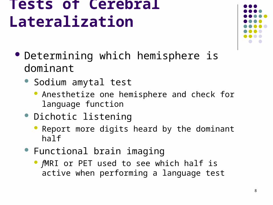
Tests of Cerebral Lateralization
Determining which hemisphere is dominant Sodium amytal test
Anesthetize one hemisphere and check for language function
Dichotic listening Report more digits heard by the dominant half
Functional brain imaging fMRI or PET used to see which half is active when
performing a language test
8

Dichotic Listening Task
9, 5, 6, 7, 2
LEFT
5
2
8
RIGHT
9
6
7
9

Discovery of the Relation between Speech Laterality and Handedness
Left hemisphere is speech dominant in almost all dextrals (right-handers) and most sinestrals (left-handers)
10

Sex Differences in Brain Lateralization
McGlone (1977, 1980) studies of unilateral stroke vicitims
Females may use both hemispheres more often for language tasks than men do (females may be less lateralized)
Mixed support for McGlone’s hypothesis
11

The Split Brain
12

Corpus Callosum
Largest cerebral commissure – 200 million axons
Transfers learned information from one hemisphere to the other When cut, each hemisphere functions
independently Early research paradox
No apparent effect when cut in laboratory animals
No apparent effect when damaged in humans
13

Groundbreaking Experiment of Myers and Sperry
Studied split-brain cats Transected the corpus callosum and optic chiasm
so that visual information could not cross to the contralateral hemisphere
14

Four Groups
1. Corpus callosum and optic chiasm severed (experimental group)
2. Corpus callosum severed control group
3. Optic chiasm severed control group
4. Intact control group
15

FIGURE 16.3 Restricting visual information to one hemisphere in cats. To restrict visual information to one hemisphere, Myers and Sperry (1) cut the corpus callosum, (2) cut the optic chiasm, and (3) blindfolded one eye. This restricted the visual information to the hemisphere ipsilateral to the uncovered eye.
16

FIGURE 16.4 Schematic illustration of Myers and Sperry’s (1953) groundbreaking split-brain experiment. There were four groups: (1) the key experimental group with both the optic chiasm and corpus callosum transected, (2) a control group with only the optic chiasm transected, (3) a control group with only the corpus callosum transected, and (4) an unlesioned control group. The performance of the three control groups did not differ, so they are illustrated together here.
17

Split-Brain Cats Continued
Each hemisphere can learn independently Split-brain cats with one eye patched
Learn task as well as controls
No memory or savings demonstrated when the patch was transferred to other eye
Intact cats or those with an intact corpus callosum or optic chiasm – learning transfers between hemispheres
Similar findings with split-brain monkeys18

Commissurotomy in Human Epileptics
Commissurotomy limits convulsive activity Many never have another major convulsion
Sperry and Gazzaniga Developed procedures to test split-brain
patients
Differ from split-brain animals in that the two hemispheres have very different abilities – most left hemispheres are capable of speech, while the right are not
19

FIGURE 16.5 The testing procedure that was used to evaluate the neuropsychological status of split-brain patients. Visual input goes from each visual field to the contralateral hemisphere; fine tactile input goes from each hand to the contralateral hemisphere; and each hemisphere controls the fine motor movements of the contralateral hand.
20

Evidence that the Hemispheres of Split-Brain Patients Can Function Independently
Left hemisphere can tell what it has seen, right hemisphere can only show it Present a picture to the right visual field (left brain)
Left hemisphere can tell you what it was
Right hand can show you, left hand can’t
Present a picture to the left visual field (right brain) Subject will report that they do not know what it was
Left hand can show you what it was, right can’t
21

Cross-Cuing Cross-cuing – facial feedback from the other
hemisphere For example, the right hemisphere might
make the face frown when the left hemisphere gives an incorrect spoken answer
22

Doing Two Things at Once
Each hemisphere of a split-brain can learn independently and simultaneously Helping-hand phenomenon – presented with two
different visual stimuli, the hand that “knows” may correct the other
Dual foci of attention – split-brain hemispheres can search for target item in array faster than intact controls
Chimeric figures task – only symmetrical version of right half of faces recognized Indicates competition between hemispheres
23

Chimeric Figures Test
24

Chimeric Figures Test
25

The Z Lens Advancing the study of split-brains with a
contact lens to restrict visual input to one hemisphere
Previous studies had to limit viewing time to less than .1 second
Can be used to assess each hemisphere’s understanding of spoken instructions by limiting essential visual information to one side of brain
26

FIGURE 16.7 The Z lens, which was developed by Zaidel to study functional asymmetry in split-brain patients. It is a contact lens that is opaque on one side (left or right), so that visual input reaches only one hemisphere.
27

Dual Mental Functioning and Conflict in Split-Brain Patients
Usually in split-brain patients the left hemisphere is dominant in most everyday activities
For some, the right is dominant and this can create conflict between hemispheres For example, the case of Peter Hemispheres often disagreed with each other
28

Independence of Split Hemispheres: Current Perspective
Discussions of split-brain patients tend to focus on examples of hemispheric independence
Still interactions between the hemispheres (via sub-cortical structures)
Emotional information somehow passed between hemispheres
Difficult tasks are more likely to enlist involvement of both hemispheres
29

Sperry, Zaidel, & Zaidel (1979)
30
Patient was shown an array of photos and asked if one was familiar. He pointed to the photo of his aunt.

Task Difficulty
Simple tasks best processed in one hemisphere
Complex tasks generally require both hemispheres
Important finding because:1. Complicates interpretation of functional-brain
imaging studies of lateralization of function
2. Explains why the elderly often display less lateralization of function
31

Differences between Left and Right Hemispheres
For many functions there are no substantial differences between hemispheres
When differences do exist, usually slight biases in favor of one hemisphere—not absolute differences
Key point: Lateralization of function is statistical rather than absolute
Media misrepresent or distort cerebral hemisphere differences – suggest there are absolute differences
32

Examples of Cerebral Lateralization of Function
Left hemisphere: superior in controlling ipsilateral movement
Left hemisphere: an “interpreter” Right hemisphere superiority:
Spatial ability Emotion Musical ability Some memory tasks
33

Table 16.1
34

What is Lateralized—Broad Clusters of Abilities or Individual Cognitive Processes?
Broad categories are not lateralized – individual tasks may be
Better to consider lateralization of constituent cognitive processes – individual cognitive elements Example: two spatial tasks – left hemisphere is better
at judging above or below, right at how close two things are
35

Anatomical Brain Asymmetries
Frontal operculum (Broca’s area) Near face area of primary motor cortex Language production
Planum temporale (Wernicke’s area) Temporal lobe, posterior lateral fissure Language comprehension
Primary auditory cortex (Heschl’s gyrus)
36

Anatomical Brain Asymmetries Continued
Although asymmetries are seen in language related areas, these regions are not all larger in the left
Left planum temporale – larger in only 65% of human brains
Heschl’s gyri – larger on the right Two gyri in the right, only one in the left
Frontal operculum – visible surface suggests right is larger, but left has greater volume
37

FIGURE 16.9 The anatomical asymmetry detected in the planum temporale of musicians by magnetic resonance imaging. In most people, the planum temporale is larger in the left hemisphere than in the right; this difference was found to be greater in musicians with perfect pitch than in either musicians without perfect pitch or controls. (Based on Schlaug et al., 1995.)
38