Embed Size (px)
Citation preview

THE JOURNAL OF BIOLOGICAL CHEMISTRY Q 1987 by The American Society for Biochemistry and Molecular Biology, Inc.
Vol. 262, No. 36, Isaue of December 25, pp. 17744-17749,1987 Printed in U. S. A .
Effects of Low pH on Influenza Virus ACTIVATION AND INACTIVATION OF THE MEMBRANE FUSION CAPACITY OF THE HEMAGGLUTININ*
(Received for publication, April 20, 1987)
Toon StegmannSS, Frank P. BooyT, and Jan WilschutSII From the $Laboratory of Physiological Chemistry, University of Groningen, Bloemsingel 10, 9712 KZ Groningen, The Netherlands and the lIBiochemica1 Luboratory, University of Groningen, Nijenbogh 16, 9747AG Groningen, The Netherlands
The hemagglutinin of influenza virus undergoes a conformational change at low pH, which results in exposure of a hydrophobic segment of the molecule, crucial to expression of viral fusion activity. We have studied the effects of incubation of the virus at low pH either at 37 or 0°C. Treatment of the virus alone at pH 5.0 induces the virus particles to become hydropho- bic, as assessed by measuring the binding of zwitter- ionic liposomes to the virus. At 37 “C this hydrophobic- ity is transient, electron microscopic examination of the virus reveals a highly disorganized spike layer, and fusion activity toward ganglioside-containing zwitterionic liposomes, measured at 3 7 ° C with a ki- netic fluorescence assay, is irreversibly lost. By con- trast, after preincubation of the virus alone at pH 5.0 and 0 ° C fusion activity remains unaffected. Yet, the preincubation at 0 ° C does result in exposure of the hydrophobic segment of hemagglutinin, but now hy- drophobicity is sustained and viral spike morphology unaltered. Hydrophobicity also remains to a significant extent upon pH neutralization, but fusion activity is negligible under these conditions. It is concluded that for optimal expression of fusion activity the virus must be bound to the target membrane before exposure to low pH. Furthermore, even after exposure of the hy- drophobic segment of hemagglutinin, fusion occurs only at low pH. Finally, fusion occurs only at elevated temperature, possibly reflecting the unfolding of hem- agglutinin trimers or the cooperative action of several hemagglutinin trimers in the reaction.
The infectious entry of influenza virus into cells occurs through a process of receptor-mediated endocytosis (Matlin et al., 1981) directing the virus particles into endosomes. Induced by the low pH inside the endosomal compartment (Tycko and Maxfield, 1982; Yoshimura and Ohnishi, 1984), the viral membrane then fuses with the endosomal membrane releasing the viral genome into the cytoplasm. This fusion process is mediated by a viral spike glycoprotein, the hemag- glutinin (White et al., 1982). Hemagglutinin is a homotrimer with a total molecular weight of 224,000 (Wilson et al., 1981). Each hemagglutinin monomer consists of two subunits, HA,
*This investigation was carried out under the auspices of the Netherlands Foundation of Chemical Research (S.O.N.) with finan- cial support from the Netherlands Organization for the Advancement of Pure Research (Z.W.O.). The costs of publication of this article were defrayed in part by the payment of page charges. This article must therefore be hereby marked “advertisement” in accordance with 18 U.S.C. Section 1734 solely to indicate this fact.
8 Present address: Dept. of Cell Biology, Yale University School of Medicine, 333 Cedar St., New Haven, CT 06510.
11 To whom correspondence should be addressed.
and HA, (Klenk et al., 1975). At the pH threshold of fusion (Doms et al., 1985, 1986; Daniels et al., 1985) an irreversible conformational change takes place in the hemagglutinin (Ske- he1 et al., 1982), leading to exposure of the hydrophobic N terminus of the HA, subunit. Thereby the isolated hemagglu- tinin acquires the ability to bind to nonionic detergents or lipid vesicles (Skehel et al., 1982; Doms et al., 1985). Strains or mutants of influenza virus with different pH dependences of fusion activity also show a corresponding shift in the pH dependence of the conformational change (Doms et al., 1986; Daniels et al., 1985). Therefore, the conformational change in the hemagglutinin is considered to play a crucial role in viral fusion. However, if influenza virus is exposed to low pH at 37°C prior to its interaction with a target membrane, its fusion or hemolytic activity is abolished (Sat0 et al., 1983; Junankar and Cherry, 1986; Stegmann et al., 1986). It has been suggested that the conformational change that is crucial to viral fusion is also involved in inactivation of the fusion capacity during a prior low pH treatment of the virus alone and that both fusion and inactivation require the hemagglu- tinin molecules to be mobile in the viral membrane (Junankar and Cherry, 1986).
We have investigated how the fusion activity, hydrophobic- ity, and spike layer morphology of the virus are influenced by the pH and temperature of preincubation of the virus alone. Fusion of the virus was measured with a fluorescence reso- nance energy transfer assay involving liposomes as target membrane vesicles (Stegmann et al., 1985); viral hydropho- bicity was assessed by measuring the binding of zwitterionic liposomes to the virus, and viral spike morphology was visu- alized by means of cryoelectron microscopy. A model for the observed loss of fusion activity upon low pH treatment of the virus alone is presented and discussed in terms of its possible implications for the mechanism of viral fusion.
MATERIALS AND METHODS
Chemicals-N-(Lissamine rhodamine B sulfonyl)- phosphatidylethanolamine (N-Rh-PE),’ N-(7-nitro-2,1,3-benzoxadi- azol-4-y1)phosphatidylethanolamine (N-NBD-PE), bovine brain phosphatidylserine (PS), dioleoylphosphatidylethanolamine (DOPE), and dioleoylphosphatidylcholine (DOPC) were purchased from Avanti Polar Lipids, Inc. (Birmingham, AL). Disialoganglioside (GD,) was from Supelco Inc. (Bellefonte, PA) and proteinase K from Boehringer (Mannheim, Federal Republic of Germany).
Virus and Liposomes-The X-47 and X-31 recombinant strains of influenza virus were propagated in the allantoic cavity of embryon-
’ The abbreviations used are: N-Rh-PE, N-(lissamine rhodamine B sulfony1)phosphatidylethanolamine; G,,, disialoganglioside; HEPES, 4-(2-hydroxyethyl)-l-piperazineethanesulfonic acid; MES, 2-(N-morpholino)ethanesulfonic acid; N-NBD-PE, N-(7-nitrobenzo- 2-oxa-l,3-diazol-4-yl)phosphatidylethanolamine; DOPC, dioleoyl- phosphatidylcholine; DOPE, dioleoylphosphatidylethanolamine; PS, phosphatidylserine.
17744

Influenza Virus and Low pH 17745
ated eggs, purified, handled, and stored essentially as described before (Stegmann et al., 1985). Viral phospholipid phosphate was deter- mined, after extraction according to Folch et al. (1957), by phosphate analysis (Bottcher et al., 1961). Protein was determined according to Peterson (1977). Liposomes (large unilamellar vesicles) were prepared by reverse-phase evaporation (Szoka and Papahadjopoulos, 1978) with modifications described by Wilschut et al. (1980) and sized by extrusion through polycarbonate filters with a pore size of 0.2 pm (Olson et al., 1979). After extrusion any remaining larger or multila- mellar liposomes were removed by centrifugation (Szoka and Papa- hadjopoulos, 1978), and phospholipid phosphate of the liposomes in the supernatant was determined by phosphate analysis (Bottcher et al., 1961).
Fusion and Binding Experiments-In the resonance energy trans- fer fusion assay 0.6 mol % each of N-NBD-PE and N-Rh-PE were incorporated in the bilayer of the liposomes (Struck et al., 1981). Measurements were carried out at 37°C in a cuvette containing a final volume of 1.4 ml of 135 mM NaCl, 15 mM sodium citrate, 10 mM MES, and 5 mM HEPES (fusion buffer), set to various pH values. The increase in N-NBD-PE fluorescence, due to dilution of the fluorophores into the viral membrane upon fusion, was recorded continuously at excitation and emission wavelengths of 465 and 530 nm, respectively, using an SLM-8000 fluorimeter (SLM/Aminco, Urbana, IL). For calibration of the fluorescence scale, the initial residual fluorescence of the liposomes was set to zero and the fluores- cence at infinite probe dilution to 100%. The latter value was deter- mined by addition of Triton X-100 (0.5% v/v) to the liposomes and subsequent correction of the fluorescence intensity for sample dilu- tion and the effect of Triton on the quantum yield of the N-NBD- PE (Struck et al., 1981). The initial rate of fluorescence increase was calculated from tangents drawn to the first part of the fusion curve and taken as a measure of fusion activity.
In the binding experiments virus was added to liposomes (contain- ing the resonance energy transfer probes) at a 1:l ratio, 5 nmol of membrane phospholipid each, and incubated for 15 min at 0 "C unless stated otherwise. The mixture was centrifuged at 0 'C for 15 min at 12,000 X g. In the absence of liposomes about 80-85% of the virus is pelleted by this procedure. The formation of aggregates upon binding of liposomes facilitates sedimentation, resulting in an essentially quantitative recovery of virus in the pellet. After addition of Triton X-100 and proper dilution, fluorescence in supernatant and pellet fractions was determined. Binding was calculated as the percentage of total fluorescence recovered in the pellet corrected for the fluores- cence of liposomes that could be pelleted in the absence of virus (usually about 3%).
Preincubation of the virus prior to addition to the liposomes was performed in a small volume (60 ~ 1 ) of fusion buffer set to the indicated pH at the desired temperature. After preincubation at 0 'C a cold pipette tip was used for transfer to the final incubation mixture.
Gel Electrophoresis-Sodium dodecyl sulfate-polyacrylamide gel electrophoresis was performed essentially as described in Laemmli (1970) on 9% slab gels. Before application to the gel, samples were precipitated with 10% trichloroacetic acid, washed with ethanol/ether (l:l, v/v) and then boiled for 5 min in sample buffer containing 10% sodium dodecyl sulfate, 5% j3-mercaptoethanol, and 8 M ureum.
Cryoelectron Microscopy-A small drop of virus, incubated for the indicated time at the appropriate temperature and pH in fusion buffer, was applied to a carbon-coated copper grid, blotted with filter paper, and rapidly frozen in liquid ethane. The grid was then trans- ferred to the cooling holder of a Philips 400 electron microscope and examined under low-dose conditions as described by Booy et al. (1985).
RESULTS
Activation and Inactivation of Viral Fusion Activity at Low pH-Influenza virus X-47 was incubated for 5 min at various pH values either at 37 or at 0 "C. After this preincubation the virus was added to liposomes, labeled with N-NBD-PE and N-Rh-PE at 37 "C and pH 5.0, and the kinetics of fusion of the virus with the liposomes were determined. Alternatively, fusion activity was determined at the respective pH values without a preincubation of the virus. The liposomes consisted of DOPC and DOPE (molar ratio, 2:l) and contained 5 mol % of the ganglioside GDI,, serving as a specific receptor for the virus. We have demonstrated previously that fusion of
influenza virus with liposomes of such a composition closely parallels the biological fusion activity of the virus (Stegmann et al., 1986).
Fig. 1 shows that a preincubation of the virus alone at low pH and 37 "C results in a rapid loss of fusion activity (open circles). The figure also shows the activation of fusion capacity upon exposure of the virus to low pH in the presence of target liposomes (closed circles). Remarkably, the pH dependence of inactivation during preincubation of the virus alone closely parallels the pH dependence of fusion, measured directly at the respective pH values. This suggests that both the fusion reaction and the inactivation of fusion capacity during a low pH treatment of the virus alone are due to the occurrence of
I
4.5 50 5.5 I'
pH of Incubation or of plncubation 6x6574
FIG. 1. Fusion activity of influenza virus X-47 with DOPC/ DOPE (molar ratio, 2:l) liposomes containing 6 mol 9i Gob. A: M, pH dependence of fusion at 37'C (left-hand scale); W, A-A, pH dependence of inactivation of fusion capacity; virus was preincubated alone at the indicated pH at 37 (0) or O'C (A) for 5 min, after which fusion activity was measured at pH 5.0 and 37 "C (right-hand scale, activity is expressed relative to that of a nontreated control). B, time course of inactivation of fusion activity, measured at pH 5.0,37 "C, during preincubation of the virus alone at pH 5.0 and 37'C (0) or 0°C (A). Fusion activity was measured, as described under "Materials and Methods," at a ratio of virus to liposomes of 1:l (5 p~ phospholipid phosphorus each).

17746 Influenza Virus and Low p H
the same conformational change in the viral hemagglutinin. There was no evidence to indicate that virus aggregation was involved in the inactivation during a low pH preincubation at 37°C; neither light scattering nor absorbance of the virus suspension changed to any significant extent. Comparable observations were made using the X-31 strain of influenza virus (results not shown).
In contrast to the effect of a low pH preincubation of the virus alone at 37°C (Fig. 1, A and B, circles), a pH 5.0 preincubation at 0°C caused very little inactivation of viral fusion activity (Fig. 1, A and B, triangles). Direct fusion of the virus with the liposomes at pH 5.0 and O T , without preincubation of the virus, was very slow, even though binding of the virus to the liposomes was not affected (results not shown). Hence, there is also a similarity between the temper- ature dependences of fusion and inactivation.
At pH 4.8 and below, both at 0 and 37 "C, fusion activity was inactivated (Fig. hi), as we have reported previously (Stegmann et al., 1986). Under these conditions inactivation of fusion capacity is probably due to denaturation of the hemagglutinin.
Evidence for a Conformational Change a t Low Tempera- ture-Since neither inactivation nor fusion occurred at pH 5.0-5.5 at 0 "C, the question arose whether the conformational change in the hemagglutinin occurs a t all under these low temperature conditions. The experiment shown in Fig. 2 provides evidence for the induction of the conformational change in the hemagglutinin at low temperature. The hem- agglutinin is quite resistant to proteolytic enzymes unless the virus is preincubated at low pH (Doms et al., 1985; Stegmann et al., 1985). Fig. 2 shows that the hemagglutinin becomes susceptible to proteolysis by a prior low pH incubation of the virus, irrespective of whether the temperature of the prein- cubation is 37 or 0 "C. The actual proteolysis was carried out after pH neutralization. This confirms the irreversible nature of the conformational change (Skehel et al., 1982).
Additional evidence for the occurrence of the conforma- tional change at 0 "C was obtained by studying the binding of liposomes to the virus. For zwitterionic liposomes consisting of DOPC and DOPE (molar ratio, 2:l) without a receptor, a
1 2 3 4 5
- DP
FIG. 2. Effect of a low pH treatment on the susceptibility of influenza virus X-47 to proteolytic digestion. Proteolysis was carried out for 2.5 h at pH 7.4 and 0 "C at 0.1 mg/ml of viral protein and proteinase K each. Lane 1, proteinase K alone; lane 2, virus treated with proteinase K, without preincubation; lane 3, virus prein- cubated at pH 5.0,37 "C, and then treated with proteinase K, lane 4, virus preincubated at pH 5.0, 0 "C, and then treated with proteinase K; lane 5, untreated virus. NP, nucleoprotein; M, matrix protein; PK, proteinase K, DP, digestion products.
pH
FIG. 3. pH dependence of liposome binding at 0 O C to influ- enza virus X-47. Composition of the liposomes: DOPC/DOPE (molar ratio, 2:l) (A); PS (0); DOPC/DOPE/PS (molar ratio, 2:1:2) (0). For comparison the fusion activity of the virus at 37°C is also shown (0, cf. Fig. lA).
TABLE I Effect of ionic strength on the binding of liposomes to influenza virus
X-47 at p H 5.0 and 0 "C Binding is expressed as a percentage of the control value, i.e.
binding in the presence of 0.15 M NaC1. Binding
NaCl concentration DOPC/DOPE DOPC/DOPE/PS
(21 ) liposomes (2:1:2) liposomes
M % of control 0.25" 95 87 0.5" 88 61 1.0" 87 57 1.Ob 90 95
' Binding in the presence of NaC1. NaCl added after binding.
pH dependence for binding at 0 "C was observed closely cor- responding to that of the fusion activity at 37 "C (Fig. 3). Since the conformational change in the hemagglutinin ex- poses a previously buried hydrophobic segment, binding in this case probably involves hydrophobic interactions between the viral hemagglutinin and the liposomal membrane. Indeed, only a minor part of the liposomes was prevented from binding to the virus at NaCl concentrations up to 1 M at pH 5.0 (Table I), indicating that the binding is not electrostatic in nature. By contrast, binding of liposomes with a negative surface charge (DOPC/DOPE/PS; molar ratio, 2:1:2) was reduced by about 50% in the presence of 1 M NaC1. Once binding was established, however, also these liposomes could not be re- moved from the virus by addition of NaCl to 1 M. Binding of the virus to liposomes containing negatively charged lipids in addition to zwitterionic lipids was almost independent of pH below pH 6.0 but decreased above that pH (Fig. 3). Probably the binding in this case being governed mainly by electrostatic interactions increases steadily as more viral amino acids be- come protonated with decreasing pH. Liposomes consisting of only negatively charged phospholipids bound almost quan- titatively to the virus irrespective of the pH (Fig. 3).
Transient Versus Sustained Viral Hydrophobicity-The binding experiments in Fig. 3, demonstrating the enhanced hydrophobicity of the virus at low pH, were carried out by adding the virus to the liposomes at various pH values and 0 "C. Preincubation of the virus alone at pH 5.0 showed that the hydrophobicity was rapidly transient at 37 "C (Fig. 4A). After a 30-s preincubation at 37°C virus particles were no longer able to bind the liposomes (Fig. 4B). On the other hand, after a preincubation at 0 "C in the pH range of 5.0- 5.5, the virus maintained the capacity to bind zwitterionic liposomes at 0°C (Fig. 4A). Only a slow decrease in hydro- phobicity was noted under these conditions (Fig. 4B). Part of

Influenza Virus and Low p H 17747
A 50-
40 - 30-
m c p 20- ._ n
10-
0- &a
O+ 0 2 4 6
timof pir~utmtim fmin) 8
FIG. 4. Effect of preincubation of influenza virus X-47 on binding of DOPC/DOPE (molar ratio, 2:l) liposomes. A , virus was either not preincubated at all (A) or preincubated for 5 min at pH 5.0 and 37 "C (0) or 0 "C (O), after which liposomes were added at 0 "C and the incubation continued for 15 min at the pH indicated. B, binding at pH 5.0, 0 "C of liposomes to virus, preincubated at pH 5.0 and 37 "C (0) or 0 "C (0) during the periods of time indicated.
this decrease can be accounted for by the fact that about 10- 15% of the virus adhered to the vessel wall after 15 min of incubation at 0 "C.
Interestingly, some binding activity was observed after preincubation of the virus at pH 5.4-5.5 and 37 "C (Fig. 5A). However, at this pH the induced hydrophobicity does not seem to be transient yet. The transient character only emerges at pH 5.3 and below.
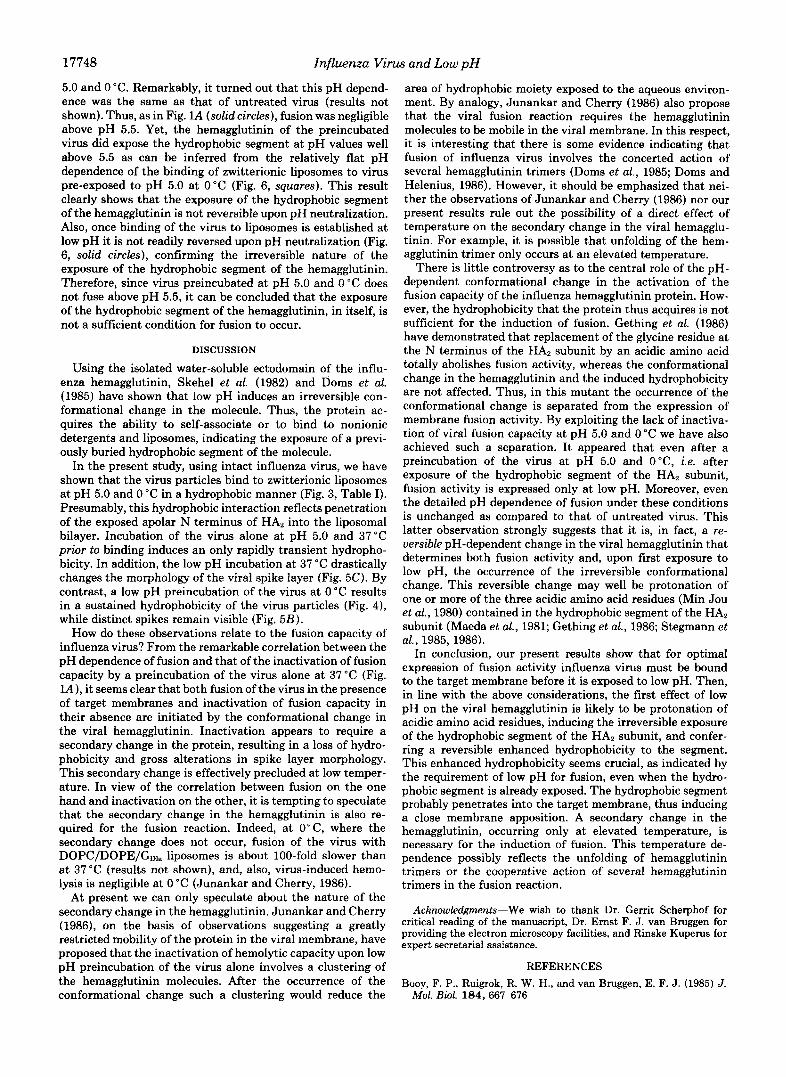
Morphology of the Viral Spike Layer at Low pH-It is well established that a low pH treatment of the virus at 37 "C induces gross alterations in the viral spike layer (Ruigrok et al., 1986). In view of the above results it was of considerable interest to examine viral spike morphology by electron mi- croscopy after low pH treatment of the virus at 0 "C. A novel vitrification technique was utilized, which does not involve staining of the samples (Lepault et al., 1983; Booy et al., 1985). At neutral pH individual spikes projecting from the periphery of the virus particles could be readily distinguished (Fig. 5A). The virus particles were often present in duplets. At pH 5.0 and 37 "C individual spikes could no longer be resolved (Fig. 5C). The morphology of the spike layer was completely dis- turbed, leading to a fuzzy appearance of the protein covering the virus. In marked contrast, at pH 5.0 and 0 "C individual spikes still could be clearly resolved and were similar in appearance to those at neutral pH (Fig. 5B). Interestingly, some of the virus particles appeared unaffected by the low pH treatment at 37°C (Fig. 5C, arrows), suggesting that the disorganization of the spike layer is a highly cooperative phenomenon.
At pH 5.3, the pH where the viral hydrophobicity just starts
FIG. 5. Electron micrographs of unstained, frozen, hy- drated influenza virus X-47. A , virus at pH 7.4; B, virus incubated for 5 min at pH 5.0 and 0 "C; C, virus incubated for 5 min at pH 5.0 and 37 "C; D, virus incubated for 13 min at pH 5.3 and 37 "C. The bar represents 100 nm. The arrows in panel C indicate unaffected virus particles.
I
50-
40 - I g m30- c ._ 3 20-
10-
0- I 1 1
4.5 5.0 55 6;"657.4 PH
FIG. 6. Effect of pH neutralization on low pH-induced bind- ing of DOPC/DOPE (molar ratio, 2:l) liposomes at 0°C to influenza virus X-47. A, binding measured directly at the pH indicated; 0, binding after incubation of virus and liposomes at the pH indicated for 15 min and a subsequent adjustment of the pH to neutral by the addition of NaOH; 0, binding of liposomes to virus, preincubated for 5 min at pH 5.0 and 0 "C, at the pH values indicated.
to become transient (Fig. 4A), viral spike morphology was still largely unchanged (Fig. 50). However, the individual spikes were not as well resolved as at neutral pH (panel D versus panel A).
Effects of pH Neutralization-The above results show that during incubation of the virus alone at pH 5.0 and 0 "C the conformational change in the hemagglutinin is induced, while at the same time the virus maintains its potential to fuse. Obviously, it was of considerable interest to examine the pH dependence of fusion of a virus preparation pretreated at pH
17748 Influenza Virus and Low pH
5.0 and 0 "C. Remarkably, it turned out that this pH depend- ence was the same as that of untreated virus (results not shown). Thus, as in Fig. lA (solid circles), fusion was negligible above pH 5.5. Yet, the hemagglutinin of the preincubated virus did expose the hydrophobic segment at pH values well above 5.5 as can be inferred from the relatively flat pH dependence of the binding of zwitterionic liposomes to virus pre-exposed to pH 5.0 at 0°C (Fig. 6, squares). This result clearly shows that the exposure of the hydrophobic segment of the hemagglutinin is not reversible upon pH neutralization. Also, once binding of the virus to liposomes is established at low pH it is not readily reversed upon pH neutralization (Fig. 6, solid circles), confirming the irreversible nature of the exposure of the hydrophobic segment of the hemagglutinin. Therefore, since virus preincubated at pH 5.0 and 0 "C does not fuse above pH 5.5, it can be concluded that the exposure of the hydrophobic segment of the hemagglutinin, in itself, is not a sufficient condition for fusion to occur.
DISCUSSION
Using the isolated water-soluble ectodomain of the influ- enza hemagglutinin, Skehel et al. (1982) and Doms et al. (1985) have shown that low pH induces an irreversible con- formational change in the molecule. Thus, the protein ac- quires the ability to self-associate or to bind to nonionic detergents and liposomes, indicating the exposure of a previ- ously buried hydrophobic segment of the molecule.
In the present study, using intact influenza virus, we have shown that the virus particles bind to zwitterionic liposomes at pH 5.0 and 0 "C in a hydrophobic manner (Fig. 3, Table I). Presumably, this hydrophobic interaction reflects penetration of the exposed apolar N terminus of HA2 into the liposomal bilayer. Incubation of the virus alone at pH 5.0 and 37°C prior to binding induces an only rapidly transient hydropho- bicity. In addition, the low pH incubation at 37 "C drastically changes the morphology of the viral spike layer (Fig. 5C). By contrast, a low pH preincubation of the virus at 0 "C results in a sustained hydrophobicity of the virus particles (Fig. 4), while distinct spikes remain visible (Fig. 5B).
How do these observations relate to the fusion capacity of influenza virus? From the remarkable correlation between the pH dependence of fusion and that of the inactivation of fusion capacity by a preincubation of the virus alone at 37 "C (Fig. lA), it seems clear that both fusion of the virus in the presence of target membranes and inactivation of fusion capacity in their absence are initiated by the conformational change in the viral hemagglutinin. Inactivation appears to require a secondary change in the protein, resulting in a loss of hydro- phobicity and gross alterations in spike layer morphology. This secondary change is effectively precluded at low temper- ature. In view of the correlation between fusion on the one hand and inactivation on the other, it is tempting to speculate that the secondary change in the hemagglutinin is also re- quired for the fusion reaction. Indeed, at O"C, where the secondary change does not occur, fusion of the virus with DOPC/DOPE/Gol. liposomes is about 100-fold slower than at 37 "C (results not shown), and, also, virus-induced hemo- lysis is negligible at 0 "C (Junankar and Cherry, 1986).
At present we can only speculate about the nature of the secondary change in the hemagglutinin. Junankar and Cherry (1986), on the basis of observations suggesting a greatly restricted mobility of the protein in the viral membrane, have proposed that the inactivation of hemolytic capacity upon low pH preincubation of the virus alone involves a clustering of the hemagglutinin molecules. After the occurrence of the conformational change such a clustering would reduce the
area of hydrophobic moiety exposed to the aqueous environ- ment. By analogy, Junankar and Cherry (1986) also propose that the viral fusion reaction requires the hemagglutinin molecules to be mobile in the viral membrane. In this respect, it is interesting that there is some evidence indicating that fusion of influenza virus involves the concerted action of several hemagglutinin trimers (Doms et al., 1985; Doms and Helenius, 1986). However, it should be emphasized that nei- ther the observations of Junankar and Cherry (1986) nor our present results rule out the possibility of a direct effect of temperature on the secondary change in the viral hemagglu- tinin. For example, it is possible that unfolding of the hem- agglutinin trimer only occurs at an elevated temperature.
There is little controversy as to the central role of the pH- dependent conformational change in the activation of the fusion capacity of the influenza hemagglutinin protein. How- ever, the hydrophobicity that the protein thus acquires is not sufficient for the induction of fusion. Gething et al. (1986) have demonstrated that replacement of the glycine residue at the N terminus of the HA, subunit by an acidic amino acid totally abolishes fusion activity, whereas the conformational change in the hemagglutinin and the induced hydrophobicity are not affected. Thus, in this mutant the occurrence of the conformational change is separated from the expression of membrane fusion activity. By exploiting the lack of inactiva- tion of viral fusion capacity at pH 5.0 and 0 "C we have also achieved such a separation. It appeared that even after a preincubation of the virus at pH 5.0 and O"C, i.e. after exposure of the hydrophobic segment of the HA2 subunit, fusion activity is expressed only at low pH. Moreover, even the detailed pH dependence of fusion under these conditions is unchanged as compared to that of untreated virus. This latter observation strongly suggests that it is, in fact, a re- versible pH-dependent change in the viral hemagglutinin that determines both fusion activity and, upon first exposure to low pH, the occurrence of the irreversible conformational change. This reversible change may well be protonation of one or more of the three acidic amino acid residues (Min Jou et al., 1980) contained in the hydrophobic segment of the HA, subunit (Maeda et al., 1981; Gething et al., 1986; Stegmann et al., 1985, 1986).
In conclusion, our present results show that for optimal expression of fusion activity influenza virus must be bound to the target membrane before it is exposed to low pH. Then, in line with the above considerations, the first effect of low pH on the viral hemagglutinin is likely to be protonation of acidic amino acid residues, inducing the irreversible exposure of the hydrophobic segment of the HA2 subunit, and confer- ring a reversible enhanced hydrophobicity to the segment. This enhanced hydrophobicity seems crucial, as indicated by the requirement of low pH for fusion, even when the hydro- phobic segment is already exposed. The hydrophobic segment probably penetrates into the target membrane, thus inducing a close membrane apposition. A secondary change in the hemagglutinin, occurring only at elevated temperature, is necessary for the induction of fusion. This temperature de- pendence possibly reflects the unfolding of hemagglutinin trimers or the cooperative action of several hemagglutinin trimers in the fusion reaction.
Acknowledgments-We wish to thank Dr. Gerrit Scherphof for critical reading of the manuscript, Dr. Ernst F. J. van Bruggen for providing the electron microscopy facilities, and Rinske Kuperus for expert secretarial assistance.
REFERENCES Booy, F. P., Ruigrok, R. W. H., and van Bruggen, E. F. J. (1985) J.
Mol. BWl. 184, 667-676

Influenza Virus and Low p H 17749
Bottcher, C. J. F., Van Gent, C. M., and Fries, C. (1961) Anal. Chim. Acta 24,203-204
Daniels, R. S., Downie, J. C., Hay, A. J., Knossow, M., Skehel, J . J., Wang, M. L., and Wiley, D. C. (1985) Cell 40, 431-439
Doms, R. W., and Helenius, A. (1986) J. Virol. 60,833-839 Doms, R. W., Helenius, A., and White, J. (1985) J. Biol. Chern. 260,
Doms, R. W., Gething, M. J., Henneberry, J., White, J., and Helenius,
Folch, J., Lees, M., and Sloane Stanley, G. H. (1957) J. Biol. Chem.
Gething, M. J., Doms, R. W., York, D., and White, J. (1986) J. Cell
Junankar, P. R., and Cherry, R. J. (1986) Biochim. Biophys. Acta
Klenk, H. D., Rott, R., Orlich, M., and Blodorn, J. (1975) Virology
Laemmli, U. K. (1970) Nature 222,680-685 Lepault, J., Booy, F. P., and Dubochet, J. (1983) J. Mierosc. (Ozf.)
Maeda, T., Kawasaki, K., and Ohnishi, S. (1981) Proc. Natl. Acad.
Matlin, K. S., Reggio, H., Helenius, A., and Simons, K. (1981) J. Cell
Min Jou, M., Verhoeyen, M., Devos, R., Samon, E., Fange, R., Huylebroek, D., Friers, W., Threlfall, G., Barber, G., Carey, N.,
2973-2981
A. (1986) J. Virol. 57, 603-613
226,497-509
Bid. 102,ll-23
854,198-206
68,426-439
129,89-102
Sci. U. S. A. 78,4133-4137
Biol. 91,601-613
and Emtage, S. (1980) Cell 19, 683-696
D. (1979) Biochim. Biophys. Acta 557,9-23 Olson, F., Hunt, C. A., Szoka, F. C., Vail, W. J., and Papahadjopoulos,
Peterson, G. L. (1977) Anal. Biochern. 83, 346-356 Ruigrok, R. W. H., Wrigley, N. G., Calder, L. J., Cusack, S., Wharton,
S. A., Brown, E. B., and Skehel, J. J. (1986) EMBO J. 5, 41-49 Sato, S. B., Kawasaki, K., and Ohnishi, S. (1983) P m . Natl. Acad.
Sci. U. S. A. 80,3153-3157 Skehel, J. J., Bayley, P. M., Brown, E. B., Martin, S. R., Waterfield,
M. D., White, J. M., Wilson, I. A., and Wiley, D. C. (1982) Proc. Natl. Acad. Sci. U. S . A. 79,968-972
Stegmann, T., Hoekstra, D., Scherphof, G., and Wilschut, J. (1985)
Stegmann, T., Hoekstra, D., Scherphof, G., and Wilschut, J. (1986)
Struck, D. K., Hoekstra, D., and Pagano, R. E. (1981) Biochemistry
Szoka, F. C., and Papahadjopoulos, D. (1978) Proc. Natl. Acad. Sci.
Tycko, B., and Maxfield, F. R. (1982) Cell 28, 643-651 White, J., Helenius, A., and Gething, M. J. (1982) Nature 300, 658-
Wilschut, J., Duzgiines, N., Fraley, R., and Papahadjopoulos, D.
Wilson, I. A., Skehel, J. J., and Wiley, D. C. (1981) Nature 289,366-
Yoshimura, A., and Ohnishi, S. (1984) J. Virol. 51,497-504
Biochemist~y 24,3107-3113
J. Bid. Chern. 261, 10966-10969
20,4093-4099
U. S. A. 75,4194-4198
659
(1980) Biochemistry 19,6011-6021
373