Embed Size (px)
Citation preview

Cloning and Characterization of the Human and Rat Islet-specificGlucose-6-phosphatase Catalytic Subunit-related Protein (IGRP)Genes*
Received for publication, February 19, 2001, and in revised form, March 27, 2001Published, JBC Papers in Press, April 10, 2001, DOI 10.1074/jbc.M101549200
Cyrus C. Martin‡§, Larry J. Bischof‡, Barbara Bergman¶, Lauri A. Hornbuckle‡i, Carl Hilliker¶,Claudia Frigeri‡, David Wahl¶, Christina A. Svitek‡, Randall Wong¶, Joshua K. Goldman‡,James K. Oeser‡, Frederic Lepretre**, Philippe Froguel**, Richard M. O’Brien‡ ‡‡,and John C. Hutton¶ ‡‡
From the ‡Department of Molecular Physiology and Biophysics, Vanderbilt University Medical School, Nashville,Tennessee 37232, ¶Barbara Davis Center for Childhood Diabetes, University of Colorado Health Sciences Center, Denver,Colorado 80262, and iInstitut de Biologie-CNRS 8090, Institut Pasteur de Lille, Lille Cedex, France
Islet-specific glucose-6-phosphatase (G6Pase) cata-lytic subunit-related protein (IGRP) is a homolog of thecatalytic subunit of G6Pase, the enzyme that catalyzesthe terminal step of the gluconeogenic pathway. Its cat-alytic activity, however, has not been defined. SinceIGRP gene expression is restricted to islets, this sug-gests a possible role in the regulation of islet metabolismand, hence, insulin secretion induced by metabolites. Wereport here a comparative analysis of the human,mouse, and rat IGRP genes. These studies aimed to iden-tify conserved sequences that may be critical for IGRPfunction and that specify its restricted tissue distribu-tion. The single copy human IGRP gene has five exons ofsimilar length and coding sequence to the mouse IGRPgene and is located on human chromosome 2q28–32 ad-jacent to the myosin heavy chain 1B gene. In contrast,the rat IGRP gene does not appear to encode a proteinas a result of a series of deletions and insertions in thecoding sequence. Moreover, rat IGRP mRNA, unlikemouse and human IGRP mRNA, is not expressed in is-lets or islet-derived cell lines, an observation that wastraced by fusion gene analysis to a mutation of the TATAbox motif in the mouse/human IGRP promoters to TGTAin the rat sequence. The results provide a framework for
the further analysis of the molecular basis for the tissue-restricted expression of the IGRP gene and the identifi-cation of key amino acid sequences that determine itsbiological activity.
Glucose-6-phosphatase (G6Pase)1 is located in the endoplas-mic reticulum (ER) and catalyzes the terminal step of thegluconeogenic pathway in liver and kidney. The enzyme isthought to be a multi-subunit complex; however, the exactnumber of subunits, their stoichiometry, and topological rela-tionships are unclear (1, 2). The 36-kDa G6Pase catalytic sub-unit spans the membrane multiple times and appears, based onstudies using microsomes, to have its catalytic site orientedtoward the lumen (1, 2). A model has therefore been proposedin which the G6Pase catalytic subunit is postulated to be as-sociated functionally with a 46-kDa glucose 6-phosphate (G6P)transporter (3) and hypothetical transporters for inorganicphosphate and glucose, which serve to deliver cytosolicallygenerated G6P to the active site and shuttle the reaction prod-ucts back to the cytosol (1, 2). Rapid kinetic data, on the otherhand, favor an alternative model that places the catalytic sitewithin the membrane and ascribes both a transport functionand catalytic activity to the 36-kDa catalytic subunit (1, 2).Mutations within the G6Pase catalytic subunit cause glycogenstorage disease type 1a (4), which is characterized by severehypoglycemia in the post-absorptive state, hepatomegaly asso-ciated with excessive glycogen deposition, growth retardation,and renal failure (4). In glycogen storage disease type 1b mu-tations in the G6P transporter give rise to a similar phenotypeand additional complications possibly related to independentfunctions of this molecule in other tissues (4).
Hepatic G6Pase activity is increased in poorly controlledhuman type 1 and 2 diabetics (5, 6) and in experimental rodentdiabetic models (7–11). Along with the elevated activity of othergluconeogenic enzymes, it contributes to an increase in hepaticglucose production and the hyperglycemia that characterizesthe disease (5, 6, 12, 13). The change in G6Pase activity has
* This work was supported by a grant from Juvenile Diabetes Foun-dation International (JDFI) and Vanderbilt Diabetes Core LaboratoryGrant P60 DK20593 (to R. O’B.), American Diabetes Association Grant9901-116 and Barbara Davis Center Diabetes and Endocrinology Re-search Center Grant P30 DK57516 (to J. C. H.), and by a grant from theNord-Pas de Calais Region (to F. L.). The costs of publication of thisarticle were defrayed in part by the payment of page charges. Thisarticle must therefore be hereby marked “advertisement” in accordancewith 18 U.S.C. Section 1734 solely to indicate this fact.
The nucleotide sequence(s) reported in this paper has been submittedto the GenBankTM/EBI Data Bank with accession number(s) AF283835(human IGRP gene excluding the promoter), AF283575 (human IGRPpromoter), AF321459-AF321463 (human IGRP exons 1–5, respectively),AF323433 (rat IGRP promoter and exon 1), AF323434-AF323436 (ratIGRP exons 2, 3, and 5, respectively), NM021331 (mouse IGRP cDNA),AF118761 (mouse IGRP promoter), and AF 118762-AF118766 (mouseIGRP exons 1–5, respectively).
§ Supported by Vanderbilt Viruses, Nucleic Acids, and Cancer Train-ing Program 5T32 CA09385-17).
i Recipient of Vanderbilt Molecular Endocrinology Training ProgramAward 5 T 32 DK07563-12).
‡‡ To whom correspondence may be addressed: Dept. of MolecularPhysiology and Biophysics, 761 MRB II, Vanderbilt University MedicalSchool, Nashville, TN 37232-0615. Tel.: 615-936-1503; Fax: 615-322-7236; E-mail: [email protected] (R. O’B.) or Tel.:303-315-8197; Fax: 303-315-4892; E-mail: [email protected](J. C. H.).
1 The abbreviations used are: IGRP, islet-specific G6Pase catalyticsubunit-related protein; G6Pase, glucose-6-phosphatase; ER, endoplas-mic reticulum; G6P, glucose 6-phosphate; ORF, open reading frame;CAT, chloramphenicol acetyltransferase; RIN and INS-1, rat insuli-noma-derived cell lines; contig, group of overlapping clones; HIT, ham-ster insulinoma tumor; bp, base pair(s); kbp, kilobase pair(s); PCR,polymerase chain reaction; RT, reverse transcription; nt, nucleotides;MES, 4-morpholineethanesulfonic acid; aa, amino acid(s).
THE JOURNAL OF BIOLOGICAL CHEMISTRY Vol. 276, No. 27, Issue of July 6, pp. 25197–25207, 2001© 2001 by The American Society for Biochemistry and Molecular Biology, Inc. Printed in U.S.A.
This paper is available on line at http://www.jbc.org 25197
by guest on September 23, 2018
http://ww
w.jbc.org/
Dow
nloaded from

been attributed to changes in expression of the genes encodingboth the G6Pase catalytic subunit and the G6P transporter.The former probably reflects the combined stimulatory effect ofglucose (10, 14, 15) and the loss of the inhibitory action ofinsulin (16, 17). Less is known about the factors that regulateexpression of the G6P transporter (18). However, experimentaloverexpression of either the G6Pase catalytic subunit (19) orthe G6P transporter (20) in hepatocytes using recombinantadenovirus leads to enhanced rates of G6P hydrolysis as well aschanges in glycogen metabolism.
Most studies show that islets also contain a hydrolytic activitythat is specific for G6P but that is present at a lower specificactivity than liver (2, 21–25). G6Pase activity is elevated in isletsisolated from ob/ob mice resulting in increased glucose substratecycling (26, 27). The question of whether islet G6Pase activity iscatalyzed by the same G6Pase catalytic subunit as in liver, how-ever, has proven controversial (2, 21). Thus, the G6P hydrolyticactivity in islets displays distinct kinetic behavior and inhibitorprofiles compared with that in hepatic extracts (21).
We recently identified a novel cDNA from mouse b cell-derived cell lines that encodes an islet-specific G6Pase catalyticsubunit-related protein (IGRP) (21). IGRP is a putative ERmembrane protein that is similar in size (38 kDa), topology,and sequence (;50% identity at the amino acid level) to theG6Pase catalytic subunit (21). The function of IGRP, however,is uncertain since its overexpression in fibroblast or endocrinecell lines does not increase rates of G6P hydrolysis in tissuehomogenates (21). The mouse IGRP gene has a chromosomallocus that is distinct from the G6Pase catalytic subunit but ithas a similar exon/intron structure, suggesting that the genesarose from an ancient gene duplication/transposition event(28). To date, IGRP gene expression has only been detected inpancreatic endocrine cells (21), a feature that is reflected in theislet-specific activity of the IGRP promoter as assessed usingfusion gene constructs. Thus, the IGRP promoter is inactive inhuman HepG2 hepatoma cells but is ;150-fold more activethan the G6Pase catalytic subunit promoter in hamster insu-linoma tumor (HIT) cells (28).
In this paper we report a comparative analysis of the struc-ture of the IGRP genes from the mouse, human, and rat and aninvestigation of their expression at the level of tissue mRNAand promoter activity. The principal objectives were to identifyconserved amino acids that are potentially critical for IGRPfunction and conserved sequences within the IGRP gene pro-moter that may specify its restricted tissue distribution. In theprocess we have uncovered a major difference in gene structureand expression between rodent species.
EXPERIMENTAL PROCEDURES
Materials—[a-32P]dATP (.3000 Ci mmol21) and [g-32P]ATP (.6000Ci mmol21) were obtained from Amersham Pharmacia Biotech, and[3H]acetic acid, sodium salt (.10 Ci mmol21), was obtained from ICN.All individually specified reagents were of analytical grade and pur-chased from Sigma.
General Cloning, DNA Isolation, and Sequencing Procedures—Plas-mid DNA purification, subcloning, and restriction endonuclease analy-ses were performed by standard protocols (29). DNA fragments used forsubcloning and labeling were isolated from agarose gels using either theQiaex II gel extraction kit (Qiagen) or Quantum Prep spin columns(Bio-Rad). Zeta-probe membranes (Bio-Rad) were used for DNA hybrid-ization analysis and both alkaline transfer and hybridization using thestandard protocol were performed according to the manufacturer’s in-structions. DNA probes were labeled by random oligonucleotide prim-ing with [a-32P]dATP using the Stratagene Prime-It II random primerlabeling kit according to the manufacturer’s instructions (final specificradioactivity, 0.2–1.2 Ci.nmol21). DNA sequencing was performed us-ing the U. S. Biochemical Corp. Sequenase kit or by automated se-quencing using an ABI 377 DNA analyzer. All DNA sequences arenumbered relative to the experimentally determined mouse IGRP genetranscription start site, designated 11 (28).
Isolation of Human IGRP Genomic Clones—An arrayed human PAClibrary2 was screened using a 32P-labeled 1028-bp cDNA probe isolatedas a PstI fragment from the pSV.SPORT 1B1 clone, which containedsequences from all 5 exons of mouse IGRP (21). Thirty PAC libraryfilters (1,105,920 clones) were incubated overnight in 200 ml of 63 SSC((13 SSC 5 0.15 M NaCl and 0.015 M sodium citrate)), 0.5% SDS, 100mg/ml salmon sperm DNA, and 100 ng of labeled probe (;500,000dpm/ml), washed five times at moderate stringency, and exposed for ;2h at 280 °C to Kodak X-Omat AR film. Specific hybridization signalswere identified as positive replicates from the filter map, and thecorresponding PAC clone was then used for large scale isolation ofplasmid by cesium chloride gradient centrifugation (29). Two independ-ent clones designated PAC 294 (;90 kbp) and PAC 299 (;110 kbp) wereisolated. They were digested with a panel of restriction enzymes, andSouthern blot analysis was performed with fragments corresponding tothe 59 or 39 end of the mouse IGRP gene (28). These were, respectively,a SalI-PvuII fragment of a mouse IGRP cDNA clone (pSV.SPORT 1B1)containing exons 1, 2, and part of exon 3 (21) and an XbaI-XbaI frag-ment of the mouse IGRP gene, isolated from the pGEM-BAC 4.5 plas-mid (28), containing exons 4 and 5 and the intervening intron sequence.The hybridization analysis indicated that both PACs contained theentire human IGRP gene, which was then isolated within two overlap-ping genomic sub-clones, a ;9-kbp XhoI-XhoI fragment containing thepromoter and exons 1–4 and an ;6-kbp EcoRI-EcoRI fragment contain-ing exons 3–5 and 39-flanking sequence. The fragments were sub-clonedfrom PAC294 into pGEM7 (Promega, Madison, WI) and sequenced onboth strands over their entire length. The identification of the exon/intron boundaries (Table I) and the sizes of the four introns in the IGRPgene (Fig. 1) were initially determined by comparison with the mouseIGRP gene sequence (28) and subsequently from the human cDNAsequence (see below).
Chromosome Mapping—Contiguous sequence data spanning ;10kbp of the cloned human IGRP gene was analyzed by the CENSORprogram (30) to identify and edit out repeated genomic sequences andused to search the unannotated high throughput-sequencing humangenome data base. Two BACs were identified; one (AC069137) contain-ing the full-length gene on a 13.3-kbp contig within a 195-kbp insert theother (AC90045) containing a series of noncontiguous sequences withina 190-kbp insert. The BAC sequences were again edited by CENSOR toremove the repeated sequence and compared with the data base toidentify genes flanking human IGRP.
As an independent approach, two PCR primer sets within exon 2(forward 59-ATGTGTGAGAGACCAAGACCTAAG-39 and reverse 59-TGAAGTTTTAGCATCCTCACTC-39) and exon 5 (forward 59-AGAAC-CTCTGTGTCTAATGC-39 and reverse 59-GGTCTGTGCCTACTCT-GTGG-39) were used to analyze the Stanford radiation hybrid panel G3,which has been mapped with 1185 markers. Reactions (10 ml) were runfor 35 cycles in PCR buffer (PerkinElmer Life Sciences) containing 3 mM
MgCl2 and 30 ng of template (95 °C 3 15 s, 55 °C 3 15 s, and 72 °C 330 s) using Taq gold polymerase (Applied Biosystems) and a GeneAmpPCR system 9700 machine (Applied Biosystems).
Isolation of Rat IGRP Genomic Clones—A fragment of the rat IGRPgene was generated using rat genomic DNA (CLONTECH) as the tem-plate in a PCR reaction with the following primers: forward (59-CG-GAATTCCTCCACAGATGGTCAGCATCACATG-39) and reverse (59-CGGAATTCGGGGTCTCCAACATTGGACATAAAATTTAG-39); EcoRIcloning sites are underlined. The primers were designed based onconserved sequences in the human and mouse IGRP genes present inthe promoter and exon 1, respectively. The PCR fragment generatedwas cloned into the EcoRI site of pGEM7 (Promega), and the 237-bpIGRP insert was subsequently used as a labeled probe to screen a ratBAC library (Genome Systems, Inc. Gene Screening Custom Service). Asingle rat BAC clone, designated 67/L18, hybridized to the probe. Thelarge scale isolation of 67/L18 BAC plasmid DNA was performed bystandard cesium chloride centrifugation (29).
BAC 67/L18 contained the entire rat IGRP gene. Restriction enzymeanalysis and Southern blotting were performed as with the humanIGRP gene using labeled fragments representing either the 59 or 39 endof the rat IGRP gene. The fragment representing the 59 end of the ratgene was the same as that used in the initial library screening (seeabove, this section). The fragment representing the 39 end of the ratIGRP gene was generated using the 67/L18 BAC plasmid as the tem-plate in a PCR reaction with the following primers: forward (59-GGA-ATTCTCACGAGTCCAGCAAAAGGCGTG-39) and reverse (59-CCCAA-
2 Children’s Hospital Oakland Research Institute BACPAC Re-sources Home Page.
Human and Rat IGRP25198
by guest on September 23, 2018
http://ww
w.jbc.org/
Dow
nloaded from

GCTTGAGGCCTTTGAACACACTCCAGG-39); the EcoRI- and HindIII-cloning sites are underlined. These primers represent IGRP exon 5 se-quences that are conserved in the human and mouse IGRP genes. The221-bp rat IGRP fragment PCR fragment generated was cloned intoand subsequently released from the EcoRI-HindIII-digested pGEM7vector.
Genomic DNA fragments that hybridized to these labeled probeswere then subcloned into the pGEM7 or pSP72 plasmid vectors (Pro-mega) for sequence analysis. The entire rat IGRP gene was isolatedwithin two overlapping genomic sub-clones; a ;6-kbp KpnI-KpnI frag-ment contained the promoter and exons 1–3, whereas a ;6-kbp PstI-PstI fragment contained exons 3 and 5. The identification of the exon/intron boundaries (Table I) was determined by direct DNA sequencingof both DNA stands and comparison with the mouse IGRP gene se-quence (28). The sizes of the three introns in the rat IGRP gene (Fig. 1)were calculated by direct sequencing (introns A and C) or estimated byPCR (intron B). In the latter case, the size of the intron was estimatedusing two separate primer pairs; the difference in the size of the PCRproducts was as expected. PCR reactions (100 ml) contained 100 pmol ofeach primer, 13 PCR buffer (PerkinElmer Life Sciences), 0.2 mM eachdNTP, 1.5 mM MgCl2, 200 ng of pGEM7 plasmid DNA containing the;6-kbp KpnI-KpnI rat IGRP fragment as the template, and 5 units ofAmpliTaq DNA polymerase (PerkinElmer Life Sciences). Reactionswere run for 94 °C for 5 min and then for 30 cycles of 30 s at 94 °C, 30 sat 47 °C, and 2 min at 72 °C using an MJ Research MiniCycler. Prod-ucts were analyzed by agarose gel electrophoresis.
Cell Culture—The pancreatic islet-derived cell lines, bTC3, Min6,RIN, INS1, HIT, and aTC1 as well as COS 7 cells were passaged assubconfluent cultures (8.5-cm dishes) in Dulbecco’s modified Eagle’smedium supplemented with 100 units/ml penicillin and 100 mg/mlstreptomycin. INS-1 cultures contained in addition 50 mM mercaptoeth-anol. All cell cultures were also supplemented with 10% (v/v) fetalbovine serum except HIT cell cultures, which were supplemented with2.5% (v/v) fetal bovine serum and 15% (v/v) horse serum.
Northern Blotting and RT-PCR Analyses—Wistar-Furth rats andBalb-c mice were obtained from Charles River and Jackson Laborato-ries, respectively. Animals were fed ad libitum and sacrificed by CO2
asphyxiation. Islets were isolated by a modification of the collagenasedigestion procedure of Lacy and Kostianovsky (31) and Guest et al. (32).Viable human islets derived from cadaveric donors were obtainedthrough the Juvenile Diabetes Foundation International human isletconsortium, shipped at room temperature in Connaught Medical Re-search Laboratory medium supplemented with 10% fetal bovine serumand 5.6 mM glucose (Life Technologies), and maintained in culture for24–72 h in the same medium under a 5% CO2 in air atmosphere beforeharvesting RNA.
Total RNA was prepared from tissues and cell lines using Trizolreagent (Life Technologies) and quantified in a fluorimetric assay byDNase-resistant SYBR green binding (RiboGreen kit; MolecularProbes, Eugene, OR). Northern blotting was performed after electro-phoresis of samples (5 mg of total RNA) on denaturing formaldehydegels and with commercially available blots prepared with 2-mg samplesof poly(A)1 mRNA from various human tissue sources (multiple tissuenorthern and multiple tissue northern 1; CLONTECH, Palo Alto, CA).Blots were hybridized for 16 h at 42 °C in 50% (v/v) formamide, 53saline/sodium phosphate/EDTA, 53 Denhardt’s reagent, and 50 mg/mlsalmon testis DNA with a 32P-radiolabeled randomly primed probecorresponding to the ORFs of human IGRP or rat G6Pase catalyticsubunit (9). Blots were subsequently washed in 23 SSC, 0.05% SDS atroom temperature for 30 min, then in 0.23 SSC, 0.1% SDS at 42 °C andvisualized by PhosphorImager (Molecular Dynamics, Palo Alto, CA).
Reverse transcription of human, mouse, and rat tissue total RNA(300 ng) was performed with oligo(dT18) or random nonamers as prim-ers using Maloney murine leukemia virus reverse transcriptase at42 °C with reagents provided in the Stratagene high fidelity RT kit.Comparative analysis of the expression of IGRP in various tissues of themouse, human, and rat was performed using a conserved primer pairwithin exons 1 (forward 59-CCAAGATGAT(A/C)TGGGTAGC-39) andexon 5 (reverse 59-TGTCAATGTGGATCCAGTC-39). The forwardprimer had a single degenerate nucleotide (internal parentheses (A/C))to accommodate a single base difference between the mouse and hu-man/rat sequences. PCR reactions were performed with a Pfu/Taqpolymerase mixture (Roche Molecular Biochemicals Expand High Fi-delity PCR system) for 5 min at 94 °C, then 40 cycles of 1 min at 94 °C,1 min at 53 °C, and 2 min at 72 °C followed by a final 20 min extensionat 72 °C. Products were analyzed on 1.5% agarose gels, and the majorbands were excised and cloned into the pTOPO PCR blunt II vector
using a zero blunt TOPO PCR cloning kit (Invitrogen, Carlsbad, CA)and then sequenced.
Human islet cDNA library screening using the mouse IGRP ORFprobe proved unsuccessful. However, once the genomic sequence andexon/intron boundaries of human IGRP were established (Table I), eachexon was amplified using a series of PCR forward and reverse primers,each incorporating 10 bp of the 59 sequence of the flanking exon and thefirst 20 bp of the exon to be amplified. The products of these reactionswere gel-purified, mixed, and subjected to PCR using a forward primerincorporating the start codon and Kozak sequence (33) (59-TCAAGAT-GGATTTCCACAGGA-39) and a compatible reverse primer located justbeyond the stop codon (59-CAGAGCACTAACTCTAGGCACC-39). Thesequence of the PCR product generated was confirmed and gave theexpected in vitro translation product. Human cadaveric islet RNA be-came available at a later date and was used to amplify the coding regionof human IGRP cDNA with 40 cycles of RT-PCR with the above forwardprimer and a reverse primer located further downstream in exon 5(59-GTGAAGTCGGATTAGAAGCC-39). The PCR products were in-serted into the pTOPO blunt vector and then subcloned into pCDNA3.1for expression studies using EcoRI and XhoI sites common to bothvectors.
Generation of Antisera and Immunoperoxidase Staining—A PstIfragment containing the majority of the mouse IGRP ORF was insertedin-frame in the pUEX vector (34), generating a fusion protein withb-galactosidase, which was purified by isolation of inclusion bodies andpreparative SDS-gel electrophoresis. Antibodies were raised in NewZealand white rabbits by immunization in complete Freund’s adjuvantfollowed by boosting in incomplete Freund’s adjuvant at six weeklyintervals. Balbc mouse pancreas was perfusion-fixed with 4%(w/v)paraformaldehyde and subjected to standard paraffin embedding andsectioning before immunoperoxidase staining using the primary anti-serum diluted 1:100 in PBS (2 h at room temperature) and a secondarydonkey anti-rabbit antibody conjugated to horseradish peroxidase(Jackson Laboratories).
Fusion Gene Plasmid Construction and Analysis—The constructionof a mouse IGRP-chloramphenicol acetyltransferase (CAT) fusion genecontaining promoter sequence from 2306 to 13 in the pCAT(An) ex-pression vector (35) has been previously described (28). A rat IGRP-CATfusion gene was constructed in the pCAT(An) expression vector asfollows. The rat IGRP gene promoter was isolated as a HindIII-PstIfragment and subcloned into HindIII-PstI-digested pSP72 (Promega).The promoter fragment was then isolated from the pSP72 plasmid as aHindIII-BamHI fragment and ligated into HindIII-BglII-digested pCA-T(An). The resulting plasmid contains rat IGRP promoter sequencefrom 2900 to 13, relative to the position of the mouse transcriptionstart site. The PstI site used in this cloning is conserved in the mouseIGRP gene, and the same strategy was used in the construction of thepreviously described full-length mouse IGRP-CAT fusion gene (28).Therefore, the same 39 polylinker sequence between position 13 and theCAT reporter gene is present in the mouse and rat fusion gene con-structs. A truncated rat IGRP-CAT fusion gene was then generated byrestriction enzyme digestion of the 2900 IGRP-CAT construct withHindIII and NheI followed by Klenow treatment of the noncompatibleends and blunt-end ligation. The resulting plasmid has a calculated 59end point of 2321. The TGTA sequence in the rat IGRP promoter wasmutated to a TATA box by site-directed mutagenesis within the contextof the 2321 to 13 promoter fragment using PCR and the followingoligonucleotide as the 39 primer: 59-AACTGCAGGGCTCAGAGTTCG-GTTGTCTTTATAGGGTCCCCTTGTGATG-39. A PstI site used for clon-ing purposes and the mutated base are underlined. The 59 PCR primer(59-CGGGATCCAAGCTCTAGCCAAGC-39), with the BamHI cloningsite underlined, was designed to conserve the junction between theIGRP promoter and pCAT(An) vector to be the same as that in thewild-type rat 2 321 IGRP-CAT fusion gene construct; the HindIII-NheIjunction is shown in italics. The PCR fragment was digested withBamHI and PstI and subcloned into BamHI-PstI-digested pSP72 forsequencing. The promoter fragment was then re-isolated from thepSP72 plasmid as a BamHI-PstI fragment and ligated into BamHI-PstI-digested 2321 rat IGRP-CAT. This BamHI site is located immediately59 of the HindIII cloning site in the pCAT(An) vector (35).
A human IGRP-CAT fusion gene was constructed in the pCAT(An)expression vector such that the 59 and 39 end points were equivalent tothose in the mouse 2306 IGRP-CAT and rat 2321 IGRP-CAT con-structs (Fig. 7). This was achieved using PCR in conjunction with thefollowing 59 (59-CCCAAGCTTCACCAAACATAGAAATTGC-39) and 39(59-AACTGCAGTGCTCTGATTCCCACCG-39) primers. HindIII andPstI sites used for cloning purposes are underlined. A single base pairchange (italics) at position 21 in the human promoter was introduced
Human and Rat IGRP 25199
by guest on September 23, 2018
http://ww
w.jbc.org/
Dow
nloaded from

into the 39 primer to restore the PstI site such that the subsequentsub-cloning of the PCR fragment generated a fusion gene construct withthe same 39 polylinker sequence between position 13 and the CATreporter gene as found in the mouse and rat fusion gene constructs.Thus, the PCR fragment was digested with HindIII and PstI andsubcloned into HindIII-PstI-digested pSP72. The promoter fragmentwas then isolated from the pSP72 plasmid as a HindIII-BamHI frag-ment and ligated into HindIII-BglII-digested pCAT(An). The resultingplasmid contains human IGRP promoter sequence from 2324 to 13,relative to the position of the mouse transcription start site. Promoterfragments generated by PCR were completely sequenced to ensure theabsence of polymerase errors, whereas promoter fragments generatedby restriction enzyme digestion were only sequenced to confirm the 59end points. All plasmid constructs were purified by centrifugationthrough cesium chloride gradients (29). For fusion gene analyses, HITcells were grown and co-transfected as previously described using acalcium phosphate precipitate containing 15 mg of a CAT construct and2.5 mg of a Rous sarcoma virus-b galactosidase fusion gene construct(36). After transfection, b-galactosidase and CAT activity were assayedas described (36). To correct for variations in transfection efficiency, theresults are expressed as a ratio of CAT:b-galactosidase activity. Inaddition, three independent preparations of each plasmid constructwere analyzed in quadruplicate in separate experiments.
IGRP Protein Expression by in Vitro Translation and Cellular Trans-fection—In vitro transcription/translation assays were performed usingrabbit reticulocyte lysate with a TNT T7 Quick translation kit (Pro-mega) as previously described (37) using T7 polymerase transcriptsfrom sequences cloned into the mammalian expression vectorpCDNA3.1 (Invitrogen). A number of mouse IGRP constructs wereanalyzed in addition to the above-mentioned human IGRP cDNA clonesto evaluate the effects of 59- and 39-untranslated region sequences onexpression levels and the activity of two alternative start codons in thesequence. These included: (i) the full-length mouse IGRP cDNA (nt1–1901) (21); (ii) a PstI fragment (nt 110–1137) incorporating the sec-ond and third AUG codons; (iii) a cloned PCR product (nt 220–1038)generated using the primers 59-TTGGAACCAAGATGATCTGG-39 (for-ward) and 59-CAGAGCACTAACTCTAGGCACC-39) (reverse), which de-leted the putative start codon and second AUG codon but retained thethird potential AUG (nt 231) embedded in a Kozak sequence; (iv) acloned PCR product (nt 59–1038) generated using the primers 59-CAAGATGGATTTCCTTCATAGGAGT-39 (forward) and 59-CAGAG-CACTAACTCTAGGCACC-39) (reverse), which contains the entire ORFbut with minimal flanking sequence.
The human IGRP protein was expressed by transient transfection ofCOS 7 cells using the pCDNA3.1 vector full-length construct (see above).A rat G6Pase catalytic subunit cDNA cloned into the same vector wasused as a positive control (21). Transfections were performed as previ-ously described using a calcium phosphate precipitate containing 15 mg ofa pCDNA 3.1 construct and 5 mg of pRSV b-galactosidase followed byculture for a further 48–72 h in Dulbecco’s modified Eagle’s medium withserum (36). Cells were harvested using a non-enzymic procedure (LifeTechnologies, Inc. cell dissociation buffer), rinsed twice in 0.3 M sucrose,10 mmol l21 MES-K1, 2 mmol l21 EGTA, 1 mmol l21 MgSO4 (pH 6.5), and
then sonicated for 20 s in 1 ml of the same media. The sonicate wascentrifuged at 800 3 g for 6 min to remove unbroken cells and debris, anda particulate fraction was prepared by further centrifugation of the su-pernatant at 214,000 3 g for 30 min in a Beckman TLN-55 rotor. Thepellet was resuspended in 300–500 ml of homogenization media (;0.3–1mg/ml protein) and assayed for G6P hydrolytic activity (21). The super-natant was assayed for b-galactosidase activity using the spectrophoto-metric assay previously described (21).
RESULTS
Isolation of the Human and Rat IGRP Genes—The humanIGRP gene was isolated from a human PAC library probed witha mouse IGRP cDNA fragment (Fig. 1). Two independent cloneswith similar restriction digestion patterns were obtained froma screen of ;1010 bp of genomic sequence. One of these clones,designated PAC 294, was selected for further analysis and wassubsequently found to contain the entire human IGRP tran-scription unit (Fig. 1). A fragment of the rat IGRP gene wasgenerated by PCR using primers representing a region of thepromoter and exon 1 conserved in the mouse and human IGRPgenes. This was used to probe a rat BAC library to obtain asingle positive clone that was found to contain the entire ratIGRP gene (Fig. 1).
Exon/Intron Structure of the Human and Rat IGRP Genes—The exon/intron structure of the human IGRP gene (Fig. 1;Table I) was initially determined by comparing the sequence ofthe human IGRP gene with that of the mouse IGRP cDNA (21)and gene (28) and confirmed by subsequent sequence analysisof human IGRP cDNAs generated by RT-PCR from human isletRNA. The human and mouse IGRP genes are both composed of5 exons, and the sizes of exons 2, 3, and 4 are identical (Fig. 1).The exon/intron splice junctions are also well conserved incomparison with the mouse gene and match the splice consen-sus sequence (38), with the exception of the boundary betweenthe 39 end of intron C and the 59 end of exon 4 (Table I). Boththe human and mouse IGRP genes exhibit a change in thenucleotide at the 59 end of exon 4 from the consensus G to an A(Table I). This change may explain the frequent removal ofexon 4 by differential splicing of the mouse (21) and humanIGRP mRNA (see below). The TATA motif identified in themouse IGRP gene promoter (28) is also conserved in the humanIGRP promoter (see below). Since this motif is critical for de-termining the location of the transcription start site (39), wewould predict that the IGRP gene transcription start site isidentical in the mouse and human genes. If correct, exon 1 inthe human gene will be 6 bp larger than in the mouse gene dueto an insertion in the 59-untranslated leader sequence (Fig. 1).
FIG. 1. Structure of the mouse, rat,and human IGRP genes. The IGRPgene exon and intron sizes were deter-mined by a combination of direct DNAsequencing and PCR as described under“Experimental Procedures.”
Human and Rat IGRP25200
by guest on September 23, 2018
http://ww
w.jbc.org/
Dow
nloaded from

Thus, the length of IGRP-coding sequence in exon 1 (218 bp) isidentical between human and mouse genes.
The exact size of human IGRP exon 5 is unknown since ahuman poly(A)1 cDNA was not isolated. However, the length ofthe IGRP-coding sequence in exon 5 (512 bp) is identical in thehuman and mouse genes, and the human genomic sequencecould be aligned with the mouse cDNA through to a conservedelement preceding a consensus poly(A) addition site in mouseIGRP. The human IGRP genomic sequence up to this pointcontained an additional 500 bp appearing as separate 400- and100-bp inserts. On this basis, the expected human IGRP mRNAwould be larger than mouse IGRP mRNA, which is consistentwith what is seen on Northern blots (see below). The fourintrons in the human IGRP gene, which were determined bydirect sequencing, were similar in size to those of the mousegene (Fig. 1).
The rat IGRP gene, by contrast, showed major differencesfrom the mouse and human genes. The exon/intron structure(Fig. 1; Table I), determined by comparison of the rat IGRPgene and mouse IGRP cDNA (21) and gene sequences (28),showed that although exons 2 and 3 are identical in size, exon4 is absent in the rat gene (Fig. 1). With the exception of exon4, the exon/intron splice junctions are otherwise conserved(Table I), and the equivalent of exon 5 was identifiable bysequence homology. Direct sequencing showed that the inter-vening sequence between exons 3 and “5” in the rat IGRP genewas 1830 bp in length; the equivalent in the mouse and humanwas 3476 and 2951 bp, respectively. Alignment of the rat andhuman gene sequences indicated that ;500 bp were deletedboth upstream and downstream of exon 4 (116 bp in bothhuman and mouse). Introns A and B were of a similar size tothe corresponding human and mouse introns (Fig. 1). The ab-sence of exon 4 is consistent with other observations (see below)that indicate that the rat gene is a non-expressed pseudogene.The sizes for exons 1 and “5” in the rat gene thus cannot beassigned (Fig. 1).
Chromosomal Mapping of the Human IGRP Gene—Analysisof the human genome high throughput sequence data baseidentified two BACs of approximately 180 kbp, one of whichcontained the human IGRP gene as a contiguous sequence. Thetwo BACS contained the human Unigene expressed sequencetag cluster markers H210260 and H101282, which placed thegene on the distal end of chromosome 2 in the interval D2S156(microsatellite AFM211yd6) to D2S376 (microsatelliteAFM319 3 g1) (NCBI GeneMap 99 170.5–180.6 centimorgan)and close to D2S399 at 174.8 centimorgan (microsatelliteAFMa131wb9). Radiation hybrid analysis placed the gene ad-
jacent to the STS marker SHGC 13934 on chromosome 2 (LODscore 11.84 and 14.90 with exon 2 and exon 5 probes, respec-tively), which again lies in the D2S156 to D2S376 interval. TheSHGC 13934 STS marker amplifies a 137-bp fragment of themyosin heavy chain 1B gene, the sequence of which was foundwithin the same BAC as the human IGRP gene. Our previousmapping studies with mouse IGRP gene (G6pc-rs) using twointerspecific back-cross DNA mapping panels located it on theproximal portion of mouse chromosome 2 near the markerD2Mit11, positioned at 39 centimorgan (28). The orthologousgene in humans would be on chromosome 2q, consistent withour observations.
Sequence Analysis and Translation of Human IGRPmRNA—Attempts to clone a cDNA for human IGRP from aseries of human pancreatic islet cDNA libraries using a1000-bp ORF probe from the mouse IGRP cDNA were unsuc-cessful. cDNAs encoding the ORF, however, could be generatedeither by PCR-based ligation of the individual exons or byRT-PCR from cadaveric human pancreatic islet total RNA. Thesynthetic and RT-PCR constructs were identical in sequenceand produced the same sized products upon in vitrotranslation.
In vitro translation of a cRNA that incorporated the deducedORF (nt 43–1020 relative to the mouse cDNA sequence) (21)generated a protein doublet of 37 kDa, a size consistent withthe predicted molecular mass (40,583 Da) (Fig. 2). In vitrotranslation of the ORF of rat G6Pase catalytic subunit from thesame vector gave a somewhat smaller product (33 kDa versus40,559 Da, predicted) and raised the question as to whether theassigned human IGRP start codon was correct. Exon 1 of hu-man IGRP has an in-frame stop codon at position 21, indicatingthat translation initiation upstream was not possible. Down-stream, however, there are two start codons that are conservedin human IGRP and mouse IGRP, although not in the humanor mouse G6Pase catalytic subunits. The second of these has astrong predicted Kozak sequence (33) that produced a proteinof 28 kDa (predicted size 33.8 kDa) from a 59-truncated con-struct (Fig. 2). It was concluded that the AUG codon at nt 43 isthe preferred start site. The size discrepancy between the pre-dicted and observed molecular mass of human IGRP probablyrelates to the hydrophobic nature of the protein. The slowerelectrophoretic mobility of IGRP versus the G6Pase catalyticsubunit is conceivably related to its more acidic nature (pre-dicted pI 8.72 versus 9.22). The molecular sizes of in vitrotranslated mouse IGRP and human IGRP were indistinguish-able and consistent with the native predicted molecular mass(40,685 Da) and pI (8.62) of mouse IGRP and its size, deter-
TABLE IComparison of the exon/intron boundaries of the mouse, rat, and human IGRP genes
The mouse IGRP exon/intron boundaries are from Ebert et al. (28); the human and rat IGRP gene exon/intron boundaries were determined asdescribed under “Experimental Procedures.” Regions of sequence divergence from the mouse IGRP gene are underlined (uppercase exon sequence)or are in bold type (lowercase intron sequence). The 59 and 39 consensus splice sequences are from Jackson (38).
Intron Gene 59 Intron junction 39 Intron junction
A Mouse IGRP TTTAAATG/gtaagact actcacag/GATATTGTHuman IGRP TTTAAATG/gtaagatt a aatacag/GATATTATRat IGRP TTTAAATG/gtaagact gttcacag/GATATTGT
B Mouse IGRP AGGCCCAG/gtaagcag cattgcag/GAAGTCCAHuman IGRP AGGTCCAG/gtaagcta tgttgcag/GAAGTCCARat IGRP AGGTCCAG/gtaagcaa caatacag/GAAGTCCA
C Mouse IGRP CTGCACAG/gtcagctt catcacag/ACTGACCTHuman IGRP CTGCACAG/gtcagctt catcgtag/ACTGACCTRat IGRP CTGCACAG/gtcagctt Exon 4 absent
D Mouse IGRP GATTGGTG/gtaaatat atccccag/GGATGCTAHuman IGRP AATTGGTG/gtaaatat atcctcag/GCATGCTGRat IGRP Exon 4 absent atccctag/GGACGCTA
Consensus sequence: (A or T)G/gtaa cag/G 39
Human and Rat IGRP 25201
by guest on September 23, 2018
http://ww
w.jbc.org/
Dow
nloaded from

mined by Western blot analysis of islets (38 kDa) (data notshown).
The deduced human IGRP ORF encoded a 355-amino acidprotein of a generally hydrophobic character (Fig. 3). Threeconsensus sites for NH2-linked glycosylation were present(amino acids 50, 92, and 287), and the protein had a COOH-terminal consensus sequence (KKXX) characteristic of an ER-resident transmembrane protein. The hydrophobic amino acidswere arranged in nine major stretches, eight of which werepredicted to be able to span a phospholipid bilayer as an a-helix(TMAP (40)). Each stretch, however, contained a chargedamino acid(s) (aa): the sequence aa 25–47, Asp34 and Arg36; aa57–77, Asp65 and Lys72; aa 116–138, His133; aa 148–173,Arg168; aa 179–193, Glu191, aa 210–230, Arg227; aa 255–280,Arg261 and Glu276; aa 288–307, Arg293; and residues 319–343,Lys327. The NH2 terminus of the protein did not bear a consen-sus signal sequence for import into the ER, but it is conceivablethat the putative transmembrane segments could function inthis regard.
The human IGRP ORF sequence could be aligned with 2 gapswith residues 7–359 of the human G6Pase catalytic subunit(359 aa) (Fig. 3). The sequence was 50.4% identical (75.2%similarity) and homologous over the full length of the moleculesincluding the putative transmembrane domains. The conserva-tion of these hydrophobic segments and the charged residueswithin them suggests that they may have a function other thansimple membrane spanning. Of the three potential sites ofN-glycosylation, only the site located in the putative secondlumenal domain (amino acid 92) was conserved. Both the hu-man IGRP and G6Pase catalytic subunit molecules contain theCOOH-terminal ER membrane protein retention motif. Thehuman IGRP ORF was identical in length to the previouslycloned mouse IGRP (21) and was closely homologous (84.8%identity; 92.9% similarity). The homology is similar to thatbetween the human and mouse G6Pase catalytic subunits andargues for conservation of function of IGRP. The G6Pase cata-
lytic subunit shares an extended sequence motif that is foundin bacterial vanadate-sensitive haloperoxidases and mamma-lian phosphatidic acid phosphatases (41, 42) (Fig. 3). This mo-tif, which incorporates the active site of these enzymes, isconserved in human IGRP (Lys72 . . . Arg-Pro80 . . . Pro-Ser-Gly-His115 . . . Ser-Arg168 . . . His174) with the same alignment.Amino acids within the G6Pase catalytic subunit sequencewhose mutation results in loss of function in glycogen storagedisease type 1a (Fig. 3) were generally conserved with theexception of Gly33 (Ala), Ser120 (Ala), and Lys209 (Leu); theamino acid present in the G6Pase catalytic subunit is shown inparentheses. Human and mouse IGRP were identical in se-quence at all these positions.
No cDNA for rat IGRP could be isolated by screening eitherrat insulinoma or rat islet cDNA libraries with a mouse IGRPprobe nor generated by RT-PCR of rat islet total RNA usingrat-specific primers deduced from the genomic sequence. Thiscontrasted with the relative ease with which the mouse IGRPcDNA was obtained from these sources (21). The alignment ofthe coding regions of the rat gene and those of mouse andhuman IGRP furthermore suggested that even if such a cDNAexisted, it would not encode a protein of similar size and se-quence to mouse or human IGRP. Thus, although regions of therat IGRP gene corresponding to exons 1, 2, 3, and 5 of mouseIGRP were highly homologous at the nucleotide level (89.9%identity), there were 3 catastrophic changes within the deducedreading frame. First, a deletion of 2 nucleotides within exon 1(mouse nt 73 and 89) changes the rat coding sequence beyondamino acid 10 even though the reading frame remains open tothe end of the exon. Second, exon 4 (116 bp) is absent, andsplicing of exons 3 and 5, if it occurred, would alter the readingframe of exon 5. Third, an additional base is present in exon 5(mouse nt 917), altering the reading frame and producing apremature stop codon. The change in sequence of exon 1 wouldbe circumvented if the alternate start site at Met57 were used;however, this would only generate a 11.8-kDa protein becauseof the exon 4 deletion.
Tissue Distribution and Expression of Human IGRPmRNA—Northern blot analyses of human tissue poly(A)1 mRNAwith a human IGRP ORF probe showed the presence of a single;3100-bp hybridizing species in pancreas (Fig. 4A). Testis pro-duced a weaker signal (10% of the pancreas signal) from hybrid-izing species of ;2400 and 1000 bp (Fig. 4B), whereas 14 othermajor human tissues were negative (,3% of the pancreas signal).The same probe used under low stringency conditions (42 °C,0.23 SSC) showed a strong signal from human pancreatic islettotal RNA, an even stronger signal with the equivalent loading ofmouse islet RNA, but no signal from rat islets or the rat insuli-noma cell lines, RIN (Fig. 4C) and INS-1 (data not shown).
IGRP mRNA expression was further investigated by RT-PCR using highly conserved primer pairs within exons 1 and 5.As in the case of Northern blotting, strong signals were de-tected from mouse and human islet RNA preparations, butnone were detected from rat islets (Fig. 4D). A series of RT-PCRproducts that were ;100, 200, and 350 bp shorter than theexpected 595-bp target were also obtained using RNA fromhuman islets and human b cell-derived cell lines. Cloning andsequencing of these products from human sample 144 and 1174(Fig. 4D) identified them as alternatively spliced variants, themost prominent of which is an exon 4 deletion equivalent tothat previously documented in mouse IGRP (21, 28). Othervariants included deletions of exon 2, exons 3 plus 4, and exons2, 3, and 4 together. Splicing occurred accurately at the donor/splice junctions shown in Table I. Only one of the alternativelyspliced products (Dexons 3 and 4) maintained the readingframe of the full-length molecule and could potentially gener-
FIG. 2. In vitro translation of human (h) and mouse (m) IGRP.T7 polymerase-derived transcripts from cloned pCDNA3.1 constructswere translated in vitro in the presence of [35S]methionine using rabbitreticulocyte lysate and analyzed by SDS-PAGE. Mouse IGRP constructsincluded the original cDNA (mIGRP cDNA; nt 1–1901), a PstI fragmentcontaining the second and third alternative start codons (mIGRP Met2&3; nt 110–1137), a PCR construct containing the third start codononly (mIGRP Met 3; nt 220–1038), a PCR construct containing the ORFbut with minimal flanking sequence (mIGRP Met1; nt 59–1038), andthe Dexon 4 form of IGRP (mIGRP exon 4).
Human and Rat IGRP25202
by guest on September 23, 2018
http://ww
w.jbc.org/
Dow
nloaded from
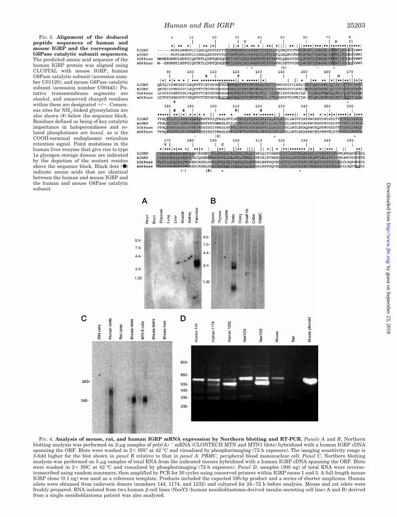
FIG. 3. Alignment of the deducedpeptide sequences of human andmouse IGRP and the correspondingG6Pase catalytic subunit sequences.The predicted amino acid sequence of thehuman IGRP protein was aligned usingCLUSTAL with mouse IGRP, humanG6Pase catalytic subunit (accession num-ber U01120), and mouse G6Pase catalyticsubunit (accession number U00445). Pu-tative transmembrane segments areshaded, and conserved charged residueswithin them are designated 1/2. Consen-sus sites for NH2-linked glycosylation arealso shown (#) below the sequence block.Residues defined as being of key catalyticimportance in haloperoxidases and re-lated phosphatases are boxed, as is theCOOH-terminal endoplasmic reticulumretention signal. Point mutations in thehuman liver enzyme that give rise to type1a glycogen storage disease are indicatedby the depiction of the mutant residueabove the sequence block. Black dots (●)indicate amino acids that are identicalbetween the human and mouse IGRP andthe human and mouse G6Pase catalyticsubunit.
FIG. 4. Analysis of mouse, rat, and human IGRP mRNA expression by Northern blotting and RT-PCR. Panels A and B, Northernblotting analysis was performed on 2-mg samples of poly(A) 1 mRNA (CLONTECH MTN and MTN1 blots) hybridized with a human IGRP cDNAspanning the ORF. Blots were washed in 23 SSC at 42 °C and visualized by phosphorimaging (72 h exposure). The imaging sensitivity range is3-fold higher for the blot shown in panel B relative to that in panel A. PBMC, peripheral blood mononuclear cell. Panel C, Northern blottinganalysis was performed on 5-mg samples of total RNA from the indicated tissues hybridized with a human IGRP cDNA spanning the ORF. Blotswere washed in 23 SSC at 42 °C and visualized by phosphorimaging (72-h exposure). Panel D, samples (300 ng) of total RNA were reverse-transcribed using random nonamers, then amplified by PCR for 30 cycles using conserved primers within IGRP exons 1 and 5. A full-length mouseIGRP clone (0.1 ng) was used as a reference template. Products included the expected 595-bp product and a series of shorter amplicons. Humanislets were obtained from cadaveric donors (numbers 144, 1174, and 1232) and cultured for 24–72 h before analysis. Mouse and rat islets werefreshly prepared. RNA isolated from two human b cell lines (NesY2 (human nesidioblastoma-derived insulin-secreting cell line) A and B) derivedfrom a single nesidioblastoma patient was also analyzed.
Human and Rat IGRP 25203
by guest on September 23, 2018
http://ww
w.jbc.org/
Dow
nloaded from

ate a 32.2-kDa protein. The relative proportions of alternatelyspliced forms varied from individual to individual and betweenoligo(dT) versus randomly primed transcripts. This variationdid not appear to correlate with the age, sex, and weight of thedonor nor the warm ischemia time before islet isolation orduration of tissue culture before RNA extraction (Fig. 4D).Without reference to fresh material from healthy individuals,the significance of these alternatively spliced products isunclear.
Islet-specific Pancreatic Expression of Mouse IGRP—Al-though Northern blotting indicated a pancreas-specific patternof IGRP mRNA expression (Fig. 4A), this analysis did notdetermine whether IGRP was expressed in pancreatic endo-crine or exocrine cells. To address this issue antibodies wereraised to recombinant mouse IGRP, and immunoperoxidasestaining of mouse pancreas was performed. The result showsthat the antigen was localized to islet cells with no reactivityevident in the acinar tissue or ductal elements (Fig. 5). Veryfew IGRP-negative cells were observed within the islet, sug-gesting that alpha and beta cells were certainly immunoreac-tive and that possibly all four endocrine cell types expressedthe protein (Fig. 5).
Enzymic Activity of Human IGRP—Enzyme activity studieswere performed by transiently transfecting COS 7 cells withvarious pCDNA 3.1 constructs; G6P hydrolytic activity wasthen assessed in a micosomal fraction prepared from lysedcells. A construct encoding the rat G6Pase catalytic subunitserved as a positive control, and the efficiency of transfectionwas evaluated by co-transfection of a Rous sarcoma virus-bgalactosidase fusion gene construct. Transfection with theG6Pase catalytic subunit construct resulted in an ;25-foldincrease in G6P hydrolysis over basal activity (Table II). Incontrast, transfection with a construct encoding human IGRPproduced no detectable change in G6P hydrolytic activity, aspreviously observed for mouse IGRP (Table II and Ref. 21).Transfection of COS 7 cells with constructs encoding truncated
forms of mouse IGRP in which the putative start codon wasdeleted but which contained either the second or third putativestart site also failed to increase basal G6P hydrolytic activity(Table II). The rates of hydrolysis of the generic phosphatasesubstrate, p-nitrophenol phosphate were not altered in humanIGRP- or mouse IGRP-transfected COS 7 cells, although theywere good substrates for the G6Pase catalytic subunit (data notshown and Ref. 21).
Transcriptional Activity of the Proximal Mouse, Rat, andHuman IGRP Gene Promoters—We have previously shownthat the proximal mouse IGRP promoter region, located be-tween 2306 and 13, is sufficient to confer maximal IGRP-CATfusion gene expression in HIT cells (28). The level of basalmouse IGRP-CAT fusion gene expression in both HIT andbTC-3 cells decreases gradually upon deletion of the IGRPpromoter sequence between 2306 and 266, indicating thatmultiple cis-acting elements contribute to maximal fusion geneexpression (36). An alignment of the equivalent human and ratIGRP promoter regions revealed multiple regions of conservedsequence (Fig. 6). We previously determined the location ofseveral transcription factor binding sites in the mouse IGRPpromoter using the ligation-mediated PCR in situ footprintingtechnique; these binding sites correlated with regions of theIGRP promoter, identified as being important for basal IGRP-CAT fusion gene expression (36). Fig. 6 shows that many of theresidues in the mouse IGRP promoter that are contacted bytranscription factors in bTC-3 cells in situ are also conserved inthe human and rat promoters. In addition, a hepatocyte nu-clear factor-3 binding site identified in the mouse IGRP pro-moter, which binds a hepatocyte nuclear factor-3 in vitro (36),is also conserved in the rat and human promoters (Fig. 6).
The observation that several putative cis-acting elementsare conserved in the rat IGRP promoter (Fig. 6) was surprisinggiven that the rat IGRP gene is not expressed (Figs. 4, C andD). In contrast, the TATA box motif identified in the mouseIGRP promoter is not conserved in the rat promoter (Fig. 6).Wobbe and Struhl (43) have shown that the sequence TGTAfound in the rat promoter directs a greater than 20-fold lowerlevel of in vitro transcription than the TATA motif. To deter-mine whether this and other sequence variations affect therelative activity of the mouse, rat, and human IGRP promoters,we constructed fusion genes in which these promoters wereligated to the CAT reporter gene. Basal IGRP-CAT fusion geneexpression was then assayed after transient transfection of theHIT cell line. Fig. 7A shows that the human IGRP promotersequence located between 2324 and 13 confers a slightlyhigher level of basal fusion gene expression than the equivalent
FIG. 5. Immunoperoxidase staining of mouse pancreas withantibodies raised to recombinant IGRP. Immunoperoxidase stain-ing was performed as described under “Experimental Procedures.”IGRP was localized by immunoperoxidase labeling (brown) and thesections were counterstained with Gill’s hematoxylin.
TABLE IIG6P hydrolytic activity in transiently transfected COS7 cells
COS7 cells were co-transfected as described under “ExperimentalProcedures,” with pCDNA3.1 constructs encoding IGRP or rat G6Pasecatalytic subunit (15 mg) together with a reference vector encodingb-galactosidase (5 mg). Enzyme activities were determined in cell ho-mogenates after 48 h. Mouse IGRP constructs (see Fig. 2 legend fordetails) included the designated full-length construct (mouse IGRP), aPstI fragment containing the second and third alternative start codons(Met 213), and a PCR construct containing the third start codon only(Met 3). Each tabulated result represents the mean 6 S.E. of duplicatedeterminations from four separate experiments.
b-Galactosidase G6P hydrolysis
nmol/min/mg nmol/min/mg
Control 307 6 103 6.1 6 2.1Human IGRP 239 6 126 5.7 6 2.7Mouse IGRP 309 6 111 6.3 6 1.8Met 213 400 6 240 6.3 6 3.1Met 3 256 6 148 6.6 6 2.7G6Pase 204 6 91 152.6 6 36.6
Human and Rat IGRP25204
by guest on September 23, 2018
http://ww
w.jbc.org/
Dow
nloaded from

mouse promoter sequence located between 2306 and 13. Bycontrast, neither the equivalent rat IGRP promoter sequencelocated between 2321 and 13 (Fig. 7B) nor a longer fragmentof the promoter containing sequence located between 2900 and13 (Fig. 7A) confers appreciable fusion gene expression. How-ever, mutating the rat TGTA motif back to the consensus TATAmotif markedly enhances basal rat IGRP-CAT fusion gene ex-pression (Fig. 7B). Nevertheless, the actual level of rat pro-moter activity remains ;3-fold lower than that of the mouse(Fig. 7B), suggesting that changes in elements other than theTATA box contribute to the low activity of the rat promoter.
A concern with all the data obtained on the rat IGRP geneand its transcription or translation was the fact that a singleBAC clone served as the source of sequence data and fusiongene constructs. It was conceivable that a pseudogene wasselected in the original screen and/or that sequence artifactswere introduced during the construction of the BAC library.These concerns were addressed by sequence analysis of criticalregions of the rat IGRP gene amplified by PCR using primersets based on conserved rat/mouse sequences with genomicDNA isolated from adult rat spleen and liver as the template.These regions included the promoter and the TGTA motif, thedeletions and insertion affecting the reading frame in exons 1and 5 and the entire intervening sequence between exons 3 and
5. In every case the BAC sequence was confirmed. The rat BACsequencing and PCR-based gene sequencing were performed inNashville, TN and Denver, CO, respectively using independentprimer sets to avoid any risk of contamination.
DISCUSSION
Two remarkable features of the IGRP molecule are the focusof the current investigation. The first is its structural similarityto the G6Pase catalytic subunit, a molecule that plays a centralrole in glucose homeostasis and the pathophysiology of diabetesmellitus. The second is its restricted expression to pancreaticislets. A comparative analysis of the human, mouse, and ratgene structures was undertaken to define the conserved pri-mary sequence of the protein, which could impact on the ex-pression of IGRP catalytic activity and conserved promotersequences that could be important in tissue-specific and phys-iological transcriptional regulation.
Multiple shared sequences were identified in the promotersof the mouse, rat, and human IGRP genes (Fig. 6) that, alongwith our previous in situ footprinting studies (36), will facili-tate the identification of cis-acting elements, which are impor-tant for basal and islet-specific IGRP gene expression. Of spe-cific interest for future mutagenesis are two putative E-boxelements (Fig. 6). Such an element is one of the major sites thatcontributes to basal insulin gene expression mediated by aheterodimeric complex of the basic helix-loop-helix proteinsBETA2/NeuroD and a ubiquitous factor, either E2A or HEB(44, 45). Surprisingly, both the putative IGRP E-box motifs are
FIG. 6. Alignment of the mouse, rat, and human IGRP genepromoter sequences. The human, mouse, and rat IGRP promotersequences were aligned using the IntelliGenetics, Inc. IFIND programand labeled relative to the experimentally determined transcriptionstart site of mouse IGRP (28) designated as 11. Increases (●) or de-creases (E) in dimethyl sulfate methylation of the mouse IGRP pro-moter comparing in situ versus in vitro methylated bTC-3 cell DNAwere determined by ligation-mediated PCR (36). The TATA box, twoE-box motifs, and a hepatocyte nuclear factor-3 binding site, which areconserved between the mouse and human promoters, are boxed.
FIG. 7. Basal activities of the mouse, rat, and human IGRPgene promoters. HIT cells were transiently co-transfected, as de-scribed under “Experimental Procedures,” with mouse, rat, or humanIGRP promoter-CAT fusion gene constructs (15 mg) containing thepromoter sequence shown together with a reference vector encodingb-galactosidase (bgal; 2.5 mg). The rat constructs had either the wild-type (TGTA) or back-mutated (TATA) box motif. After transfection, cellswere cultured for 18–20 h in serum-free medium, and CAT and b-ga-lactosidase activity was determined (36). The mean ratio of CAT:b-galactosidase activity 6S.E. is presented from three transfection exper-iments, each using independent preparations of each CAT plasmid.
Human and Rat IGRP 25205
by guest on September 23, 2018
http://ww
w.jbc.org/
Dow
nloaded from

conserved in the rat promoter despite the fact that the TATAbox in the rat IGRP promoter is mutated (Fig. 6) and the geneis not expressed (Fig. 4D). An E-box motif in the humantranscobalamin II promoter has been shown to mediate bidi-rectional transcription in the absence of a TATA box motif (46).However, in the case of the rat IGRP promoter, the two E-boxmotifs are not sufficient to confer high promoter activity in theabsence of the TATA box (Fig. 7B). The proximal region of themouse IGRP promoter that contains the two putative E-boxmotifs by itself confers very low basal fusion gene expression(36); thus, even if these elements are found to contribute tobasal IGRP gene expression, their activity will depend on ad-ditional distal elements.
Several protein binding sites have been identified in themouse IGRP promoter by in situ footprinting (36) (Fig. 6) forwhich no candidate trans-acting factor can be identified bycomputer analysis using the MatInspector software (47). Theseconceivably represent binding sites for novel islet-enrichedtrans-acting factors, a hypothesis that gains further credencefrom the observation that these regions are highly conserved inthe rat and human IGRP promoters (Fig. 6). The search forsuch elements and their associated binding proteins is of par-ticular interest given the emerging realization that such fac-tors include potential diabetogenes (48, 49), many of which alsoplay a critical role in pancreatic ontogeny (50–52).
Since the human IGRP gene, its mRNA, and the encodedprotein showed great similarity to the corresponding mouseIGRP molecules this suggests conservation of biological func-tion. The full-length human IGRP protein, like the mouse pro-tein, possessed characteristic structural features of an ER-localized transmembrane protein with hydrolytic properties(41, 42). Yet as previously observed, no catalytic activity wasobtained with either G6P or a generic phosphatase substrate(Table II). Nevertheless, the present documentation of the hu-man IGRP protein sequence will assist in future structure/function studies using site-directed mutagenesis and chimericIGRP/G6Pase catalytic subunit proteins. Immunohistochemi-cal analyses with antibodies directed toward recombinantmouse IGRP confirm that the IGRP protein is expressed endo-genously in mouse islets (Fig. 5). Moreover, Western blottinganalyses with antibodies directed toward Myc-tagged mouseIGRP show that it can be expressed in COS 7 cells by transienttransfection.3 Thus our inability to detect enzymatic activitycannot be related to failure of protein expression.
Although the data obtained on human IGRP were consistentwith the hypothesis that IGRP plays an important tissue-specific function in the adult pancreatic islet, the results withthe rat IGRP gene paradoxically indicate that IGRP is a non-functional or perhaps a vestigial gene product. We found noevidence for the existence of a second, functional IGRP gene inthe rat, and mRNA expression could not be detected in ratislets or rat islet-derived cell lines using either probes thathybridize with the highly homologous mouse sequence or byRT-PCR analyses with exact primers (Fig. 4). The rat IGRPgene nucleotide sequence was more closely related to the mousethan the human sequence, as expected from the taxonomicdivergence of these species, and clearly it was the rat equiva-lent of the IGRP gene. A possible explanation of this paradox isthat selection has occurred in the rat IGRP gene for a series ofmutational events that have ensured its inactivation. The de-letion of exon 4 in the rat IGRP gene removes sequences thatwould be essential for hydrolytic activity (53), although it couldbe argued that, like the common Dexon 4 splice variant ofmouse (21) and human IGRP (Fig. 4D), such a molecule has a
regulatory function. However, the reading frame mutations inthe 59 and 39 ends of the ORF of rat IGRP and the change in theTATA box indicate that, at some point in evolution, expressionof IGRP in the rat islet was turned off because the protein wasunnecessary, redundant, or deleterious.
Acknowledgments—We thank Roland Stein and Eva Henderson forproviding the HIT cell line, Rebecca Taub for the rat G6Pase construct,Kevin Docherty for NesY2 (human nesidioblastoma-derived insulin-secreting cell line) RNA, and Donna Curtis for assistance with the database searching. Human islets were obtained from Miami, FL, Roches-ter, MN and Boston, MA through the Juvenile Diabetes FoundationInternational islet consortium.
REFERENCES
1. Mithieux, G. (1997) Eur. J. Endocrinol. 136, 137–1452. Foster, J. D., Pederson, B. A., and Nordlie, R. C. (1997) Proc. Soc. Exp. Biol.
Med. 215, 314–3323. Gerin, I., Veiga-da-Cunha, M., Achouri, Y., Collet, J. F., and Van Schaftingen,
E. (1997) FEBS Lett. 419, 235–2384. Chou, J. Y., and Mansfield, B. C. (1999) Trends Endocrinol. Metab. 10,
104–1135. Cline, G. W., Rothman, D. L., Magnusson, I., Katz, L. D., and Shulman, G. I.
(1994) J. Clin. Invest. 94, 2369–23766. Consoli, A. (1992) Diabetes Care 15, 430–4417. Argaud, D., Zhang, Q., Pan, W., Maitra, S., Pilkis, S. J., and Lange, A. J. (1996)
Diabetes 45, 1563–15718. Liu, Z., Barrett, E. J., Dalkin, A. C., Zwart, A. D., and Chou, J. Y. (1994)
Biochem. Biophys. Res. Commun. 205, 680–6869. Haber, B. A., Chin, S., Chuang, E., Buikhuisen, W., Naji, A., and Taub, R.
(1995) J. Clin. Invest. 95, 832–84110. Massillon, D., Barzilai, N., Chen, W., Hu, M., and Rossetti, L. (1996) J. Biol.
Chem. 271, 9871–987411. Mithieux, G., Vidal, H., Zitoun, C., Bruni, N., Daniele, N., and Minassian, C.
(1996) Diabetes 45, 891–89612. Granner, D. K., and O’Brien, R. M. (1992) Diabetes Care 15, 369–39513. O’Brien, R. M., and Granner, D. K. (1996) Physiol. Rev. 76, 1109–116114. Massillon, D., Chen, W., Barzilai, N., Prus-Wertheimer, D., Hawkins, M., Liu,
R., Taub, R., and Rossetti, L. (1998) J. Biol. Chem. 273, 228–23415. Argaud, D., Kirby, T. L., Newgard, C. B., and Lange, A. J. (1997) J. Biol. Chem.
272, 12854–1286116. Streeper, R. S., Svitek, C. A., Chapman, S., Greenbaum, L. E., Taub, R., and
O’Brien, R. M. (1997) J. Biol. Chem. 272, 11698–1170117. Streeper, R. S., Eaton, E. M., Ebert, D. H., Chapman, S. C., Svitek, C. A., and
O’Brien, R. M. (1998) Proc. Natl. Acad. Sci. U. S. A. 95, 9208–921318. Li, Y., and van de Werve, G. (2000) Biochem. Biophys. Res. Commun. 272,
41–4419. Seoane, J., Trinh, K., O’Doherty, R. M., Gomez-Foix, A. M., Lange, A. J.,
Newgard, C. B., and Guinovart, J. J. (1997) J. Biol. Chem. 272,26972–26977
20. An, J., Li, Y., van De Werve, G., and Newgard, C. B. (2001) J. Biol. Chem. 276,10722–10729
21. Arden, S. D., Zahn, T., Steegers, S., Webb, S., Bergman, B., O’Brien, R. M., andHutton, J. C. (1999) Diabetes 48, 531–542
22. Perales, M. A., Sener, A., and Malaisse, W. J. (1991) Mol. Cell. Biochem. 101,67–71
23. Trandaburu, T. (1977) Acta Histochem. 59, 246–25324. Watanabe, A., and Nagashima, H. (1983) Acta Med. Okayama 37, 463–47025. Waddell, I. D., and Burchell, A. (1988) Biochem. J. 255, 471–47626. Khan, A., Chandramouli, V., Ostenson, C. G., Low, H., Landau, B. R., and
Efendic, S. (1990) Diabetes 39, 456–45927. Khan, A., Hong-Lie, C., and Landau, B. R. (1995) Endocrinology 136,
1934–193828. Ebert, D. H., Bischof, L. J., Streeper, R. S., Chapman, S. C., Svitek, C. A.,
Goldman, J. K., Mathews, C. E., Leiter, E. H., Hutton, J. C., and O’Brien,R. M. (1999) Diabetes 48, 543–551
29. Sambrook, J., Fritsch, E. F., and Maniatis, E. F. (1989) Molecular Cloning: ALaboratory Manual, 2nd Ed., Cold Spring Harbor Laboratory, Cold SpringHarbor Laboratory, Plainview, NY
30. Jurka, J., Klonowski, P., Dagman, V., and Pelton, P. (1996) Comput. Chem. 20,119–121
31. Lacy, P. E., and Kostianovsky, M. (1967) Diabetes 16, 35–3932. Guest, P. C., Pipeleers, D., Rossier, J., Rhodes, C. J., and Hutton, J. C. (1989)
Biochem. J. 264, 503-v833. Kozak, M. (1991) J. Biol. Chem. 266, 19867–1987034. Bressan, G. M., and Stanley, K. K. (1987) Nucleic Acids Res. 15, 1005635. Jacoby, D. B., Zilz, N. D., and Towle, H. C. (1989) J. Biol. Chem. 264,
17623–1762636. Bischof, L. J., Martin, C. C., Svitek, C. A., Stadelmaier, B. T., Hornbuckle,
L. A., Goldman, J. K., Oeser, J. K., Hutton, J. C., and O’Brien, R. M. (2001)Diabetes 50, 502–514
37. Arden, S. D., Roep, B. O., Neophytou, P. I., Usac, E. F., Duinkerken, G., deVries, R. R., and Hutton, J. C. (1996) J. Clin. Invest. 97, 551–561
38. Jackson, I. J. (1991) Nucleic Acids Res. 19, 3795–379839. Kollmar, R., and Farnham, P. J. (1993) Proc. Soc. Exp. Biol. Med. 203, 127–13940. Milpetz, F., Argos, P., and Persson, B. (1995) Trends Biochem. Sci. 20,
204–20541. Stukey, J., and Carman, G. M. (1997) Protein Sci. 6, 469–47242. Hemrika, W., Renirie, R., Dekker, H. L., Barnett, P., and Wever, R. (1997)
Proc. Natl. Acad. Sci. U. S. A. 94, 2145–21493 B. Bergman and J. C. Hutton, unpublished observations.
Human and Rat IGRP25206
by guest on September 23, 2018
http://ww
w.jbc.org/
Dow
nloaded from

43. Wobbe, C. R., and Struhl, K. (1990) Mol. Cell. Biol. 10, 3859–386744. Stein, R. (2001) in Handbook of Physiology. Section 7: The Endocrine System
(Jefferson, L. S., and Cherrington, A. D., eds) Vol. II, pp. 25–47, OxfordUniversity Press, New York
45. German, M. (2000) in Diabetes Mellitus: A Fundamental and Clinical Text(LeRoith, D., Taylor, S. I., and Olefsky, J. M., eds) 2nd Ed., pp. 11–19,Lippincott-Raven Publishers, Philadelphia
46. Li, N., and Seetharam, B. (1998) J. Biol. Chem. 273, 28170–2817747. Quandt, K., Frech, K., Karas, H., Wingender, E., and Werner, T. (1995) Nucleic
Acids Res. 23, 4878–488448. Winter, W. E. (2000) Pediatr. Diabetes 1, 88–11749. Habener, J. F., and Stoffers, D. A. (1998) Proc. Assoc. Am. Physicians 110, 12–2150. Sander, M., and German, M. S. (1997) J. Mol. Med. 75, 327–34051. Edlund, H. (1998) Diabetes 47, 1817–182352. Bramblett, D. E., Huang, H. P., and Tsai, M. J. (2000) Adv. Pharmacol. 47,
255–31553. Pan, C. J., Lei, K. J., Annabi, B., Hemrika, W., and Chou, J. Y. (1998) J. Biol.
Chem. 273, 6144–6148
Human and Rat IGRP 25207
by guest on September 23, 2018
http://ww
w.jbc.org/
Dow
nloaded from

HuttonJames K. Oeser, Frédéric Leprêtre, Philippe Froguel, Richard M. O'Brien and John C.Claudia Frigeri, David Wahl, Christina A. Svitek, Randall Wong, Joshua K. Goldman,
Cyrus C. Martin, Larry J. Bischof, Barbara Bergman, Lauri A. Hornbuckle, Carl Hilliker,Glucose-6-phosphatase Catalytic Subunit-related Protein (IGRP) Genes
Cloning and Characterization of the Human and Rat Islet-specific
doi: 10.1074/jbc.M101549200 originally published online April 10, 20012001, 276:25197-25207.J. Biol. Chem.
10.1074/jbc.M101549200Access the most updated version of this article at doi:
Alerts:
When a correction for this article is posted•
When this article is cited•
to choose from all of JBC's e-mail alertsClick here
http://www.jbc.org/content/276/27/25197.full.html#ref-list-1
This article cites 51 references, 26 of which can be accessed free at
by guest on September 23, 2018
http://ww
w.jbc.org/
Dow
nloaded from