Embed Size (px)
Citation preview

Brirish Journal of Plasric Surgery (1984) 31, 635642 c 1984 The Trustees of British Association of Pktic Surgeons
The intra-uterine healing of foetal muscle wounds: experimental study in the rat
A. R. ROWSELL
Department of Plastic Surgery, The Radcliffe Infirmary, Oxford
Summary-This work confirms that foetal wound healing is rapid, occurs without an apparent inflammatory response and takes place without the deposition of collagen. It is shown for the first time that foetal muscle wounds heal by regeneration. This study also shows that, immediately after injury, there is a degeneration of mesenchymal and ectodermal elements in that part of the lip between the wound and the mid line. This degeneration is followed by regeneration of only the mesenchymal elements.
For the past two decades there has been an increasing interest in the possibility of in-utero correction of life-threatening foetal abnormalities.
Intrauterine surgery is not new. Mayer in 1918 was the first to report a successful attempt at intrauterine surgery, when he described the transfer of guinea pig foetuses from the lumen of the uterus to the maternal peritoneal cavity, with survival of the foetus.
One year later, Wolff (1919) described a similar series of experiments in which he transferred foetal rabbits from the uterine lumen to the maternal peritoneal cavity, again with foetal survival.
The first reports of successful intrauterine foetal surgery, were those of Bors and Nicholas in 1925. Bors showed that in-utero amputation of limbs in foetal guinea pigs did not prevent a successful outcome to the pregnancy.
In the same year Nicholas described the long term survival of foetal rats following the in-utero ablation of eyes, limbs and tails.
Since these early attempts, there have been numerous reports of intrauterine experiments carried out on foetuses of many different species. Before any attempt can be made to correct foetal abnormalities in utero, it is necessary to have a thorough understanding of foetal wound healing.
Healing in foetal skin wounds has been described by Hess (1954), Dixon (1960), Somasundaram and Prathap (1972), Sopher (1973, Goss (1977) and Robinson and Goss (1981). Musa Ris and Wray (1972) have studied the healing of long bone fractures in the foetal rabbit, but apart from an inconclusive report by
Robinson and Goss (1981), there has been no published description of foetal muscle healing. In an attempt to correct this deficiency a study of muscle healing in foetal rats was undertaken. The experimental model chosen for this study was a through and through wound of the upper lip, and the animals were followed for 72 hours after surgery.
l”“l”“1 Icm
Fig. 1
Figure l-The experimental lip wound
635
636 BRITISH JOURNAL OF PLASTIC SURGERY
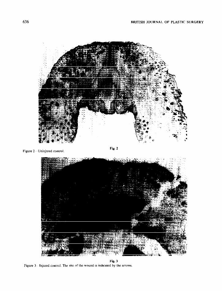
Figure 2- -Uninjured control.
Fig. 3
Figure 3-Injured control. The site of the wound is indicated by the arrows

THE INTRA-UTERINE HEALING OF FOETAL MUSCLE WOUNDS: EXPERIMENTAL STUDY IN THE RAT 637
Figure 4
Figure S-
Fig. 4
Unsutured wound at 24 hours. The site of the wound is indicated by the arrows ( x 2)
Fig. S
Unsutured wound at 24 hours. The site of the wound is indicated by the arrow ( x 8)

638 BRITISH JOURNAL OF PLASTIC SURGERY
Figu Ire
Fig. 6
Wnsutured wound at 48 hours. The site of the wound is indicated by the arrows.
Fig ur e 7- -Unsutured wound at 72 hours. The site of the wound is indicated by the arrows.
Fig. 7

THE INTRA-UTERINE HEALING OF FOETAL MUSCLE WOUNDS: EXPERIMENTAL STUDY IN THE RAT 639
Figu
Fig. 8
ured wound at 24 hours. The site of the wound is indicated by the arrows
Figul re 9-
Fig. 9
med wound at 48 hours. The site of the wound is indicated by the arrows.

640 BRITISH JOURNAL OF PLASTIC SURGERY
Figur .e l&Sutured wound at 72 hours. The site of the wound is indicated by the arrows.
Fig. 10
Materials and methods
Time-mated adult female Wistar rats of between 295 and 335 grams were anaesthetised with halothane on the 18th day of gestation. A laparotomy was performed in each animal to gain access to the pregnant uterus. Each experimental foetus was partially exteriorised through a small hysterotomy and a through and through surgical wound made in the upper lip approximately 2mm from the mid line.
Each foetus was returned to the lumen of the uterus, and the hysterotomy closed with a purse- string suture. The laparotomy wounds were closed in layers and the animals allowed to recover from the anaesthetic. The pregnancies then continued until the foetuses were harvested.
In this study a total of 60 foetal rats were each given a wound of the upper lip (Fig. 1). The injured foetuses were divided into two groups, each containing 30 animals. In the first group the lip wounds were left unsutured, while in the second group the wounds were sutured using 10/O nylon and microsurgical techniques. In each group, ten foetuses were harvested at 24 hours, ten were harvested at 48 hours, and ten were
harvested at 72 hours after wounding. Each harvested foetus was fixed in formal saline, and haematoxylin and eosin stained horizontal sections of the upper lip were prepared.
Results
The first histological section (Fig. 2) is that of an uninjured control. The epidermis, oral mucosa, vibrissae follicles, and striated muscle bundles are clearly visible.
The next section (Fig. 3) is that of an injured control, a foetus that was harvested immediately after wounding, which shows a through and through wound that is disrupting all the layers of the lip. The creation of this wound has had no effect on the structures in the lip between the wound and the midline.
Unsutured wounds
Figure 4 is a photomicrograph of an unsutured wound 24 hours post injury and shows that both the epidermis and the oral mucosa are regen- erating. In the substance of the lip, free eryth- rocytes are visible and represent a haema- toma.

THE INTRA-UTERINE HEALING OF FOETAL MUSCLE WOUNDS: EXPERIMENTAL STUDY IN THE RAT 641
An important observation is that between the wound and the midline there has been a loss both of ectodermal elements, represented by a reduction in the number of vibrissae follicles, and of differentiated mesenchymal elements, rep- resented by an absence of striated muscle.
A high-power view of the same section (Fig. 5) does not show the presence of any inflammatory cells.
By 48 hours after injury there has been some recovery of the mesenchymal elements within the lip with striated muscle bundles clearly visibie (Fig. 6). However, there appears to have been little, if any, recovery of the ectodermal elements.
By 72 hours after injury (Fig. 7) the muscle bundles have recovered to such a degree that they are comparable to those in the uninjured side of the lip. It can be also seen in this section that the regenerated muscle bundles cross the original wound site without either interruption or the interposition of scar tissue.
Sutured wounds
Figure 8 is a photomicrograph of a sutured wound at 24 hours post injury, and it can be seen in this section that the epidermis and the oral mucosa are regenerating. Again between the wound and the midline there has been a loss of ectodermal elements represented by a reduced number of vibrissae follicles. However, there has been only a partial loss of striated muscle in that part of the lip.
By 48 hours post-injury (Fig. 9) there has been such a degree of recovery that the muscle bundles on the injured side of the lip are comparable to those on the uninjured side. This section also shows the muscle bundles crossing the wound without interruption and without the interposition of scar tissue.
The histological picture of the sutured wound at 72 hours post-injury (Fig. 10) is very similar to that at 48 hours.
It was not possible to demonstrate the deposition of collagen in any of these wounds using the van Gieson staining technique.
Discussion
Rapid regeneration of the epidermis and the oral mucosa was a feature of all the wounds in this study, whether sutured or unsutured. This agrees with the findings of other workers including Hess
(1954), Sopher (1975), Goss (1977), and Robinson and Goss (1981).
The report on foetal muscle healing by Robinson and Goss in 1981 was based on a study of cheek wounds in foetal rats. These wounds involved skin, muscle and oral mucosa but, unfortunately, the authors only followed their experimental animals for 24 hours after operation. Because of this short period of observation their findings on the healing of the muscle component were incomplete. Although the muscle ends had not shown any sign of reconstitution, they were able to report that by 24 hours there was mitotic activity in the myoblasts.
Although the findings of Robinson and Goss were inconclusive they were encouraging. Because wound healing is a temporal as well as a spatial phenomenon, it was decided to follow the animals in this study for 72 hours after surgery, in an attempt to reach the end point of foetal muscle healing.
The striated muscle elements in the lip wounds took longer to heal than the epidermis and oral mucosa, and it was not until 48 hours post injury in the sutured wounds, and 72 hours in the unsutured wounds, that the muscle bundles crossed the original site of injury. There was no evidence of collagen deposition in any of the wounds and it would appear that striated muscle wounds in the foetus heal by regeneration rather than by repair.
Several previous workers, Sopher (1975), Goss (1977), and Robinson and Goss (1981). have reported the apparent absence of an inflammatory response in foetal wounds and there was an absence of inflammatory cells in all the wounds investigated in this study. Whether this lack of an inflammatory response may be due to the sterility of the intrauterine environment or to an immaturity of the foetal reticula-endothelial system is open to question. Block (1960) found that in the opossum there was no inflammatory response in neonates for the first 5 days of post-natal life. which would support the latter theory.
The phenomenon of post-injury degeneration of the ectodermal and mesenchymal derivatives in that part of the lip between the wound and the midline. followed by regeneration of the mesenchymal derivatives only, was an unexpected finding. Further work is being undertaken to investigate this and the findings of that study will be reported in a subsequent paper.

642 BRITISH JOURNAL OF PLASTIC SURGERY
Conclusion
Foetal muscle wounds heal by regeneration rather than by repair. The findings of this study would also appear to support the view that a growth factor, either humoral or cellular, influences facial tissue differentiation from lateral to medial, and that a wound or cleft placed across the tissues interferes with this process.
Goss, A. N. (1977). Intra-uterine healing of fetal rat oral mucosa, skin and cartilage wounds. Journal of Oral Pathology, 6, 35.
Hess, A. (1954). Reactions of mammalian fetal tissue to injury. 11. Skin. Anntomica/ Record, 119,435.
Mayer, A. (1918). Uber die Miiglichkeit operativer Eingriffe beim lebendem Slugetierfoetus. Zentralblatt .fiir Gynaekologie, 42, 773.
Musa Ris, P. and Wray, J. B. (1972). A histological study of fracture healing within the uterus of the rabbit. CIinical Orthopaedics, 87, 318.
Acknowledgements Nicholas, J. S. (1925). Notes on the application of
experimental methods upon mammalian embryos.
The author would like to thank Mr M. D. Poole and Mr A. M. Godfrey for their encouragement and advice, Dr B. Roach for her help with the interpretation of the histological findings and the Plastic Surgery Research Fund, Oxford, for financing this project. I would also like to thank Mr M. Christmas of the Department of Medical Illustration at the Welsh Regional Plastic Surgery, Burns and Oral Surgery Centre, Chepstow, for his invaluable help with the illustrations.
Anatomical Record, 31, 385. Robinson. B. W. and Goss. A. N. (1981). Intrauterine healing
of fetal rat cheek wounds. Cleft Palate Journal, 18, 251. _ Somaaundaram, K. and Prathap, K. (1970). Intrauterine
healing of skin wounds in rabbit fetuses, Journal of Pathology, 100, 81.
Footnote
This paper was presented at the Winter meeting of the British Association of Plastic Surgeons, December, 1983.
Sopher, D. (1975). Future prospects for fetal surgery. In Teratology: Trends and Applications. Edited by C. L. Burry and D. E. Poswillo. New York: Springer-Verlag.
Wolff, B. (1919). Experimentelle Untersuchungen iiber die Entstehung extrauteriner Schwanger-schaften und iiber die Maglichkeit operativer Eingriffe beim lebenden SBugetierfoetus. Beitraege zur Pathologischen Anatomie, 65, 423.
References Block, M. (1960). Wound healing in the new-born opossum
(Didelphys virginiana). Nature, 187, 340. Bars, E. (1925). Die methodik der intrauterinen Operation am
iiberlebenden Siugetierfoetus. Archiv fiir Entwick- lungsmechanik der Organismen (Lxipzig), 105, 655.
Dixon, J. B. (1960). Inflammation in the fetal and neonatal rat. The local reaction to skin burns. Journal of Pathology
Author
A. R. Rowsell, FRCS, FRACS. Senior Registrar in Plastic Surgery, The Department of Plastic Surgery, The Radcliffe
and Bacteriology, 80, 73. Infirmary, Oxtbrd.