Embed Size (px)
Citation preview

PRINTED BY: [email protected]. Printing is for personal, private use only. No part of this book may be reproduced or transmittedwithout publisher's prior permission. Violators will be prosecuted.
7 Skeletal System
LEARNING OBJECTIVES
1 List and describe five functions of the skeletal system.2 Explain the difference between compact and spongy bone.3 Classify bones according to size and shape.4 Identify the general features of a long bone.5 Explain the process by which long bones grow in length.6 Explain the difference between the axial and appendicular skeletons.7 Identify the bones of the skull.8 Identify the structural features of vertebrae.9 List and describe the divisions of the vertebral column.10 Describe the structural features of the sternum and ribs.11 Identify the parts of the pectoral girdle.12 Identify the bones of the upper extremities.13 Identify the parts of the pelvic girdle.14 Identify the bones of the lower extremities.15 List and describe the different types of joints.16 Describe ways in which the aging of an individual affects the skeletal system.17 Identify pathology related to the skeletal system.
106 107Key Termsamphiarthrosis (am-fee-ahr-THROH-sis)diaphysis (dye-AF-ih-sis)diarthrosis (dye-ahr-THROH-sis)epiphyseal plate (ep-ih-FIZ-ee-al PLATE)epiphysis (ee-PIF-ih-sis)osteoblast (AH-stee-oh-blast)osteoclast (AH-stee-oh-clast)osteocyte (AH-stee-oh-syte)osteon (OH-stee-ahn)synarthrosis (sin-ahr-THROH-sis)Introduction to the Skeletal System
The skeletal system consists of the bones and the cartilage, ligaments, and tendons associated with the bones. It accounts for about 20% of thebody weight. Bones are rigid structures that form the framework for the body. People often think of bones as dead, dry, inert pipes and platesbecause that is how they are seen in the laboratory. In reality, the living bones in our bodies contain active tissues that consume nutrients,require a blood supply, use oxygen and discharge waste products in metabolism, and change shape or remodel in response to variations inmechanical stress. The skeletal system is strong but lightweight. It is well adapted for the functions it must perform. It is a masterpiece ofdesign.
Overview of the Skeletal SystemFunctions of the Skeletal System
The skeletal system gives form and shape to the body. Without the skeletal components, we would appear as big “blobs” inefficiently “oozing”around on the ground. Besides contributing to shape and form, our bones perform several other functions and play an important role inhomeostasis.
Support
Bones provide a rigid framework that supports the soft organs of the body. Bones support the body against the pull of gravity, and the largebones of the lower limbs support the trunk when standing.
Protection
The skeleton protects the soft body parts. The fused bones of the cranium surround the brain to make it less vulnerable to injury. The vertebraesurround and protect the spinal cord. The bones of the rib cage help protect the heart and lungs in the thorax.
Movement
Bones provide sites for muscle attachment. Bones and muscles work together as simple mechanical lever systems to produce body movement.
Storage

The intercellular matrix of bone contains large amounts of calcium salts, the most important being calcium phosphate. Calcium is necessary forvital metabolic processes. When blood calcium levels decrease below normal, calcium is released from the bones so that there will be an adequatesupply for metabolic needs. When blood calcium levels are increased, the excess calcium is stored in the bone matrix. Storage and release aredynamic processes that go on almost continually.
Blood Cell Formation
Blood cell formation, called hematopoiesis (hee-mat-oh-poy-EE-sis), takes place mostly in the red marrow of bones. Red marrow is found in thecavities of most bones in an infant. With age, it is largely replaced by yellow marrow for fat storage. In the adult, red marrow is limited to thespongy bone in the skull, ribs, sternum, clavicles, vertebrae, and pelvis. Red marrow functions in the formation of red blood cells, white bloodcells, and blood platelets.
Structure of Bone Tissue
There are two types of bone tissue: compact and spongy. As the names imply, the two types differ in density, or how tightly the tissue ispacked together. Three types of cells contribute to bone homeostasis: osteoblasts, osteoclasts, and osteocytes. Osteoblasts are bone-formingcells, osteoclasts resorb or break down bone, and osteocytes are mature bone cells. An equilibrium between osteoblasts and osteoclastsmaintains bone tissue.
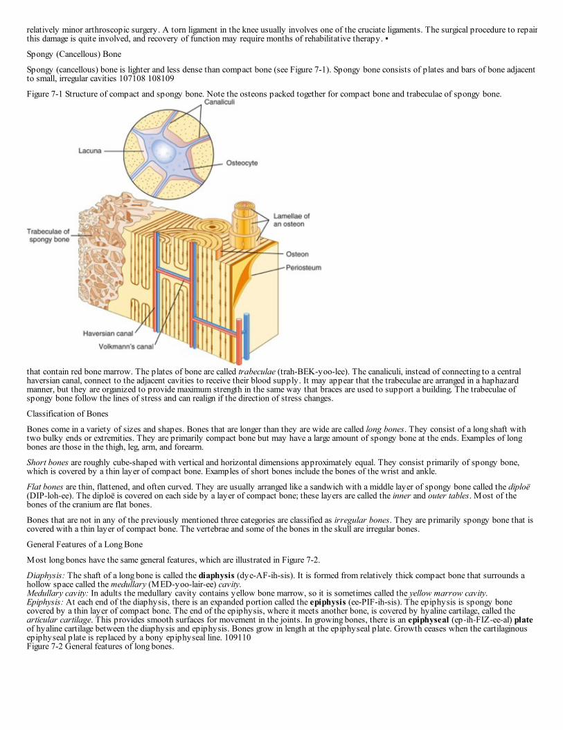
Compact Bone
The microscopic unit of compact bone is known as the osteon (haversian system). The osteon consists of a central canal called the osteonic(haversian) canal, which is surrounded by concentric rings (lamellae) of hard, calcified matrix. Between the rings of matrix, the bone cells(osteocytes) are located in spaces called lacunae. Small channels (canaliculi) radiate from the lacunae to the osteonic (haversian) canal to providepassageways through the hard matrix. In compact bone the haversian systems are packed tightly together to form what appears to be a solidmass. The osteonic canals contain blood vessels that are parallel to the long axis of the bone. These blood vessels interconnect, by way ofperforating (Volkmann) canals, with vessels on the surface of the bone. The microscopic structure of compact bone is illustrated in Figure 7-1.
Highlight on the Skeletal System
Osteoporosis: Osteoporosis is a bone disorder caused by decreased osteoblast activity. It is characterized by loss of the organic matrix,collagenous fibers, and minerals in the bone tissue. People with osteoporosis are susceptible to deformities of the vertebral column and fracturesbecause the bones are too weak to support the weight of the body. Osteoporosis occurs most frequently in postmenopausal Caucasian women.Factors that influence its occurrence are aging, malnutrition, lack of exercise, and hormone imbalance. Supplemental estrogen after menopausemay be of benefit, and exercise is always important in maintaining bone strength.Epiphyseal plate: The epiphyseal plates of specific long bones ossify at predictable times. Radiologists frequently can determine a youngperson's age by examining the epiphyseal plates to see whether they have ossified. A difference between bone age and chronologic age mayindicate some type of metabolic dysfunction.Mastoiditis: The mastoid air cells are separated from the cranial cavity by only a thin partition of bone. A middle ear infection that spreads tothe mastoid air cells (mastoiditis) is serious because there is danger that the infection will spread from the air cells to the membranes around thebrain.Sinus problems: The bones with paranasal sinuses are the frontal, the sphenoid, the ethmoid, and the two maxillae. The sinuses are lined withmucous membranes that are continuous with the nasal cavity. Allergies and infections cause inflammation of the membranes, which results insinusitis. The swollen membranes may reduce drainage from the sinuses so that pressure within the cavities increases, resulting in sinusheadaches.Soft spots: The bones in the skull of a newborn are not completely joined together but are separated by fibrous membranes. The six large areasof membranes are called fontanels, or soft spots. The anterior fontanel is on the top of the head, at the junction of the frontal and parietal bones.The posterior fontanel is at the junction of the occipital and parietal bones. On each side of the head there is a mastoid (posterolateral) fontanelnear the mastoid region of the temporal bone and a sphenoid (anterolateral) fontanel just superior to the sphenoid bone.Abnormal spinal curvatures: An abnormally exaggerated lumbar curvature is called lordosis, or swayback. This is often seen in pregnantwomen as they adjust to their changing center of gravity. An increased roundness of the thoracic curvature is kyphosis, or hunchback. This isfrequently seen in elderly people. Abnormal side-to-side curvature is scoliosis. Abnormal curvatures may interfere with breathing and other vitalfunctions.Yes and no: The atlas holds up the skull and permits you to nod “yes.” The axis allows you to rotate your head from side to side to indicate“no.”Marrow biopsy: The sternum is frequently used for a red marrow biopsy because it is accessible. The sample for biopsy is obtained byperforming a sternal puncture, in which a large needle is inserted into the sternum to remove a sample of red bone marrow.Fractured clavicle: The clavicle is the most frequently fractured bone in the body because it transmits forces from the arm to the trunk. Theforce from falling on the shoulder or outstretched arm is often sufficient to fracture the clavicle.Tennis elbow: Tennis elbow is an inflammation of the tissues surrounding the lateral epicondyle of the humerus. Six muscles that controlmovement of the hand attach in this region, and repeated contraction of these muscles irritates the attachments. The medical term for tenniselbow is lateral epicondylitis.Pelvic outlet and childbirth: The female pelvis is shaped to accommodate childbearing. Because the fetus must pass through the pelvic outlet,the physician carefully measures this opening to make sure there is enough room. The distance between the two ischial spines is a goodindication of the size of the pelvic outlet. If the opening is too small, a cesarean delivery is indicated.Broken hip: Elderly people, particularly those with osteoporosis, are susceptible to “breaking a hip.” The femur is a weight-bearing bone, andwhen it is weakened, it cannot support the weight of the body and the neck of the femur fractures under the stress. Instead of saying, “Grandmafell and broke her hip,” often it is more appropriate to say, “Grandma broke her hip, then fell.”Bunion: Poorly fitted shoes may compress the toes so that there is a lateral deviation of the big toe toward the second toe. When this occurs, abursa and callus form at the joint between the first metatarsal and proximal phalanx. This creates a bunion.Gout: Gout was commonly known as the disease of the kings because it was believed to be caused by a rich diet and fine wines. Gout is anequal-opportunity disease, however, and occurs across the entire population. A rich diet and fine wines may contribute to the disease, but theyare not the definitive cause. Gout is caused by the excessive accumulation of uric acid that forms needle-like crystals within the joint, producingpain and inflammation. The great toe is the most commonly affected joint. The disorder is diagnosed by aspirating joint fluid and observing thecrystals under the microscope. Although there is no cure for gout, it can be effectively controlled with antiinflammatory drugs and dietarymeasures.
Knee problems: The term torn cartilage refers to a damaged meniscus, usually the medial, in the knee. Frequently this can be repaired withrelatively minor arthroscopic surgery. A torn ligament in the knee usually involves one of the cruciate ligaments. The surgical procedure to repair
relatively minor arthroscopic surgery. A torn ligament in the knee usually involves one of the cruciate ligaments. The surgical procedure to repairthis damage is quite involved, and recovery of function may require months of rehabilitative therapy. ▪
Spongy (Cancellous) Bone
Spongy (cancellous) bone is lighter and less dense than compact bone (see Figure 7-1). Spongy bone consists of plates and bars of bone adjacentto small, irregular cavities 107108 108109
Figure 7-1 Structure of compact and spongy bone. Note the osteons packed together for compact bone and trabeculae of spongy bone.
that contain red bone marrow. The plates of bone are called trabeculae (trah-BEK-yoo-lee). The canaliculi, instead of connecting to a centralhaversian canal, connect to the adjacent cavities to receive their blood supply. It may appear that the trabeculae are arranged in a haphazardmanner, but they are organized to provide maximum strength in the same way that braces are used to support a building. The trabeculae ofspongy bone follow the lines of stress and can realign if the direction of stress changes.
Classification of Bones
Bones come in a variety of sizes and shapes. Bones that are longer than they are wide are called long bones. They consist of a long shaft withtwo bulky ends or extremities. They are primarily compact bone but may have a large amount of spongy bone at the ends. Examples of longbones are those in the thigh, leg, arm, and forearm.
Short bones are roughly cube-shaped with vertical and horizontal dimensions approximately equal. They consist primarily of spongy bone,which is covered by a thin layer of compact bone. Examples of short bones include the bones of the wrist and ankle.
Flat bones are thin, flattened, and often curved. They are usually arranged like a sandwich with a middle layer of spongy bone called the diploë(DIP-loh-ee). The diploë is covered on each side by a layer of compact bone; these layers are called the inner and outer tables. Most of thebones of the cranium are flat bones.
Bones that are not in any of the previously mentioned three categories are classified as irregular bones. They are primarily spongy bone that iscovered with a thin layer of compact bone. The vertebrae and some of the bones in the skull are irregular bones.
General Features of a Long Bone
Most long bones have the same general features, which are illustrated in Figure 7-2.
Diaphysis: The shaft of a long bone is called the diaphysis (dye-AF-ih-sis). It is formed from relatively thick compact bone that surrounds ahollow space called the medullary (MED-yoo-lair-ee) cavity.Medullary cavity: In adults the medullary cavity contains yellow bone marrow, so it is sometimes called the yellow marrow cavity.Epiphysis: At each end of the diaphysis, there is an expanded portion called the epiphysis (ee-PIF-ih-sis). The epiphysis is spongy bonecovered by a thin layer of compact bone. The end of the epiphysis, where it meets another bone, is covered by hyaline cartilage, called thearticular cartilage. This provides smooth surfaces for movement in the joints. In growing bones, there is an epiphyseal (ep-ih-FIZ-ee-al) plateof hyaline cartilage between the diaphysis and epiphysis. Bones grow in length at the epiphyseal plate. Growth ceases when the cartilaginousepiphyseal plate is replaced by a bony epiphyseal line. 109110Figure 7-2 General features of long bones.

Periosteum: Except in the region of the articular cartilage, the outer surface of long bones is covered by a tough, fibrous connective tissue calledthe periosteum. The periosteum is richly supplied with nerve fibers, lymphatic vessels, blood vessels, and osteoblasts.Nutrient foramina: Blood vessels enter the diaphysis of the bone through small openings called nutrient foramina.Endosteum: The surface of the medullary cavity is lined with a thinner connective tissue membrane, the endosteum, which contains osteoclasts.
In addition to the general features that are present in most long bones, all bones have surface markings and characteristics that make a specificbone unique. Bones have holes, depressions, smooth facets, lines, projections, and other markings. These usually represent passageways forvessels and nerves, points of articulation with other bones, or points of attachment for tendons and ligaments.
Bone Development and Growth
The terms osteogenesis and ossification are often used synonymously to indicate the process of bone formation. Parts of the skeleton formduring the first few weeks after conception. By the end of the eighth week after conception, the skeletal pattern is formed in cartilage andconnective tissue membranes and ossification begins. Bone development continues throughout adulthood. Even after adult stature is attained,bone development continues for repair of fractures and for remodeling to meet changing lifestyles. Three types of cells are involved in thedevelopment, growth, and remodeling of bones. Osteoblasts are bone-forming cells; osteocytes are mature bone cells; and osteoclasts breakdown and reabsorb bone.
Bone Growth in Length
Bones grow in length at the epiphyseal plate located between the diaphysis and epiphysis of a long bone. The hyaline cartilage in the region ofthe epiphyseal plate next to the epiphysis continues to grow by mitosis. The chondrocytes in the region next to the diaphysis age anddegenerate. Osteoblasts move in and ossify the matrix to form bone. This process continues throughout childhood and adolescence until thecartilage growth slows and finally stops. When cartilage growth ceases, usually in the early 20s, the epiphyseal plate completely ossifies so thatonly a thin epiphyseal line remains and the bones can no longer grow in length. Bone growth occurs under the influence of growth hormonefrom the anterior pituitary gland and sex hormones from the ovaries and testes.
Even though bones stop growing in length in early adulthood, they can continue to increase in thickness or diameter throughout life in responseto stress from increased muscle activity or to weight gain. The increase in diameter is called appositional (ap-poh-ZISH-un-al) growth.Osteoblasts in the periosteum form compact bone around the external bone surface. At the same time, osteoclasts in the endosteum break downbone on the internal bone surface, around the medullary cavity. These two processes together increase the diameter of the bone and at the sametime keep the bone from becoming excessively heavy and bulky.
Divisions of the Skeleton
The typical adult human skeleton consists of 206 named bones. For convenience, the bones of the skeleton are grouped in two divisions, asillustrated in Figure 7-3. The 80 bones of the axial skeleton form the vertical axis of the body. They include the bones of the head, vertebralcolumn, ribs, and breastbone or sternum. The appendicular skeleton consists of 126 bones and includes the free appendages and theirattachments to the axial skeleton. The free appendages are the upper and lower extremities, or limbs, and their attachments are called girdles.Table 7-1 lists the named bones of the body by category.
Bones of the Axial Skeleton
The axial skeleton, with 80 bones, is divided into the skull, hyoid, vertebral column, and rib cage.
Skull

The skull has 28 bones, as illustrated in Figures 7-4 and 7-5. The eight bones of the cranium are interlocked to enclose the brain. The anterioraspect of the skull, the face, consists of 14 bones. The remaining six bones are the 110111
Figure 7-3 Divisions of the skeleton with major bones identified. Yellow = axial skeleton. Blue = appendicular skeleton.
auditory ossicles, tiny bones in the middle ear cavity. With the exception of the lower jaw, or mandible, and the auditory ossicles, the bones inthe skull are tightly interlocked along irregular lines called sutures. Some of the bones in the skull contain sinuses, which are air-filled cavitieslined with mucous membranes. The sinuses help to reduce the weight of the skull. The paranasal sinuses are arranged around the nasal cavityand drain into it.
CraniumFrontal Bone
The frontal bone forms the anterior portion of the skull above the eyes (forehead). The paranasal frontal sinuses are cavities in the frontal bone.
Parietal Bones
The two parietal (pah-RYE-eh-tal) bones form most of the superolateral aspect of the skull.
Occipital Bone
The single occipital (ahk-SIP-ih-tal) bone forms most of the posterior part of the skull. The foramen magnum is a large opening on the lowersurface of the occipital bone. The spinal cord passes through this opening. Occipital condyles are rounded processes on each side of the foramenmagnum. They articulate with the first cervical vertebra.
Temporal Bones
The two temporal bones, one on each side of the head, form parts of the sides and base of the cranium. Near the inferior margin of the temporalbone, there is an opening, the external auditory meatus, which is a canal that leads to the middle ear. Just anterior to the external auditorymeatus, the temporal bone articulates with the mandible to form the temporomandibular joint (TMJ). Posterior and inferior to each externalauditory meatus, there is a rough protuberance, the mastoid process. The mastoid process contains air cells that drain into the middle ear cavity.111112
Table 7-1 Names of Bones of the Body Listed by Category
Bones Number
Axial Skeleton (80 Bones)
Skull (28 bones)
Cranial bones
Parietal (2)
Temporal (2)
Frontal (1)
8

Occipital (1)
Ethmoid (1)
Sphenoid (1)
Facial bones
Maxilla (2)
Zygomatic (2)
Mandible (1)
Nasal (2)
Palatine (2)
Inferior nasal concha (2)
Lacrimal (2)
Vomer (1)
14
Auditory ossicles
Malleus (2)
Incus (2)
Stapes (2)
6
Hyoid 1
Vertebral column
Cervical vertebrae (7)
Thoracic vertebrae (12)
Lumbar vertebrae (5)
Sacrum (1)
Coccyx (1)
26
Thoracic cage
Sternum (1)
Ribs (24)
25
Appendicular Skeleton (126 Bones)
Pectoral girdles
Clavicle (2)
Scapula (2)
4
Upper extremity
Humerus (2)
Radius (2)
Ulna (2)
Carpals (16)
Metacarpals (10)
Phalanges (28)
60
Pelvic girdle
Coxal, innominate, or hip bones (2)
2

Lower extremity
Femur (2)
Tibia (2)
Fibula (2)
Patella (2)
Tarsals (14)
Metatarsals (10)
Phalanges (28)
60
From Applegate E: The anatomy and physiology learning system, ed 4, St Louis, 2011, Saunders.
Sphenoid Bone
The sphenoid (SFEE-noyd) bone is an irregularly shaped bone that spans the entire width of the cranial floor. It is wedged between other bonesin the anterior portion of the cranium. The sphenoid bone contains paranasal sphenoid sinuses.
Ethmoid Bone
The ethmoid (ETH-moyd) bone is located anterior to the sphenoid bone and forms most of the bony area between the nasal cavity and theorbits. The ethmoid bone contains many small, paranasal ethmoidal sinuses.
Facial Bones
The 14 facial bones form the basic framework and shape of the face. They also provide attachments for the muscles that control facialexpression and move the jaw for chewing. All facial bones except the vomer and mandible are paired. Facial bones are illustrated in Figures 7-4and 7-5.
Maxillary Bones
The maxillary bones, or maxillae (maks-ILL-ee), form the upper jaw and the anterior part of the hard palate or roof of the mouth. Each maxillahas a large paranasal maxillary sinus. These are the largest of all the paranasal sinuses.
Palatine Bones
The palatine (PAL-ah-tyne) bones are behind, or posterior to, the maxillae and form the posterior portion of the hard palate.
Nasal Bones
The two nasal bones are small rectangular bones that form the bridge of the nose.
Lacrimal Bones
The small, thin lacrimal (LACK-rih-mal) bones are located in the medial walls of the orbits, between the ethmoid bone and the maxilla. Each onehas a small lacrimal groove that is a pathway for a tube that carries tears from the eyes to the nasal cavity.
Zygomatic Bones
The zygomatic (zye-goh-MAT-ik) bones, also called malar bones, form the prominences of the cheeks.
Inferior Nasal Conchae
The inferior nasal conchae (KONG-kee) are thin, curved bones that are attached to the lateral walls of the nasal cavity and project into the nasalcavity.
Vomer
The thin, flat vomer (VOH-mer) is in the inferior portion of the midline in the nasal cavity. It forms part of the nasal septum. 112113 113114
Figure 7-4 Skull, anterior view.

Figure 7-5 Skull, lateral view.
Mandible
The mandible (MAN-dih-bul) is the lower jaw. It articulates with the temporal bone to form the temporomandibular (tem-por-oh-man-DIB-yoo-lar) joint.
Auditory Ossicles
Three tiny bones form a chain in each middle ear cavity in the temporal bone. These are the malleus, incus, and stapes. These bones transmitsound waves from the tympanic membrane, or eardrum, to the inner ear, where the sound receptors are located.
Hyoid Bone
The hyoid bone is not really part of the skull, so it is listed separately. It is a U-shaped bone in the neck, suspended under the mandible. It isunique because it is the only bone in the body that does not articulate directly with another bone. It functions as a base for the tongue and as anattachment for several muscles associated with swallowing.
Vertebral Column
The vertebral column extends from the skull to the pelvis and contains 26 bones called vertebrae (singular, vertebra). The bones are separatedby pads of fibrocartilage called intervertebral discs. The discs act as shock absorbers and allow the column to bend. Normally there are fourcurvatures, illustrated in Figure 7-6, that increase the strength and resilience of the column. They are named according to the region in which theyare located. The thoracic and sacral curvatures are concave anteriorly and are present at birth. The cervical curvature develops when an infantbegins to hold his or her head erect. The lumbar curvature develops when an infant begins to stand and walk. Both the cervical and lumbarcurvatures are convex anteriorly.
General Structure of Vertebrae
All vertebrae have a common structural pattern, illustrated in Figure 7-7, although there are variations among them. The thick anterior, weight-bearing portion is the body or centrum. The posterior curved portion is the vertebral arch. The vertebral arch and body surround a central large

bearing portion is the body or centrum. The posterior curved portion is the vertebral arch. The vertebral arch and body surround a central largeopening, the vertebral foramen. When all the vertebrae are stacked together in a column, the vertebral foramina make a canal that contains thespinal cord. Transverse processes project laterally from the vertebral arch, and in the posterior midline there is a spinous process. Theseprocesses are places for muscle attachment. The spinous processes can be felt as bony projections along the midline of the back.
Composition of the Vertebral Column
The seven cervical vertebrae are designated C1 through C7. The 12 thoracic vertebrae are designated T1 through T12. Five lumbar vertebrae,designated L1 through L5, make up the part of the vertebral column in the small of the back. The lumbar vertebrae have large, heavy bodiesbecause they support most of the body weight and have many back muscles attached to them. 114115
Figure 7-6 Curvatures of the vertebral column. The thoracic and sacral curvatures are concave anteriorly, and the cervical and lumbar curvaturesare convex anteriorly.
Figure 7-7 General features of vertebrae, viewed from above.
The sacrum is a triangular bone just below the lumbar vertebrae. In the child there are five separate bones, but these fuse to form a single bone inthe adult. The sacrum articulates with the pelvic girdle laterally, at the sacroiliac (say-kro-ILL-ee-ak) joint, and forms the posterior wall of thepelvic cavity.
The coccyx (KOK-siks), or tailbone, is the last part of the vertebral column (see Figure 7-8). A child has four (the number varies from three tofive) separate small bones, but these fuse to form a single bone in the adult.
Thoracic CageThe thoracic cage, or bony thorax, protects the heart, lungs, and great vessels. It also supports the bones of the shoulder girdle and plays a rolein breathing. The components of the thoracic cage are the thoracic vertebrae dorsally, the ribs laterally, and the sternum and costal cartilage

in breathing. The components of the thoracic cage are the thoracic vertebrae dorsally, the ribs laterally, and the sternum and costal cartilageanteriorly.
Sternum
The sternum, or breastbone, is in the anterior midline (Figure 7-8). An important anatomic landmark, the jugular (suprasternal) notch is aneasily palpable, central indentation in the superior margin of the sternum. The superior portion of the sternum articulates with the clavicles andthe first two pairs of ribs. The body of the sternum has notches along the sides where it attaches to the cartilage of the third through seventhribs.
Ribs
Twelve pairs of ribs, illustrated in Figure 7-8, form the curved, lateral margins of the thoracic cage. One pair is attached to each of the 12thoracic vertebrae. The upper seven pairs of ribs are called true, or vertebrosternal (ver-TEE-broh-stir-nal), ribs because they attach to thesternum directly by their individual costal cartilage. The lower five
Figure 7-8 Thoracic cage.
pairs of ribs are called false ribs because their costal cartilage does not reach the sternum directly. The first three pairs of false ribs reach thesternum indirectly by joining with the cartilage of the ribs above. These are called vertebrochondral (ver-TEE-broh-kahn-dral) ribs. The bottomtwo rib pairs have no anterior attachment and are called vertebral ribs or floating ribs.
Bones of the Appendicular Skeleton
The 126 bones of the appendicular skeleton are suspended from two yokes or girdles that are anchored to the axial skeleton. They are additionsor appendages to the axis of the body. The appendicular skeleton is designed for movement. If a portion is immobilized for a period of time, lifewithout appendicular movement can be awkward.
Pectoral Girdle
Each half of the pectoral girdle, or shoulder girdle, consists of two bones: an anterior clavicle (KLAV-ih-kul) and a posterior scapula (SKAP-yoo-lah). The bones of the pectoral girdle, illustrated in Figure 7-9, form the connection between the upper extremities and the axial skeleton.The clavicles and scapulae, with their associated muscles, also form the shoulder.
The clavicle is commonly called the collarbone. It is an elongated, S-shaped bone that articulates proximally with the manubrium of the sternum.The distal end articulates with the scapula.
The scapula, commonly called the shoulder blade, is a thin, flat triangular bone on the posterior surface of the thoracic wall. It articulates withthe clavicle and the humerus. The acromion process of the scapula forms the point of the shoulder. On the lateral margin of the scapula there is a115116
Figure 7-9 Components of the pectoral girdle: clavicle and scapula.
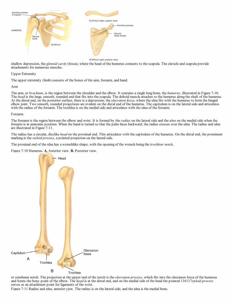
shallow depression, the glenoid cavity (fossa), where the head of the humerus connects to the scapula. The clavicle and scapula provideattachments for numerous muscles.
Upper Extremity
The upper extremity (limb) consists of the bones of the arm, forearm, and hand.
Arm
The arm, or brachium, is the region between the shoulder and the elbow. It contains a single long bone, the humerus, illustrated in Figure 7-10.The head is the large, smooth, rounded end that fits into the scapula. The deltoid muscle attaches to the humerus along the shaft of the humerus.At the distal end, on the posterior surface, there is a depression, the olecranon fossa, where the ulna fits with the humerus to form the hingedelbow joint. Two smooth, rounded projections are evident on the distal end of the humerus. The capitulum is on the lateral side and articulateswith the radius of the forearm. The trochlea is on the medial side and articulates with the ulna of the forearm.
Forearm
The forearm is the region between the elbow and wrist. It is formed by the radius on the lateral side and the ulna on the medial side when theforearm is in anatomic position. When the hand is turned so that the palm faces backward, the radius crosses over the ulna. The radius and ulnaare illustrated in Figure 7-11.
The radius has a circular, disclike head on the proximal end. This articulates with the capitulum of the humerus. On the distal end, the prominentmarking is the styloid process, a pointed projection on the lateral side.
The proximal end of the ulna has a wrenchlike shape, with the opening of the wrench being the trochlear notch,
Figure 7-10 Humerus. A, Anterior view. B, Posterior view.
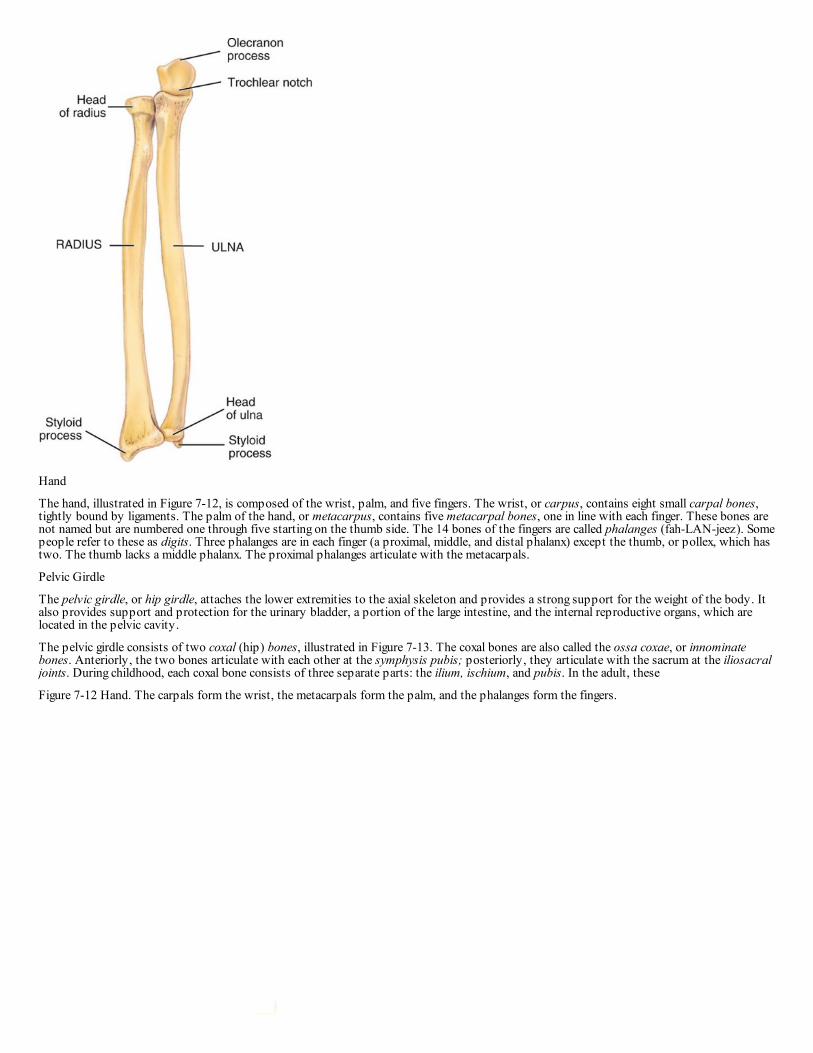
or semilunar notch. The projection at the upper end of the notch is the olecranon process, which fits into the olecranon fossa of the humerusand forms the bony point of the elbow. The head is at the distal end, and on the medial side of the head the pointed 116117styloid processserves as an attachment point for ligaments of the wrist.Figure 7-11 Radius and ulna, anterior view. The radius is on the lateral side, and the ulna is the medial bone.
Hand
The hand, illustrated in Figure 7-12, is composed of the wrist, palm, and five fingers. The wrist, or carpus, contains eight small carpal bones,tightly bound by ligaments. The palm of the hand, or metacarpus, contains five metacarpal bones, one in line with each finger. These bones arenot named but are numbered one through five starting on the thumb side. The 14 bones of the fingers are called phalanges (fah-LAN-jeez). Somepeople refer to these as digits. Three phalanges are in each finger (a proximal, middle, and distal phalanx) except the thumb, or pollex, which hastwo. The thumb lacks a middle phalanx. The proximal phalanges articulate with the metacarpals.
Pelvic Girdle
The pelvic girdle, or hip girdle, attaches the lower extremities to the axial skeleton and provides a strong support for the weight of the body. Italso provides support and protection for the urinary bladder, a portion of the large intestine, and the internal reproductive organs, which arelocated in the pelvic cavity.
The pelvic girdle consists of two coxal (hip) bones, illustrated in Figure 7-13. The coxal bones are also called the ossa coxae, or innominatebones. Anteriorly, the two bones articulate with each other at the symphysis pubis; posteriorly, they articulate with the sacrum at the iliosacraljoints. During childhood, each coxal bone consists of three separate parts: the ilium, ischium, and pubis. In the adult, these
Figure 7-12 Hand. The carpals form the wrist, the metacarpals form the palm, and the phalanges form the fingers.

bones are firmly fused to form a single bone. Where the three bones meet, there is a large depression, the acetabulum (as-seh-TAB-yoo-lum),which holds the head of the femur. The obturator foramen is a large opening between the pubis and ischium that functions as a passageway forblood vessels, nerves, and muscle tendons.
Together, the sacrum, coccyx, and pelvic girdle form the basin-shaped pelvis. The false pelvis (greater pelvis) is surrounded by the flaredportions of the ilium bones and the lumbar vertebrae. The true pelvis (lesser pelvis) is smaller and inferior to the false pelvis. It is the regionbelow the pelvic brim, or pelvic inlet, and it is encircled by bone. The large opening at the bottom of this region is the pelvic outlet. Thedimensions of the true pelvis are especially important in childbirth.
Lower Extremity
The lower extremity (limb) consists of the bones of the thigh, leg, foot, and patella, or kneecap. The lower extremities support the entire weightof the body when we are erect, and they are exposed to tremendous forces when we walk, run, and jump. With this in mind, it is not surprisingthat the bones of the lower extremity are larger and stronger than those in the upper extremity.
Thigh
The thigh is the region from the hip to the knee. It contains a single long bone, the femur, illustrated in Figure 7-14. It is the largest, longest, andstrongest bone in the body. 117118
Figure 7-13 Bones of the pelvic girdle. The right and left ossa coxae form the pelvic girdle. Posteriorly, the two bones are separated by thesacrum. Anteriorly, they meet at the symphysis pubis.
Figure 7-14 Femur and patella (right). A, Anterior view. B, Posterior view.

The large, smooth, ball-like head of the femur has a small depression called the fovea capitis. A ligament attaches here. Prominent projections atthe proximal end, the greater and lesser trochanters, are major sites for muscle attachment. The neck is between the head and the trochanters.The distal end is marked by two large, rounded surfaces, the lateral and medial condyles. These form joints with the bones of the leg. Theintercondylar notch is a depression between the condyles that contains ligaments associated with the knee joint. On the anterior surface,between the condyles, a smooth patellar surface marks the area for the kneecap.
Leg
The leg is the region between the knee and the ankle. It is formed by the slender fibula (FIB-yoo-lah) on the lateral side and the larger, weight-bearing tibia (TIB-ee-ah), or shin bone, on the medial side. The tibia articulates with the femur to form the knee joint and with the talus (one ofthe foot bones) to allow flexion and extension at the ankle.
The proximal end of the fibula is the head, and the projection at the distal end is the lateral malleolus, which forms the lateral bulge of the ankle.The superior surface of the tibia is flattened and smooth, with two slightly concave regions called the lateral and medial condyles. The condylesof the femur fit into these regions. The anterior crest is a sharp ridge on the anterior surface and forms the shin. On the medial side of the distalend, the medial malleolus forms the medial bulge of the ankle. Figure 7-15 illustrates the tibia and fibula.
Foot
The foot, illustrated in Figure 7-16, is composed of the ankle, instep, and five toes. The ankle, or tarsus, contains seven tarsal bones. Thesecorrespond to the carpals in the wrist. The largest tarsal bone is the calcaneus (kal-KAY-nee-us), or heel bone. The talus, another tarsal bone,rests on top of the calcaneus and articulates with the tibia. The instep of the foot, or metatarsus, contains five metatarsal bones, one in line witheach toe. The distal ends of these bones form the ball of the foot. These bones are not named 118119but are numbered one through five startingon the medial side. The tarsals and metatarsals, together with strong tendons and ligaments, form the arches of the foot. The 14 bones of the toesare called phalanges. Three phalanges are in each toe (a proximal, middle, and distal phalanx), except in the great (or big) toe, or hallux, which hasonly two. The
Figure 7-15 Tibia and fibula, anterior view (right). The fibula is on the lateral side of the leg, and the tibia is on the medial side.

Figure 7-16 Bones of the foot. A, Superior view. B, Lateral view.
great toe lacks a middle phalanx. The proximal phalanges articulate with the metatarsals.
Patella
The patella, or kneecap, is a flat, triangular bone enclosed within the major tendon that anchors the anterior thigh muscle to the tibia. It providesa smooth surface for the tendon as it turns the corner between the thigh and leg when the knee is flexed. It also protects the knee joint anteriorly.
Articulations
An articulation (ahr-tik-yoo-LAY-shun), or joint, is where two bones come together. In terms of the amount of movement they allow, there arethree types of joints: immovable, slightly movable, and freely movable.
Synarthroses
Synarthroses (sin-ahr-THROH-seez) are immovable joints. The singular form is synarthrosis. In these joints, the bones come in close contactand are separated by only a thin layer of fibrous connective tissue. The sutures in the skull are examples of immovable joints.
Amphiarthroses
Slightly movable joints are called amphiarthroses (am-fee-ahr-THROH-seez). The singular form is amphiarthrosis. In this type of joint, thebones are connected by hyaline cartilage or fibrocartilage. The ribs connected to the sternum by costal cartilage are slightly movable jointsconnected by hyaline cartilage. The symphysis pubis is a slightly movable joint in which there is a fibrocartilage pad between the two bones.The joints between the vertebrae, the intervertebral discs, are also of this type. 119120 120121
Figure 7-17 Generalized structure of a synovial joint.

Figure 7-18 Types of freely movable joints.
Diarthroses
Most joints in the adult body are diarthroses (dye-ahr-THROH-seez) or freely movable joints. The singular form is diarthrosis. In this type ofjoint, the ends of the opposing bones are covered with hyaline cartilage, the articular cartilage, and they are separated by a space called the jointcavity. The components of the joints are enclosed in a dense fibrous joint capsule (Figure 7-17).
The outer layer of the capsule consists of the ligaments that hold the bones together. The inner layer is the synovial membrane, which secretessynovial fluid into the joint cavity for lubrication. Because all of these joints have a synovial membrane, they are sometimes called synovialjoints.
Some diarthroses have pads and cushions associated with them. The knee has fibrocartilaginous pads, called semilunar cartilages or the lateral
meniscus (meh-NIS-kus) and medial meniscus, which rest on the lateral and medial condyles of the tibia. The pads help stabilize the joint andact as shock absorbers. Bursae are fluid-filled sacs that act as cushions and help reduce friction. Bursae are lined with a synovial membrane that

act as shock absorbers. Bursae are fluid-filled sacs that act as cushions and help reduce friction. Bursae are lined with a synovial membrane thatsecretes synovial fluid into the sac. They are commonly located between the skin and underlying bone or between tendons and ligaments.Inflammation of a bursa is called bursitis.
There are six types of diarthrotic or freely movable joints based on the shapes of their parts and the types of movement they allow. These aredescribed and illustrated in Figure 7-18.
Aging of the Skeletal System
The major age-related change in the skeletal system is the loss of calcium from the bones. Calcium loss occurs in both men and women, but itstarts at an earlier age and is more severe in women. The exact reasons for the loss are unknown and possibly involve a combination of severalfactors. These may include an imbalance between osteoblast and osteoclast activity, imbalance between calcitonin and parathormone levels,reduced absorption of calcium and/or vitamin D from the digestive tract, poor diet, and lack of exercise. Whatever the cause, there is no sure wayof preventing the loss, but adequate calcium and vitamin D in the diet may help reduce the effects.
Another change with age is a decrease in the rate of collagen synthesis. This means that the bones have less strength and are more brittle. Bonesfracture more readily in elderly individuals, and the healing process may be slow or incomplete. Tendons and ligaments become less flexiblebecause of the changes in collagen.
The articular cartilage at the ends of bones tends to become thinner and deteriorates with age. This causes joint disorders that are commonlyfound in older individuals. People also appear to get shorter as they get older. This is caused partially by loss of bone mass and partially bycompression of the intervertebral discs.
Age-related changes in the skeletal system cannot be prevented. An active and healthy lifestyle with appropriate exercise and an adequate diethelp reduce the effect of the changes in the skeletal system. 121122
Highlight on Conditions Affecting the Skeletal System
Ankylosing spondylitis (ANG-kih-loh-sing spahn-dih-LYE-tis) Inflammation of the spine that is characterized by stiffening of the spinaljoints and ligaments so that movement becomes increasingly painful and difficult; also called rheumatoid spondylitisArthritis (ahr-THRYE-tis) Inflammation of a jointBunion (BUN-yun) Abnormal swelling of the joint between the big toe and the first metatarsal bone, resulting from a buildup of soft tissuesand bone caused by chronic irritation from ill-fitting shoesCarpal tunnel syndrome (KAHR-pull TUH-nul SIN-drohm) Condition characterized by pain and burning sensations in the fingers and hand,caused by compression of the median nerve as it passes between a wrist ligament and the bones and tendons of the wristDislocation (dis-loh-KAY-shun) Displacement of a bone from its joint with tearing of ligaments, tendons, and articular capsule; also calledluxationGout (GOWT) A form of acute arthritis in which uric acid crystals develop within a joint and irritate the cartilage, causing acute inflammation,swelling, and pain; most commonly occurs in middle-aged and older menLyme disease (LYME dih-ZEEZ) A bacterial disease transmitted to humans by deer ticks; characterized by joint stiffness, headache, fever andchills, nausea, and back pain; complications include severe arthritis and cardiac problems; early stages of the disease respond well to antibioticsOsteoarthritis (ahs-tee-oh-ahr-THRYE-tis) A noninflammatory disease of the joints that is characterized by degeneration of the articularcartilage and changes in the synovial membrane; also called degenerative joint disease (DJD)Osteomalacia (ahs-tee-oh-mah-LAY-shee-ah) Softening of bone because of inadequate amounts of calcium and phosphorus; bones bend easilyand become deformed; in childhood this is called ricketsOsteomyelitis (ahs-tee-oh-my-eh-LYE-tis) Inflammation of the bone marrow caused by bacteriaOsteoporosis (ahs-tee-oh-por-OH-sis) Decrease in bone density and mass; commonly occurs in postmenopausal women as a result of increasedosteoclast activity caused by diminished estrogen levels; bones fracture easilyOsteosarcoma (ahs-tee-oh-sahr-KOH-mah) Malignant tumor derived from bone; also called osteogenic sarcoma; osteoblasts multiply withoutcontrol and form large tumors in boneRheumatoid arthritis (ROO-mah-toyd ahr-THRYE-tis) A chronic systemic disease with changes occurring in the connective tissues of thebody, especially the joints; in contrast to osteoarthritis, the symptoms are usually more generalized and severe; evidence indicates it may be anautoimmune diseaseSpina bifida (SPY-nah BIFF-ih-dah) A developmental anomaly in which the vertebral laminae do not close around the spinal cord, leaving anopening through which the cord and meninges may or may not protrudeSprain (SPRAYN) Twisting of a joint with pain, swelling, and injury to ligaments, tendons, muscles, blood vessels, and nerves; most oftenoccurs in the ankle; more serious than a strain, which is the overstretching of the muscles associated with a jointTalipes (TAL-ih-peez) Congenital deformity of the foot in which the patient cannot stand with the sole of the foot flat on the ground; alsocalled clubfoot ▪
Terminology Review
Medical Term Word Parts Definition
Amphiarthrosisarthr/o: joint
-osis: condition of
A slightly movable joint; plural, amphiarthroses.
Diaphysis dia-: through The long straight shaft of a long bone.
Diarthrosis arthr/o: joint
-osis: condition of
Freely movable joint characterized by a joint cavity; also called a synovial joint; plural, diarthroses.
Epiphysealplate
epi-: above, upon,on
The cartilaginous plate between the epiphysis and diaphysis of a bone; responsible for the lengthwise growthof a long bone.

plate on
-phys: to grow
of a long bone.
Epiphysis epi-: above, upon,on
The end of a long bone.
Osteoblast oste/o: bone
-blast: immaturecell
Bone-forming cell; immature bone cell.
Osteoclast oste/o: bone
-clast: to break
Cell that destroys, breaks down, or resorbs bone tissue.
Osteocyte oste/o: bone
-cyte: cell
Mature bone cell.
Osteon oste/o: bone Structural unit of bone; haversian system.
Synarthrosis syn-: together
arthr-: joint
-osis: condition of
An immovable joint; plural, synarthroses.
On the Web
For information on the skeletal system:
Loyola University Medical Education Network: Pick a Bone:www.meddean.luc.edu/lumen/MedEd/GrossAnatomy/learnem/bones/main_bone.htmMerck Manual: Bone, Joint, and Muscle Disorders: www.merck.com/mmhe (Click on “Bone, Joint, and Muscle Disorders”)National Osteoporosis Foundation: www.nof.org
Check out the Evolve site at http://evolve.elsevier.com/Bonewit/today/ to actively Prepare for your Certification, and to accessadditional interactive activities and exercises to help you study and prepare for success.
Pageburst Integrated Resources
As part of your Pageburst Digital Book, you can access the following Integrated Resources:
Additional ResourcesBody Spectrum Electronic Anatomy Coloring Book®Prepare for Certification Review