Embed Size (px)
Citation preview
THE INNER EAR AND BRAIN STEM FUNCTIONS AS A
SEVERITY INDEX FOR HEAD TRAUMA
* ** *** R.WILLINGER , C.M. KOPP , A. GENTINE
* INRETS-LCB, case 24, BRON, France **IMFS, URA-CNRS 854, rue Boussingault, STRASBOURG, France
***CHU Hautepierre, serv. ORL, STRASBOURG, France
ABSTRACT
Impact biomechanics research on the head has frequently looked at fatal cerebral lesions. In the head trauma population, there are patients with no brain injury, but presenting labyrinthic commotions. The important factor here is less. the probability that an inner ear lesion occurs, than the bringing in of this kind of lesion to the head tolerance limits research.
lnitially we briefly review the clinical observations, i. e. vertigo, deafness, etc . . . We then examine the mathematical models which describe the semicircular canal, cupula and macula.
Finally the results are related to the theoretical values of mechanical parameters applied to the different inner ear organs in cases of extreme linear and angular accelerations as found in the literature.
INTRODUCTION
Research on impact biomechanics of the head deals mainly with cerebral lesions such as subdural or intracerebral hematomas. These lesions, often fatal, are analyzed in autopsies and if they can inform us about the mechanisms, they have only a minor role in the detection of human shock tolerances.
In the population of patients with cranial traumatisms, we often find more or less severe cases presenting no cerebral lesion, but slight commotions of the labyrinth or the brain stem. Our study deals with these patients in order to characterize their lesions, to trace their accidentology data and to fix the tolerance limits of these serious but not fatal lesions. Thus we ar� more concerned with the contribution of these type of lesions to the research on cerebral tolerance, than with the percentage of patients with a traumatism of the inner ear or the brain stem.
Most patients who, after a cranial traumatism, take medical advice in an ENT medical department complain of hearing troubles or vertigos, due to an impairment of the vestibulo-oculomotor system.The lesions are then frequently in the whole labyrinth (cochlea, utricule, semicircular canal), or in the control of the Vestibulo-Oculomotor Reflex (VOR), situated at the brain stem periphery.
The motivation of our work is the severity of these lesions. Clinical studies ( 1 ) have shown that the date of return to work and the social rehabilitation of the patient with a cranial traumatism depend more on the patients psychological state than on the "severity" of the traumatism; the evolution of this psychological state is highly correlated with the vestibulo-cochlear recovery. On the other hand, the observation of labyrinth impairment in case of a closed cranial traumatism gives a supplementary reason for the development of this type of study. In fact, the clinical investigations and the simplicity of their qualitative and quantitative processing make out of the lesions an interesting tool for research on
- 1 27 -

lesion mechanisms and human tolerance limits. In this werk, we present the first step of collaboration between a laboratory of shock
biomechanics, a research team on the development of functional exploratory methods in vestibulometry and optocinetics and a medical ENT department.
First we present the clinical aspect of the problem with the conditions of traumatism appearance, the examinations made and the results observed. Then we insist upon the clinical and biomechanical conclusions summarizing the lesions and the possible mechanisms. ln itially however we look at the mathematical models available for the identification of the vestibulo-oculomotor system. The method is then based on the use of these models for calculating the mechanical parameters of the system and for the precise location of the observed lesion. This theoretical approach is then completed by clinical and accidentological investigations in order to establish the lesion mechanisms, as well as the corresponding tolerance limits.
CLINICAL OBSERVATIONS
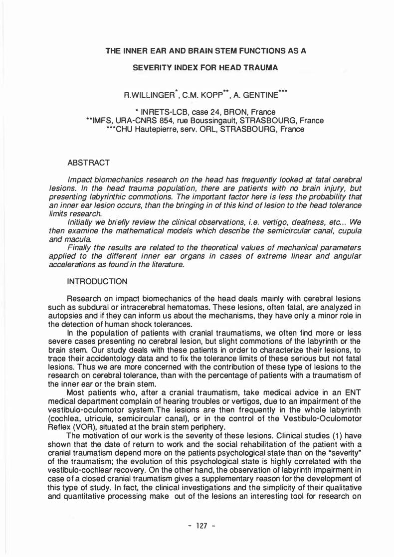
The patients with cranial traumatisms taking medical advice in ENT departments complain systematically of various types of vertigos or hearing losses (1 ,2,3). These complains are characteristic of labyrintic or central injury, because there we find the angular accelerometers formed by the semicircular canals, the linear accelerometers formed by the macula set and the microphone of the inner ear formed by the cochlea (Fig. 1 ) . In the central field, the vestibular nucleus of the brain stem manage the vestibulo-oculomotor reflex and the neuro-neural liaisons.
The sources of the traumatisms are in about 58% of the cases traffic accidents ; 26% are due to falls and 1 6% have various sources. The most exposed population is the active one (20 to 60 years old), like for other cranial traumatisms.
The peripheral and central problems making the subject of this study are very often due to skul l impacts (80%) but can also result from cervical impacts or head hyperextension (20%).
lt appears from the bibliography (2), that the severity of the lesions are difficult to determine because the medical profession states them in terms of : - shorter or langer duration of unconsciousness, - prolonged duration of comas, -vestibular, cochlear or brain stem commotions, -disappearance of otolites, -otolites settled upon the cupula of the posterior sec.
At present, only the existence of a skull fracture allows the objective identification of a certain severity of a traumatism. In order to draw to a conclusion the determination of the severity of a trauma, let us quote a statement often made in medical studies, according to which the lesion of sensory and nervous textures are most often stated without any formal proof. We will not dwell any langer on the medico-legal problems relative to experts' opinions and compensations of victims entailed by this involuntary lack of objectiveness. In cases in which no organic lesion had been observed, collaboration between ENT specialists, psychiatrist and psychologists clearly showed improvement of syndromes after the settlement of a psychological or financial problem (4).
As to recorded complains, studies often conclude that there is no correlation between the severity of the traumatism and the syndromes called forth. The main complains recorded are the following ones (1 ) :
- Hypoacousy, which is a decrease of the auditive threshold due to tympanoossicular, cochlear or auditory nerve lesion. When it exists, it is bilateral i n one case out of three.
- Tinnitus, which is a continuous buzzing in the ears at a given frequency. lt is often combined with the fracture of the otic bone, and when it exist, it is also bilateral in one
- 1 28 -

case out of three. - Vertigos represent 85% of the complains and can be :
. of a rotary type in 56% of the cases. They are then started, in 80% of the cases, by a change of the head position and continue from a few seconds to a few hours. Finally, about half of the patients suffering from this syndrome recover,
. of a non-rotary type in 1 8% of the cases. They have a very short duration in 80% of the cases and find expression in terms of an unbalancing after a movement of the head. Here too, only half of the patients recover,
. false vertigos in 1 0% of the cases. These have a non-vestibular and often psychological source.
- Otalgia in 1 4% of the cases. These are pains in the auditory meatus, often on one side, with a streng tendency to improvement.
- Cephalalgia is found in 75% of the cases ; it is the most frequent and the less inclined to improvement.
Canal vertic a l
Fig. 1 : Anatomical view of the vestibule.
Besides the pathology studies, we should list, in the clinical aspect of the problem, the investigations available to us at present. This is a main aspect of our study because these investigations are at the basis of the elaboration of the existing mathematical models and will also make possible the identification of the lesions for which we will try and trace the shock parameters.
In order to determine the integrity or the impairment of the neuro-sensory structures of a patient with cranial traumatism, they are submitted, taking medical advice in vestibular pathology, to a battery of tests including :
- The clinical examination with otoscopy, cervical auscultation and investigation of the cranial nerves,
- audiometry with : . tonal air and bone examination, . vocal examination, . tympanometric examination (impedancemetry), . brain stem evoked response audiometry (SERA),
- vestibulometry, will form the main part of our investigation. lt is based on the stimulation of the nystagmus, that is to say of the movements of the eyeball, whose characteristics are recorded by electronystagmography (ENG). This recording is based upon the existence of a peri-orbital electric field created by the corneo-retinal dipole. This electrical field varies with the displacements of the eye during the production of the
- 1 29 -
nystagmus and these variations are recorded by mean cf peri-orbital electrodes. The conventional recording process is as follows : - Spontaneous nystagmus observed at rest ; it is a function cf the direction cf the
subject's sight, -rotary test nystagmus recorded when the subject is sitting in an armchair which is
rotated, - caloric test nystagmus provoked by a flow cf cold and warm water in the external
auditory meatus (this technique allows the separate analysis cf a labyrinth without solicitating its symmetrical),
- position nystagmus, which appears when the subject changes his position (lying, standing, etc . . . ),
- cervical nystagmus provoked by a rotation movement cf the head, - multifrequencial rotary test nystagmus recorded in a similar way to that cf rotary
test, except that in this technique the speed and the direction cf the rotation vary in a pseudo-random way.
THE BIOMECHANICAL ASPECT
The analysis reported in this paragraph is relative to the correlation between the lesions and the location cf the impacts and then to the mechanisms suggested by some lesions. Here we will distinguish between vestibular and cochlear injuries, knowing that 70% cf the patients suffering from the former injury complain also cf the other one.
i) Cochlear injury: Post-trauma bi lateral deafness is cf 35%, which shows that the effect cf impact on
the only otic bone cannot be the source of the lesions. Gil let ( 1 ) shows that in case cf deafness, 48% result from lateral impacts, 38% from occipital and 1 4% from frontal impact. However, 75% of the cases of deafness present a lateral component.
The total deafness, which represents only 6% of the lesions observed, very often result from occipital shocks, rarely from front and never from side impacts.
ii) Vestibular injury : About half of the patients with cranial traumatisms taking medical advice in ENT
departments have spontaneous nystagmus, not always accompanied by dizziness. Usually, a given head position engenders vertigo. This position nystagmus is observed in 20% of the cases.
One third of the vestibular injuries are peripherat and one third are due to central impairment, the remaining representing mixed injuries. 20% of the patients have cervical nystagmus, of which 85% are uni lateral.There is a streng correlation between this nystagmus and cervical pains.
.
Before trying to establish lesion mechanisms, we can state that : -ff there exist inner ear lesions without any impact on the otic bone, this lesions only
appears in cranial impact cases -brain stem lesions can appear without any cranial impact and are thus provoked by
accelerations, - among the patients taking medical advice in ENT departments, only half had a
fracture and only in 25% of the cases, this fracture was situated on the otic bone, - this lesions are often bilateral, which goes i n the same direction as the
observations made above, - some type of lesions are obtained exclusively by non-lateral impacts, - the position nystagmus can in certain cases be attributed to a settlement of otolites
on the cupula of the posterior sec. provoking thus cupulolithiasis (5), - according to SHERRIGTON (6), the vertigos found in boxers most often results
from mono or bilateral labyrinth lesions, - three quarters of vestibular pathology are also cochlear pathology,
- 1 30 -

- the precise location of impacts in case of vestibular injury is not often given in the bibliography.
The physical parameters susceptible of intervening into the lesion mechanisms are the bone deformations with or without fracture, stresses and strains in biological textures, acceleration and pressure waves. These parameters can have various sources and are of course not mutually independent.
Bane deformation, for instance, exists locally at the impact location but is also differentiated as a function of the mode shapes determined in the modal analysis. These deformations can act on the stresses of the intra-cranial substance, or possibly generate a pressure wave, according to the characteristics of the impact.
The stresses, as we just saw, are influenced by bone deformations but can also vary as a function of acceleration when different density environments are in contact , or with the frequency characteristics of the entering acceleration signal and its vector direction. Finally acceleration induces relative displacements between density, complex rigidity or different viscosity environments.
Considering these remarks, we have drawn up an initial list of lesions and possible mechanisms :
In the cochlear field, we see endolymph oedemas due to lasses of cells provoked by stresses finding their sources in acceleration or spreading of a pressure wave. lt is the same thing for the oedemas of the Corti apparatus, in which destructions of hair cells are sometimes observed. Finally, the tearing or destruction of the cochlea take place in case of fractures of the otic bone, in which we then can observe shearing due to the relative bone elements movements.
The ossicular lesions of the inner ear are often formed by luxations provoked by streng linear accelerations or local bone deformations. Cases of fractures of the ossicles or their total pulverization are systematically accompanied by fractures of the otic bone.
In the field of the vestibule, three types of lesions with considerably different sources can be distinguished : Endolymph oedemas and destructions of hair cells which, like in the cochlea case, come from stresses provoked by acceleration or pressure wave ; displacements Of maculas Of the Otolith System or Of cupula in the SCC, engendered by acceleration ; finally the tearing or destruction of the vestibule resulting from a fracture of the petrous bone.
Lesions of the nervous central system and ·more particularly those situated on the peripheral side of the brain stem are oedemas, hemorrhages or tearing due to stretching, thus to stresses depending on the acceleration and hyperextension of the head.
The otic bone lesions can be of different types according to the characteristics of the impact. Here it is interesting to note that fractures of the otic bone are possible with a contra-lateral, an occipital or a frontal impact.
THE EXISTING MATHEMATICAL MODELS
i) The semicircular canals under angular acceleration
There are three semicircular canals in each inner ear and form together with the utricule and the cochlea, the membranous labyrinth, an assembly of canals imbedded in
the petrous bone (Fig. 1 ). The internal radius of the SCC is about 1 .5 1 o-4 m and the radius of the arch of the circle they form equals about 3.2 1 o-3 m. They end at each of their extremities in a common "pocket" called the utricule. The endolymph is the fluid filling all the membranous canals, as well as the utricule, and the whole lies in the perilymph filling the bone canals of the petrous bone.
At one of the sec extremities, quite close to the utricule, there is a diaphragm formed by the cupula and its blistered crest. This cupula plays the part of a test body in the pressure sensor thus formed and destined to measuring the angular accelerations of
- 1 3 1 -

the head. When the cupula is deformed under the influence of pressure provoked by an angular acceleration, it carries along the neuro-sensory hairs imbedded on the blistered crest, generating thus the information on angular acceleration which is then carried to the vestibular nucleus in the brain stem. There the information is processed and then transmitted to the oculomotor system to create the nystagmus.
The mathematical modelling of this sensor is based on the difference of pressure appearing on two sides of the cupule when the head is submitted to angular acceleration (7). This pressure difference and the rigidity of the cupula give a slight endolymph discharge, in its turn accompanied by inertia and viscosity effects. The putting into equation of this system was made in the following way (7) :
-80
- 1 00
0 - 1 20 :::c E - 1 40 b N - 1 60
- 1 80 f
-200 , 0 1 ' 1 1 0 1 00 1 000
2,0
1 ,5 0 e. E> «!
1 ,0
0 ,5
0,0 , 0 1 ' 1 1 0 1 00 1 000
Fig. 2a : Transfer fonction of SCC system .
..
( 1 ) Ay(t)+By(t)+Cy(t)=-D0(t) -
-D0(ro) (2) Hc(ro)=--2--
-Aro +Bro+c
- 1 32 -

He (ro) : the transfer function of the SCC system (Fig. 2a),
y(t) : the curvilinear abscissa of the endolymph discharge into the canal, 0"(t) : the head angular acceleration,
-D0"(t) : the force applied to the cupule (0=3.8 1 o-9 kgm·1 ) ,
A : the effective mass of the endolymph (A=1 .62 1 o-6 kg), B : term of viscous friction (8=2.8 1 o-4 Nms·1 ) , C : elasticity of the cupule (C=1 .3 1 o-5 Nm-1 ) .
i i ) The semicircular canals under linear acceleration
The cupula, formed by a gel, has a specific mass slightly superior to that of the enqolymph, which results in rotary information in case of linear acceleration. This "misfunctioning" is recorded in a bilateral way and then compensated at the vestibular core level. Nevertheless, in case of lass of tauch of otolites, these sometimes sattle on the cupula increasing artificially its specific mass, a pathology dealt with hereafter. This most often unilateral phenomenon then provokes vertigos as a function of the head position, called position vertigos.
The interest in such modelling also lies in the study of the effects of a drug on the equilibrium system. The consumption of alcohol, for instance, provokes a decrease in the specific mass of the cupula, causing thus rotary information in case of linear acceleration.
In the shock biomechanics field, the effect of a difference in the specific mass between the cupule and the endolymph can have dramatic consequences in case of very high acceleration. The modelling of this phenomenon is based on that of the SCC's already mentioned. The terms A, B and C vary very little (about 4%), but the term D, linking acceleration to transcupular pressure force, now takes a value D' defined by (8) :
(3) D'=So ßp Ö COSE s0 : surface of the cupule (So=0.78 1 o-6 m2),
ßp : cupule-endolymphe specific mass difference (ßp=645 kgm-3),
ö : cupule thickness (ö=0.6 1 o-3 m),
e : angle between the acceleration vector and the ordinary direction of the cupule,
D' : supposing e null (the most unfavourable case), 0'=3.0 1 0-7 kg.
iii) The otolith system
In the utricule and the saccule, supporting cells bear a so-called statolith membrane encrusted with otoconites formed by calcium carbonnade crystals, of a specific mass of
2. 7 1 o3 kgm-3. These supporting cells, equiped with sensory cells in form of hairs are directed into two directions perpendicular to the macula plane. This otolith device forms the linear accelerometer of the vertebras. lt is based on the difference of the specific masses of the otolites and the endolymph in which they swim. The response of this device to an acceleration is the shearing of the supporting cells, shearing recorded and transmitted to the sensory cells.
The modelling of this sensor is based on the appearance of volumic forces in the otolites in the presence of linear acceleration. lts putting into equation goes as follow (9)
(4) My(t)+tj(t)+Ky(t)=F(t)
F(t) : the volumic force applied to the otolites,
- 1 33 -

y(t) : the displacement of the otolites in the F(t) direction, K : rigidity due to the supporting cells (K=3.2 1 o-
4 Nm-1 , for an otolith surface element of 1 o-6 m2), M : the effective mass of such a surface element (M=1 .5 1 o-9 kg), C : viscosity due to the supporting cells (C=4.5 1 o-4 Nms-1 )
I n the hypothesis according to which the otolites are hold bac'k by a system of parallel spring and damper, the transfer function of the whole can be explained in form of mechanical .impedance as follows (Fig. 2b) :
(5) z Z3{Z1+Z� Z1+Zz+-Z3
z1 , Z2 : the spring and damper impedance (Z1 =Kljro ; Z2=C),
Z3 : the mass impedance (Z3=jrom).
- 1 34 -

iv) Cybernetic modelling of the VOR
The VOR (Vestibulo-Oculomotor Reflex) is the reflex that coordinates the ocular balls movements as a function of those of the head. In the in-vivo experimentation, we have access only to these two parameters and the transfer function between the input formed by the armchair rotation and the output in form of the ENG record, is the product of transfer functions relative to the functioning of the SCC's (He) of the mechano-neural transmission system (Hm), the neuro-neural transmission due to the longitudinal nerve (Hf), the processing of the brain stem (Ht), and finally the oculomotor system (Ho). The transfer function of the overall system is then given by the product of the subsystems transfer functions (9).
(6) H=HcH J-i 1HtHo
H ( ) G jroT m (6-1 ) m ro = m1 . T +Jro m
(6-2 ) H 1(ro)=G1
jofft (6-3) Ht(ro)=G-t --
1 +jroTt
(6-4) H0(ro)=G0 • 1 . ( 1 +JroT01 ) ( 1 +JroT02)
The experimental determination of the various time constants Ti and gains Gi, allows us then to characterize and locate the possible failure of an element intervening into the VOR. In this way, it was possible ( 1 0, 1 1) , to identify labyrinth oedemas, uni lateral destructions, cupulolithiases and cent.ral injuries.
METHOD
Our method is based upon three distinct approaches, which are the theoretical, the experimental and the accidentological approaches.
The theoretical approach uses the mathematical models shown above and consists of calculating the mechanical parameters in the different systems, in case of extreme acceleration. Accordingly, the inputs of the models will be impulses drawn from the shock tolerance curves coming from the bibliography. We will consider in particular the tolerance curve to linear accelerations given by the WSU curve ( 1 2) and the tolerance curve to angular accelerations proposed by THIBAUL T and al. ( 1 3). Our analysis is made in the frequency domain and the input acceleration, supposed to have a sinusoidal form,reads (1 4) :
(7) a(f)hoto cos(1tfto)
1t 1 - (2fto)2
.With ao the acceleration peak value in the time domain and to the impact duration.
The maximum frequency contained in such an impact equals fm=7tl2t0. In a recent study ( 1 5) , we expressed the maximum FOURIER transform amplitude of the acceleration signal for variable WSU extreme impacts. Figura 3 shows that for the two types of acceleration (WSU and THIBAUL T), the admissible maximum amplitude increases in an exponential way for shocks of maximum frequency fm inferior to 1 50 Hz.This can be explained by the brain mechanical properties and finds expression, i n the time domain, in a constant acceleration limit for shocks of a duration of more than 1 O ms. The output F is the pressure force applied to the cupula.
- 1 35 -

.g 1 00 :::i "t:)
. ....
]-< 1 0
0 2 0 0 4 0 0 6 0 0 8 0 0
Fig. 3 :
Maximum amplituds of inputs and outputs, in SI units :
THIBAULT c wsu
- '5 F(thtbaultl 10 • F(WSU) 1 c · 3
I n view o f detecting the sensitiveness of a system to a given shock duration (or maximum frequency), we determine, in a second step, the forces and displacements generated by impacts with constant amplitude but increasing duration.
For the SCC's, we are interested in the pressure force applied to the cupula and in the endolymph displacement conditioning the deformation of the cupula. In the otolites system, we express the volumic forces applied to the otolites as well as there displacements. As the transfer function of the latter is expressed in terms of mechanical impedance, the following transformations are necessary for expressing FOURIER's transform of forces F(ro) and displacements e(ro) as a function of the acceleration :
(S) F(ro) a(ro�Z(ro)
JOO (9) e(ro)= v�ro)
j(l) The experimental aspect of our method is based on the functional exploration of
given cases of cranial traumatisms. The aim of these examination is to determine the various parameters for the VOR transfer function and to locate the lesions. In this analysis we will try to distinguish between and isolate the cases of : - central injury on the brain stem, - bilateral vestibular injury in case of side, front or occipital impacts without fractures, - losses of touch or displacement of the cupula, - losses of touch of the otolites.
For each of these pathologies, we will try to understand the lesion mechanisms at stake and the specific tolerance limits, using mathematical models and the patient's accidentology data. This point forms the third indicator of our working method. In the first step, we will analyze, by means of dynamic rigidities measuring techniques, the impact zones both at the head level and at the trauma acting power level. Finally, a third step will deal with the assessment of mechanical parameters in the presence of a case of fall or other impacts, by means of accident reconstruction.
RESUL TS AND DISCUSSION
The SCC's were first of all studied in extreme angular acceleration configuration given by THIBAUL T. For several points of the tolerance curve, we determine the force spectrum due to the transcupular pressure and the endolymph displacement.
The force has an expression similar to that of the acceleration, with the exception of
- 1 3S -

one constant (F= D 0"). lts order of magnitude for an acceleration of 1 2500 rds-2 , is 4.7
1 o-5 N. lts evolution as a function of the shock duration goes like that of the acceleration. When based on the cerebral tolerance curve, the maximum amplitude of the force signal in the frequency domain , increases abruptly as soon as the maximum frequency contained in the shock spectrum (fm) is inferior to about 1 50 Hz with the results that this can have on the displacements or deformations of the cupule for such "lang" extreme impulses (Fig. 3).
The endolymph displacement, modulating the deformations of the cupula, presents a quite different pace. The energy of its signal is located under the Hertz value, which can be explained by the fact that the system is very much damped. The displacement spectrum has the same aspect, whatever may be the shock, and then we characterize it by means of its amplitude dO at 0.0 16 Hz.
� 2 < 1
o ..L.���-'--�-=:=:::=1111-.------"-------...J , 0 1 ' 1 1 0
Fig. 4 : Fourier transform amplitude of endolymph displacement. ( 12500rd/s"2 ; 4,3ms)
1 0 0
f
Under THIBAUL T's limit impacts, we observe, figure Sa, that the amplitude increases abruptly for shocks of a duration superior to 1 O ms, thus for shocks whose spectrum remains i nferior to 1 50 Hz. This result was foreseeable considering the admissible energy for long shocks, and then we can question the behaviour of the cupula in cases of shocks of this type. Experiments made on frogs showed that it was possible to displace or extract the cupule by means of a simple overpressure in the canal. In some pathological cases, this diagnosis was also suspected. The study of the evolution of dO
as a function of the duration of a constant peak amplitude impulse of 12500 rds-2, shows a increase as a function of the shock duration (and a decrease as a function of fm) see figure 5.
The study of the behaviour of the SCC's under linear acceleration is made according to the modal presented above and under sollicitations drawn from the WSU tolerance curve .Qualitatively the results are very close to those stated above, because the basic equations have the same form. The order of magnitude of the forces for an acceleration of 1 000 ms·2 applied during a 4.5 ms duration is 3.0 1 o-4 N , or about ten times superior to that engendered by an angular limit acceleration of a similar duration. Hera too and for identical reasons, the force and its spectrum develop like entering acceleration (fig.3).
The maximum FOURIER's transform amplitude of the endolymph displacement (dO) are also clearly superior to the preceding ones. This result is i l lustrated in figure 5, in which we express dO for extreme linear and angular impulses as a function of their
- 1 37 -

duration (fig. Sa) and maximum frequency contained in the shock spectrum.(fig. Sb). These results teach us that for the mechanism consisting in a displacement or an extraction of the cupule, the linear impacts are more dangerous than angular impulses. This is corroborated by the clinical observation that labyrinth lesions appear mainly in cases of a fall or a direct impact on the head.
dO
0 , 0 5 m . . .
ai . / . p .
... .o· · · · · · · . .. --
• 1 · · · · ·O- · · · 2
• 3 · · · · -Gt · · · 4
. „ • • „ - · ·
. . „ .o-· ·
o· · · · · · ·
„ „ • • • • • „ .. • • • • • • • • • • • • • • • • o· ..
.. . .
..
a
.. .. ..
0 , 00------�����--�������--������---1 0 2 0 4 0 ms 6 0
dO
, 0 1
.. .. ......
m.. ·· . .. ··m
,00 1-+-������---.....-������--������---1 1 0 1 0 0 1 0 0 0 fm 1 0 0 0 0
Fig. 5 : Maximum endolymph displacement amplitudes (log(dO) in S I units) as a function of shock duration (a) and shock spectrum maximum frequency (b},for : 1 -maximum angular accelerations (THIBAUL T) 2-constant angular acceleration peak (1 2500 rd s·2) 3-maximum linear accelerations (WSU)
4-constant linear acceleration peak (1 OOOm s·2)
The behaviour of the otolite system, sensitive to linear accelerations, is studied for shocks relative to the WSU tolerance curve. The accelerations generate volumic forces that, given the transfer function of the system, presents a pace close to that of the input
signal, with values of the order of 1 .5 1 o-6 N for an impact of 4.5 ms duration and an
- 1 38 -

amplitude of 1 000 ms·2. As above, we observe an increase of the force amplitude in the frequency domain , for shocks of a duration longer than 1 O ms (fig. 6). Once again, we can expect an increase in risk, here the loss of contact of otolites for this type of impacts. When the solicitations are acceleration of constant peak amplitude, but increasing duration, the force amplitude increases linearly with the duration.
1 2
1 0 g OI 8 < 0 ..... . LI.. 6
4
2
0,04 -E 0,03 -
-Q) 0 , 02 -a. E < 0 , 0 1
0,00
0
1
200 400 600
Fig. 6 : Maximum force amplitude in otolites, for WSU inputs.
1 0 1 0 0
Fig. 7 : Fourier transform of the otolites displacement. (1 000m/s"2 ; 4,5ms)
fm
800
f
1 0 0 0
FOURIER's transform of the otolites displacement shows that the energy is concentrated at frequencies below 1 50 Hz, while that relative to the displacement of the endolymph was concentrated between O and 1 Hz (fig. 7). So we can immediately predict that the risk of loss of contact of the otolites exists in a much broader shock range than that of loss of contact of the cupule. For all the shocks, the pace of the spectrum is identical and we characterize it by means of its amplitude eo at 1 .6 Hz (fig. 8). This figure reveals one again a critical zone for shocks with a maximum frequency lass than 1 50 Hz, while, for a constant peak acceleration amplitude, eo increases with the shock duration (fig. 8).
- 1 39 -

eO
0, 1 0
0 , 05
o , o o�����������--����������-t 0 1 0 ms 2 0
b eO • wsu
- - - - -o- - - - 1 000 m / s2
' 1
, 0 1 ...-����������__.����������---t 1 0 1 0 0 fm 1 0 0 0
Fig. 8 : Maximum otolites displacement amplitudes (SI units), as a function af shock duration (a) and maximum shock spectrum frequency (b), for :
WSU inputs and a 1 000 ms-2 canstant linear peak acceleration.
CONCLUSION
In this wark, we shaw the interest of the study of the equilibrium system far the research an head shack talerance. These often severe but rarely fatal lesians can be analyzed in vivo qualitatively and quantitatively by means of clinical instrumented examinatian. The existing mathematical models and accidentology will allow us in the lang range to define the tolerance limits far given lesions.
This first step af our collaboratian has allawed us ta establish a certain number af lesions and possible mechanisms among the patients with cranial traumatisms taking medical advice in ENT departments.
The study in the frequency damain of the forces applied to the cupule and the displacement of the endolymph, revealed that the head tolerance curves stated in the bibliography, cannot be representative of the risk of the cupula loss of touch. The forces and displacements characterized in the field af atalites far extreme acceleration, led us to similar canclusians far the lass af tauch af otalites. The mechanisms af lesians cannected
- 1 40 -

with the linear acceleration can explain the great number of bilateral phenomena observed in the vestibular injuries, as well as the almest non-influence of the impact zone.
As no analysis of patients with cranial traumatism, including the accidentology survey, has been made until now, only a qualitative estimation of the risk relative to these mechanisms can be proposed :
- For the cupula, linear accelerations are more dangerous than angular accelerations,
- otolites are sensitive to higher frequencies than the cupula, which makes them vulnerable to a broader shock range,
- the risks increase in an exponential way when the shock duration is superior to 1 O ms, or for maximum frequencies of the shock spectrum less than about 1 50 Hz.
REFERENCES
(1 ) B. GILLET : La recuperation cochleo-vestibu laire des traumatises cränio-cervicaux. These Med. Strasbourg 1 975. (2) GURDJIAN, WEBSTER : Head lnjury, Little, Brown company, Boston 1 958. (3) T. DAITO, H. NAGAI : Auditory disorders caused by head injury. lnt. Audio, Leyden, 1 966, 5/2, 1 47-1 51 . (4) R. BRUN : Traite general des nevroses ; Payot, Paris 1 956. (5) HF. SCHUKNECHT, R. DAVISON : Deafness and vertigo from head injury. Arch. of ORL, 1 956, 63 ,51 3-528. (6) CS. SHERRINGTON : The brain and its mechanisms ; Cambridge Univ. Press, 1933 (7) EN. NJEUGNA : Le canal semi-circulaire de l'oreille humaine-Modele, ldentification, Anomalies- : These Univ. Strasbourg 1 982. (8 )EN. NJEUGNA, JL. E ICHHORN, CM. KOPP, A. GENTINE : Modale mecanique du CSC et accelerations lineaires. To be published in Acta. ORL Belg. (9) A; GENTINE : Modales theoriques et physiques du vestibule chez l'homme. These d'Etat, Univ. Strasbourg 1 989. (1 O) K. KALFANE : Exploration fonctionnelle du RVO. These Univ. Strasbourg 1 977. ( 1 1 ) JL. EICHHORN, CM. KOPP, A. GENTINE : Alteration biomecanique du CSC dans la maladie de MENIERE. XXIV symp. d'Electronystagmographie, Bordeaux, juin 1 990. (1 2) JH. Mc ELHANEY, VL. ROBERTS, JF. HILYARD : Handbook of human tolerance. Man & Technology Publisher company, Tokyo 1 976. (1 3) LE. THIBAUL T, TA. GENNARELLI, SS. MARGULIES, JM. and R. EPPINGER : The strain dependent pathophysiological consequences of inertial loading on central nervous system tissue. IRCOBI conf. Lyon 1 990. ( 14) CM. HARRIS : Shock and vibration handbook. Mc Graw-Hill book company, NY. ( 15) R. WILLINGER, CM.KOPP, D. CESARI : Head tolerance curves in the frequency domain. To be published in ESV conf., Paris 1 991 .
- 1 4 1 -