Embed Size (px)
Citation preview

Asian Pacific Journal of Cancer Prevention, Vol 16, 2015 4571
DOI:http://dx.doi.org/10.7314/APJCP.2015.16.11.4571The Inhibition Effect of Triptolide on Human Endometrial Carcinoma Cell Line HEC-1B: A in vitro and in vivo Studies
Asian Pac J Cancer Prev, 16 (11), 4571-4576
Introduction
Endometrial cancer is one of the three major gynecological malignancies in Western countries. Its incidence ranks the first among malignancies of female reproductive tract. 52630 new cases took place in the United States in 2014 and 8570 cases died of it (Siegel et al., 2014). Current therapy for endometrial malignancies includes surgery, radiotherapy, chemotherapy, hormone therapy and so on. Chemotherapy is currently the more effective one for endometrial cancer but can cause severe side effects as in other malignancies, seriously degrading quality of life. So we remain need explore the new effective drug.
Tripterygium show efficacy for a variety of tumors, and the active ingredient triptolide can inhibit tumor growth and metastasis. Recent studies have proved good anti-cancer efficacy of triptolide in vitro and in vivo for various cancers, including bladder cancer (Ho et al., 2014), gastric cancer (Li et al., 2012), colorectal cancer (Liu
1Department of Gynecologic Oncology, 2Department of Radiation of Gynecologic Oncology, 3Department of Chemotherapy, the Affiliated Cancer Hospital of Nanjing Medical University and Jiangsu Cancer Hospital, Nanjing, China *For correspondence: [email protected], [email protected]
Abstract
Background: To investigate the inhibitory effect and the underlying mechanism of triptolide on cultured human endometrial carcinoma HEC-1B cells and corresponding xenograft. Materials and Methods: For in vitro studies, the inhibition effect of proliferation on HEC-1B cell by triptolide was determined by MTT assay; cell cycle and apoptosis of the triptolide-treated and untreated cells were detected by flow cytometry. For in vivo studies, a xenograft tumor model of human endometrial carcinoma was established using HEC-1B cells, then the tumor-bearing mice were treated with high, medium, and low-dose (8 μg, 4 μg and 2 μg/day) triptolide or cisplatin at 40 μg/day or normal saline as control. The mice were treated for 10-15 days, during which body weight of the mice and volume of the xenograft were weighted. Then expression of Bcl-2 and vascular endothelial growth factor (VEGF) was analyzed by SABC immunohistochemistry. Results: Cell growth was significantly inhibited by triptolide as observed by an inverted phase contrast microscope; the results of MTT assay indicated that triptolide inhibits HEC-1B cell proliferation in a dose and time-dependent manner; flow cytometry showed that low concentration (5 ng/ml) of triptolide induces cell cycle arrest of HEC-1B cells mainly at S phase, while higher concentration (40 or 80 ng/ml) induced cell cycle arrest of HEC-1B cells mainly at G2/M phase, and apoptosis of the cells was also induced. High-dose triptolide showed a similar tumor-inhibitory effect as cisplatin (-50%); high-dose triptolide significantly inhibited Bcl-2 and VEGF expression in the xenograft model compared to normal saline control (P<0.05). Conclusions: triptolide inhibits HEC-1B cell growth both in vitro and in mouse xenograft model. Cell cycle of the tumor cells was arrested at S and G2/M phase, and the mechanism may involve induction of tumor cell apoptosis and inhibition of tumor angiogenesis. Keywords: Uterine cancer-triptolide-VEGF- Bcl-2
RESEARCH ARTICLE
The Inhibition Effect of Triptolide on Human Endometrial Carcinoma Cell Line HEC-1B: a in vitro and in vivo Studies
Jing Ni1, Qiang Wu1*, Zhi-Hua Sun2*, Jian Zhong1, Yu Cai1, Xin-En Huang3
et al., 2014), breast cancer (Li et al., 2014) liver cancer (Alsaied et al., 2014), osteosarcoma (Kwon et al., 2013), malignant peripheral nerve sheath tumors (Wang et al., 2012) and bone metastases of tumors (Park et al., 2014). Triptolide also showed good efficacy for gynecological malignancies, for example, ovarian cancer (Zhao et al., 2012; Cai et al., 2012; Zhong et al., 2013) and cervical cancer (Chen et al., 2013). However, no study had assessed the efficacy of triptolide for endometrial cancers in vitro or in vivo.
In this study, we carried out in vitro and in vivo experiments to explore the efficacy of triptolide on endometrial cancer cells and the underlying mechanism.
Materials and Methods
MaterialsHuman endometrial carcinoma cell line HEC-1B was
purchased from Type Culture Collection of the Chinese Academy of Sciences, Shanghai, China. Triptolide (purity

Jing Ni et al
Asian Pacific Journal of Cancer Prevention, Vol 16, 20154572
≥98%) was purchased from National Pharmaceutical and Biological Products Institution of China, and was dissolved in DMSO as 1g/L stock solution, which was filtered with 0.22μm filter, stored at - 20°C, and diluted to desired concentration using serum-free medium before use. Tetrazolium (MTT) and DMSO were purchased from Amersco; MEM cell culture medium was purchased from GIBCO; fetal bovine serum was purchased from Evergreen, Hangzhou. BalB/C nude mice (4 months old, female, weighing 13-15g was purchased from Slake Experimental Animails, Shanghai (certificate No. 0044036); rabbit and mouse Bcl-2 and VEGF antibody, SABC kit were purchased from Boster Biology, Wuhan. Cisplatin was purchased from Qilu Pharmaceutical Co., Ltd. The mice were housed in the Animal Center of Nanjing Medical University (SPF grade, license number: SYXK-Su-2002-0013).
Cell cultureHEC-1B cell was cultured with MEM medium
supplemented with 10% fetal bovine serum and 100U of penicillin/streptomycin, and incubated at 37°C in 5% CO2. Medium was refreshed every 48 hours, and cells were digested with 0.25% trypsin for passage. The cells grow as an adherent monolayer, and cell in logarithmic growth was used for experiments.
In vitro experimentsMorphological observation: HEC-1B cells in
logarithmic growth were seeded at 2x10⁵/well in a 6-well plate, allowed adherent for 24h, and then treated with 5 or 40ng/ml triptolide for 48 hours before observation with an inverted microscope for morphological changes.
MTT assay for cell proliferationHEC-1B cells were diluted to 5x10⁴/ml and 100ul of
the solution was seeded in wells of a 96-well plate. The cells were cultured at 37°in 5% CO2 for 24 hours, then the medium was replaced with that containing different concentrations of triptolide for another 24, 48, or 72 hours. 5 duplicates were set for each experiment, and equal volume of MEM medium containing 0.01% (V/V) DMSO was used as negative control and medium without drug or cell as blank control. At the end of culture, 20uL of MTT solution (5g/L) was added to each well, incubated for another 4 hours, then the medium was aspirated and replaced with 150ul of DMSO, followed by low-speed oscillation utill all purple crystals were completely dissolved. Absorbance of the solution at 490nm was read with a microplate reader, and growth inhibition rate was calculated as: Inhibition rate (%) = [1- (Atest - Ablank)/ (Anegtive - Ablank)] x 100%, and IC50 value was calculated using the modified Kou formula.
Flow cytometry for cell cycle and apoptosis analysisHEC-1B cells were treated with 5, 10, 20, 40, or
80ng/ml of triptolide or corresponding concentrations of DMSO as control. The cells were cultured for 48h, then collected, washed with PBS twice, and fixed with 75% ethanol overnight at 4°C, stained with propidium iodide (PI) for 30min in dark, and analyzed by flow cytometry.
In vivo experimentsConstruction of the mouse model and drug
administration: Single cell suspension was adjusted to 1x10⁸ cells/mL, and 0.2ml of the cell suspension (2x10⁷) was injected subcutaneously under the armpit. When subcutaneous tumor growth of rice-grain size was observed, 40 tumor-bearing mice were randomly divided into 5 groups of 8. The groups received high, medium, low-dose triptolide (8, 4, 2ug/d) or cisplatin (40ug/d), or normal saline (0.2ml/d) as control. The drugs were administered intraperitoneally at 0.2mL per mouse; cisplatin was dissolved in normal saline and triptolide was dissolved in DMSO then diluted with normal saline to desired concentration. Drugs were administered every day, Triptolide and normal saline was administered for 15 days while cisplatin was withdrawn after 10 days of administration due to severe weight loss.
Analysis of the xenograftXenograft growth: mental status, activity, feeding, and
defecation of the mice were observed every day. Long diameter (a) and short diameter (b) of the xenograft was measured every 3 days after inoculation. All mice were sacrificed by cervical dislocation at the 16th day since drug administration, and the xenograft was stripped out and weighed.
Tumor volume was calculated as a × b2 × 0.52; and tumor-inhibition rate of each group was calculated as: Ri = (Vc-Vt)/Vc × 100%, where Vc stands for tumor volume of the normal saline control group, and Vt stands for tumor volume of the treated group. Tumor volume or weight was plotted against time to obtain a growth curve.
Bcl-2 and VEGF expression in the xenograft was detected by immunohistochemistry; the cells were stained using the SABC method as instructed by the manufacturer, and the results was graded as I-IV, meaning 0-25%, 26%-50%, 51%-75%, 76%-100% positive cells.
Statistical methodsAll data were represented as mean ± standard
deviation and analyzed by SPSS16.0 statistical software, Comparison between groups was performed by analysis of variance and rank test.
Results
In vitro experimentsMorphological observation after triptolide treatment:
Morphological observation indicated that after 48 hours of triptolide treatment, some of the HEC-1B cells detached from the plate and suspended in the medium. The cells rounded up, became smaller, and deformed. Number of floating cells grew as treatment time extended and treatment dose increased. Cells of the control showed adherent, vigorous and looked in good shape. They grow in to a monolayer, with barely any detachment of cells (Figure 1).
The growth inhibition effect of triptolide on HEC-1B cellsThe growth inhibition effect of triptolide on HEC-
1B cells was time and dose-dependent. Treatment with
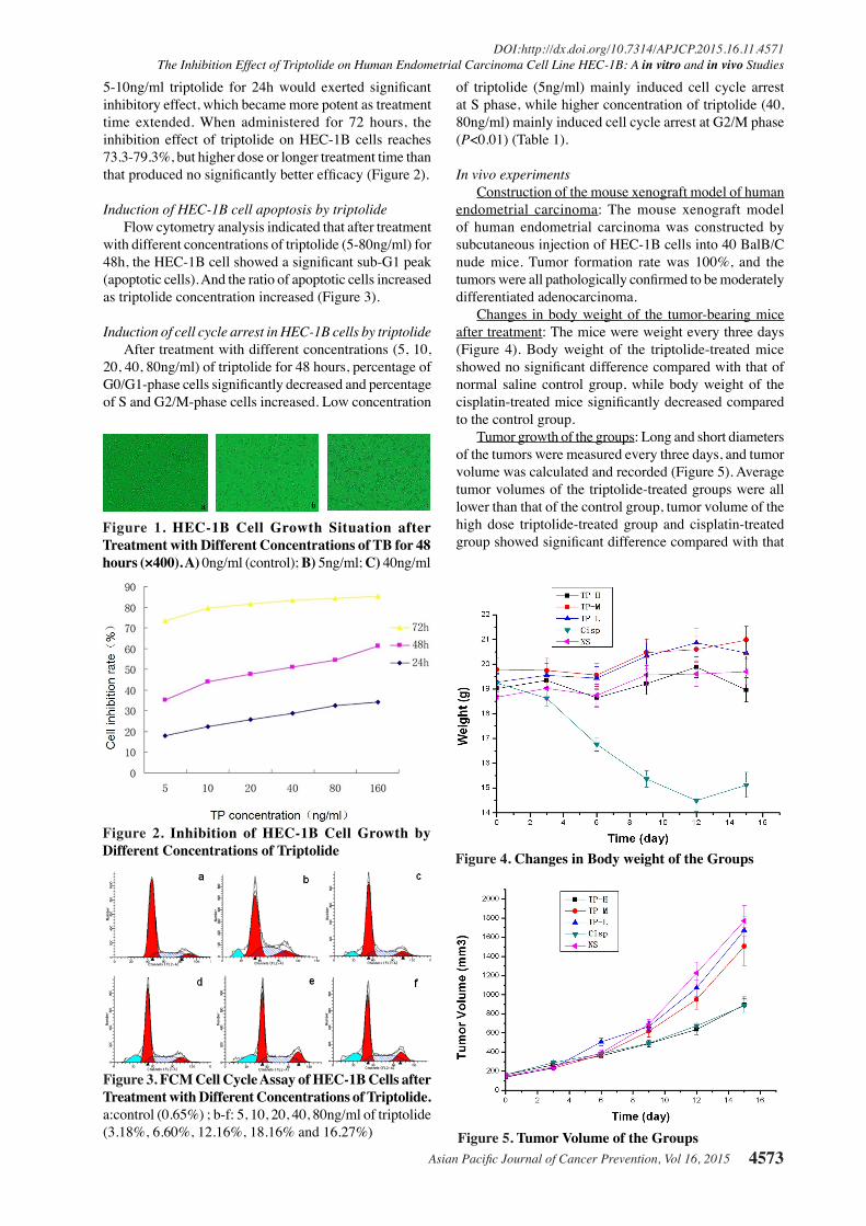
Asian Pacific Journal of Cancer Prevention, Vol 16, 2015 4573
DOI:http://dx.doi.org/10.7314/APJCP.2015.16.11.4571The Inhibition Effect of Triptolide on Human Endometrial Carcinoma Cell Line HEC-1B: A in vitro and in vivo Studies
5-10ng/ml triptolide for 24h would exerted significant inhibitory effect, which became more potent as treatment time extended. When administered for 72 hours, the inhibition effect of triptolide on HEC-1B cells reaches 73.3-79.3%, but higher dose or longer treatment time than that produced no significantly better efficacy (Figure 2).
Induction of HEC-1B cell apoptosis by triptolideFlow cytometry analysis indicated that after treatment
with different concentrations of triptolide (5-80ng/ml) for 48h, the HEC-1B cell showed a significant sub-G1 peak (apoptotic cells). And the ratio of apoptotic cells increased as triptolide concentration increased (Figure 3).
Induction of cell cycle arrest in HEC-1B cells by triptolideAfter treatment with different concentrations (5, 10,
20, 40, 80ng/ml) of triptolide for 48 hours, percentage of G0/G1-phase cells significantly decreased and percentage of S and G2/M-phase cells increased. Low concentration
of triptolide (5ng/ml) mainly induced cell cycle arrest at S phase, while higher concentration of triptolide (40, 80ng/ml) mainly induced cell cycle arrest at G2/M phase (P<0.01) (Table 1).
In vivo experimentsConstruction of the mouse xenograft model of human
endometrial carcinoma: The mouse xenograft model of human endometrial carcinoma was constructed by subcutaneous injection of HEC-1B cells into 40 BalB/C nude mice. Tumor formation rate was 100%, and the tumors were all pathologically confirmed to be moderately differentiated adenocarcinoma.
Changes in body weight of the tumor-bearing mice after treatment: The mice were weight every three days (Figure 4). Body weight of the triptolide-treated mice showed no significant difference compared with that of normal saline control group, while body weight of the cisplatin-treated mice significantly decreased compared to the control group.
Tumor growth of the groups: Long and short diameters of the tumors were measured every three days, and tumor volume was calculated and recorded (Figure 5). Average tumor volumes of the triptolide-treated groups were all lower than that of the control group, tumor volume of the high dose triptolide-treated group and cisplatin-treated group showed significant difference compared with that
Figure 3. FCM Cell Cycle Assay of HEC-1B Cells after Treatment with Different Concentrations of Triptolide. a:control (0.65%) ; b-f: 5, 10, 20, 40, 80ng/ml of triptolide (3.18%, 6.60%, 12.16%, 18.16% and 16.27%)
Figure 2. Inhibition of HEC-1B Cell Growth by Different Concentrations of Triptolide
Figure 1. HEC-1B Cell Growth Situation after Treatment with Different Concentrations of TB for 48 hours (×400). A) 0ng/ml (control); B) 5ng/ml; C) 40ng/ml
Figure 5. Tumor Volume of the Groups
Figure 4. Changes in Body weight of the Groups

Jing Ni et al
Asian Pacific Journal of Cancer Prevention, Vol 16, 20154574
of control group (P<0.05). The inhibition rate was 50% for high-dose triptolide group, 15% for medium-dose triptolide group, 6% for low-dose triptolide group, and 50% for cisplatin group.
The effect of triptolide on Bcl-2 and VEGF expression in the mouse xenograft model is shown in Table 2 and 3.
Discussion
Triptolide, one of the major active ingredients of tripterigium, has been used to treat rheumatoid arthritis, systemic lupus erythematosus, nephritis and some other autoimmune diseases for over a thousand years. Recent studies around the world had suggested broad-spectrum anti-cancer effect of triptolide, inhibiting growth of various tumors and inducing tumor cell apoptosis. In addition, it showed a synergistic effect when combined with cisplatin, which means good multi-channel anti-tumor potential.
In vitro and in vivo studies have suggested growth inhibitory and pro-apoptosis effect of triptolide in various cancers. Triptolide promotes liver cancer cell apoptosis by inhibiting NFkB activity (Alsaied et al., 2014); inhibits tumor cell growth and induces apoptosis by inhibiting nuclear transport of b-catenin and its target genes involved in cell cycle progression (Liu et al., 2014); induces SKOV3 cell apoptosis by regulating concentration of free oxygen species in mitochondria (Zhong et al., 2013); and significantly inhibits cell proliferation and induces
apoptosis in malignant peripheral nerve sheath tumors (MPNSTs) in a dose and time-dependent manner (Wang W et al., 2012).
Our results suggested that after treatment with different concentrations (5, 10, 20, 40, 80ng/ml) of triptolide for 48 hours, the HEC-1B cells showed a significant sub-G1 apoptotic peak, with percentage of apoptotic cells being 3.18%, 6.60%, 12.16%, 18.16%, and 16.27% respectively. These results suggest that triptolide induces apoptosis of the endometrial carcinoma cells by inhibiting cell proliferation. And it was observed by light microscopy that as incubation time and triptolide dose increased, number of floating cells increased, the mortality in high dose group increased.
We also treated the HEC-1B cells with 5~160 ng/ml triptolide to observe changes in cell cycle progression. It was found that treatment with low-dose triptolide (5 ng/ml) for 24h significantly inhibited HEC-1B proliferation (P<0.05), and cell cycle was arrested at the S phase. Such proliferation-inhibiting effect was obviously dose-dependent, at higher concentrations (40, 80 ng/ml), triptolide exerted stronger inhibition on cell proliferation and cell cycle was mainly arrested at the G2/M phase. The inhibition effect was also time-dependent, as incubation time prolonged, the inhibition grew stronger.
A lot of in vivo studies have proved very good tumor inhibition effect of triptolide for various cancers, for example colorectal cancer (Liu et al., 2014), liver cancer (Alsaied et al., 2014), malignant peripheral nerve sheath tumors (Wang et al., 2012), ovarian cancer (Zhao et al., 2012), gastric cancer (Li et al., 2012), and breast cancer (Li et al., 2014) .
Liu et al. (2014) also treated colorectal cancer cell line SW480 with triptolide or oxaliplatin alone, or the combination of the two, followed by MTT assay to test cell activity, and FACS to determine cell apoptosis. It was found that combination of the two can effectively inhibit proliferation of the cells and induce apoptosis. And it was revealed by western blotting and real-time PCR that such effect was achieved by inhibiting nuclear transport of b-catenin and expression of downstream cell cycle-related target genes.In addition, in the mouse xenograft model, combined therapy with both the two dramatically inhibited tumor growth without significant cytotoxicity effect, as indicated by blood test and hepatorenal function tests. Combined use of the two drugs at a low dose showed significant tumor-inhibitory activity and low cytotoxicity, suggesting great potential in clinical practice.
In this study, we constructed the mouse xenograft model of human endometrial carcinoma using human endometrial carcinoma cell line HEC-1B and BalB/C nude mice, and observed an anti-tumor activity of triptolide. We observed a 50% tumor-inhibition rate at high dose of triptolide, suggesting good efficacy at such dose. During the 15 days of drug administration, mice receiving cisplatin suffered severe weight loss, while mice receiving triptolide showed no weight loss or abnormality in mental status, activity, feeding, or defecation. Body weight of the mice of the two groups showed significant difference at the end of the experiment (P<0.05), suggesting high selectivity of triptolide against tumor cells.
Table 1. Cell Cycle Change Resulted from Different Concentrations of Triptolide (%, x ±s, n=3)Group G0/G1 S G2/M
control 70.12±4.73 21.15±4.77 8.73±1.745ng/ml 58.48±4.06** 33.52±3.30* 7.99±3.5410ng/ml 60.25±2.21** 28.76±2.09 10.99±0.6020ng/ml 57.51±4.94** 29.89±2.75 12.60±3.5140ng/ml 57.12±4.77** 28.19±4.18 16.49±1.56**80ng/ml 55.56±1.92** 28.92±3.03 15.52±1.13***compare to control group:*P<0.05, **P<0.01
Table 2. Bcl-2 Expression in Xenograft of the Groups (n=8)Group Ⅰ Ⅱ Ⅲ Ⅳ
TP-high dose* 6 2 0 0TP-mid dose 5 2 1 0TP-low dose* 6 2 0 0Cisplatin* 6 2 0 0NS-control 2 6 0 0*Compare to NS-Control Group P<0.05
Table 3. VEGF Expression in Xenograft of the Groups (n=8)Group Ⅰ Ⅱ Ⅲ Ⅳ
TP-high dose* 4 4 0 0TP-mid dose* 4 2 1 1TP-low dose 1 3 4 0Cisplatin* 5 3 0 0NS-control 0 2 2 4*compare to NS-control group P<0.05

Asian Pacific Journal of Cancer Prevention, Vol 16, 2015 4575
DOI:http://dx.doi.org/10.7314/APJCP.2015.16.11.4571The Inhibition Effect of Triptolide on Human Endometrial Carcinoma Cell Line HEC-1B: A in vitro and in vivo Studies
It has been proposed early in the 1970s by Folkman, that tumor growth and metastasis depend on angiogenesis, that is, newly formed blood vessels would provide sufficient nutrition, oxygen for tumor growth, and facilitate metastasis. Tumor angiogenesis is a complex process involving endothelial cell proliferation, migration, and extracellular matrix degradation. This process depends on a variety of antigenic factors, among which, VEGF is one of the most important. VEGF is synthesized by the tumor cells and secreted to tumor stroma, where it specifically acts on vascular endothelial cells and promotes proliferation of the target cells, thereby promoting tumor angiogenesis and accelerating tumor growth. In addition, it also increases vascular permeability, transforms extracellular matrix, and facilitates tumor cell invasion into blood vessels and metastasis. VEGF is often overexpressed in tumor tissue and is negatively correlated with prognosis. In addition, it also predicts the anti-tumor efficacy of anti-angiogenic drugs (Wang et al., 2012; Dobrzycka et al., 2013;Wang et al., 2014; Saarelainen et al., 2014).
Wang et al. (2012) performed in vitro and in vivo experiments on the efficacy of triptolide for malignant peripheral nerve sheath tumours (MPNSTs), which suggested significant inhibition of VEGF and EGFR expression, and triptolide inhibited STS-26T xenograft growth, suggesting very good efficacy for MPNST. Wang et al. (2014) measured VEGF-A, VEGFR2 and VEGFR3 expression in 76 endometrial carcinoma samples by immunohistochemistry and qRT-PCR, and revealed significant correlation between VEGF-A level and microvascular density (P<0.01), and significant correlation between VEGFR3 and tumor staging with an adverse DFS (P=0.09). VEGFR3 is a very good indicator for efficacy of anti-angiogenic treatment.
Saarelainen et al. (2014) assessed VEGF level in 80 cases of endometrial cancer, including 11 cases with metastasis, and it was found that VEGF level was significantly higher in the metastasis group than the non-metastasis group. Multivariate analysis indicated that VEGF was the only one independent risk factor of metastasis, suggesting an important role of serum VEGF in endometrial carcinoma metastasis. Immunohistochemistry indicated that the tumor marker was not correlated with any clinicopathological factors. In this study, after treatment with high-dose triptolide, VEGF expression significantly decreased compared to normal saline control (P<0.05), suggesting that inhibition of angiogenesis play an important part in the anti-tumor effect of triptolide, which is consistent with published results.
It has also been reported that the prognostic significance of bFGF was greater than VEGF, Cox multivariate regression indicated that high serum bFGF was correlated with short OS and DFS (Dobrzycka et al., 2013).
The Bcl-2 gene family is one of the most important regulators of apoptosis in mammalian cells, Bcl-2 exerts strong anti-apoptosis effect in the terminal part of the apoptosis mechanism, and is very important for development, progression, and treatment of tumors. Studies performed on osteosarcoma cell line U20S (Kwon et al., 2013), cervical cancer cell line Hela (Chen et al.,
2013), and cisplatin-resistant ovarian cancer cell line SKOV3 (Zhong et al., 2013) all indicated an important regulatory effect of Bcl-2 in tumor growth, which is also a major mechanism how triptolide inhibits tumor growth. Kwon et al. (2013) reported that Bcl-2 level was significantly reduced in U20S cells after triptolide treatment, and both the death receptor pathway and the mitochondria pathway were involved in triptolide-induced apoptosis. Chen et al. (2013) showed that triptolide induces cervical cancer cell apoptosis by regulating expression of Bcl-2 and related genes.
In our study, Bcl-2 expression in tumor-bearing animal treated with high-dose triptolide was significantly lower than that in mice of the normal saline control group (P<0.05), indicating that triptolide induces HEC-1B cell apoptosis by inhibiting Bcl-2 expression, which was consistent with Kwon HY’s results (Kwon et al., 2013).
Through in vitro and in vivo experiments, we found that triptolide dramatically inhibited HEC-1B cell proliferation and promoted apoptosis, and such efficacy grew stronger as triptolide dose increased and treatment time extended. Inhibition of angiogenesis and Bcl-2 expression may be involved in the inhibition of tumor cell growth and induction of apoptosis by triptolide.
Acknowledgements
This study is supported by the Traditional Chinese Medicine Scientific Research Project by the Jiangsu Province Administration of Traditional Chinese Medicine [No. LZ13234 (Wu), HZ07029 (Sun)] and Jiangsu Scientific Research Project for health care for cadres [BJ14019 (Wu)] , and the Research Foundation of Jiangsu Cancer Hospital [No. ZS201202 (Wu)] and National 863 Sub-project [2014AA020604-H20146809 (Wu)]
References
Alsaied OA, Sangwan V, Banerjee S, et al (2014). Sorafenib and triptolide as combination therapy for hepatocellular carcinoma. Surgery, 156, 270-9.
Cai YY, Lin WP, Li AP, Xu JY (2013). Combined effects of curcumin and triptolide on an ovarian cancer cell line. Asian Pac J Cancer Prev, 14, 4267-71.
Chen RH, Tian YJ (2013). Enhanced anti-tumor efficacy of aspirin combined with triptolide in cervical cancer cells.Asian Pac J Cancer Prev, 14, 3041-4.
Dobrzycka B, Mackowiak-Matejczyk B, Kinalski M, Terlikowski SJ (2013). Pretreatment serum levels of bFGF and VEGF and its clinical significance in endometrial carcinoma. Gynecol Oncol, 128, 454-60.
Ho JN, Byun SS, Lee S, et al (2015). Synergistic antitumor effect of triptolide and cisplatin in cisplatin resistant human bladder cancer cells. J Urol, 193, 1016-22.
Kwon HY, Kim KS, An HK, et al (2013). Triptolide induces apoptosis through extrinsic and intrinsic pathways in human osteosarcoma U2OS cells. Indian J Biochem Biophys, 50, 485-91.
Li CJ, Chu CY, Huang LH, et al (2012). Synergistic anticancer activity of triptolide combined with cisplatin enhances apoptosis in gastric cancer in vitro and in vivo. Cancer Lett. 319, 203-13.
Li J, Liu R, Yang Y, et al (2014). Triptolide-induced in vitro and

Jing Ni et al
Asian Pacific Journal of Cancer Prevention, Vol 16, 20154576
0
25.0
50.0
75.0
100.0
New
ly d
iagn
osed
with
out
trea
tmen
t
New
ly d
iagn
osed
with
tre
atm
ent
Pers
iste
nce
or r
ecur
renc
e
Rem
issi
on
Non
e
Chem
othe
rapy
Radi
othe
rapy
Conc
urre
nt c
hem
orad
iatio
n
10.3
0
12.8
30.025.0
20.310.16.3
51.7
75.051.1
30.031.354.2
46.856.3
27.625.033.130.031.3
23.738.0
31.3
0
25.0
50.0
75.0
100.0
New
ly d
iagn
osed
with
out
trea
tmen
t
New
ly d
iagn
osed
with
tre
atm
ent
Pers
iste
nce
or r
ecur
renc
e
Rem
issi
on
Non
e
Chem
othe
rapy
Radi
othe
rapy
Conc
urre
nt c
hem
orad
iatio
n
10.3
0
12.8
30.025.0
20.310.16.3
51.7
75.051.1
30.031.354.2
46.856.3
27.625.033.130.031.3
23.738.0
31.3
in vivo cytotoxicity in human breast cancer stem cells and primary breast cancer cells. Oncol Rep, 31, 2181-6.
Liu Y, Xiao E, Yuan L, Li G (2014). Triptolide synergistically enhances antitumor activity of oxaliplatin in colon carcinoma in vitro and in vivo. DNA Cell Biol, 33, 418-25.
Park B (2014). Triptolide, a diterpene, inhibits osteoclastogenesis, induced by RANKL signaling and human cancer cells. Biochimie, 105, 129-36.
Saarelainen SK, Staff S, Peltonen N, et al (2014). Endoglin, VEGF, and its receptors in predicting metastases in endometrial carcinoma. Tumour Biol, 35, 4651-7.
Siegel R1, Ma J, Zou Z, Jemal A (2014). Cancer statistics, 2014. CA Cancer J Clin, 64, 9-29.
Wang J, Taylor A, Showeil R, et al (2014). Expression profiling and significance of VEGF-A, VEGFR2, VEGFR3 and related proteins in endometrial carcinoma. Cytokine. 68, 94-100.
Wang W, Lin W, Hong B, et al (2012). Effect of triptolide on malignant peripheral nerve sheath tumours in vitro and in vivo. J Int Med Res, 40, 2284-94.
Zhao H, Yang Z, Wang X, et al (2012). Triptolide inhibits ovarian cancer cell invasion by repression of matrix metalloproteinase 7 and 19 and upregulation of E-cadherin. Exp Mol Med, 44, 633-41.
Zhong YY, Chen HP, Tan BZ, Yu HH, Huang XS (2013). Triptolide avoids cisplatin resistance and induces apoptosis via the reactive oxygen species/nuclear factor-κB pathway in SKOV3<sup>PT</sup> platinum-resistant human ovarian cancer cells. Oncol Lett, 6, 1084-92.