Embed Size (px)
Citation preview

IBIMA Publishing
Plastic Surgery: An International Journal
http://www.ibimapublishing.com/journals/PSIJ/psij.html
Vol. 2013 (2013), Article ID 973073, 13 pages
DOI: 10.5171/2013.973073
_____________
Cite this Article as: Yohei Tanaka, Yuichiro Tsunemi, Makoto Kawashima and Hiroshi Nishida (2013),
"The Impact of Near-infrared in Plastic Surgery," Plastic Surgery: An International Journal, Vol. 2013 (2013), Article ID 973073, DOI: 10.5171/2013.973073
Research Article The Impact of Near-infrared in Plastic
Surgery
Yohei Tanaka1,2,3
, Yuichiro Tsunemi2, Makoto Kawashima
2 and Hiroshi Nishida
3
1Clinica Tanaka Plastic, Reconstructive Surgery and Anti-aging Center, Matsumoto, Nagano, Japan
2Department of Dermatology, Tokyo Women’s Medical University, Tokyo, Japan
3Department of Applied Life Sciences, Niigata University of Pharmacy and Applied Life Sciences,
Niigata, Japan
Correspondence should be addressed to: Yohei Tanaka; [email protected]
Received 22 January 2013; Accepted 21 March 2013; Published 23 June 2013
Academic Editor: Maureen Susan Thorniley
Copyright © 2013 Yohei Tanaka, Yuichiro Tsunemi, Makoto Kawashima and Hiroshi Nishida.
Distributed under Creative Commons CC-BY 3.0
Abstract
Many studies regarding near-infrared, have used near-infrared resources without a water filter
or a cooling system, and have proven its thermal effects. With these methods near-infrared energy is mainly absorbed in the superficial tissues and cannot be delivered sufficiently to
deeper tissues. As solar near-infrared is filtered by atmospheric water, a water filter is essential
in order to simulate solar near-infrared. Thus, these approaches could not sufficiently evaluate
the effects of incident solar near-infrared that reaches the human tissue. We have clarified that the near-infrared that simulates solar near-infrared non-thermally affects subcutaneous
tissues, including muscle. Importantly, the biological effects of near-infrared have both
beneficial applications and deleterious effects. Near-infrared induces dermal heating thermally and non-thermally induces collagen and elastin stimulation, which results in skin tightening,
and induces long-lasting vasodilation that may prevent vasospasm and may be beneficial for
ischemic disorders and flap surgeries. Near-infrared also non-thermally relaxes and weakens dystonic or hypertrophic muscles to reduce wrinkles and myalgia. Its long-lasting induction of
subcutaneous adipocytes may have an application in volume augmentation. However,
continuous near-infrared exposure may induce photoaging and thinning of superficial muscles, which results in skin ptosis. Protection against near-infrared should be strongly considered, as
over half of the solar energy is near-infrared. Although plastic surgeons are not familiar with
the effects of near-infrared, its potential appears to be high and significant. This paper reviews
the effects of near-infrared and introduces the new findings and applications of the biological effects of near-infrared in the field of plastic surgery.
Keywords: Near-infrared; non-thermal; biological effects; photoaging.
Introduction
Many studies have demonstrated the
effects of sun and ultraviolet (UV)
exposure on human tissue. However, the
long-term effects of near-infrared (NIR) exposure on human skin and
subcutaneous tissues have not been well
Plastic Surgery: An International Journal 2
_______________
Yohei Tanaka, Yuichiro Tsunemi, Makoto Kawashima and Hiroshi Nishida (2013), Plastic Surgery: An
International Journal, DOI: 10.5171/2013.973073
investigated. Although various types of UV
blocking materials, such as sunblock, sunglasses, films, and fibers are often used
to prevent skin damage from UV exposure,
and despite the world-wide use of these blocking materials, unwanted
physiological effects such as rosacea,
erythema ab igne, long-lasting
vasodilation (Tanaka, 2009a; Tanaka, 2011a), long-lasting muscle thinning
(Tanaka, 2010a, 2011b), and sagging and
skin ptosis still occur (Tanaka, 2011c; Tanaka, 2012a, b). Both superficial muscle
thinning and skin ptosis develop with age,
and though face lift surgeries and skin tightening procedures are proven
effective treatments, the mechanisms
underlying their development remain
unclear.
We previously reported that NIR from
1100 to 1800 nm together with a water-filter that excludes wavelengths between
1400 and 1500 nm penetrates deep into
human tissue and it is absorbed by water in the skin (Tanaka, 2009a,b; Tanaka,
2010b; Tanaka, 2011d), hemoglobin in
dilated vessels (Tanaka, 2009a; Tanaka,
2011a), myoglobin in the superficial muscle (Tanaka, 2010a; Tanaka, 2011b),
and bone cortical mass, and it is scattered
by adipose cells (Tanaka, 2011e; Tanaka, 2012b).
Notably, in order to experimentally simulate solar NIR that reaches the skin
(Tanaka, 2012a, b); a water filter is
required, as solar NIR is filtered by atmospheric water (Anderson, 1981; Gates,
1966). NIR increases the surface
temperature and induces thermal effects,
so a contact cooling is needed to pursue the properties of NIR (Tanaka, 2011c; Tanaka,
2012a, b).
NIR is used to treat wound healing
disorders (Danno, 2001; Horwitz, 1999;
Schramm, 2003) and malignant tumors (Bäumler, 1999; Dees, 2002; Kelleher,
1999; Orenstein, 1999). NIR induces
dermal heating thermally and non-
thermally induces collagen and elastin stimulation, which results in skin
tightening. NIR also induces long-lasting
vasodilation that may prevent vasospasm
and may be beneficial for ischemic
disorders and flap surgeries in the field of plastic surgery. Further, NIR non-
thermally relaxes and weakens dystonic or
hypertrophic muscles to reduce wrinkles and myalgia.
In addition to its therapeutic effects,
however, NIR also induces non-thermal DNA damage (Tanaka, 2010c; Tanaka,
2012c) and cell death by apoptosis
(Tirlapur, 2001). NIR induces cell death of cancer cells and bone marrow cells
(Tanaka, 2011e), which may have a
potential application in the treatment of cancer (Tanaka, 2010c; Tanaka, 2012c).
The necessity to protect against NIR has
not been well investigated to date. Fair skin with lower concentrations of melanin
and a thin dermis might allow NIR to
penetrate deeper into human tissue, and damage superficial muscles compared to
darker skin with dense melanin and a
thick dermis (Tanaka, 2011c; Tanaka, 2012a, b). In addition to natural NIR,
human skin is increasingly exposed to
artificial NIR from medical devices and
electrical appliances (Schieke, 2003; Schroeder, 2008). Furthermore most
sunscreens only block UV, but not visible
light and NIR (Tanaka, 2011c; Tanaka, 2012a, b), and sunglasses and glasses are
unable to block NIR exposure. Thus,
individuals using sunscreens and glasses should further equip themselves for
protection against NIR (Darvin, 2010;
Meinke, 2011; Pujol, 1993; Schieke, 2003; Schroeder, 2008; Tanaka, 2011c; Tanaka,
2012a, b), as we are exposed to
tremendous amounts of NIR (Tanaka,
2011c).
Previous studies regarding NIR have
reported its application in the industrial and the agricultural fields, but have not
well investigated the effects of NIR
exposure in the medical fields, particularly in the field of plastic surgery.
Findings from our previous studies
suggest that we should consider the biological effects of NIR, which have both
potentially usefully applications and
detrimental physiological effects.
3 Plastic Surgery: An International Journal
_______________
Yohei Tanaka, Yuichiro Tsunemi, Makoto Kawashima and Hiroshi Nishida (2013), Plastic Surgery: An
International Journal, DOI: 10.5171/2013.973073
Methods to Evaluate Non-Thermal
Biological Effects of NIR
In previous studies of NIR, a light source
emitting wide wavelengths of NIR were used as an NIR source (Frank, 2004;
Tanaka, 2012a). The temperature of the
superficial layer of a culture fluid in a laboratory dish under NIR irradiation rises
immediately, as NIR is mainly absorbed by
water. The energy of NIR then decreases as it penetrates deeper, and will not reach
enough target cells in the base. Therefore,
these previous studies were only able to
describe the optical and thermal effects of NIR, and are not useful to examine non-
thermal biological effects of NIR (Tanaka,
2012a, b).
NIR induces dermal heating, which results
in the tightening of skin laxity (Bitter, 2000; Chan, 2008; Goldberg, 2000; Ross,
2000; Tanaka, 2009a, b; Zelickson, 2006).
In previous studies (Danno, 2001; Kim,
2005: Kligman, 1982) NIR devices without a water filter or contact cooling were used
to investigate thermal effects on the
human body. NIR increases the skin surface temperature and induces
perspiration and vasodilation, as NIR is
primarily absorbed by water and hemoglobin. Subsequently, a substantial
amount of energy is absorbed in the
superficial layers of skin, and only limited NIR energy can be delivered to deeper
tissues. Therefore, these previous studies
only describe superficial and thermal
effects of NIR, and are not useful to examine non-thermal biological effects
(Tanaka, 2012a, b).
Sunlight that reaches the human skin
contains solar energy composed of 6.8%
UV light, 38.9% visible light and 54.3% infrared (IR) (Kochevar, 1999). The IR
spectral region is arbitrarily divided according to wavelength into sub-regions
of NIR (760–3000 nm), middle IR (3000–
30 000nm), and far IR (30 000 nm–1 mm).
NIR from the sun is selectively filtered by atmospheric water (Anderson, 1981;
Gates, 1966), and NIR that reaches the
Earth’s surface readily penetrates the superficial layers of the skin (Tanaka,
2010a, 2011c, 2012a, 2011e) (Fig. 1, left).
Wavelengths below 1100 nm will be
absorbed by melanin in the skin.
Wavelengths between 1400 and 1500 nm
and those above 1850 nm will be absorbed by water in the skin, which
results in heating and may induce painful
sensations and burns (Kelleher, 1999). Filtering out the wavelengths below 1100
nm, around 1450 nm, and above 1850 nm
enabled the delivery of NIR to deeper tissues (Davenport, 2006) and also
simulated the solar NIR that contracts
human skin under natural conditions.
An NIR device that emits a spectrum of
NIR wavelength from 1100 to 1800 nm
together with a water-filter that excludes wavelengths between 1400 and 1500 nm
as well as those above 1850 nm simulates
the natural exposure of skin to NIR and allows for the evaluation of solar NIR that
reaches the skin under natural conditions.
NIR increases the surface temperature and induces thermal effects, thus, in order
to reduce the skin surface temperature,
perspiration, and blood vessel dilation,
contact cooling is required. These specific wavelengths and the cooling system
enables NIR to be delivered to the deeper
tissues without pain or epidermal burns
(Davenport, 2006; Goldberg, 2007), and
allows for the investigations of properties
of NIR (Tanaka, 2012a, b) (Fig. 1).

Plastic Surgery: An International Journal 4
_______________
Yohei Tanaka, Yuichiro Tsunemi, Makoto Kawashima and Hiroshi Nishida (2013), Plastic Surgery: An
International Journal, DOI: 10.5171/2013.973073
Fig. 1: A Schematic of Solar NIR (Left) and the NIR Research Strategy that Can Evaluate
Non-Thermal Biological Effects of NIR (Right)
Discussion
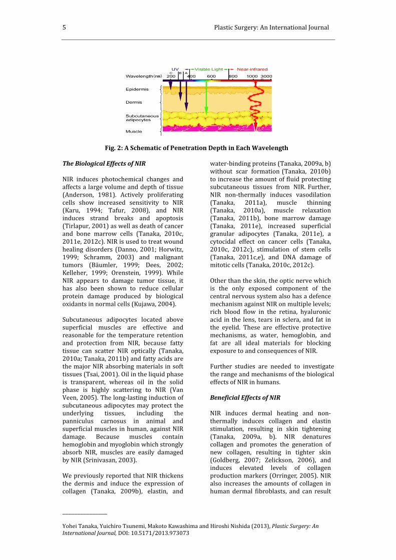
Properties and Chromophore of NIR
NIR exhibits both wave and particle properties and it is strongly absorbed by
water, hemoglobin, and myoglobin
(Tanaka, 2011c). As a consequence, NIR can affect the subcutaneous tissues,
including muscles, with its high
permeability (Tanaka, 2011c) (Fig. 2).
The NIR spectrum is a result of the
overtones and combination of bond
stretching vibrations from O-H, C-H, and N-H groups (Weyer, 1985). Water is a
polar molecule possessing hydrogen
bonds with an electrical dipole moment, and will be resonated by NIR. NIR will be
resonated and absorbed in O-H
intramolecular hydrogen bonds and the electrical dipole moment (Tsai, 2001).
NIR induces an increase in water
retention in the dermis and induces perspiration, vasodilation, and the
expression of collagen and elastin (Tanaka,
2009a). Both collagen and elastin possess helical structures rich in hydrogen bonds.
Humans have biological defense
mechanisms in which induced hydrogen
bonds and helical structures are resonated by NIR and absorb NIR to protect the
subcutaneous tissues against this
radiation.
NIR non-thermally induces degeneration
of myoglobin, resulting in apoptosis of
vascular smooth muscle cells and long-lasting vasodilation (Tanaka, 2011a). Both
hemoglobin and myoglobin have heme
binding sites and alpha helices. Our results showing long-lasting muscle
thinning and vasodilation induced by NIR
suggest that NIR might resonate and
damage heme. However, our collagen, elastin, and cancer studies suggest that
NIR may mainly resonate hydrogen bonds,
helical structures, alpha helices, and DNA (Tanaka, 2011c). Alpha helices are
thought to be resonated by NIR
(Nevskaya, 1976). Both hemoglobin and myoglobin are oxygen-carrying proteins
with many alpha helices. It is possible that
NIR induces resonance of helical
structures in the oxygen-carrying proteins and degenerates proteins containing
hydrogen bonds and helical structures,
which result in damage to the storage and transport of oxygen. This could be one of
the mechanisms of apoptosis (Tanaka,
2011c).
5 Plastic Surgery: An International Journal
_______________
Yohei Tanaka, Yuichiro Tsunemi, Makoto Kawashima and Hiroshi Nishida (2013), Plastic Surgery: An
International Journal, DOI: 10.5171/2013.973073
Fig. 2: A Schematic of Penetration Depth in Each Wavelength
The Biological Effects of NIR
NIR induces photochemical changes and affects a large volume and depth of tissue
(Anderson, 1981). Actively proliferating
cells show increased sensitivity to NIR (Karu, 1994; Tafur, 2008), and NIR
induces strand breaks and apoptosis
(Tirlapur, 2001) as well as death of cancer and bone marrow cells (Tanaka, 2010c,
2011e, 2012c). NIR is used to treat wound
healing disorders (Danno, 2001; Horwitz,
1999; Schramm, 2003) and malignant tumors (Bäumler, 1999; Dees, 2002;
Kelleher, 1999; Orenstein, 1999). While
NIR appears to damage tumor tissue, it has also been shown to reduce cellular
protein damage produced by biological
oxidants in normal cells (Kujawa, 2004).
Subcutaneous adipocytes located above
superficial muscles are effective and reasonable for the temperature retention
and protection from NIR, because fatty
tissue can scatter NIR optically (Tanaka,
2010a; Tanaka, 2011b) and fatty acids are the major NIR absorbing materials in soft
tissues (Tsai, 2001). Oil in the liquid phase
is transparent, whereas oil in the solid phase is highly scattering to NIR (Van
Veen, 2005). The long-lasting induction of
subcutaneous adipocytes may protect the underlying tissues, including the
panniculus carnosus in animal and
superficial muscles in human, against NIR
damage. Because muscles contain hemoglobin and myoglobin which strongly
absorb NIR, muscles are easily damaged
by NIR (Srinivasan, 2003).
We previously reported that NIR thickens
the dermis and induce the expression of collagen (Tanaka, 2009b), elastin, and
water-binding proteins (Tanaka, 2009a, b)
without scar formation (Tanaka, 2010b)
to increase the amount of fluid protecting subcutaneous tissues from NIR. Further,
NIR non-thermally induces vasodilation
(Tanaka, 2011a), muscle thinning (Tanaka, 2010a), muscle relaxation
(Tanaka, 2011b), bone marrow damage
(Tanaka, 2011e), increased superficial granular adipocytes (Tanaka, 2011e), a
cytocidal effect on cancer cells (Tanaka,
2010c, 2012c), stimulation of stem cells
(Tanaka, 2011c,e), and DNA damage of mitotic cells (Tanaka, 2010c, 2012c).
Other than the skin, the optic nerve which is the only exposed component of the
central nervous system also has a defence
mechanism against NIR on multiple levels; rich blood flow in the retina, hyaluronic
acid in the lens, tears in sclera, and fat in
the eyelid. These are effective protective mechanisms, as water, hemoglobin, and
fat are all ideal materials for blocking
exposure to and consequences of NIR.
Further studies are needed to investigate
the range and mechanisms of the biological
effects of NIR in humans.
Beneficial Effects of NIR
NIR induces dermal heating and non-
thermally induces collagen and elastin
stimulation, resulting in skin tightening
(Tanaka, 2009a, b). NIR denatures collagen and promotes the generation of
new collagen, resulting in tighter skin
(Goldberg, 2007; Zelickson, 2006), and induces elevated levels of collagen
production markers (Orringer, 2005). NIR
also increases the amounts of collagen in human dermal fibroblasts, and can result
Plastic Surgery: An International Journal 6
_______________
Yohei Tanaka, Yuichiro Tsunemi, Makoto Kawashima and Hiroshi Nishida (2013), Plastic Surgery: An
International Journal, DOI: 10.5171/2013.973073
in clinical improvement of skin texture
(Lee, 2006). NIR induces high collagen density in the dermis, resulting in
epidermal smoothness without scar
formation, which provides safe, consistent, and long-term effects of skin rejuvenation
(Tanaka, 2010b).
Pre-exposure of NIR prevents UV-induced toxicity (Danno, 1992; Frank, 2004;
Menezes, 1998), and this effect is
independent of heat shock protein induction and cell division (Menezes,
1998). These findings support the
hypothesis that NIR prepares skin to better resist the subsequent damage from
UV or NIR (Tanaka, 2012a, b).
NIR is known to be of therapeutic benefit in the treatment of musculoskeletal disorders
and healing of indolent wounds (Ceylan,
2003; Webb, 1998). NIR induces long-lasting vasodilation for an increase in blood
circulation by causing apoptosis of vascular
smooth muscle cells, which may prevent vasospasm and may be beneficial for
ischemic disorders and flap surgeries.
NIR increases mitochondrial metabolism (Karu, 1999; Passarella, 1984; Yu, 1997;
Wilden; 1998), facilitates wound healing,
and promotes angiogenesis in skin (Conlan; 1996), bone (Yaakobi, 1996), nerve (Assia,
1989), and skeletal muscle (Bibikova,
1994; Oron, 2006).
NIR also non-thermally relaxes and
weakens dystonic or hypertrophic muscles to reduce wrinkles and myalgia. Thus, this
simple technique of using NIR may offer an
alternative method to relax superficial
muscles.
We previously reported that NIR increased
subcutaneous adipocytes on the panniculus
carnosus as well as CD34-positive cells
surrounding the subcutaneous adipocytes. Adipogenesis is tightly associated with
angiogenesis, and the expression of
adipocytes is linked to the development of its vasculature (Christiaens, 2010). Thus,
induction of subcutaneous adipocytes may
have an application for volume
augmentation.
Our previous study revealed that NIR
increased subcutaneous and bone marrow adipocytes, CD34-positive hematopoietic
stem cells in bone marrow, and cortical
bone mass (Tanaka, 2011e). Stimulation of stem cells by NIR irradiation might be
beneficial for applications in regenerative
medicine.
NIR is also an essential tool in cancer
detection and imaging, as actively
proliferating cells show increased sensitivity to NIR (Karu, 1994; Tafur,
2008). NIR also induces non-thermal DNA
damage of mitotic cells especially in prophase, metaphase, and anaphase due to
the absence of nuclear lamin protection
(Tanaka, 2010c, 2012c), and suppresses
the proliferation of various kinds of malignant cells (Tanaka, 2010c, 2011c,
2012c).
NIR may be beneficial in multiple fields of
plastic surgery, as the schedule reduces
discomfort and side effects, deep subcutaneous tissues are accessible, and
facilitates repeated irradiations.
These findings indicate that NIR has a wide
range of biological effects, and future
studies are warranted to develop these
findings into potential useful and beneficial techniques and applications.

7 Plastic Surgery: An International Journal
_______________
Yohei Tanaka, Yuichiro Tsunemi, Makoto Kawashima and Hiroshi Nishida (2013), Plastic Surgery: An
International Journal, DOI: 10.5171/2013.973073
Fig. 3: Schematic Indicating Effects and Potentially Useful Applications of NIR
Deleterious Effects of NIR
NIR exerts biologic effects on human skin (Schieke, 2003). NIR causes skin changes
similar to those observed in solar elastosis,
and enhance UV-induced dermal damage
(Kligman, 1982). NIR activates mitogen-activated protein kinases and induces gene
transcription, and it is likely to increase
collagen degradation (Kim, 2006; Schieke, 2003; Schroeder, 2008). Epidemiological
data and clinical reports imply that NIR is
not innocuous to human skin (Dover, 1989; Kligman, 1984; Schieke, 2003).
Several diseases such as rosacea and
erythema ab igne might be induced by chronic NIR exposure. Rosacea is a chronic
cutaneous disorder characterized by
centrofacial persisting erythema, telangiectasia, papules, pustules, edema,
and ocular involvement. Rosacea affects all
races, though is more common in Caucasians and fair-skinned populations
(Berg, 1989). NIR should be considered as a
critical factor in the development and
aggravation of rosacea, since the distribution of erythema is most prominent
on the facial convexities (Bae, 2009).
Long-term exposure to sources of heat and
NIR, such as fires and stoves, results in
erythema ab igne (Findlayson, 1966), which is clinically characterized by a
reticular hyperpigmentation and
telangiectasia accompanied histologically by epidermal atrophy, vasodilation, dermal
melanin and hemosiderin deposits. Long-
term exposure of NIR from various heat
sources is thought to induce reticulated erythema and results in histopathological
changes similar to those seen in solar-
damaged skin (Page, 1988). The occurrence
of telangiectasia appeared to increase with
age, increased sunbathing, and poor
pigmentation ability (Berg, 1989).
These lesions may develop thermal
keratosis, such as hyperkeratosis,
keratinocyte dysplasia, and dermal elastosis, which are similar to the changes
that occur in actinically damaged skin
(Arrington, 1979). Several reports indicate that carcinomas arise from heat-
induced erythema ab igne (Hewitt, 1993;
Jones, 1988; Kligman, 1984). Similar to UV, NIR induces photoaging and
potentially photocarcinogenesis (Schieke,
2003). In addition, skin tumors in mice
appeared faster after irradiation with the full lamp spectrum containing UV, visible,
and NIR compared to irradiation with UV
alone (Bain, 1943).
NIR is attenuated by thick water-
containing dermis. Thus, skin with sparse melanin and a thin dermis allows NIR to
penetrate deeper into tissue than skin
with dense melanin and a thick dermis
(Tanaka, 2011c; Tanaka, 2012a, b). The mean area of the facial surface that is
covered with wrinkles is significantly
larger in Caucasians than in African Americans, and characteristics of age-
related periorbital changes seem to occur
at a more accelerated rate in Caucasians (Odunze, 2008). In addition, fair skin is
more sensitive to skin aging (Guinot,
2002; Nagashima, 1999). These findings support the observation that fair skin
tends to wrinkle and sag earlier in life
(Rawlings, 2006; Tsukahara, 2004), as fair
skin is thinner and is more susceptible to NIR damage to the underlying superficial
muscles than dark skin (Tanaka, 2011c;
Tanaka, 2012a).
Plastic Surgery: An International Journal 8
_______________
Yohei Tanaka, Yuichiro Tsunemi, Makoto Kawashima and Hiroshi Nishida (2013), Plastic Surgery: An
International Journal, DOI: 10.5171/2013.973073
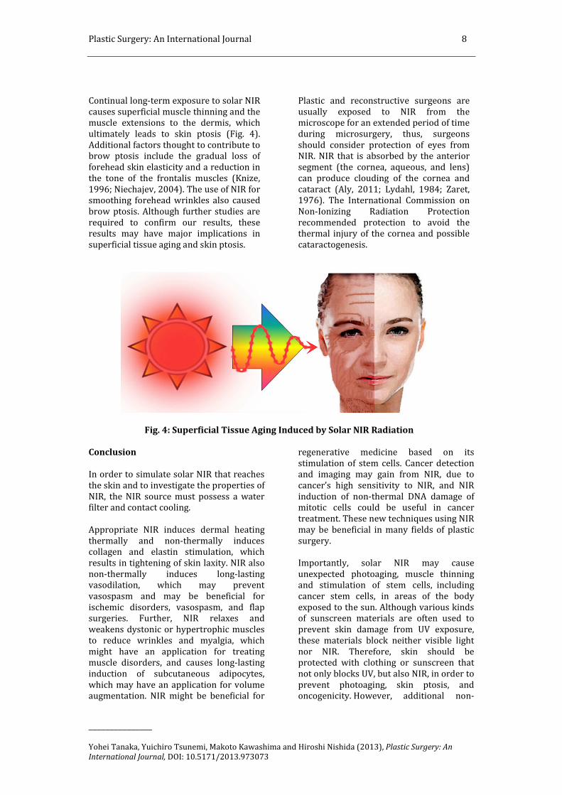
Continual long-term exposure to solar NIR
causes superficial muscle thinning and the muscle extensions to the dermis, which
ultimately leads to skin ptosis (Fig. 4).
Additional factors thought to contribute to brow ptosis include the gradual loss of
forehead skin elasticity and a reduction in
the tone of the frontalis muscles (Knize,
1996; Niechajev, 2004). The use of NIR for smoothing forehead wrinkles also caused
brow ptosis. Although further studies are
required to confirm our results, these results may have major implications in
superficial tissue aging and skin ptosis.
Plastic and reconstructive surgeons are
usually exposed to NIR from the microscope for an extended period of time
during microsurgery, thus, surgeons
should consider protection of eyes from NIR. NIR that is absorbed by the anterior
segment (the cornea, aqueous, and lens)
can produce clouding of the cornea and
cataract (Aly, 2011; Lydahl, 1984; Zaret, 1976). The International Commission on
Non-Ionizing Radiation Protection
recommended protection to avoid the thermal injury of the cornea and possible
cataractogenesis.
Fig. 4: Superficial Tissue Aging Induced by Solar NIR Radiation
Conclusion
In order to simulate solar NIR that reaches
the skin and to investigate the properties of
NIR, the NIR source must possess a water filter and contact cooling.
Appropriate NIR induces dermal heating
thermally and non-thermally induces collagen and elastin stimulation, which
results in tightening of skin laxity. NIR also
non-thermally induces long-lasting vasodilation, which may prevent
vasospasm and may be beneficial for
ischemic disorders, vasospasm, and flap surgeries. Further, NIR relaxes and
weakens dystonic or hypertrophic muscles
to reduce wrinkles and myalgia, which
might have an application for treating muscle disorders, and causes long-lasting
induction of subcutaneous adipocytes,
which may have an application for volume augmentation. NIR might be beneficial for
regenerative medicine based on its
stimulation of stem cells. Cancer detection and imaging may gain from NIR, due to
cancer’s high sensitivity to NIR, and NIR
induction of non-thermal DNA damage of mitotic cells could be useful in cancer
treatment. These new techniques using NIR
may be beneficial in many fields of plastic
surgery.
Importantly, solar NIR may cause
unexpected photoaging, muscle thinning and stimulation of stem cells, including
cancer stem cells, in areas of the body
exposed to the sun. Although various kinds of sunscreen materials are often used to
prevent skin damage from UV exposure,
these materials block neither visible light
nor NIR. Therefore, skin should be protected with clothing or sunscreen that
not only blocks UV, but also NIR, in order to
prevent photoaging, skin ptosis, and oncogenicity. However, additional non-

9 Plastic Surgery: An International Journal
_______________
Yohei Tanaka, Yuichiro Tsunemi, Makoto Kawashima and Hiroshi Nishida (2013), Plastic Surgery: An
International Journal, DOI: 10.5171/2013.973073
thermal studies are needed to investigate the biological effects of NIR in humans.
Disclosure
None of the authors of this study have a
conflict of interest.
References
Aly, E. M. & Mohamed, E. S. (2011). "Effect of Infrared Radiation on the Lens," Indian
journal of ophthalmology, 59 97-101.
Anderson, R. R. & Parrish, J. A. (1981). "The Optics of Human Skin," Journal of
Investigative Dermatology, 77 13-19.
Arrington, J. H. III. & Lockman, D. S. (1979).
"Thermal Keratoses and Squamous Cell
Carcinoma in Situ Associated with Erythema Ab Igne," Archives of
Dermatology, 115 1226-1228.
Assia, E., Rosner, M., Belkin, M., Solomon, A. & Schwartz, M. (1989). "Temporal
Parameters of Lowenergy Laser Irradiation
for Optimal Delay of Post-Traumatic Degeneration of Rat Optic Nerve," Brain
Research, 476 205-212.
Bae, Y. I., Yun, S. J., Lee, J. B., Kim, S. J., Won,
Y. H. & Lee, S. C. (2009). "Clinical Evaluation
of 168 Korean Patients with Rosacea: The Sun Exposure Correlates with the
Erythematotelangiectatic Subtype," Annals
of Dermatology, 21 (3) 243-249.
Bain, J. A, Rusch, H. P. & Kline, B. E. (1943).
"The Effect of Temperature upon
Ultraviolet Carcinogenesis with Wavelength 2,800–3,400A˚,"Cancer
Research, 3 610-612.
Bäumler, W., Abels, C., Karrer, S., Weiss, T.,
Messmann, H., Landthaler, M. & Szeimies,
R.- M. (1999). "Photo-Oxidative Killing of
Human Colonic Cancer Cells Using Indocyanine Green and Infrared Light,"
British Journal of Cancer, 80 (3-4) 360-363.
Berg, M. (1989). "Epidemiological Studies
of the Influence of Sunlight on the Skin,"
Photodermatol, 6 80-84.
Bibikova, A. & Oron, U. (1994). "Attenuation of the Process of Muscle
Regeneration in the Toad Gastrocnemius
Muscle by Low Energy Laser
Irradiation," Lasers in Surgery and
Medicine, 14 355-361.
Bitter, P. H. (2000). "Noninvasive Rejuvenation of Photodamaged Skin Using
Serial, Full-Face Intense Pulsed Light
Treatments," Dermatologic Surgery, 26 835-843.
Ceylan, Y., Hizmetli, S. & Siling, Y. (2003).
"The Effects of Infrared Laser and Medical Treatments on Pain and Serotonin
Degradation Products in Patients with
Myofascial Pain Syndrome: A Controlled Trial," Rheumatology International, 24 260-
263.
Chan, H. H., Yu, C. S., Shek, S., Yeung, C. K.,
Kono, T. & Wei, W. I. (2008). "A
Prospective, Split Face, Single-Blinded
Study Looking at the Use of an Infrared Device with Contact Cooling in the
Treatment of Skin Laxity in Asians," Lasers
in Surgery and Medicine, 40 146-152.
Christiaens, V. & Lijnen, H. R. (2010).
"Angiogenesis and Development of Adipose Tissue. Review," Molecular and Cellular
Endocrinology, 318 2-9.
Conlan, M. J., Rapley, J. W. & Cobb, C. M.
(1996). "Biostimulation of Wound Healing
by Low-Energy Laser Irradiation," Journal
of Clinical Periodontology, 23 492-496.
Danno, K., Horio, T. & Imamura, S. (1992).
"Infrared Radiation Suppresses Ultraviolet B-Induced Sunburn-Cell Formation,"
Archives of Dermatological Research, 284
92-94.
Danno, K., Mori, N., Toda, K., Kobayashi, T.
& Utani, A. (2001). "Near-Infrared
Irradiation Stimulates Cutaneous Wound Repair: Laboratory Experiments on
Possible Mechanisms," Photodermatology,
Photoimmunology & Photomedicine, 17 261-265.

Plastic Surgery: An International Journal 10
_______________
Yohei Tanaka, Yuichiro Tsunemi, Makoto Kawashima and Hiroshi Nishida (2013), Plastic Surgery: An
International Journal, DOI: 10.5171/2013.973073
Darvin, M. E., Haag, S., Meinke, M., Zastrow,
L., Sterry, W. & Lademann, J. (2010). "Radical Production by Infrared a
Irradiation in Human Tissue," Skin
Pharmacol Physiol, 23 (1) 40-46.
Davenport, S. A., Gollnick, D. A., Levernier,
M. & Spooner, G. J. R. (2006). "Method and
System for Treatment of Post-Partum Abdominal Skin Redundancy or Laxity,"
United States Patent 20060052847.
Available at: http://www.freepatentsonline.com/y2006
/0052847.html
Dees, C., Harkins, J., Petersen, M. G., Fisher,
W. G. & Wachter, E. A. (2002). "Treatment
of Murine Cutaneous Melanoma with Near
Infrared Light," Photochemistry and
Photobiology, 75 296-301.
Dover, J. S., Phillips, T. J. & Arndt, K. A. (1989). "Cutaneous Effects and Therapeutic
Uses of Heat with Emphasis on Infrared
Radiation," Journal of the American
Academy of Dermatology, 20 278-286.
Findlayson, G. R., Sams, W. M. Jr. & Smith, J.
G. (1966). "Erythema Ab Igne: A Histopathological Study," The Journal of
Investigative Dermatology, 46 104-107.
Frank, S., Oliver, L., Lebreton-De, Coster, C.,
Moreau, C., Lecabellec, M. T., Michel, L.,
Vallette, F. M., Dubertret, L. & Coulomb, B. (2004). "Infrared Radiation Affects the
Mitochondrial Pathway of Apoptosis in
Human Fibroblasts," Journal of Investigative
Dermatology, 123 823-831.
Gates, D. M. (1966). "Spectral Distribution
of Solar Radiation at the Earth's Surface," Science, 151 523-529.
Goldberg, D. J. (2000). "New Collagen Formation after Dermal Remodeling with
an Intense Pulsed Light Source," Journal of
Cosmetic and Laser Therapy, 2 59-61.
Goldberg, D. J., Hussain, M., Fazeli, A. &
Berlin, A. L. (2007). "Treatment of Skin
Laxity of the Lower Face and Neck in Older Individuals with a Broad Spectrum Infrared
Light Device," Journal of Cosmetic and Laser
Therapy, 9 35-40.
Guinot, C., Malvy, D. J.- M., Ambroisine, L.,
Latreille, J., Mauger, E., Tenenhaus, M., Morizot, F., Lopez, S., Le Fur, I. &
Tschachler, E. (2002). "Relative
Contribution of Intrinsic Vs. Extrinsic Factors to Skin Aging as Determined by a
Validated Skin Age Score," Arch Dermatol,
138 1454-1460.
Hewitt, J. B., Sherif, A., Kerr, K. M. &
Stankler, L. (1993). "Merkel Cell and
Squamous-Cell Carcinomas Arising in Erythema Ab Igne," British Journal of
Dermatology, 128 591-592.
Horwitz, L. R., Burke, T. J. & Carnegie, D.
(1999). "Augmentation of Wound Healing
Using Monochromatic Infrared Energy.
Exploration of a New Technology for Wound Management," Advances Wound
Care, 12 (1) 35-40.
Jones, C. S., Tyring, S. K., Lee, P. C. & Fine, J.
D. (1988). "Development of
Neuroendocrine (Merkel Cell) Carcinoma Mixed with Squamous-Cell Carcinoma in
Erythema Ab Igne," Archives of
Dermatology, 124 110-113.
Karu, T. (1999). "Invited Review. Primary
and Secondary Mechanisms of Action of
Visible to Near-IR Radiation on Cells," Journal of Photochemistry and photobiology
B: Biology, 49 1-17.
Karu, T., Pyatibrat, L. & Kalendo, G. (1994).
"Irradiation with He-Ne Laser Can
Influence the Cytotoxic Response of Hela Cells to Ionizing Radiation," International
Journal of Radiation Biology, 65 691-697.
Kelleher, D. K., Thews, O., Rzeznik, J., Scherz, A., Salomon, Y. & Vaupel, P. (1999).
"Hot Topic. Water-Filtered Infrared-A
Radiation: A Novel Technique for Localized Hyperthermia in Combination with
Bacteriochlorophyll-Based Photodynamic
Therapy," International Journal of
Hyperthermia, 15 467-474.
Kim, H. H., Lee, M. J., Lee, S. R., Kim, K. H.,
Cho, K. H., Eun, H. C. & Chung, J. H. (2005). "Augmentation of UV-Induced Skin
Wrinkling by Infrared Irradiation in

11 Plastic Surgery: An International Journal
_______________
Yohei Tanaka, Yuichiro Tsunemi, Makoto Kawashima and Hiroshi Nishida (2013), Plastic Surgery: An
International Journal, DOI: 10.5171/2013.973073
Hairless Mice," Mechanisms of Ageing and
Development,126 1170-1177.
Kim, M. S., Kim, Y. K., Cho, K. H. & Chung, J.
H. (2006). "Infrared Exposure Induces an Angiogenic Switch in Human Skin That is
Partially Mediated by Heat," British Journal
of Dermatology, 155 1131-1138.
Kligman, L. H. (1982). "Intensification of
Ultraviolet-Induced Dermal Damage by Infrared Radiation," Archives of
Dermatological Research, 272 229-238.
Kligman, L. H. & Kligman, A. M. (1984). "Reflections on Heat," British Journal of
Dermatology, 110 369-375.
Knize, D. M. (1996). "An Anatomically
Based Study of the Mechanism of Eyebrow
Ptosis," Plastic and Reconstructive Surgery,
97 1321-1333.
Kochevar, I. E., Pathak, M. A. & Parrish, J. A.
(1999). 'Photophysics, Photochemistry and Photobiology,' In: Freedberg, I. M, Eisen, A.
Z, Wolff, K, et al, Eds. Fitzpatrick's
Dermatology in General Medicine. New
York: Mcgraw-Hill: 220-229.
Kujawa, J., Zavodnik, I. B., Lapshina, A., Labieniec, M. & Bryszewska, M. (2004).
"Cell Survival, DNA, and Protein Damage in
B14 Cells under Low-Intensity Near-Infrared (810nm) Laser Irradiation,"
Photomedicine and Laser Surgery, 22 504-
508.
Lee, J. H., Roh, M. R. & Lee, K. H. (2006).
"Effects of Infrared Radiation on Skin
Photo-Aging and Pigmentation," Yonsei
Medical Journal, 47 (4) 485-490.
Lydahl, E. (1984). "Infrared Radiation and Cataract," Acta ophthalmologica.
Supplementum, 166 1-63.
Meinke, M. C., Haag, S. F., Schanzer, S., Groth, N., Gersonde, I. & Lademann, J.
(2011). "Radical Protection by Sunscreens
in the Infrared Spectral Range," Photochemistry and Photobiology, 87 (2)
452-456.
Menezes, S., Coulomb, B., Lebreton, C. & Dubertret, L. (1998). "Non-Coherent Near
Infrared Radiation Protects Normal Human
Dermal Fibroblasts from Solar Ultraviolet
Toxicity," Journal of Investigative
Dermatology, 111 629-633.
Nagashima, H., Hanada, K. & Hashimoto, I. (1999). "Correlation of Skin Phototype with
Facial Wrinkle Formation,"
Photodermatology, Photoimmunology &
Photomedicine, 15 2-6.
Nevskaya, N. A. & Chirgadze, Y. N. (1976).
"Infrared Spectra and Resonance Interactions of Amide-I and II Vibrations of
Alpha-Helix," Biopolymers, 15 (4) 637-648.
Niechajev, I. (2004). "Transpalpebral
Browpexy," Plastic and Reconstructive
Surgery, 113 2172-2180.
Odunze, M., Rosenberg, D. S. & Few, J. W.
(2008). "Periorbital Aging and Ethnic
Considerations: A Focus on Leteral Canthal Complex," Plastic and Reconstructive
Surgery, 121 1002-1008.
Orenstein, A., Kostenich, G., Kopolovic, Y.,
Babushkina, T. & Malik, Z. (1999).
"Enhancement of ALA-PDT Damage by IR-Induced Hyperthermia on a Colon
Carcinoma Model," Photochemistry and
Photobiology, 69 (6) 703-707.
Oron, U. (2006). "Photoengineering of
Tissue Repair in Skeletal and Cardiac
Muscles," Photomedicine and Laser
Therapy, 24 111-120.
Orringer, J. S., Voorhees, J. J., Hamilton, T., Hammerberg, C., Kang, S., Johnson, T. M.,
Karimipour, D. J. & Fisher, G. (2005).
"Dermal Matrix Remodeling after Nonablative Laser Therapy," Journal of the
American Academy of Dermatology, 53 775-
782.
Page, E. H. & Shear, N. H. (1988).
"Temperature-Dependent Skin Disorders,"
Journal of the American Academy of
Dermatology, 18 1003-1019.

Plastic Surgery: An International Journal 12
_______________
Yohei Tanaka, Yuichiro Tsunemi, Makoto Kawashima and Hiroshi Nishida (2013), Plastic Surgery: An
International Journal, DOI: 10.5171/2013.973073
Passarella, S., Casamassima, E., Molinari, S.,
Pastore, D., Quagliariello, E., Catalano, I. M. & Cingolani, A. (1984). "Increase of Proton
Electrochemical Potential and ATP
Synthesis in Rat Liver Mitochondria Irradiated in Vitro by Helium-Neon Laser,"
FEBS Letters,175 95-99.
Pujol, J. A. & Lecha, M. (1993). "Photoprotection in the Infrared Radiation
Range," Photodermatology,
Photoimmunology & Photomedicine, 9 275-278.
Rawlings, A. V. (2006). "Ethnic Skin Types: Are There Differences in Skin Structure and
Function? Review Article, " International
Journal of Cosmetic Science, 28 79-93.
Ross, E. V., Sajben, F. P., Hsia, J., et al.
(2000). "Non-Ablative Skin Remodeling:
Selective Dermal Heating with a Mid-Infrared Laser and Contact Cooling
Combination," Lasers in Surgery and
Medicine, 26 186-195.
Schieke, S. M., Schroeder, P. & Krutmann, J.
(2003). "Review Article. Cutaneous Effects
of Infrared Radiation: From Clinical Observations to Molecular Response
Mechanisms," Photodermatology,
Photoimmunology & Photomedicine, 19 228-234.
Schramm, J. M., Warner, D., Hardesty, R. A. & Oberg, K. C. (2003). "A Unique
Combination of Infrared and Microwave
Radiation Accelerates Wound Healing," Plastic & Reconstructive Surgery, 111 (1)
258-266.
Schroeder, P., Lademann, J., Darvin, M. E., Stege, H., Marks, C., Bruhnke, S. &
Krutmann, J. (2008). "Infared Radiation-
Induced Matrix Metalloproteinase in Human Skin: Implications for Protection,"
Journal of Investigative Dermatology, 128
2491-2497.
Srinivasan, S., Pogue, B. W., Jiang, S.,
Dehghani, H., Kogel, C., Soho, S., Gibson, J. J.,
Tosteson, T. D., Poplack, S. P. & Paulsen, D. K. (2003). "Interpreting Hemoglobin and
Water Concentration, Oxygen Saturation,
and Scattering Measured in Vivo by Near-
Infrared Breast Tomography," Proceedings
of the National Academy of Sciences of the
United States of America, 100 12349-12354.
Tafur, J. & Mills, P. J. (2008). "Low-Intensity Light Therapy: Exploring the Role of Redox
Mechanisms," Photomedicine and Laser
Surgery, 26 321-326.
Tanaka, Y. (2012b). "Impact of Near-
Infrared in Dermatology, Review," World
Journal of Dermatology, 1(3) 30-37.
Tanaka, Y. & Kawashima, M. (2012a). 'The
Biological Effects of Near-Infrared,' Aesthet
Dermatol, 22, 100-109.
Tanaka, Y. & Matsuo, K. (2011c). "Non-
Thermal Effects of Near-Infrared Irradiation on Melanoma," Breakthroughs
in Melanoma Research (Ed. Tanaka, Y.),
ISBN: 978-953-307-291-3, Intech, 597-628.
Tanaka, Y., Matsuo, K. & Yuzuriha, S.
(2009a). "Long-Term Evaluation of Collagen and Elastin Following Infrared
(1100 To 1800 Nm) Irradiation," Journal of
Drugs in Dermatology, 8 708-712.
Tanaka, Y., Matsuo, K. & Yuzuriha, S.
(2010a). "Long-Lasting Muscle Thinning
Induced by Infrared Irradiation Specialized with Wavelength and Contact Cooling: A
Preliminary Report," Eplasty, 10:E40.
Tanaka, Y., Matsuo, K. & Yuzuriha, S.
(2010b). "Long-Term Histological
Comparison between Near-Infrared Irradiated Skin and Scar Tissues," Clin
Cosmet Investig Dermatol, 3 143-149.
Tanaka, Y., Matsuo, K. & Yuzuriha, S. (2011a). "Near-Infrared Irradiation Non-
Thermally Induces Long-Lasting
Vasodilation by Causing Apoptosis of Vascular Smooth Muscle Cells," Eplasty,
11:E22.
Tanaka, Y., Matsuo, K. & Yuzuriha, S.
(2011b). "Long-Lasting Relaxation of
Corrugator Supercilii Muscle Contraction
Induced by Near Infrared Irradiation,"
Eplasty, 11:E6.

13 Plastic Surgery: An International Journal
_______________
Yohei Tanaka, Yuichiro Tsunemi, Makoto Kawashima and Hiroshi Nishida (2013), Plastic Surgery: An
International Journal, DOI: 10.5171/2013.973073
Tanaka, Y., Matsuo, K. & Yuzuriha, S. (2011d). "Objective Assessment of Skin
Rejuvenation Using Near-Infrared 1064-
Nm Neodymium: YAG Laser in Asians,"
Clinical, Cosmetic and Investigational
Dermatology, 4 123-130.
Tanaka, Y., Matsuo, K. & Yuzuriha, S. (2011e). "Near-Infrared Irradiation Non-
Thermally Affects Subcutaneous
Adipocytes and Bone," Eplasty, 11:E12.
Tanaka, Y., Matsuo, K., Yuzuriha, S. &
Shinohara, H. (2009b). "Differential Long-
Term Stimulation of Type I versus Type III Collagen after Infrared Irradiation,"
Dermatologic Surgery, 35 1099-1104.
Tanaka, Y., Matsuo, K., Yuzuriha, S., Yan, H.
& Nakayama, J. (2010c). "Non-Thermal
Cytocidal Effect of Infrared Irradiation on Cultured Cancer Cells Using Specialized
Device," Cancer Science, 101 1396-1402.
Tanaka, Y., Tatewaki, N., Nishida, H., Eitsuka, T., Ikekawa, N. & Nakayama, J.
(2012c). "Non-Thermal DNA Damage of
Cancer Cells Using Near-Infrared Irradiation," Cancer Science, 103 1467-
1473.
Tirlapur, U. K. & König, K. (2001).
"Femtosecond Near-Infrared Laser Pulse
Induced Strand Breaks in Mammalian Cells," Cellular and Molecular Biology, 47
131-134.
Tsai, C.- L., Chen, J.- C. & Wang, W.- J. (2001). "Near-Infrared Absorption
Property of Biological Soft Tissue
Constituents," Journal of Medical and
Biological Engineering, 21 7-14.
Tsukahara, T., Fujimura, T., Yoshida, Y., Kitahara, T., Hotta, M., Moriwaki, S., Witt, P.
S., Simion, F. A. & Takema, Y. (2004).
"Comparison of Age-Related Changes in
Wrinkling and Sagging of the Skin in Caucasian Females and in Japanese
Females," International Journal of Cosmetic
Science, 55 373-385.
Van Veen, R. L., Sterenborg, H. J., Pifferi, A., Torricelli, A., Chikoidze, E. & Cubeddu, R.
(2005). "Determination of Visible Near-IR
Absorption Coefficients of Mammalian Fat
Using Time- and Spatially Resolved Diffuse Reflectance and Transmission
Spectroscopy," Journal of Biomedical Optics,
10 054004.
Webb, C., Dyson, M. & Lewis, W. H. (1998).
"Stimulatory Effect of 660-Nm Low Level Laser Energy on Hypertrophic Scarderived
Fibroblasts: Possible Mechanisms for
Increase in Cell Counts," Lasers in Surgery
and Medicine, 22 294-301.
Weyer, L. G. (1985). "Near-Infrared
Spectroscopy of Organic Substances,"
Applied Spectroscopy Reviews, 21 1-43.
Wilden, L. & Karthein, R. (1998). "Import of Radiation Phenomena of Electrons and
Therapeutic Low-Level Laser in Regard to
the Mitochondrial Energy Transfer,"
Journal of Clinical Laser Medicine & Surgery, 16 159-165.
Yaakobi, T., Maltz, L. & Oron, U. (1996). "Promotion of Bone Repair in the Cortical
Bone of the Tibia in Rats by Low Energy
Laser (He–Ne) Irradiation," Calcified Tissue
International, 59 297-300.
Yu, W., Naim, J. O., Mcgowan, M., Ippolito, K. & Lanzafame, R. J. (1997).
"Photomodulation of Oxidative Metabolism
and Electron Chain Enzymes in Rat Liver
Mitochondria," Photochemistry and
Photobiology, 66 866-871.
Zaret, M. M., Snyder, W. Z. & Birenbaum, L. (1976). "Cataract after Exposure to Non-
Ionizing Radiant Energy," British Journal of
Ophthalmology, 60 632-637.
Zelickson, B., Ross, V., Kist, D., Counters, J.,
Davenport, S. & Spooner, G. (2006).
"Ultrastructural Effects of an Infrared Handpiece on Forehead and Abdominal
Skin," Dermatologic Surgery, 32 897-901.