Embed Size (px)
Citation preview

© Annie Leibovitz/Contact Press Images
Chapter 21
The Immune
System:
Innate and
Adaptive Body
Defenses
MDufilho 1/25/2016 1

The Immune System
• Immune system provides resistance to disease
• Made up of two intrinsic systems
– Innate (nonspecific) defense system
• Constitutes first and second lines of defense
– First line of defense: external body membranes (skin
and mucosae)
– Second line of defense: antimicrobial proteins,
phagocytes, and other cells (inhibit spread of invaders;
inflammation most important mechanism)
– Adaptive (specific) defense system
• Third line of defense attacks particular foreign
substances (takes longer to react than innate)
MDufilho 1/25/2016 2
Figure 21.1 Simplified overview of innate and adaptive defenses.
MDufilho
Surface barriers
• Skin • Mucous membranes
Internal defenses
• Phagocytes • Natural killer cells • Inflammation • Antimicrobial proteins • Fever
Innate
defenses
Adaptive
defenses
Humoral immunity
• B cells
Cellular immunity
• T cells
1/25/2016 3

Antimicrobial Proteins (cont.)
• Interferons (IFN): family of immune modulating
proteins
– Cells infected with viruses can secrete IFNs (type
(alpha) and (beta) that “warn” healthy neighboring cells
• IFNs enter neighboring cells, stimulating production of
proteins that block viral reproduction and degrade viral
RNA
• IFN- and IFN- also activate NK cells
– IFN- (gamma, also called immune interferon):
• Is secreted by lymphocytes
• Has widespread immune mobilizing effects
• Activates macrophages and NK cells, so indirectly
fight cancer
MDufilho 1/25/2016 4

Figure 21.5 The interferon mechanism against viruses.
MDufilho
Innate defenses Internal defenses
Virus
Viral nucleic acid
New
viruses
Antiviral
mRNA DNA
Nucleus
mRNA for
interferon
Interferon
receptor Interferon
Virus
enters cell.
1
2
3
4
5
Interferon
genes
switch on.
Host cell 1 Host cell 2
Binds interferon
from cell 1; interferon
induces synthesis of
protective proteins
Infected by virus;
makes interferon;
is killed by virus
Antiviral
proteins block
viral reproduction.
Interferon
binding stimulates
cell to turn on genes
for antiviral proteins.
Cell
produces
interferon
molecules.
Slide 6
1/25/2016 5

Antimicrobial Proteins (cont.)
• Complement
– Complement system consists of ~20 blood
proteins that circulate in blood in inactive form
– Includes proteins C1–C9, factors B, D, and P,
and regulatory proteins
– Provides major mechanism for destroying foreign
substances
– Activation enhances inflammation and also
directly destroys bacteria
• Enhances both innate and adaptive defenses
MDufilho 1/25/2016 6

Figure 21.6 Complement activation.
MDufilho
Activated by antibodies
coating target cell
Classical pathway Lectin pathway Alternative pathway
Activated by lectins
binding to specific sugars
on microorganism’s surface
Activated spontaneously. Lack of
inhibitors on microorganism’s
surface allows process to proceed
Together with other complement
proteins and factors
Pore
Complement
proteins
(C5b–C9)
Membrane
of target cell
MACs form from activated
complement components (C5b
and C6–C9) that insert into the
target cell membrane, creating
pores that can lyse the target cell.
Stimulates histamine
release, increases blood
vessel permeability,
attracts phagocytes by
chemotaxis, etc.
Coats pathogen
surfaces, which
enhances phagocytosis
Opsonization:
C3
C3b C3a
C3b
C5b
C6
C7
C8
C9
C5a Enhances inflammation:
MA
C
1/25/2016 7
Part 2 – Adaptive Defenses
• Adaptive immune system is a specific
defensive system that eliminates almost any
pathogen or abnormal cell in body
• Activities
– Amplifies inflammatory response
– Activates complement
• Shortcoming: must be primed by initial exposure
to specific foreign substance
– Priming takes time
MDufilho 1/25/2016 8

Part 2 – Adaptive Defenses
• Characteristics of adaptive immunity
– It is specific:
– It is systemic:
– It has memory:
– Two main branches of adaptive system
1. Humoral (antibody-mediated) immunity
2. Cellular (cell-mediated) immunity
MDufilho 1/25/2016 9

21.3 Antigens
• Antigens: substances that can mobilize adaptive
defenses and provoke an immune response
• Targets of all adaptive immune responses
• Most are large, complex molecules not normally
found in body (nonself)
• Characteristics of antigens
– Can be a complete antigen or hapten (incomplete)
– Contain antigentic determinants
– Can be a self-antigen
MDufilho 1/25/2016 10

Complete Antigens and Haptens
• Antigens can be complete or incomplete
• Complete antigens have two important
functional properties:
– Immunogenicity:
– Reactivity:
– Examples:
MDufilho 1/25/2016 11
Haptens (incomplete antigens
• Molecules too small to be seen so are not
immunogenic by themselves
– Examples: small peptides, nucleotides, some hormones
• May become immunogenic if hapten attaches to
body’s own proteins
– Combination of protein and hapten is then seen
as foreign
• Causes immune system to mount attack that is
harmful to person because it attacks self-proteins
as well as hapten
– Examples: poison ivy, animal dander, detergents, and
cosmetics
MDufilho
1/25/2016 12

Antigenic Determinants
• Antigenic determinants: parts of antigen that
antibodies or lymphocyte receptors bind to
• Most naturally occurring antigens have
numerous antigenic determinants that:
– Mobilize several different lymphocyte
populations
– Form different kinds of antibodies against them
• Large, chemically simple molecules (such as
plastics) have little or no immunogenicity
MDufilho 1/25/2016 13
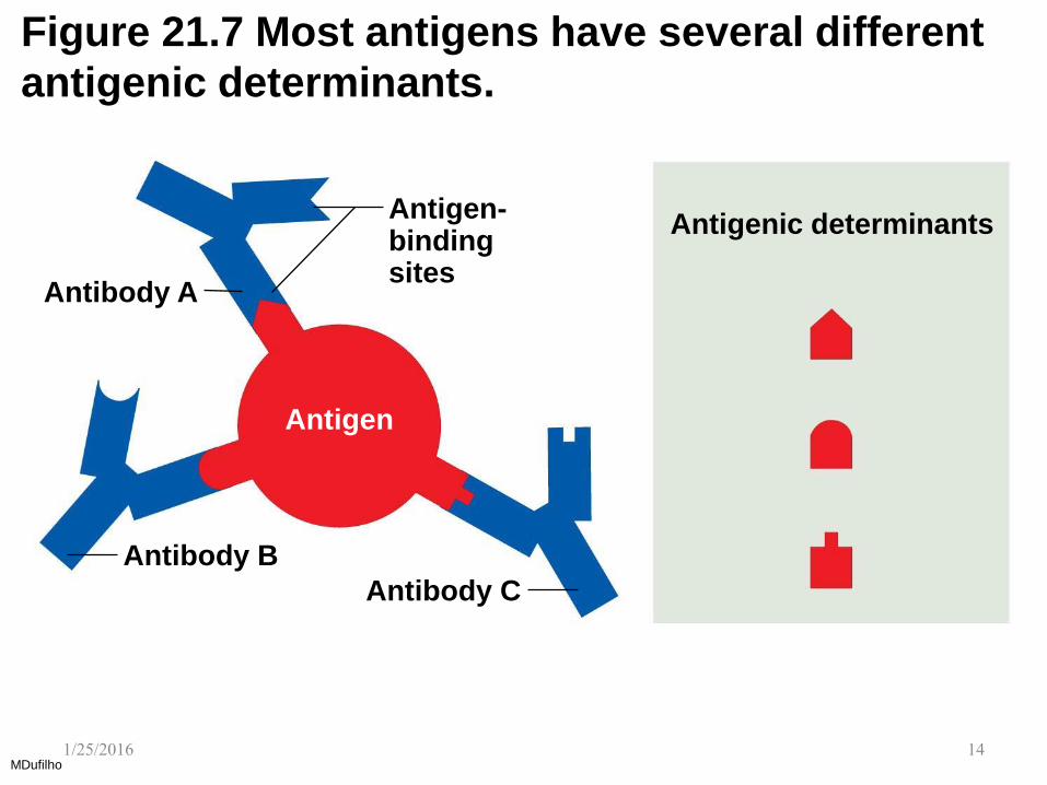
Figure 21.7 Most antigens have several different
antigenic determinants.
MDufilho
Antigenic determinants Antigen- binding sites
Antibody A
Antibody B
Antibody C
Antigen
1/25/2016 14

Self-Antigens: MHC Proteins
• Self-antigens: all cells are covered with variety
of proteins located on surface that are not
antigenic to self, but may be antigenic to others
in transfusions or grafts
• One set of important self-proteins are group of
glycoproteins called MHC proteins
– Coded by genes of major histocompatibility
complex (MHC) and unique to each individual
– Contain groove that can hold piece of self-
antigen or foreign antigen
• T lymphocytes can recognize only antigens that are
presented on MHC proteins MDufilho
1/25/2016 15

21.4 Lymphocytes and Antigen-Presenting
Cells
• Adaptive immune system involves three crucial
types of cells
– Two types of lymphocytes
• B lymphocytes (B cells)—humoral immunity
• T lymphocytes (T cells)—cellular immunity
– Antigen-presenting cells (APCs)
• Do not respond to specific antigens
• Play essential auxiliary roles in immunity
MDufilho 1/25/2016 16

Figure 21.8 Lymphocyte development, maturation, and activation.
MDufilho
1
2
3
4
5
Adaptive defenses Humoral immunity
Cellular immunity
Red bone
marrow
Lymphocyte
precursors
Thymus
Red bone marrow
Lymph node
Antigen
Primary lymphoid organs
(red bone marrow and thymus)
Secondary lymphoid organs
(lymph nodes, spleen, etc.)
Origin
Maturation
Seeding secondary lymphoid organs and
circulation
Antigen encounter and activation
Proliferation and differentiation
• Both B and T lymphocyte precursors originate in
red bone marrow.
• Lymphocyte precursors destined to become T cells
migrate (in blood) to the thymus and mature there.
• B cells mature in the bone marrow.
• During maturation lymphocytes develop
immunocompetence and self-tolerance.
• Immunocompetent but still naive lymphocytes leave
the thymus and bone marrow.
• They “seed” the secondary lymphoid organs and
circulate through blood and lymph.
• When a lymphocyte’s antigen receptors bind its
antigen, that lymphocyte can be activated.
• Activated lymphocytes proliferate (multiply) and then
differentiate into effector cells and memory cells.
• Memory cells and effector T cells circulate continuously
in the blood and lymph and throughout the secondary
lymphoid organs.
Slide 6
1/25/2016 17
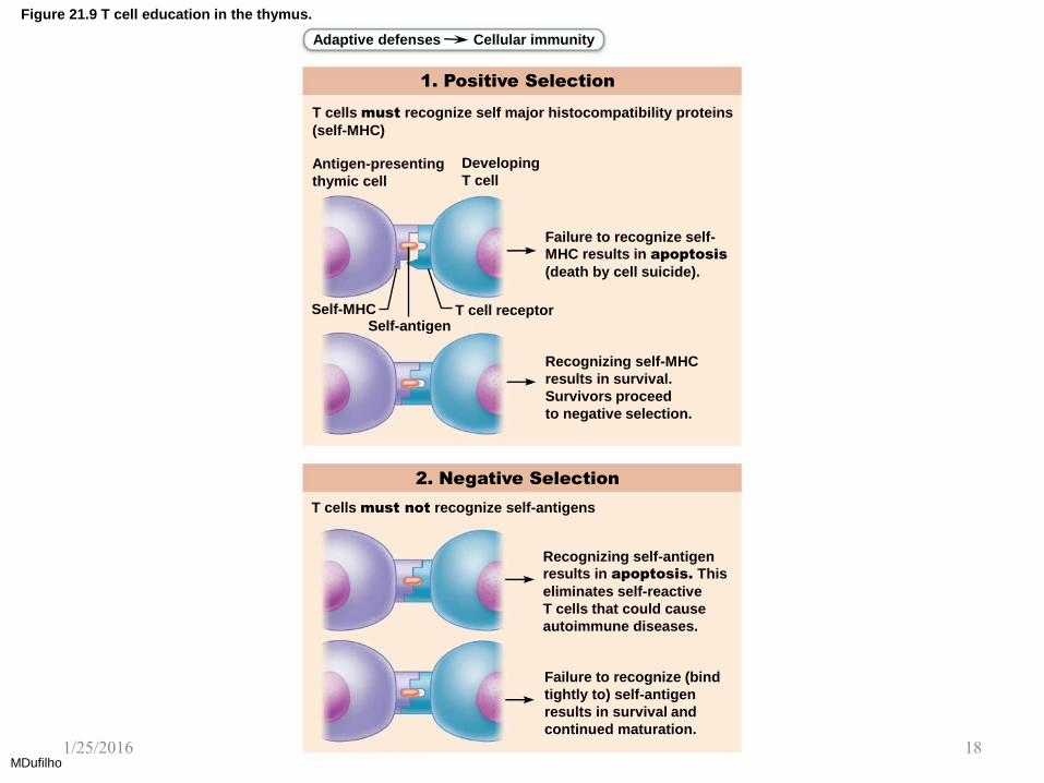
Figure 21.9 T cell education in the thymus.
MDufilho
Adaptive defenses Cellular immunity
1. Positive Selection
T cells must recognize self major histocompatibility proteins
(self-MHC)
Antigen-presenting
thymic cell
Developing
T cell
Failure to recognize self-
MHC results in apoptosis
(death by cell suicide).
T cell receptor Self-MHC Self-antigen
Recognizing self-MHC
results in survival.
Survivors proceed
to negative selection.
2. Negative Selection
T cells must not recognize self-antigens
Recognizing self-antigen
results in apoptosis. This
eliminates self-reactive
T cells that could cause
autoimmune diseases.
Failure to recognize (bind
tightly to) self-antigen
results in survival and
continued maturation.
1/25/2016 18

Lymphocytes (cont.)
• Antigen receptor diversity
– Genes, not antigens, determine which foreign
substances the immune system will recognize
• Variety of immune cell receptors are result of acquired
genetic knowledge of microbes
– ∼25,000 different genes codes for up to a billion
different types of lymphocyte antigen receptors
• Huge variety of receptors: gene segments are shuffled
around, resulting in many combinations
MDufilho 1/25/2016 19

Antigen-Presenting Cells (APCs)
• Engulf antigens and present fragments of
antigens to T cells for recognition
• Major types
– Dendritic cells in connective tissues and
epidermis
– Macrophages - widely distributed in connective
tissues and lymphoid organs
– B cells
MDufilho 1/25/2016 20
21.5 Humoral Immune Response
• When B cell encounters target antigen, it
provokes humoral immune response
– Antibodies specific for that particular antigen are
then produced
MDufilho 1/25/2016 21
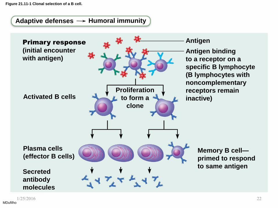
Figure 21.11-1 Clonal selection of a B cell.
MDufilho
Adaptive defenses Humoral immunity
Primary response
(initial encounter
with antigen)
Antigen
Antigen binding
to a receptor on a
specific B lymphocyte
(B lymphocytes with
noncomplementary
receptors remain
inactive)
Proliferation
to form a
clone
Activated B cells
Plasma cells
(effector B cells)
Secreted
antibody
molecules
Memory B cell—
primed to respond
to same antigen
1/25/2016 22

Figure 21.12 Primary and secondary humoral responses.
MDufilho
Primary immune
response to antigen A occurs after a delay.
Secondary immune response to antigen A is faster and larger; primary
immune response to antigen B is similar to that for antigen A.
First exposure
to antigen A
Second exposure to antigen A;
first exposure to antigen B
Time (days)
An
tib
ody titer (a
ntibody concentration)
in
p
la
sm
a (a
rb
itra
ry u
nits)
104
103
102
101
100
0 7 14 21 28 35 42 49 56
Anti-
bodies
to A
Anti-
bodies
to B
1/25/2016 23

Figure 21.11-2 Clonal selection of a B cell.
MDufilho
Memory B cell—
primed to respond
to same antigen
Secondary response
(can be years later) Clone of cells
identical to
ancestral cells
Subsequent
challenge by same
antigen results in
more rapid response
Plasma
cells
Secreted
antibody
molecules
Memory
B cells
1/25/2016 24

Figure 21.13 Active and passive humoral immunity.
MDufilho
Humoral
immunity
Active Passive
Naturally
acquired
Artificially
acquired
Naturally
acquired
Artificially
acquired
Infection;
contact with
pathogen
Vaccine; dead or attenuated pathogens
Antibodies passed from mother to fetus via placenta; or to infant in her milk
Injection of exogenous antibodies (gamma globulin)
1/25/2016 25

Antibodies
• Antibodies—also called Immunoglobulins
(Igs)—are proteins secreted by plasma cells
– Make up gamma globulin portion of blood
• Capable of binding specifically with antigen
detected by B cells
• Grouped into one of five Ig classes
MDufilho 1/25/2016 26

Figure 21.14a Antibody structure.
MDufilho
Adaptive defenses Humoral immunity
Antigen-binding site
Hinge region
Stem region
Heavy chain
variable region
Heavy chain
constant region
Light chain
variable region
Light chain
constant region
Disulfide bond 1/25/2016 27

Antibodies (cont.)
• Antibody targets and functions
– Antibodies do not destroy antigens; they
inactivate and tag them
• Form antigen-antibody (immune) complexes
– Defensive mechanisms used by antibodies
• Neutralization
• Agglutination
• Precipitation
• Complement fixation
– OYO – Learn Table 21.5 p. 789
MDufilho 1/25/2016 28

Figure 21.15 Mechanisms of antibody action.
MDufilho
Adaptive defenses Humoral immunity
Antigen Antigen-antibody
complex Antibody
Inactivates by Fixes and activates
Neutralization
(masks dangerous parts of bacterial
exotoxins; viruses)
Agglutination
(cell-bound antigens) Precipitation
(soluble antigens) Complement
Enhances Enhances Leads to
Phagocytosis Inflammation Cell lysis
Chemotaxis
Histamine
release
1/25/2016 29

Clinical – Homeostatic Imbalance 21.2
• Parasitic infections by worms such as Ascaris and
Schistosoma require different immune attack
strategies
– Worms are too big for regular PLAN attack
(Precipitation, Lysis (by complement), Agglutination,
or Neutralization)
• IgE antibodies still play a critical role in worm’s
destruction by binding to surface of worm, marking
it for destruction by eosinophils
• Eosinophils bind to exposed stems of IgE, which
triggers eosinophils to release their toxic contents
onto prey, lysing it from the outside MDufilho
1/25/2016 30

Clinical – Homeostatic Imbalance 21.2
• Monoclonal antibodies as clinical and
research tools
– Monoclonal antibodies: commercially prepared
pure antibodies that are specific for a single
antigenic determinant
– Produced by hybridomas, cell hybrids formed
from fusion of tumor cell and B cell
• Tumor cell portion allows cells to proliferate
indefinitely, while B cell portion allows production of
single type of antibody
– Used in research, clinical testing, and cancer
treatment MDufilho
1/25/2016 31
Clinical – Homeostatic Imbalance 21.2
• Summary of antibody actions
– Antigen-antibody complexes do not destroy
antigens; they prepare them for destruction by
innate defenses
– Antibodies go after extracellular pathogens; they
do not invade solid tissue unless lesion is
present
• Recent exception found: antibodies can act
intracellularly if attached to virus before it
enters cell
– Activate mechanisms that destroy virus
MDufilho 1/25/2016 32

21.6 Cellular Immune Response
• T cells provide defense against intracellular
antigens
• Some T cells directly kill cells; others release
chemicals that regulate immune response
• T cells are more complex than B cells both in
classification and function
• Two populations of T cells are based on which
cell differentiation glycoprotein receptors are
displayed on their surface
MDufilho 1/25/2016 33
21.6 Cellular Immune Response
– CD4 cells usually become helper T cells (TH)
that can activate B cells, other T cells, and
macrophages; direct adaptive immune response
• Some become regulatory T cells, which moderate
immune response
• Can also become memory T cells
– CD8 cells become cytotoxic T cells (TC) that
are capable of destroying cells harboring foreign
antigens
• Also become memory T cells
MDufilho 1/25/2016 34
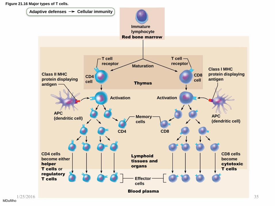
Figure 21.16 Major types of T cells.
MDufilho
Lymphoid
tissues and
organs
Thymus
Adaptive defenses Cellular immunity
Immature
lymphocyte
Class II MHC
protein displaying
antigen
CD4
cell
T cell
receptor Maturation
T cell
receptor
CD8
cell
Class I MHC
protein displaying
antigen
APC
(dendritic cell) Memory
cells
Activation Activation
APC
(dendritic cell)
CD8 cells
become cytotoxic
T cells
CD4 cells
become either helper
T cells or
regulatory
T cells Effector
cells
Blood plasma
CD8 CD4
Red bone marrow
1/25/2016 35

MHC Proteins and Antigen Presentation
(cont.)
• Two classes of MHC proteins:
– Class I MHC proteins: displayed by all cells
except RBCs
– Class II MHC proteins: displayed by APCs
(dendritic cells, macrophages, and B cells)
• Both types are synthesized in ER and bind to
peptide fragments
MDufilho 1/25/2016 36

Table 21.6 Role of MHC Proteins in Cellular Immunity
MDufilho 1/25/2016 37

Activation and Differentiation of T cells
• T cells can be activated only when antigen is
presented to them
• Activation is a two-step process
1. Antigen binding
2. Co-stimulation
• Both occur on surface of same APC
• Both are required for clonal selection of T cell
MDufilho 1/25/2016 38

Figure 21.17 Clonal selection of T cells involves simultaneous recognition of self and nonself.
MDufilho
Bacterial antigen
Dendritic
cell
Co-stimulatory
molecule receptor
CD4 T cell
T cell
receptor
(TCR)
CD4 protein
Co-stimulatory
molecule
Class lI MHC
protein
displaying
processed
bacterial antigen
Helper
T cells
Memory
CD4 T cell
Clone
formation
Adaptive defenses Cellular immunity
Dendritic cell engulfs
an exogenous
antigen, processes it,
and displays its
fragments on class II
MHC protein.
CD4 T cell
recognizes antigen-
MHC complex. Both
TCR and CD4 proteins
bind to antigen-MHC
complex.
1
2
2a
2b
3
Co-stimulatory
molecules bind their
receptors.
Clone formation
Activated CD4 T cells
proliferate (clone), and
become memory and
effector cells.
Double recognition
Antigen
presentation
Slide 4
1/25/2016 39

Activation and Differentiation of T cells
• Cytokines -Chemical messengers of immune
system
– Mediate cell development, differentiation, and
responses in immune system
– Include interferons and interleukins
• Interleukin 1 (IL-1) is released by macrophages and
stimulates T cells to release interleukin 2 (IL-2)
• IL-2 is a key growth factor, acting on same cells that
release it and other T cells to divide rapidly
– Other cytokines amplify and regulate innate and
adaptive responses
• Example: gamma interferon—enhances killing power
of macrophages
MDufilho
1/25/2016 40

21.6 Cellular Immune Response
Roles of Specific Effector T Cells
• Helper T (TH) cells
– Play central role in adaptive immune response
– Activate both humoral and cellular arms
– Once primed by APC presentation of antigen,
helper T cells:
• Help activate B cells and other T cells
• Induce T and B cell proliferation
• Secrete cytokines that recruit other immune cells
• Without TH, there is no immune response
MDufilho
1/25/2016 41

Figure 21.18 The central role of helper T cells in mobilizing both humoral and cellular immunity.
MDufilho
Adaptive defenses Humoral immunity
Cellular immunity
Helper T cells help in humoral immunity Helper T cells help in cellular immunity
Helper T cell
T cell receptor (TCR)
Helper T cell
CD4 protein
MHC II protein
of B cell displaying
processed antigen
IL-4 and other
cytokines
B cell (being activated) CD8 T cell
(becomes TC cell
after activation)
Class I
MHC protein
CD8
protein
APC (dendritic
cell)
Class II MHC
protein
CD4 protein Helper T cell
IL-2
1
2
1
2
3
TH cell binds with the self-nonself complexes of a B cell that has encountered
its antigen and is
displaying it on
MHC II on its surface.
TH cell releases
interleukins as co-
stimulatory signals to
complete B cell
activation.
TH cell binds
dendritic cell.
TH cell
stimulates dendritic
cell to express
co-stimulatory
molecules.
Dendritic cell
can now activate
CD8 cell with the
help of interleukin 2
secreted by TH cell.
1/25/2016 42
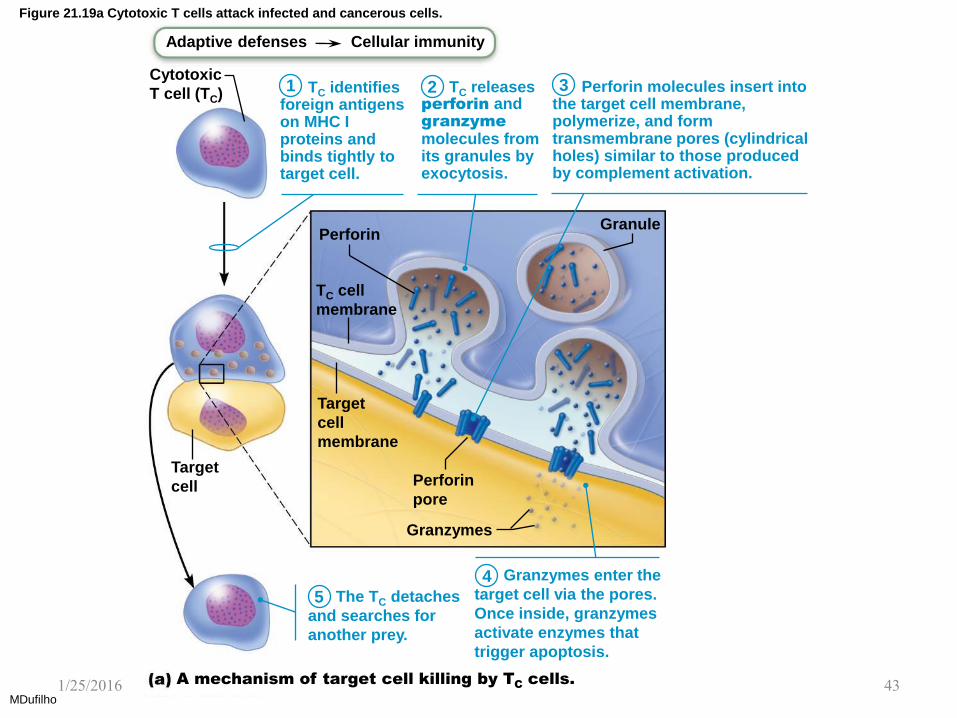
Figure 21.19a Cytotoxic T cells attack infected and cancerous cells.
MDufilho
Adaptive defenses Cellular immunity
Cytotoxic
T cell (TC)
Perforin Granule
TC cell
membrane
Target
cell
membrane
Perforin
pore
Target
cell
Granzymes
A mechanism of target cell killing by TC cells.
1 2 3
5
4
TC identifies foreign antigens on MHC I proteins and binds tightly to target cell.
TC releases perforin and granzyme
molecules from its granules by exocytosis.
Perforin molecules insert into the target cell membrane, polymerize, and form transmembrane pores (cylindrical holes) similar to those produced by complement activation.
The TC detaches
and searches for
another prey.
Granzymes enter the
target cell via the pores.
Once inside, granzymes
activate enzymes that
trigger apoptosis.
1/25/2016 43

Roles of Specific Effector T Cells (cont.)
– Subsets of TH cells
• TH1—mediates most aspects of cellular immunity
• TH2—defends against parasitic worms, mobilizes
eosinophils; activates responses dependent on
humoral immunity; promotes allergies
• TH17—links adaptive and innate immunity by
releasing IL-17
– May play role in autoimmune disease
MDufilho 1/25/2016 44
Roles of Specific Effector T Cells (cont.)
• Cytotoxic T (TC) cells (cont.)
– Natural killer cells recognize other signs of
abnormality that cytotoxic T cells do not look for,
such as:
• Cells that lack class I MHC proteins
• Antibodies coating target cell
• Different surface markers seen on stressed cells
– NK cells use same key mechanisms as TC cells
for killing their target cells
– Immune surveillance: NK and TC cells prowl
body looking for markers they each recognize
MDufilho 1/25/2016 45

Roles of Specific Effector T Cells (cont.)
• Regulatory T (TReg) cells
– Dampen immune response by direct contact or
by secreting inhibitory cytokines such as IL-10
and transforming growth factor beta (TGF-)
– Important in preventing autoimmune reactions
• Suppress self-reactive lymphocytes in periphery
(outside lymphoid organs)
• Research into using them to induce tolerance to
transplanted tissue
MDufilho 1/25/2016 46
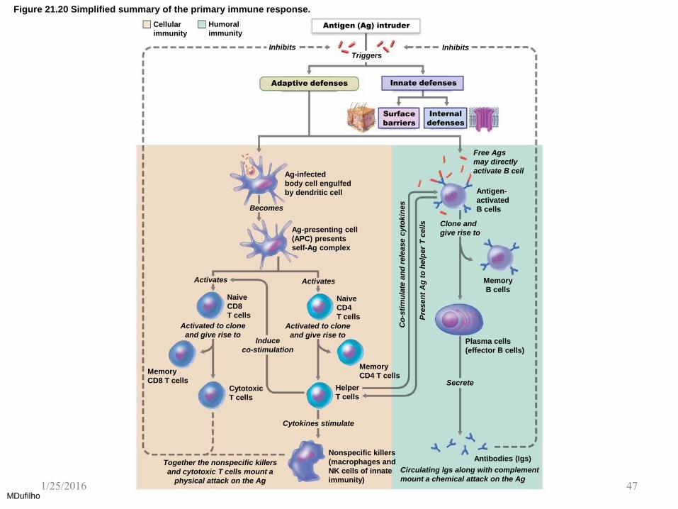
Figure 21.20 Simplified summary of the primary immune response.
MDufilho
Cellular
immunity
Humoral
immunity Antigen (Ag) intruder
Inhibits Triggers
Inhibits
Adaptive defenses Innate defenses
Surface
barriers
Internal
defenses
Free Ags
may directly
activate B cell
Antigen-
activated
B cells
Clone and
give rise to
Memory
B cells
Plasma cells
(effector B cells)
Nonspecific killers
(macrophages and
NK cells of innate
immunity)
Antibodies (Igs)
Helper
T cells
Memory
CD4 T cells
Cytotoxic
T cells
Memory
CD8 T cells
Naive
CD4
T cells
Naive
CD8
T cells
Ag-presenting cell
(APC) presents
self-Ag complex
Ag-infected
body cell engulfed
by dendritic cell
Secrete
Cytokines stimulate
Together the nonspecific killers
and cytotoxic T cells mount a
physical attack on the Ag
Circulating lgs along with complement
mount a chemical attack on the Ag
Induce
co-stimulation
Activated to clone
and give rise to
Activates
Activated to clone
and give rise to
Co
-sti
mu
late
an
d r
ele
ase c
yto
kin
es
Pre
sen
t A
g t
o h
elp
er
T c
ells
Becomes
Activates
1/25/2016 47
Organ Transplants and Prevention of
Rejection
• Most common type of organ transplant is an
allograft: transplant from same species
• Success depends on similarity of tissues
– ABO, other blood antigens, and MHC antigens
are matched as closely as possible
• After surgery
– Patient treated with immunosuppressive therapy
– Many of these therapies have severe side effects
MDufilho 1/25/2016 48
12.7 Immune Problems
Immunodeficiencies
• Congenital or acquired conditions that impair
function or production of immune cells or molecules
• Severe combined immunodeficiency (SCID)
syndrome: genetic defect with marked deficit in B
and T cells
– Defective adenosine deaminase (ADA) enzyme
allows accumulation of metabolites lethal to T
cells; fatal if untreated
• Hodgkin’s disease is an acquired
immunodeficiency that causes cancer of B cells,
which depresses lymph node cells and thus leads
to immunodeficiency
MDufilho
1/25/2016 49
Immunodeficiencies (cont.)
• Acquired immune deficiency syndrome
(AIDS) caused by Human immunodeficiency
virus (HIV)
– cripples immune system by interfering with
activity of helper T cells
• HIV is transmitted via body fluids: blood, semen,
and vaginal secretions
• HIV can enter the body via:
– Blood transfusions; blood-contaminated needles;
sexual intercourse and oral sex; mother to fetus
• HIV destroys TH cells, thereby depressing
cellular immunity
MDufilho
1/25/2016 50

Autoimmune Diseases
• Autoimmune disease results when immune
system loses ability to distinguish self from
foreign
• Autoimmunity: production of autoantibodies
and sensitized TC cells that destroys body
tissues
• Examples
– Rheumatoid arthritis: destroys joints
– Myasthenia gravis: impairs nerve-muscle connections
– Multiple sclerosis: destroys white matter myelin
– Graves’ disease: causes hyperthyroidism
– Type 1 diabetes mellitus: destroys pancreatic cells
– Systemic lupus erythematosus (SLE): affects multiple organs
– Glomerulonephritis: damages kidney
MDufilho
1/25/2016 51
Hypersensitivities
• Hypersensitivities: immune responses to
perceived (otherwise harmless) threat that
cause tissue damage
• Different types are distinguished by:
1. Their time course
2. Whether antibodies or T cells are involved
• Antibodies cause immediate and subacute
hypersensitivities
• T cells cause delayed hypersensitivity
MDufilho 1/25/2016 52
Hypersensitivities (cont.)
• Immediate hypersensitivity
– Also called acute (type I) hypersensitivities
(allergies); begin in seconds after contact with
allergen, antigen that causes allergic reaction
– Initial contact with allergen is asymptomatic but
sensitizes person
– Activated IgE against antigen binds to mast cells
and basophils
– Later encounter with same allergen causes flood
of histamine release from IgEs, resulting in
induced inflammatory response
MDufilho 1/25/2016 53

Figure 21.21 Mechanism of an acute allergic (immediate hypersensitivity) response.
MDufilho
Adaptive defenses Humoral immunity
Sensitization stage
Antigen
Mast cell with fixed
IgE antibodies
IgE
Granules containing
histamine
Subsequent (secondary)
responses
Mast cell granules
release contents
after antigen binds
with IgE antibodies
Histamine
Outpouring of fluid
from capillaries
Release of
mucus
Constriction of
small respiratory
passages (bronchioles)
1
2
3
4
5
6
Antigen (allergen)
invades body.
Plasma cells produce
large amounts of class
IgE antibodies against
allergen.
IgE antibodies attach
to mast cells in body
tissues (and to circulating
basophils).
More of same antigen
invades body.
Antigen combines with
IgE attached to mast cells
(and basophils), which triggers
degranulation and release
of histamine (and other
chemicals).
Histamine causes blood vessels to dilate and become leaky, which
promotes edema; stimulates secretion of large amounts of mucus; and
causes smooth muscles to contract. (If respiratory system is site of
antigen entry, asthma may ensue.)
Slide 7
1/25/2016 54

Hypersensitivities (cont.)
• Subacute hypersensitivities
– Caused by IgM and IgG transferred via blood
plasma or serum
– Slow onset (1–3 hours) and long duration
(10–15 hours)
– Cytotoxic (type II) reactions
• Antibodies bind to antigens on specific body cells,
stimulate phagocytosis and complement-mediated
lysis of cellular antigens
• Example: mismatched blood transfusion reaction
MDufilho 1/25/2016 55

Hypersensitivities (cont.)
• Subacute hypersensitivities (cont.)
– Immune complex (type III) hypersensitivity
• Antigens widely distributed in body or blood
• Insoluble antigen-antibody complexes form
• Complexes cannot be cleared from particular area of
body
• Intense inflammation, local cell lysis, and cell killing by
neutrophils
• Example: systemic lupus erythematosus (SLE)
MDufilho 1/25/2016 56

Hypersensitivities (cont.)
• Delayed hypersensitivities (type IV)
– Slow onset (1–3 days)
– Mechanism depends on helper T cells
– Cytokine-activated macrophages and cytotoxic T
cells cause damage
– Example: allergic contact dermatitis (e.g., poison
ivy)
– Agents act as haptens
– TB skin test depends on this reaction
MDufilho 1/25/2016 57

Developmental Aspects of Immune System
• Immune system stem cells develop in liver and
spleen in weeks 1–9
• Bone marrow becomes primary source of stem
cells later and through adult life
• Lymphocyte development continues in bone
marrow and thymus
• TH2 lymphocytes predominate in newborn; TH1
system educated as person encounters
antigens
MDufilho 1/25/2016 58

Developmental Aspects of Immune System
• Influences on immune system function
– Nervous system: depression, emotional stress,
and grief impair immune response
– Diet: vitamin D is required for activation of CD8
cells TC cells
MDufilho 1/25/2016 59

Developmental Aspects of Immune System
• With age, immune system begins to wane
– Greater susceptibility to immunodeficiency and
autoimmune diseases
– Greater incidence of cancer
– Why immune system fails is unknown, but may
be due to atrophy of thymus and decreased
production of naive T and B cells
MDufilho 1/25/2016 60