Embed Size (px)
Citation preview
THE HISTOLOGICAL DIFFERENTIATION OF
TUBERCULOSIS FROM CROHN'S DISEASE IN
MUCOSAL BIOPSIES FROM THE UPPER
GASTROINTESTINAL TRACT WITH
GRANULOMATOUS INFLAMMATION
A Dissertation submitted in part fulfilment of the rules and
regulations for the M.D. Degree Branch III (Pathology) Examinations
of the Tamil Nadu Dr.M.G.R.Medical University, Chennai to be held
in April 2017
CERTIFICATE
This is to certify that the following dissertation bearing the title ‘The histological
differentiation of Tuberculosis from Crohn's disease in mucosal biopsies from the
upper gastrointestinal tract with granulomatous inflammation’ is a bonafide work
done by Dr. Sangeetha Ramakrishna Pillai in partial fulfilment of the rules and
regulations for M.D. Branch III (Pathology) degree examination of the Tamil Nadu
Dr. M.G. R. Medical University to be held in April 2017.
Dr. Vivi M. Srivastava, MBBS, MD
Professor and Head of Department,
Department of General Pathology
Christian Medical College,
Vellore, Tamil Nadu,
India
Dr. Anna B. Pulimood, MD, PhD
Principal,
Christian Medical College,
Vellore, Tamil Nadu,
India.

CERTIFICATE
This is to certify that the following dissertation bearing the title ‘The histological
differentiation of Tuberculosis from Crohn's disease in mucosal biopsies from the
upper gastrointestinal tract with granulomatous inflammation’ is a bonafide work
done by Dr. Sangeetha Ramakrishna Pillai in partial fulfilment of the rules and
regulations for M.D. Branch III (Pathology) degree examination of the Tamil Nadu
Dr. M.G. R. Medical University, to be held in April 2017.
The candidate has independently reviewed the literature, standardised the data
collection, methodology and carried out the evaluation toward the completion of
the thesis.
Dr. Anna B. Pulimood, MD, PhD
Professor and Guide,
Christian Medical College,
Vellore, Tamil Nadu,
India
CERTIFICATE
This is to certify that the following dissertation bearing the title ‘The histological
differentiation of Tuberculosis from Crohn's disease in mucosal biopsies from the
upper gastrointestinal tract with granulomatous inflammation’ is a bonafide work
done by me under the guidance of Dr. Anna B. Pulimood in partial fulfilment of
the rules and regulations for M.D. Branch III (Pathology) degree examination of
the Tamil Nadu Dr. M.G. R. Medical University to be held in April 2017.
I have independently reviewed the literature, standardised the data collection,
methodology and carried out the evaluation toward the completion of the thesis.
Dr. Sangeetha Ramakrishna Pillai
Postgraduate Registrar,
Department of General Pathology,
Christian Medical College,
Vellore, Tamil Nadu,
India.
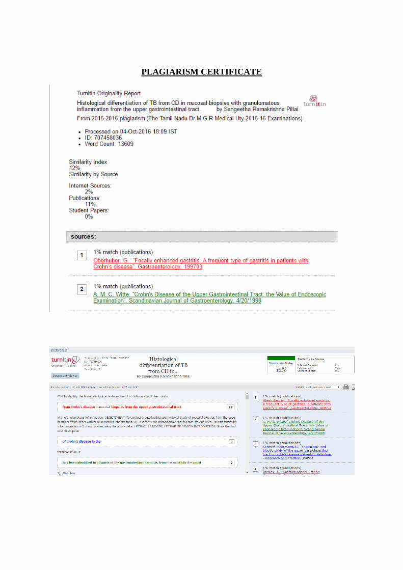
PLAGIARISM CERTIFICATE
ACKNOWLEDGEMENT
I express my heartfelt gratitude to Dr. Anna B. Pulimood, my guide and mentor for
her constant support and timely guidance towards the successful completion of this
dissertation.
I would like to thank God Almighty, my parents, my brother and all my fellow batch
mates for keeping me motivated throughout this journey.
I also wish to acknowledge Mr. Srinivasan and Mr. B. Vijayakumar for their help in
retrieval and staining of slides.
ABBREVIATIONS
TB Tuberculosis
CD Crohn’s disease
GIT Gastrointestinal tract
IEL Intraepithelial lymphocytes
AFB Acid fast bacilli
Contents
AIM ........................................................................................ 1
OBJECTIVES ........................................................................ 2
LITERATURE REVIEW ....................................................... 3
MATERIALS AND METHODS .......................................... 30
RESULTS ............................................................................. 35
IMAGES ............................................................................... 67
DISCUSSION ...................................................................... 81
CONCLUSIONS .................................................................. 87
LIMITATIONS .................................................................... 89
REFERENCES ..................................................................... 90
1
AIM
To identify the histopathological features useful in distinguishing tuberculosis from
Crohn’s disease in mucosal biopsies from the upper gastrointestinal tract with
granulomatous inflammation.
2
OBJECTIVES
A] To conduct a detailed histopathological study of mucosal biopsies from the
upper gastrointestinal tract with granulomatous inflammation.
B] To identify the pathological features that may be useful in differentiating
tuberculosis from Crohn’s disease using the above data.
LITERATURE REVIEW

3
LITERATURE REVIEW
INTRODUCTION
Since the first ever description of Crohn's disease in the terminal ileum, it has
been identified in all parts of the gastrointestinal tract i.e. from the mouth to
the anus(1) . Until few years ago, involvement of the upper gastrointestinal
tract was thought to occur only in 1-5% of Crohn's disease patients. However,
because of the more frequent use of upper gastrointestinal endoscopy, recent
reports show substantially higher incidence rates. Cameron et al demonstrated
an incidence of 71% in a prospective study involving the paediatric
population(2) and in a retrospective study, Laenart et al reported an incidence
of 30%(3). A high incidence of upper gastrointestinal involvement in an adult
population, of up to 60%, was described by Schmitz-Moormann et al(4).
Crohn's disease of the upper gastrointestinal tract generally occurs in
conjunction with involvement of the lower gastrointestinal tract. General use
of colonoscopy in recent years has led to awareness of endoscopic and
histological alterations of the lower gastrointestinal tract in Crohn's disease. In
contrast, systematic detailed studies of histological changes in the upper
gastrointestinal tract have scarcely been reported (4).
Tuberculosis involving the upper gastrointestinal tract is a rare disease and
the gastrointestinal tract is only the sixth most common site of extra
pulmonary tuberculosis. However in developing countries like India, the

4
menace of tuberculosis has never been controlled whereas in industrialized
countries, the incidence of tuberculosis has started to rise again owing to the
increased incidence of HIV/AIDS. This highlights the importance of a
comprehensive assessment of the histological changes noted in Tuberculosis
of the upper gastrointestinal tract (5).
Tuberculosis and Crohn’s disease are both granulomatous diseases of the
gastrointestinal tract with numerous overlapping features. Distinguishing
between the two entities is of paramount importance, as an incorrect
diagnosis and thereby inappropriate management is associated with increased
mortality and morbidity.
CLINICAL FEATURES
Crohn's disease
Esophagus: The esophagus is considered to be one amongst the least common
segments of the gastrointestinal tract involved in Crohn's disease and isolated
involvement is rare(1,6,7). The first ever case of esophageal CD was
described by Frank and Taylor in 1950 (8). However, even 65 years later its
true prevalence is still elusive. The prevalence of esophageal Crohn's disease
has been described to range from 1-2% in adults to upto 40% in children and
adolescents (9–12).
5
The average age at presentation is reported to be around 40 years (6), though
studies show an increasing incidence in children and adolescents (11). There
is no documented sex difference (6).
Patients with esophageal CD in the early stages may be asymptomatic.
Odynophagia, dysphagia and sometimes severe weight loss are the
presenting symptoms in patients with advanced esophageal CD and amongst
these dysphagia is the most common presenting complaint(1,6,13). Rarely,
fistulisation and bleeding may occur and when present, they generally
indicate a more threatening aspect of the disease (10).
Gastro-duodenum: The entity of gastric CD was first described by Ross et al in
1949(14). Thereafter, the prevalence of gastroduodenal disease has been reported to
range from 5-60% in adults(9). Gastroduodenal CD has also been reported to be more
common in the paediatric population than adults(15). The antrum is most commonly
involved and duodenal CD is usually associated with antral involvement (6).
Patients with gastroduodenal CD are generally asymptomatic. Epigastric abdominal
pain is the most common symptom in gastroduodenal CD. Loss of weight, general
malaise, vomiting and diarrhoea are other common symptoms(16,17). Wagtman et al
reported that the only symptoms that statistically differ between patients with upper
gastrointestinal CD and patients with distal CD are abdominal pain and malaise(18).
In more advanced diseases, particularly when obstruction has developed, epigastric

6
distress, early satiety, nausea, vomiting and weight loss are the predominant
symptoms (1,9,19).
Complications such as hematemesis or melena due to acute or chronic blood loss
from the upper GIT are rare (1).
Tuberculosis
Esophagus: Most cases of esophageal tuberculosis which have been reported are
secondary to tuberculosis elsewhere, commonly pulmonary tuberculosis. The
prevalence of esophageal tuberculosis in a large autopsy series was only 0.15% (5).
Primary esophageal tuberculosis, without evidence of tuberculosis elsewhere, is
considered to be a rarity worldwide (20), however, it may not be so uncommon in
developing countries like India.
The esophagus is considered to be intrinsically protected from TB by the following
features – the stratified squamous epithelial lining, mucosal coating by saliva and
mucous, tubular structure and rapid peristaltic transit of swallowed material that
prevents stasis and mucosal invasion(21).
The middle one third of the esophagus at the level of the carina is the most
commonly involved site in esophageal tuberculosis. This is usually caused by
spread of infection from tuberculous mediastinal lymph nodes by way of a draining

7
fistula (5,22). On rare occasions the proximal third of the esophagus can be
involved by direct extension from tubercular laryngitis or pharyngitis(5).
The most common presenting symptom in esophageal tuberculosis is dysphagia
which occurs in 90% of the cases (5,20,22). Dysphagia can be due to intrinsic
tuberculous involvement of the esophagus or due to esophageal compression by
mediastinal tuberculous lymph nodes (5,20). Other symptoms include odynophagia,
retrosternal pain, fever and weight loss (5,22,23).
Complications include esophageal strictures, stenosis, fistulae and bleeding. The
strictures may result from esophageal compression by mediastinal lymph nodes.
Esophagotracheal and esophagomediastinal fistula are frequently described in
esophageal tuberculosis (21).
Massive bleeding has been reported to occur due to deep penetration of the
tuberculous ulcer into the aorta with resultant esophago-aortic fistula (24).
Stomach: Gastric tuberculosis most commonly occurs secondary to a pre-existing
pulmonary infection and cases of isolated and primary gastric TB without evidence
of lesions elsewhere have rarely been reported (25). The reported incidence of
primary gastric TB is <0.02% on routine gastric biopsies. However, the incidence
increases dramatically to 4.5% in individuals with moderate pulmonary TB and up to
25% in those with severe disease (25).
8
Primary and isolated gastric tuberculosis is considered to be rare due to the surface
integrity of the gastric mucosa, local immunity of the stomach wall, rapid emptying
of stomach contents, bactericidal properties of gastric acid and paucity of lymphoid
tissue in the gastric wall (26–28). Possible routes of infection include direct invasion
through the mucosa, haematogenous spread, lymphatic spread or contiguous spread
through the serosa (29).
Gastric tuberculosis is seen more frequently in males as compared to females (29).
Also, it is three to four times more common in adults than in children (29).The age of
onset is 20-40 years of age.
Tuberculosis most commonly occurs in the antrum and lesser curvature of the
stomach.
The clinical manifestations of gastric TB are varied and nonspecific. Subjective
complaints like intermittent epigastric discomfort, weight loss, hematemesis and
features of gastric outlet obstruction may be present (30,31). However, gastric outlet
obstruction as the primary manifestation is rare (32). Systemic manifestations such as
low grade fever, malaise and weight loss can also be present.
As gastric tuberculous ulcers rarely penetrate the muscle layer, perforation is rare (31).

9
Duodenum: The prevalence of duodenal TB ranges from 2%-16.5% based on different
studies (33,34). Duodenal tuberculosis generally occurs secondary to pulmonary
involvement (32) and duodenum is considered to be the fourth most common site of
GI tuberculosis involvement (23). The most common site of involvement is the
proximal duodenum.
Duodenal tuberculous involvement can be either extrinsic or intrinsic(33,35). The
extrinsic type is more common than the intrinsic type and is considered to occur due
to periduodenal lymph node compression (33). Intrinsic lesions could be ulcerative,
hypertrophic or ulcero-hypertrophic (33,35).
The clinical presentation of duodenal TB is classically divided into obstructive and
dyspeptic (33). Gupta et al noted that 73% of patients presented with features of
gastric outlet obstruction and 27% had dyspeptic symptoms (36). Chavhan et al also
has described a predominance of obstructive symptoms (37). Patients with extrinsic
involvement and the hypertrophic type of intrinsic involvement generally present with
obstructive symptoms and those with the ulcerative type of lesion present with
dyspeptic symptoms (33). Most of the times the obstruction is the result of both,
extrinsic compression by the enlarged lymph nodes and the narrowing caused by
intrinsic involvement acting together (37).
The most common presenting complaints in duodenal TB are epigastric pain and
vomiting (38). Complications like fistulae, perforation and haemorrhage have been
10
reported in various case reports (33). A case report of acute perforation of
tuberculous duodenal segment has been reported by Berney et al (39). Infrequently, a
fistulous communication can occur between the duodenum and renal pelvis or bile
duct (38).
RADIOLOGY
Crohn’s disease
Esophagus: Radiological findings are generally less informative in Crohn’s disease,
however, thickened mucosal folds or cobblestones and irregular asymmetric mucosal
defects such as aphthous or deep ulcers are the few of the common radiological
features of esophageal CD(1,12). A fibrotic stenosis may develop in advanced cases
(1,9). Spontaneous fistulae can also develop because of the transmural nature of CD
(1).
Gastro-duodenum: Aphthous ulcers are the earliest detectable radiologic signs and
are demonstrated by the double contrast technique. Other common radiologic signs
include thickened folds, cobble stoning and ulcerations (1,19,40). Stricture formation
can give rise to some specific radiologic features: tubular narrowing, a 'ram horn's
sign' or a pseudo Billroth I configuration (when the stomach and duodenum are both
involved) (19,41).
11
Tuberculosis
Esophagus: CT of the thorax is recommended in cases of suspected esophageal TB,
not only to detect the secondary nature of the disease (via mediastinal lymph nodes)
but also to detect extra luminal complications like perforation and
pneumomediastinum (21).
Stomach: The radiological appearances in gastric tuberculosis are predominantly
ulcerative and hyperplastic (31). Thickening of the gastric wall, with associated
enlarged lymph nodes, often visible via CT, may sometimes be the only clue to the
diagnosis (38).
Duodenum: Duodenal TB lacks specific radiological features however, barium studies
help in the localization and recognition of the site and extent of narrowing and
ulcerations (33,37). The radiological findings noted in duodenal TB with an
obstructive presentation are luminal narrowing of varying degrees and a sharp, band
like cut off of the duodenum at the D2-D3 junction. In patients with the dyspeptic
presentation, the radiological findings include luminal narrowing at the second and
third part of the duodenum, ulcerations in the second to fourth parts and extrinsic
compressions at the third to fourth parts of duodenum (37). Other findings include
scarred and deformed duodenal cap and widening of the C loop of the duodenum
(33,37). Occasionally, duodenal TB may present as polypoidal intraluminal soft tissue
12
mass (33). CT may show thickening of the duodenal wall with lymph node
enlargement (42).
ENDOSCOPY
Crohn's disease
Esophagus: The morphologic changes in Crohn's disease is mainly noted in the lower
third of the esophagus (1,6). There are no pathognomic endoscopic features for
esophageal Crohn’s disease(12). One of the common endoscopic changes, however,
are the nonspecific aphthoid ulcerations which are small discrete ulcers surrounded by
a red halo of edematous tissue(6,12,43). Multiple aphthoid ulcerations with
intervening normal mucosa gives rise to a 'cobblestone appearance'(6,12). Other
endoscopic findings include mucosal erythema, slight friability of mucosa,
granularity, nodular thickening and mucosal defects such as erosions and deep
ulcerations (1,6,44,45). Marked fibrosis leading to stenosing lesions has been reported
to be one of the complications of esophageal CD(6). Huchzermeyer described two
stages in esophageal CD: Stage I, wherein inflammatory changes in the form of a mild
or more frequently erosive-ulcerative esophagitis are noted, and Stage II, wherein a
stenosing form is observed (46).
Stomach: Abdullah et al reported the incidence of endoscopic abnormalities in
gastric CD to be about 64%(47).The mucosal lesions are most commonly seen in the
13
antro-pyloric region (1,4,6,19,48).The most frequent gastric mucosal lesions in CD
are mucosal redness, mucosal edema(frequently with curtain folds), linear and
serpiginous ulcerations and aphthous ulcers along with acute and chronic
erosions(4,48). Other lesions usually noted are nodularity, granularity, cobble
stoning and thickened mucosal folds (1,4,6,19,48,49). Amongst these, superficial
ulcerations and aphthous ulcers have been described as probably the earliest
recognisable manifestations of gastric CD. In gastric CD, the ulcers and erosions are
generally longitudinal or serpiginous. A feature that helps to differentiate them from
peptic ulcers(50).
Schmitz-Moormann et al and Gad et al demonstrated that chronic erosions have a
significant correlation with the presence of granulomas. This indicates that only
chronic erosions constitute a characteristic endoscopic lesion for gastric CD, whereas
other generally cited lesions do not reveal such specificity (4,51). Occasionally, a
linitis plastica appearance with luminal narrowing and rigidity may be noted and is
suggestive of diffuse gastric involvement (19). Obstruction in the pyloric region due
to scarring is also seen to occur in majority of gastroduodenal Crohn’s disease(18).
It must be remembered that normal endoscopic findings, however, does not rule out
the histological presence of focal gastritis(48). Oberhuber et al, noted that 25% of
patients with Crohn's disease who had normal endoscopic findings showed
histological evidence of focal gastritis (26).

14
Duodenum: Mucosal redness, mucosal edema and aphthous lesions are the most
frequent endoscopic lesions noted in duodenal CD. Other lesions noted are
granularity, nodular mucosal lesions, ulcers, stenosis and insufficient distensibility.
However, only three lesions namely mucosal redness, ulcers and stenosis correlate
significantly with the presence of granulomas. These lesions resemble the
endoscopic features of CD in the lower gastrointestinal tract (4).
Tuberculosis
Esophagus: Three distinct morphological mucosal lesions are noted in
esophageal tuberculosis - ulcerative, hypertrophic or granular.
Ulcerative lesion - The most common macroscopic finding is a midesophageal linear
ulceration with smooth edges and a membranous necrotic base. The ulcers are
generally multiple, fairly large in size (24). The surrounding mucosa appears normal
with or without evidence of stricture. A similar endoscopic picture is not usually seen
with any other condition and therefore in patients with appropriate histories and
distinctive midesophageal ulcerative lesions, the diagnosis of esophageal tuberculosis
should be considered (5,20,24).
The mycobacteria bacilli initially involve the esophageal submucosa
followed by the formation of tubercles. With disease progression, caseous
necrosis occurs within the nodules, eventually leading to ulceration (52).

15
Generally, the tuberculous ulcers are superficial. The more serious ulcers which
rarely occur, often can penetrate the muscularis layer and breakthrough the adventitia
resulting in perforation and fistulae. Such deep ulcers also have been described to
bleed easily (52).
Hyperplastic lesion – This lesion occurs due to excessive amount of tuberculous
inflammatory granulation tissue and fibrous hyperplasia. Occasionally massive
hyperplasia can result in a false tumour like mass (pseudo tumour) formation in the
esophageal lumen and cause luminal narrowing.
Granular lesion – This is the least common lesion and is seen in severe systemic
disease wherein the mucosa and submucosa show many gray-white nodules
(52).
Stomach: In gastric TB, the gross lesions may be of 4 types, as described by
Broders– ulcerative, hypertrophic, tumour type or inflammatory
type(25,27,53,54).
Ulcerative lesion - The most common lesion is the ulcerative lesion (80%) which is
usually seen along the lesser curvature (27,53,54). Typically the ulcer has a
serpiginous outline. The ulcer also has characteristic undermined edges which are not
seen in any other gastric disease. The undermined edges are typically ragged,

16
edematous and grossly inflamed. Discrete tubercles are generally present at the edge
of the ulcer. The granulomatous base is characteristically dirty gray brown and may
be covered with discernible tubercles(54).
The ulcer is believed to develop as a result of fistula formation from a submucosal
focus. Small submucosal tubercles undergo caseation and suppuration and produce
multiple fistulae through the mucosa into the gastric lumen. Ulcers further develop
by the numerous irregular ruptures of the mucosa, giving rise to the serpiginous
outline of the ulcer (54).
Hypertrophic lesion - The hypertrophic lesion is seen more frequently in the antrum
and is characterized by submucosal infiltration. The infiltration may spread through
the wall resulting in a linitis plastica like appearance. Cicatrisation with gastric
obstruction is a common sequelae. The mucosal surface generally remains intact in
an early lesion. However, as a consequence of the mass of the lesion and
mechanical interference with the action of the muscularis mucosae, the gastric
rugae get obliterated.
Tumour type lesion - The main characteristic of this lesion is the discrete nature of
the mass. This lesion tends to caseate early and is associated with liquefaction,
fistulisation and ulceration.
Superficial tubercles are common in the neighbourhood of all tuberculous lesions(54).

17
Duodenum: .The macroscopic lesions noted in duodenal TB are described to be
ulcerative, hyperplastic or ulcerohypertrophic(31,39,42) . Ulcerative lesions are the
most common findings and accounts for 60%. The ulcers are generally
circumferential and surrounded by inflamed mucosa. Ulcerohypertrophic accounts
for 30% and show and inflammatory mass with a thickened and ulcerated mucosa.
The hypertrophic lesion is the least common lesion (10%) and is characterized by
scar, fibrosis and pseudo tumour formation (42).
HISTOPATHOLOGY
Crohn's disease
In the diagnosis of CD in the upper gastrointestinal tract, histopathological findings
are of much greater predictive significance than endoscopic findings (4). Many
investigators have used the criteria proposed by Nugent and Roy for the definition of
Crohn's disease of the upper GI tract:-
i. Non caseating granulomatous inflammation on endoscopic biopsies in the
upper gastrointestinal tract, with or without Crohn's disease elsewhere in
the intestinal tract, and without evidence of a systemic granulomatous
disorder.

18
ii. Crohn's disease of the small or large intestine and radiological and/or
endoscopic findings of diffuse inflammatory change in the upper
gastrointestinal tract consistent with Crohn's disease (1).
Esophagus: Isolated involvement of the esophagus is rare in Crohn's disease.
Esophageal CD is generally associated with advanced ileo-colonic disease (11). At
the outset, it is important to note that the transmural nature of CD cannot be
accurately assessed on superficial mucosal biopsies and moreover there are no
pathognomic histological features for esophageal CD (12,55).
Nevertheless, esophageal mucosal biopsies on histological evaluation usually
demonstrate focal inflammation and infrequently granulomas (4).
A lymphocytic infiltrate within the lamina propria is one of the most consistent
findings in esophageal CD(12). Focal infiltration with mononuclear cells and
histiocytes into the lamina propria extending up to the muscular layer is considered
to be histologic evidence of esophageal CD(11).
Non caseating epithelioid granulomas, are generally uncommon, but when seen, are
considered to be diagnostic for esophageal CD (1,6,11,12,56). Esophageal
granulomas are situated generally in the submucosa, at a depth, which is only barely
reached by regular esophageal mucosal biopsies. It has been postulated that with
additional sectioning, the detection rate of granulomas can be increased (4).
19
Additionally, Rubio et al observed that 40% of cases with lymphocytic esophagitis
were diagnosed to have Crohn's disease (57)whereas Purdy et al observed only 12%
to have CD (58). The main criteria for diagnosing lymphocytic esophagitis is the
occurrence of high number of intraepithelial lymphocytes (IEL) and not more than
rare granulocytes (57,58). Both the peripapillary and inter papillary areas must be
examined to assess the severity of IELs. Peripapillary is defined as the first five
layers of squamous epithelium surrounding the esophageal papillae. The lymphocytic
esophagitis was graded mild if it predominantly involved the peripapillary area and
severe if it involved both the interpapillary and peripapillary areas (58).
Another important feature is the presence of intraepithelial lymphocytes with
irregular nuclear contours (11,56). The peripapillary areas are usually rich in round
IELs and the interpapillary regions are rich in IELs with irregular nuclear contours
(57). It has been postulated that these irregular lymphocytes mirror cell
deformation brought about by emperipolesis, which is one of the mechanisms of
lymphocyte transmigration within the squamous epithelium. This may reflect a
mechanism that is necessary to circumvent to the barrier of strong desmosomes
present in the intercellular spaces of the squamous epithelium. Rubio et al observed
that the transmigration seems to take place from the peripapillary fields to the
interpapillary fields (57).
Stomach: To date, demonstration of non caseating epithelioid granulomas has been
considered to be the histological hallmark for the definitive diagnosis of

20
gastroduodenal Crohn's disease(26,48). However, in addition to the granulomas,
focally enhanced gastritis has found to be a significant pattern of inflammation that is
frequently found in gastric mucosal biopsies of patients with Crohn's disease
(4,26,48). And these focal lesions are comparable to similar lesions that have long
been described as an important histological feature in lower GI Crohn's disease (48).
The presence of mucosal edema, foveolar abscesses, erosions, ulcerations, fibrosis
and lymphoid aggregates have also been noted consistently in Crohn's disease.
However, none of these mucosal findings by itself indicates a diagnosis of Crohn's
disease (1,51). It has been postulated that the analysis of the localization and
morphological features of the granulomas together with associated surrounding
changes yields much more information than mere granulomas alone (26).
GRANULOMAS – The prevalence of granulomas in gastroduodenal Crohn's
disease varies tremendously from study to study ranging from 6.5%-
68%(48,51,59,60). Schmitz Moormann reported the prevalence of non caseating
epithelioid granulomas to be only 22.4% in gastric CD and Oberhuber reported a
prevalence of 16%(4,26).The differences in the prevalence of granulomas amongst
different studies have been attributed to the different methodological factors as well
as the nature and extent of the disease (48,51). Granulomas are seen more
commonly in the stomach as compared to the duodenum. Gastric granulomas are
significantly more common in young patients and more frequently involve the
antrum (48,51).
21
Biopsies from endoscopically visualized erosions generally yield a high rate of
granulomas. However, granulomas have also been described in endoscopically normal
mucosa. In a series reported by Gad et al, granulomas were detected in 16% of cases
with endoscopically normal mucosa(4,51)
The granulomas are characteristically small in size and are preferentially described as
micro granulomas (51). They are usually superficially located in the lamina propria
and are associated with few or no giant cells(51).
Location of the granulomas in the foveolar isthmi/glandular areas, and direct contact
between the granuloma and a damaged epithelial structure are more suggestive of
Crohn's disease. Furthermore, if the granulomas are small/loosely arranged or
associated with focally enhanced gastritis, a diagnosis of Crohn's disease is more
likely(26).
In gastroduodenal mucosal biopsies from previously diagnosed lower GI Crohn's
disease, the presence of granulomas, especially micro-granulomas, is conclusive of
proximal GI involvement. However, in primary Crohn's disease of the gastroduodenal
region other causes of granulomatous inflammation such as TB, sarcoidosis, foreign
body reaction and primary granulomatous gastritis have to be considered in the
differential diagnosis(51).
FOCALLY ENHANCED GASTRITIS – In previously diagnosed cases of lower
22
GI Crohn's disease, Oberhuber et al observed focally enhanced gastritis in 71.4%
of H.pylori negative gastric mucosal biopsies, Meining et al described it in
63.9% of gastric biopsies, Schmitz-Moorman et al in 38% cases and Parente et
al in 40% cases (4,26,61,62)
Endoscopically hyperemia and mucosal lesions may indicate focally enhanced
gastritis, however, it must be stressed that focally enhanced gastritis may also
be found in endoscopically normal mucosa (4,26).
Focally enhanced gastritis is described as a lesion wherein one foveolus or small
clusters of foveolae/ gastric glands are surrounded by a sharply demarcated
inflammatory infiltrate and occasionally infiltrated by inflammatory cells
predominantly composed of lymphocytes, histiocytes and few granulocytes. These
lesions are usually detectable at low power magnification(26,61,62). They are located
either at the level of the foveolar isthmus or deep within the glandular layer (26). It
has been observed that such lesions, if present in the antrum and angulus (incisura),
appear to be highly associated with Crohn's disease (26,61).
On immunohistochemical examination, Oberhuber et al noted that the hallmark of
focally enhanced gastritis was a periglandular or perifoveolar collection of CD3+
lymphocytes and CD68R+ histiocytes, which was consistent with CD associated
lesions in the lower GI. These lesions are generally devoid of CD20+ lymphocytes.
This feature was not observed in any other type of gastritis (26,62).
23
Parente et al have also described that focally enhanced gastritis shows a good
specificity (84%) and a positive predictive value of 71% in cases of gastric CD,
however it is not a sensitive feature of gastric CD (62).
Helicobacter pylori: The prevalence of H.pylori is generally observed to be low in
patients with Crohn's disease(60,61). However, it is important to document the
presence of Helicobacter pylori in gastric mucosal biopsies, as H.pylori associated
gastritis may veil focally enhanced gastritis because H.pylori colonization can itself
induce a focal accumulation of neutrophilic granulocytes. Focally enhanced gastritis
can, therefore, only be appreciated in H.pylori negative cases (26,60).
The positive predictive value of focally enhanced gastritis is 97.5%. Therefore, the
index of suspicion of Crohn's disease should be high in patients with H.pylori
negative focally enhanced gastritis (26,61). However, since these inflammatory
reactions are only focally distributed in the gastric mucosa and can be even
associated with endoscopically normal mucosa, bias due to sampling error during
endoscopy may occur. It has, therefore, been suggested that if upper GI endoscopy
is performed to diagnose Crohn's disease, biopsy samples should be taken from the
antrum (preferably lower curvature), corpus (preferably lower third of the anterior
wall) and the angulus (6,61).
Duodenum: Inclusion of a duodenal biopsy is considered to be an essential
component of the diagnostic workup for upper GI Crohn's disease, as isolated

24
duodenal involvement is more frequent than isolated Crohn's disease of the stomach
(1,48). Duodenal biopsy specimens are obtained from both the bulbar and postbulbar
mucosa. An important advantage of including duodenal tissue, particularly the post
bulbar duodenal mucosa, is that it is rarely affected by H.pylori infection thereby
eliminating any confounding factors (48).
The second part of the duodenum is most commonly affected, although any part can
be involved (1). The histopathological features of duodenal Crohn's disease are
considered to be similar to those in the stomach. The presence of granulomas were
noted in only 5% of duodenal CD cases in a series conducted by Hardee et al(15).
With the exclusion of granulomas, focal cryptitis seems to be the most distinctive
feature in duodenal biopsies of patients with Crohn's disease (15,63). Tobin et al
noted the incidence of duodenal cryptitis to be 26%(64) and suggested that cryptitis
could be a significant finding in duodenal CD. Focal cryptitis shows a specificity of
99% and positive predictive value of 93% (63).
Hardee et al also noted that villous atrophy and crypt hyperplasia of varying degrees
were frequently observed in duodenal CD(15) .
The prevalence of increase in intraepithelial lymphocytes in duodenal mucosal
biopsies of patients with CD was noted to be 15% in a study by Tobin et al (64).
Patterson et al postulated that Crohn's disease should be included in the differential
25
diagnosis of increase in intraepithelial lymphocytes with normal villous architecture
in duodenal mucosal biopsies as they noted that 5% of patients with increase in
intraepithelial lymphocytes were subsequently diagnosed with Crohn's disease(65).
They also suggested that a combination of increased duodenal IELs and focal gastritis
increase the chances of CD as a diagnosis(65).
Additionally, lymphangectasia, crypt abscess and focal gastric metaplasia of the
duodenal mucosa have also been noted (51).
Tuberculosis
Histopathologic criteria for diagnosis of gastric tuberculosis described by Broders:
1. A positive diagnosis can be made, if tubercle bacilli can be demonstrated in the
depths of the histologically tuberculous lesion.
2. Diagnosis is probable, if there is a good histological picture of tuberculosis
without bacilli
3. Diagnosis is possible, if there is a good description of the gross lesions of
tuberculosis.
Patients are histologically diagnosed to have Tuberculosis if they show evidence of
epithelioid granulomas with or without caseation and/or the presence of acid fast

26
bacilli on histopathological examination (4,22,39). Zeil Neelsen staining has to be
performed for identification of acid fast bacilli (5).
With respect to gastric TB, granulomas are usually present in the submucosal and sub
serosal layers and usually a nonspecific inflammatory lesion is seen in the mucosa.
These biopsy findings are probable reasons for failure of endoscopic diagnosis (27).
Microscopy, although cheap and sensitive, has a low sensitivity (40-60%) and can
identify acid fast bacilli in only around 25% of patients with extra pulmonary
disease (24,66). It has been noted that identification of acid fast bacilli from
endoscopic biopsy samples is difficult due to the paucibacillary nature of GI
tuberculosis(32,38).
TUBERCULOSIS AND CROHN’S DISEASE IN THE LOWER
GASTROINTESTINAL TRACT.
Endoscopy
Lower GI endoscopy can provide crucial information to help in differentiating
between lower GI tuberculosis and Crohn’s disease. The frequency of endoscopic
involvement of the different sites in the lower GIT is similar in tuberculosis and
Crohn’s disease except for the rectum that is generally not involved in intestinal
tuberculosis(67). Many features namely odematous mucosa, mucosal ulcerations,
nodularity, luminal narrowing, strictures and pseudopolyps can occur in both
conditions(68).
One of the most distinguishing feature is that ulcers in CD occur on a normal
27
appearing mucosa, whereas in TB colitis the mucosa surrounding the ulcer has
reactive changes such as erythema, nodularity or edema(68,69). Also, a cobblestone
appearance of mucosa is strongly suggestive of Crohn’s disease, although it has also
been reported in TB as well. Skip lesions, aphthous ulcers and longitudinal deep,
fissuring ulcers are significantly more frequent in CD. Transverse ulcers are
classically observed in intestinal TB(70,71).
Pathology
Crohn’s disease: Terminal ileum with proximal right colon is the most common site
involved in CD(72). Diseased areas are admixed with uninvolved areas of normal
bowel leading to the rise of ‘skip-lesions’. The earliest grossly visible lesions are
small aphthous ulcers that typically develop over lymphoid follicles. As the aphthous
ulcers enlarge, they coalesce to form larger serpiginous ulcers with heaped up
edematous margins(73). In CD, the pathology extends transmurally and hence, serosa
may show adhesions or serosal exudates. As the disease becomes chronic, stricture
formation occurs at sites of transmural inflammation
Histologically, CD is associated with chronic inflammation and architectural changes
in the mucosa, though the changes tend to be patchy or focal. Architectural alteration,
cryptitis, crypt abscesses, crypt distortion and basal plasmacytosis are common
features of CD. Another characteristic feature is the presence of characteristic non
caseating, small granulomas composed of epithelioid histiocytes. Pericryptal
granulomas found in isolation are more likely found in CD(71,74,75).
Intestinal tuberculosis: ITC causes ulceration, short strictures and marked thickening
of the bowel wall due to inflammation, fibrosis and adhesions. The ulcers are
28
transverse, often circumferential, with ill-defined, sloping or overhanging edges.
The histological hallmarks of ITB are confluent, caseating granulomas containing acid
fast bacilli and surrounded by a lymphoid cuff. These are found in all layers of the
intestinal wall and in the regional lymph nodes(68). Other common microscopic
features are aphthous ulcers over lymphoid follicles, fissuring ulcers that extend into
muscularis propria or deeper, mucosal architectural distortion, pyloric gland
metaplasia, cryptitis and crypt abscess formation with moderate to severe chronic
inflammation(68).
OTHER INVESTIGATIONS
Elevated ESR and C reactive protein are common findings in the active phases of both
tuberculosis and Crohn’s disease(70,76).
AFB smear and culture: Culture is the only definitive diagnostic test to isolate
Mycobacterium tuberculosis bacilli. Another unerring test is the demonstration of acid
fast bacilli by smear examination. However, it must be noted that tubercle bacilli are
seldom isolated from esophageal lesions and gastric secretions(5).
PCR Assay: Polymerase chain reaction amplification reaction of Mycobacterial DNA
has been noted to improve the rate of detection (38). TB PCR is highly specific for GI
tuberculosis, but has poor sensitivity (77).
QuantiFERON TB GOLD: A recent study reported high sensitivity (86%), specificity
(100%), positive predictive value (100%) and negative predictive value (88%) for the
QuantiFERON TB GOLD test in detecting extra pulmonary tuberculosis(78).
29
Conclusion
Crohn’s disease and tuberculosis are chronic granulomatous diseases with many
overlapping features. The shifting global map of the two disease with increasing
numbers of cases of TB being recognised in the developed countries in the backdrop
of the HIV epidemic and the materialisation of CD in developing nations where it was
previously not recognised, has made distinguishing these two diseases crucial with
respect to patient management. To the best of our knowledge, no studies have
attempted to distinguish tuberculosis from Crohn’s disease in the upper
gastrointestinal tract. Our study is unique, in that we aim to conduct a detailed
histopathological study of mucosal biopsies with granulomatous inflammation of
patients with upper gastrointestinal TB and CD, and identify the pathological features
that may be useful in differentiating these two conditions.

MATERIALS AND METHODS

30
MATERIALS AND METHODS
The study was performed in the Department of General Pathology during the time
period December 2015-August 2016 using the archival stained and mounted
Haematoxylin and Eosin (H&E) slides and formalin fixed paraffin embedded blocks.
Prior to initiation, our study was approved by the Institutional Review Board and
Ethics committee of Christian Medical College.
Forty cases were selected from a total of 98 mucosal biopsies with granulomatous
inflammation from the upper gastrointestinal tract (esophagus, stomach and
duodenum) over a ten year period (January 2005-December 2014).
Inclusion criteria: Only cases with a final clinical diagnosis of Crohn’s disease or
Tuberculosis based on a combination of clinical history, laboratory parameters,
radiological findings, endoscopic findings, response to treatment and with a minimum
of 1 year of follow up information were selected.
Exclusion criteria:
Cases in which final diagnosis is ambiguous
Cases that do not have 1 year follow up.
Referral cases – slides and blocks for review
The clinical, endoscopic, radiological and histopathological data of these cases were
reviewed by a gastroenterologist. A database was created in the Epidata software for
the entry of clinical, endoscopic and histopathological data of each case.

31
CLINICAL DATA
Clinical data and laboratory data of the patients, including ESR (Erythrocyte
sedimentation rate), CRP (C-reactive protein), AFB smear, Xpert TB PCR,
QuantiFERON Gold, TB culture, endoscopic findings and radiological investigations
were obtained from the Computerised Hospital Information System and patient charts
with the help of a gastroenterologist.
The clinical symptoms recorded included the presence or absence of dyspepsia,
vomiting, diarrhoea, melena, fever, loss of weight, loss of appetite, abdominal pain,
fistula in ano and extra-intestinal TB.
ENDOSCOPY
The endoscopic findings from the esophagus, stomach and duodenum were reviewed
In the stomach, endoscopic findings from both the fundus/body and antrum/incisura
were documented separately. The various parameters recorded were the presence or
absence of erosions, ulcerations, edema, erythema, nodularity, deformity, thickening,
submucosal nodules and ulcero-proliferative growth.
MICROSCOPY
The 20 TB cases included 6 esophageal, 17 gastric and 12 duodenal mucosal biopsies.
All the 20 CD cases had gastric biopsies (total number of 24 sites) and 16 had biopsies
from duodenum. The CD cases had no esophageal mucosal biopsies

32
The H&E slides were retrieved from the archives, re stained when necessary and the
histological parameters were assessed and entered into the database. The following
features were assessed in all three locations
Chronic inflammation – The increase in the number of chronic inflammatory
cells in the lamina propria as compared to the normal inflammatory infiltrates
in each mucosal biopsy was evaluated and classified subjectively by a three tier
system into mild, moderate and marked inflammation.
Focally enhanced inflammation – A lesion wherein one foveolus or small
clusters of foveolae/ gastric glands were surrounded by a sharply demarcated
inflammatory infiltrate, predominantly composed of lymphocytes, histiocytes
and few granulocytes, with the epithelium occasionally infiltrated by
inflammatory cells
Activity – The measure of disease activity based on neutrophilic infiltration of
the epithelium and lamina propria in each biopsy was classified as mild,
moderate and marked.
Presence of erosions (involving part of the mucosal thickness) and ulceration
(full mucosal thickness).
Lymphoid aggregates – The presence and number of lymphoid aggregates in
each biopsy was recorded. The number of lymphoid aggregates with germinal
centers was also noted.
Fibrosis
Granulomas – An epithelioid cell granuloma was defined as ‘a discrete
collection of at least five epithelioid cells which are activated histiocytes with

33
homogenous eosinophilic cytoplasm and elongated slipper shaped nuclei, with
or without accompanying multinucleated giant cells’.
The maximum diameter of the granulomas was measured using an ocular
graticule in a standard Olympus microscope at 400x magnification. At this
magnification, one unit of graticule measured 2.5 micrometre.
Granulomas were classified as ‘small’, ‘medium’ and ‘large’ based on whether
they were less than 200µm, 200-400µm or >400µm in maximum dimension.
Other parameters recorded for each granuloma included number, location,
presence of a surrounding cuff, presence of necrosis, caseation, acid fast bacilli
and organisation.
Granulomas were considered ‘well organised’ if they were well contoured with
the histiocytes oriented circumferentially around the centre. They were
considered ‘loose/disorganised’ if the histiocytes were loosely arranged with
poorly demarcated outlines.
Granulomas were labelled as confluent when the boundaries of adjacent
granulomas merged with each other.
Caseation was defined as structureless necrosis with karyorrhetic debris.
Histiocytic aggregates were defined as relatively small, loosely arranged
collections of histiocytes without the other features of activation seen in
epithelioid histiocytes.
Each biopsy had a unique identification number assigned at the time it was received in
the Department of Pathology. The biopsies of TB and CD were jumbled at the start of
the study. Each case was then reviewed and data entered against biopsy numbers.

34
Only after all the biopsies were seen, was the final diagnosis matched to each case and
the data analysed. This ensured that the pathologists were blinded to the diagnosis at
the time of reviewing the slides.
Statistical methods
Categorical variables were summarised using counts and percentages. Quantitative
variables were summarised using mean and standard deviation or median and
range. Chi square test was used to compare the proportions between the groups and
two sample t tests were used to compare the means between the two groups. For non-
normal variables, Mann-Whitney U test was carried out. For all the analysis, 5% level
of significance was considered to be significant. Software STATA version 13.1 for
Windows was used for the statistical analysis.

RESULTS

35
Forty cases with granulomatous inflammation in mucosal biopsies from the upper
gastrointestinal tract that included 20 cases with a final diagnosis of Tuberculosis and
20 cases with a final diagnosis of Crohn’s disease were studied.
PATIENT DEMOGRAPHICS
The median age of patients with TB was 27 years (range 4-56 years) with the peak
incidence in the third decade. In the patient population with Crohn’s disease, the
median age was 17 years (range 5-55 years) with the peak incidence in the second
decade.
The male: female ratio was 1.2:1 for TB and 3:1 for CD.
Figure 1: Age distribution of TB and CD patients.
0
1
2
3
4
5
6
7
8
9
10
0-10 11 to 20 21-30 31-40 41-50 51-60
Nu
mb
er
of
pat
ien
ts
Age of patients (in years)
TB (n=20)
CD (n=20)

36
CLINICAL DATA
Among the 20 patients with TB, the clinical symptoms were unavailable for one
patient. The remaining 19 patients presented with symptoms for a median duration of
4 months (1-36 months). The patients with CD were clinically symptomatic for a
median duration of 9 months (range1-48 months).
The salient clinical features of all the cases are outlined in Table 1.
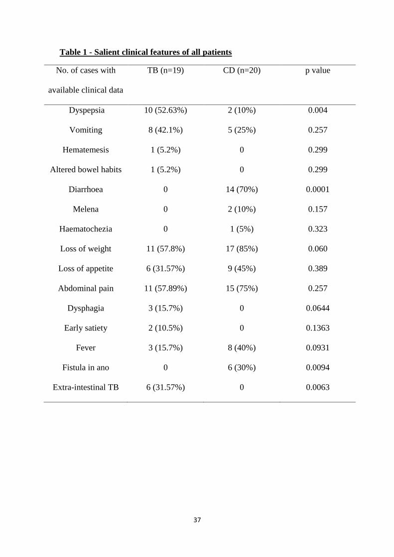
Many features such as abdominal pain, loss of weight, loss of appetite and fever were
prevalent to a similar extent in both the diseases. However, dyspepsia as a presenting
symptom was more common in TB (53%) as compared to CD (10%). Another
significant difference noted was that almost 70% of CD patients presented with
diarrhoea, while none of TB patients had diarrhoea.
Evidence of extra-intestinal TB was identified in six of the TB patients. Amongst
these, 4 patients had tuberculous lymphadenopathy (omental, cervical and axillary),
one patient had hepatic TB and one had pulmonary TB.
Six (30%) of the CD patients were detected to have fistula in ano at the time of
presentation. One patient with CD was also diagnosed with uveitis.
37
Table 1 - Salient clinical features of all patients
No. of cases with
available clinical data
TB (n=19) CD (n=20) p value
Dyspepsia 10 (52.63%) 2 (10%) 0.004
Vomiting 8 (42.1%) 5 (25%) 0.257
Hematemesis 1 (5.2%) 0 0.299
Altered bowel habits 1 (5.2%) 0 0.299
Diarrhoea 0 14 (70%) 0.0001
Melena 0 2 (10%) 0.157
Haematochezia 0 1 (5%) 0.323
Loss of weight 11 (57.8%) 17 (85%) 0.060
Loss of appetite 6 (31.57%) 9 (45%) 0.389
Abdominal pain 11 (57.89%) 15 (75%) 0.257
Dysphagia 3 (15.7%) 0 0.0644
Early satiety 2 (10.5%) 0 0.1363
Fever 3 (15.7%) 8 (40%) 0.0931
Fistula in ano 0 6 (30%) 0.0094
Extra-intestinal TB 6 (31.57%) 0 0.0063
38
INVESTIGATIONS
Blood borne viruses
Two of the TB patients were identified to have hepatitis B and one patient was
positive for HIV. All the CD patients tested negative for blood borne viruses.
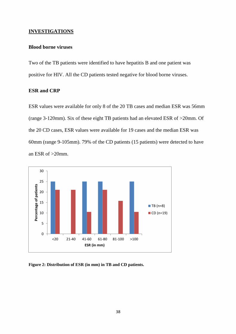
ESR and CRP
ESR values were available for only 8 of the 20 TB cases and median ESR was 56mm
(range 3-120mm). Six of these eight TB patients had an elevated ESR of >20mm. Of
the 20 CD cases, ESR values were available for 19 cases and the median ESR was
60mm (range 9-105mm). 79% of the CD patients (15 patients) were detected to have
an ESR of >20mm.
Figure 2: Distribution of ESR (in mm) in TB and CD patients.
0
5
10
15
20
25
30
<20 21-40 41-60 61-80 81-100 >100
Pe
rce
nta
ge o
f p
atie
nts
ESR (in mm)
TB (n=8)
CD (n=19)
39
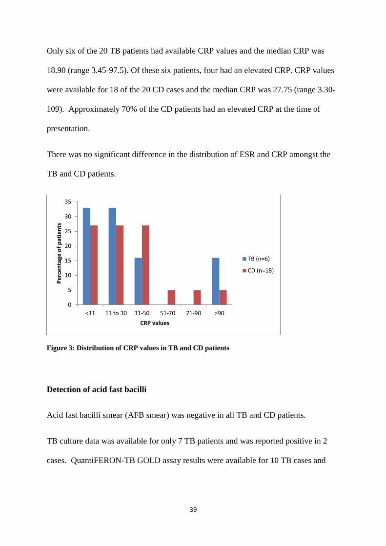
Only six of the 20 TB patients had available CRP values and the median CRP was
18.90 (range 3.45-97.5). Of these six patients, four had an elevated CRP. CRP values
were available for 18 of the 20 CD cases and the median CRP was 27.75 (range 3.30-
109). Approximately 70% of the CD patients had an elevated CRP at the time of
presentation.
There was no significant difference in the distribution of ESR and CRP amongst the
TB and CD patients.
Figure 3: Distribution of CRP values in TB and CD patients
Detection of acid fast bacilli
Acid fast bacilli smear (AFB smear) was negative in all TB and CD patients.
TB culture data was available for only 7 TB patients and was reported positive in 2
cases. QuantiFERON-TB GOLD assay results were available for 10 TB cases and
0
5
10
15
20
25
30
35
<11 11 to 30 31-50 51-70 71-90 >90
Pe
rce
nta
ge o
f p
atie
nts
CRP values
TB (n=6)
CD (n=18)
40
were positive in 3 of these cases. Of the ten TB cases wherein Xpert TB PCR results
were available, 2 cases were reported as positive.
TB culture, QuantiFERON-TB GOLD assay and Xpert TB PCR were negative in all
CD cases.
RADIOLOGY
The radiological investigation data were available only for 13 TB cases and 10 CD
cases. The salient radiological features are enlisted in Table 2.
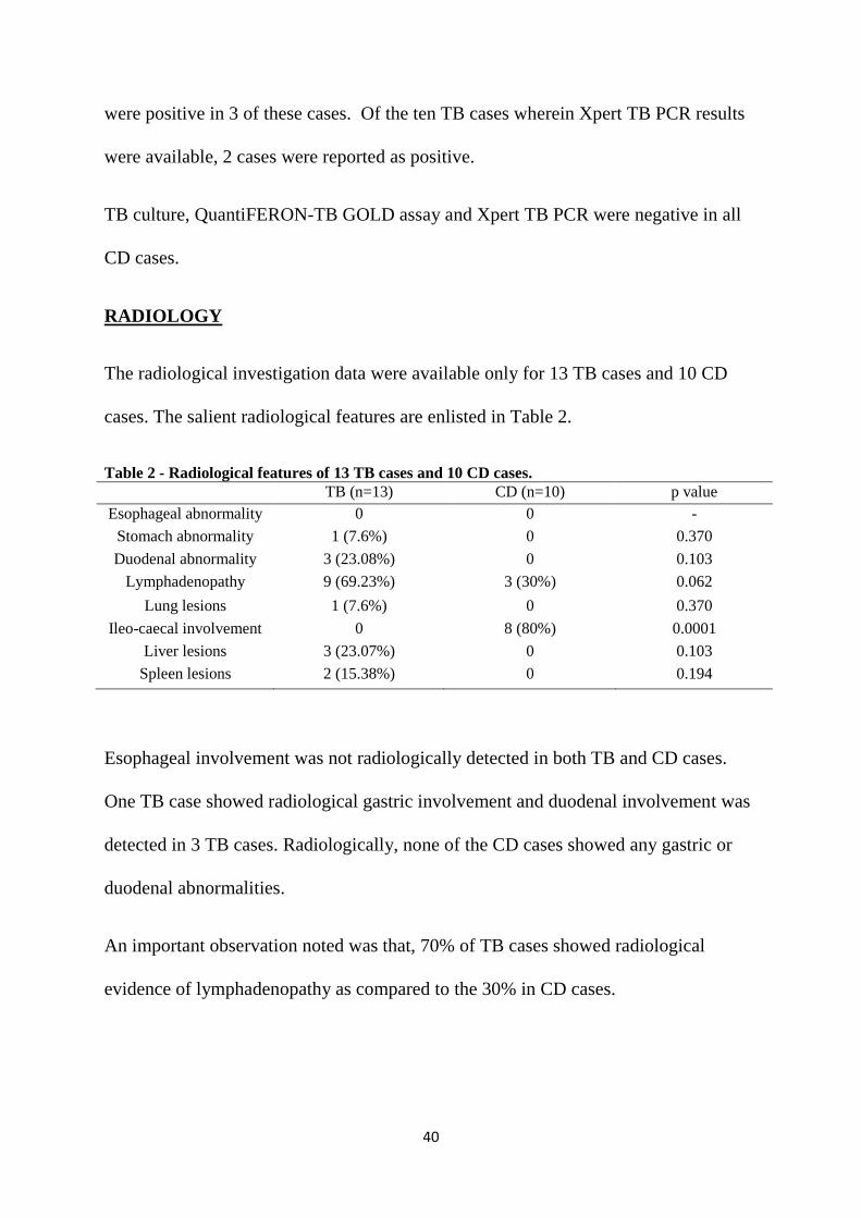
Table 2 - Radiological features of 13 TB cases and 10 CD cases.
TB (n=13) CD (n=10) p value
Esophageal abnormality 0 0 -
Stomach abnormality 1 (7.6%) 0 0.370
Duodenal abnormality 3 (23.08%) 0 0.103
Lymphadenopathy 9 (69.23%) 3 (30%) 0.062
Lung lesions 1 (7.6%) 0 0.370
Ileo-caecal involvement 0 8 (80%) 0.0001
Liver lesions 3 (23.07%) 0 0.103
Spleen lesions 2 (15.38%) 0 0.194
Esophageal involvement was not radiologically detected in both TB and CD cases.
One TB case showed radiological gastric involvement and duodenal involvement was
detected in 3 TB cases. Radiologically, none of the CD cases showed any gastric or
duodenal abnormalities.
An important observation noted was that, 70% of TB cases showed radiological
evidence of lymphadenopathy as compared to the 30% in CD cases.

41
Table 3 - Distribution of lymphadenopathy in TB and CD.
No. of cases with
available radiological
data
TB (n=13) CD (n=10) p value
Necrosis 7 (53.85%) 0 0.005
Peri-esophageal 2 (15.38%) 0 0.194
Peri-gastric 1 (7.69%) 0 0.369
Periduodenal 0 0 -
Lesser omental 4 (30.77%) 0 0.054
Greater omental 4 (30.77%) 0 0.054
Mesenteric 6 (46.15%) 3(30%) 0.431
Para-aortic 5 (38.46%) 0 0.027
Necrotic lymphadenopathy was noted in ~54% TB cases whereas necrosis was absent
in all of the CD cases. This difference was found to be statistically significant with a
p-value of 0.005. In TB, lymph nodes from peri-esophageal, peri-gastric, lesser
omentum, greater omentum, para-aortic and mesenteric regions were involved.
However, in CD only the mesenteric lymph nodes were involved.
Another striking observation was that radiological ileo-caecal involvement,
predominantly in the form of wall thickening, was detected in 80% of CD cases
whereas this finding was absent in all TB cases. This difference was found to be
statistically significant with a p-value of 0.0001.
Furthermore, 3 TB cases showed radiological evidence of liver lesions and splenic
lesions were reported in 2 TB cases.

42
UPPER GASTROINTESTINAL ENDOSCOPY
Abnormal endoscopic findings were significantly more common (p value=0.014) in
TB as compared to CD. Nineteen of the 20 TB cases (95%) had endoscopic
abnormalities. Interestingly, in contrast, 10 of 20 CD cases (50%) were normal
endoscopically.
Figure 4: Proportion of cases with abnormal endoscopic findings.
Table 4 - Distribution of cases with abnormal endoscopy findings in esophagus,
fundus/body, antrum/incisura and duodenum
Number of cases TB (n=20) CD (N=20) p value
Esophagus 7 (35%) 0 0.0036
Stomach 10 (50%) 8 (40%) 0.7512
Fundus/Body 3 (15%) 2 (10%)
Antrum/Incisura 5 (25%) 6 (30%)
Both 2 (10%) 0
Duodenum 11 (55%) 5 (25%) 0.053
Esophagus – On endoscopy, seven of the TB cases demonstrated abnormal
esophageal findings. In four of these cases (20%), the esophagus was involved in
isolation. However, the esophagus was spared in all the CD cases. This difference was
significant statistically with a p value of 0.0036.
0
5
10
15
20
Abnormal Normal
Nu
mb
er
of
pat
ien
ts
TB (n=20) CD (n=20)

43
Among the TB cases, the middle third of the esophagus was the site most frequently
involved (5 of the 7 endoscopically abnormal esophageal cases). The most common
esophageal endoscopic finding in TB was ulceration (30%). The ulcerations were
generally single in number, with a median size of 0.87cm (range 0.4-2.5cm). Other
esophageal endoscopic findings documented in TB were erosions, edema, erythema,
nodularity and ulcero-proliferative growth.
Fundus/Body - Five of the TB cases (25%) and 2 CD cases (10%) had abnormal
endoscopic findings in the fundus/body.
Table 5 - Proportion of various abnormal endoscopic findings in fundus/body (n=Total number
of cases)
Total number of cases TB=20 CD=20 p value
Erosions 1 (5%) 1 (5%) 1.00
Edema 4 (20%) 1 (5%) 0.151
Erythema 1 (5%) 2 (10%) 0.548
Thickening 1 (5%) 0 0.311
Submucosal nodules 2 (10%) 0 0.146
Erosions, erythema and edema were seen in both TB and CD. However, edema was
seen in 20% of TB cases as compared to the 5% in CD. Thickening and submucosal
nodules were seen in one TB case each.
Antrum/Incisura: Seven of the TB cases (35%) and 6 CD cases (30%) had abnormal
endoscopic findings in the antrum/incisura.
Table 6 - Proportion of various abnormal endoscopic findings in antrum/incisura (n=Total
number of cases)
Total number of cases TB=20 CD=20 p value
Erosions 2 (10%) 3 (15%) 0.632
Ulcerations 3 (15%) 1 (5%) 0.292
Edema 3 (15%) 1 (5%) 0.292
Erythema 1 (5%) 3 (15%) 0.292
Thickening 0 1 (5%) 0.311
Ulcero-proliferative
growth
1 (5%) 0 0.311
Deformed pylorus 3 (15%) 1 (5%) 0.292

44
Erosions, ulcerations, edema, erythema and deformed pylorus were the common
findings reported in both TB and CD cases with no significant statistical difference in
their prevalence.
Duodenum: Abnormal duodenal endoscopic findings were significantly more
common (p value=0.05) in TB (55%) as compared to CD (25%) cases. In both the TB
(45%) and CD (60%), the second part of the duodenum was the site most frequently
involved.
Table 7- Proportion of various abnormal endoscopic findings in duodenum (n=Total number of
cases)
Number of cases TB=20 CD=20 p value
Erosions 3 (15%) 1 (5%) 0.292
Ulcerations 2 (10%) 1 (5%) 0.548
Edema 5 (25%) 1 (5%) 0.077
Erythema 3 (15%) 2 (10%) 0.635
Nodular mucosa 4 (20%) 2 (10%) 0.375
Thickening 1 (5%) 1 (5%) 1.00
Deformed duodenum 4 (20%) 1 (5%) 0.152
Granularity 1 (5%) 0 0.312
Erosions, ulcerations, nodular mucosa, thickening, erythema and edema were the
common findings reported in both TB and CD cases with no significant statistical
difference in their prevalence. However, edema was slightly more common in TB
(25%) as compared to CD (5%).
Correlation of upper GI endoscopy with lower GI endoscopy and histology
Among the 20 TB cases, lower GI endoscopy data was available for only 4 patients.
Ironically, the only case with an endoscopically normal upper GI endoscopy had a
corresponding abnormal lower GI endoscopy, moderate chronic inflammation and
granulomatous inflammation. Among the 19 cases with an endoscopically abnormal

45
upper GI, lower gastrointestinal details were available for only 3 cases and all these
three cases had an endoscopically normal lower GI, no significant chronic
inflammation or granulomatous inflammation
Among the 10 CD cases wherein upper GI endoscopy was normal, 80% of them had
an abnormal lower GI endoscopy, and 60% cases each had lower GI moderate chronic
inflammation and granulomatous inflammation. Of the CD cases with abnormal upper
GI endoscopy, lower GI data was unavailable for one case and among the remaining 9
cases, 66% has a corresponding abnormal lower GI endoscopy, 22% had lower GI
moderate chronic inflammation and 55% had granulomatous inflammation.

46
HISTOLOGICAL FEATURES
STOMACH
Of the 20 TB cases, only 11 cases had at least one gastric biopsy for assessment.
However, all the 20 CD cases had at least one gastric biopsy for evaluation.
The 11 TB cases together had 17 gastric biopsies and the 20 CD cases had 24 gastric
biopsies. The various histological parameters were studied and contrasted between the
17 TB gastric biopsies and the 24 CD gastric biopsies.
Chronic inflammation – TB cases (47%) had a higher prevalence of moderate chronic
lamina propria inflammation as compared to CD (29%). However, this difference was
not statistically significant. The inflammation extended to the deeper mucosa in up
95% of TB cases. In 21% of CD cases, the inflammation was restricted to the
superficial parts of the mucosa (Table 10).
Patchy/discontinuous inflammation was present in an equal frequency in both TB and
CD (Table 11).
Fibrosis - Fibrosis was significantly higher in TB biopsies (82.35%) as compared to
CD biopsies (41.67%). The difference was statistically significant with a p value of
0.009.
Lymphoid aggregates - Interestingly, the presence of lymphoid aggregates was also
more common in TB biopsies (70%) in contrast to the CD cases (33.33%). This
difference was also proved to be significant statistically with a p value of 0.019.
However, the median number of lymphoid aggregates per biopsy was not significantly
different between TB and CD.
47
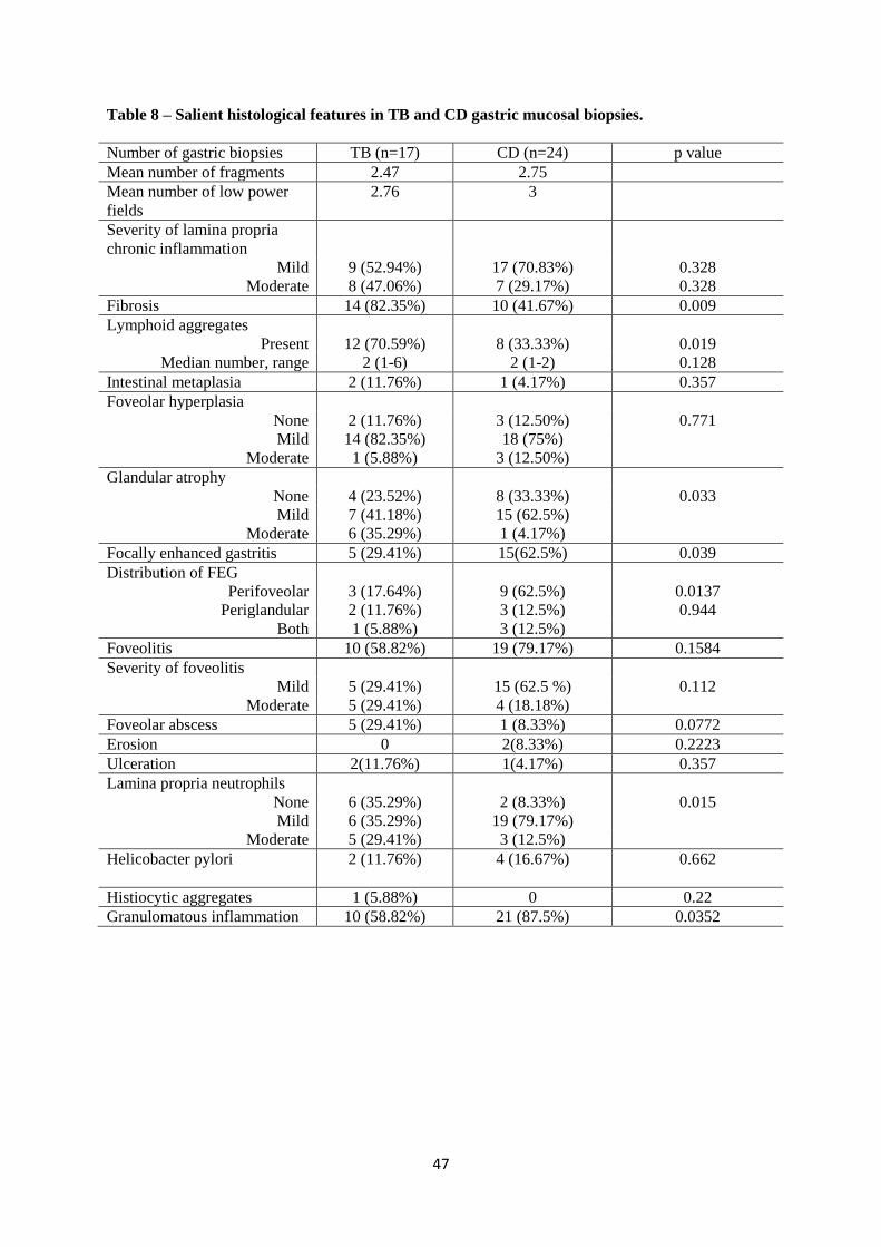
Table 8 – Salient histological features in TB and CD gastric mucosal biopsies.
Number of gastric biopsies TB (n=17) CD (n=24) p value
Mean number of fragments 2.47 2.75
Mean number of low power
fields
2.76 3
Severity of lamina propria
chronic inflammation
Mild 9 (52.94%) 17 (70.83%) 0.328
Moderate 8 (47.06%) 7 (29.17%) 0.328
Fibrosis 14 (82.35%) 10 (41.67%) 0.009
Lymphoid aggregates
Present 12 (70.59%) 8 (33.33%) 0.019
Median number, range 2 (1-6) 2 (1-2) 0.128
Intestinal metaplasia 2 (11.76%) 1 (4.17%) 0.357
Foveolar hyperplasia
None 2 (11.76%) 3 (12.50%) 0.771
Mild 14 (82.35%) 18 (75%)
Moderate 1 (5.88%) 3 (12.50%)
Glandular atrophy
None 4 (23.52%) 8 (33.33%) 0.033
Mild 7 (41.18%) 15 (62.5%)
Moderate 6 (35.29%) 1 (4.17%)
Focally enhanced gastritis 5 (29.41%) 15(62.5%) 0.039
Distribution of FEG
Perifoveolar 3 (17.64%) 9 (62.5%) 0.0137
Periglandular 2 (11.76%) 3 (12.5%) 0.944
Both 1 (5.88%) 3 (12.5%)
Foveolitis 10 (58.82%) 19 (79.17%) 0.1584
Severity of foveolitis
Mild 5 (29.41%) 15 (62.5 %) 0.112
Moderate 5 (29.41%) 4 (18.18%)
Foveolar abscess 5 (29.41%) 1 (8.33%) 0.0772
Erosion 0 2(8.33%) 0.2223
Ulceration 2(11.76%) 1(4.17%) 0.357
Lamina propria neutrophils
None 6 (35.29%) 2 (8.33%) 0.015
Mild 6 (35.29%) 19 (79.17%)
Moderate 5 (29.41%) 3 (12.5%)
Helicobacter pylori
2 (11.76%)
4 (16.67%)
0.662
Histiocytic aggregates 1 (5.88%) 0 0.22
Granulomatous inflammation 10 (58.82%) 21 (87.5%) 0.0352

48
Table 9 - Extent of lamina propria chronic inflammation.
No. of gastric biopsies TB(n=17) CD(n=24) p value
Superficial 1 (5.89%) 5 (20.83%) 0.182
Both superficial and
deep 16 (94.11%) 19 (79.17%)
Table 10- Distribution of lamina propria inflammation – Patchy/Diffuse
No. of gastric biopsies TB(n=17) CD(n=24) p value
Patchy/Discontinuous 5 (29.41%) 7 (29.17%) 0.986
Diffuse 12 (70.59%) 17 (70.83%)
Focally enhanced gastritis was a statistically significant finding in CD cases (62.5%)
in contrast to TB. However, it must be emphasized that the entity of focally enhanced
gastritis was also noted in 5% of TB biopsies, thereby highlighting that this finding is
not exclusive to CD.
Focally enhanced gastritis in CD biopsies (62.5%) had a predominant perifoveolar
distribution in contrast to TB biopsies (23.53%). This difference was statistically
significant with a p value of 0.013.
The prevalence of foveolar hyperplasia was similar in TB and CD. However, the
prevalence of moderate glandular atrophy was significantly higher in TB (35%),
unlike CD wherein majority of the cases had mild glandular atrophy (62%).
CD biopsies (79%) had a higher prevalence of foveolitis and foveolar mucin depletion
in contrast to the 59% in TB biopsies. However, moderate foveolitis was significantly
higher in TB (30%), in contrast to CD wherein majority of the cases had mild
foveolitis (62%). Foveolar abscess formation was also more prevalent in TB (29%) as
compared to CD (8.33%).
49
Ulcerations were present in 2 TB gastric biopsies, of which one biopsy had histiocytic
aggregates, epithelioid granulomas and palisading histiocytes within the ulcer base.
The prevalence of moderate lamina propria activity was significantly higher in TB
(29.41%), unlike CD wherein majority of the cases had mild activity (79.17%). This
difference was statistically significant with a p value of 0.015.
Granulomatous inflammation:
Granulomatous inflammation was present in 58% of TB biopsies (10 of 17) and
87.5% of the CD gastric biopsies (21 of 24 cases). Majority of the CD gastric biopsies
(22 of 24) were taken from the antrum with only few fundus/body biopsies (2 of 24).
Due to this uneven distribution of biopsies, the exact prevalence of granulomatous
inflammation between TB and CD in different parts of the stomach could not be
assessed.
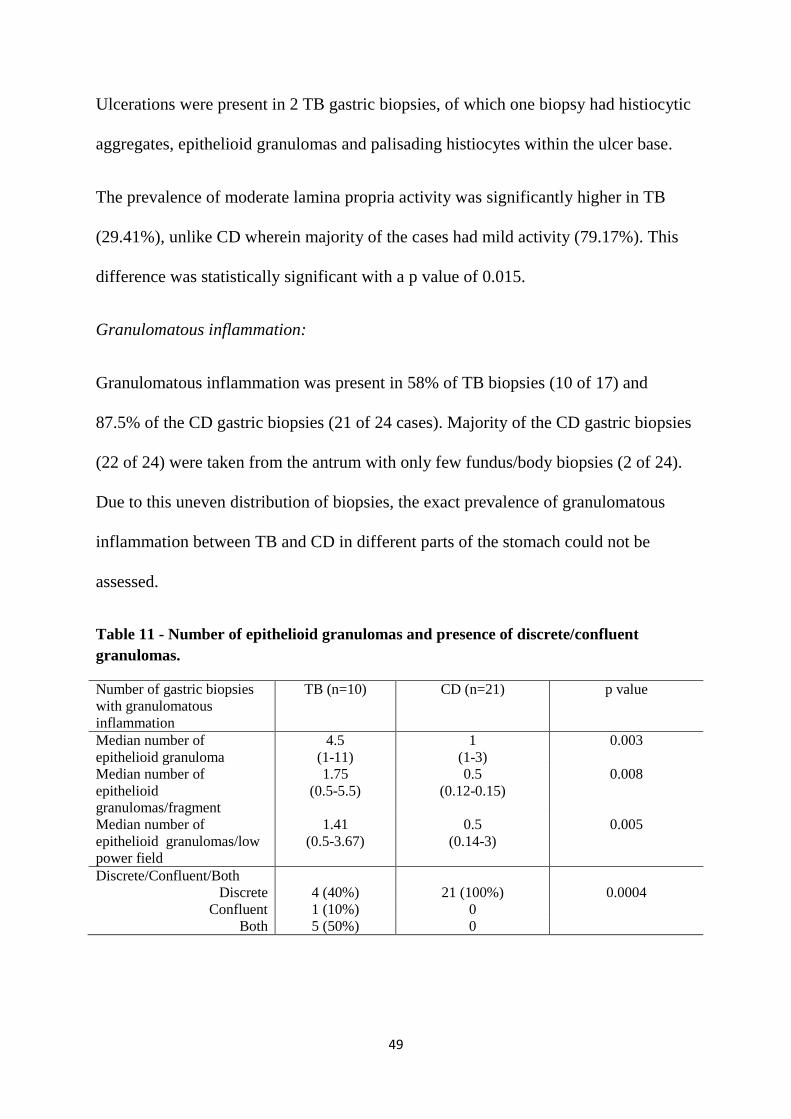
Table 11 - Number of epithelioid granulomas and presence of discrete/confluent
granulomas.
Number of gastric biopsies
with granulomatous
inflammation
TB (n=10) CD (n=21) p value
Median number of
epithelioid granuloma
4.5
(1-11)
1
(1-3)
0.003
Median number of
epithelioid
granulomas/fragment
1.75
(0.5-5.5)
0.5
(0.12-0.15)
0.008
Median number of
epithelioid granulomas/low
power field
1.41
(0.5-3.67)
0.5
(0.14-3)
0.005
Discrete/Confluent/Both
Discrete 4 (40%) 21 (100%) 0.0004
Confluent 1 (10%) 0
Both 5 (50%) 0
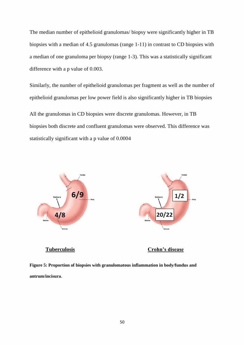
50
The median number of epithelioid granulomas/ biopsy were significantly higher in TB
biopsies with a median of 4.5 granulomas (range 1-11) in contrast to CD biopsies with
a median of one granuloma per biopsy (range 1-3). This was a statistically significant
difference with a p value of 0.003.
Similarly, the number of epithelioid granulomas per fragment as well as the number of
epithelioid granulomas per low power field is also significantly higher in TB biopsies
All the granulomas in CD biopsies were discrete granulomas. However, in TB
biopsies both discrete and confluent granulomas were observed. This difference was
statistically significant with a p value of 0.0004
Tuberculosis Crohn’s disease
Figure 5: Proportion of biopsies with granulomatous inflammation in body/fundus and
antrum/incisura.

51
Table 12 – Characteristics of granulomas in the 11 TB and 20 CD cases which have at least one
gastric biopsy
No. of cases with at least one
gastric biopsy.
TB (n=11) CD (n=20) p value
Granulomatous inflammation
present
8 (72.73%) 19 (95%) 0.4079
≥4 granulomas
4 (36.36%)
0
0.0039
At least one large granuloma
3 (27.27%)
0
0.0140
At least one disorganised
granuloma
4 (36.36%)
12 (60%)
0.2077
All compact granulomas
4 (36.36%)
7 (35%)
0.9395
At least one granuloma with
lymphoid cuff
8 (72.73%)
10 (50%)
0.2198
At least one granuloma with giant
cells
3 (27.27%)
9(45%)
0.3323
At least one granuloma with
necrosis
1 (9.09%)
0
0.171
At least one perifoveolar
granuloma
0
4 (20%)
0.112
At least one periglandular
granuloma
1 (9.09%)
2 (10%)
0.935
At least one granuloma with AFB
1 (9.09%)
0
0.171
Table 12 enlists the salient features of the granulomatous inflammation in the 11 TB
cases and 20 CD cases which have at least one gastric biopsy. The significant findings
observed were that ≥4 granulomas (36% vs 0) and large granuloma (27% vs 0) were
more common in TB as compared to CD. The presence of at least one disorganised
52
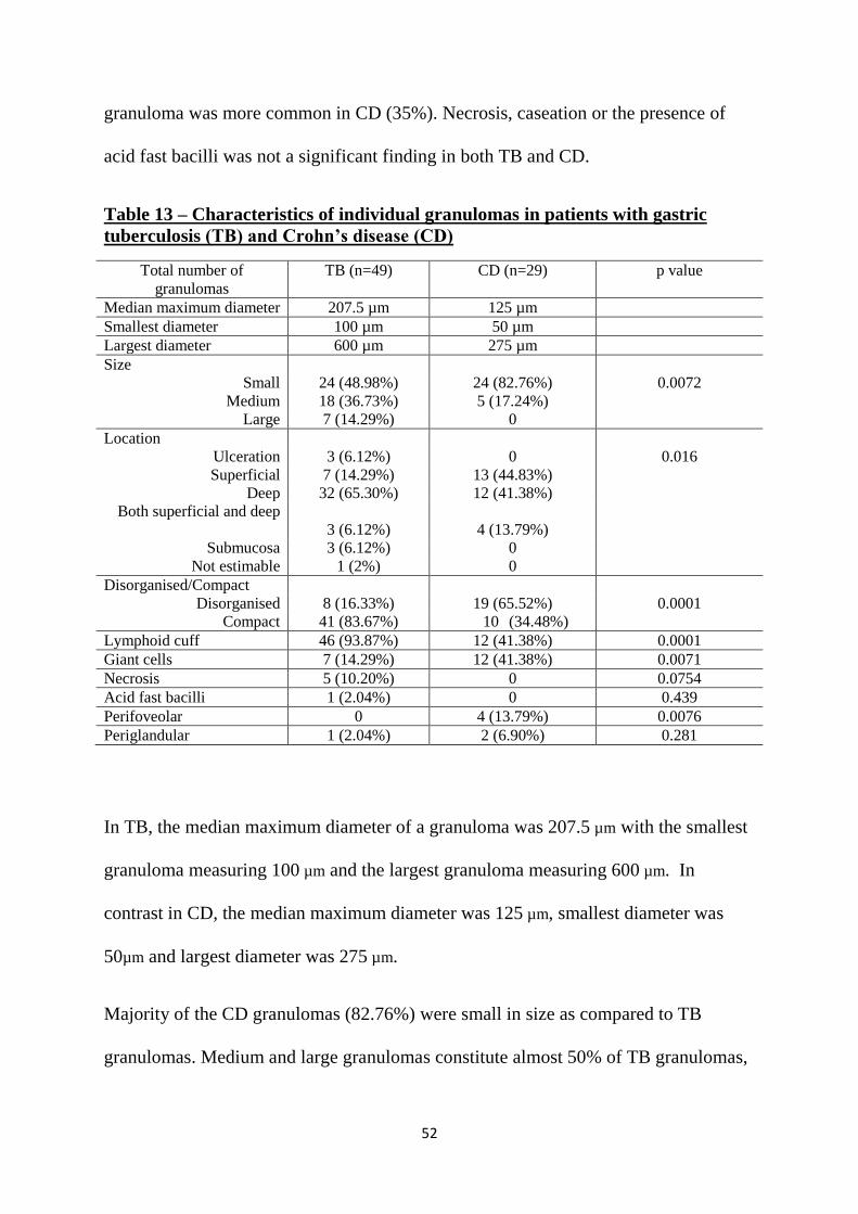
granuloma was more common in CD (35%). Necrosis, caseation or the presence of
acid fast bacilli was not a significant finding in both TB and CD.
Table 13 – Characteristics of individual granulomas in patients with gastric
tuberculosis (TB) and Crohn’s disease (CD)
Total number of
granulomas
TB (n=49) CD (n=29) p value
Median maximum diameter 207.5 µm 125 µm
Smallest diameter 100 µm 50 µm
Largest diameter 600 µm 275 µm
Size
Small 24 (48.98%) 24 (82.76%) 0.0072
Medium 18 (36.73%) 5 (17.24%)
Large 7 (14.29%) 0
Location
Ulceration 3 (6.12%) 0 0.016
Superficial 7 (14.29%) 13 (44.83%)
Deep 32 (65.30%) 12 (41.38%)
Both superficial and deep
3 (6.12%)
4 (13.79%)
Submucosa 3 (6.12%) 0
Not estimable 1 (2%) 0
Disorganised/Compact
Disorganised 8 (16.33%) 19 (65.52%) 0.0001
Compact 41 (83.67%) 10 (34.48%)
Lymphoid cuff 46 (93.87%) 12 (41.38%) 0.0001
Giant cells 7 (14.29%) 12 (41.38%) 0.0071
Necrosis 5 (10.20%) 0 0.0754
Acid fast bacilli 1 (2.04%) 0 0.439
Perifoveolar 0 4 (13.79%) 0.0076
Periglandular 1 (2.04%) 2 (6.90%) 0.281
In TB, the median maximum diameter of a granuloma was 207.5 µm with the smallest
granuloma measuring 100 µm and the largest granuloma measuring 600 µm. In
contrast in CD, the median maximum diameter was 125 µm, smallest diameter was
50µm and largest diameter was 275 µm.
Majority of the CD granulomas (82.76%) were small in size as compared to TB
granulomas. Medium and large granulomas constitute almost 50% of TB granulomas,

53
in contrast to only 18% in CD granulomas. This difference was statistically significant
with a p value of 0.007.
TB granulomas were more commonly located within ulcerations and in the
submucosa.
Other significant observations include the higher prevalence of compact granulomas
(83%) and granulomas with lymphoid cuff (46%) in TB. Disorganised granulomas
and perifoveolar granulomas were significantly more common in CD (13.79%).

54
Endoscopic correlation
Table 14 – Comparison of salient histological features between endoscopically normal
and abnormal gastric mucosa in both TB and CD. Total number
of cases with
gastric biopsies
11 TB cases 20 CD cases
Endoscopy
Normal (3)
Endoscopy
Abnormal (n=8)
Endoscopy Normal
(13)
Endoscopy Abnormal
(n=7)
Focally
enhanced
gastritis present
3 (100%) 2 (25%) 9 (69.23%) 4 (57.14%)
Moderate
chronic
inflammation
0 (%) 4 (50%) 2 (18.18%) 5 (71.43%)
Granulomatous
inflammation
present
3 (100%) 5 (62.5%) 12 (92.30%) 7 (100%)
The endoscopically normal gastric biopsies were compared with endoscopically
abnormal gastric biopsies for various parameters for both TB and CD
Three of 11 TB cases had endoscopically normal mucosa. Strikingly, these cases also
had histological changes like focally enhanced gastritis and granulomatous
inflammation. The prevalence of moderate chronic inflammation in TB cases was
higher in endoscopically abnormal mucosa.
Of the 20 CD cases, the 13 cases which had an endoscopically normal mucosa had a
high frequency of focally enhanced gastritis (69%) and granulomatous inflammation
(92%). The prevalence of moderate chronic inflammation in CD cases was
significantly higher in endoscopically abnormal mucosa (71.43%).

55
DUODENUM
Of the 20 TB and 20 CD cases included in our study, the various histological
parameters were studied and contrasted between the 12 TB and 16 CD cases which
had duodenal biopsies.
Table 15 - Salient histological features in TB and CD duodenal mucosal biopsies.
Number of cases with duodenal
biopsies
TB (n=12) CD (n=16) p value
Crypt villous alteration with
villous atrophy
Absent 4 (33.33%) 13 (81.25%) 0.036
Mild 6 (50%) 2 (12.50%)
Moderate 2 (16.67%) 1 (6.25%)
Increased IELs 3 (25%) 2 (12.5%) 0.392
Severity of lamina propria
chronic inflammation
Mild 7 (58.33%) 15 (93.75%) 0.073
Moderate 5 (41.66%) 1 (6.25%)
Lymphoid aggregates 8 (66.67%) 4 (25%) 0.027
Cryptitis 4 (33.33%) 3 (18.75%) 0.378
Crypt hyperplasia 9 (75%) 8 (50%) 0.180
Brunner’s gland hyperplasia 6 (50%) 4 (25%) 0.277
Pyloric metaplasia 3 (25%) 1 (6.25%) 0.161
Fibrosis 5 (41.67%) 5 (31.25%) 0.569
Surface neutrophilic infiltration 5 (41.67%) 5 (31.25%) 0.569
Lamina propria neutrophils
Absent 6 (50%) 12 (75%) 0.174
Mild 6 (50%) 3 (18.75%)
Moderate 0 1 (6.25%)
Erosions 3 (25%) 0 0.034
Ulceration 1 (8.33%) 0 0.240
Granulomatous inflammation 7 (58.23%) 1 (6.25%) 0.0025
Of the 12 TB cases, crypt villous ratio alteration and villous atrophy was seen in 8
cases (50% mild, 16% moderate). However, in CD cases, crypt villous ratio alteration
and villous atrophy was observed in only 3 cases. This difference was statistically
significant with a p value of 0.036.

56
Figure 6: Distribution of intraepithelial lymphocytes
25% of TB cases and 12.5% of CD cases had increased intraepithelial lymphocytes.
The distribution of IELs is diagrammatically represented in Figure 6. In both TB and
CD, majority of the patients had an IEL count of <25 and there was no significant
statistical difference.
The prevalence of moderate chronic inflammation was significantly higher in TB
(41.66%) in contrast to CD (6.25%). In CD, the majority of cases only had mild
inflammation (93.75%)
The presence of lymphoid aggregates was strikingly more common in TB biopsies
(66.67%) in contrast to the CD cases (25%). This difference was also proved to be
significant statistically with a p value of 0.027.
The prevalence of cryptitis, crypt hyperplasia, Brunner’s gland hyperplasia, pyloric
metaplasia and surface neutrophilic infiltration was higher in TB as compared to CD,
however, these differences were not statistically significant.
Lamina propria activity was seen more in TB cases as compared to CD. Also, 3 of the
TB cases had erosions, however erosions were absent in all of the CD cases.
0
2
4
6
8
10
12
0-25 26-40 >40
Nu
mb
er
of
pat
ien
ts
Intraepithelial lymphocytes
TB(n=12)
CD (n=16)

57
Granulomatous inflammation - Granulomatous inflammation was more frequent in
TB than CD. Only 6.25% of CD cases had granulomatous inflammation as compared
to the 58.3% TB cases. This difference was statistically significant with a p value of
0.0025.
Table 16 - - Number of epithelioid granulomas and presence of discrete/confluent
granulomas.
Number of biopsies with
granulomatous inflammation
TB (n=7) CD (n=1) p value
Median number of epithelioid
granuloma
1
(1-5)
1
(1-1)
0.7055
Median number of epithelioid
granulomas/fragment
0.50
(0.14-1)
0.25
(0.25-0.25)
0.4966
Median number of epithelioid
granulomas/low power field
0.33
(0.14-1)
0.33
(0.33-0.33)
0.8220
Discrete/Confluent/Both
Discrete 6 (85.71%) 1 (100%) 0.686
Confluent 0 0
Both 1 (14.29%) 0
The median number of granulomas was one in both TB and CD. However, the
maximum number of granulomas in TB duodenal biopsies was more (up to 5) as
compared to CD duodenal biopsies.
In both TB and CD, the granulomas were predominantly discrete.
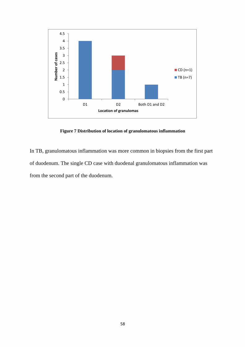
58
Figure 7 Distribution of location of granulomatous inflammation
In TB, granulomatous inflammation was more common in biopsies from the first part
of duodenum. The single CD case with duodenal granulomatous inflammation was
from the second part of the duodenum.
0
0.5
1
1.5
2
2.5
3
3.5
4
4.5
D1 D2 Both D1 and D2
Nu
mb
er
of
case
s
Location of granulomas
CD (n=1)
TB (n=7)

59
Table 17 – Characteristics of granulomas in the 12 TB and 16 CD cases which have at least one
duodenal biopsy
No. of cases with duodenal biopsies TB(n=12) CD(n=16) p value
Granulomatous inflammation
7 (58.33%)
1 (6.25%)
0.0025
≥4 granulomas
1 (8.33%)
0
0.2396
At least one large granuloma
2 (16.67%)
0
0.0901
At least one disorganised granuloma
1 (8.33%)
0
0.2396
All compact granulomas
6 (50%)
1(6.25%)
0.0082
At least one granuloma with a lymphoid
cuff
6 (50%)
1 (6.25%)
0.0082
At least one granuloma with giant cells
7 (58.33%)
0
0.0004
At least one granuloma with inclusion
1 (8.33%)
0
0.2396
At least one granuloma with necrosis
1 (8.33%)
0
0.2396
Table 17 enlists the salient features of the granulomatous inflammation in the 12 TB
cases and 16 CD cases which have at least one duodenal biopsy. Duodenal mucosal
biopsies with ≥4 granulomas, large granulomas, compact granulomas and granulomas
with a lymphoid cuff were more frequent in TB as compared to CD. Necrosis,
caseation or the presence of acid fast bacilli was not a significant finding in both TB
and CD.

60
Characterisation of granulomas
Table 18 – Characteristics of individual granulomas in patients with duodenal
tuberculosis (TB) and Crohn’s disease (CD)
Total number of
granulomas
TB (n=11) CD (n=1) p-value
Median maximum diameter 200 µm 212.50 µm
Smallest diameter 115 µm
Largest diameter 462.50 µm
Size
Small 5 (45.45%) 0 0.466
Medium 4 (36.36%) 1 (100%)
Large 2 (18.18%) 0
Location
Ulceration 0 0 0.460
Villous 0 0
Deep 7 (63.63%) 1 (100%)
Submucosa 4 (36.37) 0
Disorganised/Compact
Disorganised 1 (9.09%) 0 0.753
Compact 10(90.90%0 1 (100%)
Lymphoid cuff 10 (90.91%) 1 (100%) 0.753
Giant cells 6 (60%) 0 0.251
Necrosis 1 (9.09%) 0 0.753
Inclusion 1 (9.09%) 0 0.753
Perifoveolar 0 4 (13.79%) 0.0076
Periglandular 1 (2.04%) 2 (6.90%) 0.281
In TB, the median maximum diameter was 200 µm with the smallest granuloma
measuring 115 µm and the largest granuloma measuring 462.5 µm. In CD, the only
granuloma present had a maximum diameter of 212.50 µm.
TB granulomas were more commonly located in the deep mucosa and submucosa.
Another significant observation was the higher prevalence of compact granulomas
(90%) in TB.
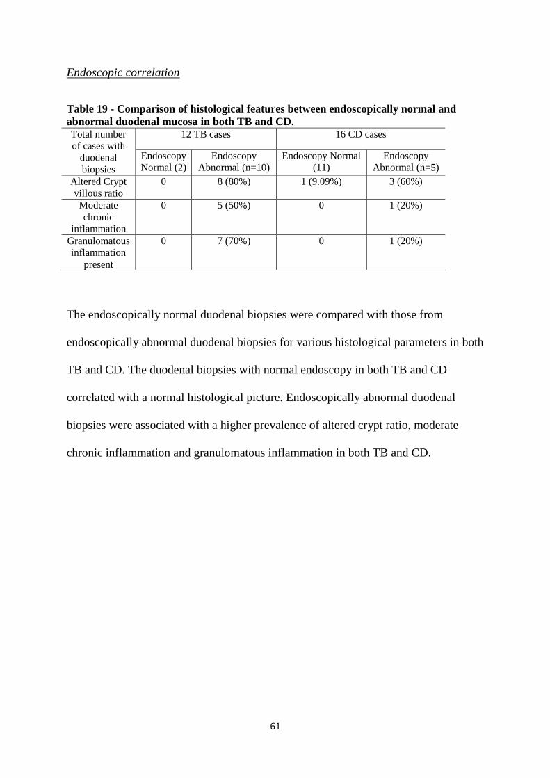
61
Endoscopic correlation
Table 19 - Comparison of histological features between endoscopically normal and
abnormal duodenal mucosa in both TB and CD. Total number
of cases with
duodenal
biopsies
12 TB cases 16 CD cases
Endoscopy
Normal (2)
Endoscopy
Abnormal (n=10)
Endoscopy Normal
(11)
Endoscopy
Abnormal (n=5)
Altered Crypt
villous ratio
0 8 (80%) 1 (9.09%) 3 (60%)
Moderate
chronic
inflammation
0 5 (50%) 0 1 (20%)
Granulomatous
inflammation
present
0 7 (70%) 0 1 (20%)
The endoscopically normal duodenal biopsies were compared with those from
endoscopically abnormal duodenal biopsies for various histological parameters in both
TB and CD. The duodenal biopsies with normal endoscopy in both TB and CD
correlated with a normal histological picture. Endoscopically abnormal duodenal
biopsies were associated with a higher prevalence of altered crypt ratio, moderate
chronic inflammation and granulomatous inflammation in both TB and CD.

62
ESOPHAGUS
Only six of the 20 TB cases had esophageal biopsies, while none of the 20 CD cases
had esophageal biopsies. Therefore, the histological parameters were studied and
analysed for only the six TB esophageal biopsies.
Table 20 - Salient histological feature of esophageal TB.
Number of esophageal biopsies Number of cases Percentage
Ulceration 5 83.33%
Parakeratosis 2 33.33%
Basal cell hyperplasia 3 50%
Papillary elongation 3 50%
Papillary congestion 3 50%
Acanthosis
Absent 3 50%
Mild 1 16.67%
Moderate 1 16.67%
Cannot be assessed 1 16.67%
Intraepithelial lymphocytes 6 100%
Severity of IELs
Mild 4 83.33%
Moderate 1 16.67%
Marked 1 16.67%
Location of the IELs
Peripapillary 1 16.67%
Interpapillary 0 0
Both 5 83.33%
Intraepithelial eosinophils 1 16.67%
Severity of lamina propria chronic
inflammation.
Mild 1 16.67%
Moderate 4 66.66%
Marked 1 16.67%
Severity of lamina propria activity
Absent 1 16.67%
Moderate 5 83.33%
Fibrosis 4 66.67%
Lymphoid aggregates 3 50%
Granulomatous inflammation 6 100%
The most common site of involvement within the esophagus in TB was the middle
third (66%). One of the common findings was ulceration which was seen in 5 of the 6

63
TB esophageal biopsies (83.33%). The ulceration extended up to the submucosa in
one of these cases, while in the others depth of the ulceration could not be assessed.
Basal cell hyperplasia, papillary elongation and papillary congestion were present in 3
of 6 TB esophageal biopsies (50%). Two cases had acanthosis and only one case had
intraepithelial eosinophils.
All the 6 cases had intraepithelial lymphocytes. The intraepithelial lymphocytes were
predominantly located in both the peripapillary and interpapillary region (83.33%). In
four of the cases, there was only a mild increase in IELs and only one case each had
moderate and marked increase in IELs respectively.
Five of the six cases had moderate to marked lamina propria chronic inflammation as
well as moderate lamina propria activity (83%).
Granulomatous inflammation
Granulomatous inflammation was present in all the six cases. The median number of
granulomas per biopsy was 3.5 (range 1-9). The median number of epithelioid
granulomas/fragment and number of epithelioid granulomas/ low power field is 0.55
(0.08 - 1) and 0.14 (0.14 - 1.20) respectively.
Table 21 – Median number of granulomas in esophageal TB.
Median, Range
Number of epithelioid granulomas/
biopsy
3.5 (1-9)
Number of epithelioid granulomas/
fragment
0.55 (0.08-1)
Number of epithelioid granulomas/low
power field
0.63
(0.14-1.20)

64
Table 22 – Salient features of granulomas in the 6 TB esophageal biopsies
No. of cases with esophageal biopsies TB (n=6)
≥4 granulomas
6 (100%)
At least one large granuloma
3 (50%)
At least one disorganised granuloma
4(66.67%)
All compact granulomas
2 (33.33%)
At least one with a lymphoid cuff
5 (83.33%)
At least one granuloma with giant cells
6 (31.58%)
At least one granuloma with necrosis
3 (50%)
All six cases had ≥4 granulomas. Three of the biopsies had at least one large
granuloma and at least one granuloma with necrosis. Lymphoid cuff was present in 5
of the cases and necrosis was present in 3 of the cases.
Table 23 – Characteristics of individual granulomas in esophageal TB.
Total number of granulomas (n=25)
Median maximum diameter 250 µm
Smallest diameter 132.50 µm
Largest diameter 590 µm
Size
Small 10 (40%)
Medium 12 (48%)
Large 1 (12%)
Location
Lamina propria 9 (36%)
Submucosa 6 (24%)
Ulceration 10 (40%)
Lymphoid cuff 8 (32%)
Giant cells 9 (36%)
Necrosis 5 (20%)
Compact 16 (64%)

65
The 6 biopsies together had 25 granulomas. The median maximum diameter of these
25 granulomas was 250 micrometres (range 132.50-590).
60% of the granulomas were medium to large sized.. The granulomas were located in
decreasing frequency within ulceration (40%), lamina propria (36%) and submucosa
(24%). Majority of the granulomas were compact (64%)
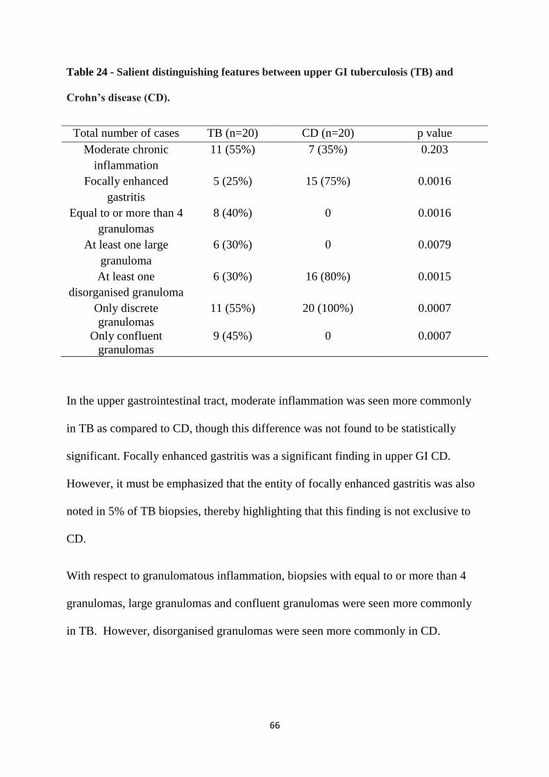
66
Table 24 - Salient distinguishing features between upper GI tuberculosis (TB) and
Crohn’s disease (CD).
Total number of cases TB (n=20) CD (n=20) p value
Moderate chronic
inflammation
11 (55%) 7 (35%) 0.203
Focally enhanced
gastritis
5 (25%) 15 (75%) 0.0016
Equal to or more than 4
granulomas
8 (40%) 0 0.0016
At least one large
granuloma
6 (30%) 0 0.0079
At least one
disorganised granuloma
6 (30%) 16 (80%) 0.0015
Only discrete
granulomas
11 (55%) 20 (100%) 0.0007
Only confluent
granulomas
9 (45%) 0 0.0007
In the upper gastrointestinal tract, moderate inflammation was seen more commonly
in TB as compared to CD, though this difference was not found to be statistically
significant. Focally enhanced gastritis was a significant finding in upper GI CD.
However, it must be emphasized that the entity of focally enhanced gastritis was also
noted in 5% of TB biopsies, thereby highlighting that this finding is not exclusive to
CD.
With respect to granulomatous inflammation, biopsies with equal to or more than 4
granulomas, large granulomas and confluent granulomas were seen more commonly
in TB. However, disorganised granulomas were seen more commonly in CD.

IMAGES

67
Photomicrograph 1: Mild chronic inflammation with a single discrete granuloma in a
case of Crohn's disease (H&E, 40X magnification).
Photomicrograph 2: Moderate chronic inflammation in a case of Tuberculosis (H&E,
40X magnification).
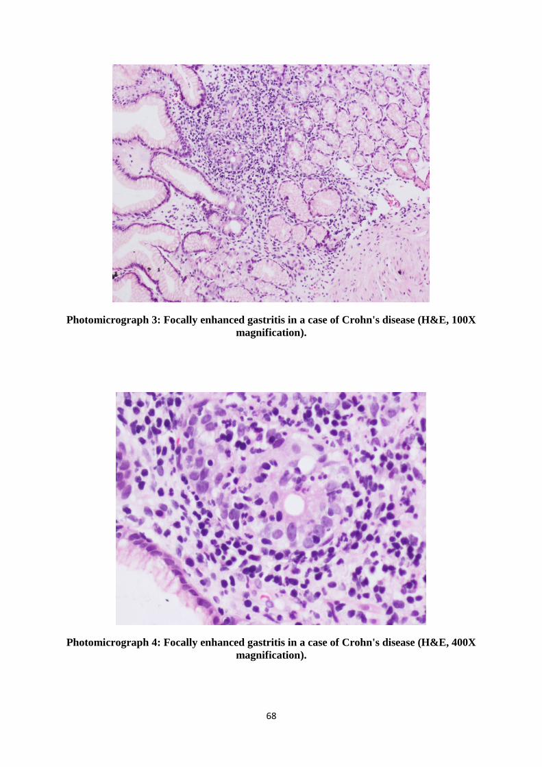
68
Photomicrograph 3: Focally enhanced gastritis in a case of Crohn's disease (H&E, 100X
magnification).
Photomicrograph 4: Focally enhanced gastritis in a case of Crohn's disease (H&E, 400X
magnification).

69
Photomicrograph 5: Patchy chronic inflammation in a case of Crohn's disease (H&E,
40X magnification).
Photomicrograph 6: Presence of lymphoid aggregates in a case of Tuberculosis (H&E,
40X magnification).
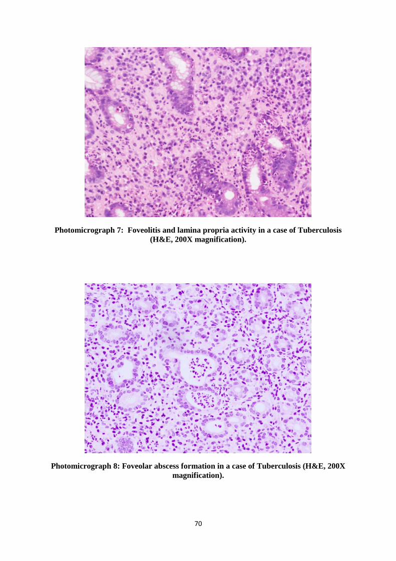
70
Photomicrograph 7: Foveolitis and lamina propria activity in a case of Tuberculosis
(H&E, 200X magnification).
Photomicrograph 8: Foveolar abscess formation in a case of Tuberculosis (H&E, 200X
magnification).
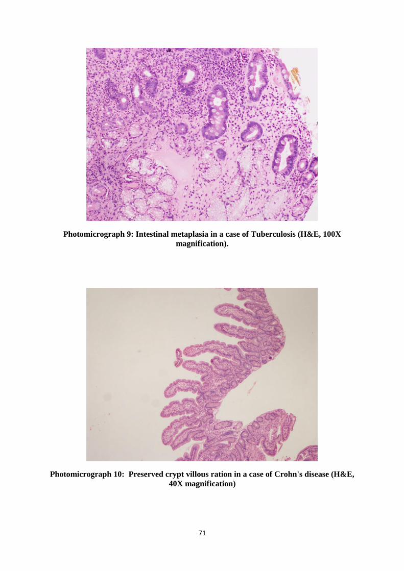
71
Photomicrograph 9: Intestinal metaplasia in a case of Tuberculosis (H&E, 100X
magnification).
Photomicrograph 10: Preserved crypt villous ration in a case of Crohn's disease (H&E,
40X magnification)

72
Photomicrograph 11: Mild villous atrophy with increase in intraepithelial lymphocytes
in a case of Tuberculosis (H&E, 200X magnification).
Photomicrograph 12: Increase in intraepithelial lymphocytes in a case of Tuberculosis
(H&E, 400X magnification)
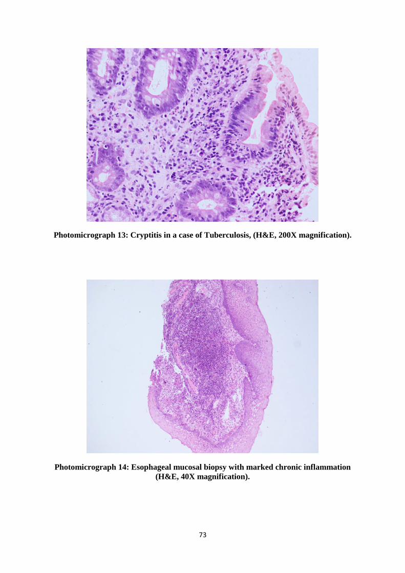
73
Photomicrograph 13: Cryptitis in a case of Tuberculosis, (H&E, 200X magnification).
Photomicrograph 14: Esophageal mucosal biopsy with marked chronic inflammation
(H&E, 40X magnification).
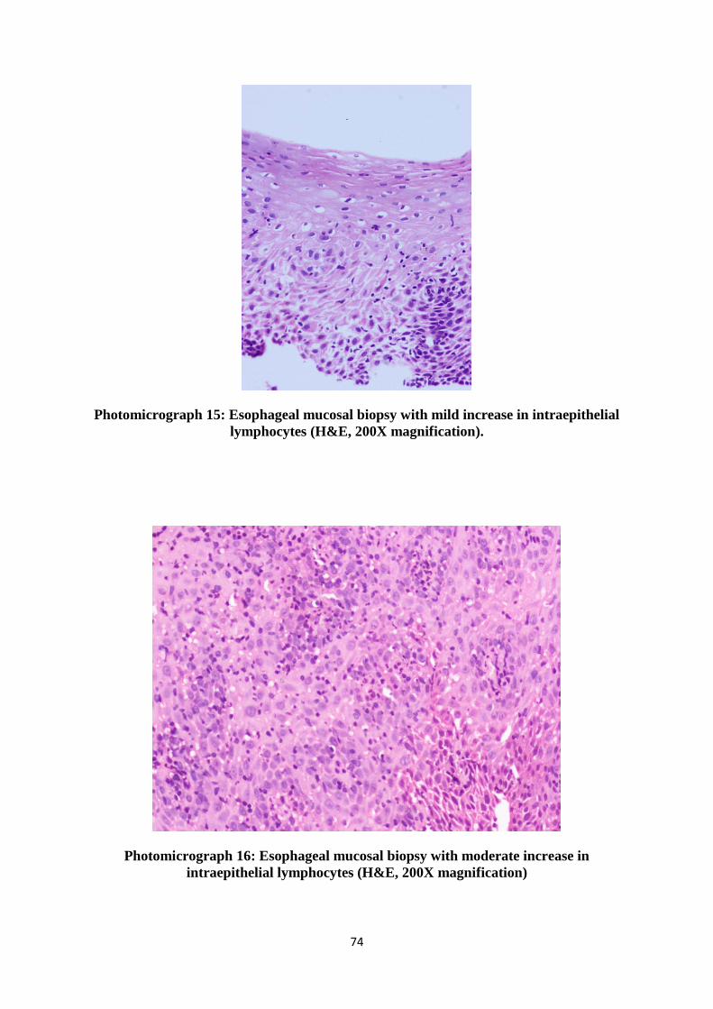
74
Photomicrograph 15: Esophageal mucosal biopsy with mild increase in intraepithelial
lymphocytes (H&E, 200X magnification).
Photomicrograph 16: Esophageal mucosal biopsy with moderate increase in
intraepithelial lymphocytes (H&E, 200X magnification)

75
Photomicrograph 17: Gastric mucosal biopsy with four granulomas in a case of
Tuberculosis (H&E, 40X magnification).
Photomicrograph 18: Confluent granulomas (H&E, 40X magnification).
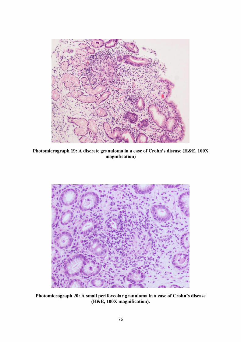
76
Photomicrograph 19: A discrete granuloma in a case of Crohn’s disease (H&E, 100X
magnification)
Photomicrograph 20: A small perifoveolar granuloma in a case of Crohn’s disease
(H&E, 100X magnification).
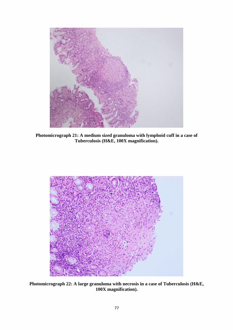
77
Photomicrograph 21: A medium sized granuloma with lymphoid cuff in a case of
Tuberculosis (H&E, 100X magnification).
Photomicrograph 22: A large granuloma with necrosis in a case of Tuberculosis (H&E,
100X magnification).

78
Photomicrograph 23: A disorganised granuloma in a case of Crohn's disease (H&E,
100X magnification).
Photomicrograph 24: Compact granulomas in a case of Tuberculosis (H&E, 100X
magnification).
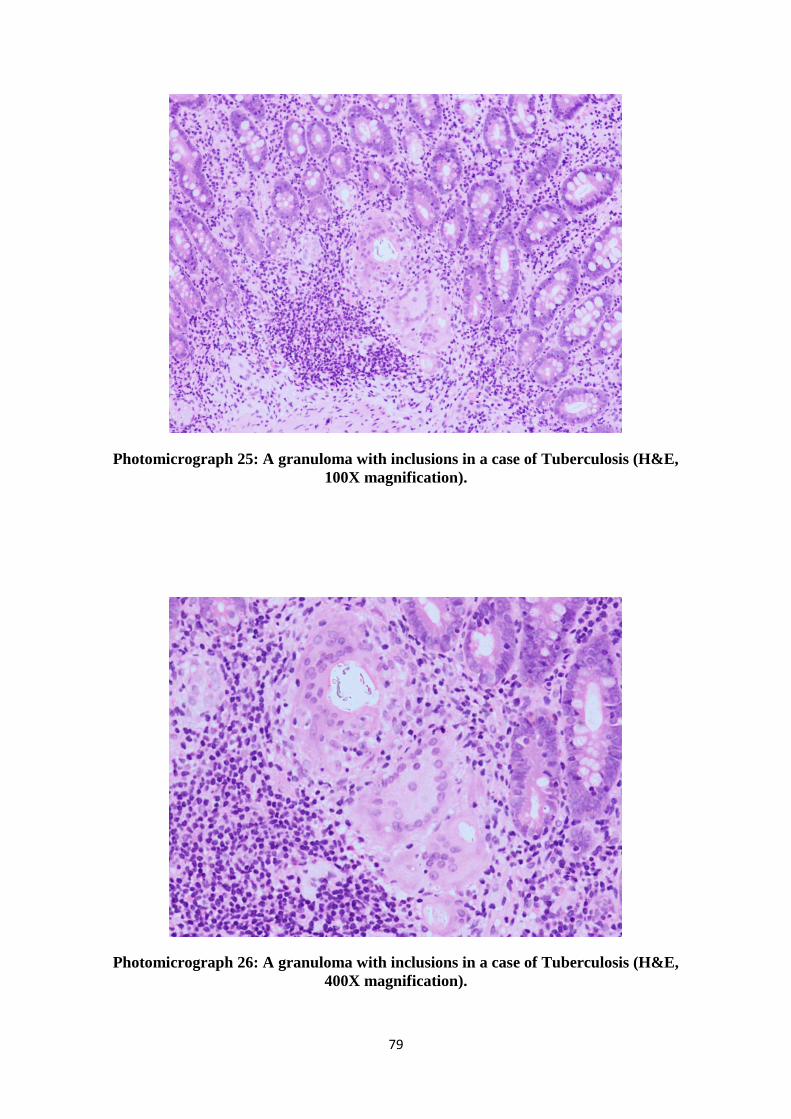
79
Photomicrograph 25: A granuloma with inclusions in a case of Tuberculosis (H&E,
100X magnification).
Photomicrograph 26: A granuloma with inclusions in a case of Tuberculosis (H&E,
400X magnification).

80
Photomicrograph 27: Duodenal mucosal biopsy with granulomatous inflammation in a
case of Crohn's disease (H&E 100X magnification).
Photomicrograph 28: Esophageal biopsy with a large submucosal granuloma in a case
of Tuberculosis (H&E, 100X magnification).

DISCUSSION
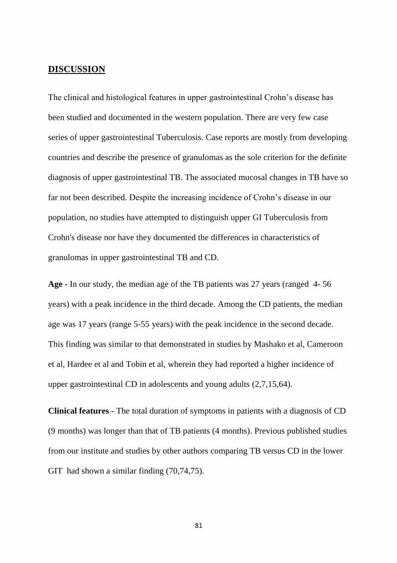
81
DISCUSSION
The clinical and histological features in upper gastrointestinal Crohn’s disease has
been studied and documented in the western population. There are very few case
series of upper gastrointestinal Tuberculosis. Case reports are mostly from developing
countries and describe the presence of granulomas as the sole criterion for the definite
diagnosis of upper gastrointestinal TB. The associated mucosal changes in TB have so
far not been described. Despite the increasing incidence of Crohn’s disease in our
population, no studies have attempted to distinguish upper GI Tuberculosis from
Crohn's disease nor have they documented the differences in characteristics of
granulomas in upper gastrointestinal TB and CD.
Age - In our study, the median age of the TB patients was 27 years (ranged 4- 56
years) with a peak incidence in the third decade. Among the CD patients, the median
age was 17 years (range 5-55 years) with the peak incidence in the second decade.
This finding was similar to that demonstrated in studies by Mashako et al, Cameroon
et al, Hardee et al and Tobin et al, wherein they had reported a higher incidence of
upper gastrointestinal CD in adolescents and young adults (2,7,15,64).
Clinical features - The total duration of symptoms in patients with a diagnosis of CD
(9 months) was longer than that of TB patients (4 months). Previous published studies
from our institute and studies by other authors comparing TB versus CD in the lower
GIT had shown a similar finding (70,74,75).
82
Clinical symptoms such as abdominal pain, loss of weight, loss of appetite and fever
had a similar prevalence in both TB and CD. The only striking differences noted were
that dyspepsia was more common in TB and diarrhoea, as a presenting symptom, was
more common in CD . However, it is important to note that, patients with upper GI
CD nearly always have concomitant lesions in the lower GIT and therefore it is
difficult to determine whether the symptoms were solely due to the upper GI
involvement(1).
Radiology - On CT imaging, involvement of the upper GIT was rare to absent in both
TB and CD. However, in our study, radiological evidence of lymphadenopathy was
significantly more common in TB as compared to CD (p value-0.062). Also, necrotic
lymph nodes were a significant finding in TB, as compared to CD where it was absent
(p value – 0.005). In our study, the lymph node groups involved in TB only included
peri-esophageal, peri-gastric, lesser omentum, greater omentum, mesenteric and para-
aortic lymph nodes. Further, mesenteric lymph nodes were more frequent in TB than
CD in our study.
Concomitant radiological ileo-caecal involvement was present in majority of CD
cases. On the other hand, few of TB cases had concomitant liver and splenic lesions.
Endoscopy
Abnormal upper GI endoscopic lesions were significantly more common in TB as
compared to CD (p value – 0.014).
In our study, abnormal endoscopic esophageal findings were detected in 35% TB
cases but, were not present in any of the 20 CD cases (p value - 0.003). The low

83
frequency of esophageal endoscopic involvement in Crohn’s disease was in
concordance with other studies by Wagtman et al (0.7%) and Schmitz Moorman et al
(15%).
Among the cases with esophageal involvement, the middle third of the esophagus was
the site most frequently involved (5 of 7 cases) and the most common endoscopic
finding in TB was ulcerations (30%). The ulcerations were generally single in number,
with a median size of 0.87cm, with the largest measuring 2.5cm. Nagi et al, Jain et al
and Abid et al had also reported similar findings in their studies (5,24,33). In our
study, one case also showed ulcero-proliferative tumour like growth as described by
Nagi et al and Li Q et al (33,79).
The frequency of gastric endoscopic abnormalities was almost the same in both TB
(50%) and CD (40%). The abnormal endoscopic gastric findings included erosions
edema, erythema, thickening, deformity and submucosal nodules. One case with TB
shows an ulcero-proliferative tumour like growth. However, there were no significant
difference between TB and CD.
Abnormal duodenal endoscopic findings were significantly more common in TB as
compared to CD (p value=0.05). Erosions, ulcerations, nodular mucosa, thickening,
erythema and edema were the common findings reported in both TB and CD cases
with no significant statistical difference in their prevalence.

84
It must also be mentioned that in our study, among the CD cases, the stomach was the
most common site to show endoscopic abnormalities and this finding was similar to
that demonstrated in other studies (4,7,47).
Histology
Granulomatous inflammation –
In the upper gastrointestinal tract, granulomatous inflammation due to TB and CD in
all the sites showed features similar to those in the lower gastrointestinal tract as
described in previous published studies from our institute(71,75).
In our study, in the upper GIT, higher numbers of granulomas were observed in TB as
compared to CD. More than or equal to 4 granulomas per biopsy was significantly
more common in TB than CD.
The presence of large granulomas was another differentiating feature between TB and
CD. Biopsies with at least one large granuloma were significantly more common in
TB as compared to CD. Also, all the granulomas were either small or medium sized.
In our study, disorganised granulomas were significantly more common in CD than in
TB, although it must be noted that disorganised granulomas were also present in TB.
Confluence of granulomas was a significant finding in TB. All the granulomas in CD
were discrete. Furthermore, the presence of lymphoid cuff was also more common in
TB than CD; however this difference was not statistically significant.

85
Another distinguishing feature between TB and CD was the differential localisation of
granulomas. TB granulomas were more commonly located within ulcerations, deep
mucosa and submucosa.
Also, in gastric mucosal biopsies, perifoveolar granulomas were more common in CD
than TB, however, this difference was not statistically significant.
Schmitz – Moorman et al and van Hogezand et al reported that granulomatous
inflammation was more frequent in stomach as compared to other sites in upper
gastrointestinal TB (4,17). A similar finding was demonstrated in our study too.
Other mucosal changes
Esophagus – Ulcerations were a common histological finding in esophageal TB.
Increase in intraepithelial lymphocytes was present in all the TB esophageal biopsies.
The intraepithelial lymphocytes were predominantly located in both the peripapillary
and interpapillary region (83.33%). In four of the cases, there was only a mild increase
in IELs and only one case each had moderate and marked increase in IELs
respectively.
Moderate to marked lamina propria chronic inflammation as well as moderate lamina
propria activity were also common histological findings in esophageal.
Stomach - Moderate chronic lamina propria inflammation was more common in TB
(47%) than CD (29%), however this difference was not statistically significant.
Features like fibrosis, lymphoid aggregates and moderate glandular atrophy were
significantly more common in TB than CD.
86
Focally enhanced gastritis was a statistically significant finding in CD (62%) in
contrast to TB (p value – 0.39). This finding was in concordance with studies by
Oberhueber et al (76%) and Sharif et al (65%) (26,80).
However, it must be also be emphasized that the entity of focally enhanced gastritis
was also noted in 5% of TB biopsies, thereby highlighting that this finding is not
exclusive to CD.
Moderate foveolitis and moderate lamina activity were more common in TB as
compared to CD.
Duodenum – Crypt villous alteration with villous atrophy was significantly more
common in TB (50% mild, 16% moderate) than CD (p value – 0.036).
Moderate chronic lamina propria inflammation was more common in TB (42%) than
CD (6.25%), however this difference was not statistically significant (p value –
0.072). Lymphoid aggregates were also significantly more common in TB than CD.
Other mucosal changes such as cryptitis, crypt hyperplasia, pyloric metaplasia and
fibrosis were more frequent in TB than CD, but none of these differences were
statistically significant.

CONCLUSIONS

87
CONCLUSIONS
We studied 40 cases with granulomatous inflammation in mucosal biopsies from the
upper gastrointestinal tract that included 20 cases with a final diagnosis of
Tuberculosis and 20 cases with a final diagnosis of Crohn’s disease. We found the
following-
1. A younger age at presentation and longer duration of symptoms were more
commonly associated with CD.
2. Necrotic lymphadenopathy on CT imaging was significantly more common in
TB. The mesenteric lymph nodes were more commonly involved in TB than
CD. In our study, the lesser omental, greater omental and para-aortic lymph
node groups were involved only in TB.
3. Abnormal endoscopic findings in the upper gastrointestinal tract were more
frequent in TB than CD. In Crohn’s disease, a few of the cases had small and
linear erosions. Tumour like lesions were present in two cases of Tuberculosis.
4. The salient distinguishing features of TB at any site were the presence of ≥4
granulomas, large granulomas, confluent granulomas and the location of
granulomas within ulcer bases and in the submucosa. Disorganised granulomas
were more common in Crohn’s disease.
5. Focally enhanced gastritis was more common in Crohn’s disease than in
Tuberculosis. However, glandular atrophy, activity, fibrosis and lymphoid
aggregates were more common in the gastric mucosa affected by Tuberculosis
than Crohn’s disease.
88
6. Involvement of the esophagus and duodenum by granulomatous inflammation
was more common in TB than CD.
7. Esophageal involvement by Tuberculosis was associated with ulceration,
moderate to marked chronic inflammation, activity and increase in
intraepithelial lymphocytes.

89
LIMITATIONS
The sample size was limited as a large number of patients with an initial
diagnosis of Crohn’s disease or Tuberculosis were lost to follow up.

REFERENCES
1. Am W, Ra V, Ra VH, Hw V, Cb L. Crohn’s disease of the upper gastrointestinal tract: the value
of endoscopic examination. Scand J Gastroenterol Suppl. 1997 Dec;225:100–5.
2. Cameron DJS. Upper and lower gastrointestinal endoscopy in children and adolescents with
Crohn’s disease: A prospective study. J Gastroenterol Hepatol. 1991 Aug;6(4):355–8.
3. C L, Cc R, M V, Am W, Cl M, E S. High incidence of upper gastrointestinal tract involvement
in children with Crohn disease. Pediatrics. 1989 1989;83(5):777–81.
4. Schmitz-Moormann P, Malchow H, Pittner P. Endoscopic and bioptic study of the upper
gastrointestinal tract in crohn’s disease patients. Pathol - Res Pract. 1985 Jan 1;179(3):377–87.
5. Jain SK, Jain S, Jain M, Yaduvanshi A. Esophageal tuberculosis: is it so rare? Report of 12
cases and review of the literature. Am J Gastroenterol. 2002 Feb;97(2):287–91.
6. H D, B F. Crohn’s disease of the upper gastrointestinal tract. Endoscopy. 1992 Aug;24(6):555–
8.
7. Mashako MN, Cezard JP, Navarro J, Mougenot JF, Sonsino E, Gargouri A, et al. Crohn’s
disease lesions in the upper gastrointestinal tract: correlation between clinical, radiological,
endoscopic, and histological features in adolescents and children. J Pediatr Gastroenterol Nutr.
1989 May;8(4):442–6.
8. Rh F, S T. Nonspecific granulomatous (regional) esophagitis. J Thorac Surg. 1950
1950;19(2):292–7.
9. Reynolds Jr HL, Stellato TA. Crohn’s Disease of the Foregut. Surg Clin North Am. 2001 Feb
1;81(1):117–35.
10. I R, F G, Jr DA. Crohn’s disease of the esophagus: Three cases and a literature review. Can J
Gastroenterol J Can Gastroenterol. 2001 Feb;15(2):117–22.
11. Ramaswamy K, Jacobson K, Jevon G, Israel D. Esophageal Crohn disease in children: a clinical
spectrum. J Pediatr Gastroenterol Nutr. 2003 Apr;36(4):454–8.
12. Decker GAG, Loftus EV, Pasha TM, Tremaine WJ, Sandborn WJ. Crohn’s Disease of the
Esophagus: Clinical Features and Outcomes: Inflamm Bowel Dis. 2001 May;7(2):113–9.
13. Rahhal RM, Banerjee S, Jensen CS, Bishop WP. Pediatric Crohn Disease Presenting as an
Esophageal Stricture: J Pediatr Gastroenterol Nutr. 2007 Jul;45(1):125–9.
14. Jr R. Cicatrizing enteritis, colitis and gastritis; a case report. Gastroenterology. 1949
1949;13(4):344–50.
15. Hardee S, Alper A, Pashankar DS, Morotti RA. Histopathology of duodenal mucosal lesions in
pediatric patients with inflammatory bowel disease: statistical analysis to identify distinctive
features. Pediatr Dev Pathol Off J Soc Pediatr Pathol Paediatr Pathol Soc. 2014 Dec;17(6):450–
4.

16. Am G, E A, P S. Clinical features of gastroduodenal Crohn’s disease in adolescents. J Pediatr
Gastroenterol Nutr. 1989 1989;8(2):166–71.
17. van Hogezand RA, Witte AMC, Veenendaal RA, Wagtmans MJ, Lamers CBHW. Proximal
Crohn’s Disease: Review of the Clinicopathologic Features and Therapy: Inflamm Bowel Dis.
2001 Nov;7(4):328–37.
18. Wagtmans MJ, Verspaget HW, Lamers CB, van Hogezand RA. Clinical aspects of Crohn’s
disease of the upper gastrointestinal tract: a comparison with distal Crohn’s disease. Am J
Gastroenterol. 1997 Sep;92(9):1467–71.
19. Ch K. Gastroduodenal Crohn’s disease. Proc Bayl Univ Med Cent. 2003 Apr;16(2):147–51.
20. Park JH, Kim SU, Sohn JW, Chung IK, Jung MK, Jeon SW, et al. Endoscopic findings and
clinical features of esophageal tuberculosis. Scand J Gastroenterol. 2010 Nov;45(11):1269–72.
21. Devarbhavi HC, Alvares JF, Radhikadevi M. Esophageal tuberculosis associated with
esophagotracheal or esophagomediastinal fistula: report of 10 cases. Gastrointest Endosc. 2003
Apr;57(4):588–92.
22. Gomes J, Antunes A, Carvalho A, Duarte R. Dysphagia as a manifestation of esophageal
tuberculosis: a report of two cases. J Med Case Reports. 2011;5(1):447.
23. Huang Y. Esophageal tuberculosis mimicking submucosal tumor. Interact Cardiovasc Thorac
Surg. 2004 Jun;3(2):274–6.
24. Abid S, Jafri W, Hamid S, Khan H, Hussainy A. Endoscopic features of esophageal
tuberculosis. Gastrointest Endosc. 2003 May;57(6):759–62.
25. Gill RS, Gill SS, Mangat H, Logsetty S. Gastric Perforation Associated with Tuberculosis: A
Case Report. Case Rep Med. 2011;2011:1–3.
26. Oberhuber G, Puspok A, Oesterreicher C, Novacek G, Zauner C, Burghuber M, et al. Focally
enhanced gastritis: A frequent type of gastritis in patients with Crohn’s disease.
Gastroenterology. 1997 Mar;112(3):698–706.
27. Ishii N, Furukawa K, Itoh T, Fujita Y. Primary Gastric Tuberculosis Presenting as Non-Healing
Ulcer and Mimicking Crohn’s Disease. Intern Med. 2011;50(5):439–42.
28. Lin O, Wu S-S, Yeh K-T, Soon M-S. CASE REPORT: Isolated gastric tuberculosis of the
cardia: Isolated gastric TB of the cardia. J Gastroenterol Hepatol. 2002 Apr 19;14(3):258–61.
29. Sullivan RC, Francona NT, Kirshbaum JD. TUBERCULOSIS OF THE STOMACH. A
CLINICAL AND PATHOLOGIC STUDY: Ann Surg. 1940 Aug;112(2):225–33.
30. S J, N K, Dk D, Sk J. Esophageal tuberculosis. Endoscopic cytology as a diagnostic tool. Acta
Cytol. 1998 Dec;43(6):1085–90.
31. S A, Sv S, G B. Primary hypertrophic tuberculosis of the pyloroduodenal area: report of 2 cases.
J Postgrad Med. 1998 Dec;45(1):10–2.
32. Padmanabhan H, Rothnie A, Singh P. An unusual case of gastric outlet obstruction caused by
tuberculosis: challenges in diagnosis and treatment. Case Rep. 2013 May 22;2013(may22
1):bcr2012008277-bcr2012008277.

33. Nagi B, Lal A, Kochhar R, Bhasin DK, Gulati M, Suri S, et al. Imaging of esophageal
tuberculosis: a review of 23 cases. Acta Radiol Stockh Swed 1987. 2003 May;44(3):329–33.
34. Kolawole TM, Lewis EA. A radiologic study of tuberculosis of the abdomen (gastrointestinal
tract). Am J Roentgenol Radium Ther Nucl Med. 1975 Feb;123(2):348–58.
35. Upadhyaya V, Kumar B, Lal R, Sharma M, Singh M, Rudramani. Primary duodenal
tuberculosis presenting as gastric-outlet obstruction: Its diagnosis. Afr J Paediatr Surg.
2013;10(2):83.
36. Gupta SK, Jain AK, Gupta JP, Agrawal AK, Berry K. Duodenal tuberculosis. Clin Radiol. 1988
Mar;39(2):159–61.
37. Chavhan GE, Ramakantan R. Duodenal tuberculosis: radiological features on barium studies
and their clinical correlation in 28 cases. J Postgrad Med. 2003 Sep;49(3):214–7.
38. Rao YG, Pande GK, Sahni P, Chattopadhyay TK. Gastroduodenal tuberculosis management
guidelines, based on a large experience and a review of the literature. Can J Surg J Can Chir.
2004 Oct;47(5):364–8.
39. Berney T. Duodenal tuberculosis presenting as acute ulcer perforation. Am J Gastroenterol.
1998 Oct;93(10):1989–91.
40. Wagtmans MJ, van Hogezand RA, Griffioen G, Verspaget HW, Lamers CB. Crohn’s disease of
the upper gastrointestinal tract. Neth J Med. 1997 Feb;50(2):S2-7.
41. J F, D F, S D, Ck C. Crohn’s disease of the stomach: the “ram’s horn” sign. Am J Roentgenol
Radium Ther Nucl Med. 1975 1975;123(2):242–51.
42. Jung JH, Kim SH, Kim MJ, Cho YK, Ahn SB, Son BK, et al. A Case Report of Primary
Duodenal Tuberculosis Mimicking a Malignant Tumor. Clin Endosc. 2014 Jul;47(4):346–9.
43. Gohel V, Long B, Richter G. Aphthous ulcers in the esophagus with Crohn colitis. Am J
Roentgenol. 1981 Oct;137(4):872–3.
44. Ammoury RF, Pfefferkorn MD. Significance of Esophageal Crohn Disease in Children: J
Pediatr Gastroenterol Nutr. 2011 Mar;52(3):291–4.
45. Tandon A, Lombard C, Triadafilopoulos G. Double-Edged Sword: Esophageal and Colonic
Crohn’s Disease. Dig Dis Sci. 2016 Jun;61(6):1487–90.
46. H H, F P, E S, H F, Cw R. Endoscopic results in five patients with Crohn’s disease of the
esophagus. Endoscopy. 1977 May;8(2):75–81.
47. Abdullah BA, Gupta SK, Croffie JM, Pfefferkorn MD, Molleston JP, Corkins MR, et al. The
Role of Esophagogastroduodenoscopy in the Initial Evaluation of Childhood Inflammatory
Bowel Disease: A 7-Year Study: J Pediatr Gastroenterol Nutr. 2002 Nov;35(5):636–40.
48. Yardley J, Hendrix T. Gastroduodenal Crohn’s disease: The focus is on focality.
Gastroenterology. 1997 Mar;112(3):1031–3.
49. Nugent FW, Richmond M, Park SK. Crohn’s disease of the duodenum. Gut. 1977 Feb
1;18(2):115–20.

50. P R, E O, G V, K G, L B, L T. Crohn’s disease of the stomach and duodenum: A clinical study
with emphasis on the value of endoscopy and endoscopic biopsies. Endoscopy. 1980
1980;12(6):288–94.
51. Gad A. The diagnosis of gastroduodenal Crohn’s disease by endoscopic biopsy. Scand J
Gastroenterol Suppl. 1989;167:23–8.
52. Jain SS. Esophageal tuberculosis presenting with hematemesis. World J Gastrointest Endosc.
2013;5(11):581.
53. Ed P. Tuberculosis of the stomach and the stomach in tuberculosis; a review with particular
reference to gross pathology and gastroscopic diagnosis. Am Rev Tuberc. 1950 Jan;61(1):116–
30.
54. Gupta B, Mathew S, Bhalla S. Pyloric obstruction due to gastric tuberculosis--an endoscopic
diagnosis. Postgrad Med J. 1990 Jan 1;66(771):63–5.
55. Dyer NH, Cook PL, Kemp Harper RA. Oesophageal stricture associated with Crohn’s disease.
Gut. 1969 Jul 1;10(7):549–54.
56. G D, P R, K G, G V. The natural history of esophageal Crohn’s disease: three patterns of
evolution. Gastrointest Endosc. 1993 1994;40(3):296–300.
57. Rubio CA, Sjödahl K, Lagergren J. Lymphocytic esophagitis: a histologic subset of chronic
esophagitis. Am J Clin Pathol. 2006 Mar;125(3):432–7.
58. Purdy JK, Appelman HD, Golembeski CP, McKenna BJ. Lymphocytic esophagitis: a chronic or
recurring pattern of esophagitis resembling allergic contact dermatitis. Am J Clin Pathol. 2008
Oct;130(4):508–13.
59. Danzi JT, Farmer RG, Sullivan BH, Rankin GB. Endoscopic features of gastroduodenal Crohn’s
disease. Gastroenterology. 1976 Jan;70(1):9–13.
60. Halme L, Kärkkäinen P, Rautelin H, Kosunen TU, Sipponen P. High frequency of helicobacter
negative gastritis in patients with Crohn’s disease. Gut. 1996 Mar;38(3):379–83.
61. Meining A, Bayerdörffer E, Bästlein E, Raudis N, Thiede C, Cyrus B, et al. Focal Inflammatory
Infiltrations in Gastric Biopsy Specimens Are Suggestive of Crohn’s Disease. Scand J
Gastroenterol. 1997 Aug 1;32(8):813–8.
62. Parente F, Cucino C, Bollani S, Imbesi V, Maconi G, Bonetto S, et al. Focal gastric
inflammatory infiltrates in inflammatory bowel diseases: prevalence, immunohistochemical
characteristics, and diagnostic role. Am J Gastroenterol. 2000 Mar;95(3):705–11.
63. Hummel TZ, ten Kate FJW, Reitsma JB, Benninga MA, Kindermann A. Additional Value of
Upper GI Tract Endoscopy in the Diagnostic Assessment of Childhood IBD: J Pediatr
Gastroenterol Nutr. 2012 Jun;54(6):753–7.
64. Tobin JM, Sinha B, Ramani P, Saleh AR, Murphy MS. Upper gastrointestinal mucosal disease
in pediatric Crohn disease and ulcerative colitis: a blinded, controlled study. J Pediatr
Gastroenterol Nutr. 2001 Apr;32(4):443–8.
65. Patterson ER, Shmidt E, Oxentenko AS, Enders FT, Smyrk TC. Normal villous architecture
with increased intraepithelial lymphocytes: a duodenal manifestation of Crohn disease. Am J
Clin Pathol. 2015 Mar;143(3):445–50.
66. Peixoto PC, Ministro PS, Sadio AD, Cancela EM, Araújo RN, Machado JL, et al. Esophageal
tuberculosis: an unusual cause of dysphagia. Gastrointest Endosc. 2009 May;69(6):1173–6.
67. Cv F, Jo S, Ma A. Colonoscopic diagnosis and medical treatment of ten patients with colonic
tuberculosis. Endoscopy. 1988 1988;20(2):62–5.
68. Hd T, A P. Pathology of intestinal tuberculosis and its distinction from Crohn’s disease.,
Pathology of intestinal tuberculosis and its distinction from Crohn’s disease. Gut Gut. 1972
Apr;13, 13(4, 4):260, 260–9.
69. Kirsch R, Pentecost M, de M Hall P, Epstein DP, Watermeyer G, Friederich PW. Role of
colonoscopic biopsy in distinguishing between Crohn’s disease and intestinal tuberculosis. J
Clin Pathol. 2006 Aug;59(8):840–4.
70. Amarapurkar D-N. Diagnosis of Crohn’s disease in India where tuberculosis is widely
prevalent. World J Gastroenterol. 2008;14(5):741.
71. Pulimood AB, Amarapurkar DN, Ghoshal U, Phillip M, Pai CG, Reddy DN, et al.
Differentiation of Crohn’s disease from intestinal tuberculosis in India in 2010. World J
Gastroenterol WJG. 2011 Jan 28;17(4):433–43.
72. Warren BF. Classic Pathology of Ulcerative and Crohn???s Colitis: J Clin Gastroenterol. 2004
May;38(Supplement 1):S33–5.
73. Magro F, Langner C, Driessen A, Ensari A, Geboes K, Mantzaris GJ, et al. European consensus
on the histopathology of inflammatory bowel disease. J Crohns Colitis. 2013 Nov;7(10):827–51.
74. Makharia GK, Srivastava S, Das P, Goswami P, Singh U, Tripathi M, et al. Clinical,
Endoscopic, and Histological Differentiations Between Crohn’s Disease and Intestinal
Tuberculosis. Am J Gastroenterol. 2010 Mar;105(3):642–51.
75. Pulimood AB, Ramakrishna BS, Kurian G, Peter S, Patra S, Mathan VI, et al. Endoscopic
mucosal biopsies are useful in distinguishing granulomatous colitis due to Crohn’s disease from
tuberculosis. Gut. 1999 Oct 1;45(4):537–41.
76. Almadi MA, Ghosh S, Aljebreen AM. Differentiating Intestinal Tuberculosis From Crohn’s
Disease: A Diagnostic Challenge. Am J Gastroenterol. 2009 Apr;104(4):1003–12.
77. Dn A, Nd P, Ad A, S A, R B, P G. Tissue polymerase chain reaction in diagnosis of intestinal
tuberculosis and Crohn’s disease. J Assoc Physicians India. 2004 Nov;52:863–7.
78. Kim YY, Lee J, Lee YJ, Lee SY, Lee YH, Choi KJ, et al. Sensitivity of Whole-Blood
Interferon-Gamma Release Assay According to the Severity and the Location of Disease in
Patients with Active Tuberculosis. Tuberc Respir Dis. 2011;70(2):125.
79. A Case Report of Mimicking Sub mucosal Tumor on Esophageal Tuberculosis | Open Access |
OMICS International [Internet]. [cited 2016 Sep 28]. Available from:
http://www.esciencecentral.org/journals/a-case-report-of-mimicking-sub-mucosal-tumor-on-
esophagealtuberculosis-2329-6925-1000268.php?aid=75224
80. Sharif F, McDermott M, Dillon M, Drumm B, Rowland M, Imrie C, et al. Focally enhanced
gastritis in children with Crohn’s disease and ulcerative colitis. Am J Gastroenterol. 2002
Jun;97(6):1415–20.
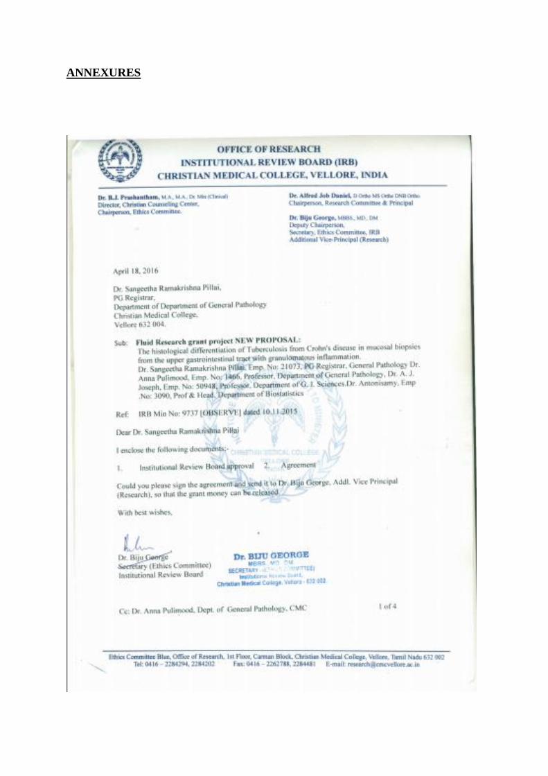
ANNEXURES
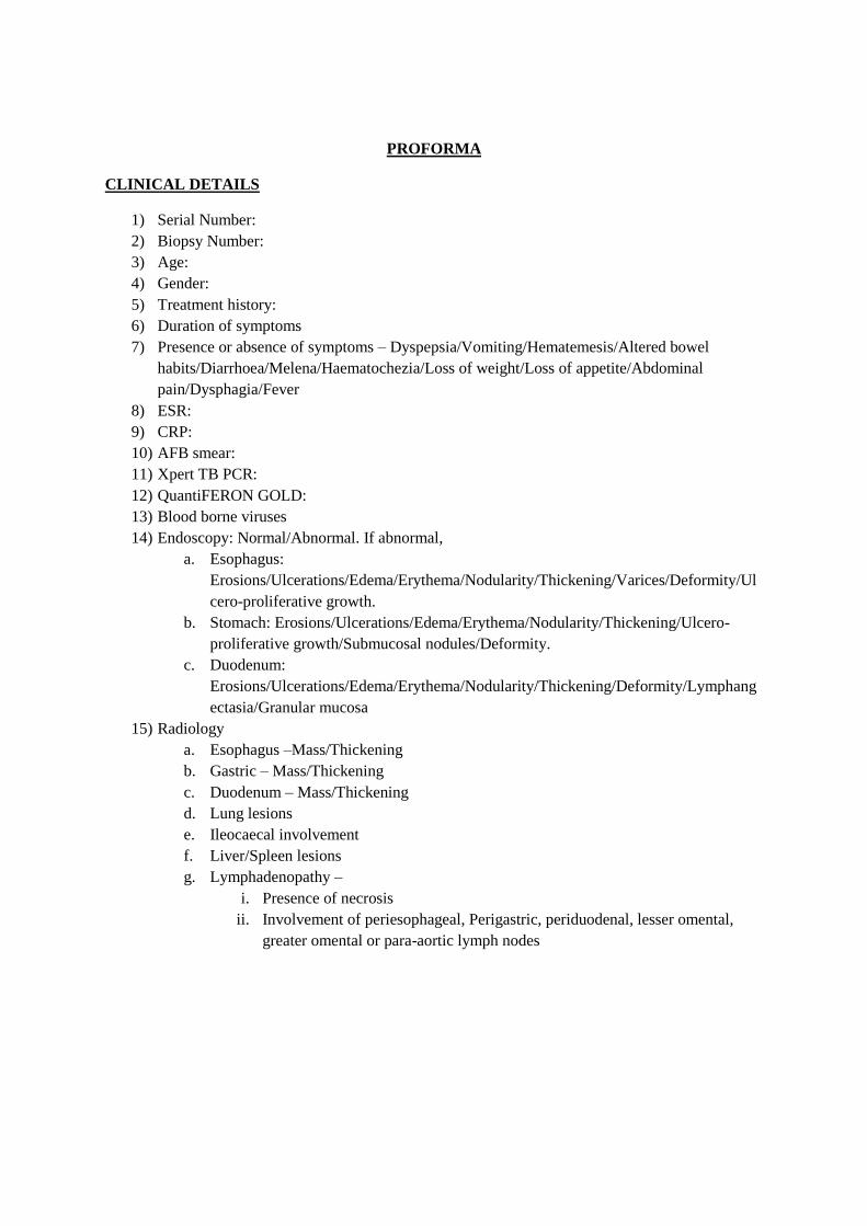
PROFORMA
CLINICAL DETAILS
1) Serial Number:
2) Biopsy Number:
3) Age:
4) Gender:
5) Treatment history:
6) Duration of symptoms
7) Presence or absence of symptoms – Dyspepsia/Vomiting/Hematemesis/Altered bowel
habits/Diarrhoea/Melena/Haematochezia/Loss of weight/Loss of appetite/Abdominal
pain/Dysphagia/Fever
8) ESR:
9) CRP:
10) AFB smear:
11) Xpert TB PCR:
12) QuantiFERON GOLD:
13) Blood borne viruses
14) Endoscopy: Normal/Abnormal. If abnormal,
a. Esophagus:
Erosions/Ulcerations/Edema/Erythema/Nodularity/Thickening/Varices/Deformity/Ul
cero-proliferative growth.
b. Stomach: Erosions/Ulcerations/Edema/Erythema/Nodularity/Thickening/Ulcero-
proliferative growth/Submucosal nodules/Deformity.
c. Duodenum:
Erosions/Ulcerations/Edema/Erythema/Nodularity/Thickening/Deformity/Lymphang
ectasia/Granular mucosa
15) Radiology
a. Esophagus –Mass/Thickening
b. Gastric – Mass/Thickening
c. Duodenum – Mass/Thickening
d. Lung lesions
e. Ileocaecal involvement
f. Liver/Spleen lesions
g. Lymphadenopathy –
i. Presence of necrosis
ii. Involvement of periesophageal, Perigastric, periduodenal, lesser omental,
greater omental or para-aortic lymph nodes
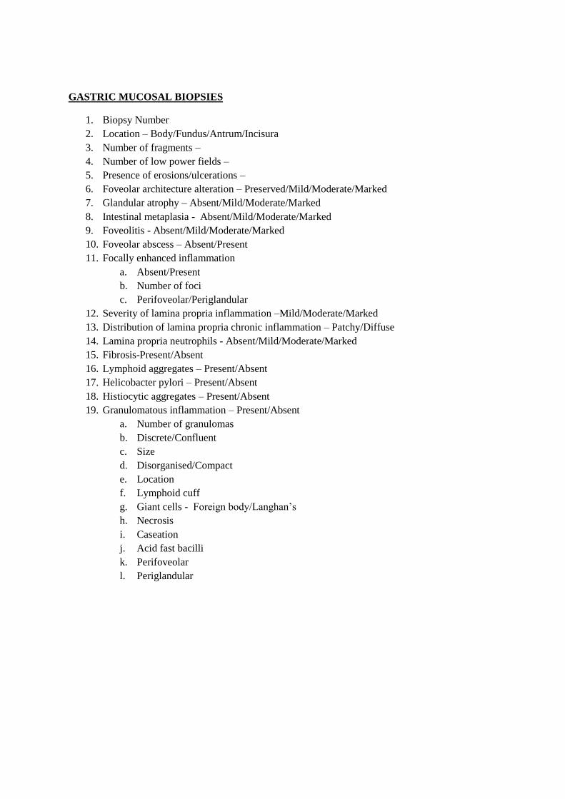
GASTRIC MUCOSAL BIOPSIES
1. Biopsy Number
2. Location – Body/Fundus/Antrum/Incisura
3. Number of fragments –
4. Number of low power fields –
5. Presence of erosions/ulcerations –
6. Foveolar architecture alteration – Preserved/Mild/Moderate/Marked
7. Glandular atrophy – Absent/Mild/Moderate/Marked
8. Intestinal metaplasia - Absent/Mild/Moderate/Marked
9. Foveolitis - Absent/Mild/Moderate/Marked
10. Foveolar abscess – Absent/Present
11. Focally enhanced inflammation
a. Absent/Present
b. Number of foci
c. Perifoveolar/Periglandular
12. Severity of lamina propria inflammation –Mild/Moderate/Marked
13. Distribution of lamina propria chronic inflammation – Patchy/Diffuse
14. Lamina propria neutrophils - Absent/Mild/Moderate/Marked
15. Fibrosis-Present/Absent
16. Lymphoid aggregates – Present/Absent
17. Helicobacter pylori – Present/Absent
18. Histiocytic aggregates – Present/Absent
19. Granulomatous inflammation – Present/Absent
a. Number of granulomas
b. Discrete/Confluent
c. Size
d. Disorganised/Compact
e. Location
f. Lymphoid cuff
g. Giant cells - Foreign body/Langhan’s
h. Necrosis
i. Caseation
j. Acid fast bacilli
k. Perifoveolar
l. Periglandular
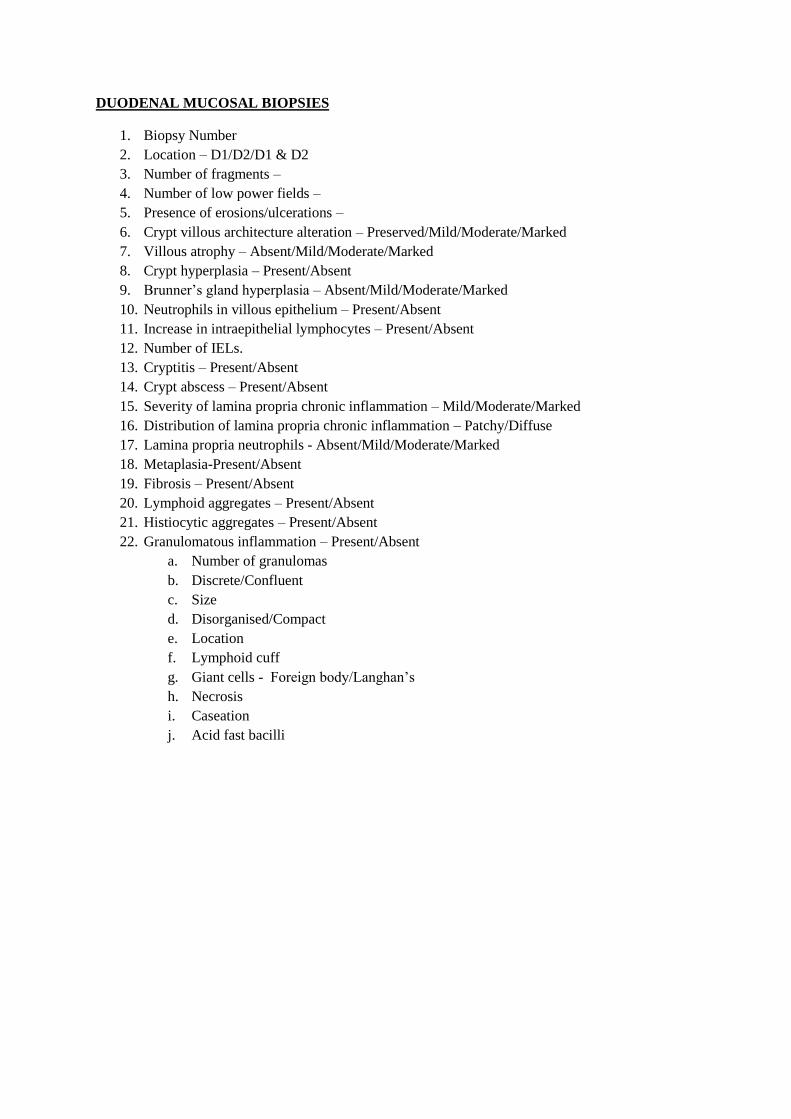
DUODENAL MUCOSAL BIOPSIES
1. Biopsy Number
2. Location – D1/D2/D1 & D2
3. Number of fragments –
4. Number of low power fields –
5. Presence of erosions/ulcerations –
6. Crypt villous architecture alteration – Preserved/Mild/Moderate/Marked
7. Villous atrophy – Absent/Mild/Moderate/Marked
8. Crypt hyperplasia – Present/Absent
9. Brunner’s gland hyperplasia – Absent/Mild/Moderate/Marked
10. Neutrophils in villous epithelium – Present/Absent
11. Increase in intraepithelial lymphocytes – Present/Absent
12. Number of IELs.
13. Cryptitis – Present/Absent
14. Crypt abscess – Present/Absent
15. Severity of lamina propria chronic inflammation – Mild/Moderate/Marked
16. Distribution of lamina propria chronic inflammation – Patchy/Diffuse
17. Lamina propria neutrophils - Absent/Mild/Moderate/Marked
18. Metaplasia-Present/Absent
19. Fibrosis – Present/Absent
20. Lymphoid aggregates – Present/Absent
21. Histiocytic aggregates – Present/Absent
22. Granulomatous inflammation – Present/Absent
a. Number of granulomas
b. Discrete/Confluent
c. Size
d. Disorganised/Compact
e. Location
f. Lymphoid cuff
g. Giant cells - Foreign body/Langhan’s
h. Necrosis
i. Caseation
j. Acid fast bacilli

ESOPHAGEAL MUCOSAL BIOPSIES
1. Biopsy Number
2. Location – D1/D2/D1 & D2
3. Number of fragments –
4. Number of low power fields –
5. Presence of erosions/ulcerations –
6. Acanthosis – Absent/Mild/Moderate/Marked
7. Parakeratosis – Absent/Mild/Moderate/Marked
8. Basal cell hyperplasia – Present/Absent
9. Papillary elongation – Present/Absent
10. Increase in intraepithelial lymphocytes – Absent/Mild/Moderate/Marked
11. Intraepithelial eosinophils - Absent/Mild/Moderate/Marked
12. Severity of lamina propria chronic inflammation – Mild/Moderate/Marked
13. Distribution of lamina propria chronic inflammation – Patchy/Diffuse
14. Lamina propria neutrophils - Absent/Mild/Moderate/Marked
15. Fibrosis – Present/Absent
16. Lymphoid aggregates – Present/Absent
17. Histiocytic aggregates – Present/Absent
18. Granulomatous inflammation – Present/Absent
a. Number of granulomas
b. Discrete/Confluent
c. Size
d. Disorganised/Compact
e. Location
f. Lymphoid cuff
g. Giant cells - Foreign body/Langhan’s
h. Necrosis
i. Caseation
j. Acid fast bacilli