Embed Size (px)
Citation preview

The functional and molecular consequences of
oxidation in the skeletal muscle myofilament
Timothy Spencer
Discipline of Physiology / School of Medical Sciences
University of Adelaide
November 2010
i
Table of contents
Thesis Declaration vi
Acknowledgements vii
Thesis Abstract viii
Common Abbreviations x
Literature Review
Introduction 2
Oxidative balance 6
Free radical production and oxidation 6
Reactive oxygen species 7
Reactive nitrogen species 11
Maintaining the balance: Antioxidants 13
Redox state 13
Endogenous antioxidants 14
Exogenous antioxidants 15
Oxidation as a cellular signal 16
Role of ROS in cellular signaling 17
Role of RNS in cellular signaling 18
Pathological roles and sources of free radicals 20
Inflammation and neutrophil activation 21
Muscle dysfunction following sepsis 21
Ischemia reperfusion injury 23
Muscle dysfunction during ischemia reperfusion injury 24

ii
Fundamental mechanisms of regulating striated muscle contraction and
relaxation 25
Cross-bridge cycling 25
The role of ion channels and Ca2+
28
Contractile Apparatus 33
Variations in muscle types 33
The myofilament 34
Myosin 36
Actin 44
Tropomyosin 46
The troponin complex 49
Troponin I 50
Troponin T 51
Troponin C 52
General Methods
Single muscle myography 58
Premise 58
Muscle dissection and mounting 58
Apparatus 62
Ca2+ activation and contractile function 64
Experimental treatments 65
Sr2+
mediated indentification of fast- vs slow-twitch fibers 65
Nitric oxide donor treatment 65
Hydrogen peroxide treatment 66
Troponin C extraction and replacement 66
Data analysis 67

iii
Proteomic techniques and analysis 68
Separation of proteins using SDS-PAGE 68
Premise 68
Polyacrylamide gel composition and preparation 68
SDS-PAGE loading and running 72
Isoelectric focusing 73
Premise 73
IEF sample preparation 75
Isoelectric focusing protocol 76
Western blot analysis 77
Premise 77
Transfer of proteins to nitrocellulose 77
Incubation with primary and secondary antibody 78
Chemilumensence detection of specific proteins 79
Data analysis 79
Recombinant mutant TnC expression 81
Premise 81
Mutagenesis 83
Transformation and expression 84
Purification 84
Acknowledgements 87
Sequential effects of GSNO and H2O2 on the Ca2+
-sensitivity of the
contratile apparatus of fast- and slow-twitch skeletal muscle fibers
from the rat
Statement of contribution 89
Abstract 90
Introduction 91
Methods 95
Muscle preparation 95

iv
Skinned fiber solutions 95
Ca2+ activation of contractile apparatus 97
Spectrophotometric analysis of SH content in skinned muscle bundles 99
Analysis 100
Results 101
Separate effects of NO and H2O2 on Ca2+-sensitiivity 101
Evidence of glutathionylation of the contractile apparatus by GSH and GSSG 106
Sequential treatment with NO and H2O2 on Ca2+-sensitivity 108
Discussion 120
Individual and sequential effects of GSNO and H2O2 on Ca2+-sensitivity of fast-twitch muscle 120
Effects of GSNO and H2O2 on slow-twitch skeletal and relevance to fatigue 123
Conclusions 124
Myofibril basis for oxidative dysfunction in skeletal muscle
Abstract 127
Introduction 129
Methods 133
Muscle preparation 133
Functional analysis 134
Ca2+
activation of contractile apparatus 134
TnC extraction and reconstitution with recombinant TnC 135
Data Analysis 136
Proteomic Analysis 137
Isoelectric focusing 137
Second-Dimensional SDS-PAGE 138
Western blot analysis of TnC and LC20 139
Recombinant protein expression 139
Results 141
Ca2+-Sensitivity of fast-twitch skeletal muscle 141

v
GSNO mediated modification of muscle fiber proteins 143
Substitution of native TnC with recombinant TnC 145
The role of C84 TnC in muscle fiber Ca2+sensitivity 149
Discussion 152
Future directions and implications 156
General Discussion
Discussion 159
Limitations and future directions 163
Physiological sources of NO 165
Significance 166
References 168

vi
THESIS DECLARATION
This work contains no material which has been accepted for the award of any other
degree or diploma in any university or other tertiary institution to Timothy Spencer
and, to the best of my knowledge and belief, contains no material previously
published or written by another person, except where due reference has been made in
the text.
I give consent to this copy of my thesis, when deposited in the University Library,
being made available for loan and photocopying, subject to the provisions of the
Copyright Act 1968.
The author acknowledges that copyright of published works contained within this
thesis (*) resides with the copyright holder(s) of those works.
I also give permission for the digital version of my thesis to be made available on the
web, via the University‟s digital research repository, the Library catalogue, the
Australasian Digital Theses Program (ADTP) and also through web search engines,
unless permission has been granted by the University to restrict access for a period of
time.
Signed,
Timothy Spencer
Spenecr, T. and G. Posterino (2009). "Sequential effects of GSNO and H2O2 on the Ca2+." American Journal of Physiology- Cell Physiology 296: C1015-C1023.
Spencer TN, Cooke J, Brown L and Wilson DP (2010). “Evidence for C84 Troponin C as the target for GSNO-mediated myofilament Ca2+ desensitization” ISHR Australian National Conference

vii
ACKNOWLEDGEMENTS
I wish to thank my primary supervisor, Dr David Wilson. His mentorship and
guidance during this time has been immeasurable and I will benefit from for the rest
of my life.
I wish to thank Dr Giuseppe Posterino (La Trobe University, Victoria) for his
supervision of Chapter 3 “Sequential effects of GSNO and H2O2 on the Ca2+-
sensitivity of the contractile apparatus of fast- and slow-twitch skeletal muscle fibers
from the rat”.
I wish to thank Dr Louise Brown and James Cooke at the Department of Chemistry
and Biomolecular Sciences, Macquarie University, Sydney, Australia. Recombinant
proteins used in the current study were a gift from Louise Brown‟s laboratory.
Finally I would like to thank my laboratory colleagues; Dr. Scott Copley, Kanchani
Rajopadhyaya, Jessica Dunn, Ksenya Wojewidka, Yann Chan, Amenah Jaghoori and
Joanne Eng, for creating such a friendly and productive laboratory environment.

viii
THESIS ABSTRACT
It is becoming increasingly evident that redox state leading to post-translational
modifications of structural proteins, enzymes and ion channels can cause activation or
inhibition of cellular function (Andrade et al., 1998a, Jackson, 2008, Kelly et al.,
1996). While low levels of nitric oxide (NO) synthesised by endothelial and neuronal
nitric oxide synthase have been shown to provide a beneficial effect to tissues, the
elevated release of NO accompanying inflammation has a detrimental effect, resulting
in dysfunction (Khanna et al., 2005). We investigated the functional consequence and
molecular substrate of NO and another potentially harmful reactive oxygen species,
H2O2, on the skeletal muscle myofilament.
In a rat model we used functional myography of demembranated single fast- and
slow-twitch skeletal muscle fibers to examined the consequence of the addition of the
free radical NO and reactive oxygen species H2O2 on the Ca2+ sensitivity of the
myofilament. The reversibility of oxidative modifications following NO or H2O2
treatment was examined using the general anti-oxidant dithiothreitol. Isoelectric
focusing combined with SDS-PAGE separation of proteins investigated the post-
translational modification of free-radical exposed myofilament proteins. Molecular
substitution of endogenous troponin C (TnC) with WT cardiac/slow TnC or C84S
TnC, incapable of being oxidized at Cys84, investigated the molecular and functional
consequence of oxidation of TnC at Cys84.

ix
Exposure of fast-twitch muscle fibers to NO resulted in a decrease in Ca2+ sensitivity,
while H2O2 had the opposite effect, increasing Ca2+ sensitivity. In contrast, slow-
twitch fibers were insensitive to both NO and H2O2. Following myofilament exposure
to NO (~2 M) proteomic analysis revealed that many proteins underwent post-
translational modification, including myosin light chain (LC20) and TnC. Molecular
substitution of endogenous fast-twitch TnC with WT-cardiac/slow TnC demonstrated
a similar sensitivity to NO as WT skeletal muscle. In contrast TnC, non-oxidizable at
Cys84, rendered fast-twitch skeletal muscle insensitive to NO.
Many myofilament proteins, including myosin light chains were identified as being
post-translationally modified by NO exposure, however, molecular substitution
experiments clearly identify TnC, specifically residue Cys84 as the functional
substrate responsible for fast-twitch skeletal muscle sensitivity to NO. Although
slow-twitch muscle contains the same isoform of TnC, it was insensitive to NO. This
suggests that slow-twitch muscle may have a greater capacity for anti-oxidant defense
than fast-twitch muscle. The contrasting increase in Ca2+ sensitivity following H2O2
to the decline caused by NO demonstrates that not all oxidative molecules act alike,
possibly targeting differing substrates and causing differing post-translational
modifications.
x
COMMON ABBREVIATIONS
BH4 tetrahydrabiopterin
[Ca2+]cyt cytoplasmic calcium concentration
CICR calcium-induced-calcium-release
DHPR dihydropyridine receptor
DTT dithiothreitol
GSNO S-nitroso-glutathione
H2O2 hydrogen peroxide
IEF isoelectric focusing
IRI ischemia-reperfusion injury
MHC myosin heavy chain
MI myocardial infarct
NOS nitric oxide synthase
O2·- super oxide
OH- hydroxyl radical
ONOO peroxynitrite
pCa log [Ca2+]
RNS reactive nitrogen species
ROS reactive oxygen species
RyR ryanodine receptors
SDS-PAGE sodium dodecyl sulfate polyacrylamide gel electrophoresis
SNAP sodium nitroprusside
SOD super oxide dismutase
SR sarcoplasmic reticulum
TnC troponin C
TnI troponin I
TnT troponin T

Literature Review

2
Introduction
Contraction of cardiac and skeletal muscle is a highly regulated process involving;
the generation of ATP, regulation of extracellular and sarcoplasmic reticulum (SR)
Ca2+ entry and Ca2+-dependent conformational changes of structural, regulatory and
motor proteins. It is becoming increasingly evident that redox state leading to post-
translational modifications of structural proteins, enzymes and ion channels can cause
activation or inhibition of cellular function (Andrade et al., 1998a, Jackson, 2008,
Kelly et al., 1996). However, many cells are able to maintain their redox balance
despite the generation of reactive oxygen (ROS) and reactive nitrogen species (RNS)
primarily due to the production of endogenous anti-oxidants and reductants. Anti-
oxidants convert oxidized molecules into harmless electron stable molecules or
remove modifications caused by an oxidative reaction. For example superoxide
dismutase (SOD) enzymatically converts the reactive oxygen species superoxide (O2·-
) to the less reactive hydrogen peroxide (H2O2) and oxygen (McCord and Fridovich,
1969).
O2·- + O2·- + 2H+ → O2 + H2O2
Catalase is a selective anti-oxidant enzyme involved in the catalytic conversion of
H2O2 to H2O and oxygen (Murrant and Reid, 2001).
H2O2 → 2H2O + O2
It therefore follows that molecular, cellular and systems function can be regulated by
an imbalance of oxidant and/or reductant.
Following from the Nobel Prize winning work of Furchgott, Murad and Ignaro
(Furchgott, 1999) it is well recognized that endothelial derived nitric oxide (NO) is an

3
important vasodilator which improves blood flow and consequently cardiac and
skeletal muscle perfusion and function (Kelm and Schrader, 1990, Rees et al., 1989,
Stamler and Meissner, 2001). In contrast, following myocardial infarction or skeletal
muscle damage infiltrating neutrophils and mast cells activate their inducible nitric
oxide synthase (NOS) to generate a very large release of NO (Khanna et al., 2005,
Laroux et al., 2001).
In addition, several studies have demonstrated that application of exogenous NO
donors to functional cardiac and skeletal muscle preparations have a negative
influence on the contractile function. Using the mixed fiber-type diaphragm muscle
Kobzik et al. identified that nitric oxide synthase inhibitors such as nitro-L-arginine
improve contractile function including the force-frequency relationship, and that these
increases in contractile function could be reversed by the exogenous NO donors S-
nitroso-N-acetylcysteine (SNAC) and sodium nitroprusside (SNAP) (Kobzik et al.,
1994b). Similarly, in electrically stimulated cardiomyocytes, the application of SNAP
or superfusion with an NO containing physiological solution attenuated contraction
(Brady et al., 1993). These data illustrate that high levels of NO can have a
detrimental effect on one or more elements involved in excitation-contraction
coupling in muscle.
In fast-twitch fibers, the contractile apparatus is sensitive to ROS including H2O2.
Exogenous application of H2O2 has been shown to enhance the contractile function of
myocytes, increasing the Ca2+-sensitivity of the contractile apparatus likely through
oxidation of sulfhydryl groups (Andrade et al., 1998a, Lamb and Stephenson, 1994).

4
It is important to recognise that, as well as species specific differences, fast- and
slow-twitch skeletal muscles are functionally different in several respects for
example: fast-twitch muscle have a higher peak amplitude of Ca2+ release (fast,
18.50.5 vs. slow, 6.41.0 µM ∆[Ca2+] ) and a shorter time between ½ maximum
contraction and ½ maximum relaxation (fast, 4.90.3 vs. slow, 7.70.6 ms) (Baylor
and Hollingworth, 2003). Herein, all studies were conducted with rat fast- and slow-
twitch skeletal muscle. Fast-twitch fibers are more sensitive to fatigue, whereas slow-
twitch fibers show a fatigue resistance that appears to be due in part to a lack of
sensitivity to various metabolites involved in fatigue in fast-twitch fibers. This key
feature enables one to functionally discriminate between the fast- and slow-twitch
fiber types. To date, there has been no systematic comparison of the effects of both
ROS and NO on the Ca2+-sensitivity of both fast- and slow-twitch fiber types,
moreover there is very little known about the role of NO in slow-twitch fiber Ca2+-
sensitivity.
While several studies have shown the capacity of proteins involved in excitation-
contraction coupling to be oxidised, and documented an association of contractile
dysfunction with elevated ROS/RNS, very few studies have demonstrated a direct
link between the observed contractile dysfunction and oxidation of specific proteins.
In this study we identify the functional effects of both NO, through the use of
exogenous donors, and the ROS H2O2, on fast- and slow-twitch skeletal muscle
myofilament function. Importantly we have extended the study to include

5
identification of the functional substrate that causes the NO-mediated decline in
myofibril Ca2+-sensitivity in fast twitch skeletal muscle.

6
Oxidative balance
Free radical production and oxidation
Free radicals are chemical species possessing an unpaired electron. They are formed
in a variety of ways including; 1) homolytic cleavage of a covalent bond; 2) the loss
of a single electron; 3) the addition of a single electron. Electron transfer is the more
common process, as homolytic cleavage requires high-energy input, such as high
temperature or UV light.
Free radicals or broadly, oxidants cause either the addition of a charged molecule like
oxygen or the loss of electrons from a substrate. The consequences of which include
changes in protein conformation and protein-protein interactions altering cellular
function. Similar post-translational modifications, such as phosphorylation (the
addition of a phosphoryl group, PO4), are important signaling mechanisms, however,
unlike phosphorylation many oxidative reactions are not catalyzed by an enzyme and
may not have evolved with the same degree of target specificity (Mannick and
Schonhoff, 2002).

7
Reactive oxygen species
As a consequence of basal muscle metabolism oxidants are always being generated,
in fact Davies et al. (1982) have provided evidence of lipid peroxidation and oxidant
production during rest, which become elevated following submaximal, exhaustive
activity independent from inflammation. Although free radical production was known
to occur well before 1970, the diversity of specific oxidant species produced within
muscle were not identified till almost 20 years later. Superoxide and hydrogen
peroxide were found to be released in contracting diaphragm muscle, as identified by
the use of the selective anti-oxidant enzymes SOD and catalase, reducing 2‟,7‟-
dichlorofluorescein, an intracellular fluorochrome probe for oxidation (Reid et al.,
1992). The hydroxyl radical was also detected in contracting skeletal muscle (O'Neill
et al., 1996) and nitric oxide generation has been identified in skeletal muscle (Balon
and Nadler, 1994).
Molecular sources of reactive oxygen species in skeletal muscle are varied and
depend on the physiological state of the muscle. Glycolytic metabolism results in the
production of ATP from glucose and ADP but, as a by-product of the conversion of
glyceraldehyde 6-phosphate to 1,3-bisphosphoglycerate, also produces two unstable
hydrogen cations which can go on to form H2O2. Significantly, interruption of the
electron transport chain, resulting in incomplete reduction of oxygen to H2O, also
produces oxidants in the form of the superoxide anion (Boveris et al., 1972, Loschen
et al., 1971). Superoxide is generated at complex I and III of the electron transport
chain by electron transport intermediates such as ubisemiquinone (Nishikawa et al.,
2000). Changes in the mitochondrial environment such as increased [Ca2+] following

8
ischemia or a high potential difference in the proton gradient which drives ATP
synthase, can disrupt electron transport and actuate O2- generation (Korshunov et al.,
1997, Turrens, 1997). It was widely reported that 2-5% of total oxygen consumed by
mitochondria result in the generation superoxide (Boveris and Chance, 1973, Loschen
et al., 1974), more recent reports suggest this value may be lower (St-Pierre et al.,
2002) but nevertheless still present. As striated muscles have a high density of
mitochondria, oxidants are largely produced during cell metabolism.
A number of other sources of O2- generation have been identified (Fig A.1).
Superoxide is also generated by a cardiac and skeletal muscle sarcoplasmic reticulum
(Cherednichenko et al., 2004, Xia et al., 2003)) and plasma membrane (Javesghani et
al., 2002) associated enzyme, NAD(P)H oxidase. Reid and colleagues have described
a phospholipase A2-dependent superoxide generation distinct from the calcium-
dependent phospholipase A2 ROS production also identified in skeletal muscle (Gong
et al., 2006, Nethery et al., 1999). Xanthine oxidase is also a source of O2-
generation, however, its specific role in physiological or pathophysiological oxidative
stress remains equivocal.
Superoxide is highly unstable, with a half-life (t 1/2) of 1 sec (Reth, 2002) before
reacting with either H2O to form H2O2 or with H2O2 form the hydroxyl anion (Fig
A.1). Peroxide is membrane permeant and more stable with a relatively long t 1/2 of
1 msec (Reth, 2002). However, in a pure solution it reaches a stable equilibrium due
to the limited available molecules in the buffer to be oxidized. Although H2O2 is
cytotoxic it is considered a relatively weak oxidizing agent, nevertheless, it readily

9
generates other very toxic free radicals such as the hydroxyl radicals (OH-)
particularly in the presence of catalytic transition metals iron (Fe2+) commonly
released during ischemia (Pattwell and Jackson, 2004).
Figure A.1. Free radical sources within the muscle and associated cells. Nitric
oxide is released by nitric oxide synthase (NOS) enzymes such as endothelial NOS
(eNOS) and inducible NOS (iNOS). Superoxide (O2-) has multiple cellular sources
including; NADPH oxidase, xanthine oxidase, phospholipase A2 and mitochondria.
The uncoupling of eNOS through limitation of substrate or co-factors such as
tetrahydrabiopterin (BH4) leads to O2- production. Superoxide and NO react to form
peroxynitrite (ONOO). The endogenous anti-oxidant, super oxide dismutase (SOD)
catalyses the conversion of O2- to H2O2 which may go on to form OH- or in the
presence of catalase break down to H2O and O2 .

10
The relative potency of reactive oxygen species (Halliwell, 1987).
OH- (highly reactive) > O2- (selectively reactive) > H2O2 (weak non-radical)

11
Reactive nitrogen species
As every cell in the body contains mitochondria, superoxide production is ubiquitous.
Similarly the free radical, nitric oxide (NO), is produced in most organs in the body.
Nitric oxide is synthesized by the enzyme, nitric oxide synthase (NOS) from the
substrates L-arginine and oxygen, utilizing several co-factors including NADPH and
tetrahydrobiopterin (BH4), to form L-citrilline and NO (Fig A.1). There are three
known isoforms of nitric oxide synthase, two constitutively expressed Ca2+/CaM
dependent isoforms, endothelial-NOS and neuronal-NOS and the Ca2+ independent,
inducible-NOS. Endothelial-NOS mediated NO is produced in every organ of the
body simply because every organ contains blood vessels and endothelial cells.
Similarly most organs in the body are innervated and consequently produce NO
through a neuronal NOS-dependent mechanism. However, under normal physiology
this NO is used as a direct vasodilator or as a neurotransmitter.
Evidence of the expression of multiple isoforms of NOS has also been found in
skeletal muscle. Staining with a monoclonal antibody for nNOS, identified its
presence in the sarcolemma membrane, while staining for eNOS displayed co-
localization with mitochondrial markers suggesting eNOS expression in
mitochondrial membranes (Kobzik et al., 1994b, Kobzik et al., 1995). Along with
endothelial and neuronal cells, many tissues including muscle contain another
potential source of NO synthesized from resident neutrophils expressing iNOS,
activated via cytokines liberated during the inflammatory response.

12
Nitric oxide is continuously generated in skeletal muscle, with production increasing
during contraction (Balon and Nadler, 1994) likely due to increased mitochondrial
activity, neuronal activation, or endothelial release as one increases vasodilatation
during exercise. Nitric oxide is a membrane permeable free radical and readily reacts
with other molecules and proteins as it attempts to reach a ground state. Importantly,
in the presence of superoxide, nitric oxide will preferentially react to form
peroxynitrite (Fig A.1) a potent vasoconstrictor and highly reactive and damaging
free radical, (Halliwell et al., 1999).
O2-• + NO ONOO-

13
Maintaining the balance: anti-oxidants
Redox state
Anti-oxidants function to delay, prevent or reverse the oxidation of cellular substrates
by oxidants. The balance of anti-oxidants and oxidants in a cellular system
determines the redox state, or level of oxidative post-translational modification
present within the cell. The redox state of a cellular system is linked to many
pathological states particularly when genes encoding anti-oxidant enzymes have been
mutated. The pathological disorder is manifest in a variety of systems and includes;
idiopathic cardiomyopathy, neurological disorders and cancer. Particularly relevant in
our aging population is the finding that diabetics are reported to have decreased levels
of protective anti-oxidants and an increase in oxidative stress (Borcea et al., 1999,
Maritim et al., 2003).
Levels of anti-oxidants can also vary with age, diet and exercise. Anti-oxidant
production is up regulated in response to prolonged muscle conditioning and down
regulated with de-conditioning (Clanton et al., 1999). Dietary intervention has also
been studied and used to alter anti-oxidant levels within the body; however, the
benefits of dietary interventions remain equivocal (Bjelakovic et al., 2004). This may
be complicated by the administration of anti-oxidants, which normally act
synergistically with endogenous anti-oxidants and that alone, have the potential to act
as oxidants themselves once initially oxidized (Herbert, 1994).

14
Endogenous anti-oxidants
A number of anti-oxidants act on specific ROS and are compartmentalized
throughout the cytoplasm and within organelles such as the mitochondria, enabling
them to act near the source of the specific ROS production or important target
proteins. The major anti-oxidants in the body include catalase, superoxide dismutase,
glutathione and peroxidase. Several isoforms of superoxide dismutase are found
within the cell and in the extracellular space as well as within the mitochondrial
matrix (Weisiger and Fridovich, 1973a, Weisiger and Fridovich, 1973b). Superoxide
dismutase works specifically on the superoxide radical to form the less reactive
oxidant, hydrogen peroxide (Sen, 1995). Hydrogen peroxide can be further reduced
by the hydrogen peroxide dismutase, catalase, to O2 and H2O (Deisseroth and
Dounce, 1970). Glutathione is a less specific anti-oxidant, spontaneously scavenging
a wide variety of ROS. It is synthesized within the body from the amino acids L-
cysteine, L-glutamic acid and glycine. Glutathione itself has a free cysteine moiety
that donates an electron to a substrate, with the consequence of reducing the substrate.
During this reaction glutathione becomes an unstable, oxidative form, but it readily
reacts with other reactive glutathione molecules to form stable glutathione disulfide.
Glutathione reductase and NADPH are responsible for regenerating glutathione.
Glutathione acts in concert with other anti-oxidants, reducing them from their radical
form allowing them to continue at act as anti-oxidants (Sen, 1995).
Other anti-oxidants function to remove the post-translational modifications caused by
some oxidants. Protein disulfide reductase, glutaredoxin and thioredoxin reductase
remove intramolecular and mixed di-sulfide bonds caused by oxidation.

15
Exogenous anti-oxidants
Exogenous anti-oxidants form a second line of defense against oxidative changes and
are primarily obtained as nutrients or supplements in the diet. Exogenous anti-
oxidants have become the focus of a large number of clinical studies as evidenced in
a variety of published meta-analysis (Bjelakovic et al., 2004, Miller et al., 2005,
Vivekananthan et al., 2003). However, many exogenous anti-oxidants are consumed
as part of normal dietary intake. Increasing the intake of exogenous anti-oxidants
such as omega-3 and 6 fatty acids, vitamins C, D and E and lutein have been
suggested to increase anti-oxidant levels within the body, with a potential beneficial
effect (Heinonen and Albanes, 1994, Landrum et al., 1997, Morris et al., 1998,
Simopoulos, 1991, Zandi et al., 2004). However, meta-analysis of exogenous anti-
oxidant clinical trials provides evidence that the efficacy of such treatments remains
equivocal (Bjelakovic et al., 2004, Miller et al., 2005, Vivekananthan et al., 2003).

16
Oxidation as a cellular signal
Similar to enzyme mediated phosphorylation, spontaneous oxidation can cause post-
translational modifications of proteins and lipids leading to conformational changes
and alterations in protein or lipid function. For example NO is a cellular signal often
associated with vascular control. Nitric oxide produced from Ca2+-dependent eNOS
in the endothelium is a potent vasodilator. Nitric oxide reacts with transition metals
such as heme iron or iron-sulfur centers to form metal-NO products. Specifically this
NO-dependent reaction is used to increase the activity of enzymes including
guanylate cyclase leading to increased cyclic guanosine monophosphate production
(cGMP) (Furchgott, 1999). The signal propagated by activating cGMP-dependent ion
channels, protein kinases and phosphodiesterases mediates among other functions, a
reduction in intracellular [Ca2+] leading to smooth muscle relaxation and increase in
organ blood flow. On the other-hand, peroxynitrite derived from NO has the opposite
effect. Specifically, the uncoupling of eNOS through oxidation or the lack of
substrates or co-factors such as BH4, as seen with aging and sedentary lifestyle leads
to the production of superoxide rather than NO (Stroes et al., 1998). Superoxide
reacts with the residual NO present to produce peroxynitrite. This limits the
bioavailibility of NO reducing vasodilatation, amplified by the fact that peroxynitrite
is also a vasoconstrictor.

17
The role of ROS in cellular signaling
Reactive oxygen species and the resulting oxidation can promote the generation of an
anti-oxidant defense mechanism which results in the up regulation of anti-oxidants in
mitochondria. Specifically oxidation has been shown to stimulate mitochondrial
biogenesis via activation of transcription factors for genes encoding glutathione S-
transferase, glutathione peroxidase and glutathione reductase, all involved in
glutathione biosynthesis and cycling (Motohashi and Yamamoto, 2004, Piantadosi
and Suliman, 2006). Reactive oxygen species are also thought to regulate SOD and
catalase activity. In fact, muscle contraction increases ROS production, while post-
exercise is associated with increase in catalase and SOD activity in both mouse
(McArdle et al., 2001) and human muscle (Khassaf et al., 2001). The increased
activity of catalase and SOD may represent an increase in the amount of enzyme
present. Interestingly prior supplementation with Vitamin C (an anti-oxidant) reduces
these responses (Khassaf et al., 2001), suggesting the level of oxidation and ROS is
directly involved in increasing the amount of catalase and SOD present following
activity.

18
The role of RNS in cellular signaling
Nitric oxide, or more specifically NO derived species, readily react with protein
thiols, forming a S-NO bond or nitrosylation. The formation of nitrosothiols is
favoured in cysteine thiols, however, not all free-thiol cysteines become nitrosylated
(Lane et al., 2001, MartÌnez-Ruiz and Lamas, 2004). A level of specificity is seen in
protein nitrosylation, enabling it to be involved in cellular signaling. Cysteines with
an “acid-base motif” either in the primary sequence (Stamler et al., 1997) or in
proximity in the tertiary structure provide a more favorable reaction (Ascenzi et al.,
2000), providing specificity through a consensus motif , e.g. Hist-Cys96-Asp forms
the NO binding site of the -subunit of haemoglobin (Hess et al., 2001). As expected
the consequences of nitrosylation are also concentration dependent and the co-
localization of reactive proteins with eNOS and nNOS provides some specificity in
signaling, however, it appears that specificity may be lost in iNOS mediated reactions
due to NO generation being several orders of magnitude greater than eNOS or nNOS.
Nitrosylation has been identified as a potential mediator in a number of signaling
pathways. Nuclear magnetic resonance of P21RAS, an activator of NF-B, has
identified a nitrosylated cysteine located in a nucleotide-binding motif. Although
nitrosylation did not apparently alter the protein structure the enzyme activity is
increased during the process of nitrosylation (Williams et al., 2003). Nitrosylation has
also been implicated in modification of the p50 subunit of NF-B inhibiting DNA
binding (delaTorre et al., 1999, Marshall et al., 2004, Matthews et al., 1996),
activation of hypoxia-inducible factors (Sumbayev, 2003, Yasinska and Sumbayev,

19
2003) and the inhibition of the ryanodine receptor (MÈsz·ros et al., 1996,
ZahradnÌkov· et al., 1997).

20
Pathophysiological roles and sources of oxidants
Oxidants are produced under a number of pathophysiological conditions including
heat stress, enzyme uncoupling and inflammation resulting from conditions including
sepsis, ischemia-reperfusion injury, trauma or activity induced muscle injury. Under
these conditions the production of oxidizing agents is in excess of the ability of
endogenous reductants to quench them and consequently results in the cellular ROS
and RNS to interact with a number of cellular proteins causing post-translational
modifications that may cause cellular activation or dysfunction (De Belder et al.,
1993, Li and Shah, 2004, Paulus et al., 1994).
Proteomic assays of blood serum have shown proteins of the troponin complex to be
modified. Analysis of troponin T and troponin I released from the myocardium, have
identified both as being post-translationally modified (Labugger et al., 2000,
McDonough et al., 1999, McDonough et al., 2001), indicating the proteins of the
contractile myofilament may be modified during ischemia reperfusion injury and
other pathologies of excess NO and ROS production.

21
Inflammation and neutrophil activation
Inflammation and sepsis are both associated with an increase in ROS and RNS
(Callahan et al., 2001, Vane et al., 1994). Both pathologies activate invading
neutrophils that lead to the activation of iNOS and consequent NO generation and
substantial amounts of ROS. Muscle injury, in particular crush injury triggers a
similar local inflammatory response, activating invading neutrophils and
macrophages. The release of ROS and NO at the site of muscle crush injury is
associated with preparation of the tissue to allow for regeneration (Malech and Gallin,
1987). In the inflammatory response triggered by sepsis and IRI, the increased NO
and ROS are considered a means to destroy any invading pathogens and damaged
cells, however, the magnitude of the ROS release is very often overwhelming
resulting in collateral damage to previously healthy tissue (Bone, 1991).
Muscle dysfunction during sepsis
Nitric oxide and ROS production during sepsis is associated with muscle dysfunction
(Callahan et al., 2001, Fujimura et al., 2000, Nethery et al., 1999, Supinski et al.,
1993). Cecal ligation and perforation, or endotoxin induced sepsis have been shown
to decrease both the maximum force produced and Ca2+ sensitivity of muscle strips
and single muscle fibers isolated from the diaphragm (Callahan et al., 2001, Fujimura
et al., 2000, Nethery et al., 1999), EDL and soleus (Supinski et al., 1993). The
addition of NOS inhibitors, such as L-NAME and free-radical scavengers, such as
polyethylene glycol-superoxide dismutase (PEG-SOD) and apocynin prevents muscle
dysfunction

22
suggesting that oxidant production is associated with muscle dysfunction (Callahan et
al., 2001, Shindoh et al., 1992, Supinski et al., 1993).

23
Ischemia reperfusion injury
The physiological response to local ischemia as a result of coronary artery disease,
myocardial infarction, claudication or during heavy exercise in skeletal muscle is for
the vascular endothelium to release the potent vasodilator, NO. However, with
prolonged ischemia, tissue damage occurs leading to an inflammatory response and
subsequent release of large amounts of NO and ROS from inflammatory cells (Jordan
et al., 1999). This damage is further exacerbated during reperfusion as invading
neutrophils enter the area accompanying the restored blood flow. The larger
proportion of cellular damage during IRI is associated with the NO and ROS
production during reperfusion (Khanna et al., 2005).
Limited substrate supply or oxidation of enzymes can also lead to the production of
free radicals through the uncoupling of enzymes, including uncoupling of NOS. In
addition when the co-factor BH4 becomes limited as often occurs in aging, (Sindler et
al., 2009) NOS produces the superoxide free radical rather than NO (Heinzel et al.,
1992). Production of the superoxide anion by NOS further decreases the
bioavailability of NO due to the preferential reaction of NO with superoxide forming
peroxynitrite (Beckman and Koppenol, 1996).
Importantly, the interaction of NO and superoxide to form peroxynitrite or NO with
heme proteins to form heme protein adducts is more favorable than the interaction of
superoxide with superoxide-dismutase (SOD) leading to the production of H2O and
O2. The formation of peroxynitrite leads to the nitration of proteins and the reduced

24
bioavailability of NO, and reduces its ability to function as a physiological
vasodilator and signaling molecule. If substrate supply is restricted due to vascular
constriction or ischemia, the reduced bioavailability of NO, will further compound
the ischemic condition, as the peroxynitrite formed functions as a potent
vasoconstrictor.
Muscle dysfunction during ischemia-reperfusion injury
Ischemia-reperfusion injury (IRI) occurs when blood flow is restored to a previously
ischemic tissue. Although necessary to salvage ischemic tissue, the restoration of
blood flow actually causes further, possibly greater, damage than the ischemic event
as a result of the inflammatory response to reperfusion (Khanna et al., 2005). Nitric
oxide, although cytoprotective during ischemia, is also responsible for the damaging
inflammatory response in IRI. As with sepsis and other inflammatory responses,
iNOS is activated and NO is produced in excess (Khanna et al., 2005). Nitric oxide
production has been shown to peak following 15 minutes of ischemia, persisting for
2-3 hours in skeletal muscle within a mouse hind limb IRI model (Brovkovych et al.,
1999). Nitric oxide production has been shown to surge again beginning two hours
into reperfusion as iNOS from invading neutrophils and macrophages is activated
(Barker et al., 2001, Messina et al., 2000). Nitric oxide inhibitors, when administered
during the last 5 minutes of ischemia and the first 2 hours of reperfusion have
beneficial effects on contractile function and post-ischemic blood flow (Ikebe et al.,
2002). The beneficial effects of NOS inhibition have also been confirmed through
administration at time periods just prior to reperfusion and up to 3 hours post-
reperfusion (Zhang et al., 1997).

25
Fundamental mechanisms regulating striated muscle contraction and
relaxation
Cross-bridge cycling
Cross-bridge cycling describes the repeated process by which the myosin molecular
motor causes sliding of the thin filament, leading to contraction. As the understanding
of interactions of myosin, actin and other associated regulatory proteins has improved
so has the model of cross-bridge cycling evolved. Early studies using light
microscopy, observed changes in the size of I-band and H-zone during contraction
and produced a model of two states; 1) steric hindrance by ATP, prevented myosin
from binding to actin and prevented contraction, 2) ATP hydrolysis by the myosin
ATPase and release of the hydrolyzed phosphate imparted a conformational change
which enabled actin and myosin to bind forming the cross-bridge (Huxley and
Hanson, 1943).
The two-state model evolved into the three state model of: blocked, closed and open
(McKillop and Geeves, 1993). Biochemical and structural studies suggested that, in
the absence of Ca2+ tropomyosin prevented actin-myosin binding, the „blocked‟ state
(McKillop and Geeves, 1993). Upon Ca2+ binding troponin C on the filament moves
into the „closed‟ state (McKillop and Geeves, 1993) in which TnI detaches from actin
(Tao et al., 1990), shifting tropomyosin by ~30 and myosin is able to bind actin
through a weak electrostatic interaction. With myosin binding weakly to actin, the
filament moves into the „open‟ state (McKillop and Geeves, 1993). Myosin binding

26
weakly to actin shifts the tropomyosin a further 10 allowing for a strong bond
formation and a conformational change in the myosin head, the power stroke, shifting
the actin filament ~4nm or more (Molloy et al., 1995), accompanied by the release of
Pi (Gordon et al., 2000).
Hydrolysis of ATP by the myosin-ATPase generates the energy required to drive the
cross-bridge cycling and contraction. The movement of myosin along the actin
filament is a multi-step process; 1) myosin ATPase binds ATP, 2) the myosin head
dissociates from actin 3) ATP is hydrolyzed forming myosin-ADP-Pi, 4) the myosin
head associates with actin in a weakly bound form, 5) in a Ca2+-troponin dependent
manner, the weak association isomerizes to a strong bond, 6) in the strongly bound
configuration myosin-ATPase produces a rotation in the myosin head and neck region
(Cooke and Holmes, 1986), generating movement, a force of ~1.7pN under isometric
conditions (Molloy et al., 1995) and releases Pi, 7) myosin-ATPase, bound to actin,
releases ADP returning to the initial state (Gordon et al., 2000).

27
Figure A.2. Cross-bridge cycling. 1) Myosin ATPase binds ATP, 2) the myosin
head dissociates from actin 3) ATP is hydrolyzed forming myosin-ADP-Pi, and in a
Ca2+-troponin dependent manner binds to the actin-myosin binding site, 4) myosin-
ATPase produces a rotation in the myosin head and neck region and releases Pi, 6)
myosin-ATPase, bound to actin, releases ADP returning to the initial state. Modified
from a web based animation http://www.unmc.edu/physiology/Mann/mann14.html
(08/02/2011)
NOTE: This figure is included on page 27 of the print copy of the thesis held in the University of Adelaide Library.

28
The role of ion channels and Ca2+
Although not the focus of the following studies it is important to recognize the Ca2+
entry pathways essential to Ca2+-mediated regulation of contraction. Neuronal signals
originating from the central nervous system or in the case of cardiac muscle, from the
sinoatrial node are transmitted to the motor neurons. At the motor neuron the signals
are chemically transmitted across neuromuscular junction creating action potentials
that then propagate along the muscle cell surface membrane and t-tubule system.
Action potentials trigger a conformation change in surface membrane L-type Ca2+
channels. The conformational change, either directly or via a Ca2+ intermediate,
transmit the signal to the intracellular Ca2+ store, which then releases free Ca2+ into
the cytosol resulting in contraction. Striated muscle cells contract uniformly due to
the rapid conductance of an action potential and subsequent influx of Ca2+ along the
sarcolemma and the t-tubules, which extend internally in both transverse and
longitudinal orientations. (Franzini-Armstrong and Porter, 1964, Posterino et al.,
2000).
High resolution electron microscopy and deconvolution confocal microscopy has
confirmed that the t-tubules form tight junctions with the terminal cisternae of the
sarcoplasmic reticulum (SR), a large Ca2+ store within the myocyte which envelopes
the myofilament (Block et al., 1988). At the tight junctions of the t-tubular
membrane, voltage sensitive L-type Ca2+ channels, also known as dihydropyridine
receptors (DHPR) become activated by membrane depolarization. In turn activation
of L-type Ca2+ channels on the t-tubule membrane activate the Ca2+ channels of the
SR, known as Ryanodine receptors (RyR) due to their sensitivity to this alkaloid. The

29
precise mechanism by which the L-type Ca2+ channel activates the RyR-mediated
Ca2+ release from the SR is not fully understood in skeletal muscle and differs
somewhat from cardiac muscle due to differing expression of RyR isoforms (Berridge
et al., 2000, Niggli, 1999).
In cardiac muscle the plasma membrane L-type Ca2+ channels are activated and allow
for extracellular Ca2+ entry. Extracellular Ca2+ binds to and activates the RyR2 in a
process known as Ca2+ induced Ca2+ release (CICR) (Fabiato, 1983). It is theorized
that as local concentrations of Ca2+ increase in the vicinity of the RyR Ca2+ competes
with and displaces an inhibitory Mg2+ bound to the RyR causing channel opening and
SR Ca2+ release into the cytosol (Lamb and Stephenson, 1992, Laver et al., 1997).
The release of SR Ca2+ further induces RyR2 opening and further Ca2+ release. The
close proximity of the L-type Ca2+ channels and the RyR limits diffusion distance and
allows for rapid signal transduction (Block et al., 1988, Carl et al., 1995, Franzini-
Armstrong et al., 1999).
In skeletal muscle no or exceedingly low levels of extracellular Ca2+ is required to
initially activate the RyR1, although CICR is thought to aid in the propagation and
amplification of the signal (Jacquemond et al., 1991, Stern et al., 1997). The L-type
Ca2+ channels and RyR of the SR are embedded in the membranes in a patterned
array with direct contact either between the L-type Ca2+ channel and the RyR or via
intermediate linking proteins including the FK506 proteins (Block et al., 1988,
Protasi et al., 1998). Similar to the cardiac RyR2, skeletal RyR1 opening is inhibited

30
by Mg2+ binding (Lamb and Stephenson, 1992). Activation of the L-type Ca2+
channel removes this inhibition, possibly through a conformational change.
Exclusive of cross sectional area, the strength of a skeletal muscle cell‟s contraction
is largely governed by the quantity of cytosolic Ca2+ ([Ca2+]cyt) released from the SR.
The free Ca2+ concentration inside the SR is as high as ~1mM or more and additional
Ca2+ is sequestered and stored by the Ca2+ binding proteins calsequestrin and
calreticulin (Berridge et al., 2003) and is released upon activation of the RyR .
Activation of the SR bound Ca2+-ATPase (SERCA2) enables reuptake of [Ca2+]cyt
into the SR at which point free SR Ca2+ becomes bound to calsequestrin anchored to
scaffolding proteins including triadin and junctin, near the RyR at the terminal
cisternae (Berridge et al., 2003). This compartmentalization of Ca2+ allows for a high
concentration of Ca2+ to be localized to the Ca2+ release channels away from the site
of Ca2+ sequestration. Single cell Ca2+ fluorescence, site directed mutagenesis and
gene knock-down studies have shown calsequestrin and associated proteins, coupled
to the RyR also act as an internal Ca2+ sensor, modifying Ca2+ release based on the
SR Ca2+ content (Kalyanasundaram et al., Koizumi et al., 1999, Oddoux et al., 2009).
Following repolarization of the T-tubules and inactivation of DHPR and RyR Ca2+
release channels, the SERCA2 pumps activity transports free Ca2+ back into the SR,
ceasing the contraction and relaxing the muscle (Melzer et al., 1995).

31
As described above, skeletal muscle contraction is a complex function involving Ca2+
signaling via L-type Ca2+ channels, activation of RyR and conformational changes in
myofilament proteins. Changes in the function of the proteins and channels involved
regulate contraction.
At the same time, historical and recent evidence has demonstrated that oxidants have
the ability to modify skeletal muscle channels and myofilament proteins. These
modifications can either positively or negatively regulate muscle function.
Proteomic techniques have identified that a variety of the myofilament proteins
become modified when in an oxidising environment (Dalla Libera et al., 2005,
Vescovo et al., 2000). Exposure to a NO donor such as SNAP and NEM has been
shown to reduce ATP hydrolysis by the myosin ATPase (Perkins et al., 1997),
however, the functional consequence to force production was not investigated. Spin
labelled myosin ATPase in a rabbit psoas myofilament, showed that when over 75%
of the SH-1 sites are modified a significant change in contractile function was
observed (Crowder and Cooke, 1984). In addition the light chains reconstituted with
myosin, following 5,5′-Dithiobis(2-nitrobenzoic acid) (DTNB) exposure, form
intramolecular disulfide bonds (Wolff-Long et al., 1993) however, again no
functional impact was investigated. Actin in its globular form has several cysteine
residues that can be oxidised, however, when polymerised into functional actin
filament the detection of oxidation is reduced or abolished (Liu et al., 1990). These
data illustrate while many studies have shown the capacity of myofilament proteins to
be oxidised, few experiments directly link contractile dysfunction with oxidation of a
specific myofilament protein.

32
To simplify our investigation of the myofilament basis for oxidant mediated muscle
dysfunction, we chose to use a demembranated myofibril, isolating the contractile
apparatus from the Ca2+ signaling and metabolic activity of the muscle. Combined
with proteomic and molecular substitution techniques we are able to directly link
post-translational modification of a specific myofilament protein with a decline in
skeletal muscle myofilament function.

33
The Contractile Apparatus
Variation in muscle types
Skeletal and cardiac muscle share the similar Ca2+-dependent activation of
contraction, however, isotype specific differences in the expression of contractile
protein isoforms leads to alterations in the kinetic properties of the distinct muscle
isotypes. Consequently, while cardiac muscle is easily identified as tissue specific,
protein isoform expression provides a mechanism for characterizing skeletal muscle
types.
Historically several different approaches have been used to classify muscle fiber type
including; myosin ATPase rate, metabolic substrate use and oxidative potential
(Essen et al., 1975, Peter et al., 1972). Skeletal muscle fibers, broadly classed as
either fast- or slow-twitch muscle, are further delineated into subgroups that can be
identified by metabolic activity. Type 1, slow-twitch fibers utilize oxidative
phosphorylation while fast twitch fibers can be divided into two classes: 1) type 2A,
oxidative-glycolytic, and 2) type 2B, glycolytic. Interestingly, the metabolic pathway
used reflects the muscle fibers susceptibility to fatigue following prolonged used.
Type I, slow-twitch muscle are fatigue resistant, type 2A, fast-twitch are also fatigue
resistant, while type 2B, fast-twitch are fatigue-sensitive (Armstrong, 1988).

34
The myofilament
While Ca2+ entry into the cytosol is the primary signaling mechanism activating
cardiac and skeletal muscle contraction, the molecular machinery that drives the
contraction of a muscle cell are the myofilaments. Cardiac and skeletal muscle
myofilaments span the muscle cell longitudinally and are a structured into a complex
of motor and regulatory proteins arranged as bidirectional filaments. The thick
filament consists predominantly of the motor protein, myosin, while the thin filament
consists of the Ca2+-sensitive regulatory troponin protein complex, positioned on a
backbone of filamentous actin.
Myofilaments are arranged into sarcomere units, visible under light microscopy (Fig
A.3). The ends of the sarcomere, Z-lines, attach neighbouring sarcomeres and anchor
the thin filaments. Myosin thick filaments, are arranged in parallel, and make up the
A band, the ends of which overlap the thin filament, where cross-bridge cycling
occurs. The I-band and H-zone describe areas in which the thin and thick filaments do
not overlap, respectively. During contraction these zones are compressed as the Z-
lines, attached to the thin filaments, are pulled towards the centre of the sarcomere
while the thin filament slides over the myosin thick filament (Huxley and
Niedergerke, 1954).

35
Figure A.3. The myofilament sarcomere unit. Z-lines, attach neighboring
sarcomeres and anchor the thin filaments. Myosin thick filaments, running in parallel,
make up the A band, the ends of which overlap the thin filament. The I-band and H-
zone describe areas in which the thin and thick filaments do not overlap, respectively.
Modified from (Sherwood, 2009).
NOTE: This figure is included on page 35 of the print copy of the thesis held in the University of Adelaide Library.

36
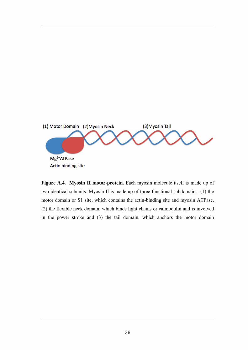
Myosin
Myosins consist of a family of 15 or more classes of motor proteins (Hodge and
Cope, 2000). The thick filament of both cardiac and skeletal muscle is comprised of
myosin II. In muscle, the thick filament of the myofilament is composed of a bipolar
polymer containing several hundred myosin proteins and associated myosin binding
proteins including the myosin light chain (LC20)(Gordon et al., 2000).
Myosin II is made up of three functional subdomains: (1) the motor domain or S1
site, which interacts with actin and hydrolyses ATP, (2) the flexible neck domain,
which binds light chains or calmodulin and is involved in the power stroke and (3) the
tail domain, which serves to anchor and positions the motor domain.
The thick filament is a bipolar polymer, in which the myosin proteins lie in parallel,
half orientated in one direction and the other half in the opposite direction such that
the myosin tail subdomains line up end-to-end in the centre (Fig A.5). Each myosin
molecule itself is made up of two identical subunits. The subunits consist of the three
functional subdomains, the tail subdomains entwine, pairing the subunits, with the
motor domain heads protruding toward the actin-binding site (Fig A.4).
The molecular machinery of the myosin head drives the movement of actin along the
myosin filament. Myosin ATPase-mediated hydrolysis of ATP to ADP and Pi
provides the free energy that powers the conformational change in the myosin head
known as the power stroke (Molloy et al., 1995). During the power stroke myosin,
bound to the actin thin filament, slides the thin filament towards the tail of the myosin

37
protein. As the thick filaments are arranged in a bipolar orientation, the myosin heads
at either end slide the thin filaments closer together, causing the cell to shorten and
contract (Fig A.5).
38
Figure A.4. Myosin II motor-protein. Each myosin molecule itself is made up of
two identical subunits. Myosin II is made up of three functional subdomains: (1) the
motor domain or S1 site, which contains the actin-binding site and myosin ATPase,
(2) the flexible neck domain, which binds light chains or calmodulin and is involved
in the power stroke and (3) the tail domain, which anchors the motor domain

39
Figure A.5. Actin movement during contraction. During the power stroke myosin,
bound to the actin thin filament slides the thin filament towards the tail of the myosin
protein. As the thick filaments are arranged in a bipolar orientation, the myosin heads
at either end slide the thin filaments closer together.

40
The Ca2+ dependent binding of myosin to actin and the subsequent conformational
change mediated by ATP hydrolysis is a rate limiting step and important site of
regulation of contraction. Muscle myosins are regulated in a number of ways
depending on isoform expressed and the tissue in which it is found. Within cardiac
and skeletal muscle, actomyosin binding and myosin ATPase activity is regulated by
the troponin-tropomyosin complex of proteins associated with the thin filament and
myosin motor domain, binding site on actin. In the absence of Ca2+ the troponin-
tropomyosin complex prevents actin-myosin binding, necessary for ATP turn-over at
the S1 site and contraction. As intracellular Ca2+ increases it binds to the TNC
complex causing a conformational change which permits acto-myosin interaction and
cross-bridge cycling.
Myosin associated proteins can also alter myosin function. In smooth muscle it is
well recognized that phosphorylation of the light chains of myosin by myosin light
chain kinase causes contraction (Kamm and Stull, 1985, Kureishi et al., 1997). In
contrast, phosphorylation of the myosin light chains of skeletal muscle, by addition of
calmodulin (2 M and 6 M) and myosin light chain kinase (0.15 M and 0.5 M
respectively), in both rabbit and rat skinned, skeletal muscle fibers has been shown to
increase the sensitivity to Ca2+ and submaximal isometric force generation, although
not cause contraction alone (Metzger et al., 1989, Sweeney and Stull, 1990). More
recent studies have speculated that the single cystein on myosin LC20 could be a
target of oxidation and contribute to dysfunction.

41
The major isoforms of myosin heavy chain (MHC) found in skeletal muscle are:
MHCI in slow-twitch, and MHCIIa, MHCIId and MHCIIb in the three subgroups of
fast-twitch fibers. Although generally grouped into distinct classes, muscle fibers can
co-express two or more major MHC isoforms, forming a continuum between fiber
types (Schiaffino et al., 1989, Billeter et al., 2005).
Other contractile myofilament proteins are expressed to match the myosin heavy
chain isoform. Myosin light chains are expressed as six different isoforms in skeletal
muscle: MLC1f, MLC2f and MLCf3 in fast-twitch muscle and MLC1sa, MLC1sb
and MLC2s in slow-twitch muscle (Pette and Staron, 2000, Schiaffino et al., 1989,
Billeter et al., 2005). Troponin T and troponin I isoform expression also varies based
predominantly on the MHC isoform present (Bottinelli et al., 1991).

42
Table A.1. Fiber type, myosin and myosin associated protein isoform
expression.
Fiber Type Myosin Heavy Chain Myosin Light Chain
Slow MHC1 MLC2s
MLC1sa
MLC1sb
Fast MHCIIa MLC1f
MHCIId MLC2f
MHCIIb MLC3f

43
Isoform specific differences in myosin expression affect the rate of ATP hydrolyses.
For example, human skinned muscle fibers classified as type I or slow (type I MHC
isoform), type II A (type II A MHC) and type II B (type IIB MHC) from biopsies of
the rectus abdominus and vastus lateralis, had ATPase activity (mmol l-1 s-1) ranging
from 0.620.08 (I MHC), 1.160.13 (II A MHC), to 2.460.35 (II B MHC) at 35°C
(Bottinelli et al., 1994, Stienen et al., 1996).
44
Actin
Actin is a common cytoskeletal and scaffolding protein found in all eukaryotic cells
and provides a path along which the many different forms of myosin motor proteins
travel. The actin in striated muscle is made up of globular actin (G-actin) molecules
that form a polymer of filamentous actin (F-actin). Actin contributes to a range of
cellular processes including cell motility, division and signaling, as well as providing
structure and shape to the cell (Doherty and McMahon, 2008).
The polymerization of G-actin into F-actin is a complex and tightly regulated process.
At the core of polymerization are four steps: 1) the activation of G-actin through the
binding of ATP, 2) the formation of an actin nucleus consisting of a dimer or trimer
of ATP-bound G-actin, 3) bidirectional elongation of the actin nucleus as activated G-
actin monomers bind at both the barbed and pointed ends of the actin nucleus, and 4)
the annealing of and coiling of paired, elongated filaments (Pollard and Cooper,
1986).
Associated with the actin filament and an important regulator of actin polymerization
are capping proteins, such as gelsolin, villin and severin. The binding of capping
proteins to ends of the forming actin filament have been show to regulate the
elongation of the filament, inhibiting further actin binding, but also preventing the
reserve or loss of G-actin from the filament (Pollard and Cooper, 1986). Within
striated muscle, capping of the actin filament occurs at the Z-line where apposing

45
actin filaments meet. These barbed ends are capped by CapZ and are linked by -
actinin, forming the Z-line (Littlefield and Fowler, 1998).
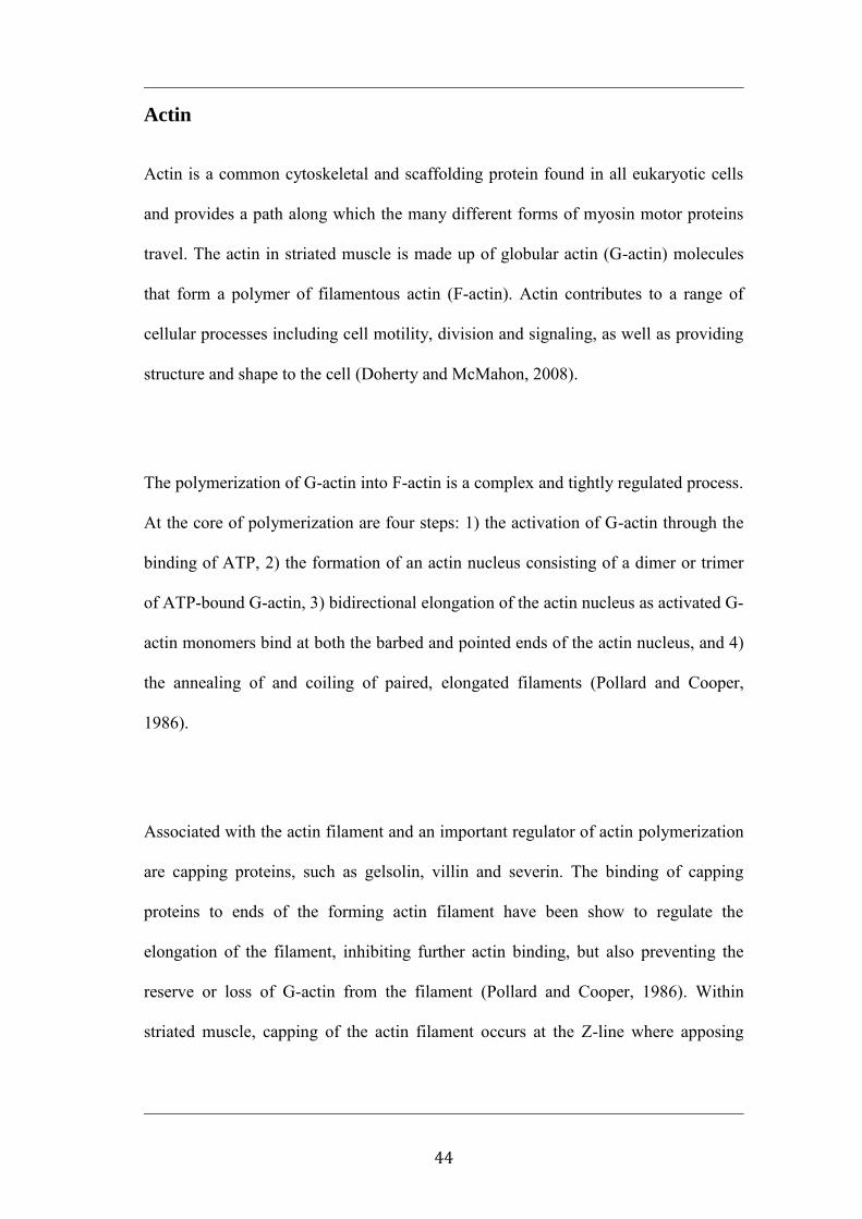
Actin makes up a major constituent of the muscle myofilament. The thin filament of
striated muscle is made up of two parallel filamentous F-actin polymers arranged in a
tight alpha helical pattern (Fig A.6a). Associated with the actin filaments are several
actin-bound, regulatory proteins including tropomyosin and the troponin complex that
regulate actin-myosin binding and thus contraction in a Ca2+-dependent manner.

46
Tropomyosin
Tropomyosin is primarily found in association with actin filaments. Over 40 isoforms
exist and tropomyosin is widely distributed in the cytoskeletal network among cell
types but has a prominent role in the contractile myofilament of smooth, cardiac and
skeletal muscle. There are at least four isoforms found in muscle, two in striated
muscle and two in smooth muscle (Lees-Miller and Helfman, 1991).
Tropomyosin exists as either a homo- or heterodimer depending on the muscle type
(Lehrer, 1975, Yamaguchi et al., 1974). Two alpha-helical chains are arranged as a
coiled-coil (Yamaguchi et al., 1974), with the long tropomyosin chains overlapping in
a head to tail fashion as a continuous polymer along the thin filament in a close
association with F-actin. (Fig A.5b)
Tropomyosin lies in the long pitch groove of F-actin, partially obstructing the myosin
binding sites (Moore et al., 1970). Electron microscopy and three-dimensional image
reconstruction has identified that, through steric hindrance, tropomyosin regulates the
strength of actin-myosin binding and hence contraction in cardiac and skeletal muscle
(Vibert et al., 1997).
The position of tropomyosin along the actin filament can be regulated through post-
translational modifications or associated proteins. Partially phosphorylated
tropomyosin has been found in both cardiac and skeletal, rat and rabbit, muscle
homogenates (Heeley et al., 1982). Phosphorylation causes a greater avidity for head-

47
tail polymerization in a purified protein preparation and promotes myosin-ATPase
activity to a greater extent, in a reconstituted actin and myosin S1 fragment ATPase
assay (Heeley et al., 1989). In skeletal and cardiac muscle, tropomyosin binds to the
F-actin associated, Ca2+-sensitive troponin complex. Upon Ca2+ binding, a
conformational change in the troponin complex alters the binding affinity of
tropomyosin to troponin, shifting tropomyosin, allowing strong actin-myosin binding
and contraction (Vibert et al., 1997, Xu et al., 1999).
48
Figure A.6. The arrangement of actin and tropomyosin polymers. a) Globular
actin polymerizes to form the actin filament, which consists of two parallel
filamentous F-actin arranged in a helical pattern. The thin filament of striated muscle
is made up of two parallel F-actin polymers. Associated with the actin filament are
regulatory proteins the inhibit actin-myosin binding in a Ca2+-dependent manner. b)
The continuous tropomyosin polymer, overlapping in a head to tail fashion sits in the
long-pitch groove of F-actin. Tropomyosin partially obstructs actin-myosin binding in
a Ca2+-troponin dependent fashion.

49
The Troponin Complex
The troponin complex is found in both skeletal and cardiac muscle in association with
the thin filament. The troponin complex binds to F-actin and tropomyosin at regular
intervals with a stoichiometry of one troponin complex to one tropomyosin. The
troponin complex is made up of three subunits; 1) troponin I (TnI), 2) troponin T
(TnT) and 3) troponin C (TnC) (Greaser and Gergely, 1973). Calcium-binding to TnC
causes a series of conformational changes relayed through the troponin complex that
ultimately alter protein binding-affinities. Although not fully understood, the process
subsequently displaces tropomyosin from the myosin-binding site of actin, which
enables actin-myosin binding and muscle contraction (Tao et al., 1990, Tobacman,
1996, Vibert et al., 1997, Xu et al., 1999).

50
Troponin I
Troponin I binds to actin and the other troponin subunits, TnT and TnC.
Reconstituted myofilament experiments have shown TnI functions to inhibit the
myosin ATPase in a Ca2+-independent manner, preventing the detachment of the
myosin motor head from the actin-myosin binding site (Potter et al., 1995).
Myofilament consisting of the actomyosin S-1 fragment and Tm exhibit a basal level
of ATPase activity, this activity is abolished when TnI is included (Potter et al.,
1995). The addition of TnC reverses the TnI inhibition of ATPase activity but does
not raise ATPase activity above basal levels, even in the presence of Ca2+ (Potter et
al., 1995). This was initially believed to be the mechanism of regulating contraction.
Troponin I functions as an anchor for the troponin-tropomyosin-actin complex. In the
presence of Ca2+ the binding of TnC to TnI is strengthened, reportedly increasing
between 15-100 fold in skeletal muscle (Tobacman, 1996), while the TnI-actin
interaction weakens, and fluorescence resonance energy transfer and photo cross-
linking experiments indicate the distance between actin and TnI increases (Tao et al.,
1990). The altered binding affinity and movement of TnI causes the movement of the
tropomyosin-troponin complex away from the actin-myosin binding site, allowing for
actin-myosin binding and contraction (Fig A.6).
Cellular damage to cardiac or skeletal muscle results in TnI leaking from the cell into
the extracellular space and eventually the blood. Currently blood TnI is used as a
clinical diagnostic for cellular damage following myocardial infarct and crush
injuries.

51
Troponin T
Troponin T is the largest of the troponin subunits and binds to actin, tropomyosin and
the troponin complex. The COOH-terminus of TnT binds to TnC, TnI and
tropomyosin (Perry, 1998). The NH2-terminus sits at the region where the
tropomyosin tail and preceding tropomyosin head overlap (Gordon et al., 2000). In
the presence of Ca2+ the binding of the COOH-terminus of TnT and TnC is
strengthened, while weakening the binding to Tm (Gordon et al., 2000).
Although TnT has a structural role it also appears to potentiate acto-myosin ATPase
activity in the presence of Ca2+. Reconstitution experiments including Tm, TnI and
TnC and actomyosin-S1 ATPase assays in the presence of Ca2+ show only a basal
level of ATPase activity (Potter et al., 1995). However, the inclusion of TnT,
completing the troponin complex, potentiates acto-myosin ATPase activity in a Ca2+
dependent manner to a maximal increase of ~170% over basal activity (Potter et al.,
1995). The increased activity may be facilitated by either a Ca2+-dependent
interaction between TnT and TnC or a Ca2+ dependent change in the interaction
between TnC and TnI, transmitted through TnT (Potter et al., 1995).

52
Troponin C
The troponin C subunit is the Ca2+ sensor protein of the troponin complex. The
binding of Ca2+ to low affinity binding sites of TnC initiates a cascade of
conformational changes that lead to actin-myosin binding and contraction. Two
isoforms of TnC are found in skeletal and cardiac muscle. All fast-twitch fibers
express skeletal TnC (skTnC) while slow-twitch fibers and cardiac muscle both
express the cardiac isoform (cTnC) (Wilkinson, 1980). Unlike other contractile
proteins, co-expression of both isoforms of TnC in one tissue has not been observed.
The distinct TnC isoform expression difference in slow- and fast-twitch muscle is
utilized in identifying fiber type. Muscle fibers contract in response to the divalent
cation, Strontium (Sr2+), however, slow-twitch fibers on average have a sevenfold
greater sensitivity to Sr2+ (O'Connell et al., 2004). SDS-PAGE analyses of single
fibers and reconstitution experiments have demonstrated that this increase in
sensitivity is associated with the presences of cardiac/slow-twitch TnC (Morimoto
and Ohtsuki, 1987, O'Connell et al., 2004, Yamamoto, 1983, Hoar et al., 1988).
Both isoforms of TnC are EF hand proteins with a typical dumbbell shape having two
EF hands linked by a flexible linker region and are both Ca2+-sensitive proteins
required for the initiation of contraction (Fig A.7). In fact, molecular substitution
experiments i.e., the exchange of skeletal TnC with cardiac TnC, in a skeletal muscle
model, indicates that the TnC isoforms function similarly (Moss et al., 1986a, Moss et
al., 1991).

53
The COOH-terminus of TnC is anchored to the NH2-terminus of TnI independent of
[Ca2+]. In contrast, the NH2-terminus of TnC binds to the inhibitory and COOH-
terminus of TnI in a Ca2+ -dependent manner (Farah and Reinach, 1995).
The COOH-terminal contains two high affinity structural Ca2+ binding sites, while the
NH2-terminal EF hand contains the regulatory, low affinity binding site(s). In skeletal
muscle the two high affinity Ca2+ binding sites have an affinity of ~2.0 x 10⁷ M-
1(Potter and Gergely, 1975), while in cardiac TnC the affinity, is 3x 10⁸ M-1
(Holroyde et al., 1980).
Cardiac/slow TnC contains a single low affinity binding-site, while fast skeletal TnC
contains two low-affinity binding sites (Holroyde et al., 1980, Potter and Gergely,
1975) which cause the conformational change leading to contraction. The large Ca2+
mediated conformational change in the NH2-terminus exposes an extensive number
of hydrophobic residues or a „sticky‟ patch, which interacts with the inhibitory and
COOH-terminal regions of TnI (Gagne et al., 1995).
Interestingly, when substituted into a fast-twitch skeletal muscle fiber, purified
cardiac TnC infers a higher Ca2+-sensitivity in the contractile apparatus than skeletal
TnC, with the original sensitivity restored when the cardiac TnC was subsequently
removed and skeletal TnC re-introduced (Moss et al., 1986a, Moss et al., 1986b).

54
Small differences in species-specific homologs of TnC regulate Ca2+-sensitivity. For
example, the Ca2+-sensitivity of the contractile apparatus of trout cardiac muscle is
>10 fold greater than that of a mammalian heart at the same temperature (Churcott et
al., 1994), in part due to a two-fold increase in Ca2+-affinity of trout TnC. This
difference comes about from a five amino acid difference in the NH2-terminal
domain (Gillis et al., 2007), none of which actually reside within the low-affinity
binding domain but likely still functions to cause conformational changes in protein-
protein interactions. It is also possible that post-translational modifications will alter
the affinity of TnC regulatory, low affinity Ca2+-binding sites and Ca2+-sensitivity of
the contractile apparatus.

55
Figure A.7. Troponin C ribbon structure. Cardiac/slow-skeletal (left - 1AJ4 (Sia et
al., 1997)) and fast-twitch skeletal muscle (right - 1TN4 (Houdusse et al., 1997))
troponin C in the Ca2+ bound structure. Both isoforms of TnC are EF hand proteins
with a typical dumbbell shape having two EF hands linked by a flexible linker region.
Cysteine sites are highlighted in grey.
NOTE: This figure is included on page 55 of the print copy of the thesis held in the University of Adelaide Library.

56
As skeletal muscle contraction is the result of a complex signaling pathway involving
many potential points of regulation, the following studies utilize a simplified, isolated
single fiber model. This simplified model allows for the analysis of the functional
consequence of ROS/RNS on the contractile proteins without considering the
complexity of regulatory actions due to plasma membrane/SR of Ca2+ signaling or
metabolic activity.
Oxidative stress includes modification of a variety of redox sensitive molecules,
discussed above. The reactive nature and oxidative modification of the oxidative
molecules varies and as such we utilize two very distinct oxidants, H2O2 and NO.
Although functionally similar, slow- and fast-twitch muscle are known to have
distinctly different responses to stimuli that cause fatigue and some forms of
oxidative stress. We investigate the effects of exogenous oxidants on both fast- and
slow-twitch muscle types with an aim to identify different susceptibility to NO-
mediated oxidative dysfunction.
We move beyond the basic functional experiments and utilize molecular substitution
and proteomic techniques to identify the functional substrate responsible for the NO-
mediated decline in Ca2+-sensitivity in fast-twitch muscle fibers.
This technique not only identified the functional, myofilament associated substrate
but also allows for speculation on the basis for the differences in slow- and fast-
twitch fiber responses to oxidative stress. The finding that the substitution of the
slow-twitch isoform of the functional substrate into a fast-twitch muscle fiber does
not abolish the NO-mediated decline in Ca2+-sensitivity suggests that a greater
abundance of anti-oxidant enzyme(s) or molecules may be associated with slow-
twitch muscle fibers.

General Methods

58
Single muscle fiber myography
Premise
Demembranated single muscle fiber myography allows for the measurement of
contractile forces independent from metabolism, regulation of Ca2+ entry pathways,
central nervous system and the influence of cytosolic proteins. This is in stark
contrast to the complexity of whole muscle perfusion techniques which are useful in
providing information on the response of an entire muscle organ including neural
influence, muscle blood flow and cellular metabolites. In the presence of exogenously
applied ATP, demembranted single muscle fiber myography produces accurate,
reproducible measurements of isometric contractile force in response to an external
Ca2+ stimulus. A significant advantage of the demembranted single fiber myography
is the near microscopic size of the fiber (50m) that allows for rapid diffusion of
exogenous substances throughout the sample.
Muscle Dissection and Mounting
All experiments were performed according to the guidelines and with the approval of
the University of Adelaide Animal Ethics committee. Male, Hooded Wistar rats; 4-6
months old or 300-350g were euthanized in a CO2 chamber. Forceps, surgical
scissors and a scalpel were used to remove hind limb muscles overlaying the extensor
digitorum longus (EDL) and soleus. Following exposure of the EDL or soleus the
connecting tendons were cut and the muscle removed, carefully to grip only the

59
tendons. Isolated muscles were immediately pinned in a petri dish at slight tension
(<95 % resting tension) under paraffin oil (Chem-supply, Australia) and kept on ice.
Paraffin oil prevents the muscle dehydrating.
Under a light microscope single fibers were isolated by making an incision into the
muscle belly. Fine surgical forceps were used to gently separate bundles of multiple
fibers away from the main muscle body. Subsequently, single fibers were then
isloated from these smaller muscle bundles. Using fine forceps the sarcolemmal
membrane was removed, 10-0 suture silk (Deknatel), was tied to the free end. The
single muscle fiber was cut free from the muscle bundle and a second suture tied to
the remaining free end. The single fibers were kept under slight tension (<5% stretch)
before being mounted on the force transducer.
Individual fibers were mounted on an isometric force transducer by placing the
surgical sutures around either end of a stationary pin and force transducer. The resting
length of the fiber was measured using a graticule mounted on to the microscope and
the fiber stretched to 120% resting length before the diameter of the muscle fiber was
measured as previously documented (Lamb and Stephenson, 1994, Dutka et al., 2005,
Endo, 1972). Once attached the fiber was immersed in a Perspex bath containing a
K-EGTA (in mM: K+, 126; Na+, 36; EGTA2-, 50; total ATP, 8; creatine phosphate
(CP), 10; total Mg2+, 10.3; HEPES, 90; pH 7.10; pCa (-log10 [Ca2+]), <9.0) solution
containing 2% Triton-X 100 for 10 minutes to solubilize the remaining membranous
compartments. The fiber was then washed free of cytosol and remnant membranes

60
and organelles in fresh K-EGTA buffer for 2 minutes to remove the residual Triton-X
100 prior to the commencement of experiments.

61
Figure B.1. A single muscle fiber dissected from the muscle body. The muscle
body is pinned at slight tension and under paraffin oil. A single fiber is isolated from
the muscle body and suture silk tied to either end before mounting into a force-
transducer.

62
Apparatus
The muscle fiber myograph consisted of a force transducer (AME801, 2kHz
resonance frequency, SensoNor Horten, Norway) with an adjustable slide. Using
suture silk single fibers were attached to a stationary pin, on the transducer with a
second suture used to connect the free end of the fiber to the adjustable slide which
enabled one to adjust the resting length of the fiber (Fig B.2).
Beneath the force transducer was a height adjustable platform. A series of Perspex
2 ml baths on a linear slide enabled one to rapidly change the muscle bathing
solution.

63
Figure B.2. A single muscle fiber mounted on a force-transducer. The single
muscle fiber is mounted between a stationary pin and a pin connected to a force-
transducer on an adjustable slide. The adjustable slide allows for adjustment of the
muscle length.

64
Ca2+
activation and contractile function
The force-Ca2+ relationship was determined by activation of the fiber in a series of
fixed Ca2+ concentration solutions buffered with EGTA. These covered a pCa range
of pCa 7.0 to pCa 4.5. Fibers were initially incubated in nominally zero Ca2+, K-
EGTA solution and prior to treatment the Ca2+ chelating EGTA was removed by
briefly washing in a K-HDTA solution (in mM: K+, 126; Na+, 36; HDTA2-, 50; total
ATP, 8; creatine phosphate (CP), 10; total Mg2+, 8.5, EGTA, 1; HEPES, 90; pH 7.10;
pCa (-log10 [Ca2+]), <9.0). HDTA is a non Ca2+ chelating analog of EGTA. The fiber
was then sequentially immersed in a series of solutions of increasing [Ca2+], the fiber
was moved to the next higher [Ca2+] once tension reached a plateau. Following peak
force was achieved the fiber was completely relaxed in a K-ETGA solution for at
least 1 minute prior to repeating the activation sequence. The activation sequences
were highly reproducible with only a small reduction in maximum Ca2+ activated
force occurring with successive stimulation (~2% per sequence). The activation
sequence was repeated at least twice per treatment.

65
Experimental treatments
Sr2+
mediated identification of fast- vs slow-twitch fibers
The soleus muscle contains predominantly slow-twitch fibers, however, there are a
small number of fast-twitch fibers. To verify the fiber type isolated from both EDL
and soleus muscle, single fibers were exposed to a Sr2+-EGTA solution (pSr 5.4).
Fast-twitch fibers produce little or no force in Sr2+-EGTA solution while slow-twitch
muscle fibers produce near maximal force (Bortolotto et al., 2000, O'Connell et al.,
2004).
Nitric oxide donor treatment
The nitric oxide donor GSNO was made fresh each hour as a 10 mM stock in K-
HDTA and further diluted to 1 mM. Fibers were exposed to a 1 mM GSNO solution
for 2 minutes, followed by a washout period of 2 minutes in K-EGTA solution.
The amount of NO liberated from 1 mM GSNO was measured in K-HDTA solution
at 23 C using an Apollo 4000 NO analyzer system (World Precision Instruments)
with a NO sensor. The peak NO concentration liberated immediately upon dissolving
1 mM GSNO in the K-HDTA solution was 14 M with a half life of approximately
10 minutes. Following 20 minutes, NO production was such that concentration was 1-
4 M and stable for a further 40 minutes. SNAP (2 mM; an alternate NO donor)
liberated a peak [NO] of 2.1 M after 25 minutes and then gradually decayed with a

66
half-life of 30 minutes. Experiments were conducted during the period where the NO
concentration was between 1-4 M.
Hydrogen peroxide treatment
Hydrogen peroxide was diluted to a 1 M stock (in water) from a 3 M aqueous
solution and then diluted to 10 mM in 2 ml of K-HDTA solution. Fibers were
exposed to H2O2 whilst being simultaneously contracted in a pCa 6.0 solution to
enhance the Ca2+-sensitivity effects of H2O2 (Posterino et al., 2003). Hydrogen
peroxide was added to the K-EGTA, K-HDTA and pCa 6.0 solutions. Following from
Posterino et.al (2003), the fiber was activated and relaxed repeatedly for 5 minutes,
resulting in approximately 2 minutes of exposure during contraction.
Troponin C extraction and replacement
Troponin C was extracted and replaced using a method modified from Moss et al.
1985. Following an initial Ca2+-activation control sequence the fiber was relaxed in
K-EGTA solution for 2 minutes. Partial extraction was completed by bathing the fiber
in a 1 ml low ionic strength solution containing Tris (20 mM ), EDTA (5 mM) and
DTT (1mM) for 10 minutes. Following washout with K-HDTA buffer the force-Ca2+
relationship was re-determined as described above. Recombinant TnC was combined
into the fiber by bathing in K-EGTA solution containing ~1 mg/mL recombinant TnC
and 25mM DTT for 1 hour. This was followed by a 2 minutes wash in K-EGTA
solution to remove excess TnC.

67
Data analysis
The data for each activation sequence within a fiber was individually analysed. Sub-
maximal responses were normalized to the maximum Ca2+-activitated force in each
sequence. The data obtained from each pair of activation sequences was then
averaged and plotted using GraphPad Prism v4.01 (GraphPad Software, San Diego,
CA). The force-pCa data was fitted with a modified Hill equation with parameters of
maximum and minimum set to 100% and 0% respectively. From the resulting curve
the pCa50 (the pCa at which 50% maximum force is produced) along with the pCa20
and pCa80, and the Hill Coefficient were determined. The effect of a treatment was
assessed by measuring the change in pCa20, pCa50 and pCa80 relative to the values
before treatment in the same fiber (Spencer and Posterino, 2009).
Similarly, in recombinant TnC substitution experiments the forces generated by fibers
were normalized to the maximum force produced in the initial Ca2+ activation
sequence prior to TnC extraction. Measuring the pCa50 of treated fibers in
comparison to untreated fibers enabled assessment of the treatment effects.
The results are presented as means ± SEM for a sample of n fibers. Statistical
significance was examined using either a student‟s unpaired t-test or repeated
measures one-way analysis of variance where appropriate with a significance level of
P<0.05.

68
Proteomic techniques and analysis
Separation of proteins using SDS-PAGE
Premise
SDS-PAGE is the separation of proteins according to their size within a
polyacrylamide gel matrix. In the presence of the ionic detergent, sodium dodecyl
sulfate (SDS), proteins become coated in the detergent anions and become largely
negatively charged. When an electric field is applied to protein samples in the
polyacrylamide matrix, the negative charge flows from the negative cathode in the
upper buffer chamber, through the gel into the lower buffer chamber, attached to the
positive anode. Since SDS imparts a near equal negative charge to all proteins, this
technique causes globular proteins to be fractionated based on size with smaller
proteins migrating more rapidly.
Polyacrylamide gel composition and preparation
Polyacrylamide gels were prepared by polymerizing monomeric acrylamide with
N,N-methylene-bis-acylamide (bis-acrylamide), as the cross-linker. The
polymerization is initiated by the addition of a free radical generator ammonium
persulfate (APS) and catalyzed by N,N,N,N-tetramethylethylenediamine (TEMED).
Separation of proteins is largely dependent on the size of pores formed in cross-linked

69
polyacrylamide. Gels are usually referred to as total acrylamide as a percentage (w/v).
In general, pore size decreases with increasing % total acrylamide. In general 7.5%
polyacrylamide gels provide reasonable separation of proteins 300 - 100 kilodaltons
(kDa), with 15% matrix being suitable for proteins less than 100 kDa, individual
porosity can be further tailored to meet the needs of the experiment by changes to the
percentage acrylamide or acrylamide relative to bis-acrylamide cross-linker.
Gels were cast 0.75 – 1.5 mm in thickness between glass plates. Gels were either a
15% acrylamide composition or a gradient gel (outlined below). Gradient gels consist
of a 20% acrylamide solution slowly mixed with a 7.5% acrylamide solution with the
aid of a peristaltic pump that provides for a more linear separation of proteins.
Briefly and referring to Figure B.4, Perspex chambers connected in series were
filled with 17 and 16ml of 7.5% (a) and 20% (b) polyacrylamide solutions. Solutions
mix in chamber (b), reducing the % polyarcylamide present. Tubing attached to
chamber (b) dispensed the mixed solution via a peristaltic pump into the Biorad
Protean II XI casting plates slowly to avoid further mixing in the casting plates
(Biorad Protean II XI Cell – Instruction Manual).
To ensure a uniformly smooth interface at the gel surface, the gel was overlaid with a
thin layer of water-saturated butanol and allowed to fully polymerize for several
hours. Following removal of butanol and several rinses a stacking gel was cast on top
of the separating gel. Typically gels were cast using 15 well combs for sample

70
loading or a single large well for experiments involving isoelectric focusing in the
second SDS-PAGE dimension.
Polyacrylamide separating gels consisted of; Tris-HCl (pH 8.8) 375 mM,; SDS 0.10%
(w/v); acrylamide 7.5 % (w/v) in 7.5% gels, 15 % (w/v) in 15% gels or 19.9% (w/v)
in 20% gels; bis-acryliamide 0.20 % (w/v) in 0.40% gels, 15 % (w/v) in 15% gels or
0.50% (w/v) in 20% gels; glycerol 0.70% (v/v) in 7.5% gels, 1.30 % (v/v) in 15%
gels or 1.30% (v/v) in 20% gels; APS 2.2 mM; and TEMED 0.04% (v/v).
Polyacrylamide stacking gels consisted of; Tris-HCl (pH 8.8) 125 mM; SDS 0.10%
(w/v); acrylamide 4.50% (w/v); bis-acrylamide 0.12% (w/v); APS 0.13-0.26 mM; and
TEMED 0.10 % (v/v). Tris-HCl (pH 8.8) and SDS were prepared as a stock solution
at 4x concentration for lower gels (7.5% - 20%) and a separate stock for the stack.
Acrylamide and bis-acrylamide prepared as a stock solution of 30% (w/v) acrylamide
and 0.8% (w/v) bis-acrylamide. Ammonium Persulfate was prepared as a 10% (w/v)
(438 mM) stock as a daily stock.

71
Figure B.4. Gradient gel casting. Perspex chambers containing (a) 7.5% and (b)
20% poly-acrylamide solutions are connected in series. The polycrylamide solutions
slowly combine in (b), reducing the percentage polyacrylamide over time. The
combined solution is pumped into the gel casting plates resulting in a gradient of
polyacrylamide, greater at the bottom than at the top.

72
SDS-PAGE loading and running
Gels were inserted in either a Biorad mini-PROTEAN electrophoresis module
(BioRad, Australia) or a large format Biorad PROTEAN II XI cell. The
electrophoresis module is filled with a SDS-PAGE “running buffer” containing Tris
25 mM, glycine 192 mM and SDS 0.1 % (w/v).
Protein samples were solubilized in an SDS sample buffer consisting of; Tris-HCl
25mM, glycine 30% (v/v) and SDS 1% (w/v) and bromophenol blue 0.01%
(Laemmli, 1970) prior to loading.
Electrophoresis was carried out using the Biorad mini-PROTEAN module, run at a
constant voltage of 200 V for one hour. Gels loaded in the Biorad PROTEAN II XI
cell were run at 35 mAmps per gel for 4.5 hours or until the bromophenol blue dye
front reach the bottom of the gel.

73
Isoelectric focusing
Premise
Proteins consist of both positively and negatively charged amino acids, the
combination of which give individual proteins a characteristic inherent charge or
isoelectric point (Pi). Isoelectric focusing separates proteins based on their isoelectric
point. Samples are prepared in a non ionic sample buffer, often urea based, and
electrophoresed in a 4% polyacrylamide matrix into which a pH gradient is
established. In experiments conducted herein we made use of commercially available
isoelectric focusing gels manufactured with an immobilized pH gradient (IPG)
(BioRad, Australia). When a strong electrical current is place across the isoelectric
focusing gel proteins migrate to the pH in the gel that corresponds to their isoelectric
point at which their inherent charge is neutral (Fig B.5).
Modifications to a protein such as phosphorylation or oxidation usually only impart
very small changes in molecular mass that may not be resolved using traditional SDS-
PAGE, however, post-translational modifications that induce a charge bias to the
protein can be effectively separated from their unmodified counterparts using
isoelectric focusing. Following isoelectric focusing based on protein charge, proteins
are further resolved along a 2nd dimension (size) using standard SDS-PAGE to
separated proteins based on mass.

74
Figure B.5. An immobilized pH gradient (IPG) strip containing a protein sample
before and after isoelectric focusing. The protein sample (dashed lines) is initially
distributed evenly along the IPG strip prior to isoelectric focusing (top). During
isoelectric focusing (bottom), each protein migrates to its Pi (eg. pH > 4.1).

75
IEF sample preparation
Samples, either snap frozen single fibers or a crushed muscle homogenate, were
prepared in rehydration buffer. The rehydration buffer contained; thiourea 2 M; urea 7
M; 3-[(3-Cholamidopropyl)dimethylammonio]-1-propanesulfonate (CHAPS) 2%
(non-ionic detergent) and bromophenol blue 0.005% and was stored as 500 L
aliquots at -80C. Ampholytes, pH 3-10 0.05% (Biorad) and pH 4-7 1% (GE
Healthcare), were added directly before use. The chaotropic agents urea and thiourea
denature proteins and inactivate proteases, consequently no further attempt was made
to inhibit proteases. To load the isoelectric focusing gels 345 L of rehydration
buffer, containing the protein sample was distributed evenly along the well of a
rehydration tray and the exposed 18 cm pH 3-6 IPG strip laid, gel side down, onto the
sample. Air bubbles were removed before overlaying the IPG strip with 2-3mL of
mineral oil to prevent dehydration. The rehydration tray was covered and left
overnight to allow complete loading of the sample.
Prior to isoelectric focusing the rehydrated IPG gel was drained of oil by placing on
parafilm and placed gel side down in the PROTEAN IEF (Biorad, Australia)
focusing tray. Lightly wetted 1 cm paper wicks were installed over each electrode to
trap any residual salts. The strip was again covered with 2-3 ml of mineral oil and air
bubbles removed.

76
Isoelectric focusing protocol
Following sample incorporation into the IEF strips a three-step method was used to
effect isoelectric focusing of proteins: 1) 0-250 V rapid linear voltage ramp over 15
minutes; 2) 250-10 000 V ramp over 3 hours; 3) 10 000 V for a total of 60 000 V
hours and 4) a hold step at 500 V.
Following isoelectric focusing the IPG strip was placed on parafilm to remove
residual mineral oil followed by brief incubation in a series of buffers designed to
enable SDS incorporation into proteins while removing the urea and thiourea and
CHAPS. Step 1 involved a 10 minute equilibration in 6 ml of a solution containing
urea 6 M, Tris-HCl 0.375 M (pH 8.8), SDS 2% and, glycerol 20% for 10 minutes.
Step 2, 10 minute equilibration in 6 ml a solution containing urea 4 M, Tris-HCl 0.375
M (pH 8.8), SDS 2% and, glycerol 20% followed by briefly immersing in SDS-PAGE
running buffer prior to loading onto a large format SDS-PAGE gel fitted with a
preparatory well for 2nd dimension separation by SDS-PAGE (see above).

77
Western blot analysis
Premise
Western blot analysis was used to identify specific proteins that had previously been
separated by SDS-PAGE or IEF and SDS-PAGE, then transferred and immobilized
onto a nitrocellulose membrane. Proteins were visualized using polyclonal or
monoclonal antibodies specific to proteins of interest and secondary antibody
conjugated to the enzyme, horse-radish peroxidase, which reacts with luminol
emitting light enabling detection of the proteins of interest using autoradiographic
film.
Transfer of proteins to nitrocellulose
Following separation using SDS-PAGE, proteins were transferred from the poly-
acrylamide gel to a nitrocellulose membrane. The Biorad Mini-Protean II transfer
system was used for smaller gels, or a Biorad Transblot cell for larger gels. Proteins
were transferred in a transfer buffer composed of; Tris 25 mM, glycine 192 mM and
methanol 20% (v/v). The polyacrylamide gel and nitrocellulose were placed together,
carefully removing all air space between. Filter paper is place on the outside as well
as Scotch-Brite pads. The bundle is held together in a transfer cassette that fits
vertically into the transfer system. The cassette is orientated with the nitrocellulose
membrane towards the positive electrode of the transfer unit encouraging transfer of
SDS coated, negatively charged proteins from the polyacrylamide onto the

78
nitrocellulose. The entire cassette was immersed in transfer buffer and cooled during
the transfer process, either by a manufacturer-supplied ice pack or by conducting the
transfer in a refrigerated cold room. Mini-Protean II transfers were run at 100 V for
one hour while larger Transblot cell transfers were run at 30 mAmps for 16 hours. It
is important to recognize that following transfer not all proteins were transferred
equally, largely this is dependent on molecular weight. Consequently following
transfer gels were also stained with coomassie brilliant blue R-250 which provided
one with the opportunity to normalize protein load to the relative amount of myosin
heavy chain remaining in the polyacrylamide gel.
Incubation with primary and secondary antibody
Nonspecific binding of primary and secondary antibodies to the nitrocellulose was
prevented by incubating the nitrocellulose in 50 ml blocking solution (in mM); TBS
(Tris-HCl (pH 7.4) 50, NaCl 150); Tween-20 0.05% (v/v); and nonfat milk powder
3% (w/v) for one hour.
Primary antibodies to Troponin C and light chain (LC20) (MRCL3/MRLC2/MYL9)
were diluted to 1:1000 and obtained from (Santa Cruz Biotechnology, USA) in a
solution containing 10ml TBS, tween 0.05%, nonfat dried milk 1% solution. The
nitrocellulose was then incubated in the antibody solution for one hour with constant
agitation.

79
In order to visualize the primary antibody-bound protein a secondary anti-body
conjugated to horseradish peroxidase (HRP) was used. The secondary antibody,
specific for either rabbit IgG (TnC) or mouse IgG (LC20), binds to the primary
antibody and in the presence of a chemiluminescent solution, generates light
detectible by X-ray film. Secondary antibody was diluted 1:10000 (rabbit IgG) or
1:5000 (mouse IgG) in TBS, 0.05% tween-20 solution and the nitrocellulose
incubated for one hour, gently agitated.
Chemiluminescence detection of specific proteins
Chemiluminescent solution (Thermo Scientific, Super Signal West Pico) was
prepared immediately prior to application to the nitrocellulose membrane as per the
manufactures instructions. Following application excess reagent was removed and the
membrane covered in clear, commercial plastic wrap. In the dark autoradiographic
film (medic grade X-ray film, AGFA) was exposed to the western blot to produce a
series of exposure lengths ranging from 30 sec to 10 minutes. Film was developed on
an AGFA CP1000 processor as per the manufacturers instructions.
Data analysis
Developed film and coomassie stained gels were scanned on a densiometric scanner
(GS-710, Biorad) and optical density (OD) analyzed using Quantity One software
(Biorad).

80
Optical density is the result of intensity of a given pixel in the digital scan. Quantity
One detects the average OD for a selected area, multiplied by the area (OD*mm2) and
is referred to as a volume. To remove background signal, an area of film or gel that
does not contain signal is selected and the resulting background volume subtracted
from signals of interest.

81
Recombinant mutant TnC expression
Premise
Recombinant proteins used in the current study were a gift from Louise Brown‟s
laboratory (Macquarie University, Sydney). Nevertheless for completeness in the
thesis the basic procedures are outlined below. E coli mediated recombinant protein
expression was used to produce a wild type and mutant TnC containing a C84S
substitute. Plasmid vectors containing the target gene were used as templates.
Oliogonucleatide primers containing the desired mutation were annealed to the
parental DNA plasmid and extended and incorporated into new plasmid strands. The
parental DNA was digested leaving only the new mutated plasmid strands. These
strands were then transformed into competent cells and grown, producing the desired
mutated protein (Fig B.6).

82
Figure B.6. Overview of site-directed mutagenesis using a QuikChange
Mutagenesis II site directed mutagenesis kit. Step 1: Parental DNA plasmid
containing the target protein (cTnC) was prepared. Step 2: Temperature cycling
incorporated the mutagenic primer into polymerized circular mutated DNA plasmid
strands. Step 3: The parental DNA plasmid was digested and the mutated DNA
plasmid was transformed into competent cells. (Diagram modified from QuikChange
Mutagenesis Site-Directed Mutagenesis Kit instruction manual, Revision A.01).
NOTE: This figure is included on page 82 of the print copy of the thesis held in the University of Adelaide Library.

83
Mutagenesis
Single cysteine cTnC mutants, Cys35 and Cys84 were engineered by substituting the
triplet of nucleotide bases encoding cysteine for a serine encoding triplet.
Oligonucelotide primers containing the desired mutation for both the forward and
reverse strands of the plasmid had previously been designed (Brown et al., 2002).
Mutagenesis was carried out in a pET-3d expression vector (Novagen), containing rat
cTnC, using the Quickchange Mutagenesis II site directed mutagenesis kit
(Stratagene) as per manufacturer‟s protocol (Fig B.6).
Temperature cycling on a thermal cycler incorporates the mutagenic primer into
polymerized circular plasmid strands. Briefly; the plasmid containing cTnC was
denatured, the oliogonucleatide primers containing the desired mutation were
annealed to the plasmid, and a DNA polymerase extended and incorporated the
mutagenic primer into new plasmid strands. To remove the nonmutated cTnC plasmid
strands and ensure only mutated cTnC was expressed the parental DNA plasmid was
digested with Dpn I. The Dpn I endonuclease was specific for the methylated and
hemimethylated DNA of most strains of E. coli used as expression vectors, including
pET-3d. All PCR was conducted by Dr Brown‟s laboratory.
84
Transformation and expression
The cTnC plasmid was transformed into competent Escherichia coli BL21 (DE3)
cells. Dpn I treated DNA were mixed with competent cells and incubated for 30
minutes on ice. The transform reactions were heat pulsed for 45 seconds at 42°C and
placed on ice for 2 minutes. Preheated NZY+ broth, prepared as per the
manufacturer‟s protocol, was added to the transform reaction and incubated at 37°C
for 1 hour. The transform reaction was then plated on agar plates containing
ampicillin to select for cells containing the plasmid vector.
Protein expression was achieved through overnight cell growth at 37°C. Cells were
harvested by centrifugation and suspended in Tris 50 mM (pH 7.5), DTT 1 mM, NaCl
100 mM and CaCl2 5 mM.
Purification
Cells were lysed using a french press at 15000 psi and centrifugation was used to
remove cell waste. The supernatant was brought to CaCl2 10 mM (in the presence of
Ca2+ the hydrophobic surfaces become exposed) and dialyzed against the suspension
buffer and loaded onto an equilibrated hydrophobic affinity interaction
chromatography column, Phenyl Sepharose 6B (Pharmacia, GE Healthcare). Proteins
bound non-specifically were removed using a salt wash (in mM: Tris 50 (pH 7.5),
DTT 1, NaCl 200 and CaCl2 1) and a EDTA solution used to sequester Ca2+ from
TnC causing it to be release from the column (Tris 50 mM (pH 7.5), DTT 1 mM,
NaCl 200 mM and EDTA 10 mM). Samples were assessed by UV spectroscopy.
85
Absorption peaks obtained from UV spectroscopy correlate to bond types present and
was used to confirm correct tertiary structure. Nucleic acid contaminants were
removed using a DEAE Sephadex column (Sigma-Aldrich) and a salt gradient (NaCl
0.35 M-1 M). Purity of cTnC and cTnC mutants was confirmed with SDS-PAGE (Fig
B.7). Samples were lyophilized and stored at -20°C. Lyophilized recombinant TnC
was dialyzed against HEPES 25 mM (pH 7.4) prior to use. Protein concentration
following dialysis was determined using a Pierce BCA assay (Thermo Scientific,
USA).

86
Figure B.7. Purity of cTnC and cTnC mutants confirmed with coomassie
brilliant blue stained SDS-PAGE. DTT was the only reducing agent included in the
SDS-PAGE samples, at concentrations of 0, 10 mM and 25 mM. Samples included in
the gel are wild-type cTnC (WT) and TnC C84 (C84S). Dimerisation was observed in
no DTT samples (*). Modified from an image provided by collaborators, Dr Louise
Brown and James Cooke at the Department of Chemistry and Biomolecular Sciences,
Macquarie University, Sydney, Australia.

87
Acknowledgements
Recombinant wild-type and mutant cTnC expression and purification was carried out
by collaborators, Dr Louise Brown and James Cooke at the Department of Chemistry
and Biomolecular Sciences, Macquarie University, Sydney, Australia.

Sequential effects of GSNO and H2O2 on the Ca2+
-
sensitivity of the contractile apparatus of fast- and
slow-twitch skeletal muscle fibers from the rat
Timothy Spencer1, and Giuseppe Posterino2
1Discipline of Physiology, School of Molecular and Biomedical Sciences, The University of Adelaide, Adelaide, South Australia, Australia
2Department of Zoology, School of Life Science, Faculty of Science and Technology, La Trobe University, Melbourne, Victoria, Australia
American Journal of Physiology- Cell Physiology (2009) 296: C1015-C1023.

89
STATEMENT OF CONTRIBUTION
This chapter in whole was published in the American Journal of Physiology – Cell
Physiology, vol 296 (2009): C1015-C1023. Timothy Spencer and Dr Giuseppe
Posterino jointly authored the paper.
Dr Posterino gives permission for the paper to be included in the thesis. It is
acknowledged that in the conceptualization, spectrophotometric experimentation and
analysis and editing of the paper Dr Posterino contributed 25 % of the paper.
The remaining work (75 %) is to be attributed to Timothy Spencer.
Dr Giuseppe Posterino Timothy Spencer

Spencer, T. and Posterino, G. (2009) Sequential effects of GSNO and H2O2 on the Ca²+ - sensitivity of the contractile apparatus of fast- and slow-twitch skeletal muscle fibers from the rat. American Journal of Physiology – Cell Physiology, v.296 (5), pp. C1015-C1023, May 2009
NOTE: This publication is included in the print copy of the thesis
held in the University of Adelaide Library.
It is also available online to authorised users at:
http://dx.doi.org/10.1152/ajpcell.00251.2008

Myofibril basis for oxidative dysfunction in skeletal
muscle

127
Abstract
It is evident the redox state can potentiate or inhibit cellular muscle function. Our
previous studies in demembranated fast-twitch skeletal muscle fibers have shown that,
the oxidant H2O2 increases Ca2+-sensitivity, while NO decreases the Ca2+-sensitivity
impairing contraction. We chose to investigate the myofilament basis for the decline
in Ca2+ sensitivity in the fast-twitch skeletal muscle following NO exposure.
Muscle myography was used to analyse the functional consequence of NO exposure
on single demembranated fast-twitch muscle fibers. Isoelectric focusing and SDS-
PAGE proteomic analysis coupled with western blot analysis identified modified
muscle fiber proteins following NO exposure. Molecular substitution of endogenous
TnC with recombinant cardiac/slow WT-TnC and cardiac/slow C84S TnC, containing
a non-oxidisable serine residue at position 84, identified the functional myofilament
substrate of NO mediated Ca2+ desensitisation.
In demembranated, fast-twitch skeletal muscle, the NO donor GSNO reduced the
Ca2+-sensitivity of the muscle fiber. Proteomic analysis showed that among a number
of muscle fiber proteins, myosin light chains and TnC were modified in the presence
of GSNO. Molecular substitution of endogenous TnC with cardiac/slow WT-TnC had
a similar sensitivity to NO as fast-twitch skeletal muscle. In contrast, substitution with
C84S TnC was insensitive to NO. These results indicate that although multiple
muscle fiber proteins are modified following NO exposure, C84S TnC is the

128
functional substrate resulting in the GSNO mediated decline in muscle fiber Ca2+-
sensitivity.

129
Introduction
Contraction of cardiac and skeletal muscle is a highly regulated process involving;
regulation of extracellular and sarcoplasmic reticulum (SR) Ca2+ entry, generation of
ATP, and Ca2+ dependent conformational changes of structural, regulatory and motor
proteins. It is becoming increasingly evident that redox state leading to post-
translational modifications of structural proteins, enzymes and ion channels can cause
activation or inhibition of cellular function (Andrade et al., 1998a, Jackson, 2008,
Kelly et al., 1996). Cells are able to maintain their redox balance despite the
generation of reactive oxygen (ROS) and reactive nitrogen species (RNS) primarily
due to the production of endogenous anti-oxidants and reductants that convert these
molecules to harmless electron stable molecules or remove modifications caused by
an oxidative reaction. Therefore it follows that molecular, cellular and systems
function can be regulated by an excess of oxidant or reductant.
The physiological response to ischemia as a consequence of coronary artery disease,
heart failure, claudication or during heavy exercise in skeletal muscle is for the
vascular endothelium to release the potent vasodilator, nitric oxide (NO).
Interestingly, with prolonged ischemia tissue damage occurs leading to an
inflammatory response and subsequent release of large amounts of NO from
inflammatory cells, neutrophils (Jordan et al., 1999).
In addition, studies have demonstrated that the administration of exogenous NO
donors to functional cardiac and skeletal muscle preparations have a negative effect
130
on the contractile function. Using the diaphragm muscle mixed fiber type, Kobzik et
al., (1994a) have shown that nitric oxide synthase (NOS) inhibitors such as nitro-L-
arginine improve contractile function including the force-frequency relationship, and
that these improvements in contractile function could be reversed by exogenous NO
donors. Similarly, in isolated, electrically stimulated cardiomyocytes, the application
of an NO donor or superfusion with an NO solution attenuated contraction (Brady et
al., 1993). The suggestion has been that NO can detrimentally affect one or more
elements of excitation-contraction coupling in muscle. The control of cytoplasmic
Ca2+ concentration by the ryanodine receptor (RyR) has been a focus of a number of
studies (Aracena et al., 2003). In whole muscle preparations the functional impact of
oxidation on the RyR is a significant reduction in Ca2+ release and diminished
contractile function (Andrade et al., 1998a, Posterino and Lamb, 1996). In addition,
proteins within the muscle fiber may also be oxidized and contribute directly to
reduced muscle function.
Several components of the muscle fiber have the potential to be, or have been shown
to be modified when in an oxidising environment (Dalla Libera et al., 2005).
However, the majority of studies have either focussed on associated changes ie.,
relatively indirect assessment of altered function, or biochemical changes. Exposure
of permeablized rabbit psoas single muscle fibers to an NO donor such as SNAP and
NEM has been shown indirectly to reduce ATP hydrolysis by the myosin ATPase
through analysing the conversion of a fluorescent NADH to nonfluorescent NAD+
(Perkins et al., 1997). The functional consequence of NO exposure was only
indirectly assessed. Spin labelled myosin ATPase in a rabbit psoas muscle fiber,
showed that when over 75% of the SH-1 sites are modified a significant decrease in

131
contractile function was observed (Crowder and Cooke, 1984). The light chains of
myosin (LC20) have also been identified as a possible target of oxidation. Light chains
reconstituted with myosin, oxidised following 5,5′-Dithiobis(2-nitrobenzoic acid)
(DTNB) exposure, form intramolecular disulfide bonds (Wolff-Long et al., 1993),
however, the study used an incomplete myofilament preparation precluding
investigation of the functional importance of LC20 oxidation. Actin in its globular
form has several cysteine residues that can be oxidised, however, when polymerised
into a functional actin filament there is less oxidation or it is abolished (Liu et al.,
1990). Therefore, although many studies have shown the capacity of myofilament
proteins to be oxidised, few experiments have directly linked contractile dysfunction
with oxidation of a specific myofilament protein.
Different muscle fiber types display isoform specific differences in many
myofilament proteins, but nevertheless share similar Ca2+-dependent myofilament
regulation mediated by the troponin complex (Bottinelli and Reggiani, 2000). The
functional similarity of skeletal and cardiac muscle myofilament proteins has allowed
for substitution of native skeletal muscle proteins with cardiac isoforms, while
maintaining myofilament function (Davis and Tikunova, 2008, Moss et al., 1986b,
Moss et al., 1991).
In the current study we made use of a simplified, demembranated fast-twitch muscle
fiber preparation to enable the identification of the functional importance of NO
mediated oxidative stress on the contractile apparatus independent from other
components of excitation-contraction coupling. We have used functional force Ca2+

132
measurement, protein substitution experiments using oxidisable and non-oxidisable
(at Csy84) forms of TnC and biochemical analysis of myofilament proteins to
investigate the functional, myofilament basis for GSNO mediated fast-twitch skeletal
muscle dysfunction and the role of specific cysteine residues on cardiac TnC.

133
Methods
Muscle preparation
All experiments were performed according to the guidelines and with approval of the
University of Adelaide Animal Ethics Committee. Male rats (Sprague-Dawley,
approximately 400g) were euthanized in a CO2 chamber. The EDL muscles were
excised, immersed under paraffin oil, and kept on ice. Under a dissecting microscope,
single fibers were isolated from whole muscle using fine forceps and the sarcolemmal
membrane removed. The single fiber was then mounted between a force transducer
(2kHz resonance frequency; AME801, SensoNor, Horten, Norway) and stationary pin
and stretched to 120% of its resting length. The adjusted length and diameter were
then remeasured. Following from Posterino and Lamb (1996) the fiber was immersed
in a Perspex bath containing a K-EGTA (in mM: K+, 126; Na+, 36; EGTA2-, 50;
total ATP, 8; creatine phosphate (CP), 10; total Mg2+, 10.3; HEPES, 90; pH 7.10; pCa
(-log10 [Ca2+]), <9.0) solution containing 2% Triton-X 100 for 10 minutes to
solubilize the remaining membranous compartments.

134
Functional analysis
Ca2+
activation of the contractile apparatus
Single fibers were mechanically skinned using forceps and were subsequently
chemically skinned in K-EGTA solution containing 2% Triton-X 100 for 10 min to
destroy all remaining membranous compartments. The fiber was then washed free of
cytosolic proteins and residual membranous compartments using K-EGTA solution
for 2 min. Prior to Ca2+ activation fully demebranated single muscle fibers were
equilibrated in K-HDTA (a non-chelating analog of EGTA) solution which contained
(in mM); K+ 126, Na+ 36, HDTA 50, total ATP 8, creatine phosphate 10, total Mg2+
8.5, EGTA 1, HEPES 90, pH 7.10, pCa (-log10[Ca2+]) <9.0. The force-pCa
relationship was determined by activation of the fiber in Ca2+-EGTA solutions (in
mM); K+ 126, Na+ 36, EGTA 50, total ATP 8, creatine phosphate 10, total Mg2+ 8.1,
HEPES 90 mM, pH 7.10, Ca2+ 50, pCa 4.5. To effect full relaxation of the contractile
apparatus, fibers were fully relaxed in a heavily buffered K-EGTA solution. Both the
K-EGTA and Ca2+-EGTA solutions were mixed in a series of ratios to give solutions
with a pCa range of between 7.0 and 4.5. Fibers were activated through a sequence of
these solutions ranging in pCa from <9 to 4.5 to establish the full Ca2+-dependent
force (force-Ca2+) relationship. Previous experiments have determined the free [Ca2+]
for ratios used (Spencer and Posterino, 2009).
The experimental protocol with GSNO involved three successive control activation
sequences fibers followed by exposure to GSNO (1mM for 2 min in a Ca2+ free K-

135
HDTA solution. Fibers where then washed in a K-HDTA solution for 2 min followed
by generation of the force-pCa relationship using fixed [Ca2+] solution for pCa 7.0 -
4.5 .
The nitric oxide donor, GSNO was made fresh every hour as a 10 mM stock and
further diluted, to 1 mM. Previous experiments have determined the amount of NO
liberated from GSNO 1 mM in solution at 23 C (Spencer and Posterino, 2009).
Specifically the peak NO concentration immediately upon dissolving GSNO 1 mM in
solution was 14 M with a half-life of approximately 10 mins. At approximately 20
mins after addition, a low level NO concentration of between 1-4 M was elicited for
a further 40 mins. Experiments were conducted at the stage where the NO
concentration was ranging from 1-4 M. The reducing agent Dithiothreitol (DTT)
was made as a 1 M stock and then further diluted to the required concentration during
experiments. All chemicals were obtained from Sigma.
TnC extraction and reconstitution with recombinant TnC
Following an initial force-pCa series in single skinned muscle fiber troponin C was
extracted using a modified protocol from Moss et.al (1986). Briefly, fibers were
incubated in a low salt solution (in mM: Tris 20, EDTA 5, DTT 1) for 1 hour. The
extraction solution was retained, frozen and lyophilized for later analysis of extracted
proteins. The fiber was next washed in K-HDTA solution for 2 min followed by a
second Ca2+ series for pCa 7.0 – 4.5. Fibers were subsequently incubated in K-EGTA
solution with recombinant TnC (~1 mg/ml) and DTT (25 mM) for 1 hour. A high

136
DTT concentration was required to prevent dimerisation of recombinant TnC (see Fig
B.7 in General Methods). Following reconstitution of recombinant TnC or C84S TnC
the force-Ca2+ relationship was again redetermined. A subpopulation of fiber,
reconstituted with either WT cardiac/slow or C84S cardiac/slow TnC were exposed to
GSNO (1 mM) in K-HDTA for 2 min prior to repeating the force-Ca2+ relationship.
Data analysis
The activation sequence within a fiber was individually analysed. Sub-maximal
responses were normalized to the maximum Ca2+-activitated force produced in the
initial Ca2+ activation sequence. The data obtained from each pair of activation
sequences was then averaged and plotted using GraphPad Prism v4.01 (GraphPad
Software, San Diego, CA). The force-Ca2+ data were fitted with a modified Hill
equation. From the resulting curve the pCa50 (the pCa at which 50% maximum force
is produced) was determined as a measure of Ca2+-sensitivity.
The results are presented as means ± SEM for a sample of n fibers. Statistical
significance was examined using either a student‟s unpaired t-test or repeated
measures one-way analysis of variance where appropriate with a significance level of
P<0.05.

137
Proteomic analysis
Isoelectric focusing
Fast-twitch muscle homogenate was prepared by snap freezing EDL under liquid
nitrogen and homogenzised in liquid nitrogen and immobilised pH gradient (IPG)
rehydration buffer.
Fast-twitch skeletal muscle homogenates were prepared for isoelectric focusing (IEF)
by dilution of ~1 mg (total protein) in 315 L immobilised pH gradient (IPG)
rehydration buffer containing urea 7 M, thio urea 2 M, CHAPS 2%, bromophenol
blue 0.0001%, 3-10 pH carrier ampholyte 0.05% (Bio-Rad) and 4-7 pH carrier
ampholyte 1% (GE Healthcare). Prior to dilution in 315 L IPG rehydration buffer
GSNO 1 mM or DTT 10 mM was added to the muscle homogenate for 2 min.
Isoelectric focusing was carried out according to the manufacture‟s protocol (Bio-
Rad). Immobilised pH gradient strips (18cm pH 3-10 linear gradient, Bio-Rad) were
rehydrated overnight at room temperature. Isoelectric focusing was carried out using a
PROTEAN IEF cell (Bio-Rad) and included a 15 min 250 V first step to remove salts,
a second voltage ramp from 250-10 000 V over 3 hours and a final 10 000 V focusing
step for 60 000 Vhours. Fully focused proteins were placed on a 500 V hold cycle.

138
Second-Dimension SDS-PAGE
Prior to running the second dimension SDS-PAGE, IPG strips were equilibrated in
SDS-containing buffers in a protocol modified from the manufacturer‟s instructions
(Bio-Rad). Briefly, IPG strips were initially equilibrated in buffer containing; urea 6
M, Tris-HCl 0.375 M (pH 8.8), SDS 2% and glycerol 20% for 10min. A second
equilibration in buffer containing; urea 4 M, Tris-HCl 0.375 M (pH 8.8), SDS 2% and
glycerol 20%, iodoacetamide 2.5% (w/v) for a further 10min. Prior to loading the IPG
strip onto the SDS-PAGE, the IPG strip was placed in SDS running buffer containing
Tris 25 mM, glycine1 92 mM and SDS 0.1 % (w/v).
IPG strips were embedded in a 5% acrylamide stacking gel and proteins resolved by
7.5 - 20% gradient SDS-PAGE on a large format Biorad PROTEAN II XI module
(Biorad, Australia). Samples were electrophoresed at 35 mAmp per gel for 4.5 hours.
Gels were either protein stained in coomassie blue overnight and excess stain was
removed by washing in 10% acetic acid, or for western blot analysis, transferred to
0.2 m nitrocellulose (Biorad) using a Biorad Mini PROTEAN II transfer unit as 100
V for 1 hour or a large format Biorad Transblot apparatus at 100 V for 3 hrs in SDS-
free transfer buffer composed of; Tris 25 mM, glycine 192 mM and methanol 20%
(v/v).

139
Western blot analysis of TnC and LC20
Following transfer of proteins to nitrocellulose, the western blots were blocked with
3% non-fat dried milk powder (NFDM) in TBS (in mM: Tris-HCl (pH 7.4) 50, NaCl
150) with Tween 0.05% (TBS-T) for 1 hour to minimise non-specific antibody
binding. Following three 5 min washes in TBS-T western blots were incubated with
the primary antibody, either anti-TnC (H-110) (1:1000 dilution, Santa Cruz) or anti-
LC20 (1:1000 dilution, Santa Cruz) in 1% NDFM in TBS-T for 1 hour. Western blots
were again washed three times in TBS-T, to remove antibody not specifically bound
to ligand, prior to incubation with the secondary antibody; anti-rabbit IgG-horseradish
peroxidase-conjugated (1:10000 dilution, Santa CruZ) or anti-mouse IgG-horseradish
peroxidase-conjugated (1:5000 dilution, Santa Cruz), in TBS-T for 1 hour. Three 5
min wash steps to remove non-specifically bound secondary antibody preceded the
exposure to enhanced chemiluminescence reagents (Thermo Scientific). Luminescent
signals were detected using autoradiographic film (AGFA).
Recombinant protein expression
Mutagenesis was carried out in a pET-3d expression vector (Novagen) containing rat
cTnC using the Quickchange Mutagenesis II site directed mutagenesis kit
(Stratagene) as per manufacturer‟s protocol. Single cysteine cTnC mutants, Cys35
and Cys84 were engineered by removing native cysteine residues C84S and C35S,
respectively (Brown et al., 2002).

140
Cardiac TnC plasmid was transformed into competent Escherichia coli BL21 (DE3)
cells. Protein expression was achieved through overnight growth of transfected cells
on a rotorary shaking platform at 37 °C. Cells were harvested by centrifugation and
the pellet suspended in Tris 50 mM (pH 7.5), DTT1 mM , NaCl 100 mM and CaCl2
5 mM. Cells were lysed using a French Press at 15000 psi and centrifugation was
used to remove cell debri. The supernatant was brought to 10 mM CaCl2 and dialysed
against the suspension buffer and loaded onto an equilibrated hydrophobic interaction
chromatography Phenyl Sepharose 6B column (Pharmacia, GE Healthcare). Proteins
bound non-specifically were removed using a Ca2+ containing wash buffer (in mM:
Tris 50 (pH 7.5), DTT 1, NaCl 200 and CaCl2 1). Sequestration of Ca2+ using EDTA
containing elution buffer (Tris 50 mM (pH 7.5), DTT 1 mM, NaCl 200 mM and
EDTA 10 mM) enabled elution of TnC. Fractions containing TnC were assessed
using UV spectroscopy. Nucleic acid contaminants were removed using a DEAE
Sephadex column (Sigma-Aldrich) and a (NaCl 0.35 - 1 M) salt gradient. Purity of
cTnC and cTnC mutants was confirmed with SDS-PAGE (see Fig B.7). Samples were
lyophilized and stored at -20°C. Lyophilized recombinant TnC was dialysed with
HEPES 25mM (pH 7.4) prior to use. Protein concentration following dialysis was
determined using the Pierce bicinchoninic acid protein assay (Thermo Scientific).

141
Results
Ca2+
-sensitivity of fast-twitch skeletal muscle
Exposure of skinned fast-twitch skeletal muscle to the nitric oxide donor GSNO (1
mM) for 2 min, significantly decreases the Ca2+-sensitivity of the contractile
apparatus (initial 6.26 0.02, GSNO treated 6.18 0.01 n=6) without significantly
decreasing the maximal force at pCa 4.5 (Fig D.1). Previous work has demonstrated
that this change in Ca2+-sensitivity can be reversed by incubation in K-HDTA
solution containing DTT (10 mM) for 20 min (Spencer and Posterino, 2009)(Chapter
3 Fig C.1) .

142
Figure D.1. GSNO treatment decreases the Ca2+
-sensitivity of demembranated
fast-twitch skeletal muscle fibers. The force-Ca2+ relationship was determined in
demembranated fast-twitch muscle fibers. Muscle fibers were subsequently exposed
to GSNO (1 mM) for 2 min in K-HDTA solution and the force-Ca2+ relationship
examined. Exposure to GSNO significantly reduced the Ca2+-sensitivity of the
demembranated, fast-twitch skeletal muscle fiber (initial () EC50 pCa 6.26 0.02,
GSNO treated () 6.18 0.01 n=6). (*) P <0.05.
-6.5 -6.0 -5.5 -4.50
25
50
75
100
log [Ca2+]
Fo
rce
(%
of M
ax
imu
m)
*

143
GSNO mediated modification of muscle fiber proteins
Homogenates of EDL exposed to DTT (10mM) or GSNO (1 mM) were analysed
using 2-D PAGE coomassie brilliant blue R250 staining, followed by western blot
analysis of TnC and LC20. Coomassie blue staining of the SDS-PAGE gradient gel
identified several altered protein spots. Western blot analysis of TnC identified a
single spot for TnC was observed in the DTT reduced sample while following GSNO
(1mM) for 2 min a shift in isoelectric point was seen in a proportion of TnC with a
second distinct spot being identified, indicating a post-translational modification has
occurred. The Western blot was subsequently probed with an antibody to LC20. A
similar shift the isoelectric point of LC20 was observed with an altered proportion of
LC20 among three visible spots in the presence of GSNO, not DTT (Fig D.2).

144
Figure D.2. 2-Dimensional electrophoresis followed by western blot analysis of TnC and LC20 in reduced (DTT) and GSNO treated
skeletal muscle. Exposure of the fast-twitch muscle homogenates to GSNO (1 mM) resulted in a shift in isoelectric point of (b) TnC in
comparison to DTT (10 mM) reduced muscle (a). The nitrocellulose was reprobed for LC20. Western blot analysis of the LC20 also identified an
altered spot pattern of (d) the GSNO (1 mM) treated muscle in comparison to (c) DTT (10 mM) reduced. Data represents 1 of 3 independent
experiments.

145
Substitution of native troponin C with recombinant troponin C
Following a 10 minute incubation in a low salt extraction buffer, the maximum force
in single skinned muscle fibers was reduced to 48.0 3.4% of the initial maximum.
Incubation for a further 1 hour in K-EGTA solution with DTT (25mM) with
recombinant TnC (~1 mg/ml) partially restored the maximum force (pCa 4.5). Upon
incubation with wild-type TnC, maximum force was restored to 69.3 3.5% of the
initial maximum force prior to TnC extraction. Incubation with TnC C84S restored
force from 41.3 2.1% after TnC extraction to 61.8 2.2% after reconstitution. The
restoration of force suggests successful reconstitution of the myofilament with both
wild-type and C84S mutant recombinant TnC.
Western blot analysis identified that low salt extraction buffer removed some TnC
from the demembranated single muscle fibers but did not remove LC20 (Fig D.4).
Ratio analysis of TnC vs LC20 is consistent with the removal of TnC (demembranated
control: 0.73, following TnC extraction: 0.36 n=1) Taken together these data indicate
that low salt extraction buffer facilitates partial removal of TnC consistent with
observed changes in force (Fig D.3).

146
Figure D.3. The role of native and cardiac WT TnC and cardiac C84S TnC,
insensitive to oxidation of C84S, in mediating Ca2+ dependent activation of force
in demembranated skeletal muscle fibers. (a) Force Ca2+ relationship of;
demembranated fast-twitch skeletal muscle fibers (), demebranated muscle fibers
following extraction of TnC (), and demembranated muscle fibers following
consitution of TnC using WT cardiac TnC (). (b) Force Ca2+ relationship of;
demembranated fast-twitch skeletal muscle fibers (), demebranated muscle fibers
following extraction of TnC (), and demembranated muscle fibers following
constitution of TnC using C84S cardiac TnC ().
-6.5 -6.0 -5.5 -4.50
25
50
75
100
*
**
log [Ca2+]
Fo
rce
(%
of
Ma
xim
um
)
-6.5 -6.0 -5.5 -4.50
25
50
75
100
*
**
log [Ca2+]
Fo
rce
(%
of M
ax
imu
m)

147
Table D.1. Changes in the pCa50 of demembranated muscle fibers prior to and
following recombinant TnC substitution.
Initial TnC extracted Recombinant TnC GSNO n
WT (EC50) 6.05 ± 0.02 5.89 ± 0.04 * 6.01 ± 0.03** 5.87 ± 0.03# 7
C84S (EC50) 5.96 ± 0.02 5.79 ± 0.03 * 5.86 ± 0.02** 5.82 ± 0.03 5
The table shows the EC50 values of demembranated muscle fibers initially. Following
TnC extraction the EC50 is significantly reduced (*). Recombinant WT and C84S TnC
was incorporated following native TnC extraction, significantly increasing the EC50
(**). Exposure of demembranated muscle fibers with recombinant TnC incorporated
to GSNO significantly reduced the EC50 of WT (#) but not C84S recombinant TnC.

148
Figure D.4. Western blot analysis of demebranated single fibers before and
following extraction of TnC. Western blot analysis using anti-TnC identified TnC in
the demembranated single fibers following TnC extraction, as well as the low salt
extraction buffer. Western blot analysis with anti-LC20 identified LC20 present in
demembranated single fibers and demembranted single fibers following TnC
extraction, but no LC20 in the extraction buffer. Ratio analysis of TnC vs LC20 is
consistent with the removal of TnC (demembranated single myofibril: 0.73, TnC
extracted myofibril: 0.36 ).

149
The role of C84 TnC in muscle fiber Ca2+
-sensitivity
Muscle fibers in which native TnC had been extracted and replaced with WT
cardiac/slow TnC demonstrated a decrease in Ca2+-sensitivity following exposure to
GSNO (1 mM) for 2 min compared to control muscle fibers, in which TnC had been
substituted but no GSNO added (untreated EC50 6.01 0.03 n=7, GSNO treated 5.87
0.03 n=5) (Fig D.5.a). The maximum force produced by treated muscle fibers was
not significantly different from untreated controls (untreated 69.9 3.5% n=7, GSNO
treated 64.03.6% n=5). In contrast muscle fibers containing the C84S mutant TnC
showed no significant decrease in Ca2+-sensitivity following exposure to GSNO (1
mM) for 2 min (untreated EC50 5.86 0.02 n=6, GSNO treated 5.82 0.03 n=5) (Fig
D.5.b). The maximum force of treated C84S muscle fibers was not significantly
different from untreated C84S fibers (untreated 62.8 2.2% n=6, GSNO treated 62.4
4.1% n=5). The lack of change in Ca2+-sensitivity in reconstituted muscle fibers
containing C84S TnC suggests NO mediated Ca2+-sensitivity changes are due, at least
in part, to modification of the cysteine residue of TnC.

150
Figure D.5. Mutation of cysteine 84 of recombinant troponin C to a non-
oxidisable serine prevents the GSNO mediated reduction in muscle fiber Ca2+
-
sensitivity. Demembranated, fast-twitch skeletal muscle fibers following recombinant
troponin C substitution. Following substitution of endogenous troponin C with
recombinant cardiac/slow troponin C the muscle fiber was either reassessed for the
second force-Ca2+ relationship (untreated) or treated with GSNO (1 mM) for 2 min.
(a) The Ca2+-sensitivity of muscle fibers containing recombinant troponin C
decreased following exposure to GSNO (1 mM) () compared to untreated
recombinant TnC but without GSNO exposure () (untreated EC50 6.01 0.03, n=7,
GSNO 5.86 0.03 n=5). (b) Substitution of mutant, cardiac/slow C84S troponin C
into muscle fibers showed no significant change in Ca2+-sensitivity following
exposure to GSNO (1mM) () compared to muscle fibers containing C84S TnC but
not exposed to GSNO () (untreated EC50 5.86 0.02, n=6, GSNO 5.82 0.03,
n=5). (*) P<0.05.
-6.5 -6.0 -5.50
25
50
75
100
-4.5
log [Ca2+]
Fo
rce
(%
of M
ax
imu
m)
-6.5 -6.0 -5.50
25
50
75
100
-4.5
log [Ca2+]
Fo
rce
(%
of M
ax
imu
m)
*
(a)
(b)

151

152
Discussion
Muscle weakness associated with ischemic conditions may not be limited to
metabolic constraints but also myofilament dysfunction mediated by nitric oxide.
Ischemic conditions often cause endothelial cells to increase NO production causing
vasodilation. Prolonged ischemia activates inflammatory cells such as neutrophils
leading to a further, release of NO.
There is clear evidence that the addition of high levels of exogenous NO is associated
with an impairment of skeletal and cardiac muscle contraction (Gath et al., 1996,
Kobzik et al., 1994a, Morrison et al., 1996). It is generally recognised that oxidative
modification may affect ion channels involved in action-potential propagation and ion
gradients, the coupling of L-type Ca2+ channels to the RyR of the SR, the RyR and
SERCA2 pumps that control cytoplasmic Ca2+ concentration and mitochondrial
proteins involved in metabolism (Lamb, 2002, Jackson, 2008, Zima and Blatter, 2006,
Stamler and Meissner, 2001, Marechal and Gailly, 1999). However, in this study the
focus has been the myofilament basis of NO mediated dysfunction. Our study not
only illustrates a muscle fiber-dependent functional change in Ca2+-sensitivity, but
using molecular substitution, we have identified that TnC is the regulatory sensor
mediating muscle fiber sensitivity to GSNO.
While the changes in Ca2+-sensitivity have been observed using other NO donors
(Perkins et al., 1997, Spencer and Posterino, 2009, Heunks et al., 2001, Andrade et
al., 1998b), the same decline has not been observed with the oxidant H2O2 (Posterino
and Lamb, 1996, Andrade et al., 1998a, Spencer and Posterino, 2009). Although we

153
have identified the specific substrate of oxidation we have not yet identified the
endogenous reductant that would normally reverse this process following restoration
of the normal redox environment.
Previous studies have identified that the myosin ATPase, actin, troponin I and T, as
well as light chains of myosin (LC20) are all substrates of oxidation (Crowder and
Cooke, 1984, Perkins et al., 1997, Labugger et al., 2000, Dalla Libera et al., 2005,
Wolff-Long et al., 1993). Specifically, Crowder et al. (1984) found that when 75% or
more of the SH-1 sites of myosin were spin-labelled there was an observable change
in contractile function, however, the authors concluded that in order to achieve a 75%
saturation of SH-1, labelling was likely to become non-specific and that the change in
contractile function was attributed to non-specific targets. Other myofilament proteins
including globular actin have been shown to be oxidised but when placed in
association with either myofilament proteins, or in the case of G-Actin as polymeric
F-Actin, are no longer a substrates of oxidation (Liu et al., 1990). Consequently,
although several direct biochemical targets have been identified, physiologically they
are not likely functional targets of oxidation nor would they be expected to mediate
muscle fiber oxidative dysfunction.
Troponin C was successfully extracted from functional skeletal muscle using a
classical approach of low salt buffer extraction (Moss et al., 1985), reducing the
hydrophobic interactions of myofilament associated proteins. The passive extraction
technique lead to a significant drop in Ca2+-sensitivity and force, consistent with the
loss of native TnC. The fact that not all force was lost illustrates that extraction was

154
likely incomplete and is consistent with our biochemical analysis. However upon
addition of exogenous WT TnC force was significantly improved suggesting that both
extraction and replacement of TnC was successful and is consistent with the work of
Moss et al. 1986. The partial restoration of function, lost after the extraction,
demonstrates that despite structural differences cardiac/slow TnC (Fig A.6) functions
similarly to native fast-twitch TnC. Incomplete restoration of force may suggest that
not all the lost TnC is replaced. Without the presence of chaperones or buffers that
significantly modulate ionic strength incomplete restoration of TnC is not surprising
particularly if one considers that the tight packing of myofilament proteins are likely
to provide considerable steric hindrance to impede the reincorporation of TnC protein
into the myofilament. However, SDS-PAGE, coomassie brilliant blue and silver
staining analysis of the extraction buffer was unable to detect any significant loss of
other myofilament proteins other than TnC. Other groups have successfully achieved
complete extraction of TnC using chelators such as trifluoperazine to remove
Troponin, however, this may also remove associated LC20 (Trybus et al., 1994).
Despite these limitations with the biochemical analysis our functional data provides
compelling evidence that demonstrates 1) the loss of function following TnC
extraction and 2) the restoration of function following TnC replacement and 3)
insensitivity to GSNO following replacement of extracted C84S TnC but not
following WT TnC.

155
The fact that demembranated single muscle fibers reconstituted with mutant C84S
TnC were insensitive to GSNO-mediated reduction in muscle fiber Ca2+-sensitivity
despite the oxidation of other proteins, including LC20, clearly demonstrates that the
reduction in Ca2+-sensitivity following GSNO exposure is mediated by TnC.
The GSNO-mediated reduction in Ca2+-sensitivity by muscle fibers containing
recombinant WT TnC further demonstrates that recombinant cardiac TnC is
functioning in a manner similar to native fast-twitch skeletal TnC and is consistent
with previous substitution experiments (Moss et al., 1986b, Moss et al., 1991).
Cysteine residue along with the amino acids methionine and tyrosine are also
potentially susceptible to oxidation. However, the fact that muscle fibers were
insensitive to GSNO in the presence of C84S but not WT TnC suggests other
oxidisable amino acid residues are; 1) not available, 2) not accessible, or 3) not
functionally relevant to the GSNO mediated reduction in Ca2+-sensitivity. The Cys84
residue of cardiac TnC sits at the low affinity Ca2+ binding site (Tikunova et al., 2010)
making it an ideal target to modulate Ca2+-sensitivity as alterations may further
reduce the Ca2+-binding affinity. Although cardiac TnC has a second cysteine residue
at position 35 our C84S TnC data suggests that it is either not available or not
regulatory. In fast-twitch TnC there is a single cysteine located at position 98 near but
not in the low affinity Ca2+ binding region. The single cysteine residue at position 98
may be the site of oxidation mediating a decline in Ca2+-sensitivity following GSNO
exposure. Although Cys98 is not situated in the low affinity binding site comparison
of species specific homologs of TnC has demonstrated that as little as a 5 amino acid
difference, distal from the low affinity binding site, can impart significant changes to
Ca2+ binding affinities (Gillis et al., 2007). Future studies aimed at investigating the

156
role of residue cys98 in skeletal muscle TnC will evaluate this possibility. However,
taken together these data suggests that the oxidative modification of the cys84
residue, and not other residues of TnC or other proteins are responsible for the
decrease in Ca2+-sensivity in fibers exposed to GSNO. An s-nitrosylation (Hess et al.,
2005, Hess et al., 2001), of the cys84 residue is perhaps the most likely modification
which will be explore by future mass-spectroscopy analysis.
Future directions and implications
The manner in which these studies were conducted also has several important
implications to cardiac muscle in which similar single fiber experiments less easily
conducted due to limitations in the ability to isolate individual cardiac fibers. Others
have investigated cardiac muscle function using rat trabeculae (~100µm diameter)
that are far larger in comparison to rat skeletal muscle fibers (~50µm diameter),
requiring longer perfusion time (Kentish et al., 1986, Konhilas et al., 2002). By using
single skeletal muscle fibers we have reduced diffusion distance enabling more
efficient molecular substitution, which otherwise would have been exceedingly
difficult to achieve in larger preparations.
Although the slow skeletal and cardiac TnC isoforms are identical, and our data
demonstrates that cardiac TnC, when in fast-twitch muscle, can be oxidised and
causes Ca2+-sensitivity contractile dysfunction, in contrast Ca2+-sensitivity did not
change in permeablized slow-twitch muscle following GSNO treatment (Chapter 3.
Fig C.5 and Table C.3). These data suggest that the endogenous anti-oxidants

157
associated with the native slow-twitch muscle myofibril may have a greater capacity
to quench excess NO or remove modifications. Whether this also occurs in the cardiac
muscle remains an open question, however, studies in cardiac tissue following
ischemia-reperfusion injury have found modification to TnI and TnT (Labugger et al.,
2000, McDonough et al., 1999) suggesting that endogenous reductants can be
overwhelmed by oxidative stressors. Direct proteomic analysis of TnC at C84 in
intact cardiomyocytes exposed to NO will assist in establishing whether this residue is
responsible for NO mediated cardiomyocyte dysfunction.

General Discussion

159
Discussion
Oxidative stress is often associated with muscle dysfunction, but it is important to
recognize not all oxidizing agents have the same impact. Specific oxidants may
modify selective proteins or lipids, leading to a different functional outcome.
Hydrogen peroxide and NO are two common oxidants with very different impacts and
substrates. For example, preventing NO generation by the application of NOS
inhibitors to whole muscle preparations results in an increase in force output for a
given level of electrical stimulation (Kobzik et al., 1994a). In contrast, the addition of
NO donors including SNAC, SNAP, GSNO and diethylamine-NO, consistently
decrease the force-frequency relationship (Kobzik et al., 1994a). Data indicate that,
independent of blood flow, low levels of NO cause a reduction in muscle fiber force
output. In addition, in skinned, fast-twitch muscle fibers the NO donors, GSNO and
SNAP reduced Ca2+-sensitivity without affecting maximum contraction (Table C.1).
Furthermore, data presented herein, are consistent with published data (Andrade et al.,
1998a, Posterino et al., 2003), reporting that H2O2 applied to the fast-twitch muscle
fiber during submaximal contraction, increases Ca2+-sensitivity. Interestingly,
following H2O2-mediated oxidative stress, Ca2+-sensitivity can also be further
increased by the exogenous application of the reductant, glutathione (Table C.1).
These results suggest that in vivo, ROS specifically H2O2, at low levels may be
beneficial.

160
Both RNS and ROS target free sulfhydryl groups on proteins, including proteins
associated with the contractile apparatus. It had been suggested that NO and ROS
may interact at similar sites on proteins and lipids and through competition, weak
oxidants may confer protection from more damaging potent oxidizing agents (Reid,
1998). However, when we examined the functional effects of sequential addition of
GSNO and H2O2 they retain their distinct effects, indicating that both molecules act
independently leading to a net shift in Ca2+-sensitivity (Fig C.4).
Interestingly the consequences of GSNO and H2O2 administration were different in
fast- and slow-twitch fiber types. Fast-twitch fibers showed a small increase in Ca2+-
sensitivity when exposed to H2O2 and a decrease in Ca2+-sensitivity when exposed to
GSNO. However, slow-twitch fibers showed no changes in Ca2+-sensitivity or
maximum force when subject to the same protocols (Fig C.5). In a physiological
setting, slow-twitch fibers are also more resistant to fatigue than fast-twitch fibers
(Stephenson et al., 1998). Considering that protein isoform expression and
metabolism define the two fiber types, two possible explanations may account for
these differences including that; 1) slow-twitch fibers contain more endogenous anti-
oxidants, including proteins which contain free thiols that may act as a buffer; 2)
differences in reactivity of myofilament proteins‟ to oxidizing agents.
However, using a mutant C84S TnC that is not subject to sulfhydryl oxidation at
position cys84 enabled one to identify that the C84 TnC is the functional substrate for
GSNO-mediated Ca2+-desensitization (Fig D.6). The recombinant TnC used in
substitution experiments was the cardiac/slow twitch isoform. When incorporated into

161
the fast-twitch fiber, sensitivity to GSNO was observed, indicating that other elements
associated with the slow-twitch muscle myofilament were responsible for its
insensitivity to GSNO. These results provide evidence that troponin isoform
expression is not responsible for slow-twitch fiber insensitivity to GSNO leaving one
to consider more seriously a role for endogenous myofilament associated antioxidants
in slow-twitch fibers as a key mechanism providing protection against oxidative
stress. Future studies will test this directly by substitution of skeletal muscle TnC into
slow-twitch muscle fibers, followed by exposure to a NO donor.
Slow-twitch fibers rely primarily on oxidative phosphorylation to generate ATP and
as a result generate a large amount of free radicals. So-called glycolytic fast-twitch
fibers rely on glycolysis and to a lesser extent oxidative phosphorylation to generate
ATP. To counter the higher production of oxidative molecules it seems plausible that
slow-twitch fibers may have evolved with a greater complement of endogenous
antioxidants associated with the myofilament. Even with the high concentration of
NO donor and H2O2 used, slow-twitch fibers were able to overcome the oxidative
insult. The presence of myofilament associated antioxidants, such as non-function
protein free thiols, may also explain the slow-twitch fibers insensitivity to other
oxidative molecules (Plant et al., 2001) and the “fatigue resistance” of slow-twitch
muscle fibers (Stephenson et al., 1998). The results support the concept of
endogenous anti-oxidants such as GSH or catalase being directly associated with the
myofilament. An alternative hypothesis would be that slow-twitch muscle has a
reserve of non-functional sulfhydryls which act as a sink to buffer oxidation.

162
The work of Van Eyk‟s laboratory has demonstrated that cardiomyocyte derived TnI
and TnT, lost to the blood serum following a MI or IRI, undergo a shift in isoelectric
point (Labugger et al., 2002, Labugger et al., 2000, McDonough et al., 1999,
McDonough et al., 2001). This suggests that the troponin complex is subject to
oxidative modification following ischemia. However, our TnC substitution
experiments wherein the reactive cysteine 84 residue of TnC was replaced with the
less reactive serine, demonstrated that TnC was the functional substrate of GSNO
mediated decline in Ca2+-sensitivity (Fig D.6). Two-dimensional electrophoresis of
GSNO treated skeletal muscle (Fig D.3) demonstrated that TnC is not the only
myofilament protein modified during GSNO exposure. Myosin light chains have been
suggested to be a possible substrate for GSNO, in fact we confirmed they were indeed
modified. However, that C84S TnC substituted fibers were insensitive to GSNO
despite this and other proteins in the filament being oxidized, indicates that in fast-
twitch fibers, the functional substrate for GSNO leading to myofilament dysfunction
is C84 TnC.
Moreover the fact that we used cardiac/slow isoform of recombinant TnC in these
experiments may provide grounds for an extension of implications of this work to
include possible outcomes in cardiac tissue. Slow-twitch muscle was insensitive to
GSNO, likely due to a high endogenous anti-oxidant content preventing oxidation.
However, many studies have provided evidence of oxidative damage in cardiac tissue
following myocardial infarct (Labugger et al., 2000, McDonough et al., 1999,
McDonough et al., 2001). The fact that it has been demonstrated that TnI oxidation
follows MI, indicates that some oxidation is occurring and that cardiac muscle does
not have the same anti-oxidant reserve as we observe in slow-twitch skeletal muscle.

163
This leads one to speculate that, following myocardial infarction C84 TnC oxidation
may also be the substrate responsible for myocardial contractile dysfunction.
Although the specific nature of the modification of TnC was not directly assessed in
this thesis, the GSNO mediated change in C84 TnC is likely due to a nitrosylation of
the cysteine residue, as the GSNO mediated decline in Ca2+-sensitivity was also seen
when exposed to other NO donors such as SNAP and both are reversed with the
addition of DTT (Hess et al., 2005).
Limitations and future directions
The skinned muscle fiber model allows for the isolation of the muscle fiber and the
exclusion of the ion channels and soluble signaling proteins involved in EC-coupling.
Although this study focuses on the identification of the myofilament basis for
functional changes, other elements, found in intact muscle fiber models including ion
channels, proteins involved in SR Ca2+ regulation, membrane potential and neuronal
input are also likely to contribute to the NO mediated decline in muscle function.
Although we have identified C84 TnC as the functional substrate for GSNO mediated
muscle fiber Ca2+-desensitiztion the study could be improved through the use of
authentic NO, DET NONOate, and nitrates or nitrites (Lundberg et al., 2008, Trottier
et al., 1998). Nitric oxide donors have the potential to give spurious results as a bi-
product of muscle fiber reaction with substrates produced during NO liberation.
Although limited by the use of NO donors we reduced the possibility of spurious

164
results by confirming the results with two different exogenous NO sources, GSNO
and SNAP, suggesting that NO mediates the changes in Ca2+-sensitivity.
Evidence of TnI and TnT modification has been identified in MI, however, the
specific modification of TnC was not investigated. We have shown both biochemical
and functional evidence of TnC modification following NO exposure. Further studies
are aimed to identify whether TnC is modified during pathologies such as IRI,
coronary ligation and chronic limb ischemia in both skeletal muscle and cardiac
muscle models. Protein isolation followed by mass-spectrometry of native TnC
following IRI would confirm: 1) if TnC is post-translationally modified; 2) the
specific target residue and 3) the form of modification present.
Calcium-binding to TnC is known to cause a series of conformational changes relayed
through the troponin complex, altering protein binding affinities, specifically the
binding affinity of tropomyosin to troponin. The decrease in tropomyosin-troponin
binding affinity causes strong actin-myosin binding. Post-translational modification of
TnC may result in the modification of protein binding affinities within the
tropomyosin-troponin complex, specifically oxidation of Cys84 TnC may increase the
tropomyosin-troponin binding affinity. These possiblilities could be investigated
using isolated proteins coupled with isothermal titration calorimetry.

165
Physiological sources of NO
There are three endogenous sources of NO; the Ca2+-calmodulin dependent eNOS;
nNOS; and Ca2+-independent iNOS. To demonstrate the clinical relevance of NO
production and muscle dysfunction, future studies will aim to identify the source of
harmful, endogenous NO. During IRI several NOS isoforms are believed to be active
during various stages. Briefly: 1) eNOS activity is increased in response to the
ischemic event to vasodilate vessels and improve blood flow; 2) following reperfusion
activated neutrophils release NO at several orders of magnitude greater via iNOS as a
component of the inflammatory response. Nitric oxide production may be prevented
by NOS blockers, such as L-NAME, however, they are non-specific, leading to
blockade of all NOS isoforms. Nitric oxide synthase blocker administration has the
potential to identify temporal importance of NO production during IRI through
administration prior to, during and following ischemia. Nitric oxide synthase
knockout mice of both the iNOS and eNOS isoforms have been developed and
provide a specificity not seen in NOS blockade. However, the essential nature of
eNOS and iNOS can lead to adaption of knockout mice through increased production
of NO via the remaining NOS isoforms. The most effective knockout mouse model
will be one that employs an inducible knockout, allowing for normal physiological
function of NOS isoforms until the targeted blockade of iNOS, or eNOS expression is
required.
Determining the extent to which endogenous NO contributes to muscle dysfunction in
pathologies such as IRI and coronary occulsion is complicated by the formation of the
vasoconstrictor, peroxynitrite from NO and O2•-. The abundance of the important

166
NOS co-factor BH4 is reduced as physical activity declines (Sindler et al., 2009)
commonly associated with age, and can become limited during ischemia leading to
O2•- generation by NOS rather than NO. However, future spin paramagnetic resonance
studies would do well to consider the formation of peroxynitrite rather than NO itself
as the potentially most harmful product during IRI. Peroxynitrite generation may be
limited by reducing the abundance of O2•- present through the addition of SOD or, a
membrane permeant superoxide dismutase mimetic, TEMPOL. However,
concentrations of SOD or TEMPOL must be able to effectively compete with the
favorable reaction of NO and O2•-. Prevention of peroxynitrite formation may improve
vasodilatation during ischemia through increased bioavailability of NO and reducing
the abundance of a vasoconstrictive RNS. Managing the balance between ROS and
NO formation, presents a formidable challenge in the management of IRI (Matthews
et al., 1996, Motohashi and Yamamoto, 2004, Reid, 1998).
Significance
The identification that slow-twitch muscle is relatively insensitive to GSNO and H2O2
may be of therapeutic value. Specifically, exercise and activity are important tools in
the management of health. However, poor cardiac and vascular function may impair
the ability to tolerate anaerobic exercise, mediated by fast-twitch skeletal muscle, due
to oxidative stress further impairing muscle function. The fact that slow-twitch is
insensitive to oxidative stress suggests the potential for a successful return to activity
should employ slow-twitch muscle. By limiting exercise to slow-twitch muscle
groups, the decline in Ca2+-sensitivity, leading to contractile dysfunction in fast-
twitch muscle, resulting from an increased ROS and RNS, may be reduced.

167
Nitrosylation is increasingly being recognized as an important component of cellular
signaling likened to phosphorylation. Like phosphorylation, mediated by the balance
of kinase and phosphatase activities, effective nitrosylation mediated signaling is
dependent on maintaining appropriate redox status. Maintaining the redox balance in
the face of an increased generation of NO, such as during IRI, will ultimately require
the identification of novel therapies. As the addition of exogenous anti-oxidants
currently remains equivocal (Bjelakovic et al., 2004, Miller et al., 2005,
Vivekananthan et al., 2003) a novel target may be to enhance the production of
endogenous anti-oxidants.

References

169
Andrade, F., Reid, M., Allen, D. & Westerblad, H. 1998a. Effect of hydrogen peroxide and dithiothreitol on contractile function of single skeletal muscle fibres from the mouse. The Journal of Physiology, 509, 565.
Andrade, F., Reid, M., Allen, D. & Westerblad, H. 1998b. Effect of nitric oxide on single skeletal muscle fibres from the mouse. The Journal of Physiology, 509, 577.
Aracena, P., S·nchez, G., Donoso, P., Hamilton, S. & Hidalgo, C. 2003. S-glutathionylation decreases Mg2+ inhibition and S-nitrosylation enhances Ca2+ activation of RyR1 channels. Journal of Biological Chemistry, 278, 42927.
Armstrong, R. B. 1988. Muscle Fiber Recruitment Patterns and Their Metabolic Correlates. In: HORTON, E. S. & TERJUN, R. L. (Eds.) Exercise, Nutrition, and Energry Metabolism Ney York, MacMillan Publishing Company.
Ascenzi, P., Colasanti, M., Persichini, T., Muolo, M., Polticelli, F., Venturini, G., Bordo, D. & Bolognesi, M. 2000. Re-evaluation of amino acid sequence and structural consensus rules for cysteine-nitric oxide reactivity. Biological chemistry, 381, 623-627.
Balon, T. & Nadler, J. 1994. Nitric oxide release is present from incubated skeletal muscle preparations. Journal of applied physiology, 77, 2519.
Barclay, J. & Hansel, M. 1991. Free radicals may contribute to oxidative skeletal muscle fatigue. Canadian journal of physiology and pharmacology, 69, 279-284.
Barker, J., Knight, K., Romeo, R., Hurley, J., Morrison, W. & Stewart, A. 2001. Targeted disruption of the nitric oxide synthase 2 gene protects against ischaemia/reperfusion injury to skeletal muscle. The Journal of Pathology, 194, 109-115.
Baylor, S. & Hollingworth, S. 2003. Sarcoplasmic reticulum calcium release compared in slow-twitch and fast-twitch fibres of mouse muscle. The Journal of Physiology, 551, 125.
Beckman, J. & Koppenol, W. 1996. Nitric oxide, superoxide, and peroxynitrite: the good, the bad, and ugly. American Journal of Physiology- Cell Physiology, 271, C1424.
Berridge, M., Bootman, M. & Roderick, H. 2003. Calcium signalling: dynamics, homeostasis and remodelling. Nature Reviews Molecular Cell Biology, 4, 517-529.
Berridge, M., Lipp, P. & Bootman, M. 2000. The versatility and universality of calcium signalling. Nature Reviews Molecular Cell Biology, 1, 11-21.
Billeter, R., Heizmann, C., Howald, H. & Jenny, E. 2005. Analysis of myosin light and heavy chain types in single human skeletal muscle fibers. European Journal of Biochemistry, 116, 389-395.
Bjelakovic, G., Nikolova, D., Simonetti, R. & Gluud, C. 2004. Antioxidant supplements for prevention of gastrointestinal cancers: a systematic review and meta-analysis. The Lancet, 364, 1219-1228.
Block, B., Imagawa, T., Campbell, K. & Franzini-Armstrong, C. 1988. Structural evidence for direct interaction between the molecular components of the

170
transverse tubule/sarcoplasmic reticulum junction in skeletal muscle. Journal of Cell Biology, 107, 2587.
Bone, R. 1991. The pathogenesis of sepsis. Annals of Internal Medicine, 115, 457. Borcea, V., Nourooz-Zadeh, J., Wolff, S., Klevesath, M., Hofmann, M., Urich, H.,
Wahl, P., Ziegler, R., Tritschler, H. & Halliwell, B. 1999. [alpha]-lipoic acid decreases oxidative stress even in diabetic patients with poor glycemic control and albuminuria. Free Radical Biology and Medicine, 26, 1495-1500.
Bortolotto, S., Cellini, M., Stephenson, D. & Stephenson, G. 2000. MHC isoform composition and Ca2+-or Sr2+-activation properties of rat skeletal muscle fibers. American Journal of Physiology- Cell Physiology, 279, C1564.
Bottinelli, R., Canepari, M., Reggiani, C. & Stienen, G. 1994. Myofibrillar ATPase activity during isometric contraction and isomyosin composition in rat single skinned muscle fibres. The Journal of Physiology, 481, 663.
Bottinelli, R. & Reggiani, C. 2000. Human skeletal muscle fibres: molecular and functional diversity. Prog Biophys Mol Biol, 73, 195-262.
Bottinelli, R., Schiaffino, S. & Reggiani, C. 1991. Force-velocity relations and myosin heavy chain isoform compositions of skinned fibres from rat skeletal muscle. The Journal of Physiology, 437, 655.
Boveris, A. & Chance, B. 1973. The mitochondrial generation of hydrogen peroxide. General properties and effect of hyperbaric oxygen. Biochemical Journal, 134, 707.
Boveris, A., Oshino, N. & Chance, B. 1972. The cellular production of hydrogen peroxide. Biochemical Journal, 128, 617.
Brady, A., Warren, J., Poole-Wilson, P., Williams, T. & Harding, S. 1993. Nitric oxide attenuates cardiac myocyte contraction. American Journal of Physiology- Heart and Circulatory Physiology, 265, H176.
Brovkovych, V., Stolarczyk, E., Oman, J., Tomboulian, P. & Malinski, T. 1999. Direct electrochemical measurement of nitric oxide in vascular endothelium. Journal of pharmaceutical and biomedical analysis, 19, 135-143.
Brown, L., Sale, K., Hills, R., Rouviere, C., Song, L., Zhang, X. & Fajer, P. 2002. Structure of the inhibitory region of troponin by site directed spin labeling electron paramagnetic resonance. Proceedings of the National Academy of Sciences of the United States of America, 99, 12765.
Callahan, L. A., Nethery, D., Stofan, D., DiMarco, A. & Supinski, G. 2001. Free radical-induced contractile protein dysfunction in endotoxin-induced sepsis. Am J Respir Cell Mol Biol, 24, 210-7.
Carl, S., Felix, K., Caswell, A., Brandt, N., Ball, W., Vaghy, P., Meissner, G. & Ferguson, D. 1995. Immunolocalization of sarcolemmal dihydropyridine receptor and sarcoplasmic reticular triadin and ryanodine receptor in rabbit ventricle and atrium. The Journal of cell biology, 129, 673.
Chance, B., Sies, H. & Boveris, A. 1979. Hydroperoxide metabolism in mammalian organs. Physiol Rev, 59, 527-605.
Cherednichenko, G., Zima, A., Feng, W., Schaefer, S., Blatter, L. & Pessah, I. 2004. NADH oxidase activity of rat cardiac sarcoplasmic reticulum regulates calcium-induced calcium release. Circulation research, 94, 478.
Churcott, C., Moyes, C., Bressler, B., Baldwin, K. & Tibbits, G. 1994. Temperature and pH effects on Ca2+ sensitivity of cardiac myofibrils: a comparison of

171
trout with mammals. American Journal of Physiology- Regulatory, Integrative and Comparative Physiology, 267, 62.
Clanton, T., Zuo, L. & Klawitter, P. 1999. Oxidants and skeletal muscle function: physiologic and pathophysiologic implications. Proceedings of the Society for Experimental Biology and Medicine, 222, 253.
Close, R. 1967. Properties of motor units in fast and slow skeletal muscles of the rat. The Journal of Physiology, 193, 45.
Cooke, R. & Holmes, K. 1986. The mechanism of muscle contraction. Critical Reviews in Biochemistry and Molecular Biology, 21, 53-118.
Crowder, M. & Cooke, R. 1984. The effect of myosin sulphydryl modification on the mechanics of fibre contraction. Journal of muscle research and cell motility, 5, 131-146.
Dalla Libera, L., Ravara, B., Gobbo, V., Betto, D., Germinario, E., Angelini, A. & Vescovo, G. 2005. Skeletal muscle myofibrillar protein oxidation in heart failure and the protective effect of carvedilol. Journal of molecular and cellular cardiology, 38, 803-807.
Davies, K., Quintanilha, A., Brooks, G. & Packer, L. 1982. Free radicals and tissue damage produced by exercise. Biochemical and Biophysical Research Communications, 107, 1198-1205.
Davis, J. P. & Tikunova, S. B. 2008. Ca2+ exchange with troponin C and cardiac muscle dynamics. Cardiovasc Res, 77, 619-626.
De Belder, A., Why, H., Richardson, P., Bucknall, C., Martin, J., Radomski, M., Salas, E. & Moncada, S. 1993. Nitric oxide synthase activities in human myocardium. The Lancet, 341, 84-85.
Deisseroth, A. & Dounce, A. 1970. Catalase: Physical and chemical properties, mechanism of catalysis, and physiological role. Physiological reviews, 50, 319.
delaTorre, A., Schroeder, R., Punzalan, C. & Kuo, P. 1999. Endotoxin-mediated S-nitrosylation of p50 alters NF-{kappa} B-dependent gene transcription in ANA-1 murine macrophages. The Journal of Immunology, 162, 4101.
Doherty, G. & McMahon, H. 2008. Mediation, modulation, and consequences of membrane-cytoskeleton interactions.
Dutka, T. L., Cole, L. & Lamb, G. D. 2005. Calcium phosphate precipitation in the sarcoplasmic reticulum reduces action potential-mediated Ca2+ release in mammalian skeletal muscle. Am J Physiol Cell Physiol, 289, C1502-12.
Edwards, J., Macdonald, W., van der Poel, C. & Stephenson, D. 2007. O2bullet production at 37 {degrees} C plays a critical role in depressing tetanic force of isolated rat and mouse skeletal muscle. American Journal of Physiology- Cell Physiology, 293, C650.
El Dwairi, Q., Guo, Y., Comtois, A., Zhu, E., Greenwood, M., Bredt, D. & Hussain, S. 1998. Ontogenesis of nitric oxide synthases in the ventilatory muscles. American journal of respiratory cell and molecular biology, 18, 844-852.
Endo, M. 1972. Stretch-induced increase in activation of skinned muscle fibres by calcium. Nature: New biology, 237, 211.
Essen, B., Jansson, E., Henriksson, J., Taylor, A. & Saltin, B. 1975. Metabolic characteristics of fibre types in human skeletal muscle. Acta Physiologica Scandinavica, 95, 153-165.

172
Fabiato, A. 1983. Calcium-induced release of calcium from the cardiac sarcoplasmic reticulum. American Journal of Physiology- Cell Physiology, 245, C1.
Farah, C. & Reinach, F. 1995. The troponin complex and regulation of muscle contraction. The FASEB Journal, 9, 755.
Franzini-Armstrong, C. & Porter, K. 1964. Sarcolemmal invaginations constituting the T system in fish muscle fibers. The Journal of cell biology, 22, 675.
Franzini-Armstrong, C., Protasi, F. & Ramesh, V. 1999. Shape, size, and distribution of Ca2+ release units and couplons in skeletal and cardiac muscles. Biophysical journal, 77, 1528-1539.
Fujimura, N., Sumita, S., Aimono, M., Masuda, Y., Shichinohe, Y., Narimatsu, E. & Namiki, A. 2000. Effect of free radical scavengers on diaphragmatic contractility in septic peritonitis. American journal of respiratory and critical care medicine, 162, 2159.
Furchgott, R. 1999. Endothelium-derived relaxing factor: discovery, early studies, and identifcation as nitric oxide (Nobel Lecture). Angewandte Chemie International Edition, 38, 1870-1880.
Gagne, S. M., Tsuda, S., Li, M. X., Smillie, L. B. & Sykes, B. D. 1995. Structures of the troponin C regulatory domains in the apo and calcium-saturated states. Nat Struct Biol, 2, 784-9.
Gath, I., Closs, E., Godtel-Armbrust, U., Schmitt, S., Nakane, M., Wessler, I. & Forstermann, U. 1996. Inducible NO synthase II and neuronal NO synthase I are constitutively expressed in different structures of guinea pig skeletal muscle: implications for contractile function. The FASEB Journal, 10, 1614.
Gillis, T., Marshall, C. & Tibbits, G. 2007. Functional and evolutionary relationships of troponin C. Physiological genomics, 32, 16.
Gong, M., Arbogast, S., Guo, Z., Mathenia, J., Su, W. & Reid, M. 2006. Calcium-independent phospholipase A2 modulates cytosolic oxidant activity and contractile function in murine skeletal muscle cells. Journal of applied physiology, 100, 399.
Gordon, A. M., Homsher, E. & Regnier, M. 2000. Regulation of contraction in striated muscle. Physiol Rev, 80, 853-924.
Greaser, M. & Gergely, J. 1973. Purification and properties of the components from troponin. Journal of Biological Chemistry, 248, 2125.
Halliwell, B. 1987. Oxidants and human disease: some new concepts. The FASEB Journal, 1, 358.
Halliwell, B., Zhao, K. & Whiteman, M. 1999. Nitric oxide and peroxynitrite. The ugly, the uglier and the not so good. Free Radical Research, 31, 651-669.
Heeley, D., Moir, A. & Perry, S. 1982. Phosphorylation of tropomyosin during development in mammalian striated muscle. FEBS letters, 146, 115-118.
Heeley, D., Watson, M., Mak, A., Dubord, P. & Smillie, L. 1989. Effect of phosphorylation on the interaction and functional properties of rabbit striated muscle alpha alpha-tropomyosin. Journal of Biological Chemistry, 264, 2424.
Heinonen, O. & Albanes, D. 1994. The effect of vitamin E and beta carotene on the incidence of lung cancer and other cancers in male smokers. The New England journal of medicine (USA).

173
Heinzel, B., John, M., Klatt, P., Bˆhme, E. & Mayer, B. 1992. Ca2+/calmodulin-dependent formation of hydrogen peroxide by brain nitric oxide synthase. Biochemical Journal, 281, 627.
Herbert, V. 1994. The antioxidant supplement myth. American Journal of Clinical Nutrition, 60, 157.
Hess, D., Matsumoto, A., Kim, S., Marshall, H. & Stamler, J. 2005. Protein S-nitrosylation: purview and parameters. Nature Reviews Molecular Cell Biology, 6, 150-166.
Hess, D., Matsumoto, A., Nudelman, R. & Stamler, J. 2001. S-nitrosylation: spectrum and specificity. Nature cell biology, 3, E46-E49.
Heunks, L., Cody, M., Geiger, P., Dekhuijzen, P. & Sieck, G. 2001. Nitric oxide impairs Ca2+ activation and slows cross-bridge cycling kinetics in skeletal muscle. Journal of applied physiology, 91, 2233.
Hoar, P., Potter, J. & Kerrick, W. 1988. Skinned ventricular fibres: troponin C extraction is species-dependent and its replacement with skeletal troponin C changes Sr 2+ activation properties. Journal of muscle research and cell motility, 9, 165-173.
Hodge, T. & Cope, M. 2000. A myosin family tree. Journal of Cell Science, 113, 3353.
Holroyde, M., Robertson, S., Johnson, J., Solaro, R. & Potter, J. 1980. The calcium and magnesium binding sites on cardiac troponin and their role in the regulation of myofibrillar adenosine triphosphatase. Journal of Biological Chemistry, 255, 11688.
Houdusse, A., Love, M. L., Dominguez, R., Grabarek, Z. & Cohen, C. 1997. Structures of four Ca2+-bound troponin C at 2.0 ≈ resolution: further insights into the Ca2+-switch in the calmodulin superfamily. Structure, 5, 1695-1711.
Huxley, A. & Hanson, J. 1943. Changes in the cross-striations of muscle during contraction and stretch and their structural interpretation. Acta Physiol. Scani, 6, 123.
Huxley, A. & Niedergerke, R. 1954. Interference microscopy of living muscle fibres. Nature, 173, 971-973.
Ikebe, K., Kato, T., Yamaga, M., Tsuchida, T., Irie, H., Oniki, Y. & Takagi, K. 2002. Effect of nitric oxide on the contractile function of rat reperfused skeletal muscle. Journal of Surgical Research, 106, 82-85.
Jackson, M. J. 2008. Redox regulation of skeletal muscle. IUBMB Life, 60, 497-501. Jacquemond, V., Csernoch, L., Klein, M. & Schneider, M. 1991. Voltage-gated and
calcium-gated calcium release during depolarization of skeletal muscle fibers. Biophysical journal, 60, 867-873.
Javesghani, D., Magder, S., Barreiro, E., Quinn, M. & Hussain, S. 2002. Molecular characterization of a superoxide-generating NAD (P) H oxidase in the ventilatory muscles. American journal of respiratory and critical care medicine, 165, 412.
Jordan, J., Zhao, Z. & Vinten-Johansen, J. 1999. The role of neutrophils in myocardial ischemiañreperfusion injury. Cardiovascular research, 43, 860.
Kalyanasundaram, A., Bal, N., Franzini-Armstrong, C., Knollmann, B. & Periasamy, M. The Calsequestrin Mutation CASQ2D307H Does Not Affect Protein Stability and Targeting to the Junctional Sarcoplasmic Reticulum but

174
Compromises Its Dynamic Regulation of Calcium Buffering. Journal of Biological Chemistry, 285, 3076.
Kamm, K. & Stull, J. 1985. The function of myosin and myosin light chain kinase phosphorylation in smooth muscle. Annual review of pharmacology and toxicology, 25, 593-620.
Kelly, R., Balligand, J. & Smith, T. 1996. Nitric oxide and cardiac function. Circulation research, 79, 363.
Kelm, M. & Schrader, J. 1990. Control of coronary vascular tone by nitric oxide. Circulation research, 66, 1561.
Kentish, J., Ter Keurs, H., Ricciardi, L., Bucx, J. & Noble, M. 1986. Comparison between the sarcomere length-force relations of intact and skinned trabeculae from rat right ventricle. Influence of calcium concentrations on these relations. Circulation research, 58, 755.
Khanna, A., Cowled, P. A. & Fitridge, R. A. 2005. Nitric oxide and skeletal muscle reperfusion injury: current controversies (research review). J Surg Res, 128, 98-107.
Khassaf, M., Child, R., McArdle, A., Brodie, D., Esanu, C. & Jackson, M. 2001. Time course of responses of human skeletal muscle to oxidative stress induced by nondamaging exercise. Journal of applied physiology, 90, 1031.
Kobzik, L., Reid, M., Bredt, D. & Stamler, J. 1994a. Nitric oxide in skeletal muscle. Nature, 372, 546-8.
Kobzik, L., Reid, M., Bredt, D. & Stamler, J. 1994b. Nitric oxide in skeletal muscle. Nature, 372, 546.
Kobzik, L., Stringer, B., Balligand, J., Reid, M. & Stamler, J. 1995. Endothelial-type nitric oxide synthase (ec-NOS) in skeletal muscle fibers: mitochondrial relationships. Biochemical and Biophysical Research Communications, 211, 375-381.
Koizumi, S., Lipp, P., Berridge, M. & Bootman, M. 1999. Regulation of ryanodine receptor opening by lumenal Ca2+ underlies quantal Ca2+ release in PC12 cells. Journal of Biological Chemistry, 274, 33327.
Konhilas, J., Irving, T. & de Tombe, P. 2002. Myofilament calcium sensitivity in skinned rat cardiac trabeculae: role of interfilament spacing. Circulation research, 90, 59.
Korshunov, S., Skulachev, V. & Starkov, A. 1997. High protonic potential actuates a mechanism of production of reactive oxygen species in mitochondria. FEBS letters, 416, 15-18.
Kureishi, Y., Kobayashi, S., Amano, M., Kimura, K., Kanaide, H., Nakano, T., Kaibuchi, K. & Ito, M. 1997. Rho-associated kinase directly induces smooth muscle contraction through myosin light chain phosphorylation. Journal of Biological Chemistry, 272, 12257.
Labugger, R., McDonough, J. L., Neverova, I. & Van Eyk, J. E. 2002. Solubilization, two-dimensional separation and detection of the cardiac myofilament protein troponin T. Proteomics, 2, 673-8.
Labugger, R., Organ, L., Collier, C., Atar, D. & Van Eyk, J. 2000. Extensive troponin I and T modification detected in serum from patients with acute myocardial infarction. Circulation, 102, 1221.
Laemmli, U. 1970. Cleavage of structural proteins during the assembly of the head of bacteriophage T4. Nature, 227, 680-685.

175
Lamb, G. & Posterino, G. 2003. Effects of oxidation and reduction on contractile function in skeletal muscle fibres of the rat. The Journal of Physiology, 546, 149.
Lamb, G. & Stephenson, D. 1992. Importance of Mg2+ in excitation-contraction coupling in skeletal muscle. Physiology, 7, 270.
Lamb, G. & Stephenson, D. 1994. Effects of intracellular pH and [Mg2"] on excitation-contraction coupling in skeletal muscle fibres of the rat. Journal ofPhysiology, 478.2.
Lamb, G. D. 2002. Excitation-contraction coupling and fatigue mechanisms in skeletal muscle: studies with mechanically skinned fibres. J Muscle Res Cell Motil, 23, 81-91.
Landrum, J., Bone, R., Joa, H., Kilburn, M., Moore, L. & Sprague, K. 1997. A One Year Study of the Macular Pigment: The Effect of 140 Days of a Lutein Supplement* 1. Experimental Eye Research, 65, 57-62.
Lane, P., Hao, G. & Gross, S. 2001. S-nitrosylation is emerging as a specific and fundamental posttranslational protein modification: head-to-head comparison with O-phosphorylation. Science's STKE, 2001.
Laroux, F. S., Pavlick, K. P., Hines, I. N., Kawachi, S., Harada, H., Bharwani, S., Hoffman, J. M. & Grisham, M. B. 2001. Role of nitric oxide in inflammation. Acta Physiologica Scandinavica, 173, 113-118.
Laver, D., Baynes, T. & Dulhunty, A. 1997. Magnesium inhibition of ryanodine-receptor calcium channels: evidence for two independent mechanisms. Journal of Membrane Biology, 156, 213-229.
Lawler, H. 2000. Interaction of nitric oxide and reactive oxygen species on rat diaphragm contractility. Acta Physiologica Scandinavica, 169, 229-236.
Lees-Miller, J. & Helfman, D. 1991. The molecular basis for tropomyosin isoform diversity. Bioessays, 13, 429-437.
Lehrer, S. 1975. Intramolecular crosslinking of tropomyosin via disulfide bond formation: evidence for chain register. Proceedings of the National Academy of Sciences, 72, 3377.
Li, J. & Shah, A. 2004. Endothelial cell superoxide generation: regulation and relevance for cardiovascular pathophysiology. American Journal of Physiology- Regulatory, Integrative and Comparative Physiology, 287, R1014.
Littlefield, R. & Fowler, V. 1998. Defining actin filament length in striated muscle: rulers and caps or dynamic stability? Annual review of cell and developmental biology, 14, 487-525.
Liu, D., Wang, D. & Stracher, A. 1990. The accessibility of the thiol groups on G-and F-actin of rabbit muscle. Biochemical Journal, 266, 453.
Loschen, G., Azzi, A., Richter, C. & FlohÈ, L. 1974. Superoxide radicals as precursors of mitochondrial hydrogen peroxide. FEBS letters, 42, 68.
Loschen, G., Flohe, L. & Chance, B. 1971. Respiratory chain linked H (2) O (2) production in pigeon heart mitochondria. FEBS letters, 18, 261.
Lundberg, J., Weitzberg, E. & Gladwin, M. 2008. The nitrateñnitriteñnitric oxide pathway in physiology and therapeutics. Nature Reviews Drug Discovery, 7, 156-167.
MacDonald, W. & Stephenson, D. 2006. Effect of ADP on slow-twitch muscle fibres of the rat: implications for muscle fatigue. The Journal of Physiology, 573, 187.

176
Malech, H. & Gallin, J. 1987. Current concepts: immunology. Neutrophils in human diseases. New England Journal of Medicine, 317, 687.
Mannick, J. & Schonhoff, C. 2002. Nitrosylation: the next phosphorylation? Archives of biochemistry and biophysics, 408, 1.
Marechal, G. & Gailly, P. 1999. Effects of nitric oxide on the contraction of skeletal muscle. Cell Mol Life Sci, 55, 1088-102.
Maritim, A., Sanders, R. & Watkins III, J. 2003. Diabetes, oxidative stress, and antioxidants: a review. Journal of Biochemical and Molecular Toxicology, 17, 24-38.
Marshall, H., Hess, D. & Stamler, J. 2004. S-nitrosylation: Physiological regulation of NF- B. Proceedings of the National Academy of Sciences of the United States of America, 101, 8841.
MartÌnez-Ruiz, A. & Lamas, S. 2004. S-nitrosylation: a potential new paradigm in signal transduction. Cardiovascular research, 62, 43.
Matthews, J., Botting, C., Panico, M., Morris, H. & Hay, R. 1996. Inhibition of NF-kappaB DNA binding by nitric oxide. Nucleic acids research, 24, 2236.
McArdle, A., Pattwell, D., Vasilaki, A., Griffiths, R. & Jackson, M. 2001. Contractile activity-induced oxidative stress: cellular origin and adaptive responses. American Journal of Physiology- Cell Physiology, 280, C621.
McCord, J. & Fridovich, I. 1969. Superoxide dismutase. An enzymic function for erythrocuprein (hemocuprein). Journal of Biological Chemistry, 244, 6049.
McDonough, J., Arrell, D. & Van Eyk, J. 1999. Troponin I degradation and covalent complex formation accompanies myocardial ischemia/reperfusion injury. Circulation research, 84, 9.
McDonough, J., Labugger, R., Pickett, W., Tse, M., MacKenzie, S., Pang, S., Atar, D., Ropchan, G. & Van Eyk, J. 2001. Cardiac troponin I is modified in the myocardium of bypass patients. Circulation, 103, 58.
McKillop, D. & Geeves, M. 1993. Regulation of the interaction between actin and myosin subfragment 1: evidence for three states of the thin filament. Biophysical journal, 65, 693-701.
Melzer, W., Herrmann-Frank, A. & Luttgau, H. 1995. The role of Ca2+ ions in excitation-contraction coupling of skeletal muscle fibres. Biochimica et biophysica acta, MR. Reviews on biomembranes, 1241, 59-116.
Messina, A., Knight, K., Dowsing, B., Zhang, B., Phan, L., Hurley, J., Morrison, W. & Stewart, A. 2000. Localization of inducible nitric oxide synthase to mast cells during ischemia/reperfusion injury of skeletal muscle. Laboratory Investigation, 80, 423-431.
MÈsz·ros, L., Minarovic, I. & Zahradnikova, A. 1996. Inhibition of the skeletal muscle ryanodine receptor calcium release channel by nitric oxide. FEBS letters, 380, 49-52.
Metzger, J., Greaser, M. & Moss, R. 1989. Variations in cross-bridge attachment rate and tension with phosphorylation of myosin in mammalian skinned skeletal muscle fibers. Implications for twitch potentiation in intact muscle. Journal of General Physiology, 93, 855.
Miller, E., Pastor-Barriuso, R., Dalal, D., Riemersma, R., Appel, L. & Guallar, E. 2005. Meta-analysis: high-dosage vitamin E supplementation may increase all-cause mortality. Annals of Internal Medicine, 142, 37.

177
Molloy, J., Burns, J., Kendrick-Jones, J., Tregear, R. & White, D. 1995. Movement and force produced by a single myosin head.
Moopanar, T. & Allen, D. 2005. Reactive oxygen species reduce myofibrillar Ca2+ sensitivity in fatiguing mouse skeletal muscle at 37∞ C. The Journal of Physiology, 564, 189.
Moopanar, T. & Allen, D. 2006. The activity-induced reduction of myofibrillar Ca2+ sensitivity in mouse skeletal muscle is reversed by dithiothreitol. The Journal of Physiology, 571, 191.
Moore, P., Huxley, H. & DeRosier, D. 1970. Three-dimensional reconstruction of F-actin, thin filaments and decorated thin filaments. Journal of molecular biology, 50, 279-288.
Morimoto, S. & Ohtsuki, I. 1987. Ca2+-and Sr2+-sensitivity of the ATPase activity of rabbit skeletal myofibrils: effect of the complete substitution of troponin C with cardiac troponin C, calmodulin, and parvalbumins. Journal of biochemistry, 101, 291.
Morris, M., Beckett, L., Scherr, P., Hebert, L., Bennett, D., Field, T. & Evans, D. 1998. Vitamin E and vitamin C supplement use and risk of incident Alzheimer disease. Alzheimer Disease & Associated Disorders, 12, 121.
Morrison, R., Miller 3rd, C. & Reid, M. 1996. Nitric oxide effects on shortening velocity and power production in the rat diaphragm. Journal of applied physiology, 80, 1065.
Moss, R. L., Allen, J. D. & Greaser, M. L. 1986a. Effects of partial extraction of troponin complex upon the tension-pCa relation in rabbit skeletal muscle. Further evidence that tension development involves cooperative effects within the thin filament. J. Gen. Physiol., 87, 761-774.
Moss, R. L., Giulian, G. G. & Greaser, M. L. 1985. The effects of partial extraction of TnC upon the tension-pCa relationship in rabbit skinned skeletal muscle fibers. J Gen Physiol, 86, 585-600.
Moss, R. L., Lauer, M. R., Giulian, G. G. & Greaser, M. L. 1986b. Altered Ca2+ dependence of tension development in skinned skeletal muscle fibers following modification of troponin by partial substitution with cardiac troponin C. J Biol Chem, 261, 6096-9.
Moss, R. L., Nwoye, L. O. & Greaser, M. L. 1991. Substitution of cardiac troponin C into rabbit muscle does not alter the length dependence of Ca2+ sensitivity of tension. J Physiol, 440, 273-89.
Motohashi, H. & Yamamoto, M. 2004. Nrf2-Keap1 defines a physiologically important stress response mechanism. Trends in molecular medicine, 10, 549-557.
Murphy, R., Dutka, T. & Lamb, G. 2008a. Hydroxyl radical and glutathione interactions alter calcium sensitivity and maximum force of the contractile apparatus in rat skeletal muscle fibres. The Journal of Physiology, 586, 2203.
Murphy, R. M., Dutka, T. L. & Lamb, G. D. 2008b. Hydroxyl radical and glutathione interactions alter calcium sensitivity and maximum force of the contractile apparatus in rat skeletal muscle fibres. J Physiol, 586, 2203-16.
Murrant, C. & Reid, M. 2001. Detection of reactive oxygen and reactive nitrogen species in skeletal muscle. Microscopy Research and Technique, 55, 236-248.

178
Nethery, D., Stofan, D., Callahan, L., DiMarco, A. & Supinski, G. 1999. Formation of reactive oxygen species by the contracting diaphragm is PLA2 dependent. Journal of applied physiology, 87, 792.
Niggli, E. 1999. Localized intracellular calcium signaling in muscle: calcium sparks and calcium quarks. Annual Review of Physiology, 61, 311-335.
Nishikawa, T., Edelstein, D., Du, X., Yamagishi, S., Matsumura, T., Kaneda, Y., Yorek, M., Beebe, D., Oates, P. & Hammes, H. 2000. Normalizing mitochondrial superoxide production blocks three pathways of hyperglycaemic damage. Nature, 404, 787-790.
O'Connell, B., Stephenson, D., Blazev, R. & Stephenson, G. 2004. Troponin C isoform composition determines differences in Sr2+-activation characteristics between rat diaphragm fibers. American Journal of Physiology- Cell Physiology, 287, C79.
O'Neill, C., Stebbins, C., Bonigut, S., Halliwell, B. & Longhurst, J. 1996. Production of hydroxyl radicals in contracting skeletal muscle of cats. Journal of applied physiology, 81, 1197.
Oddoux, S., Brocard, J., Schweitzer, A., Szentesi, P., Giannesini, B., FaurÈ, J., Pernet-Gallay, K., Bendahan, D. & Lunardi, J. 2009. Triadin deletion induces impaired skeletal muscle function. Journal of Biological Chemistry, 284, 34918.
Pattwell, D. & Jackson, M. 2004. Contraction-induced oxidants as mediators of adaptation and damage in skeletal muscle. Exercise and sport sciences reviews, 32, 14.
Paulus, W., Vantrimpont, P. & Shah, A. 1994. Acute effects of nitric oxide on left ventricular relaxation and diastolic distensibility in humans. Assessment by bicoronary sodium nitroprusside infusion. Circulation, 89, 2070.
Perkins, W., Han, Y. & Sieck, G. 1997. Skeletal muscle force and actomyosin ATPase activity reduced by nitric oxide donor. Journal of applied physiology, 83, 1326.
Perry, S. 1998. Troponin T: genetics, properties and function. Journal of muscle research and cell motility, 19, 575-602.
Peter, J., Barnard, R., Edgerton, V., Gillespie, C. & Stempel, K. 1972. Metabolic profiles of three fiber types of skeletal muscle in guinea pigs and rabbits. Biochemistry, 11, 2627-2633.
Pette, D. & Staron, R. 2000. Myosin isoforms, muscle fiber types, and transitions. Microscopy Research and Technique, 50, 500-509.
Piantadosi, C. & Suliman, H. 2006. Mitochondrial transcription factor A induction by redox activation of nuclear respiratory factor 1. Journal of Biological Chemistry, 281, 324.
Plant, D., Gregorevic, P., Williams, D. & Lynch, G. 2001. Redox modulation of maximum force production of fast-and slow-twitch skeletal muscles of rats and mice. Journal of applied physiology, 90, 832.
Pollard, T. & Cooper, J. 1986. Actin and actin-binding proteins. A critical evaluation of mechanisms and functions. Annual Review of Biochemistry, 55, 987-1035.
Posterino, G., Cellini, M. & Lamb, G. 2003. Effects of oxidation and cytosolic redox conditions on excitationñcontraction coupling in rat skeletal muscle. The Journal of Physiology, 547, 807.

179
Posterino, G. & Dunn, S. 2008. Comparison of the effects of inorganic phosphate on caffeine-induced Ca2+ release in fast-and slow-twitch mammalian skeletal muscle. American Journal of Physiology- Cell Physiology, 294, C97.
Posterino, G. & Lamb, G. 1996. Effects of reducing agents and oxidants on excitation-contraction coupling in skeletal. The Journal of Physiology, 496, 809-825.
Posterino, G., Lamb, G. & Stephenson, D. 2000. Twitch and tetanic force responses and longitudinal propagation of action potentials in skinned skeletal muscle fibres of the rat. The Journal of Physiology, 527, 131.
Potter, J. & Gergely, J. 1975. The calcium and magnesium binding sites on troponin and their role in the regulation of myofibrillar adenosine triphosphatase. Journal of Biological Chemistry, 250, 4628.
Potter, J., Sheng, Z., Pan, B. & Zhao, J. 1995. A Direct Regulatory Role for Troponin T and a Dual Role for Troponin C in the Ca Regulation of Muscle Contraction. Journal of Biological Chemistry, 270, 2557.
Protasi, F., Franzini-Armstrong, C. & Allen, P. 1998. Role of ryanodine receptors in the assembly of calcium release units in skeletal muscle. The Journal of cell biology, 140, 831.
Rees, D., Palmer, R. & Moncada, S. 1989. Role of endothelium-derived nitric oxide in the regulation of blood pressure. Proceedings of the National Academy of Sciences of the United States of America, 86, 3375.
Reid, M. 2001. Plasticity in Skeletal, Cardiac, and Smooth Muscle: Invited Review: Redox modulation of skeletal muscle contraction: what we know and what we don't. Journal of applied physiology, 90, 724.
Reid, M., Khawli, F. & Moody, M. 1993. Reactive oxygen in skeletal muscle. III. Contractility of unfatigued muscle. Journal of applied physiology, 75, 1081.
Reid, M., Shoji, T., Moody, M. & Entman, M. 1992. Reactive oxygen in skeletal muscle. II. Extracellular release of free radicals. Journal of applied physiology, 73, 1805.
Reid, M. B. 1998. Role of nitric oxide in skeletal muscle: synthesis, distribution and functional importance. Acta Physiol Scand, 162, 401-9.
Reth, M. 2002. Hydrogen peroxide as second messenger in lymphocyte activation. nature immunology, 3, 1129-1134.
Schiaffino, S., Gorza, L., Sartore, S., Saggin, L., Ausoni, S., Vianello, M., Gundersen, K. & Lÿmo, T. 1989. Three myosin heavy chain isoforms in type 2 skeletal muscle fibres. Journal of muscle research and cell motility, 10, 197-205.
Sen, C. 1995. Oxidants and antioxidants in exercise. Journal of applied physiology, 79, 675.
Sherwood, L. 2009. Human physiology: from cells to systems, Brooks/Cole Pub Co. Shindoh, C., DiMarco, A., Nethery, D. & Supinski, G. 1992. Effect of PEG-
superoxide dismutase on the diaphragmatic response to endotoxin. American journal of respiratory and critical care medicine, 145, 1350-1354.
Sia, S. K., Li, M. X., Spyracopoulos, L., GagnÈ, S. M., Liu, W., Putkey, J. A. & Sykes, B. D. 1997. Structure of cardiac muscle troponin C unexpectedly reveals a closed regulatory domain. Journal of Biological Chemistry, 272, 18216.
Simopoulos, A. 1991. Omega-3 fatty acids in health and disease and in growth and development. American Journal of Clinical Nutrition, 54, 438.
180
Sindler, A., Delp, M., Reyes, R., Wu, G. & Muller-Delp, J. 2009. Effects of ageing and exercise training on eNOS uncoupling in skeletal muscle resistance arterioles. The Journal of Physiology, 587, 3885-3897.
Smith, M. & Reid, M. 2006. Redox modulation of contractile function in respiratory and limb skeletal muscle. Respiratory physiology & neurobiology, 151, 229-241.
Spencer, T. & Posterino, G. 2009. Sequential effects of GSNO and H2O2 on the Ca2. American Journal of Physiology- Cell Physiology, 296, C1015-C1023.
St-Pierre, J., Buckingham, J., Roebuck, S. & Brand, M. 2002. Topology of superoxide production from different sites in the mitochondrial electron transport chain. Journal of Biological Chemistry, 277, 44784.
Stamler, J. & Meissner, G. 2001. Physiology of nitric oxide in skeletal muscle. Physiological reviews, 81, 209.
Stamler, J., Toone, E., Lipton, S. & Sucher, N. 1997. (S) NO signals: translocation, regulation, and a consensus motif. Neuron, 18, 691-696.
Stephenson, D. G., Lamb, G. D. & Stephenson, G. M. 1998. Events of the excitation-contraction-relaxation (E-C-R) cycle in fast- and slow-twitch mammalian muscle fibres relevant to muscle fatigue. Acta Physiol Scand, 162, 229-45.
Stern, M., Pizarro, G. & RÌos, E. 1997. Local control model of excitationñcontraction coupling in skeletal muscle. The Journal of general physiology, 110, 415.
Stienen, G., Kiers, J., Bottinelli, R. & Reggiani, C. 1996. Myofibrillar ATPase activity in skinned human skeletal muscle fibres: fibre type and temperature dependence. The Journal of Physiology, 493, 299.
Stroes, E., Hijmering, M., Van Zandvoort, M., Wever, R., Rabelink, T. & Van Faassen, E. 1998. Origin of superoxide production by endothelial nitric oxide synthase. FEBS letters, 438, 161-164.
Sumbayev, V. 2003. S-nitrosylation of thioredoxin mediates activation of apoptosis signal-regulating kinase 1. Archives of biochemistry and biophysics, 415, 133-136.
Supinski, G., Nethery, D. & DiMarco, A. 1993. Effect of free radical scavengers on endotoxin-induced respiratory muscle dysfunction. The American review of respiratory disease, 148, 1318.
Sweeney, H. & Stull, J. 1990. Alteration of cross-bridge kinetics by myosin light chain phosphorylation in rabbit skeletal muscle: implications for regulation of actin-myosin interaction. Proceedings of the National Academy of Sciences, 87, 414.
Tao, T., Gong, B. & Leavis, P. 1990. Calcium-induced movement of troponin-I relative to actin in skeletal muscle thin filaments. Science, 247, 1339.
Tikunova, S., Liu, B., Swindle, N., Little, S., Gomes, A., Swartz, D. & Davis, J. 2010. Effect of Calcium-Sensitizing Mutations on Calcium Binding and Exchange with Troponin C in Increasingly Complex Biochemical Systems. Biochemistry, 49, 1975-1984.
Tobacman, L. 1996. Thin filament-mediated regulation of cardiac contraction. Annual Review of Physiology, 58, 447-481.
Trottier, G., Triggle, C., O'Neill, S. & Loutzenhiser, R. 1998. Cyclic GMP-dependent and cyclic GMP-independent actions of nitric oxide on the renal afferent arteriole. British journal of pharmacology, 125, 563-569.

181
Trybus, K., Waller, G. & Chatman, T. 1994. Coupling of ATPase activity and motility in smooth muscle myosin is mediated by the regulatory light chain. The Journal of cell biology, 124, 963.
Turrens, J. 1997. Superoxide production by the mitochondrial respiratory chain. Bioscience reports, 17, 3-8.
Vane, J., Mitchell, J., Appleton, I., Tomlinson, A., Bishop-Bailey, D., Croxtall, J. & Willoughby, D. 1994. Inducible isoforms of cyclooxygenase and nitric-oxide synthase in inflammation. Proceedings of the National Academy of Sciences of the United States of America, 91, 2046.
Vescovo, G., Volterrani, M., Zennaro, R., Sandri, M., Ceconi, C., Lorusso, R., Ferrari, R., Ambrosio, G. & Dalla Libera, L. 2000. Apoptosis in the skeletal muscle of patients with heart failure: investigation of clinical and biochemical changes. Heart, 84, 431.
Vibert, P., Craig, R. & Lehman, W. 1997. Steric-model for activation of muscle thin filaments1. Journal of molecular biology, 266, 8-14.
Vivekananthan, D., Penn, M., Sapp, S., Hsu, A. & Topol, E. 2003. Use of antioxidant vitamins for the prevention of cardiovascular disease: meta-analysis of randomised trials. The Lancet, 361, 2017-2023.
Weisiger, R. & Fridovich, I. 1973a. Mitochondrial superoxide dismutase. Journal of Biological Chemistry, 248, 4793.
Weisiger, R. & Fridovich, I. 1973b. Superoxide dismutase. Organelle specificity. Journal of Biological Chemistry, 248, 3582.
Wilkinson, J. 1980. Troponin C from rabbit slow skeletal and cardiac muscle is the product of a single gene. European Journal of Biochemistry, 103, 179-188.
Williams, J., Pappu, K. & Campbell, S. 2003. Structural and biochemical studies of p21Ras S-nitrosylation and nitric oxide-mediated guanine nucleotide exchange. Proceedings of the National Academy of Sciences of the United States of America, 100, 6376.
Wolff-Long, V., Saraswat, L. & Lowey, S. 1993. Cysteine mutants of light chain-2 form disulfide bonds in skeletal muscle myosin. Journal of Biological Chemistry, 268, 23162.
Xia, R., Webb, J., Gnall, L., Cutler, K. & Abramson, J. 2003. Skeletal muscle sarcoplasmic reticulum contains a NADH-dependent oxidase that generates superoxide. American Journal of Physiology- Cell Physiology, 285, C215.
Xu, C., Craig, R., Tobacman, L., Horowitz, R. & Lehman, W. 1999. Tropomyosin positions in regulated thin filaments revealed by cryoelectron microscopy. Biophysical journal, 77, 985-992.
Yamaguchi, M., Greaser, M. & Cassens, R. 1974. Interactions of troponin subunits with different forms of tropomyosin. Journal of ultrastructure research, 48, 33-58.
Yamamoto, K. 1983. Sensitivity of actomyosin ATPase to calcium and strontium ions. Effect of hybrid troponins. Journal of biochemistry, 93, 1061.
Yasinska, I. & Sumbayev, V. 2003. S-nitrosation of Cys-800 of HIF-1 [alpha] protein activates its interaction with p300 and stimulates its transcriptional activity. FEBS letters, 549, 105-109.

182
ZahradnÌkov·, A., Minarovic, I., Venema, R. & Meszaros, L. 1997. Inactivation of the cardiac ryanodine receptor calcium release channel by nitric oxide. Cell Calcium, 22, 447-453.
Zandi, P., Anthony, J., Khachaturian, A., Stone, S., Gustafson, D., Tschanz, J., Norton, M., Welsh-Bohmer, K. & Breitner, J. 2004. Reduced risk of Alzheimer disease in users of antioxidant vitamin supplements: the Cache County Study. Archives of Neurology, 61, 82.
Zhang, B., Knight, K. R., Dowsing, B., Guida, E., Phan, L. H., Hickey, M. J., Morrison, W. A. & Stewart, A. G. 1997. Timing of administration of dexamethasone or the nitric oxide synthase inhibitor, nitro-L-arginine methyl ester, is critical for effective treatment of ischaemia-reperfusion injury to rat skeletal muscle. Clin Sci (Lond), 93, 167-74.
Zima, A. V. & Blatter, L. A. 2006. Redox regulation of cardiac calcium channels and transporters. Cardiovasc Res, 71, 310-21.