Embed Size (px)
Citation preview
The Function of the Tectum for Attention to Auditory Stimuli in the Cat ’
JOHN A. JANE: R. BRUCE MASTERTON3 AND IRVING T. DIAMOND Department of PsychoEogy, Duke University, Durham, h’orth Carotina
ABSTRACT Cats were trained to avoid a shock by moving from one to the other compartment of a double grill box at the onset of a compound signal which consisted of a soft low tone and a bright flashing light. After training on the compound signal unreinforced test trials were givcn on the tone alone and the light alone. By this pro- cedure the relative potency of sound and light was established for normal cats and cats deprived of various structures in their auditory system. On the basis of this and other subsidiary experiments also reported here, several conclusions seem warranted:
1. In normal cats sound is a prepotent stimulus over light. 2. The prepotency of sound does not depend on the intensities of the competing stimuli. 3. Even with ap- proximately 90% of the second order auditory fibers destroyed, sound is still prepotent over light. 4. Bilateral removal of auditory cortex does not affect the prepotency of sound. 5 . Deep bilateral section of the brachia of the inferior colliculus and small bi- lateral lesions in the apices of the inferior colliculi abolish the prepotency of sound. 6. The loss of the prepotency of sound after midbrain lesions is not the result of a change in absolute threshold for the sound.
‘ I . . . It is not usual for the organism to be exposed to the action of only one stimulus at a time. It is more usual for the organism to be acted on by many stimuli concurrently, and to be driven re- flexly by some group of stimuli which is at any particular moment prepotcnt. . .” (Sherrington, ’47). A significant portion of Sherrington’s inquiry into the integra- tive action of the central nervous system is devoted to demonstrating the way in which the spinal cord resolves the com- petition between concurrent stimuli for the control of the motor system. To be sure, concurrent stimuli do not necessarily compete but may reinforce each other. The condition for competition is met when each of the two stimuli applied separately tends “. . . to evoke reflex action that for its end-effect employs the same final common path but employs it in a different way from the other. . .” (Sherrington, ’47). For ex- ample, when a noxious injurious stimulus and a harmless tactual stimulus are simul- taneously applied to the planta of a dog’s foot the withdrawal reflex is prepotent, and as a rule the noci-ceptive reflexes dominate postural reflexes, an outcome which has obvious adaptive significance.
While Sherrington does not attcmpt to explain the mechanisms for prepotency at the cellular level, he does develop the prin-
J. COMP. NEUR., 125: 165192.
ciple of the final common path to account for the fact that at any one time there is a unity of action: unlike reflexes may have successive but never simultaneous use of the final common path.
Sherrington defines the prepotent stim- ulus in terms of the reflex which is elicited. It is noteworthy that one can speak of the prepotent stimulus or the prepotent reflex interchangeably because of the rigid rela- tionship between stimulus and response in the spinal animal.
The definition of the prepotent stimulus might be broadened to include that stim- ulus which is the crucial one for any ac- tion. The organism is exposed to many stimuli concurrently, and these are not equally effective, i.e., there is a hierarchy of stimuli. The hierarchy can be con- sidered as the result of neural mechanisms of selection and the stimulus which is the highest in the hierarchy may properly be called prepotent. These ideas are appar- eritly consistent with the everyday notion of attention since it is usually granted that attention to one particular stimulus is accomplished at the expense of an equal
1 This research was supported by the National Insti. tute of Mental Health, grant M-4849.
2 Present address: Department of Neurosurgery, Western Reserve University Hospitals Cleveland, Ohio.
USPHS Postdoctoral Fellow. Preient address: De. partment of Psychology, Vanderbilt University, Nash- ville, Tennessee.
165

166 JOHN A. JANE, R. BRUCE MASTERTON AND IRVING T. DIAMOND
degree of attention to other stimuli. The competition between concurrent stimuli which is implicit in this extension of the meaning of prepotent clearly cannot turn on the control of the same final common path in Sherrington’s sense because any number of responses can be elicited by a prepotent stimulus. It follows from this argument that there is further competition between concurrent stimuli at levels of the central nervous system above the spinal cord.
The present inquiry grew out of an attempt to investigate higher neural mech- anisms of selection by combining the abla- tion method with the technique of simul- taneous stimulus presentation. In a pre- liminary experiment lesions were restricted to sensory neocortex since many results from both clinical and experimental stud- ies can be interpreted as evidence for lo- calizing selection mechanisms there.
A total of nine cats, five normal and four deprived on auditory cortex, were trained in a sound localization apparatus (for de- scription of apparatus see Neff, Fisher, Diamond and Yela, ’56) to choose the one of a pair of food boxes which contained a flashing light and a periodic buzzer. The light appeared through a circular window in the door to the food box, while the buzzer was located just behind the food box. After the animals had learned to run in the direction of the combined light and sound, test trials were carried out in which either the light alone, or the sound alone, was presented. We reasoned that the performance on such equivalence tests (as this term is used by Kliiver, ’33) would reflect which modality, if any, had been prepotent in the original learning.
A more direct test of the relative impor- tance of sound and light was provided by conflict trials in which the light was pre- sented in the food box on one side of the localization apparatus and the sound in the other food box. In such a situation the response toward the light was incompatible with a response toward the sound and therefore more closely approximates Sher- rington’s experiments on prepotency.
The results of this study are presented in figure 1 which shows that to the five normal cats the sound was a much more important cue than the flashing light. In
every case the percentage correct on the sound alone was higher than on the light alone, and further, the sound was chosen much more often than the light in the con- flict tests. The four other cats in which auditory cortical areas AI, A11 and Ep had been removed used the light cue instead of the sound. We know now that the post- operative failure to select the sound can be explained by an incapacity to localize sound, and may have nothing whatever to do with the hierarchy of effective cues (Neff, ’62; Masterton and Diamond, ’64). In other words, the behavior of the four cats without auditory cortex reflects a sen- sory incapacity and not necessarily an alteration in a selective mechanism.
This ambiguity in the interpretation of the results of the preliminary study led to the design of the experiment to be re- ported here. The requirement of recogniz- ing the location of the sound source was eliminated by the use of shock-avoidance training in a double grill box. Such train- ing is easily accomplished; and more im- portant, a cat’s capacity to respond to the onset of a sound in a double grill box is not abolished by massive destruction of auditory pathways, including lesions of the trapezoid body, inferior colliculus or audi-
Fig. 1 Performance of five normal cats and four cats without auditory cortex in a twothoice localization apparatus. The height of the three bars labeled “equivalence” depict the percent approaches to the one of the two goal boxes which contained both light and buzzer (t + B), light alone (L), or buzzer alone (B). The re- sponse level expected by chance is indicated by a horizontal line at the 50% point of the ordi- nate. The fourth bar labeled “confiict” depicts the percent responses to the buzzer and the flash- ing light when the two stimuli are in separate goal boxes.
Note that for each of the normal cats, the height of the hatched bar (light and buzzer) is nearly the same as the height of the black bar (buzzer alone), while the height of the white bar (light alone) fluctuates about the chance level. We interpret this to mean that the audi- tory part of the combined light and sound stimu- lus has been selected as the relevant cue. Simi- larly, the height of the blackened portion of the “conflict” bar illustrates that the buzzer is se- lected at the expense of the light,
A reversal of the normal result by cats without auditory cortex is illustrated in the lower portion of the figure. But, the apparent light dominance in these cats reflects an incapacity to localize the buzzer and not necessarily a Ioss of sound prepotency.

Nor
mal
534
529
47 3
486
493
O w
L4
B L II B Lvsl
Equi
vale
nce
Con
flict
549
'O01
Equi
vale
nce
Con
flict
n
Equi
vale
nce
Con
flict
L*
B
L 6
Lvsl
Equi
vale
nce
Con
flicl
With
out
Audi
tory
Cor
tex
532
533
n
B
LvsB
Eq
uiva
lenc
e Co
nflic
t EQ
uiva
lenc
e C
onfli
ct
Figu
re 1
u
LtB
L
Equi
vale
nce
Con
flict
535
L*B
L
B
LvsB
c,
Equi
vale
nce
Con
flict
9
,-j t: 6" # z
Com
bina
tion
Ligh
t and
Buz
ze
n Lighl,L
Eoui
vole
nce
Conf
lict

168 JOHN A. JANE, R. BRUCE MASTERTON A N D IRVING T. DIAMOND
tory cortex. The plan in the present ex- periment was first to prepare several groups of cats by removing different portions of the auditory system. These naive operated animals were then trained to respond at the simultaneous presentation of sound and light. Just after attaining a learning criterion, tests were administered to deter- mine which of the two cues, light or sound, was prepotent. Results of these tests were then compared with results obtained in a similar way in normal cats.
METHODS
Training procedures. Four normal cats and 17 cats with various lesions of the auditory system were trained in a double grill box to respond at the onset of a com- pound stimulus, light combined with sound. The grill box is made of two iden- tical chambers (18” X 24” X 18”) sepa- rated by a partition with a doorway. A trial consisted in the simultaneous presentation of the sound and light followed in five seconds by repeated mild shocks delivered to the feet through the grill floor. The cat was able to escape shock by crossing the partition to the opposite chamber and could avoid shock by crossing within five seconds of the onset of the light and sound. The conditioned stimulus remained on during the unconditioned stimulus and was ter- minated only when the cat crossed the bar- rier.
The time between trials varied from 1 to 5 minutes to prevent temporal condition- ing. In the early stages of training, and occasionally in later stages, the cats crossed the barrier between trials. These spontaneous responses were never pun- ished by shock, but, at the same time it would not serve our purpose to reward the spontaneous crossing by postponing the next trial. Therefore, in early training, spontaneous crossings were followed by a shortened intertrial interval. Later, when spontaneous activity was reduced, a cross- ing between trials was the signal for the experimenter to begin once again the tim- ing of the intertrial interval.
Daily sessions consisting of ten trials continued until the cat reached a perfor- mance level of 90% in one session.
Testing procedures. After completing training, the cats were now ready for the
crucial part of the experiment - the de- termination of the efficacy of each modal- ity alone. At first glance it would seem to be a simple matter to present sound alone or light alone and see how the animals re- acted.
However, a special problem is created by the circumstance that any test trial in which a part of the compound is presented alone might also serve as a basis for fur- ther training. On the one hand, the pun- ishment of a failure to respond to sound alone, or light alone, would clearly defeat the purpose of the experiment since the animals could now learn to respond to both cues. On the other hand, if a failure to respond to the sound or light remained unpunished, the animals might learn to respond only to the combined sound and light. In order to minimize the likelihood of this last outcome we decided to present some additional trials during the test ses- sions in which the compound stimulus was presented but a failure to respond was not followed by the punishing shock.
Therefore, each test session contained two sound trials, two light trials, and eight trials on the compound light plus sound. Of the eight light-sound combination trials, four were “training trials” in the sense that a failure to respond was followed by shock. The remaining four light-sound combination trials were test trials and a failure to respond was not followed by the shock. In all test trials the stimulus was presented for five seconds during which period the animal had the opportunity to cross or remain at rest. The order of trials within a test session was varied system- atically to prevent one particular test, e.g., sound alone, from appearing in the same place in every test session. One of the possible sequences of trials is shown in table 1. In another sequence the order among test trials would be changed, but the number and type of test trials and training trials always remained the same.
Thus, after five test sessions, sound alone would be presented in ten test trials, light in ten test trials, and the combination of light and sound in 20 test trials. The main behavioral results to be presented are the number of responses made by operated and normal animals on these test trials.
CAT TECTUM 169
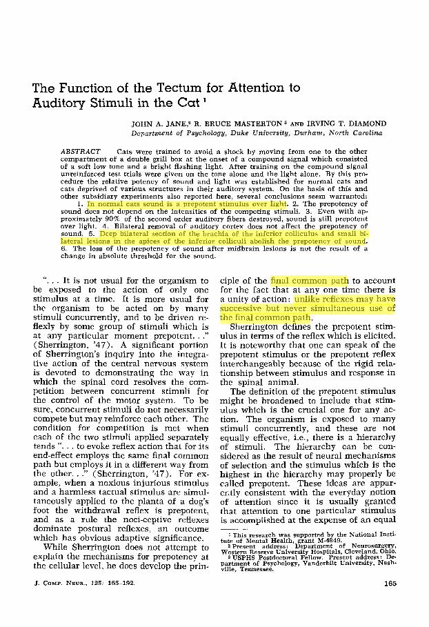
TABLE I Sequence of trials and tests in typicul test session
Consequence
response Trial Stimulus of no
1 2 3 4 5 6 7 8 9
1 0 11 1 2
Training Test Test Training Test Test Test Training Test Test Training Test
light and sound light and sound light alone light and sound light and sound sound alone light and sound light and sound light alone light and sound light and sound sound alone
shock no shock no shock shock no shock no shock no shock shock no shock no shock shock no shock
In the case of animals selecting light in preference to sound, it was necessary to show that the lesion had not produced an incapacity to respond to sound. Therefore, after completion of training and testing, several of the animals which had selected light as the relevant cue were trained to respond to sound alone. These cases were then tested on low intensity tones to deter- mine whether the lesion had caused a gross change in auditory sensitivity.
Characteristics of the stimuli. Since in- tensity must play a major part in deter- mining the prepotency of a stimulus, it was very important to arrive at sensible intensity levels of light and sound. Clearly it is no fair contest to match the sound of a cannon with a barely detectable change in illumination. Unfortunately we know of no means of equating the subjective in- tensities of light and sound. The best solu- tion seemed to be to rely on our previous experience with the preliminary study re- viewed in the introduction. In those experi- ments we found that a buzzer was strongly favored in competition with a flashing light. Therefore, it appeared reasonable to attempt to reduce the intensity of the sound by replacing the raucous buzz with a soft, low-pitched tone and to make the flashing light stimulus even brighter. During all training and testing the cats were alone in a dimly lit sound-treated room, with the experimenter sitting at the controls outside and observing through a one-way vision window. As a further precaution against the intrusion of an unwanted sound we put an electric fan in the training room. The constant humming sound of the fan may
be considered as a crude way of balancing the dim illumination of the room. The lat- ter served as a constant background for the light component of the compound stimulus, while the former provided a con- stant background for the tone.
The sound stimulus was a 300 cps tone emanating from a speaker four feet from the grill box. The sound pressure level at a point two feet from the speaker was 27 db above the background level. Since the grill box had solid plexiglass walls, stand- ing waves existed and nodes were detected with a sound level meter. Therefore, the intensity of the tone reaching the ear de- pended to some extent on the location of the animal within the grill box. In order to obtain a more useful estimate of the ef- fective sound intensity, threshold measure- ments were made on normal cats and com- pared to the intensity of the standard tone. That is, normal cats which had been trained to respond to the tone were pre- sented with tones of successively less in- tensity until they no longer responded. The intensity of the tone at which the ani- mals responded 50% of the time was taken as the “threshold.” Using this definition, the standard tone was 25 db above the threshold of normal cats.
The light stimulus was produced by flashing a 24-inch fluorescent bulb at a rate of ten per second about two feet above the cat’s head. Since the sound-treated room was only dimly lighted, human ob- servers found the flashing light a disturb- ing stimulus. Precautions were taken to assure that the onset of the light was silent. If there was any sound created in the bulb it was certainly below human threshold. As a further safeguard the hum of the fan masked any very slight sound.
Surgical and histological methods. First it is necessary to emphasize that all 17 cats with lesions of the auditory system were operated upon before receiving any training. This is the essential feature of the design since it is necessary that the original learning of the compound reflect the altered state of the nervous system. Clearly any experience received before the operation might prejudice the way in which the operated cats perceived the compound stimulus.
170 JOHN A. JANE, R. BRUCE MASTERTON AND IRVING T. DIAMOND
Auditory cortex : A standard bilateral craniectomy was performed through a curved incision and the cortex removed by subpial aspiration. An attempt was made to remove areas AI, AII, Ep and the Tem- poral-Insular cortex and at the same time spare the radiation from the lateral genicu- late body.
Trapezoid body: In two cases, the trape- zoid body was approached via a midline ventral neck incision after tracheal cannu- lation. The longus capiti muscles were deinserted and the basiocciput removed by rongeurs. The trapezoid was identified by the emerging root of the sixth cranial nerve and a longitudinal incision was made with a knife blade. In a single ani- mal (no. 228) a stereotaxic lesion was accomplished following a small dorsal craniectomy .
Midbrain at the level of the brachiurn of the inferior colliculus : Small occipital craniectomies were made and the occipital lobe gently retracted. The midbrain was approached by following the tentorium. Bilateral lesions were then made by aspira- tion. The postoperative course was occa- sionally difficult in these large one-stage procedures, and there were periods in which the cats assumed abnormal pos- tures. During such periods they responded to stimulation by extension of the limbs.
The operative ap- proach was through the posterior fossa just behind the bony tentorium. The mid- brain was readily identified by gentle re- traction of the cerebellum and the apex of each inferior colliculus was aspirated. A cerebellar deficit was noted in one case (no. 222).
Since we were not faced with the prob- lem of measuring retention we felt no special need to equalize the period of post- operative recovery. It seemed to us that our purposes would be best served by wait- ing long enough after surgery so that all of the acute symptoms would pass and only residual and permanent defects re- mained. Such a period was about three weeks in the case of the cortical surgery but was somewhat longer in the case of brain stem lesions. Thus the animals with the lesions of the brachium of the inferior colliculus recovered for about five weeks before training procedures began.
Inferior colliculus :
Following completion of the experi- ments, the animals were perfused with isotonic saline followed by 10% formalin. The brains were removed, embedded in celloidin or paraffin and serially sectioned in a frontal plane. The brain sections of cats with cortical lesions were stained with thionin and the resulting thalamic degen- eration charted. The brain sections of cats with lesions of the brain stem were stained by the Weil method. Reconstructions of both cortical and brain stem lesions were made with the aid of a projection appa- ratus. In addition, sections throughout the brain stem lesions were photographed.
RESULTS
Before presenting the chief results which were obtained in the test sessions we should say a few words about the original learning of the four normal cats and 17 cats with lesions of the auditory nervous system. Two animals (nos. 251 and 253) with midbrain lesions at the level of the brachium of the inferior colli- culus did not reach the learning criterion within a reasonable period of time. These two cases were given some tests even though learning was incomplete; and once it was clear that their performance on these tests was identical to that of the five other cats in the lesion group we felt justified in eliminating them from further consideration.
The remaining 19 cats attained the learning criterion in less than 120 trials. The two slowest learners in this popula- tion were cats with cortex lesions. The 17 cats which were relatively faster learned to respond to the compound stimulus in 70 or less trials.
The main results of the experiment are contained in the performance of the sev- eral groups during the five test sessions in which sound and light were presented alone. Table 2 shows the number of re- sponses for each of 19 cats to the light alone, to the sound alone, and to the 20 test trials with sound and light combined.
In anticipating the cumulative results for each animal, we saw four possible patterns: (1) responses only to light alone; (2) responses only to sound alone; (3 ) responses to neither sound alone nor light

CAT TECTUM 171
alone; and ( 4 ) responses to both sound alone and light alone.
The outstanding result, which can be readily seen in the table, is that, for every cat except nos. 224 and 229, there was a clearly defined prepotent stimulus. That is, virtually the entire population can be divided into two groups - one which se- lected sound and one which selected light as the relevant cue for the learned re- sponse. The two exceptional cases fell into the third pattern described above, in which neither the sound alone nor the light alone was sufficient stimulus for a response. Apparently these two animals, nos. 224 and 229, learned to respond only when the combination of light and sound was presented. The failure of any animal to exhibit the fourth pattern, i.e., a large number of responses to both light alone and sound alone, does not appear to re- quire an explanation, and we can only re- emphasize that 17 of the 19 cases selected one and not the other element of the com- pound stimulus as the basis for respond- ing. Thus a clear-cut determination of the prepotent stimulus can be easily made.
Another striking feature illustrated by the table is the consistent performance within each group. A cursory inspection of table 2 shows that three normal ani- mals selected sound, two cortical cases selected sound, three cases with trapezoid body lesions selected sound, while all nine cases with midbrain lesions selected light.
Three of the four normal cats selected the sound over light to an overwhelming degree. The fourth normal cat is one of the two excep- tional cases described above that had learned to respond primarily to the light- sound compound. Summing the scores of the three normal animals which did make a selection shows that there were five re- sponses out of 30 to light alone in contrast to 21 out of 30 to sound alone.
This result is entirely consistent with the findings of the preliminary study using the localization apparatus. Combining the results of the two experiments we gain the additional information that the prepotency of sound in normal cats does not depend on the particular behavioral training appa- ratus or on the type of reinforcement,
Performance of normal cats.
TABLE 2
Performance of all animals on test sessions ~ ~~
No. of No. of No. of responses responses responses
to light to light to sound and sound alone in alone in in 20 tests 10 tests I0 tests
Cat number
Normal 223 18/20 1/10 9/10 224 17/20 1/10 1/10 227 16/20 1/10 6/10 304 17/20 3/10 6/10
Cortex 225 19/20 1/10 8/10 229 20/20 3/10 2/10 252 13/20 1/10 7/10
Trapezoid body 226 12/20 0/10 8/10 228 18/20 0/10 7/10 24 1 20/20 1/10 9/10
of inferior colliculus 237 18/20 10/10 0/10 250 18/20 9/10 1/10 255 16/20 8/10 0/10 300 17/20 lO/lO 0/10 301 13/20 8/10 0/10
Inferior colliculus 222 13/20 7/10 0/10 232 16/20 9/10 1/10 243 17/20 7/10 1/10 24 7 15/20 9/10 2/10
Midbrain including brachium
1 Intensity of sound 9 db instead of standard 27 db.

172 JOHN A. JANE, R. BRUCE MASTERTON AND IRVING T. DIAMOND
whether punishment or reward. A further point to be emphasized is that between the preliminary study and the present study the visual cue was made more compelling. Therefore, the similarity of the results ob- tained in the two experiments as well as the low absolute intensity of the sound in the present experiment, indicate that the normal prepotency of sound does not de- pend only on the relative intensities of the stimuli.
As further evidence for the conclusion that the prepotency of the sound does not depend merely on the relative intensities of the stimuli, the case of one of the nor- mal cats, no. 304, should be noted. In that case we departed slightly from the proce-
dures already outlined by reducing the intensity of the tone from the standard value of 27 db to a sound pressure level of only 9 db above the background level. In spite of this further reduction in intensity the sound remained prepotent.
Behavioral and anatomical results of cats without auditory cortex. The per- formance of three cats, nos. 225, 229 and 252, after removal of auditory cortex is shown in table 2 and again in figures 2, 3 and 4A. In these figures the relative po- tency of sound and light is depicted by a bar graph. In addition to the results of the test sessions, figures 2, 3 and 4 A show the extent of the cortical ablations.
1oo-l CAT J-225
Fig. 2 Performance in equivalence tests and lesion reconstruction of cat no. 225. The bar graph depicts the relative potency of light alone (L) and tone alone (T) after avoid- ance training in the combined stimuli (L + T).
CAT J-229
Fig. 3 Performance on equivalence tests and lesion reconstruction of cat no. 229.

CAT TECTUM 173
The reconstructions of the cortical le- sions show that in every case there was a total removal of A1 and AII, but there was some variation in the degree to which the lesion extended ventrally to the rhinal fis- sure. Also, in a couple of cases the dorsal portion of the posterior ectosylvian gyrus is preserved as a result of an effort to leave the optic radiations intact. This point should be emphasized since the abla- tion of auditory cortex often undercuts the middle and posterior suprasylvian gyri with the result that degeneration in the lateral geniculate and pulvinar is severe. A drastic destruction of the lateral genicu- late fibers would complicate any interpre- tation of the selection of sound or light. Total removal of the visual radiations is known to produce blindness and severe visual impairment results from less than complete section. Figure 4B illustrates that in the case of animal no. 252 the principal division of the medial geniculate body is severely degenerated while the lat- eral geniculate body is relatively spared. On the basis of many other studies it can be asserted with confidence that this small amount of damage to the visual radiations would have no demonstrable effect upon visual discrimination. The locus and ex- tent of thalamic retrograde degeneration in the two other animals are similar to case no. 252. In all three cats the prin- cipal division of the medial geniculate was severely degenerated except for the ex- treme caudal tip which was partially pre-
served on one or both sides. There were some minor variations among the three brains. The degree of preservation of the magnocellular division of the medial gen- iculate shown in case no. 252 was greater than in the other two cases. In animal no. 225 anterior Po was severely degen- erated on both sides while in the case illustrated in figure 4B there was a partial preservation of anterior Po on one side.
Most important for the present purpose is the fact that in all three animals the amount of degeneration in the pulvinar and lateral geniculate was kept to a mini- mum as a result of special precautions taken to preserve the depth of the supra- sylvian sulci.
Figures 2 and 4 illustrate that two of the three cortical cases show a prepotency of sound in every way similar to normal cats. The one exceptional case, no. 229, showed no preference for either modality alone, responding only to the combined stimuli. It can be seen from figure 3 that this cat does not have a distinguishing lesion nor is the thalamic degeneration in this case substantially different from the other cortical cases. In the previous sec- tion we have described one of the normal animals, no. 224, which also displayed a failure to select one of the stimuli. Conse- quently, the cortical case, no. 229, does not serve to differentiate the normal from the cortical group either. On these grounds we conclude that the performance of cats without auditory cortex is not essentially
' O 3 8 0
CAT MJ-252
Fig. 4A Performance on equivalence tests and lesion reconstruction of cat no. 252.
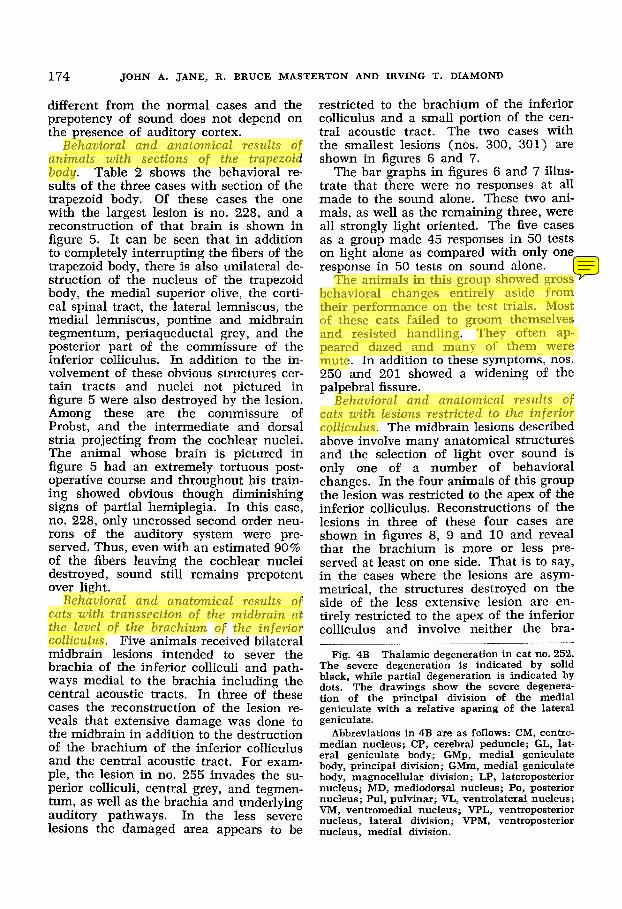
174 JOHN A. JANE, R. BRUCE MASTERTON AND IRVING T. DIAMOND
different from the normal cases and the prepotency of sound does not depend on the presence of auditory cortex.
Behavwral and anatomical results of animals with sectims of the trapezoid body. Table 2 shows the behavioral re- sults of the three cases with section of the trapezoid body. Of these cases the one with the largest lesion is no. 228, and a reconstruction of that brain is shown in figure 5. It can be seen that in addition to completely interrupting the fibers of the trapezoid body, there is also unilateral de- struction of the nucleus of the trapezoid body, the medial superior olive, the corti- cal spinal tract, the lateral lemniscus, the medial lemniscus, pontine and midbrain tegmentum, periaqueductal grey, and the posterior part of the commissure of the inferior colliculus. In addition to the in- volvement of these obvious structures cer- tain tracts and nuclei not pictured in figure 5 were also destroyed by the lesion. Among these are the commissure of Probst, and the intermediate and dorsal stria projecting from the cochlear nuclei. The animal whose brain is pictured in figure 5 had an extremely tortuous post- operative course and throughout his train- ing showed obvious though diminishing signs of partial hemiplegia. In this case, no. 228, only uncrossed second order neu- rons of the auditory system were pre- served. Thus, even with an estimated 90% of the fibers leaving the cochlear nuclei destroyed, sound still remains prepotent over light.
Behavioral and anatomical results o f cats with transsection o f the midbrain at the level o f the brachium of the inferior colliculus. Five animals received bilateral midbrain lesions intended to sever the brachia of the inferior colliculi and path- ways medial to the brachia including the central acoustic tracts. In three of these cases the reconstruction of the lesion re- veals that extensive damage was done to the midbrain in addition to the destruction of the brachium of the inferior colliculus and the central acoustic tract. For exam- ple, the lesion in no. 255 invades the su- perior colliculi, central grey, and tegmen- tum, as well as the brachia and underlying auditory pathways. In the less severe lesions the damaged area appears to be
restricted to the brachium of the inferior colliculus and a small portion of the cen- tral acoustic tract. The two cases with the smallest lesions (nos. 300, 301) are shown in figures 6 and 7.
The bar graphs in figures 6 and 7 illus- trate that there were no responses at all made to the sound alone. These two ani- mals, as well as the remaining three, were all strongly light oriented. The five cases as a group made 45 responses in 50 tests on light alone as compared with only one response in 50 tests on sound alone.
The animals in this group showed gross behavioral changes entirely aside from their performance on the test trials. Most of these cats failed to groom themselves and resisted handling. They often ap- peared dazed and many of them were mute. In addition to these symptoms, nos. 250 and 201 showed a widening of the palpebral fissure.
Behavioral and anatomical results o f cats with lesions restricted to the inferior colliculus. The midbrain lesions described above involve many anatomical structures and the selection of light over sound is only one of a number of behavioral changes. In the four animals of this group the lesion was restricted to the apex of the inferior colliculus. Reconstructions of the lesions in three of these four cases are shown in figures 8, 9 and 10 and reveal that the brachium is more or less pre- served at least on one side. That is to say, in the cases where the lesions are asym- metrical, the structures destroyed on the side of the less extensive lesion are en- tirely restricted to the apex of the inferior colliculus and involve neither the bra-
Fig. 4B Thalamic degeneration in cat no. 252. The severe degeneration is indicated by solid black, while partial degeneration is indicated by dots. The drawings show the severe degenera- tion of the principal division of the medial geniculate with a relative sparing of the lateral geniculate.
Abbreviations in 4B are as follows: CM, centro- median nucleus; CP, cerebral peduncle; GL, lat- eral geniculate body; GMp, medial geniculate body, principal division; GMm, medial geniculate body, magnocellular division; LP, lateroposterior nucleus; MD, mediodorsal nucleus; Po, posterior nucleus; Pul, pulvinar; VL, ventrolateral nucleus; VM, ventromedial nucleus; VPL, ventroposterior nucleus, lateral division; VPM, ventroposterior nucleus, medial division.

CAT TECTUM 1
MJ-252 I
GMP P
Figure 4B

CA
T 5
-22
8
1 3
'0°1
1.1 L
T
Fig
. 5
Per
form
ance
on
equ
ival
ence
tes
ts a
nd l
esio
n re
cons
truc
tion
of
cat
no.
228.
T
rans
vers
e se
ctio
ns
thro
ugh
mid
brai
n,
pons
, an
d m
edul
la a
re 2
mm
apa
rt.
Abb
revi
atio
ns f
or f
igur
es 5
-10
are
as f
ollo
ws:
BC
, br
achi
um c
onju
ncti
vum
; B
IC,
brac
hium
of
th
e in
feri
or
coll
icul
us; BP,
brac
hium
pon
tis;
CP,
cer
ebra
l pe
dunc
le;
GM
, m
edia
l ge
nicu
latc
bod
y; I
C,
infe
rior
col
licu
lus;
IP
, in
terp
edun
cula
r nu
cleu
s; L
L,
late
ral
lem
nisc
us;
LSO
, la
tera
l su
pcri
or
oliv
e; M
L,
med
ial
leni
nisc
us;
MSO
, m
edia
l su
peri
or
oliv
e;
nIII
, oc
ulom
otor
ne
rve;
nV
II,
faci
al n
erve
; nL
L,
nucl
eus
of
the
late
ral
lem
nisc
us;
RN
, re
d nu
cleu
s; S
C,
supe
rior
co
llic
ulus
; SN
, su
bsta
ntia
nig
ra;
TB
, tr
apez
oid
body
.
? 4
$- "3 U

CAT TECTUM 177
CAT J -300
6 5 4 3 2 1
LIT L -
T
Fig. 6 Performance on equivalence tests and lesion reconstruction of cat no. 300. Transverse sec- tions through the midbrain are 1 mm apart.
chium, the base of the inferior colliculus, the dorsal nucleus of the lateral lemniscus, nor fiber tracts below the inferior colli- culus.
A fourth animal, no. 222, is included in table 2, but the brain was not available for histological analysis. As a group the four cases with lesions restricted to the inferior colliculus made 32 responses out of 40 light alone tests in contrast with four responses out of 40 sound alone tests.
The measurement of auditory sensitivity
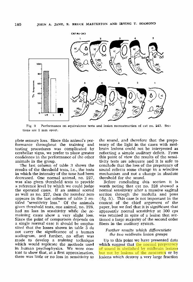
A change in the normal stimulus hier- archy produced by certain lesions in the auditory system would indeed be a trivial finding if this change were accompanied by a loss in auditory sensitivity. Complete section of the cochlear nerves would re- sult in a selection of the light cue instead of the sound without providing any insight into neural mechanisms of prepotency. To rule out the possibility that the results could be explained by a sensory deficit, all of the animals with lesions of the inferior colliculus (nos. 222, 232, 243 and 247), one case from the brachium group (no. 250), and one animal from the trape- zoid body group (no. 228) were given further tests by which their sensitivity could be compared with normal animals.
These tests began by training the ani- mal to respond to the 300 cps tone at the standard intensity of 27 db. The training on the sound alone was given in daily sessions of ten trials (just as in the case of the original training with the combina- tion light and sound) until a criterion of nine correct avoidances in one session was achieved. When the cats demonstrated the ability to respond to the sound alone, the intensity of the tone was gradually decreased in subsequent trials until an intensity was reached at which the ani- mal responded on about one-half of the trials. During these threshold tests a fail- ure to respond was always followed by a punishing shock forcing the animal to per- form as well as possible.
The results of the sensitivity tests are shown in table 3. The third column of table 3 shows the number of sessions re- quired to reach the learning criterion on the sound alone. Cat no. 222 failed to reach criterion while the five remaining animals achieved the criterion in a very few sessions. The inability of cat no. 222 to achieve the nine out of ten criterion is not easily interpreted. This cat performed consistently, making six or seven correct responses out of ten trials in session aftcr session. Therefore, the failure to reach criterion is not an indication of a com-

178 J O H N A. JANE, R. BRUCE MASTERTON AND IRVING T. DIAMOND

CAT TECTUM
h
179
R
I-
d
I- : g z s e f . 4 0 0 0
reruodraa ol0
03
bb E
180 JOHN A. JANE, R. BRUCE MASTERTON AND IRVING T. DIAMOND
CAT MJ-143
100
ao
$ 40 0
B D!
40 $
20
0 L*T T
5 4 3 2 1
Fig. 9 Performance on equivalence tests and lesion reconstruction of cat no. 243. Sec- tions are 1 mm apart.
plete sensory loss. Since this animal’s per- formance throughout the training and testing procedures was complicated by cerebellar signs, we prefer to place greater confidence in the performance of the other animals in the group.
The last column of table 3 shows the results of the threshold tests, i.e., the tests in which the intensity of the tone had been decreased. One normal animal, no. 227, was also given threshold tests to provide a reference level by which we could judge the operated cases. If an animal scored as well as no. 227, then the number zero appears in the last column of table 3 en- titled “sensitivity loss.” Of the animals given threshold tests, one animal, no. 228, had no loss in sensitivity while the re- maining cases show a very slight loss. Since the point of comparison depends on a single normal case it should be empha- sized that the losses shown in table 3 do not carry the significance of a human audiogram, and further, no effort was made to develop a training technique which would replicate the methods used in human psychophysics. We were con- tent to show that, at a first approximation, there was little or no loss in sensitivity to
the sound, and therefore that the prepo- tency of the light in the cases with mid- brain lesions could not be interpreted as reflecting a simple auditory deficit. From this point of view the results of the sensi- tivity tests are adequate and i t is safe to conclude that the loss of the prepotency of sound reflects some change in a selective mechanism and not a change in absolute threshold for the sound.
Before concluding this section it is worth noting that cat no. 228 showed a normal sensitivity after a massive sagittal section through the medulla and pons (fig. 5). This case is not important in the context of the chief argument of the paper, but we feel that it is significant that apparently normal sensitivity a t 300 cps was retained in spite of a lesion that sec- tioned a large majority of the second order fibers in the auditory system.
Further results which differentiate the two midbrain lesion groups
Up to this point we have presented data which suggest that the normal prepotency of sound is abolished by midbrain lesions but not by lesions of the neocortex or by lesions which destroy a very large fraction

CA
T J-
247
'"1 i
n
L+T
L T
6
5 4
3
2
7
1
I
Fig
. 10
P
erfo
rman
ce o
n eq
uiva
lenc
e te
sts
and
lesi
on r
econ
stru
ctio
n of
cat
no.
247
. Se
ctio
ns a
re 1
mm
apa
rt.

182 JOHN A. JANE, R . BRUCE MASTERTON AND IRVING T. DIAMOND
TABLE 3 Sensitivity to sound a f ter lesions of trapezoid body, inferior colliculus and
brachiurn o f inferior colliculus
Sensitivity loss as compared to performance of
normal cat (227)
Sessions to 9/10 Cat Lesion criterion on
sound alone number
228 TB 250 BIC 222 IC
232 IC 243 IC 247 IC
1 0 4 5 db
failed to reach no threshold criterion
5 2 4
obtained 5 db 1 db
no threshold obtained
of the second order neurons of the audi- tory pathway. In this section we wish to present results from further experiments which show that the two midbrain groups are by no means alike. In the first por- tion, evidence will be presented to show that the cats with deep lesions of the brachium are behaviorally different from those animals receiving lesions in the apex of colliculus. In the second portion of this section we shall describe electrophysiolog- ical results which also differentiate the two midbrain groups. We shall ultimately at- tempt to explain these differences between the lesion groups in terms of the degree by which the auditory lemniscal system has been disrupted.
Behavioral differences between cases with apical collicular lesions and cases with destruction of auditory pathways at the leuel of the inferior colliculus. An op- portunity for comparing the preceptual capacities of the two midbrain lesion groups was provided by an entirely differ- ent experiment which was in progress at the same time as the one reported here (Masterton and Diamond, '64). In that experiment cats were first trained to dis- criminate between a train of clicks pre- sented to the left ear and in identical train of clicks presented to the right ear. After the cats mastered that task, a second dis- crimination was presented. In the second task a pair of clicks, one cIick to each ear, was substituted for the single clicks of the first task. In place of the left click the pair- left-right was presented and in place of the right click the pair right-left was pre- sented. Time difference between the clicks of a pair was 0.5 msec. duplicating the
time difierence between the N, responses of the two auditory nerves to a click op- posite one ear. Thus the second task is simply a synthetic approximation of the first task. Figure 11A shows that once a normal cat has learned the first discrimina- tion (depicted by circles in the figure) lit- tle or no further training is required to perform the second discrimination (de- picted by triangles in the figure). This result is to be expected if the cat in fact perceives the two tasks as similar. Figure 11 also shows the typical performance of a cat deprived of auditory cortex (fig. 11B). It can be seen that the second task has to be learned anew, i.e., that the per- ceptual similarity between the two discrim- inations has been lost. Presumably, the basis for the similarity between the tasks, apparent location in space. is no longer
~~
Fig. 11 Performance of cats on Left vs. Right and Left-right vs. Right-left click discriminations. I n the performance curves the following symbols were used: circles, Left vs. Right; triangles, Left- right vs. Right-left; squares, Silence vs. Right. I n each of the three sets of symbols the open symbol represents responses to the safe signal, while the closed symbol represents reponses to the warning signal.
A - Performance of a normal cat, showing transfer on sessions 5-8.
B - Performance of a cat without auditory cortex, showing lack of transfer in sessions 1417 .
C - Performance of two cats without inferior colliculus, showing normal transfer.
D - Performance of a cat after brachium section. All lesions are bilateral. Note the simi- larity between the records of the normal cat ( A ) and the two cases without inferior colliculus (C) . The brachium case ( D ) seems to be an exaggeration of the deficit revealed in the cortical case (B) .

CAT TECTUM 183
n n I -
= E k g u 41
8-0 S3SNOdS3Y %
S3SNOdS3U %
0
P .-
0 ~ 0 1 0 0 0 O N S3SNOdS3U %

184 JOHN A. JANE, R. BRUCE MASTERTON AND IRVING T. DIAMOND
available to the cat without auditory cor- tex.
We can now turn to the second portion of the figure which shows the results ob- tained from two cases after completing the training and testing described above (fig. 1 l C ) . It can be seen that each of the ani- mals with lesions in the apical colliculus, no. 232 and no. 243, performed in a man- ner very similar to that of normal cats. The left vs. right discrimination was quickly learned and little further training on the second discrimination was required. On the other hand, the brachium case, no. 301, (fig, l l D ) not only failed to discrimi- nate between the paired clicks of the sec- ond task but even had extreme difficulty in the first task, i.e., discriminating clicks to the left ear from clicks to the right ear. Other preparations similar to that shown in figure 11D suggest that a complete in- ability to discriminate left ear from right ear results from a midbrain section deeper than that shown for no. 301 or in an in- ferior colliculus lesion which continues ventrally to interrupt the lateral lemniscus.
On behavioral grounds alone, then, it is possible to differentiate between cats with brachium sections and cats with apical col- liculus lesions. The apical colliculus le- sion results in no apparent sensory deficit in the sound localization tasks while the brachium lesion disrupts even the simple left ear vs. right ear discrimination.
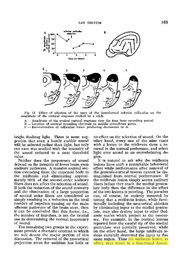
Electrophysiological difference between lesions of the apical inferior colliculus and deep sections o f the brachium o f the in- ferior colliculus. A clue to the behavioral differences between the two midbrain groups is provided by a study of the effect of midbrain lesions upon the amplitude of the cortical evoked response. Galambos, Myers and Sheatz ('61) and Goldberg and Neff ('6la) have shown that cortical evoked potentials in cats under pentobar- bital anesthesia can be abolished by brach- ium section if these sections are sufficiently deep. We followed a procedure similar to that used by these investigators in order to see whether the two groups distinguished in terms of behavior could also be distin- guished on an electrophysiological basis.
Evoked potentials to clicks were recorded from the auditory cortex of decerebellate cats deeply anesthetized with Dial. After
the amplitude of the primary evoked re- sponse became stable the apex of the ho- molateral inferior colliculus was removed. The amplitude of the cortical response was again measured from time to time. In this manner the change in the amplitude of the cortical response could be roughly cor- related with the extent of the colliculus le- sion. A typical result is illustrated in fig- ure 12.
Since the data in figure 12 were recorded from a preparation in which the lesion was very similar to the largest of the colliculus lesions in the behavioral group (no. 243), it would appear that none of the lesions in the inferior colliculus group were large enough to abolish the cortical evoked po- tential even under barbiturate anesthesia. On the other hand, many of the brachium cases are similar to those reported by Gal- ambos et al. ('61) and Goldberg and Neff ('61a). We conclude that the cats in our brachium group would have shown little or not cortical response to clicks.
These results imply that the apex of the central nucleus of the inferior colliculus does not contain a large portion of fibers of the lemniscal system which synapse in the inferior colliculus. It would appear that the relay function of the inferior colliculus is concentrated in the more basal regions. Indeed, experience with other preparations like the one illustrated in figure 12 has shown that, in order to reduce the cortical response to the level accomplished by a deep brachium lesion, the central nucleus of the inferior colliculus must be entirely removed and the lesion extended to near the depth of the nucleus of the lateral lemniscus.
DISCUSSION
The results of two experiments employ- ing entirely different training procedures indicate that for normal cats sound is a prepotent stimulus over light. In learned tasks requiring either the avoidance of pun- ishment or an approach for food, normal cats select sound as the relevant cue rather than light.
The prepotency of sound in normal cats does not seem to depend on the intensities of the competing stimuli, at least within reasonable limits. The evidence is that a tone of low intensity is selected over a
CAT TECTUM 185
C - 1 3 4 5
Fig. 12 Effect of ablation of the apex of the homolateral inferior colliculus on the
A - Amplitude of the evoked cortical response over the four hour recording period. B - Location of cortical recording electrode in middle ectosylvian gyrus. C - Reconstruction of colliculus lesion producing decrement in A.
amplitude of the cortical response evoked by a click.
bright flashing light. There is some sug- gestion that even a barely audible sound will be selected rather than light, but only one case was studied with the intensity of the sound reduced to a near threshold value.
Neither does the prepotency of sound depend on the integrity of lower brain stem auditory pathways. A massive sagittal sec- tion extending from the trapezoid body to the midbrain and eliminating approxi- mately 90% of the second order auditory fibers does not affect the selection of sound. If both the reduction of the sound intensity and the elimination of a large proportion of second order fibers are considered as simply resulting in a reduction in the total number of impulses passing up the main afferent pathways of the brain stem, then it can be concluded that this factor, i.e., the number of impulses, is not the crucial one in determining the normal prepotency of sound.
The remaining two groups in the experi- ment provide a dramatic contrast to which we will devote the major portion of this discussion. The removal of the neocortical projection areas for audition has little or
no effect on the selection of sound. On the other hand, every one of the nine cases with a lesion in the midbrain show a re- versal in the normal preference, and select light over sound to an overwhelming de- gree.
It is natural to ask why the midbrain lesions have such a remarkable behavioral effect while performance after removal of the geniculocortical system cannot be dis- tinguished from normal performance. If the midbrain lesion simply severs auditory fibers before they reach the medial genicu- late body then the difference in the effect of the two lesions is puzzling. The paradox can, of course, be entirely removed by noting that a midbrain lesion, while func- tionally including the neocortical ablation by eliminating input to the medial genicu- late, may also destroy input to other thal- amic nuclei which project to the neocor- tex. For example, in the cortical lesions reported here the caudal tip of the medial geniculate was partially preserved, while on the other hand, the large midbrain le- sions certainly destroyed the input to this same region. Thus the midbrain lesion, in effect, may result in a functional discon-

186 JOHN A. JANE, R. BRUCE MASTERTON AND IRVING T. DIAMOND
nection of a neocortical area much larger than the classical auditory cortex. Rccord- ing to this line of reasoning, the prepotency of sound might yet depend on neocortex or the geniculo-cortical system.
A similar explanation was considered at length by Goldberg and Neff ('61a) to ac- count for the difference in the behavioral effects of lesions of the midbrain and neo- cortex, These authors found severe and lasting impairment in frequency discrim- ination following a very deep lesion pene- trating both the brachium of the inferior colliculus and the underlying central acoustic tract. In contrast, the removal of the geniculo-cortical system alone by means of ablating auditory cortex has no permanent effect on frequency discrimina- tion (Butler, Diamond and Neff, '57; Gold- berg and Neff, '61b). The authors discuss the evidence for auditory pathways medial to the brachium of the inferior colliculus that project to thalamic nuclei of the dif- fuse projection system. Goldberg and Neff ('61a) conclude their argument with the suggestion that the diffuse thalamic proj- ection system and its neocortica1 target may play a critical role in the absence of the medial geniculate and its cortical tar- get.
Such a conclusion might also serve to explain our cases with deep sections at the level of the brachium. Indeed if our mid- brain lesions were entirely restricted to this group there would be no motive for seeking an alternative explanation. How- ever, in the experiment reported here, a similar behavioral reversal was found after a much more restricted lesion in the infe- rior colliculus. This finding suggests a second alternative; namely, that the infe- rior colliculus itself is responsible for the prepotency of sound.
In order to support this interpretation it is first necessary to establish that the in- ferior colliculus is not simply a relay sta- tion for auditory activity directed toward the neocortex. Such an argument is remin- iscent of an earlier paper by Ades ('44) in which he mustered electrophysiological, be- havioral, and anatomical evidence to sup- port the conclusion that the inferior col- liculus is an independent reflex center en- tirely aside from its role as a relay. In the following discussion we shall consider
these lines of supporting evidence, adding to the argument of Ades more recent stud- ies in anatomy, behavior and electrophys- iology.
Electrophysiological evidence that the in- ferior colliculus is an independent integrat- ing center. Ades ('44) reports that a siz- able response in the superior colliculus of cats can be evoked by clicks. The ampli- tude of the evoked response in the superior colliculus is markedly decreased after this nucleus has been surgically separated from the inferior colliculus. Ades concludes that the evoked response must therefore result from efferent fibers from the inferior col- liculus and notes that a pathway from the inferior to the superior colliculus would bring the former ultimately in connection with the motor centers of the ventral teg- mentum, medulla and spinal cord.
In addition to Ades' conclusions, we have shown that lesions in the apex of the inferior colliculus fail to abolish evoked potentials in auditory cortex even under deep barbiturate anesthesia. In contrast, very deep sections at the level of the brachium of the inferior colliculus elimi- nate evoked potentials at the cortex in cats deeply anesthetized with barbiturates (Goldberg and Neff, '61a; Galambos et al., '61). These facts must mean that the major portion of the primary afferent path- way to cortex remains intact after the api- cal colliculus lesion.
Behavioral evidence that the inferior col- liculus is an independent integrating center. Behavioral evidence supporting the conclusion that the inferior colliculus is more than just a relay nucleus has also been provided by studies by Ades and his co-workers. Raab and Ades ('46) have shown that a lesion of the inferior col- liculi has little or no effect on relative in- tensity discriminations. Kryter and Ades ('43) report a partial loss in absolute threshold of pure tones after a similar le- sion. However, this loss is small in com- parison with the effects of a lateral lem- nisci section. Ades ('44) interprets these behavioral findings as evidence for the by- passing of the colliculus by fibers in the lateral lemniscus. If the inferior colliculus was entirely devoted to relaying impulses to the forebrain then ablation of the infe- rior colliculus alone would functionally

CAT TECTUM 187
eliminate the geniculo-cortical system as well. However, the behavioral effects of lesions of the inferior colliculus do not replicate a lesion of cortex, a combined lesions of cortex and colliculus, or a section of the lateral lemniscus.
The concept that there are fibers by- passing the inferior colliculus and project- ing directly to the medial geniculate has not received support from Marchi studies (Barnes, Magoun and Ranson, '43), but it is worth noting that such a by-passing would be expected from a recent account of the phylogeny of the main afferent sys- tems (Bishop, '59). According to Bishop, there is a tendency for sensory fibers to project directly to ncw targets as they de- velop in the dicncephalon and telenceph- alon resulting in a by-passing of phylogene- tically older centers of the midbrain.
An alternative to Ades' interpretation that the inferior colliculus is by-passed by some auditory fibers is suggested by the fact that the lesions in the present study spared the basal portion of the inferior col- liculus. All we need assume is that the majority of fibers of the lateral lemniscus which synapse in the inferior colliculus do so in the spared basal portion.
To the evidence presented by Ades and his co-workers we have additional be- havioral evidence that a lesion restricted to the inferior colliculus is far less severe in its effects than would be expected if the input to the entire auditory forebrain were interrupted. We have shown, for example, that lesions of the apex of the inferior col- liculus do not produce a deficit in sound localization, a task which is known to be dependent on neocortex in cats (Neff, '62; Masterton and Diamond, '64). On the other hand, the deep lesions of the midbrain at the level of the brachium produce a strik- ing inability even in the most simple locali- zation discrimination.
In summary, i t can be concluded that two entirely different syndromes result from removal of the geniculo-cortical sys- tem and restricted removal of the inferior colliculus. This conclusion would be ex- tremely unlikely if the inferior colliculus were merely a link in a chain terminating in cortex. On the one hand, many symp- toms following neocortical lesions are ab- sent after restricted ablation of the inferior
colliculus. On the other hand, the principal symptom reported in this paper follows the collicular lesion but not the cortical lesion. Such a dissociation rules out the idea of a strictly serial system.
Anatomical evidence for the inferior col- liculus as a n independent integrating center. Still another line of evidence which supports the conclusion that the in- ferior colliculus is an independent organi- zation center and not simply a relay station to the neocortex comes from com- parative anatomy. In fish and primitive am- phibians the posterior part of the optic tectum receives proprioceptive impulses (Herrick, '48). The differentiation of the predominantly proprioceptive posterior tec- tum in these animals into the predominant- ly auditory inferior colliculus of mammals is closely correlated with the development of the cochlea. A cochlea first appears in higher amphibians and is accompanied by an increase in size of the lateral bulbo- tectal tract or primordial lateral lemniscus, which terminates in the posterior tectum.
Necturus lacks a cochlear rudiment, and hence the lateral bulbolemniscus here prob- ably has a very imperfect auditory function, if any. In Amblystoma and most other urodeles there is such a rudiment, and here audition of a primitive type may be repre- sented, along with other functions, in this tract. In adult Anura there is a well de- veloped primordium of the cochlea, from which a cochlear division of the VIII nerve arises. Correlated with this, there are coch- lear nuclei, from which a true lateral lemnis- cus passes to the inferior colliculus. Paral- lel to this differentiation, both general and lateral lemniscus tracts, as seen in urodeles are radically reorganized (Herrick, '48, p. 165).
According to Herrick, then, the auditory involvement of the primordial inferior col- liculus is as primitive in vertebrates as audition itself.
From the time of the appearance of the cochlea in land amphibians, homologues of the mammalian cochlear nucleus, supe- rior olive, lateral lemniscus, and inferior colliculus developed together and presum- ably constituted the main central pathways of audition. Throughout the reptilian stage, before the appearance of the neocortex and the relay nuclei of the dorsal thalamus, the torus semi-circularus (the primordial in- ferior colliculus) and the nucleus isthmus (the primordial dorsal nucleus of the lat-

188 JOHN A. JANE, R. BRUCE MASTERTON AND IRVING T. DIAMOND
eral lemniscus) were probably the high- est central integrative ganglia princi- pally devoted to audition (Le Gros Clark, '33). In extant reptiles, these nuclei send fibers to many diencephalic and mesen- cephalic structures including the nucleus rotundus and nucleus reuniens posterior of the thalamus for relay to the corpus striatum; the ventral medial geniculate of the ventral thalamus for relay to the hy- pothalamus; the superior colliculus, and the midbrain tegmentum (Kappers, Huber and Crosby, '36; Papez, '36; Kruger and Berkowitz, '60).
The neocortex in mammals differenti- ated as the rostral projections of the infe- rior colliculus found new targets in the dorsal thalamus which, in turn, projected onto the cortex. The diffuse connections of the submammalian tectum became over- laid by the true mammalian brachium of the inferior colliculus linking the colliculus to the medial geniculate body and posterior nuclei of the thalamus. The brachium, to be sure, is by far the most impressive ros- tral projection of the inferior colliculus in mammals and has gained the most atten- tion in anatomical descriptions. However, Moore and Goldberg ('63) studying the rostral projections of the colliculus by the Nauta technique have identified several nonthalamic regions of termination :
Fibers of the inferior brachium derived from the ipsilateral inferior colliculus ter- minate in several midbrain regions, includ- ing the capsule of the inferior colliculus, the parabrachial region of the lateral tegmentum and the interstitial nucleus of the inferior brachium. . . .
In addition to the major projections a few fibers originating in the inferior colliculus terminate in the central gray and, probably, in deep layers of the superior colliculus (Moore and Goldberg, '63, pp. 117 and 118).
The lesions which produced the terminal degeneration described involve more ante- rior portions of the colliculus than those in our apical colliculus group. A more poste- rior or apical lesion might very well pro- duce even more widespread degeneration in both the rostral and caudal directions.
Since the medial geniculate body and the posterior nuclei of the mammalian thalamus are dependent on the neocortex, the brachium, relaying auditory impulses to these structures, is part of the system whose chief target is the neocortex. How-
ever, the several connections of the colli- culi to widely separated nonthalamic brain stem structures cannot be considered as even indirectly dependent on the existence of neocortex in mammals because they also occur in submammalian vertebrates. Such nonthalamic connections are more likely a structural development that paralleled or closely followed the invasion of the brain stem by auditory input. Thus, the relay function of the inferior colliculus as repre- sented by the mammalian brachium is a relatively late acquisition in vertebrates brought about by the appearance of a higher integrative system, the thalamo- neocortical complex. With the connections viewed in this way, behavioral and elec- trophysiological changes resulting from an ablation of the apex of the inferior colliculi can be expected to be quite different from those produced either by severance of the brachium or by the removal of neocortex, which indeed has been demonstrated.
To sum up, the conclusion, supported by phylogenetic and anatomical considera- tions, is that any function of the mam- malian inferior colliculus as a relay for cortically directed activity developed long after the more primitive integrative func- tions. Consequently the effect of collicular activity on surrounding midbrain and non- thalamic diencephalic structures must be an important, and not necessarily second- ary, substrate for the mediation and ap- preciation of audible phenomena in mam- mals. Bringing this conclusion to bear on the central results of this paper leads us to make the suggestion that this primitive integrative function may include a selec- tion mechanism, the mechanism which in cats makes sound prepotent over light.
The superior colliculus and visual attention
We have argued that the mechanism which mediates the normal prepotency of sound in cats can be localized in the infe- rior colliculus. There is some evidence that the superior colliculus may play a parallel role for vision in primates, in the sense that these nuclei are necessary for normal attention to visual cues. Denny- Brown ('62) has reported that after a bilat- eral removal of the superior colliculi in monkeys the animals suffer a temporary

CAT TECTUM 189
loss of vision. Denny-Brown goes on to note that “. . . even more extraordinary than the loss of vision following bilateral ablation of the colliculi was the change in general behavior of these monkeys. Whether some vision for movement and placing remained or not the animals ap- peared to be totally unaware of events in their environment” (Denny-Brown, ’62, p. 536) .
Denny-Brown concludes that the pri- mate tectum is essential for reactions which may be called visual awarness and that in general the tectum has an initiating or facilitating function. It is especially noteworthy that the lesion of the superior colliculus does no damage to the pathway to the neocortex via the lateral geniculate body and, as the author points out, the nor- mal cortical evoked potential is preserved after the tectal ablation.
These experiments provide some clue concerning the function which is retained by the tectum after the ascendancy of the thalamo-cortical system. In submammal- ian vertebrates the otpic tectum is the chief center for all visual sensory functions. When the geniculo-striate system became the dominant organ for visual sensory functions with the evolution of mammals, the tectum apparently continues to play some role in visual attention. It is worth considering that the function of auditory and visual midbrain centers have under- gone similar restrictions or specializations as a result of the phylogenetic develop- ment of neocortex.
Selective mechanisms in the spinal COT^, midbrain, and cmtex
If the inferior colliculus serves as a se- lective mechanism we may now return to the inquiry of Sherrington and ask how this selection compares with that accom- plished at the spinal cord. At the spinal cord selection depends on certain inter- nuncial neurons which are tuned to spe- cific afferent input and which, in turn, cap- ture the final common path. The outcome of any competition between stimuli is pre- established by the nature of the afferent- internuncial-efferent pathway. Presumably both the prepotency of the stimulus and the response to the stimulus are fixed by innate connections (fig. 13A).
The inferior colliculus of the cat also appears to resolve a competition between stimuli with the result that sound cues emerge naturally prepotent over light cues. Whatever the tectal mechanism for stim- ulus selection, the tectum and the spinal cord are alike in the sense that the relative potency of competing stimuli is preestab- lished. However, in contrast to the mech- anism at the spinal cord, a sound stimulus gaining its prepotency at the tectum can be used by the animal in any number of
- A Spinal cord
S n rn
B Midbrain
SI RI
C Cortex
Fig. 13 Diagrammatic iuustration of selec- tion mechanisms in the spinal cord, midbrain and cortex operating at the simultaneous. presen- tation of a series of stimuli, SI, SZ, SS . . . S,. A: spinal cord level, rigid and innate selection of prepotent stimulus (SS) and prepotent response (R3); B: midbrain level, rigid and innate selec- tion of prepotent stimulus (S3) but modsable selection of responses; C: cortical level, modifi- able seIection of stimuli and responses.

190 JOHN A. JANE, R. BRUCE MASTERTON AND IRVING T. DIAMOND
ways. It is therefore possible to speak of the resolution of the competition at the tectum in psychological terms, for when a compelling stimulus can be used for any number of responses it might properly be said that the stimulus has captured the animal‘s attention. We may conclude, then, that at the level of the tectum, stim- ulus prepotency is fixed but the response to that stimulus is not fixed (fig. 13B).
Clearly there must exist even further selective mechanisms in mammals in which both the prepotency and the re- sponse are plastic and can be modified by experience. It is commonplace that ani- mals can learn to anticipate or wait for a specific cue in a particular modality. In such a circumstance it is possible to de- scribe the individual as exhibiting a highly selective degree of alertness.
We must conclude that selective mech- anisms in the nervous system have not been exhausted by describing the func- tions at the level of the spinal cord and the tectum. Presumably, these more subtle forms of selection which are evident in some mammals take place at the neocortex (fig. 13C).
Behavioral methods. Because the pres- ent behavioral methods are described as an adaptation of Sherrington’s investigation of the competition between simultaneous stimuli, an impression of novelty could be created which is not merited. The use of an unreinforced test following training is a simple application of Kluver’s (’33) method of equivalent stimuli, and a similar logic applies to the interpretation of re- sults obtained with both techniques. In a typical Kluver experiment a monkey is trained to choose one of a pair of visual pat- terns, e.g., a circle as opposed to a square. Then a new pair of stimuli, such as an oval and a triangle, are substituted for the original pair. If the monkey selects the oval in the test it is fair to conclude that curvilinearity was an important feature in learning to make the discrimination be- tween the original pair of stimuli.
Credit for adapting Sherrington’s method of compound simultaneous stimulation to the field of animal learning probably be- longs to Pavlov. Pavlov’s problem, method, and interpretation of results parallels the present experiments and the chief differ-
ence appears to depend upon the distinc- tion between classical and instrumental conditioning. After conditioning a re- sponse to a compound stimulus, the strength of each component is tested with the following result :
“When the stimuli making up the com- pound act upon different analysers, the ef- fect of one of them when tested singly was found very commonly to overshadow the effect of the others almost completely, and this independently of the number of rein- forcements of the compound stimulus. For example, a tactile component of a stimula- tory compound was usually found to ob- scure a thermal component, a n auditory component t o obscure a visual component, and so on” (Pavlov, ’27, p. 141, italics are ours).
When a salivary reflex was established in dogs to the simultaneous presentation of a tone and light, the light alone evoked no secretion of saliva while the effectiveness of tone alone approached that of the com- pound. Pavlov’s interpretation of these results is that the stronger component pre- vails, and stronger is just another way of saying prepotent as we have used this term. In support of this conclusion Pavlov notes that: (1) the ineffective component, when reinforced alone, is capable of acquiring a powerful conditioned response: ( 2 ) it is possible to create a compound of equal components, e.g. two equally intense tones, in the sense that each component alone produces a similar amount of salivation; ( 3 ) otherwise equal components of a com- pound can be made unequal by increasing the intensity of one of them.
When Pavlov’s results are added to those of the present study the following conclu- sions appear warranted. The prepotency of sound over light may be characteristic of many classes of carnivores, possibly all groups which retain certain features of a night-hunting common ancestor. This pre- potency can be demonstrated in several behavioral situations, using classical as well as instrumental conditioning, and using food as well as an aversive uncondi- tioned stimulus.
Further adaptations of Sherrington’s method of simultaneous stimulation. The competition between simultaneously pre- sented stimuli has been studied with the

CAT TECTUM 191
aid of chronically implanted electrodes (Jane, Smirnov and Jasper, '62). In this study, evoked responses were recorded in the lateral and medial geniculate bodies and the auditory and visual cortex, while cats were presented with a compound sound and light stimulus. Efforts were then made to favor one or the other modal- ity by distracting and attention-catching stimuIi such as a squeaking noise of a rat. While these attempts did not result in a differential increase or decrease in the amplitude of potentials evoked in auditory and visual centers, even so it would appear that the method of evoked potentials pro- vides an opportunity for the study of selec- tive mechanisms in higher levels of the nervous system.
ACKNOWLEDGMENTS
We are grateful to Dr. Harlow Ades for reading this manuscript and for his help- ful criticism.
It is a pIeasure to acknowledge the tech- nical assistance of Miss Stephanie Peach, Mrs. Verble Roberts, and Mr. Lloyd Ross.
Throughout this investigation we re- ceived encouragement, support and advice from the late Karl Zener, Professor and Chairman of the Department of Psychol- ogy. Duke University.
LITERATURE CITED Ades, H. W. 1944 Midbrain auditory mecha-
nisms in cats. J. Neurophysiol., 7: 415-424. Barnes, W. T., H. W. Magoun and S. W. Ranson
1943 The ascending auditory pathway in the brainstem of the monkey. J. Comp. Neur., 79: 129-152.
Bishop, G . H. 1959 The relation between nerve fiber size and sensory modality: phylogenetic implications of the afferent innervation of cor- tex. J. Nerv. Ment. Dis., 128: 89-114.
Butler, R. A,, I. T. Diamond and W. D. Neff 1957 Role of auditory cortex in discrimination of changes in frequency. J. Neurophysiol., 25: 223-235.
Clark, W. E. LeGros 1933 The medial genicu- late body and the nucleus isthmi. J. Anat., London, 67: 536-548.
Denny-Brown, D. 1962 The midbrain and motor integration. Proc. Roy. SOC. Med., 55: 527-537.
Galambos, R., R. E. Myers and G. C. Sheatz 1961 Extralemniscal activation of auditory cortex in cats. Amer. J. Physiol., 200: 23-28.
Goldberg, J. M., and W. D. Neff 1961a Fre- quency discrimination after bilateral section of the brachium of the inferior colliculus. J. Comp. Neur., 116: 265-290.
1961b Frequency discrimination after bilateral ablation of cortical auditory areas. J. Neurophysiol., 24: 119-128.
Herrick, C. J. 1948 The Brain of the Tiger Salamander. The University of Chicago Press, Chicago, Illinois.
Jane, J. A,, G. D. Smirnov and H. H. Jasper 1962 Effects of distraction upon simultaneous auditory and visual evoked potentials. Elec- troenceph. clin. Neurophysiol., 14: 344-358.
Kappers, C. U. A,, G. C. Huber and E. C. Crosby 1936 The Comparative Anatomy of the Nerv- ous System of Vertebrates Including Man. Hafner, New York.
Kliiver, H. 1933 Behavior Mechanisms in Mon- kevs. The Universitv of Chicaeo Press. Chi- - cage, lllinois.
Kruger, L., and E. C. Berkowitz 1960 The main afferent connections of the reptilian telenceph- alon as determined by degeneration and elec- trophysiological methods. J. Comp. Neur., 115: 125-142.
Kryter, K. D., and H. W. Ades 1943 Studies on the function of the higher acoustic nervous centers in the cat. Amer. J. Psychol., 56: 501-536.
Masterton, R. B., and I. T. Diamond 1964 Ef- fects of auditory cortex ablation on discrimina- tion of small binaural time differences. J. Neurophysiol., 27: 15-36.
Moore, R. Y., and J. M. Goldberg 1963 Ascend- ing projections of the inferior colliculus in the cat. J. Comp. Neur., 121: 109-136.
Neff, W. D. 1962 Neural structures concerned in localization of sound in space. Psycholo- gische Beitrage, 6: 492-500.
Neff, W. D., J. F. Fisher, I. T. Diamond and M. Yela 1956 Role of auditory cortex in dis- crimination requiring localization of sound in space. J. Neurophysiol., 19: 500-512.
Papez, J. W. 1936 Evolution of the medial geniculate body. J. Comp. Neur., 64: 41-61.
Pavlov, I. P. 1927 Conditioned Reflexes. Ox- ford University Press, Oxford, England.
Raab, D. H., and H. W. Ades 1946 Cortical and midbrain mediation of a conditioned discrimi- nation of acoustic intensities. Amer. J. Psy-
Sherrington, C. S. 1906 The Integrative Action of the Nervous System, second ed. 1947. Yale University Press, New Haven, Connecticut.
chol., 59: 59-83.