Embed Size (px)
Citation preview

The Formation of the Second Maturation Spindle inthe Eggs of Succinea, Physa, and Planorbis
by CHR. P. RAVEN1
From the Zoological Laboratory, University of Utrecht
WITH TWO PLATES
IN a previous communication (Raven, Escher, Herrebout, & Leussink, 1958) thematuration of the egg was described in Agriolimax reticulatus, Limax flavus,and Limnaea stagnalis, with special regard to the formation of the secondmaturation spindle. In the three species the second maturation spindle arises bydirect transformation from the deep centrosphere of the first maturation amphi-aster. In A. reticulatus and Limax flavus this centrosphere contains a pair ofcentrioles which move apart to opposite poles of the developing spindle. This issymmetrical and develops an aster at both ends, arising in the cytoplasm outsidethe original centrosphere.
In Limnaea stagnalis the centrosphere of the first maturation amphiastercontains only one centriole, which remains undivided. When the centrosphereelongates, the centriole moves as a whole to its outer pole. The spindle develop-ing from the centrosphere is therefore asymmetrical and egg-shaped, and asterformation occurs at its pointed outer end only. The inner aster of the secondmaturation spindle is provided by the sperm aster, which fuses secondarily withthe blunt inner end of the spindle.
In discussing this aberrant course of the second maturation division in L. stag-nalis, it was concluded that its primary cause is the failure of the egg cytocentreto divide properly. This causes the lack of astral radiations at the deep end of thedeveloping second maturation spindle, and enables the sperm aster to fuse withthis end of the spindle. It was argued that a mutation bringing about a precociousinactivation of the egg cytocentre, in conjunction with the circumstance that asperm aster, free to take over the task of the failing deep maturation aster, ispresent at the right moment, might have led to this course of events. As itappeared conceivable that a modification of development occurring at such anearly stage might have a profound evolutionary significance, it seemed impor-tant to investigate the taxonomic distribution of this peculiar mode of formationof the second maturation spindle.
1 Author's address: Zoologisch Laboratorium der Rijksuniversiteit, Janskerkhof 3, Utrecht,Netherlands.[J. Embryol. exp. Morph. Vol. 7, Part 3, pp. 344-60, September 1959]

SECOND MATURATION SPINDLE 345
Since the completion of our previous paper eggs of various pulmonates, bothBasommatophora and Stylommatophora, have therefore been fixed at variousmoments during the course of the maturation divisions. The study of these eggsin sections soon revealed that the above formulation of the problem is ratherover-simplified. We had tacitly assumed that the mode of formation of the secondmaturation spindle as observed in Umax and Agriolimax is the 'normal' one inpulmonates, while the relationships found in Limnaea stagnalis constitute adeviation of the process occurring in a limited number of species of this subclass.However, to our surprise it appeared that this early and quite fundamental pro-cess exhibits a bewildering variation among the species. Its study in as largea number of species as possible seems necessary before any generalizations canbe framed. In view of the fundamental importance of all data relative to the pro-cesses of meiosis and mitosis, such a study may yield important results.
In this paper the formation of the second maturation spindle in four pul-monates will be described: the stylommatophore Succinea putris L., and thebasommatophore species Physa acuta Drap., Planorbis planorbis L., and P.corneus L.
MATERIAL AND METHODS
The eggs of Succinea putris, Planorbis planorbis, and P. corneus were obtainedfrom snails caught in the neighbourhood of Utrecht and kept for some time inthe laboratory. Those of Physa acuta were from specimens living in the aquariaof the Institute.
Since the formation of the second maturation spindle is a process which,especially in some species, takes place very soon after oviposition, it was im-portant to be able to obtain the eggs at will at the desired moment. To thatend, methods to stimulate oviposition in these snails have been worked out byLucy P. M. Timmermans (1959).
The eggs of the land snail Succinea putris are laid, preferentially within moistmoss, in irregular masses, containing from 20 to 60 egg capsules. These capsulesare each surrounded by a layer of jelly (Jura & George, 1958), by which they arecemented together into a loose aggregate. The spherical egg capsules have adiameter of about 1 to 1-3 mm. They have a thick, elastic, transparent wall,which is not encrusted with lime. The egg cells have a diameter of about 130^.
The egg-masses of Physa acuta are gelatinous, oblong, and from 10 to 18 mm.in length. They may contain from 15 to 50 egg capsules, lying rather freely in thecommon jelly. The egg capsules are oblong and about 0-7 x 0-9 mm. in size.
The egg-masses of Planorbis planorbis are flat and disk-shaped, and have adiameter of from 3 to 5 mm. The egg capsules lie side by side in one layer, closetogether. There are about 15 to 25 egg capsules, each about 0-5 mm. in diameter.The egg cells have a diameter of 110-30//. In the first 15 minutes after ovi-position the egg masses are whitish and opaque; then they become transparent,with a yellowish tinge.
346 CHR. P. R A V E N — F O R M A T I O N OF SECOND
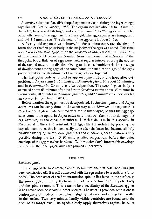
P. corneus also has flat, disk-shaped egg-masses, containing one layer of eggcapsules (cf. Jura & George, 1958). The egg-masses are about 8 to 10 mm. indiameter, have a reddish tinge, and contain from 15 to 25 egg capsules. Theouter jelly layer of the egg-mass is rather rigid. The egg capsules are transparentand 1-4-1-6 mm. in size. The diameter of the egg cells is about 140 /x.
A freshly laid egg-mass was observed under a microscope, and the time offormation of the first polar body in the majority of the eggs was noted. This timewas taken as the starting-point of the subsequent observations; all indicationsof time mentioned below are counted from the moment of extrusion of thefirst polar body. Batches of eggs were fixed at regular intervals during the courseof the second maturation division. Owing to the considerable variations in stageof development among eggs of the same batch, the maturation age of the eggsprovides only a rough estimate of their stage of development.
The first polar body is formed in Succinea putris about one hour after ovi-position; in Physa acuta 5-15 minutes, in Planorbis planorbis about 15 minutes,and in P. corneus 10-20 minutes after oviposition. The second polar body isextruded about 65 minutes after the first in Succinea putris; about 35 minutes inPhysa acuta; 50 minutes in Planorbis planorbis, and 55 minutes in P. corneus (atan average temperature of 20° C).
Before fixation the eggs must be decapsulated. In Succinea putris and Physaacuta this can be easily done in the same way as in Limnaea: the egg-mass isrolled out on a glass plate covered with moist filter-paper, so that the egg cap-sules come to lie apart. In Physa acuta care must be taken not to damage theegg capsules, as the capsule membrane is rather delicate in this species; inSuccinea it is thick and resistant. The egg cells are isolated by pricking thecapsule membrane; this is most easily done after the latter has become slightlywrinkled by drying. In Planorbis planorbis and P. corneus, decapsulation is onlypossible during the first 15-20 minutes after oviposition, before the outerenvelope of the egg-mass has hardened. With watchmaker's forceps this envelopeis removed; then the egg capsules are pricked under water.
RESULTSSuccinea putris
In the eggs of the first batch, fixed at 15 minutes, the first polar body has justbeen constricted off. It is still connected with the egg surface by a stalk or a 'mid-body'. The deep aster of the first maturation spindle lies beneath the surface atthe animal pole, often slightly to one side of the attachment of the polar bodyand the spindle remnant. This seems to be a peculiarity of the Succinea egg, asit has never been observed in other species. The aster is provided with a densecentrosphere of moderate size. This is slightly flattened and elongated parallelto the surface. Two very minute, hardly visible centrioles are found near theends of its longer axis. The dyads closely apply themselves against its outer

MATURATION SPINDLE 347
surface in a loose group; they even seem partly to have invaded the area of thecentrosphere (Plate 1, fig. A). The aster and spindle remnant are surrounded bya dense area of the cytoplasm; many mitochondria have accumulated in thisregion.
In the next few minutes the spindle remnant disappears. The centrospherewith the dyads approaches the surface and comes to lie immediately beneath theegg cortex, flattening still more against it. The astral radiations of the oldmaturation aster become blurred and indistinct, but in some cases a new orienta-tion of astral fibres is indicated near the ends of the elongated centrospherewhere the centrioles are barely visible as ill-defined and somewhat darker dots.
Apparently this does not yet give rise to the asters of the second maturationspindle, however, for in subsequent batches all astral radiations have dis-appeared. As a matter of fact, in the eggs fixed at 30 minutes, the dyads are foundin an area of clear protoplasm, lying immediately beneath the surface near theanimal pole but often somewhat eccentric with respect to the first polar body(Plate 1, fig. B). This clear area, which may be rather irregular in outline, pos-sibly represents the shrunken centrosphere. Subsequently it once more shifts itsposition and sinks somewhat below the surface but remains in the region ofdense animal cytoplasm (Plate 1, fig. C). Here two small asters appear at oppositeends of the clear area, and become connected by a spindle. Part of the spindlefibres traverse the clear area and connect with the dyads, while other fibres runalong its outer border (Plate 1, fig. D). The spindle, which may at first have atangential or oblique position, then rises to the surface and places itself at rightangles to it (Plate 1, fig. E). The dyads arrange themselves in the equatorial planeof the spindle, while the latter increases considerably in size. The aster at theouter end of the spindle flattens against the surface. The inner aster growsconsiderably in size and develops a large clear centrosphere, in the middle ofwhich the centriole is now distinctly visible (Plate 1, fig. F).
During the early stages of the formation of the second maturation spindle nosperm aster is present in the egg. It appears for the first time when the secondmaturation spindle is in metaphase. It is situated at an arbitrary place in thecytoplasm, occasionally quite near to the second maturation spindle (Plate 1,fig. F), but mostly at a greater distance. During anaphase of the second matura-tion division it grows somewhat in size and develops a distinct centrosphere,which may often show a tendency to reduplicate. Immediately after the extru-sion of the second polar body this centrosphere becomes strongly vacuolized,and the whole sperm aster rapidly disappears without leaving any traces.
Physa acuta
Early development in Physa acuta is much more rapid than in other species.The second polar body is extruded 35 minutes after the first. The formation ofthe second maturation spindle takes place in the first 20 minutes after the extru-sion of the first polar body.
348 CHR. P. RAVEN — F O R M A T I O N OF SECOND
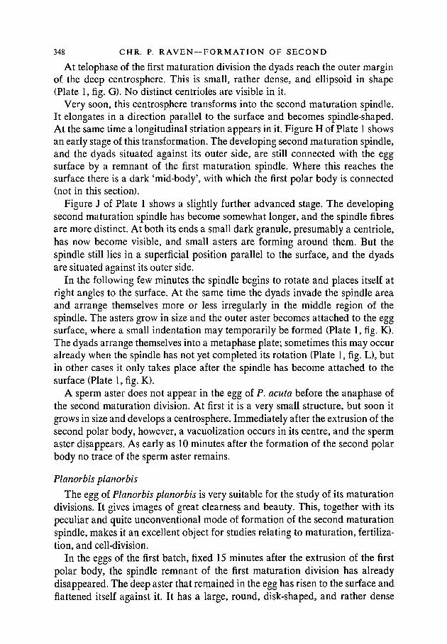
At telophase of the first maturation division the dyads reach the outer marginof the deep centrosphere. This is small, rather dense, and ellipsoid in shape(Plate 1, fig. G). No distinct centrioles are visible in it.
Very soon, this centrosphere transforms into the second maturation spindle.It elongates in a direction parallel to the surface and becomes spindle-shaped.At the same time a longitudinal striation appears in it. Figure H of Plate 1 showsan early stage of this transformation. The developing second maturation spindle,and the dyads situated against its outer side, are still connected with the eggsurface by a remnant of the first maturation spindle. Where this reaches thesurface there is a dark 'mid-body', with which the first polar body is connected(not in this section).
Figure J of Plate 1 shows a slightly further advanced stage. The developingsecond maturation spindle has become somewhat longer, and the spindle fibresare more distinct. At both its ends a small dark granule, presumably a centriole,has now become visible, and small asters are forming around them. But thespindle still lies in a superficial position parallel to the surface, and the dyadsare situated against its outer side.
In the following few minutes the spindle begins to rotate and places itself atright angles to the surface. At the same time the dyads invade the spindle areaand arrange themselves more or less irregularly in the middle region of thespindle. The asters grow in size and the outer aster becomes attached to the eggsurface, where a small indentation may temporarily be formed (Plate 1, fig. K).The dyads arrange themselves into a metaphase plate; sometimes this may occuralready when the spindle has not yet completed its rotation (Plate 1, fig. L), butin other cases it only takes place after the spindle has become attached to thesurface (Plate 1, fig. K).
A sperm aster does not appear in the egg of P. acuta before the anaphase ofthe second maturation division. At first it is a very small structure, but soon itgrows in size and develops a centrosphere. Immediately after the extrusion of thesecond polar body, however, a vacuolization occurs in its centre, and the spermaster disappears. As early as 10 minutes after the formation of the second polarbody no trace of the sperm aster remains.
Planorbis planorbis
The egg of Planorbis planorbis is very suitable for the study of its maturationdivisions. It gives images of great clearness and beauty. This, together with itspeculiar and quite unconventional mode of formation of the second maturationspindle, makes it an excellent object for studies relating to maturation, fertiliza-tion, and cell-division.
In the eggs of the first batch, fixed 15 minutes after the extrusion of the firstpolar body, the spindle remnant of the first maturation division has alreadydisappeared. The deep aster that remained in the egg has risen to the surface andflattened itself against it. It has a large, round, disk-shaped, and rather dense

MATURATION SPINDLE 349
centrosphere; the boundary between centrosphere and astral rays is not verysharp. In the middle of the centrosphere lies a somewhat darker but rathervaguely delineated body, apparently a centriole. It is always single. The dyadsare arranged in a more or less regular circle around the centrosphere, at theboundary between the latter and the astral rays (Plate 1, fig. M).
In addition to the maturation aster, a second large aster is present in each egg.It has a variable location, often near the centre of the egg, but sometimes at ashort distance beneath the maturation aster. The fact that it is connected withthe sperm tail in most cases (Plate 1, fig. P; Plate 2, fig. A) proves that it is thesperm aster. It is more or less reduplicated in all cases. Sometimes it has anelongate or biscuit-shaped centrosphere in which two rather large, but ill-defined,centrioles can often be distinguished (Plate 1, fig. N). In the middle, between thetwo centrioles, a longitudinal striation may appear, forming a short centralspindle (Plate 1, fig. P). Finally, the centrioles may move farther apart, the centralspindle elongates, and the aster divides, in this way forming a beautiful spermamphiaster (Plate 2, fig. A). It must be emphasized that this is an achromaticspindle, as the sperm nucleus is still lying as a compact dark body somewherebeneath the egg cortex and remains so until after the extrusion of the secondpolar body, as it does in all pulmonates.
The mitochondria of the egg are accumulated around both the maturationaster and the sperm aster, often penetrating in rows between the astral rays.
Starting from this situation, which prevails shortly after the extrusion of thefirst polar body, the following processes lead to the formation of the secondmaturation spindle. The dyads begin to move centrifugally along the astral raysof the maturation aster, thereby widening their circle (Plate 1, figs. O, P).
At the same time the sperm amphiaster rises to the animal pole and comes tolie close beneath the maturation aster. Now delicate fibres are 'spun' betweenthe centre of the maturation aster and the sperm amphiaster, spanning the inter-vening stretch of cytoplasm. When the sperm amphiaster lies more or lesshorizontally and its two poles are at about the same distance from the centreof the maturation aster, these spindle fibres may at first run divergently fromthe latter to both sperm centrioles (Plate 1, fig. P). Later, two different possibili-ties may be distinguished. When the sperm aster is more or less horizontal andits centrioles lie near together, the fibres of the maturation spindle seem to endindiscriminately along the whole middle region of the sperm amphiaster (Plate 2,fig. B). When the poles of the sperm amphiaster are farther apart, however, andespecially when it is situated obliquely or vertically, the maturation spindle isformed between the centre of the old maturation aster and the nearest pole ofthe sperm amphiaster (Plate 2, figs. A, C). So we get the uncommon situation ofa spindle one of the poles of which is itself a (more or less well-developed)spindle.
The maturation spindle formed in this way is at first achromatic, but soon itbecomes 'colonized' by the dyads. In their outward migration along the rays of

350 CHR. P. RAVEN — F O R M A T I O N OF SECOND
the maturation aster the dyads move at first immediately beneath the egg cortex,against which the aster remains closely applied (Plate 2, fig. A). But then the endsof the astral rays bend inwards, taking along the dyads (Plate 2, fig. B), and thelatter reach the outer surface of the developing second maturation spindle,which has in the meantime increased in girth and has become more distinctlyvisible. In some eggs several of the dyads have reached the spindle (Plate 2,fig. C), whereas other dyads still lie in the peripheral parts of the maturationaster. Finally, all dyads have reached the outer surface of the maturation spindle,and begin to invade the spindle area. Then they arrange themselves into a meta-phase plate.
Figure D of Plate 2 shows the second maturation spindle in early anaphase. Itsinner pole is formed by the sperm amphiaster, which has two distinct centresconnected by a central spindle. The latter at this stage begins to disintegrate, itsspindle fibres becoming more or less blurred and apparently breaking up intosmaller fragments. The maturation spindle is mainly centred towards the leftpole of the sperm amphiaster. The outer aster of the maturation spindle hasdeveloped from the original deep aster of the first maturation spindle. As amatter of fact, during the later phases of the migration of the dyads the astralradiations have become somewhat less distinct, but at no stage have they dis-appeared altogether, and now they begin to become more pronounced again.
A late anaphase stage is shown in Plate 2, fig. E. The sperm amphiaster hastwo large clear centrospheres, each containing a dark rather ill-defined centriole,and lying obliquely one above the other. They are separated by the remnants ofthe central spindle, which run as a dark granular band between them. Thematuration spindle is connected with the upper half of the sperm amphiaster.
In the next few minutes, while the second polar body is being formed, the twocentrospheres of the sperm amphiaster unite to a single spherical clear area witha finely reticular structure, in which a band of coarse granules forms the lastremnant of the former central spindle. On either side of this band a centriole isstill seen; they have even become much more clearly defined than at previousstages, and present themselves as compact, rather large, and globular bodies.Both sperm centrioles are visible in Plate 2, fig. F, the second polar body havingjust been pinched off.
Immediately after the extrusion of the second polar body the egg chromo-somes begin to swell into karyomeres. At the same time the sperm nucleus alsoswells. Temporarily it has the appearance of a closely packed group of karyo-meres, forming a morula-like body; with further swelling these karyomeres uniteto a polymorphic male pronucleus. This stage has been reached 20 minutes afterthe extrusion of the second polar body. The egg karyomeres are still separatevesicles at this time, but 10 minutes later they too have fused to a polymorphicpronucleus.
After the extrusion of the second polar body the deep aster of the maturationspindle (the former sperm amphiaster) moves somewhat below the surface. Its

MATURATION SPINDLE 351
astral radiations soon begin to disappear. Its centrosphere forms a large, clear,spherical area, finely reticulate in structure, but interspersed with coarsegranules. Two centrioles may still be seen in it as distinct dark globules (Plate 2,fig. G). Simultaneously with the swelling of the sperm nucleus, the latter beginsto migrate from its subcortical position towards the centre of the egg, where itcomes to lie in the centrosphere of the sperm aster. This stage has been reached20 minutes after the extrusion of the second polar body. Then the centrospherewith the male pronucleus migrates towards the animal pole, where it forms aclear crescent-shaped zone surrounding the two pronuclei. Twenty minutes laterthis clear zone has disappeared and is replaced by the dense animal pole plasmwhich has now accumulated at the animal pole.
Planorbis corneus
Although the formation of the second maturation spindle in Planorbiscorneus occurs essentially in the same way as in P. planorbis, the former speciesis much less favourable material owing to different staining properties.
In all eggs fixed 10 minutes after the extrusion of the first polar body, thedeep aster of the first maturation spindle lies closely beneath the surface at theanimal pole. It is of moderate size and has, as a rule, no clear centrosphere,the astral rays continuing towards the centre of the aster. The dyads are situatedin a narrow circle around this centre (Plate 2, fig. H).
In most eggs a small sperm aster is found. It has no distinct centrosphere. Ithas a variable location; sometimes it lies at a short distance beneath the matura-tion aster (Plate 2, fig. H), but as a rule it is situated in other parts of the egg andshows no topographic relations to the maturation aster. In most cases the spermaster is a single structure; only in a few instances does it show a beginningreduplication of its centre.
In the next 10 minutes the dyads begin to move centrifugally along the rays ofthe maturation aster, arranging themselves in a wide circle in its periphery(Plate 2, fig. J). At the same time the sperm aster slowly approaches the matura-tion aster and comes to lie beneath it. Simultaneously, it grows in size, and aclear centrosphere appears in its centre (Plate 2, fig. J). The number of spermasters showing reduplication increases; in extreme cases this may lead alreadyat this stage to the formation of a small sperm amphiaster, consisting of twoasters with distinct centrospheres and connected by a short achromatic centralspindle.
When the sperm aster has approached the maturation aster to within a smalldistance, delicate spindle fibres begin to be 'spun' between the two. If the spermaster is double, they are directed to one of its centres. At the same time the astralrays of the maturation aster bend inwards and the dyads, moving along them,reach the outer surface of the developing second maturation spindle. In the eggof Plate 2, fig. K, this has occurred with some of the dyads, but the majority ofthem still lie in the peripheral parts of the maturation aster.

352 CHR. P. RAVEN —FORMATION OF SECOND
In this way the second maturation spindle becomes 'colonized' by the dyads.This stage has been reached at about 25 minutes. The dyads, first lying at thesurface of the spindle, then gradually penetrate into its substance and arrangethemselves more or less irregularly along the spindle fibres (Plate 2, fig. L). At40 minutes they begin to concentrate in the equatorial region (Plate 2, fig. M),and so the metaphase stage is reached. The outer aster of the spindle is theformer deep aster of the first maturation spindle; its inner aster is formed by thesperm aster, which is now more or less reduplicated in all cases, ranging froma slight dumb-bell shape of its centrosphere to a well-developed sperm amphi-aster with central spindle.
DISCUSSION
It has already been remarked in the introduction to this paper that the presentobservations have brought to light a quite unexpected variability in the course ofthe maturation divisions within the group of the pulmonates. They show thatsome conclusions drawn from our previous investigations were premature, andnecessitate a renewed consideration of some points discussed previously.
In a previous paper (Raven, Escher, Herrebout, & Leussink, 1958) it was con-cluded that in the pulmonates generally the second maturation spindle arises bya direct transformation from the centrosphere of the deep aster of the firstmaturation spindle. This was shown to be the case in Limax flavus, Agriolimaxreticulatus, and Limnaea stagnalis. A similar mode of formation of the secondmaturation spindle seemed highly probable for other pulmonates from the datafound in the literature. Especially the descriptions and illustrations given byByrnes (1900) for Limax {Agriolimax) agrestis and by Lams (1910) for Avionempiricorum leave little doubt that the process in these species occurs in essen-tially the same way as in Limax flavus and Agriolimax reticulatus (cf. figs. 10,15,& 16 of Byrnes, and figs. 42, 48, 56, & 57 of Lams with the photographs in theabove-mentioned paper by Raven et al).
The present observations show, however, that we have been overhasty inextending this generalization to the pulmonates in general. It is true that theformation of the second maturation spindle in Physa acuta, though differing insome respects, belongs essentially to the same category as that of Limax, Agrio-limax, and Avion. In Succinea putris this is much less obvious; only with somereservation can we adduce it to the same scheme. But as regards Planorbis thereis no indication at all of a material continuity between the centrosphere of thefirst maturation aster and the developing second maturation spindle. Here, there-fore, our generalization breaks down altogether; we have to reconcile ourselveswith the thought that the second maturation spindle among the pulmonates mayapparently be formed in at least two fundamentally different ways.
In Limax, Agriolimax, and Arion, the deep centrosphere of the first matura-tion spindle at a certain stage contains two centrioles. Some time after the

MATURATION SPINDLE 353
extrusion of the first polar body these centrioles begin to move apart within thearea of the centrosphere. At the same time the latter elongates and becomesellipsoid, with its long axis coinciding with the line connecting the centrioles.Some connecting fibres between the centrioles may form the beginning of acentral spindle. The main mass of the spindle arises by the appearance of alongitudinal striation in the substance of the centrosphere, occurring when thecentrioles have reached the ends of its long axis.
The formation of the second maturation spindle in Physa acuta apparentlyoccurs along the same lines. However, the process does not take place here withsuch diagrammatic clearness as in the above species. The centrosphere is smalland dense, and rather ill-defined with respect to the surrounding aster and cyto-plasm. The centrioles do not become visible until they have reached the polesof the developing spindle. Moreover, the whole process occurs very rapidly, thetransformation of the centrosphere into the spindle already beginning when it isstill connected with the surface by the remnant of the first maturation spindle.But on the whole there is no reason to doubt that the formation of the secondmaturation spindle in P. acuta essentially agrees with that in the slugs.
From the work of Kostanecki & Wierzejski (1896) we may draw the same con-clusion with regard to P. fontinalis. If one compares figs. 7-12 of their paperwith Plate 1, figs. G-L of the present paper, it is apparent that the formation ofthe second maturation spindle in the two species of Physa shows a greatresemblance, though some structures, e.g. the centrioles, may appear withgreater clearness in P. fontinalis.
In Succinea putris there are greater deviations from this general scheme. Asa matter of fact, it does seem at first that the process will follow the same course.There is a distinct, elongate centrosphere, with minute centrioles near the endsof its longer axis; small asters may even begin to form around these centrioles.But this centrosphere rises to the surface and flattens itself against it. All astralradiations vanish. There remains an area of clear, presumably strongly vacuo-lated, cytoplasm, in which the dyads are situated. This may either represent theformer centrosphere, or it is a new formation due to the accumulation of fluidaround the dyads; in the latter case we must conclude that the centrosphere hasdisappeared altogether. After some time this clear area with the dyads sinks intothe depth and gives rise to the spindle, two asters again appearing at oppo-site ends.
It apparently depends on our interpretation of the 'clear area' just mentioned,whether or not we can compare the formation of the second maturation spindlein Succinea with that in the slugs and in Physa. If the clear area represents thecentrosphere, then there is a fundamental agreement. The first and last phasesof the transformation of the centrosphere into the spindle take place more or lessaccording to schedule; but the process is interrupted by a kind of 'rest period'immediately beneath the egg cortex, during which the asters temporarily dis-appear, and the centrosphere transforms by vacuolization into the 'clear area'.
354 CHR. P. RAVEN — F O R M A T I O N OF SECOND
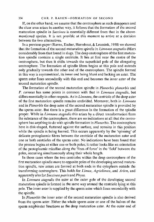
If, on the other hand, we assume that the centrosphere as such disappears andthe clear area arises in another way, it follows that the formation of the secondmaturation spindle in Succinea is essentially different from that in the above-mentioned species. It is not possible at this moment to arrive at a decisionbetween the two alternatives.
In a previous paper (Raven, Escher, Herrebout, & Leussink, 1958) we showedthat the formation of the second maturation spindle in Limnaea stagnalis differsconsiderably from that found in slugs. The deep centrosphere of the first matura-tion spindle contains a single centriole. It lies at first near the centre of thecentrosphere, but then it shifts towards the superficial pole of the elongatingcentrosphere. The formation of spindle fibres begins at this pole and extendsonly gradually towards the other end of the centrosphere. The spindle formedin this way is asymmetrical, its inner end being blunt and lacking an aster. Thesperm aster fuses secondarily with this end and becomes the inner aster of thesecond maturation spindle.
The formation of the second maturation spindle in Planorbis planorbis andP. corneus has some points in common with that in Limnaea stagnalis, butdiffers essentially in other respects. As in Limnaea, the centriole of the deep asterof the first maturation spindle remains undivided. Moreover, both in Limnaeaand in Planorbis the deep aster of the second maturation spindle is provided bythe sperm aster. But there is a great difference in the formation of the spindleproper. While in Limnaea stagnalis this arises by a direct transformation fromthe substance of the centrosphere, there are no indications at all that the centro-sphere has anything to do with spindle formation in Planorbis. The centrospherehere is disk-shaped, flattened against the surface, and remains in this positionwhile the spindle is being formed. This occurs apparently by the 'spinning' ofdelicate protoplasmic fibres between the centriole of the maturation aster andone or both centrioles of the sperm aster. No indications have been found thatthe process begins at either one or both poles; it rather looks like an orientationof the protoplasmic micellae along the 'lines of force' in the 'field' between thepoles, occurring simultaneously along their whole length.
In those cases where the two centrioles within the deep centrosphere of thefirst maturation spindle move to opposite poles of the developing second matura-tion spindle, new asters are formed at both ends in the cytoplasm outside thetransforming centrosphere. This holds for Limax, Agriolimax, and Arion, andapparently also for Succinea putris and Physa.
In Limnaea stagnalis the aster at the outer pole of the developing secondmaturation spindle is formed in the same way around the centriole lying at thispole. The inner aster is supplied by the sperm aster which fuses secondarily withthe spindle.
In Planorbis the inner aster of the second maturation spindle is also derivedfrom the sperm aster. Either the whole sperm aster or one of the halves of thesperm amphiaster functions as the deep maturation aster. At the outer end of

MATURATION SPINDLE 355
the spindle a large aster is found; this is no other than the original deep aster ofthe first maturation spindle, which is taken over as such by the second spindle.As a matter of fact, the astral radiations may temporarily become somewhatvague, so that it is possible that a kind of rejuvenation of the aster occurs; butit is out of the question that the aster disappears entirely and is replaced by anew one in between the two maturation divisions.
Apparently, therefore, variable as is the formation of the second maturationspindle in the pulmonates, the origin of its asters is equally so.
In a previous paper (Raven, Escher, Herrebout, & Leussink, 1958) we havesummarized the variations in the development of the sperm aster in variouspulmonates. The present observations give new evidence of this variability.
In Physa acuta the sperm aster appears during anaphase of the second matura-tion division. It remains always undivided, and disappears shortly after theextrusion of the second polar body.
In Succinea putris the sperm aster becomes visible when the second matura-tion spindle is in metaphase. It may show a tendency to reduplicate, but again itdisappears immediately after the extrusion of the second polar body.
In Planorbis corneus a small sperm aster without a centrosphere is alreadypresent a short time after the extrusion of the first polar body. It grows rapidly insize and develops a centrosphere; at the same time it becomes more or lessreduplicated, ranging from a slightly dumb-bell shaped centrosphere to a well-developed sperm amphiaster. In the meantime it has approached the maturationaster and participated in the formation of the second maturation spindle, whosedeep aster it becomes.
Finally, in P. planorbis the sperm aster apparently is formed already beforethe extrusion of the first polar body. Fifteen minutes after first polar body forma-tion it possesses a large centrosphere and is reduplicated in all cases; sometimesit has even at this moment developed into a sperm amphiaster with centralspindle. It then approaches the maturation aster, initiates the formation of thesecond maturation spindle, and supplies its deep aster. Its two centrospheres,each containing a distinct centriole, fuse into a spherical clear area during theextrusion of the second polar body. This area migrates to the animal pole,temporarily forms a crescent-shaped clear zone around the pronuclei, and isthen replaced by the developing animal pole plasm.
It is evident that the four species, in the order mentioned, form a series ofincreasing importance of the sperm aster. In P. planorbis it not only appearsvery early, but reaches a stage of development unprecedented in any pulmonatestudied so far. Only the evolution of the sperm aster in Physa fontinalis, asdescribed by Kostanecki & Wierzejski (1896), shows some resemblance to thatin Planorbis. According to these authors, the sperm aster in Physa fontinalisdivides and forms an amphiaster with central spindle. This may occur at varioustimes, either before or after the extrusion of the second polar body, even as lateas the stage at which the male pronucleus approaches the female pronucleus at
5584.7 A a

356 CHR. P. R A V E N — F O R M A T I O N OF SECOND
the animal pole. Anyhow, the sperm amphiaster in all cases gives rise to the firstcleavage spindle.
In a previous paper (Raven, Escher, Herrebout, & Leussink, 1958) and in mybook (Raven, 1958) I have expressed some doubt on the accuracy of Kostanecki& Wierzejski's observations. However, in view of the great variability whichprevails in the course of the maturation divisions in pulmonates, there seems atpresent to be no reason to think that these observations are incorrect. A re-investigation of the maturation divisions in P. fontinalis appears desirable.
Anyhow, the process as it occurs in Planorbis planorbis provides a strongargument in favour of the view that, in pulmonates, the poles of the cleavagespindle are also derived by division from the sperm cytocentre. As a matter offact, the egg cytocentre in this species remains undivided, forms the outer poleof the second maturation spindle, and becomes presumably extruded with thesecond polar body. In the sperm amphiaster two distinct centrioles are visible.When the centrospheres of the sperm amphiaster have fused to a single sphericalclear area after the extrusion of the second polar body, the two centrioles are stillclearly visible in it. Presumably they shift together with the clear area and thesperm nucleus towards the animal pole, where the copulation of the two pro-nuclei occurs. However, at this stage it is impossible to distinguish the two cen-trioles from other basophil granules in this area. For about 45 minutes, therefore,the centrioles are invisible; then two small asters appear near the pronuclei andgive rise to the cleavage spindle. One can hardly doubt that the poles of thisspindle are formed by the sperm centrioles. This is the more remarkable as thesetwo centrioles have had a different history, as a rule: for one of the two hasformed for some time the deep pole of the second maturation spindle. In view ofthis circumstance it is conceivable that one of the two sperm centrioles dis-integrates, while the other divides once more and forms the poles of the cleavagespindle. Up till now no indications of such a process have been found, however.
A few words remain to be said about the behaviour of the dyads. In Agrio-limax reticulatus and Limax flavus the dyads, after having reached the marginof the deep centrosphere at telophase of the first maturation division, remain asa tightly packed group against the outer surface of the developing second matura-tion spindle, as a rule near its equatorial region. They do not penetrate into thespindle area until the spindle has been fully formed and has begun its rotation.
The behaviour of the dyads in Physa acuta is quite similar to this. In Succineaputris, however, it appears that the dyads begin to penetrate into the centro-sphere at an early stage. When the latter rises to the surface they come to lieentirely in the area of clear protoplasm which possibly represents the centro-sphere. Later, when the second maturation spindle is formed, the spindle fibrespenetrate secondarily into this area and connect with the dyads.
In Limnaea stagnalis the dyads, at first forming a compact group against theouter side of the centrosphere, become arranged into a more or less regular ring.When the centrosphere begins to elongate, the dyads form a crown around its

MATURATION SPINDLE 357
outer pole. This moves along the outer surface of the developing spindle until ithas reached its equatorial region. Only after the sperm aster has fused with thedeep end of the spindle do the dyads begin to invade the spindle area.
Finally, in Planorbis planorbis and P. corneus the dyads first form a narrowring around the centre of the centrosphere. Then they begin to move centri-f ugally along the astral rays, in this way widening their circle. At first they moveimmediately beneath the surface, but then they are taken along by the astralrays bending inwards towards the spindle which has formed in the meantime. Sothey reach the outer surface of the spindle, after which they penetrate into it.
It is evident, therefore, that the course of the second maturation division inthe pulmonates shows a great deal of variation. Various components of the pro-cess may vary more or less independently provided, of course, that they fittogether in such a way as to ensure the normal completion of the maturationdivision. Every species shows a species-specific pattern, although smaller varia-tions may occur within each species. This species-specificity is such that it wouldbe possible to construct a key for the identification of the species here describedmerely on the basis of their second maturation divisions.
This is the more remarkable as we are here dealing with a process taking placeat such an early stage of development. In general we are accustomed to the factthat developmental processes in related species show a greater similarity theearlier they take place, in accordance with v. Baer's principle. I know of noother example where such fundamental differences between developmental pro-cesses occur at such an early stage.
Of course it might be argued that the development of the new individual doesnot begin before the formation of the zygote nucleus, so that the maturation ofthe egg does not represent an early, but rather a very late, stage of development.But such a formal point of view does not discount the fact that differences indevelopmental processes during egg maturation might profoundly affect thecourse of subsequent development, e.g. by influencing the localization or thesynthesis of cytoplasmic substances. In this way they might be of decisive impor-tance for the determination of species-specific differences. Perhaps such a viewmight be substantiated by cytochemical investigations during maturation inpulmonates.
SUMMARY
1. Egg maturation has been studied in Succinea putris, Physa acuta, Planorbisplanorbis, and P. corneus, with special regard to the formation of the secondmaturation spindle.
2. In Physa acuta the second maturation spindle arises by direct transforma-tion from the deep centrosphere of the first maturation amphiaster. This processbegins already when the centrosphere is still connected with the surface by theremnant of the first maturation spindle.
3. In Succinea putris the deep centrosphere of the first maturation spindle

358 CHR. P. R A V E N — F O R M A T I O N OF SECOND
rises to the surface and flattens itself against it. The dyads come to lie in an areaof clear cytoplasm, which subsequently sinks into the depth and gives rise tothe spindle.
4. In Planorbis planorbis and P. corneus the centriole of the deep aster of thefirst maturation spindle remains undivided. This aster flattens against the surfaceand becomes the outer aster of the second maturation spindle. Its inner aster isprovided by the sperm aster, while the second maturation spindle itself is formedby the 'spinning' of protoplasmic fibres between the centriole of the maturationaster and one or both centrioles of the sperm aster.
5. The sperm aster in Physa acuta is a rudimentary structure which appearslate, disappears soon, and always remains undivided. In Succinea putris thesperm aster appears slightly earlier and may show a tendency to reduplicate. InPlanorbis corneus the sperm aster appears when the first polar body is extruded.It grows rapidly in size, becomes more or less reduplicated, and participates inthe formation of the second maturation spindle. In P. planorbis the sperm asteris formed before the extrusion of the first polar body. It reduplicates and formsa beautiful sperm amphiaster with central spindle. It participates in the forma-tion of the second maturation spindle and becomes its deep aster. During theextrusion of the second polar body its two centrospheres, containing distinctcentrioles, fuse to a spherical clear area. The sperm nucleus comes to lie in thisarea, then migrates together with it towards the animal pole. The sperm cen-trioles presumably form the poles of the first cleavage spindle.
6. In Physa acuta the dyads lie at first as a compact group on one side againstthe developing second maturation spindle, and penetrate only secondarily intothe spindle when this is nearly completed. In Succinea putris the dyads pre-sumably penetrate into the centrosphere at an early stage. In Planorbis planorbisand P. corneus the dyads first form a narrow ring around the centre of thematuration aster, then move centrifugally along its astral rays, and are conveyedby the latter bending inwards towards the outer surface of the second maturationspindle.
7. It is concluded that fundamental differences in the course of the secondmaturation division occur in the pulmonates.
R E F E R E N C E S
BYRNES, E. F. (1900). The maturation and fertilization of the egg of Limax agrestis (Linne").J. Morph. 16, 201-36.
JURA, CZ., & GEORGE, J. C. (1958). Observations on the jelly mass of the eggs of three molluscs,Succinea putris, Limnaea stagnalis, and Planorbis corneus, with special reference to meta-chromasia. Proc. Acad. Sci. Amst. C 61, 590-4.
KOSTANECKI, K., & WIERZEJSKI, A. (1896). Ober das Verhalten der sogen. achromatischen Sub-stanzen im befruchteten Ei. Nach Beobachtungen an Physa fontinalis. Arch. mikr. Anat. 47,309-86.
LAMS, H. (1910). Recherches sur l'ceuf d'Arion empiricorum (Fer.). (Accroissement, maturation,fe"condation, segmentation). Mem. Acad. R. Belg. 2, 1-144.
MATURATION SPINDLE 359
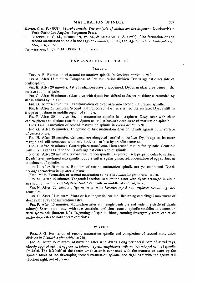
RAVEN, CHR. P. (1958). Morphogenesis. The analysis of molluscan development. London-NewYork-Paris-Los Angeles: Pergamon Press.
ESCHER, F. C. M., HERREBOUT, W. M., & LEUSSINK, J. A. (1958). The formation of thesecond maturation spindle in the eggs of Limnaea, Limax, and Agriolimax. J. Embryol. exp.Morph. 6,28-51.
TIMMERMANS, LUCY P. M. (1959). In preparation.
E X P L A N A T I O N OF PLATES
PLATE 1
FIGS. A-F. Formation of second maturation spindle in Succinea putris. x910.FIG. A. After 15 minutes. Telophase of first maturation division. Dyads against outer side of
centrosphere.FIG. B. After 20 minutes. Astral radiations have disappeared. Dyads in clear area beneath the
surface at animal pole.FIG. C. After 30 minutes. Clear area with dyads has shifted to deeper position; surrounded by
dense animal cytoplasm.FIG. D. After 40 minutes. Transformation of clear area into second maturation spindle.FIG. E. After 55 minutes. Second maturation spindle has risen to the surface. Dyads still in
irregular position in middle region of spindle.FIG. F. After 80 minutes. Second maturation spindle in metaphase. Deep aster with clear
centrosphere and distinct centriole. Sperm aster just beneath deep aster of maturation spindle.FIGS. G-L. Formation of second maturation spindle in Physa acuta. x910.FIG. G. After 15 minutes. Telophase of first maturation division. Dyads against outer surface
of centrosphere.FIG. H. After 20 minutes. Centrosphere elongated parallel to surface. Dyads against its outer
margin and still connected with 'mid-body' at surface by spindle remnant.FIG. J. After 20 minutes. Centrosphere transformed into second maturation spindle. Centriole
with small aster at either end. Dyads against outer side of spindle.FIG. K. After 20 minutes. Second maturation spindle has placed itself perpendicular to surface.
Dyads have penetrated into spindle, but are still irregularly situated. Indentation of egg surface atattachment of spindle.
FIG. L. After 20 minutes. Rotation of second maturation spindle not yet completed. Dyadsarrange themselves in equatorial plane.
FlGS. M-P. Formation of second maturation spindle in Planorbis planorbis. x910.FIG. M. After 15 minutes. Tangential section. Maturation aster with dyads arranged in circle
in circumference of centrosphere. Single centriole in middle of centrosphere.FIG. N. After 25 minutes. Sperm aster with biscuit-shaped centrosphere containing two
centrioles.FIG. O. After 25 minutes. More or less tangential section. Beginning centrifugal movement of
dyads along rays of maturation aster.FIG. P. After 15 minutes. Maturation aster with single centriole and widening circle of dyads
(above). Sperm amphiaster with two centrioles and short central spindle (middle) in connexionwith sperm tail (bottom left). Beginning of spindle fibres, running divergently from centre ofmaturation aster to both sperm centrioles.
PLATE 2
FIGS. A-G. Formation of second maturation spindle and completion of second maturationdivision in Planorbis planorbis. x 910.
FIG. A. After 15 minutes. Maturation aster with dyads along peripheral part of astral rays,closely applied against egg cortex (above). Sperm amphiaster with well-developed central spindle(middle). The left half of the sperm amphiaster is connected with the maturation aster by thespindle fibres of the developing second maturation spindle, the right half with the sperm tail(bottom right, out of focus).

360 CHR. P. RAVEN—SECOND MATURATION SPINDLE
FIG. B. After 25 minutes. Maturation aster with single centriole (above), connected by secondmaturation spindle with sperm amphiaster possessing two centrioles (middle). Rays of maturationaster bending inwards, taking along dyads.
FIG. C. After 25 minutes. Second maturation spindle, connected with left half of reduplicatedsperm aster containing two centrioles. Dyads have reached outer surface of maturation spindle.
FIG. D. After 20 minutes. Early anaphase of second maturation division. Sperm amphiasterwith central spindle, showing beginning disintegration of spindle fibres. Deep end of secondmaturation spindle centred towards left pole of sperm amphiaster.
FIG. E. After 60 minutes. Late anaphase of second maturation division. Sperm amphiaster withtwo centrospheres, each containing a centriole, and separated by remnants of central spindle.Maturation spindle connected with upper half of sperm amphiaster.
FIG. F. After 60 minutes. Telophase of second maturation division. Centrospheres of spermamphiaster fused to spherical clear area, containing remnants of central spindle. One spermcentriole just beneath telophase chromosomes, the other one in middle of lower half of clear area.
FIG. G. After 60 minutes. Remnant of sperm amphiaster as spherical clear area, still containingthe two sperm centrioles (globular bodies in upper half of clear area).
FIGS. H-M. Formation of second maturation spindle in Planorbis corneus. x910.FIG. H. After 10 minutes. Maturation aster with dyads near centre of aster (above). Small sperm
aster without centrosphere (middle).FIG. J. After 25 minutes. Maturation aster with dyads along peripheral part of astral rays
(above). Sperm aster with centrosphere (middle).FIG. K. After 15 minutes. Formation of second maturation spindle between maturation aster
(above) and sperm aster (below). Some dyads have reached outer surface of maturation spindle (atleft), others still in peripheral part of maturation aster (upper right).
FIG. L. After 25 minutes. Dyads have penetrated into maturation spindle. Partial reduplicationof sperm aster.
FIG. M. After 40 minutes. Dyads begin to concentrate in equatorial region of maturationspindle.
(Manuscript received 3: ii: 59)

J. Eiubryol. exp. Morph. Vol. 7, Part 3
CHR. P. RAVENPlate 1

J. Embryol. exp. Morph. Vol. 7, Part 3
CHR. P. RAVENPlate 2