Embed Size (px)
Citation preview
y. Cell Set. 3, 573-578(1968) 573Printed in Great Britain
THE FORMATION OF THE GENERATIVE CELL
IN THE POLLEN GRAIN OF ENDYMION
NON-SCRIPTUS (L)
R.E.ANGOLDBotany School, University of Cambridge"
SUMMARY
The generative cell wall in the pollen grain of Endymion non-scriptus is formed, as insomatic cells, from a cell plate between the vegetative and generative nuclei. This wall curvesaround the generative nucleus, and fuses with the intine to enclose the generative cell. Thegenerative cell is subsequently freed from the intine by the constriction of the generative cellwall between the generative nucleus and the intine.
INTRODUCTION
Until the use of the electron microscope in the study of pollen development, thestatus of the generative cell within the pollen grain has been a subject for controversy.Bopp-Hassenkamp (i960), using the electron microscope, confirmed Maheshwari's(1949) observation that, since the cytoplasm around the generative nucleus stainsdifferently from the bulk of the pollen grain cytoplasm, it is a separate cell. She didnot, however, consider that a wall was present between the two cells, but only a doubleplasma membrane. Larson (1963, 1965) also expressed this view. Sassen (1964a, b)considered that a wall is present, but he did not speculate on its mode of development.Maruyama, Gay & Kaufmann (1965) observed the deposition of the generative cellwall, and suggested that it was by the fusion of pectin vesicles. Permanganate fixationwas used in their study, so that they did not see cell-plate microtubules.
The work reported here formed part of a study of the development of the pollen ofthe bluebell, Endymion non-scriptus, from meiosis to germination. The stages describedoccupy a very short period during the developmental sequence, occurring just beforeanthesis. Figure 1 A is a diagram of the mature pollen grain.
METHODS
Buds at the appropriate stage of development were removed from the bluebellinflorescence, and the anthers were excised from the bud below the surface of theglutaraldehyde fixative, where they were cut in half to aid fixative access to the pollen
• Present adress: Electron Microscope Laboratory, Lord Rank Research Centre, LincolnRoad, High Wycombe, Buckinghamshire.

574 R- E. Angold
grains. There are six anthers in two whorls in the bluebell flower, and the developmentof the three anthers in the outer whorl is synchronous. One half anther from theouter whorl was removed, and an acetocarmine squash was prepared for referencepurposes, while the remaining anthers were transferred to fresh glutaraldehyde.
Fixation was in an isotonic series. The first fixative was glutaraldehyde, 2-5 %, ino-i M cacodylate buffer at pH 7-0. The solution also contained o-oi M calcium chlorideand a trace of teepol. After an hour in this solution at 4 °C, the anthers were rinsedseveral times in a solution in which the glutaraldehyde was replaced by 0-22 M sucrose,and post-fixation was at 4 °C in i-o% osmium tetroxide, buffered to pH 7-0 witho-i M cacodylate, the solution also containing 0-22 M sucrose. Osmium fixation wasallowed to continue for 30 min. After a distilled water rinse, dehydration was by agraded ethanol series. The anthers were embedded in Araldite (Glauert, 1962)containing no plasticizer, using 1:2 epoxy-propane as a vehicle for the Araldite. Thinsections, cut on glass knives, were mounted on naked copper grids, and stained inuranyl acetate (Huxley & Zubay, 1961) and lead citrate (Reynolds, 1963). A layer ofcarbon was then evaporated on to the sections to stabilize them, and they wereexamined in an AEI EM6 electron microscope.
RESULTS
The pollen of Endymion non-scriptus is two-celled at anthesis. Shortly beforeanthesis, the single-celled microspore undergoes an asymmetric mitotic division, onedaughter nucleus, the vegetative or tube nucleus, remaining near the centre of thegrain, while the other, the generative nucleus, comes to lie close to the spore wall. It isthe generative nucleus that subsequently divides to form the two sperm nuclei thatparticipate in the fertilization of the egg sac.
After mitosis (Fig. 2) a cell plate is formed between the two daughter nuclei (Fig. 3).At higher magnification (Fig. 4) the component vesicles, tubular arrays of endoplasmicreticulum, and cell-plate microtubules can be seen. The cell plate curves around thegenerative nucleus, and meets the intine, thus forming a hemispherical enclosurearound the generative nucleus, the rim of the hemisphere abutting the inner layer ofthe spore wall, the intine, and fusing with it (Fig. 5). The first-formed wall is ofirregular thickness, and is electron-transparent, with a discontinuous electron-densecore (Figs. 5, 9). The mean thickness of this wall is about 0-2 /«, and a radial sectionthrough the generative cell at this stage shows that the generative cell wall fuses atright angles with the intine (Fig. 9).
In the mature grain, the generative cell lies free in the vegetative cytoplasm, and isseparated from the intine. The mechanism by which the generative cell becomesdetached from the intine is unusual and interesting.
Shortly after the establishment of the generative cell wall, differences begin toappear in the organelle populations of the two cells within the pollen grain. In thevegetative cytoplasm, the amyloplasts become packed with starch, and osmiophiliclipid droplets are formed. These lipid droplets accumulate in the vegetative cytoplasm,lining the generative cell wall (Figs. 6-8), and they provide a useful marker for the

The generative cell of Endymion 575
position of the generative cell wall, for it becomes difficult to see at later stages ofdevelopment, when it is much thinner.
During the process of detachment of the generative cell from the intine, thegenerative cell wall becomes appressed to the intine, this appression progressing fromthe original junction of the generative cell wall and the intine, and extending inwards,
ExineIntine
Vegetativecytoplasm
Vegetativenucleus
Generative cell:wallcytoplasmnucleus
IntineGenerativecell wall
Lipid droplets
Fig. 1. A, diagram of the mature pollen grain, B-F, stages in the formationof the generative cell.
in a ring, between the generative nucleus and the spore wall. This phenomenon isshown diagrammatically in Fig. 1, B-F, and can be seen in progress in Figs. 6-8. Theregion of fusion proceeds inward, Like a closing iris diaphragm, pinching off a discretesac of cytoplasm, containing the generative nucleus itself, and leaving a disc ofgenerative cell wall appressed to the intine, marking the position occupied by thegenerative nucleus at the end of the mitotic division. Figure 8 shows this clearly;the generative nucleus has moved toward the centre of the pollen grain, and is en-closed by the generative cell wail. Around the outside of the generative cell, there canbe seen a row of the osmiophilic lipid droplets, and a strand of them links those aroundthe generative cell with a row lining the pollen wall, and marking the presence of theportion of the generative cell wall which has been left, attached to the intine. Thegenerative cell wall becomes much thinner during this process, and by its completion,

576 R. E. Angold
the wall is only about 200 A thick (Fig. 10). Figure 11 shows, at lower magnification,a portion of the generative cell wall where it is laid along the intine, together withtrapped fragments of generative cell cytoplasm.
DISCUSSION
There is evidently considerable basis for controversy as to whether or not there is awall around the generative cell. A glance at Fig. 10 would confirm, rather than refute,the suggestion put forward by Bopp-Hassenkamp (i960) and supported by Larson(1963, 1965) that there is not a wall, and that the two cells are merely separated bydouble plasma membranes. It is only when the developmental sequence is followedthat it becomes clear that a wall is present, at least initially, between the cell membranes.
The composition of the wall has not been determined. The vesicles forming thecell plate (Figs. 3, 4) are electron-dense, and have a similar appearance to the calciumpectate of the cell plate in vegetative tissue. The wall thickens rapidly after its firstformation, and it is electron-transparent. During the formation of the cellulosic intine,numerous microtubules are apparent, running in a plane parallel to, and close to, thesurface of the intine, but none are seen during the phase of rapid thickening of thegenerative cell wall. There is, however, a peak in the quantity of endoplasmic reti-culum during the formation of this wall (Fig. 4). During callose deposition in themeiocyte, there is a phase of marked activity in the endoplasmic reticulum (Angold,1967). This suggests that callose may be involved in the structure of the generative cellwall, but lacmoid staining does not positively confirm this. Lacmoid, however, is not assensitive as aniline blue fluorescence techniques although it is more specific, and asthe wall is only 0-2 /i thick it is difficult to tell, in the light microscope, whether or notstaining has occurred. Callose is certainly laid down on the generative cell wall atgermination (this observation forms part of a further report). In the light of investiga-tions by Heslop-Harrison (1966) and Heslop-Harrison & Mackenzie (1967) on thepassage of labelled nucleic acid precursors through the callose into meiocytes, it istempting to speculate that the presence of a layer of callose, which has been suggestedby Currier (1957) to act as a sealant, may enable the generative nucleus to exertgreater control over the generative cell, surrounded as it is by vegetative cytoplasm.
Whatever the composition of the generative cell wall, it undergoes considerableexpansion and, in the process, becomes much thinner. The mechanism involved inpulling the wall inwards, between the generative nucleus and the intine, is not ap-parent. Microtubules are observed at the leading edge of the ring of fusion (Fig. 10)but there is no evidence that they are involved in the process.
The behaviour of the lipid droplets is interesting. They provide a useful marker,in that they indicate the position of the generative cell wall, and it is evident that,even when part of it no longer surrounds the generative cell itself, but lines the intine,there is still a preferential distribution of the lipid near to the wall.
This work, which formed part of a project undertaken toward the degree of Ph.D. in Cam-bridge, was carried out under the supervision of Professor H. Godwin, F.R.S. I am grateful to

The generative cell of Endymion 577
him, and also to Dr Patrick EchJin and Mr Brian Chapman, for guidance and encouragement.The plates were prepared by Mr Paul Curtis.
The microscope and ancillary equipment used in this study were provided by the ScienceResearch Council.
REFERENCESANGOLD, R. E. (1967). The ontogeny and fine structure of the pollen grain of Endymion non-
scriptus (L.). Rev. Palaeobot. Palynol. 3, 205-212.BOPP-HASSENKAMP, G. (i960). Elektronenmikroskopischen Untersuchungen an Pollen-
schlSuchen zweier Liliacean. Z. Naturf. 15 B, 91-94.CURRIER, H. (1957). Callose substances in plant cells. Am.jf. Bot. 44, 478-488.GLAUERT, A. (1962). A survey of embedding media for electron microscopy. JIR. microsc. Soc.
80, 269-277.HESLOP-HARRISON, J. (1966). Cytoplasmic continuities during spore formation in plants.
Endeavour 25, 65-72.HESLOP-HARRISON, J. & MACKENZIE, A. (1967). Autoradiography of soluble [2-16C]thymidine
derivatives during meiosis and microsporogenesis in Lilium anthers. J. Cell Set. 2,387-400.
HUXLEY, H. E. & ZUBAY, G. (1961). Preferential staining of nucleic-acid containing structuresfor electron microscopy. J. biophys. biochem. Cytol. 11, 273-296.
LARSON, D. A. (1963). Cytoplasmic dimorphism within pollen grains. Nature, Lond. 200, 911-912.
LARSON, D. A. (1965). Fine-structural changes in the cytoplasm of germinating pollen. Am.J. Bot. 52, I39-I5S-
MAHESHWARI, P. (1949). The male gametophyte of angiosperms. Bot. Rev. 15, 1-75.MARUYAMA, K., GAY, H. & KAUFMANN, B. P. (1965). The nature of the wall between generative
and vegetative nuclei in Tradescantia paludosa. Am. J. Bot. 52, 605-610.REYNOLDS, S. (1963). The use of lead citrate at high pH as an electron-opaque stain in electron
microscopy. ,7. Cell Biol. 17, 208-211.SASSEN, M. M. A. (1964a). Fine structure of Petunia pollen grain and pollen tube. Ada bot.
neerl. 13, 175-181.SASSEN, M. M. A. (1964 J). Fine structure of germinated Petunia pollen. In Pollen Physiology
and Fertilisation (ed. H. F. Linskens), pp. 167-169. Amsterdam: Elsevier.
{Received 13 December 1967)
578 R. E. Angold
Fig. 2. Anaphase of pollen grain mitosis, showing generative nucleus chromosomes(chr) at the edge of the grain, x 6250.Fig. 3. The formation of the generative cell plate (gcp) which can be seen curving roundthe generative nucleus (gn). x 4750.

Journal of Cell Science, Vol. 3, No. 4
R. E. ANGOLD {Facing p. 578)

Fig. 4. Part of the cell plate {gcp) showing the constituent vesicles. Numerous profilesof tubular endoplasmic reticulum (ter) can be seen, and the region is transversed bylarge numbers of microtubules {mi), x 22000.Fig. 5. The completed generative cell wall (gciu) separating the vegetative nucleus (iti)from the generative nucleus (gn). x 6500.

Journal of Cell Science, Vol. 3, No. 4
ter
R. E. ANGOLD
Fig. 6. The generative cell shortly after the onset of the unfolding of the generative cellwall (gcw). The lipid droplets (/) can be seen lining the generative cell wall, in thevegetative cytoplasm (vc), and marking the extent of the generative cell wall against theintine. x 6250.Fig. 7. The generative cell {gc) at the completion of the separation from the intine.Increasing quantities of starch (s) are apparent in the vegetative cytoplasm (vc) at thisstage, (gn, generative nucleus.) x 3000.Fig. 8. The generative cell (gc) has moved toward the centre of the pollen grain. A lineof the lipid droplets (/) may be seen, linking it with those remaining on the portion ofthe generative cell wall appressed to the intine. Starch (s) is now packing the amylo-plasts in the vegetative cytoplasm, x 5000.

Journal of Cell Science, Vol. 3, No. 4
R. E. ANGOLD

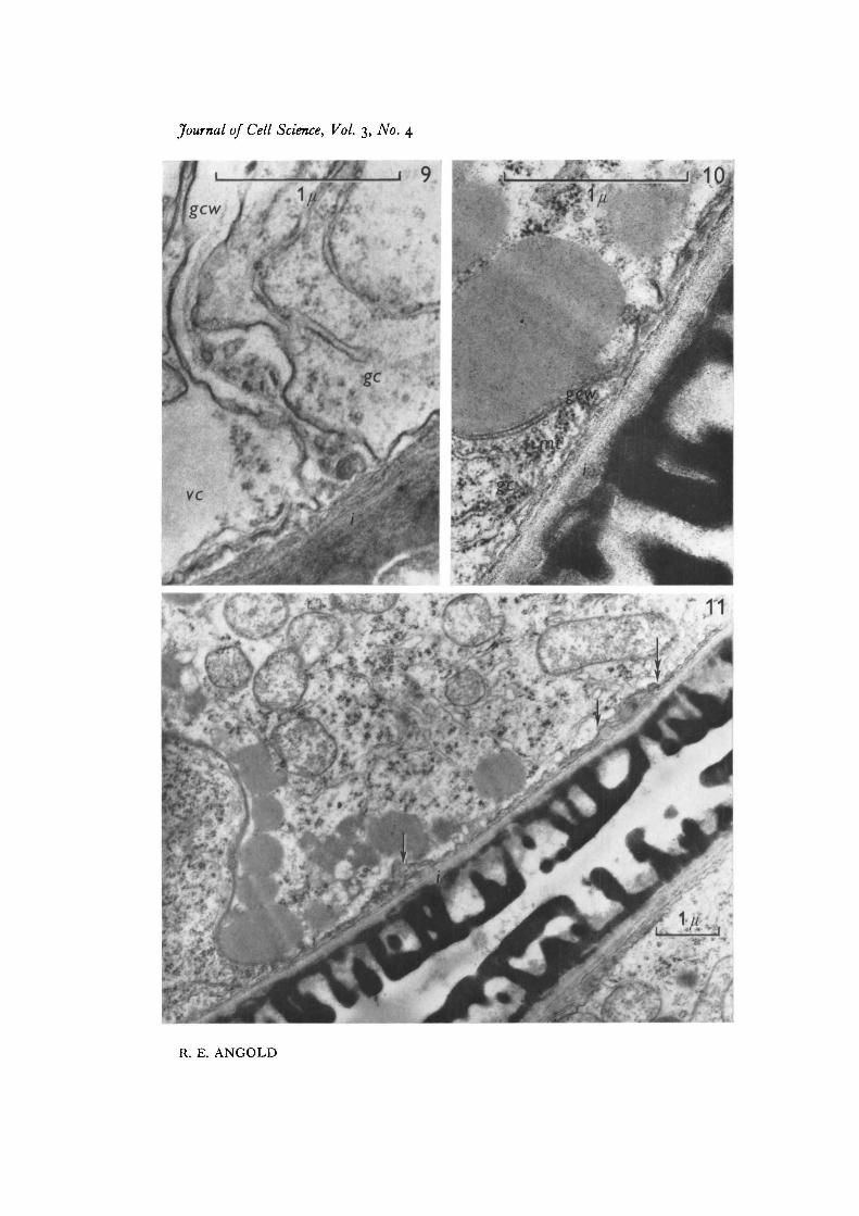
Fig. 9. Generative cell wall (gctv) soon after its formation, separating generative cellcytoplasm (gc) from vegetative cytoplasm (vc). The wall is fused with the intine (i).x 45000.
Fig. 10. Generative cell wall (gctv) during the separation of the generative cell (gc)from the intine (t). Microtubules (mt) are visible where the wall folds back. 45000.
Fig. 11. A portion of the generative cell wall laid along the surface of the intine (1).The process began at the point marked by the double arrow. Remnants of generativecell cytoplasm have been trapped between the generative cell wall and the intine(arrows), x 15000.
Journal of Cell Science, Vol. 3, No. 4
R. E. ANGOLD