Embed Size (px)
Citation preview

THE FOOT AND ANKLE
Compiled by Laurence Hattersley
2014

Compiled by Laurence Hattersley 2014
i
Contents Foot and Ankle .................................................................................................................................. 1
Bones of Ankle and Foot ............................................................................................................... 1
Muscles moving the ankle and foot ...................................................................................... 6
Anterior compartment .......................................................................................................... 7
Posterior compartment ........................................................................................................ 8
Lateral Compartment ......................................................................................................... 10
Ankle Injuries ........................................................................................................................... 10
Sprains .................................................................................................................................. 10
Pott’s Fracture ..................................................................................................................... 12
Stress Fractures .................................................................................................................. 14
Talo-calcaneal (sub-talar) joint ......................................................................................... 14
Sub-Talar Ligamentous Injury .......................................................................................... 17
The Painful Foot .................................................................................................................. 17
Arches of the Foot .............................................................................................................. 24
Referred Symptoms ............................................................................................................ 26
Metatarsalgia ....................................................................................................................... 28
Hallux Valgus ....................................................................................................................... 29
Morton’s Syndrome ............................................................................................................ 29
Gout ....................................................................................................................................... 29
March Fracture .................................................................................................................... 30
Achilles Tendon Rupture ................................................................................................... 30
Diabetic foot ......................................................................................................................... 31
Orthotic Devices.................................................................................................................. 32
Ankle and Foot Manipulations ............................................................................................. 33
Sub-talar joint ...................................................................................................................... 33
Distraction of talo-calcaneal joint .................................................................................... 33
Cuboid ................................................................................................................................... 34
Cuneiforms ........................................................................................................................... 35

Compiled by Laurence Hattersley 2014
ii
Figure 1 Three functional areas of foot .............................................................................................. 1
Figure 2 The Right Ankle seen from the front ..................................................................................... 1
Figure 3 The Right Ankle seen from the Lateral Side .......................................................................... 2
Figure 4 Bones of the Foot ................................................................................................................. 2
Figure 5 Joints of the Foot seen from lateral aspect ........................................................................... 2
Figure 6 Ligaments of medial and lateral ankle .................................................................................. 3
Figure 7 Plantar ligaments ................................................................................................................. 3
Figure 8 Movements of the ankle ...................................................................................................... 4
Figure 9 Superior aspect of the Calcaneus, showing articular surfaces ............................................... 4
Figure 10 Talocalcaneal ligaments ..................................................................................................... 5
Figure 11 Eversion and Inversion of ankle .......................................................................................... 5
Figure 12 Axes of movement in the ankle .......................................................................................... 5
Figure 13 Tibio-fibular interosseous membrane with plantar and dorsiflexion ................................... 6
Figure 14 Compartments of leg.......................................................................................................... 6
Figure 15 Tibialis anterior .................................................................................................................. 7
Figure 16 Extensor digitorum longus.................................................................................................. 7
Figure 17 Extensor hallucis longus ..................................................................................................... 7
Figure 18 Peroneus tertius ................................................................................................................. 8
Figure 19 Gastrocnemius ................................................................................................................... 8
Figure 20 Soleus ................................................................................................................................ 8
Figure 21 Tibialis Posterior................................................................................................................. 9
Figure 22 Flexor Digitorum Longus..................................................................................................... 9
Figure 23 Flexor Hallucis Longus ........................................................................................................ 9
Figure 24 Peroneus longus ............................................................................................................... 10
Figure 25 Peroneus Brevis ............................................................................................................... 10
Figure 26 Ankle sprain - lateral collateral ligament........................................................................... 11
Figure 27 Medial collateral ligament tear......................................................................................... 11
Figure 28 Pott's fracture ................................................................................................................... 12
Figure 29 Stress fracture of 3rd metatarsal ...................................................................................... 14
Figure 30 Subtalar joint ................................................................................................................... 14
Figure 31 Talo-Calcaneal (subtalar) joint .......................................................................................... 15
Figure 32 The sub-talar joint and tarsal tunnel ................................................................................. 16
Figure 33 the left foot seen form the front ...................................................................................... 16
Figure 34 Schematic of inversion sprain ........................................................................................... 17
Figure 35 Tender areas in painful foot strain .................................................................................... 17
Figure 36 Jogger's foot pain ............................................................................................................. 18
Figure 37 Mechanisms of foot strain ................................................................................................ 19
Figure 38 Pronation - evertion ......................................................................................................... 19
Figure 39 Sites of heel pain .............................................................................................................. 20
Figure 40 Plantar ligaments ............................................................................................................. 20
Figure 41 Mechanics of plantar fascia on the longitudinal arch ........................................................ 21
Figure 42 Mechanism of plantar fasciitis .......................................................................................... 21
Figure 43 Calcaneal spur on X-ray .................................................................................................... 22
Figure 44 Shoe modification with calcaneal spur ............................................................................. 22
Figure 45 Continuum of fascia of back ............................................................................................. 23
Figure 46 Achilles tendon with plantar fascia ................................................................................... 23

Compiled by Laurence Hattersley 2014
iii
Figure 47 Arches of foot .................................................................................................................. 24
Figure 48 Transverse arches of foot ................................................................................................. 25
Figure 49 Longitudinal arches .......................................................................................................... 25
Figure 50 Splay foot ......................................................................................................................... 26
Figure 51 Plantar callous formation ................................................................................................. 26
Figure 52 Visceral referred pain ....................................................................................................... 26
Figure 53 Pes cavus ......................................................................................................................... 27
Figure 54 Talipes equinus ................................................................................................................ 28
Figure 55 High heels ........................................................................................................................ 28
Figure 56 Talipes calcaneus ............................................................................................................. 28
Figure 57 Mild bunion ...................................................................................................................... 29
Figure 58 Severe bunions with deformation ..................................................................................... 29
Figure 59 Gout................................................................................................................................ 30
Figure 60 March fracture ................................................................................................................. 30
Figure 61 Technetium 99 bone scan ................................................................................................. 30
Figure 62 Achilles tendon rupture .................................................................................................... 31
Figure 63 Basic orthotic shapes ........................................................................................................ 32
Figure 64 HVT Talo-calcaneal joint ................................................................................................... 33
Figure 65 Distraction of talo-calcaneal joint ..................................................................................... 33
Figure 66 HVT location of cuboid before thrust ................................................................................ 34
Figure 67 Cuboid thrust - prone ....................................................................................................... 34
Figure 68 Cuboid MIT - prone .......................................................................................................... 35
Figure 69 Medial cuneiform - supine................................................................................................ 35
Figure 70 Cuneiforms thrust supine ................................................................................................. 36

Compiled by Laurence Hattersley 2014
1
Foot and Ankle The foot is a complex structure at the end of the leg that is made of more than 26 bones and
33 joints. It provides balance, assists in mobility, and performs other essential functions for
humans.
The foot is the lowest point of the human leg. The foot’s shape, along with the body’s natural balance-keeping systems, make humans capable of not only walking, but also running, climbing, and countless other activities. The ankle joint acts like a hinge, but is much more than a simple hinge. The ankle is made
up of several important structures. The unique design makes it a very stable joint. It has to
be stable to withstand 1.5 times your body weight when standing and 8 times your weight
when running.
Figure 1 Three functional areas of foot
The foot’s complex structure contains more than 100 tendons, ligaments, and muscles that move nearly three dozen joints while bones provide structure. The structure of the foot is similar to that of the hand, but because the foot bears more weight, it is stronger and less mobile.
The foot functions at right-angles to the leg, so the terminology relating to the foot and ankle movements is also different to the wrist and hand.
Bones of Ankle and Foot The ankle region (posterior, as seen in fig. 1) consists of
three bones:
The inferior ends of the tibia and fibula
The superior surface of the talus
Also included here is the Calcaneum, or heel
bone.
Figure 2 The Right Ankle seen from the front
Note this joint looks rather like a mortise and tenon joint with the
articular cartilage being about 6mm thick.

Compiled by Laurence Hattersley 2014
2
Figure 3 The Right Ankle seen from the Lateral Side
The rest of the foot consists of
the middle and anterior regions:
This shows the foot seen from
above. It includes the middle
section:
Navicular
Cuneiform bones
o Middle
o Intermediate
o Lateral
Cuboid
The anterior region consists of the metatarsals and the phalanges
The metatarsals are bones ‘inside’ the foot
The phalanges are the digits; the toes
Between these bones are 33 joints:
Tibial/taloid
o Hinge
Cubo-calcaneal
o Saddle
Talo-calcaneal-navicular
o Functional ball and socket
Cubo-cuneiform-metatarsals
o Gliding
Navicular cuneiforms
o Gliding
Intercuneiforms
o Gliding
Metatarsal-phalangeal
o Hinge
Interphalangeal
o Hinge
Figure 4 Bones of the Foot
Figure 5 Joints of the Foot seen from lateral aspect

Compiled by Laurence Hattersley 2014
3
All these joints create a relatively mobile structure but, understandably, the foot requires
great stability – the ligaments
Figure 6 Ligaments of medial and lateral ankle
The main ligaments on the
medial and lateral sides of the
ankle are there to limit excess
motion across those sides of the
joint.
Medial ligament Lateral Ligaments
Deltoid ligament Named after its shape
o Anterior talo-tibial o Tibio-navicular o Calcaneo-fibular o Posterior talo-tibial
Stabilises the medial side of the joint
Anterior tibio-fibular (weakest) Calcaneofibular Posterior talofibular (21/2 times stronger cf. anterior Talocalcaneal Stabilises Lateral side of joint
In addition to these are the plantar ligaments
Figure 7 Plantar ligaments
The plantar ligaments are of particular significance as they act to support and maintain the
arches of the foot.

Compiled by Laurence Hattersley 2014
4
Figure 8 Movements of the ankle
The medial and
lateral ligaments
create a plane of
movement. As the
foot is at right angles
to the leg, there is no
flexion/extension, per
se. Instead it has
Plantar flexion
o Pointing the toes distally
Dorsiflexion
Pulling the toes proximally
The talocalcaneal
joint, or sub-talar
joint, is between
the inferior
surface of the
talus and the
superior surface
of the calcaneum.
There are three
articular surfaces
with a distinct gap
between the
middle and
posterior
surfaces.
The middle articular surface is on the sustentaculum tali, a small shelf-like projection on the
medial side of the calcaneum supporting the medial aspect of the talus.
Figure 9 Superior aspect of the Calcaneus, showing articular surfaces

Compiled by Laurence Hattersley 2014
5
The talocalcaneal ligaments, here, hold the two bones
together, though they are included in the lateral ankle
ligaments.
Broadly speaking, the top of the calcaneum is convex,
with a concave lower talus; even wedge shaped. This
joint configuration allows:
Inversion
o Pulling the foot inwards
o Eversion
o Pulling the foot outwards
Some books and sports shops will
have you believe that the ankle
undergoes supination and pronation,
but this is incorrect; have you ever
seen a person in the standing
position with their foot sole facing up?
The degree of movement in the ankle
joint depends upon the talus/mortise
relationship. Here the left picture shows
the axis of rotation passing through the
fibula below the tip of the tibia. The right
picture shows the joint from above and
the medial malleolus is anterior to the
lateral; the axis of rotation is 16o toe out
stance. The stance is broader anteriorly
than posteriorly. This arrangement locks
up the ankle joint with dorsiflexion.
Figure 10 Talocalcaneal ligaments
Figure 11 Eversion and Inversion of ankle
Figure 12 Axes of movement in the ankle

Compiled by Laurence Hattersley 2014
6
In addition to the locking of
the ankle joint with
dorsiflexion, there is the effect
on the interosseous
membrane between the tibia
and fibula.
With dorsiflexion the fibula is
pushed laterally and
proximally, pushing the tibia
and fibula apart and
tightening the I/O membrane.
With neutral and plantar
flexion, the I/O membrane
reverts to ‘normal’ with the
fibres directed down and
laterally.
Muscles moving the ankle and foot All the large muscles moving the foot, toes and ankle originate in the calf. Here the leg
(everything below the knee and above the ankle) is divided in three distinct compartments:
Anterior
Posterior
Lateral
Figure 14 Compartments of leg
Figure 13 Tibio-fibular interosseous membrane with plantar and dorsiflexion
Compiled by Laurence Hattersley 2014
7
Anterior compartment
Muscles of the anterior compartment are:
Tibialis anterior
o Originates upper 2/3 of tibia and I/O membrane
o Passes down and medially, crossing the front of the
ankle
o Inserts medial aspect of medial cuneiform and first
metatarsal
o Dorsiflexes and inverts foot
Extensor digitorum
o Originates upper fibula and I/O membrane
o Passes down and inserts into distal phalanx of 4 lateral
toes
o Extends toes
Extensor hallucis longus
o Originates from the medial half of the fibula,
between Tibialis anterior and extensor digitorum
longus
o Passes down and medially to the distal phalanx
of the big toe
o Extends the big toe, Dorsiflexes and inverts foot
Figure 15 Tibialis anterior
Figure 16 Extensor digitorum longus
Figure 17 Extensor hallucis longus

Compiled by Laurence Hattersley 2014
8
Peroneus tertius
o Originates lower end of fibula and I/O membrane
o Passes down and inserts on dorsum of proximal shaft of 5th
metatarsal
o Everts foot
o Even though it is an anterior compartment muscle, it is named
in terms of its function (see peroneal muscles; lateral
compartment)
Posterior compartment
Gastrocnemius
o Originates from lower end of Femur, just over
medial and lateral condyles
o The two heads converge, pass down and insert
onto calcaneus via Achilles tendon
o Plantar flexes the foot
Soleus
o The upper end of the posterior aspects of the fibula and
the tibia (from the soleal line) and the I/O membrane,
deep to gastrocnemius
o It passes down and inserts onto the calcaneus via the
Achilles tendon
o It plantar flexes the foot
Figure 18 Peroneus tertius
Figure 19 Gastrocnemius
Figure 20 Soleus

Compiled by Laurence Hattersley 2014
9
Tibialis Posterior
o It originates from the upper
tibia and fibula and I/O
membrane
o It passes down, with the
tendon passing around the
medial aspect of the ankle
joint
o It inserts onto the medial side
of the navicular and medial
cuneiform bones
o It inverts and plantar flexes
the foot
Flexor Digitorum Longus
o It originates from the posterior aspect of the tibia
o It passes down, past the medial side of the ankle
o It inserts into the distal phalanges to the digits 2-4
o It acts to flex the 4 lateral toes and plantar flex the foot
Figure 22 Flexor Digitorum Longus
Flexor Hallucis Longus
o It arises from the lower end of the fibula
o It passes down, passed the medial side of the ankle
o It inserts on the distal phalanx of the big toe
o It flexes the big toe and the foot
Figure 23 Flexor Hallucis Longus
o
Figure 21 Tibialis Posterior

Compiled by Laurence Hattersley 2014
10
Lateral Compartment
Peroneus Longus
o It originates from the head of the lateral aspect of
the top of the fibula
o It passes down behind the lateral malleolus and
through its own groove on the plantar aspect of
the foot
o It inserts onto the first metatarsal and medial
cuneiform bones
o It everts and plantar flexes the foot
Figure 24 Peroneus longus
Peroneus Brevis
o It originates from the lower lateral aspect of the fibula
o It passes down posterior to the lateral malleolus
o It inserts onto the proximal end of the 5th metatarsal
o It everts and plantar flexes the foot
Figure 25 Peroneus Brevis
Ankle Injuries Ligamentous injuries of the ankle usually occurs at the moment of impact with the ground,
when the foot is plantar flexed and inverted. The ligaments absorb the bulk of the stress
because the peroneal muscles are unable to contract sufficiently rapidly to absorb the
impact.
Sprains
Injury may vary from a simple strain, i.e. a mere elongation of the ligaments with
microtraumata, to major disruption of the ligamentous cords with or without bony avulsion.
The greatest traumatic injury is a total dislocation. Complete ligamentous tear occurs with
75% of common sprains associated with capsular tears. It has been said that it is worse to
sprain the ankle than to break it.

Compiled by Laurence Hattersley 2014
11
Figure 26 Ankle sprain - lateral collateral ligament
Fig 25 shows a
lateral collateral tear. The left drawing shows an anterior view of a normal ankle and the right
is a view of a normal from the lateral aspect. The muddle drawing shows a severe inversion
injury of the talus and calcaneum (curved arrow), which causes a talar tilt. This can cause a
tearing of the lateral collateral ligament (T), a possible avulsion (A) of the lateral [fibular]
malleolus, and a possible tear of the interosseous membrane (L).
Ankle sprains have been classified:
Grade 1: Partial interstitial tearing of ligaments
Grade 2: More severe, but incomplete tearing. Gross stability maintained
Grade 3: Gross instability of the ankle
Another classification has been suggested:
1. Only lateral ligaments are involved (Fig 25)
2. Both medial and lateral ligaments are involved (fig 26)
3. Both medial, lateral and distal talo-fibular ligaments are involved
Figure 27 Medial collateral ligament tear
Looking at Fig 26 and a
medial collateral (deltoid)
tear, (A) shows a normal
ankle and its ligaments. (B)
Shows a severe eversion
injury (curved arrow) causing
a talar tilt and tearing of the
interosseous membrane and
medial ligaments. (C) Shows
the result of the injury as a
widening of the ankle mortise
from the tearing of these
ligaments.
Patients often describe the injury as a feeling of the ankle ‘giving way’ with the precise
mechanics of the injury remaining unclear as it happened so quickly. Pain is usually present
with selling and ecchymosis. Pain is increased with eversion.

Compiled by Laurence Hattersley 2014
12
Treatment
Grade 1 and 2 usually do well with conservative (non-surgical) treatment.
RICE, ultrasound with gentle plantar and dorsiflexion exercises; these with gradual
increased resistance.
Grade 3 treatments are more controversial. Young, high performance, athletes, workers
who do heavy manual labour and patients who have avulsion fractures are considered for
surgery. This could be 5-6 weeks of short leg casting with the foot in slight eversion, followed
by rehabilitation exercises o the peroneal muscles. Achilles stretching and proprioceptive
training.
Rehabilitation has 3 phases:
1. Limitation of injury
2. Restoration of motion (along with proprioception)
3. Regaining agility and endurance
Eversion strains also occur, though are less common. They can be associated with
ligamentous instability at the lower tib/fib joint and also with Pott’s fractures (inferior end of
the fibula, primarily, with the tibia secondarily).
Pott’s Fracture
A Pott’s fracture is a fracture affecting one or both of the malleoli. During activities such as
landing from a jump (volleyball, basketball) or when rolling an ankle, a certain amount of
stress is placed on the tibia and fibula and the ankle joint. When this stress is traumatic, and
beyond what the bone can withstand, a break in the medial, lateral or posterior malleolus
may occur. Also activities involving a sudden change of direction, such as football and rugby,
can cause fractures around the malleoli. When this happens this condition is known as a
Pott's fracture.
It is a fracture of the lower end of the fibula and medial malleolus of the tibia with rupture of
the internal lateral ligament of the ankle, and is caused by outward and backward
displacement of the leg while the foot is fixed.
A Pott's fracture often occurs in combination with
other injuries such as an inversion injury, a dislocation
of the ankle or other fractures of the foot, ankle or
lower leg. Pott's fractures can vary in location, severity
and type including displaced fractures, un-displaced
fractures, bimalleolar (both malleoli) fractures or
compound fractures. It can be difficult to distinguish
clinically between a fracture and a moderate to severe
ligament sprain. Both conditions may result from
inversion injuries, with severe pain and varying
degrees of swelling and disability.
Figure 28 Pott's fracture

Compiled by Laurence Hattersley 2014
13
Symptoms
Patients with a Pott's fracture typically experience a sudden sharp and intense pain around
the ankle or lower leg immediately at the time of injury. The pain is situated at the front,
back, inner or outer part of the ankle or lower leg. The patient may have heard a “crack” as
well. Due to the pain the patient limps to protect the injury. In severe cases weight bearing
may be impossible. Patients with a Pott's fracture usually experience swelling, bruising and
pain on firmly touching the affected region of bone. Pain may also increase during certain
movements of the foot or ankle or when attempting to stand or walk. When it is a displaced
fracture, an obvious deformity may be noticeable.
Medical and physiotherapy management
One of the most important components of rehabilitation following a Pott's fracture is that the
patient rests sufficiently from any activity that increases their pain. Activities which place
large amounts of stress through the ankle should also be avoided, particularly excessive
weight bearing activity such as running, jumping, standing or walking.
Displaced Pott’s fractures where the anatomical relationship of the bones of the ankle has
been disrupted need to be surgical fixed. This may be followed by the use of a protective
boot, brace, or a plaster cast, and/or crutches for a number of weeks. Fractures that are not
displaced, treatment may involve a plaster cast immobilization and the use of crutches,
followed by the use of a protective boot or brace for a number of weeks. Patients with a
Pott's fracture should perform pain-free flexibility, strengthening and balance exercises as
part of their rehabilitation to ensure an optimal outcome. The physiotherapist may utilize
techniques such as massage and joint mobilization which is essential to ensure optimal
range of movement and flexibility. The aim of massage is to fight the formation of heterotopic
ossification. This is the process by which bone tissue forms outside of the skeleton. The
treatment may also involve electrotherapy, taping and bracing, exercises to improve
strength, flexibility and balance, and hydrotherapy.
Lengthening of the tendon Achilles is proved to be a treatment for complicated Pott’s
fractures. Lengthening or stretching of this tendon can be done by performing a passive
dorsiflexion of the foot while the leg/knee is straight. In the most severe cases of a Pott's
fracture, patients usually make a full recovery with appropriate management. Return to
activity or sport can usually take place in a number of weeks to months.

Compiled by Laurence Hattersley 2014
14
Stress Fractures (see March fracture later)
Over activity in sports of some uncoordinated people
can cause overuse syndromes and also can cause
stress fractures. These can appear in the fibula, tibia
calcaneum and metatarsals.
Symptoms of this can appear as deep pain and
tenderness over the affected area, which recur with
resumption of the activity. Diagnosis is by (Tn99) bone
scan, which shows any increased vascular activity at the
site of the fracture, or an X-ray at a later date, revealing
signs of healing.
Figure 29 Stress fracture of 3rd metatarsal
Treatment is rest and ice with a soft orthotic where
appropriate. The healing process can be facilitated with
vitamin C, glucosamine and/or Comfrey cream (bone
knit)
Talo-calcaneal (sub-talar) joint
The talo-calcaneal joint is a potentially complicated joint between the talus and the
calcaneum. It consists of three synovial joints (Fig 8):
A large posterior one – concave on the talus
Two smaller ones – convex on the talus
o These all have reciprocally shaped facets on the calcaneum
Figure 30 Subtalar joint

Compiled by Laurence Hattersley 2014
15
Figure 31 Talo-Calcaneal (subtalar) joint
The talus and calcaneum are joined by three facets:
AF – anterior facet
MF – middle facet
PF – posterior facet
The tarsal tunnel in its oblique course (sulcus) contains the talo-calcaneal ligament.

Compiled by Laurence Hattersley 2014
16
Figure 32 The sub-talar joint and tarsal tunnel
The talus and calcaneum form a tunnel, which
forms a 45o angle with the A/P axis of the foot.
The lateral opening is under the lateral malleolus
and is readily palpable. A firm ligament joins the
two bones. To lock up the
To articulate the talo-calcaneal joint, the foot must
be fully dorsiflexed to lock up the tib-talar joint.
Figure 33 the left foot seen form the front
Inversion/eversion
of the foot during
rotation of the leg.
C – cuboid, N –
navicular facets.
In the weight bearing stance, internal rotation causes valgus of the foot (left).during weight
bearing in the stance phase, external rotation causes the foot to rotate at the sub-talar joint
and thus inverts the foot for the ultimate swing through phase.

Compiled by Laurence Hattersley 2014
17
Sub-Talar Ligamentous Injury
In plantar flexion the anterior talofibular
ligament is vulnerable to an inversion injury
at the ankle
Figure 34 Schematic of inversion sprain
If this is severe, it is followed by tearing of
the talo-navicular ligaments and the talo-
navicular capsule.
The sustentaculum tali (Fig 8) becomes the
fulcrum about which the foot moves and the
talus can dislocate laterally, the calcaneum
moving medially, thus tearing the talo-
calcaneal ligament. Rotation of the leg on
the foot can also cause these injuries.
The Painful Foot
To be normal, the foot has to conform to certain criteria:
1. It is pain free in its function of weight bearing and ambulation
2. Normal muscle function
3. The heel should be central and reasonable sagittal
4. The toes should be straight and mobile
5. During gait and stance there must be three points of weight bearing (see arches)
6. There must be normal nerve supply to the foot
Pain is normally indicated by the patient, form there the practitioner must evaluate the
motion to assess any dysfunction.
Acute problems
Dysfunction normally results from a specific activity, excessive activity of persistence of
activity to which the patient is unaccustomed. Pain and tenderness can be muscular,
ligamentous, periosteal or all of these.
Chronic problems
These can lead from
acute problems if they are
not treated, or rom
anatomical deviation from
the normal. Here the foot
must be examined for
structural deviations in
born, muscular and
ligamentous structures
Figure 35 Tender areas in painful foot strain
One effective treatment
for painful feet and ankles

Compiled by Laurence Hattersley 2014
18
is via gentle traction to the foot. This can be through the foot and calcaneum, or even via the
talus itself, along the axis of the leg. The important thing here is to ‘make contact’ with the
tissues of the whole leg, not just the foot. It can be used as part of a ‘balanced ligamentous
technique’. This addresses the fascia around the whole foot and leg, allowing the tissues to
unwind. It is slow, but effective.
All good sports shops have means of assessing gait of joggers to advise and recommend,
possibly more expensive, footwear.
Figure 36 Jogger's foot pain
The foot, ankle and lower leg pains sustained by joggers are:
a) Shin splints (myositis of Tibialis anterior)
b) Calf pain
c) Talo-tibial ligamentous sprain
d) Achilles tendon strain
e) Inflamed heel pad
f) Plantar fasciitis, leading to ‘spur’ type pain
g) Bunion pain if a hallux valgus exists
h) Arthralgia of the big toe M/P joint
The everted foot is a frequent cause of foot strain (fig 32) because of its deviations from
normal, though debate is ongoing on how the foot ‘got there’ in the first place. Here the foot
is being supported by muscular activity, which becomes fatigued.in eversion, the talus slides
medially, the forefoot abducts and broadens, depressing the metatarsal arch. Then the three
metatarsal heads become weight bearing (Fig 33).

Compiled by Laurence Hattersley 2014
19
Figure 37 Mechanisms of foot strain
The upper drawing depicts the normal foot with a central heel and a good longitudinal arch.
In the lower drawing, weight bearing (1) on misaligned structures causes the talus (2) to
slide forward and medially (5) and causes the calcaneum (3) to rotate posteriorly. The
plantar fascia (4) elongates, which places strain on it calcaneal site of insertion. This
eversion causes a strain on the deltoid ligament.
The muscle most involved here is Tibialis anterior, which is an invertor and can elongate
under stress, it then becomes tender and is rendered ineffective in weight bearing.
Figure 38 Pronation - evertion
In Fig 33 the left drawing depicts a
normal foot with a good metatarsal
arch. The right depicts a pronated
foot, with the talus sliding forward
and medially. This spreads the
forefoot, which causes a
depressed metatarsal arch.
However the mechanics of how
the foot begins to experience this
state is unclear; is it in the foot, or
an expression of mechanics from
higher up the leg and expressing
themselves in the foot?
Either way, in the long term the
body still tried to cope. Here the
evertors take up the slack. Then

Compiled by Laurence Hattersley 2014
20
the toe extensors change their alignment and become ankle-foot evertors. The talo-
calcaneal ligament, taught in inversion, becomes inflamed and painful do to stress. The
longitudinal arch becomes flatter and stresses the plantar fascia, causing heel and spur type
pain.
The heel is s frequent source of pain. These can occur at the site where the plantar fascia
attaches to the calcaneum where there is a repetitive stress during weight bearing.
Figure 39 Sites of heel pain
The plantar fascia is a multi-layered fibrous aponeurosis originating from the medial
calcaneal tuberosity and inserting into the complex of tissues of the metatarsophalangeal
joints, the flexor sheaths and the bases of the proximal phalanges. It is one of five factors
that help support the plantar arches (see later)
Figure 40 Plantar ligaments

Compiled by Laurence Hattersley 2014
21
Figure 41 Mechanics of plantar fascia on the longitudinal arch
(A) The large arrow depicts the body weight on the foot. The smaller arrows depict
weight bearing on the heel (left) and the toes (right).
(B) The arch is maintained by the articular structures with the plantar fascia merely
reinforcing the strength of the arch.
When the toes are dorsiflexed (Fig 38), the complex of tissues at the base of the toes
causes increased tension along the plantar fascia.
Figure 42 Mechanism of plantar fasciitis
A. Is the normal relationship of the plantar fascia with its tendon attached to the
calcaneum periosteum
B. Depicts traction (arrow) pulling the periosteum from the calcaneum
C. Depicts the bony changes that occur with chronic tension patterns in the plantar
fascia. Bony changes are always indicative of long term soft tension patterns; here
forming a calcaneal spur
Tension patterns in the plantar fascia pull on the periosteum of the calcaneum. Over time
microtears can occur in the fascia and some wearing away of the attachments occurs.

Compiled by Laurence Hattersley 2014
22
Fatigue fractures and periostitis occurs, which can be seen radiologically. It manifests as
local pain at its attachment to the calcaneum, possibly spreading from there distally towards
the toes.
Figure 43 Calcaneal spur on X-ray
The pain is experienced
mainly with weight
bearing, but in severe
cases can be more
constant. If the
dysfunction is persistent,
the body tries to help the
healing process at the
point of the RSI by
causing calcification of the proximal end of the plantar fascia. Thus appears a calcaneal
spur.
As was said, calcaneal spur is always evidence of long term soft tissue tension, but by the
time it is a calcaneal spur, it is a structural entity that needs structural remedies.
Figure 44 Shoe modification with calcaneal spur
Here a sponge pad
can be inserted into
the heel, which
reduces the pressure
on the calcaneum. \in
addition to this, a
section can be cut
out from the insole of
the heel of the shoe.
Treatment for calcaneal spur is usually conservative, with ice, local massage friction and
ultrasound as well as prescribed exercises to help the relaxation of the tissues down the
back of the legs. The reason for the posterior muscle groups is the principle of continuity of
tissues down the back of the legs. The back muscles are continuous with the hamstrings, on
to the calf muscles down to the Achilles tendon to the calcaneum. Here the books will have
you believe they stop, but other opinions claim the fascia continues ‘around the corner’ of the
calcaneum and continues as the plantar fascia.

Compiled by Laurence Hattersley 2014
23
Figure 45 Continuum of fascia of back
Figure 46 Achilles tendon with plantar fascia
This continuum of fascia from (at least) the posterior compartment of the calf to the base of
the toes assists the walking mechanism. This is especially significant in the latter stage of
the three stages of walking:
Heel strike
Mid stance
Toe off
It transmits the forces of plantar flexion directly into the M/P joint. As the foot plantar flexes
with weight bearing, the toes are forced into extension. This creates tension along the
plantar fascia creating a reciprocal tension at the M/P joints resulting in a ‘spring in the step’
with the ‘toe off’ phase.
Hence tension patterns a long way away can manifest in the locality of the plantar aspect of
the foot, or even just the back of the calcaneum causing an increased bony size there with
consequent problems in footwear.
The focus of this tends to be that of disease, with good footwear as a preventative factor.
However barefoot running challenges this belief system.
Barefoot running remains a controversial topic and many argue it might not be suitable for all
runners. Runner’s World Editor-in-Chief, David Willey, wrote about barefoot running in a
2011 editorial and summed up barefoot running by stating, “There's no single answer or
prescription that's right for every runner when it comes to footwear and running form.”
In 1960 Ethiopia's Abebe Bikila, the greatest Olympic marathoner of all time, won the first of
his consecutive gold medals sans shoes in a world record 2:15:17. While Bikila was making

Compiled by Laurence Hattersley 2014
24
Olympic history, England's Bruce Tulloh was running European record times from 1955 to
1967, almost always in bare feet. He ran 13:12 for three miles on grass, and 27:23 for six
miles on cinders. Later, Tulloh taught in Africa, coached, wrote books, and ran solo across
America (2,876 miles, albeit in shoes).
Charlie "Doc" Robbins and Zola Budd are two more important contributors to barefoot
running. Robbins, winner of two USA National Marathon Championships in the late 1940s,
completed 50 straight Thanksgiving Day Road Races in Manchester, Connecticut. Most
Thanksgivings, Robbins went shoeless, though he would resort to a pair of socks if the
temperature dipped below 20 degrees. Mention the name Zola Budd to the casual track fan
and you'll likely get one (or all) of three responses: Barefoot. South African. Tripped Mary
Decker. Budd set a track world record in January 1984 when, just 16, she ran 5000 meters
in South Africa in 15:01.83, more than six seconds under Mary Decker's existing record.
Still, it wasn't until 2009 that barefoot running became a hot topic. The biggest impetus was
Christopher McDougall's book Born to Run. While ostensibly the story of Mexico's
Tarahumara Indians—who run barefoot or in tire-tread huaraches—controversial chapters in
the book conclude that running shoes have done little to prevent injuries. The popularity of
minimal shoes, such as the Nike Free and Vibram FiveFingers, has fuelled the fire.
Arches of the Foot
There are three main arches of the foot
Medial longitudinal
Lateral longitudinal
Transverse
The function of these arches is to create space and protect the vessels and nerves on the
plantar aspect of the foot and so create an amount of spring and elasticity.
Figure 47 Arches of foot

Compiled by Laurence Hattersley 2014
25
Fig 44 shows the arches and the consequent three points of contact with the ground
The base of the big toe
The base of the little toe
The heel
These arches are supported by:
Bony configuration
Plantar ligaments
Plantar fascia
Short (intrinsic) muscles
Long muscles
o Tibialis anterior
o Tibialis posterior
o Peroneus longus
Figure 48 Transverse arches of foot
The left drawing depicts the fixed tarsal
and metatarsal arches and the flexible
anterior metatarsal arch.
The drawing on the right depicts the
second receded cuneiform, which
creates a mortise for the seating of the
base of the second metatarsal.
Figure 49 Longitudinal arches
The longitudinal arch is formed
by the continuity of the talus (T)
with the calcaneum (C) and
anteriorly with the navicular bone
(N). Viewed medially, the
navicular bone articulates with
the cuneiforms (CU), then with
the metatarsals (M) and the
phalanges (P).
Compiled by Laurence Hattersley 2014
26
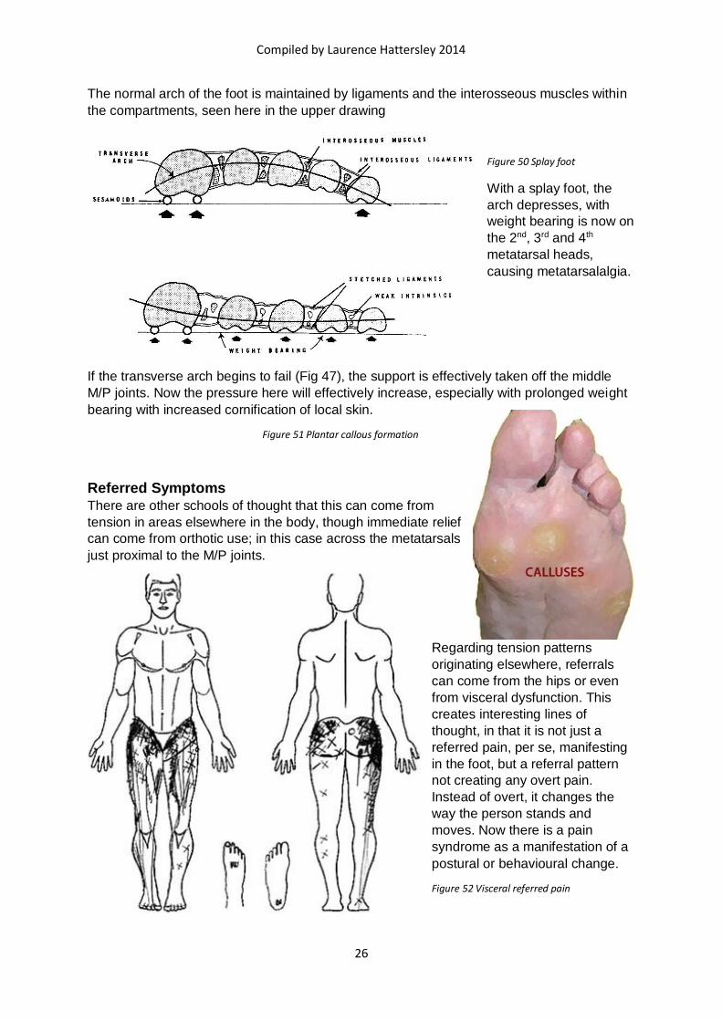
The normal arch of the foot is maintained by ligaments and the interosseous muscles within
the compartments, seen here in the upper drawing
Figure 50 Splay foot
With a splay foot, the
arch depresses, with
weight bearing is now on
the 2nd, 3rd and 4th
metatarsal heads,
causing metatarsalalgia.
If the transverse arch begins to fail (Fig 47), the support is effectively taken off the middle
M/P joints. Now the pressure here will effectively increase, especially with prolonged weight
bearing with increased cornification of local skin.
Figure 51 Plantar callous formation
Referred Symptoms
There are other schools of thought that this can come from
tension in areas elsewhere in the body, though immediate relief
can come from orthotic use; in this case across the metatarsals
just proximal to the M/P joints.
Regarding tension patterns
originating elsewhere, referrals
can come from the hips or even
from visceral dysfunction. This
creates interesting lines of
thought, in that it is not just a
referred pain, per se, manifesting
in the foot, but a referral pattern
not creating any overt pain.
Instead of overt, it changes the
way the person stands and
moves. Now there is a pain
syndrome as a manifestation of a
postural or behavioural change.
Figure 52 Visceral referred pain

Compiled by Laurence Hattersley 2014
27
Fig 49 shows one such referral pattern, here possibly from the large intestine. The large
intestine can refer pain to literally anywhere to which the bowel has a relationship:
The low back
o The ascending and descending colon are retroperitoneal, having a direct
connection with the low back
The pelvis
o The caecum has ligamentous attachments here and the sigmoid has a line of
mesentery
The shoulders
o Both the hepatic and splenic flexures have ligamentous attachments to the
diaphragm, referring back up to the shoulders via the phrenic nerve
The lower limb
o The bowel has relations with psoas, iliacus and the kidneys (via the fascia of
Toldt)
The sacroiliac joints
o Always involved with bowel and psoas
Referrals to the outside of the hips can affect the function of the muscles there, involving the
iliotibial tract. This, in theory only stops just below the knee, but the pain can continue down
to the lateral aspect of the calf. If it involves the peroneal muscles, evertion of the foot with
consequent splaying can be easily seen.
Treatment
Longitudinal arch problems can show initially as plantar fasciitis. Here the quality of
movements of the tarsal bones is important. People with flat feet (pes planus) can feel as if
they have very ‘soft’ feet, floppy almost, when the person is younger and possibly ‘hard’ and
generally very restricted in the older person from long term positional alignment. People with
complain of tarsal pain – almost a presence in the foot. Massage to the plantar aspect of the
foot can bring relief and help stimulate the intrinsic muscles of the foot, so that they
contribute more to the arch support.
Other treatments include ‘home’ massage using coarse sand (builders gravel); this creates a
diffuse but penetrating massage to the plantar side of the foot 9walking along a beach of wet
sand is a good second best). Also an exercise of trying to pick up a pencil using just the
toes, though orthotic can be very valuable with symptomatic relief.
Other conditions can occur in foot mechanics, like pes cavus.
Figure 53 Pes cavus
Pes cavus results in an abnormally high arch in
the foot. This results in an increased angle
between the metatarsals and the ground; they are
now effectively pointing towards the ground. Now
the distal ends of the metatarsals are being
‘pressed’ into the ground creating symptoms to
transverse arch problems. They can also be
associated with ‘hammer toes’; an increased flexion of the toes, producing a ‘claw’ effect.
This can also show as thickening of skin at the pressure points on the top of the toes.

Compiled by Laurence Hattersley 2014
28
Other rare conditions are talipes equinus and talipes calcaneus
Figure 54 Talipes equinus
Talipes equinus can be from a congenital shortening of the
posterior calf muscles producing a persistent plantar flexion
of the ankle. Here, therefore, the foot will only be weight
bearing on the M/P joints, with the heel not coming in contact
with the ground with normal use.
Functionally, this is similar to wearing high heels
Figure 55 High heels
Another rare condition is pes calcaneus. This occurs
when there is paralysis of the posterior calf muscles
or hyperplasia of the anterior group.
Figure 56 Talipes calcaneus
If the calf muscles are paralyzed the contraction of the
tibialis anterior and tibialis posterior pull up the arch and the
contraction of the flexor brevis digitorum pulls the pillars
closer together, therefore the heel descends, the arch
ascends, and the plantar ligaments contract. If the extensor
muscles are also paralyzed the toes drop and the anterior
deformity is increased.
Foot usage and general posture during gait can be established by examination of the
person’s feet, whilst standing, and their shoes; e.g. people who an inverted foot when weight
bearing and walking will show wear and tear around the heel and sole.
Metatarsalgia
Metatarsalgia is a loosely defined condition in which the pain is noted in the metatarsal
heads. When the pain is in the lateral four metatarsal heads, it is called Metatarsalgia. If the
pain is at the joint of the big toe, it is called a hallux valgus, or bunion; although it is also
called sesamoid disease, or localised arthritis.

Compiled by Laurence Hattersley 2014
29
Hallux Valgus
Hallux valgus is a subluxation of the big toe outward. There is usually a deformity of the
bone, the joint surface of the head of the first metatarsal being inclined obliquely out. As the
toe becomes displaced outward the extensor hallucis longus by its contraction tends to
increase the deformity. On the side of the head of the protruding metatarsal bone a bursa
develops and becomes painful, forming a bunion. This bursa sometimes suppurates.
Figure 57 Mild bunion
Figure 58 Severe bunions with deformation
In some cases hallux valgus is due
apparently to ill-shaped shoes, but in
many cases, and these the worst, a
rheumatic-gouty condition is the main
factor. It may even have reflections on the arrangement and tension patterns of the intrinsic
muscles of the foot, having a similarity with those of the thumb. The big toe has muscles that
create ‘opposition’, as with the grip activity of the hand. The muscles of the foot are still there
but their function are not as specifically defined as in the hand. Persistent tension patterns
can pull the big toe across the foot, even pushing the adjacent toes out of the way (Fig 55).
In treatment the articular surface of the head of the first metatarsal bone is first resected.
This enables the toe to be brought straight. To keep it straight the tendon of the extensor
hallucis is displaced inward so that by its contraction it keeps the toe from again going
outward.
Morton’s Syndrome
Morton’s syndrome (not neuroma) is a metatarsalgia of the second metatarsal head, which
becomes weight bearing when the first metatarsal bone is too short. Examination will reveal
tenderness and this can be confirmed, if necessary, by X-ray. Treatment can be via the
appropriate orthotic to relieve the pressure there.
Gout
Another cause of pain at the base of the big toe is gout. Gout is a congenital condition, with
a flaw in purine metabolism, resulting in the production of uric acid. Uric acid is very
insoluble and crystallises out of the body’s tissues, forming microscopic needle shaped
crystals in and around the joint. Macrophages ingest these crystals but cannot digest them.
The macrophages die and lyse, releasing inflammatory cytokines and other chemicals into
the tissues, causing inflammation. In principle gout can occur anywhere, but ‘traditionally’
affects the base of the big toe.
Compiled by Laurence Hattersley 2014
30
Figure 59 Gout
Gout is usually treated via
allopurinol tablets. However
the person can help
themselves via frequenting
alkaline foods.
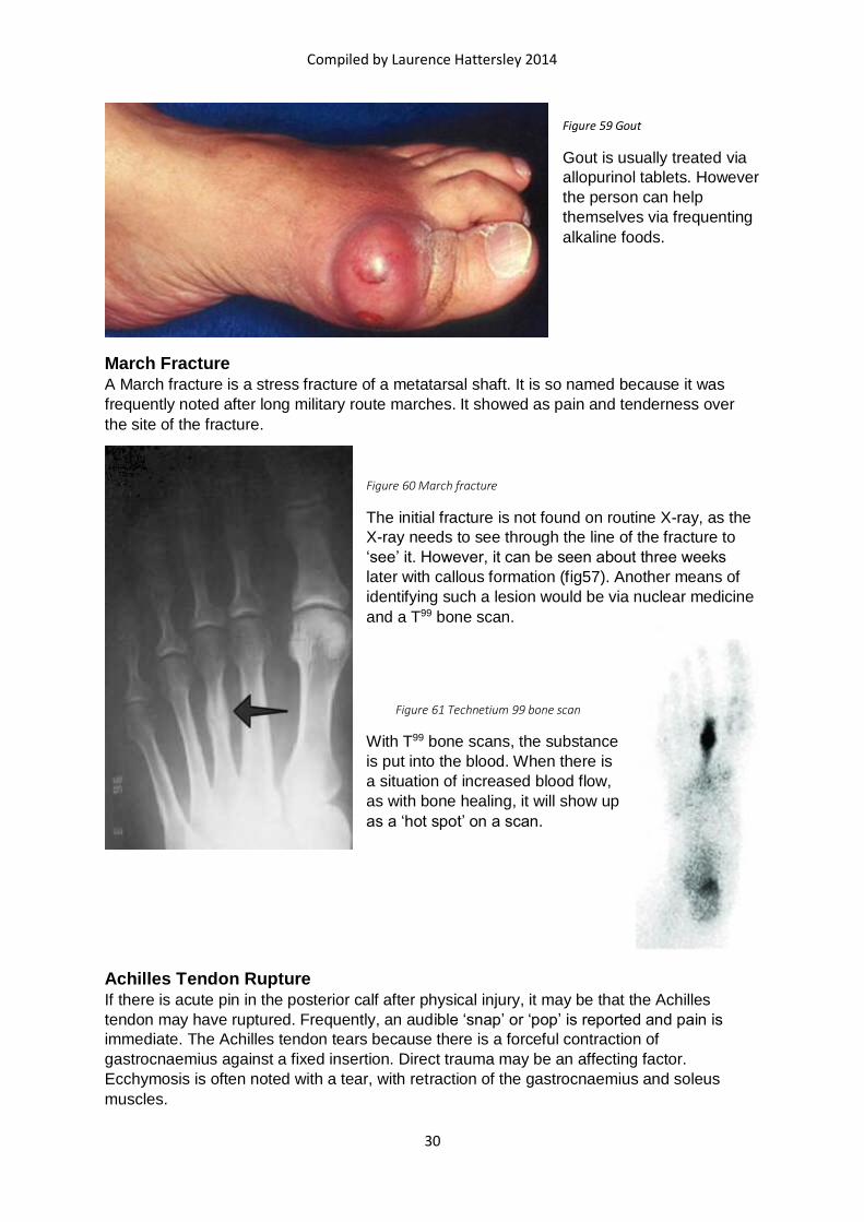
March Fracture
A March fracture is a stress fracture of a metatarsal shaft. It is so named because it was
frequently noted after long military route marches. It showed as pain and tenderness over
the site of the fracture.
Figure 60 March fracture
The initial fracture is not found on routine X-ray, as the
X-ray needs to see through the line of the fracture to
‘see’ it. However, it can be seen about three weeks
later with callous formation (fig57). Another means of
identifying such a lesion would be via nuclear medicine
and a T99 bone scan.
Figure 61 Technetium 99 bone scan
With T99 bone scans, the substance
is put into the blood. When there is
a situation of increased blood flow,
as with bone healing, it will show up
as a ‘hot spot’ on a scan.
Achilles Tendon Rupture
If there is acute pin in the posterior calf after physical injury, it may be that the Achilles
tendon may have ruptured. Frequently, an audible ‘snap’ or ‘pop’ is reported and pain is
immediate. The Achilles tendon tears because there is a forceful contraction of
gastrocnaemius against a fixed insertion. Direct trauma may be an affecting factor.
Ecchymosis is often noted with a tear, with retraction of the gastrocnaemius and soleus
muscles.

Compiled by Laurence Hattersley 2014
31
Figure 62 Achilles tendon rupture
Most Achilles tears occur about 2ins above the calcaneal insertion. If it is a complete tear, it
can be tested via the Simmond’s (Thompson) test (Fig 59): with the patient prone, squeeze
the gastrocnaemius muscle. If the tendon is intact, the foot will plantar flex; if it has ruptured,
it will not. Even with this condition, the patient may still be able to walk, so lameness will not
be diagnostic. It is possible that the person may not be able to stand on tip toe.
Treatment usually involves surgical repair. This is followed by placing the ankle in a cast for
about 8 weeks, set with the foot in plantar flexion, followed by gradual rehabilitation (daily
ultrasound) and physiotherapy.
Partial tears cannot be measured and diagnosis is made by tenderness, local pain and
aggravation by stretching the tendon by dorsiflexion or trying to stand on tip toe. In these
cases, treatment is by avoidance of physical activity for at least three weeks. Use of ¾ inch
elevated heels help relieve the stretch of the tendon in the healing process.
Diabetic foot
With diabetes, foot problems should be carefully evaluated, as complication can be
devastating. Hospitalisations for diabetic foot problems increased 25% in 1965 to 50% in
1985. The causes of foot damage are from ischaemia, neuropathic changes and neuropathic
arthropathy. Ischaemia may include small or large vessels and sensory, motor or autonomic
neuropathy. Large vessels pathology can appear as claudication and large, non-healing,
ulcers. Diabetic ulcers have been classified:
Grade I – a superficial ulcer involving only skin and subcutaneous tissue
Grade II – ulcers extending to underlying tendon, bone and joint capsule
Grade III – deep ulcers with associated osteomyelitis or abscess
Grade IV – gangrene of toe or distal mid-foot
Grade V – gangrene of mid-foot and hind-foot

Compiled by Laurence Hattersley 2014
32
The treatment of ulcers is beyond the scope of physical therapy. Diabetic neuropathy can be
treated with drugs (carbamazepine), and all diabetes must be controlled by either diet,
Glucophage or insulin treatment.
Orthotic Devices
Orthotics are many and various.
Figure 63 Basic orthotic shapes
There are adherents to the concept and use
of orthotics, but their use revolves around
the belief system of the problem is in the
foot and everything above is an effect of it.
But it has already been said that it could be
that the ‘problem’ is one in the body, even
the viscera, creating effects in the foot.
One example of their use is the correction of a valgus (as seen above). The picture on the
left shows a deformity of the foot stance within the shoe. The right picture shows an inner
heel wedge that corrects the deformity.

Compiled by Laurence Hattersley 2014
33
Ankle and Foot Manipulations Sub-talar joint (talo-calcaneal joint)
Patient side lying
Medial side of ankle uppermost
Dorsiflex the ankle (to lock it)
Fix one hand across the foot, including the navicular, up beside the medial malleolus
o Direct force cephalad and towards the table (here on the left)
Fix the heel of the other hand on the medial side of the calcaneum
o Direct the arm pedad and towards the table (here on the right)
Find the point of tension
The thrust is through the hand fixed on the calcaneum
Figure 64 HVT Talo-calcaneal joint
Distraction of talo-calcaneal joint
Patient supine
Wrap both hands
around the talus and
calcaneum
Traction gently
pedad, using body
weight
Follow any
spontaneous
releases
Figure 65 Distraction of talo-calcaneal joint

Compiled by Laurence Hattersley 2014
34
Cuboid
This is generally seen as one manipulation, but there are 5 joints around the cuboid, the
force and focus can be adjusted accordingly
Supine - Combined leverage and thrust (CLT)
Locate the plantar aspect off the cuboid; fix
Dorsiflex the foot (i.e. push both
foot and cuboid cephalad)
With the other hand fix all the
metatarsals and support the tibia
with the forearm
Thrust in the line of the joint
Figure 66 HVT location of cuboid before thrust
Prone (CLT)
Flex knee to 90o
Dorsiflex the foot and fix the metatarsals
Fix on the plantar aspect of the cuboid
Thrust in the line of the joint
Figure 67 Cuboid thrust - prone

Compiled by Laurence Hattersley 2014
35
Prone – Momentum induced thrust (MIT)
Grasp foot and fix on plantar aspect of cuboid with both thumbs
Dorsiflex foot and flex knee to 900
Take knee towards extension
At same time plantar flex foot with slight inversion
Thrust cuboid in the line of the joint
Figure 68 Cuboid MIT - prone
Cuneiforms
Only the medial cuneiform can be manipulated from the plantar aspect; all the others cannot
be reached for specific fixation
Medial cuneiform – supine
Grasp foot with both hands
o Crossing index fingers across plantar aspect of medial cuneiform
Grasp the foot to create a slight traction
Gently oscillate eh foot to feel the point of tension
Thrust towards the dorsal aspect of the foot
Figure 69 Medial cuneiform - supine

Compiled by Laurence Hattersley 2014
36
Intermediate and lateral cuneiforms
Patient supine
Stand pedad to foot
Locate affected cuneiform and cross fingers over the dorsal aspect of the bone
Cross thumbs over relevant metatarsal on its plantar side
Thrust the bone pedad
Figure 70 Cuneiforms thrust supine