Embed Size (px)
Citation preview

Int. J. Speleol. 9 (1977(78), pp. 153-165.
. The Fine Structure of Hamann's Organ inLeptodirus hohenwarti, a Highly Specialized Cave Bathysciinae
(Coleoptera, Catopidae)
byFiorenza ACCORDI* and Valerio SBORDONI*' **
INTRODUCTION
Particular sense organs are to be found in the 7th, 9th and 10th antennal seg-ments of the Catopid beetles and of some other related families of Staphyl-inoidea. Because of their structural complexity and puzzling function, thesestructures have, in recent years, attracted the attention of a number of authors.
It seems of interest to trace an outline of studies so far conducted on theseorgans which promise a greater insight into both the phylogenetic relationshipamong some staphylinoid beetle families and the evolutionary patterns ofCatopidae in adapting to the cave environment. The discovery and first de-scription of these structures was ascribed to Jeannel (1911). However, carefulreading of a paper by Hamann (1898) on sense receptors of some Bathysciinaeconvinced us of the importance of the studies by this author. Perhaps some ofJeannel's misunderstanding of Hamann's work stemmed from his way of num-erating the antennal articles. In fact Hamann reported olfactory vesicles asoccurring in the 2nd, 3rd and 5th antennal segments in Bathysciinae. Thiswould appear to correspond to the actual location of the receptors, whencounting the articles starting from the antennal tip. J eannel (1908, 1911) wasunaware of the sensory organs in the 9th and 10th antennal articles and so wasperhaps induced into rejecting Hamann's findings.
Hamann was able to describe the antennal receptors of Speophilus kiesen-wetteri, Centhmonocharis jreyeri and Leptodirus hohenwarti in some detail,in spite of the limitations of the light microscope. He even noted the spindleshape of the vesicle and periarticular gutter sensilla.
The afore-going facts would seem to suggest that this sensory structure,otherwise known as "vesicule olfactive", "antennal organ" or "antennal ves-icle", be referred to in future as Hamann's organ, in honour of its discoverer.
Jeannel (1911) reported on the vesicular organ in the 7th article of theBathysciinae. Unfortunately he gave a misleading description of the sensilla
* Institute of Zoology = University of Rome, Viale dell' Universita 32, 00100 Rama, Italy .• * To whom reprint requests should be address('d.
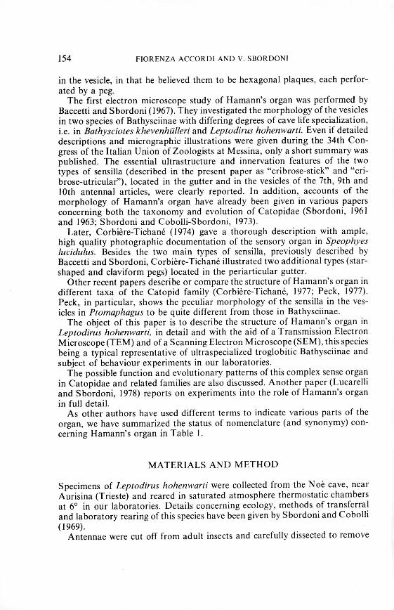
154 FIORENZA ACCORDI AND V. SBORDONI
in the vesicle, in that he believed them to be hexagonal plaques, each perfor-ated by a peg.The first electron microscope study of Hamann's organ was performed by
Baccetti and Sbordoni (1967). They investigated the morphology of the vesiclesin two species of Bathysciinae with differing degrees of cave life specialization,i.e. in Bathysciotes khevenhiilleri and Leptodirus hohenwarti. Even if detaileddescriptions and micrographic illustrations were given during the 34th Con-gress of the Italian Union of Zoologists at Messina, only a short summary waspublished. The essential ultrastructure and innervation features of the twotypes of sensilla (described in the present paper as "cribrose-stick" and "cri-brose-utricular"), located in the gutter and in the vesicles of the 7th, 9th and10th antennal articles, were clearly reported. In addition, accounts of themorphology of Hamann's organ have already been given in various papersconcerning both the taxonomy and evolution of Catopidae (S bordoni, 1961and 1963; Sbordoni and Cobolli-Sbordoni, 1973).Later, Corbiere-Tichane (1974) gave a thorough description with ample,
high quality photographic documentation of the sensory organ in Speophyeslucidu Ius. Besides the two main types of sensilla, previously described byBaccetti and Sbordoni, Corbiere-Tichane illustrated two additional types (star-shaped and c1aviform pegs) located in the periarticular gutter.
Other recent papers describe or compare the structure of Hamann's organ indifferent taxa of the Catopid family (Corbiere-Tichane, 1977; Peck, 1977).Peck, in particular, shows the peculiar morphology of the sensilla in the ves-icles in Ptomaphagus to be quite different from those in Bathysciinae.The object of this paper is to describe the structure of Hamann's organ in
Leptodirus hohenwarti. in detail and with the aid of a Transmission ElectronMicroscope (TEM) and of a Scanning Electron Microscope (SEM), this speciesbeing a typical representative of ultraspecialized troglobitic Bathysciinae andsubject of behaviour experiments in our laboratories.The possible function and evolutionary patterns of this complex sense organ
in Catopidae and related families are also discussed. Another paper (Lucarelliand Sbordoni, 1978) reports on experiments into the role of Hamann's organin full detail.As other authors have used different terms to indicate various parts of the
organ, we have summarized the status of nomenclature (and synonymy) con-cerning Hamann's organ in Table I.
MATERIALS AND METHOD
Specimens of Leptodirus hohenwarti were collected from the N oe cave, nearAurisina (Trieste) and reared in saturated atmosphere thermostatic chambersat 6° in our laboratories. Details concerning ecology, methods of transferraland laboratory rearing of this species have been given by Sbordoni and Cobolli(1969).
Antennae were cut off from adult insects and carefully dissected to remove

Table
I.Nomenclature
concerning
Hamann's
organandits
partasreferred
toby
various
authors.
Hamann
(1898)
Geruchsorgane
KugeligeGrube
Canal
Ringformig
Walle
KolbfOrmig
Zapfen
*notcorresponding.
Jeannel(1911)
Vesicule
olfactive
Vesicule
Guttiere
Periarticulaire
Plaqueshexagona
uxavec
biitonnet
sensorial
Baccetti
&Sbordoni
(1967)
Organo
antennaIe
Camera
sensoriale
Setola
ramificata
Utricoli.
sensilliutricolari
Sensilli
basiconici
Corbiere-
Tichane
Peck
(1977)
Presentpaper
:::J Z(1974.
1977)
m VJ
Vesicule
Sense
Hamann's
-1olfactive
;:creceptor
organ
C (")
Spheric
room
Vesicle
Sensory
vesicle
-1 CCanal
Tube
Vestibulum
;:c mIntermediary
room
Atrium
Z
Periarticular
Circular
canal
Periarticular
gutter
:t: :>gutter
3:Black
peg
Branching
seta
:> ZPine-cone
Bluntly-tapering
Cribrose-utricolar
Z 0sensilla
with
!lutedwalls
sensilla
;:csensilla*
Cl :>Pine-cone
Sensillabasiconica
Cribrose-stick
Z
sensilla
orblunt,elongated
sensilla
Zsmooth
sidedpapillae*
r mClaviform
pegs
Claviform
pegs
~(10thsegment)
(9th
and
10thsegment)
0 0Star-shaped
pegs
Star-shaped
;:c(sensilla
coeloconica)
sensilla
C VJ

156 FIORENZA ACCORDI AND V. SBORDONI
the external cuticular wall of the 7th antennal article, in order to expose theorgan, which was then fixed in 4% glutharaldehyde, in 70% alcohol or inBarber's fluid, and then transferred into a 0.1 M cacodylate buffer.
Specimens for SEM observations were fixed in 1% osmium tetroxide, de-hydrated in gradual ethanol series and in absolute acetone, then allowed to airdry and were subsequently stored in a dry atmosphere. Some organs were dis-sected before dehydrating, in order to observe the internal features. Antennaewere mounted on standard mica squares and metal stubs, gold coated in vacuumand observed with a Cambridge Stereoscan Electron Microscope.
Specimens for TEM observations were fixed in 1% osmium tetroxide, de-hydrated and embedded in Epon or Araldite. Thin sections, obtained with aLKB Ultrotome III, were stained with 10% uranyl acetate and lead cytrate andobserved with a Siemens Elmiscope I A.
Observations under a light microscope were also performed and micrographstaken, mounting the antennae preserved in alcohol after clarifying them withclove oil or lactic acid.
RESULTS
7th article sensory organ:The receptor organ occurring in the 7th antennal article of Leptodirus has avery complex structure. Its consists of the following parts (see table I):
a) a sensory vesicle. deeply invaginated into the antennal segment and pro-vided with a number of crib rose-utricular sensilla,
b) a vestibulum with the shape of a cylindrical neck flaring distally and bear-ing a long branching seta,c) a periarticular gutter containing a ring of crib rose-stick and star-shapedsensilla.
After extracting the organ from the cuticular wall of the 7th antennal article,the entire structure was examined in detail by SEM; its maximum length wasfound to be about 100j.Land fully covered by a cuticular lining. The sensoryvesicle was seen to be nearly spherical in shape, rather compressed laterally,with a longitudinal diameter of 40j.L and a thickness of 27j.L. Its external surfacewas seen to be perforated by almost 60 pores with an invaginated inside cuti-cular lining. An internal spongy structure was recognizable within the poresfrom which some filaments and stubs also emerged probably indicating stumpsof nerve fibers. The cylindrical neck, about 14j.L long, corresponding to thevestibulum, eccentrically connects the sensory vesicle to the medial side of theperiarticular gutter.
Under TEM examination, the sensory vesicle appeared to be lined by a thickcuticular wall breaking off in correspondence with the previously describedpores. From each pore a cribrous utricular sensillum penetrates into the inter-nal cavity of the vesicle. The number of sensilla therefore appeared to corre-spond to the number of pores. The crib rose utricular sensilla were seen to bebarely elongated with a length of I0-12j.Land a breadth of 5-6j.L. They were per-

FINE STRUCTURE IN HAMANN ORGAN IN LEPTODIRUS 157
i )
Fig. L Light micrograph of a whole antenna of Leprodirus hohenwarli, 7th, 9th and 10th articlesare indicated by arrows (x 42).
Fig. 2. The whole Hamann's organ of the 7th antennal article, as observed under SEM, A: 8thantennal segment. PG: periarticular gutter, Y: vestibulum, SY: sensory vesicle (x 510).
Fig. 3. Sensory vesicle under SEM examination: detail of the external surface. Each pore (PO)corresponds to one internal cribrose-utricular sensillum (x 2250).
Fig. 4. Periarticular gutter of the 7th antennal segment by SEM, detail of the branching seta (BS),CS: crib rose-stick sensilla (x 2050).
Fig. 5. Cross section of the sensory vesicle (7th antennal segment) by SEM, showing cribrose-utricular sensilla (CU) (x 1100).

158
I\. 1
'I
FIORENZA ACCORDI AND V. SBORDONI

FINE STRUCTURE IN HAMANN ORGAN IN LEPTODIRUS 159
Fig. 6. Cribrose-utricular sensilhim from 7th article sensory vesicle, as seen under TEM in lon-gitudinal section. Tubules are seen in transverse section (x 8300).
Fig. 7. Detail of a cribrose utricular sensillum. High magnification of tubules in longitudinalsection allows to distinguish pores (PO) in the cuticular lining (x 27,000).
Fig. 8. Detail of a cribrose utricular sensillum showing cross section of the tubules (TU). Den-dritic membranes (D) are visible among them (x 89,650).
Fig. 9. Cribrose-stick sensilhim from the 7th article periarticular gutter in longitudinal section(x 8300).
Fig. 10. The same sensillum as observed under SEM (x 5800).
forated by a number of tubules rising up from the inner part of the sensillumand opening at the surface of it. The entire sensillum and the tubules werenoted to be covered by a thin cuticle. High magnification of the tubules revealeda more complex structure of the lining which in fact appeared to be perforatedby a number of small pores, which could be interpreted as evaginations of thecuticle. The diameter of each tubule was estimated to be 0.1-0.2M, its lengthvarying according to position with a mean of 3.5M; the small pores were seento have a diameter of about 150 A. The lumen of the tubules appeared to bemainly empty. Membranes were seen to be widely distributed among the tubulesand closely adhered to the small evaginations. We also found them at the baseof the sensilla: these were the branches of the dendrite entering the sensoryvesicle through the pores, as previously reported. Each crib rose utricular sen-sillum was seen to be innervated by a single neuron which assumed a lamellarstructure before entering the vesicle.The vestibulum was seen to be cylindrical in shape and elongating into an
enlargement (atrium) giving on to the periarticular gutter. In the middle of thevestibulum internal wall, a long branching seta, with its ramifications, wasseen to emerge from the periarticular gutter.The periarticular gutter surrounds the base of the 8th antennal article.
Through it the whole organ communicates with the external environment.Both SEM and light microscope examination showed an irregularly cristatedfloor, circumscribed by a prominent scalloped rim. A ring consisting of twotypes of sensilla was seen in the gutter. The cribrose-stick sensilla appeared tobe more numerous, 30-40 having been counted. Being externally perforated,they were seen to resemble the previously described cribrose-utricular sensilla.When compared with the latter, however, they appeared to be thinner andmore elongated (10M long and 3M thick). The pores were regularly distributedin 15-20 vertical lines and the tubules were seen to be shorter and larger. Morediverse internal structures such as vacuoles and membranes also appeared toexist among the tubules.The second type of sensilla occurring in the gutter corresponds to the star-
shaped pegs described by Corbiere-Tichane in Speophyes. They were seen tohave cylindrical base which became increasingly stellate towards the tip. FromSEM examination the sensilla appeared to be similar to short trichoid sensillabasiconica with grooved surface tapering pronouncedly towards the tip. Inthis respect the sensilla would appear to be quite different from the interpreta-tive diagrams of Corbiere-Tichane. Star-shaped sensilla in Leptodirus were

160 FIORENZA ACCORDI AND V. SBORDONI
seen to be about twice as long as the previously described sensilla and only10-15 in number.
9th and 10th articles sensory organs:Two additional sensory receptor organs occur in the 9th and 10th antenna Isegments. Each segment was seen to bear two rather invaginated vesicles in-side. The vesicles were noted to be of different sizes, the medial one being deeperand longer than the lateral one and having a more developed vestibular part.They were seen to contain about 20 and 8 cribrose-stick sensilla respectively.In the former, the vestibulum was observed to exceed the vesicle in diameter.Branching setae emerged from the vesicles. These were seen to be characterizedby a short apical brush.Both the vesicles communicate with a periarticular gutter where crib rose-
stick sensilla are present together with some sensilla which we relate to theclavi/orm pegs that Corbiere-Tichane only founded in the 10th antenna I articleof Speophyes. In Leptodirus these sensilla were seen to vary both in length andshape, some of them being very long and slender with a peduncle up to threetimes shorter than the club. Close pores were noted to range throughout theclub, but not to be present on the peduncle.
DISCUSSION
The fine structure of Hamann's organ in Leptodirus hohenwarti closely re-sembles that in other Bathysciinae species studied under the electron micro-scope (Baccetti and Sbordoni, 1967; Corbiere-Tichane, 1974, 1977). Evidencedrawn from light microscope observations on several species of Bathysciinae(Hamann, 1898; Jeannel, 1911; Sbordoni, 1971, and unpublished data; Cor-biere- Tichane, 1973) further supports the idea that this is a unique organ amonginsects and structurally uniform throughout the entire subfamily.
However, differences do exist among various sectiones, genera and species.The differences concern the number and complexity of the vesicles and thestructure of the sensilla and appear to depend on the following factors:
I) the phyletic distance between taxa;2) the degree of specialization to cave environment within a particular phyl-
etic group.A single sensory vesicle is present in the 7th antenna I article of all the Bathys-
ciinae so far examined, although some aberrant individuals with complemen-tary vesicles have been detected both in Leptodirus hohenwarti and in Speon-omus lostiai (unpublished data). Conversely the 9th and 10th articles offersubstantial differences in the number of vesicles. While most Bathysciinaehave a single vesicle on each 9th and 10th article, Leptodirus hohenwarti as
Fig. I r. The periarticular gutter of the 7th antennal article under SEM examination. Cribrose-stick sensilla (CS), star-shaped sensilla (SS) and the branching seta (BS) are recognizable(x 900).

FINE STRUCTURE IN HAMANN ORGAN IN LEPTODIRUS 161
•.~\
(J)(J)
<0.•... "-.•...
Fig. 12. Longitudinal section of cribrose-stick sensilla (CS) and star-shaped sensillum (SS) in theperiarticular gutter of the 7th article (x 2150).
Fig. 13. Star-shaped sensillum in longitudinal section (x 11,000).Figs. 14 and 15. Two cross sections at different levels of the star-shaped sensillum. Dendrites
are visible in the lumen (x 16,250).Fig. 16. A cross section of the periarticular gutter showing two crib rose-stick sensilla (CS) and.
two star-shaped sensilla (SS) (x 3900).Fig. 17. Cross section of a cribrose-stick sensillum (x 9750).

162 FIORENZA ACCORDI AND V. SBORDONI
well as Bathysciotes khevenhiilleri were seen to have two vesicles as previouslyreported by Baccetti and Sbordoni (1967). The sensilla observed in Lep-todirus do not exactly correspond to those described in Bathysciotes (Bac-cetti and Sbordoni, 1967) or Speophyes. Bathysciola. Troglodrornus. fsereus(Corbiere-Tichane, 1977). The c1avi/orm pegs in the 9th and 10th articles ofLeptodirus appear to be somewhat particular. In Speophyes comparable struc-tures are located in the periarticular gutter of the 10th antennal article. Theywere not, however, detected in Bathysciola. Troglodromus or fsereus (Cor-biere- Tichane, 1977). The branching setae in the Leptodirus vesicles (also de-tected in the highly specialized As/agobius angus/alUS (unpublished data» arenot present in the other species where simple bristles (Corbiere-Tichane's "blackpegs") are reported. The star shaped sensilla also seem to present some shapeand structure differences among the genera studied. SEM observations would,however, be required to actually prove said differences do exist.The above reported differences which occur to various extents among Lep-
todirus. Bathysciotes. Astagohius. Speophyes. Troglodromus and fsereus couldbe related to the degree of phylogenetic relationships within these taxa. Onthe other hand, differences in number and structure of the crib rose utricularsensilla among the various species is clearly related to the degree of specializa-tion to the cave environment. In fact, Leptodirus hohenwarti. the most spec-ialized among the species studied, shows the highest number of sensilla (50-60)in the vesicle of the 7th article. As reported in its original description, Ochri-diola marinae. a poorly specialized, soil-dwelling species, only displays 6-8utricular sensilla (S bordoni, 1971). Intermediate numbers of sensilla have beenfound in unrelated species exhibiting increasing degrees of morphologicaladaptation to cave environment: Bathysciola dero.l'asi (20 sensilla), Troglo-dromus bucheti (20-25), Oryotus schmidti (25-30), Astagobius angustatus(40-60) etc. (S bordoni et al. in preparation).
Differences in the structure of utricular sensilla mainly relate to the numberand the diameter of tubules. So, for instance, Leptodirus. compared with Speo-phyes. shows thinner and more numerous tubules. No difference has been de-tected in the size of the utricular sensilla.
More interesting and clear-cut differences emerge when Hamann's organ inBathysciinae is compared with corresponding structures in other subfamiliesof Catopidae (sensu Jeannel, 1936). Available data reported by Peck (1977)on Ptomaphagus (subfam. Eucatopinae) and by Corbiere-Tichane (1977) onCholeva sp. (subfam. Catopinae) reveal considerable differences in the sensillalining the sensory vesicles. In Ptomaphagus hollow, fluted, apparently un-perforated sensilla occur. In Clwleva the sensilla are neither perforated bytubules nor fluted, presenting only scattered pores directly connected with adense dendritic network. These findings account for a strong evolutionaryradiation in the Catopidae family, indicating clear-cut differences among thethree reported subfamilies.
In spite of the structural diversities among sensilla, parallel evolution in con-nt:ction with adaptation to cave life occurs in both Bathysciinae and in thePtomaphagus genus.

FINE STRUCTURE IN HAMANN ORGAN IN LEPTODIRUS 163
The function of Hamann's organ was previously supposed to be mainly ol-factory (Jeannel, 1911; Baccetti and Sbordoni, 1967; Corbiere-Tichane, 1977;Peck, 1977), although Argano et al. (1969) hypothesised a role in hygrorecep-tion for Bathysciinae. Difficulties in understanding the possible role of Ha-mann's organ mainly stem from its widespread occurrence in species belongingto different families, and occupying remarkably diverse niches (from pholeoph-iles to cavernicoles) and having different behaviour patterns. However, thereported development in structural complexity as both Bathysciinae and Plom-aphagus become troglobitic, seems to indicate a strong dependence on someparticular factor which characterizes the cave environment. Now, if olfactionbe important as one of the senses involved in the orientation of beetles in cavesan even greater olfactory efficiency would be expected to characterize surfacebeetles, such as some Plomaphagus species, that largely depend on olfaction tofind food. Evidence shows to the contrary, however, that the organ in theepigean species is invariably less complex.Among other possible functions, hygroreception could be more directly in-
volved in adapting to the cave environment. This role of Hamann's organ hasbeen tested by comparing the humidity responses of both intact and antennect-omized subjects in two species of Bathysciinae, LeplOdirus hohenwarti andBalhysciola derosasi, under various alternative R.H. conditions (Lucarelli andSbordoni, 1978). The results clearly show that important hygroreceptors arelocated in the 7th, 9th and 10th antennal articles. So, it is very likely thatHamann's organs be responsible for hygroreception. A structure in the processof evolving as a hygroreceptor would be expected to exhibit a progressive in-crease in air-contacting surfaces. Such a process does actually occur in Bath-ysciinae both through the increase in number of sensilla and through structuralcomplication of each sensillum where the area of the invaginated cuticularsurface has been seen to increase. A similar process also occurs in PlOmapha-gus where the external cuticular surface of the sensillum has become fluted.The evolution in the utricular sensilla could be so hypothesised as being dueto a progressive modification and deepening of the tubules in the crib rose-sticksensilla. Likewise, we could hypothesise that the fluted sensilla of Plomaphagusderive from some trichoid, cristate sensilla, not unlike the starshaped ones.However, more research into the structure of Plomaphagus fluted sensilla iscalled for. .
Due to the high degree of structural complexity other functions of this organapart from the hygroreception cannot be excluded. It is worthy noting thatHamann's organ, which appears to be one of the most complex receptor struc-ture in insects is not particular to the Catopidae, as suggested by Corbiere-Tichane (1973) as it also appears in some related families of Staphilinoidea,such as Anisotomidae (Crowson, 1967), Liodidae (sensu stricto), Colonidae,Leptinidae (Sbordoni et aI., in preparation).

164 FIORENZA ACCORDI AND V. SBORDONI
ACKNOWLEDGEMENTS
We are indebted to Prof. Baccio Baccetti who collaborated with the seniorauthor (V.S.) in previous research into the Hamann's organ. We thank alsoMarina Cobolli Sbordoni for her assistance and Franco Ventola for his tech-nical help. Thanks are due also to the Institute of Geology, Rome University,for the use of the SEM.
SUMMARY
Hamann's organ in LeplVdirtls hohel1l\'{Jrti. a highly specialized cave Bathysciinae, has been studiedunder the TEM, SEM and light microscope. This receptor organ, located in the 7th, 9th and 10thantennal articles and previously referred to as the "vesieule olfaetive" and as the "antennal organ"or "antennal vesicle", reaches its highest degree of structural complexity in Lepwdims. This paperattempts to establish some degree of synonymy among the terms used by earlier authors in de-scribing the various antennal parts and sensilla. Five types of sensilla to be found in the organarc described, namely crihrose-stick seltsil/a. crihrose-lilriCli/ar seltsil/a. star-shaped scmil/a. c/al'i-.I0rtll semil/a and hraltchiltg sctae. Comparisons within Bathysciinae species and among the latterand other subfamilies of Catopidae reveal differences in the number of vesicles and in the numberand structures of sensilla. these differences appear to depend on I) the degree of phylogenetic re-lationships among taxa and 2) the degree of specialization to cave environment. The considerablecomplexity of Hamann's organ, unrivalled by other insects organs. apart from light receptors,suggests that it has a plurality of functions. Its hygroreceptor role, supported by rccent experi-mental work, is discussed here.
RIASSUNTO
Viene studiata la morfologia e la fine struttura dell'organo di Hamann (precedentemente indicatocome "vesicule olfactive" 0 organo antenna Ie) nelle antenne di I.cpwdims hohelt\l'arti, un Cole-ottero Batiscino cavernicolo ultraspecializzato. Tra Ie specie di Catopidi finora studiate, l.. /lOhclt-",arti raggiunge il massimo grado di complicazione strutturale dell'organo di Hamann. Vengonodescritti c illustrati 5 tipi di sensilli presenti negli organi situati sui 7°, 9° e 10° segmento dell'antenna (sensilli cribrosobastoneellari, cribroso-utricolari. a stella, c1aviformi e setole ramificate)e viene stabilita una sinonimia con i reccttori corrispondenti precedentemente illustrati da variautori. Vengono discusse Ie differenze nella struttura dell'organo di Hamann tra varie specie diCatopidi. Tali differenze risultano correlate in parte con il grado di affinit,i filogenetica e in partecon il grado di specializzazione per I'ambiente cavernicolo. Viene infine discusso, sulla base direcenti ricerche. il ruolo igrorecettivo dell'organo di Hamann che costituisce una delle strutturesensoria Ii piu complesse finora studiate negli insetti.
REFERENCI:S
ARGANO, R., M. COBOlL! SBORDONI and V. SBORDONI, 1969. The humidity responsesof troglobitic Bathysciinae (Coleoptera, Catopidae) at various degrees of specialilation.5. lnt. Kongr. Speliiologie Stuttgart 1969, Abh., Munchen 4.
BACCETTI, B. and V. SBORDONI, 1967. Prime osservazioni ultrastrutturali sull'organo anten-nale dei Bathysciinae. Boll. Zool., 34: 84-85.
CORBIERE-TICHANE, G., 1973. Etude comparative de la "vesicule olfactive" de I'antenne chezles Coleoptcres cavernicoles de la famille des Catopidae. C. R. Acad. Sci., 277: 2049-2051.
CORBIERE-TICHANE, G., 1974. Fine structure of an antennal sensory organ ("vcsiculc olfac-tive") of Speophyes /tlcidtl/tls Delar. (cave Coleoptera of the Bathysciinae subfamily).Tissue & Cell, 6 (3): 535-550.

FINE STRUCTURE IN HAMANN ORGAN IN lEPTODIRUS 165
CORBIERE-TICHANE, G., 1977. Etude comparative au microscope electronique de la "vesiculeolfactive" des Catopidae cavernicoles (Coleopteres). Ann. Sc. Nat., Zoologie, Paris, (12)19: 89-110. .
CROWSON, R. A., 1967. The natural classification of the families of Coleoptera. Reprint by E.W. Classey ltd., Hampton, England.
HAMANN, 0., 1898. Mittheilungen zur Kenntnis der Hiihlenfauna. Zool. Anzeiger, 21: 529-531.JEANNEl, R., 1908. Biospeologica V. Coleoptcres (premiere serie). Arch. Zool. expo et gen.,
Paris, (4) 8: 267-326.JEANNEl, R., 191 I. Biospeologica XIX. Revision des Bathysciinae (Coleoptcres Silphides).
Arch. Zool. expo et gen., Paris, (5) 7: 1-641.JEANNEL, R., 1936. Monographie des Catopidae. Mem. Mus. Nat. Hist. Nat., (N.S.) I: 1-433.LUCARELLI, M. and V. SBORDONI, 1978. Humidity responses and the role of Hamann's organ
of cavernicoIous Bathysciinae (Coleoptera Catopidae). Int. J. Speleol. 9: 167-177.PECK, S. 8., 1977. An unusual sense receptor in internal antenna I vesicles of PWlI1aphagus (Cole-
optera: Leiodidae). Can. Ent., 109: 81-86.SBORDONI, V., 1971. Oehridio/a marina£': nuovo genere e nuova specie di Bathysciinae endogei
della Macedonia (Coleoptera, Catopidae). Fragm. Entom., 8: 29-40.SBORDONI, V., 1973. A new cave dwelling PlOlI1ap/wgus (Coleoptera Catopidae) from Tabasco,
Mexico. In "Subterranean Fauna of Mexico", Parte II, Acc. Naz. lincei, Quaderno N.171: 363-367.
SBORDONI, V. and M. COBOlLI, 1969. Note sull'allevamento sperimentale degli animali caver-nicoli in laboratorio. Arch. Zool. It., 54: 33-57.
SBORDONI, V. and M. COBOLLI SBORDONI, 1973. Aspetti ecologici ed evolutivi del popola-mento di grotte temperate e tropicali: Osservazioni sui cicio biologico di alcune speciedi PlOlI1aphagus (Coleoptera Catopidae). Int. J. Speleol., 5: 337-347.