Embed Size (px)
Citation preview
THE FAMILY TIES OF HEREDITARY MYELOID MALIGNANCY SYNDROMES
Prapti Patel MD
Internal Medicine Grand Rounds
University of Texas Southwestern Medical Center
June 8, 2018
This is to acknowledge that Dr. Patel has disclosed a financial interest or other relationships with commercial concerns related directly or indirectly to this program. Dr. Patel will NOT be discussing “off-label” uses in this presentation.

Prapti Patel, MD, PhD
Assistant Professor of Internal Medicine
Division of Hematology/Oncology
Hematologic Malignancies and Bone Marrow Transplant Program
Interests:
Acute Myeloid Leukemia
Myelodysplastic Syndrome
Targeted Therapies for Acute Myeloid Leukemia
Hematopoietic Stem Cell Transplantation
Inherited Hematologic Malignancies
Purpose and Overview
The purpose of this discussion is to review hereditary hematologic malignancy syndromes and their associated germline mutations, clinical presentation, diagnosis, and management.
Objectives
1. At the conclusion of this lecture, the listener should be able to: 2. To identify patients who require a workup for an inherited predisposition to hematologic
malignancies based on a thorough personal and family history 3. To identify physical features and associated lab abnormalities that are associated with
hereditary hematologic malignancy syndromes 4. To be familiar with the most common inherited leukemia syndromes and inherited bone
marrow failure syndromes 5. To understand the basic testing for germline mutations and the implications of the
results

Introduction
When patients are given a diagnosis of an acute myeloid leukemia (AML), often times their first question is, “Doctor, I have young children. Do you think this is hereditary?” An overwhelming majority of the time, the answer given back to them is a quick, “No, this is just bad luck.” What follows immediately is the necessary discussion about bone marrow biopsies, chemotherapy, and stem cell transplant. Oftentimes taking an appropriate family history and looking for physical signs of inherited leukemia syndromes are overlooked amongst the litany of life threatening issues that present themselves in an acute leukemia patient. Also, given the perceived rarity of hereditary hematologic malignancy syndromes (HHMS), clinicians often do not recognize the need for germline testing. This results in a missed opportunity to diagnose a HHMS, which can have implications for donor selection in stem cell transplant, family planning, and management of secondary complications such as bleeding, pulmonary fibrosis, cirrhosis, and subsequent malignancies.
However, with increased awareness of these existing syndromes, novel methods of genetic testing, and collaboration amongst hematologists across the world, HHMS are being diagnosed and discovered at a rapid pace. This review will discuss the common HHMS, their presentations, diagnosis, and management.
When to consider an HHMS
After taking a thorough family history, the clinician should be able to assess if a HHMS should be considered. There are three instances in which a patient’s personal or family history should prompt a workup for an HHMS.
• A patient with a hematologic malignancy who has a personal history of multiple malignancies, such as breast cancer, brain cancer, adrenocortical cancer or sarcomas.
• A patient with a hematologic malignancy who has organ manifestations of a HHMS, such as low platelets or bleeding propensity, dysmorphic nails, premature greying, hyperpigmentation of the skin, oral leukoplakia, idiopathic pulmonary fibrosis, unexplained liver disease, lymphedema, recurrent atypical infections such as nontuberculous mycobacterium or extra-genital warts (figure 1).
• A patient with a hematologic malignancy and >2 first or secondary relatives a hematologic malignancy or long standing history of organ manifestations of HHMS as listed above.
Germline testing should also be done for any patient with an acute leukemia or MDS who has a related stem cell donor with unexplained cytopenias, HHMS organ manifestations, or failure to mobilize stem cells for transplant, which is indicative of a bone marrow failure state.
Lastly, if on routine mutation testing for new diagnosis of a hematologic malignancy, a patient is found to have a germline HHMS mutation, he/she should undergo germline testing to

differentiate between an acquired versus germline mutation. Examples include biallelic CEPBPA mutations or a GATA2 mutation (discussed below).
If any of these conditions are met, patients should undergo a history and physical that is focused on HHMS followed by germline testing. Gold standard to assess for germline mutations is skin fibroblast culture obtained by a skin punch biopsy to assess for the established HHMS discussed below.
Established hereditary AML syndromes
Familial Platelet Disorder (FPD) with Propensity to AML: RUNX1 and ANKRD26
There are two syndromes in which patients can present with AML and either mild to moderate thrombocytopenia or abnormal platelet function. Prior to presenting with AML, patients will note frequent bleeding episodes or bleeding out of proportion to injuries sustained. If this is the case, patients should be tested for both RUNX1 and ANKRD26 mutations as described below.
Familial Platelet Disorder with Propensity to AML (Germline RUNX1 Mutation) The RUNX1 gene is located on chromosome 21q22 and encodes one subunit of a
heterodimeric transcription factor that controls genes essential for hematopoiesis and stem cell function. Mutations of RUNX1 are usually hemizygous and due to point mutations. RUNX1 is a
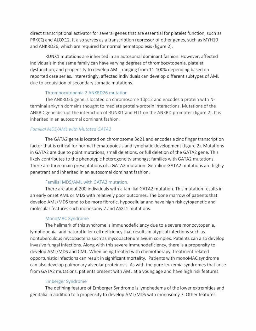
direct transcriptional activator for several genes that are essential for platelet function, such as PRKCQ and ALOX12. It also serves as a transcription repressor of other genes, such as MYH10 and ANKRD26, which are required for normal hematopoiesis (figure 2).
RUNX1 mutations are inherited in an autosomal dominant fashion. However, affected individuals in the same family can have varying degrees of thrombocytopenia, platelet dysfunction, and propensity to develop AML, ranging from 11-100% depending based on reported case series. Interestingly, affected individuals can develop different subtypes of AML due to acquisition of secondary somatic mutations.
Thrombocytopenia 2 ANKRD26 mutation The ANKRD26 gene is located on chromosome 10p12 and encodes a protein with N-
terminal ankyrin domains thought to mediate protein-protein interactions. Mutations of the ANKRD gene disrupt the interaction of RUNXI1 and FLI1 on the ANKRD promoter (figure 2). It is inherited in an autosomal dominant fashion.
Familial MDS/AML with Mutated GATA2
The GATA2 gene is located on chromosome 3q21 and encodes a zinc finger transcription factor that is critical for normal hematopoiesis and lymphatic development (figure 2). Mutations in GATA2 are due to point mutations, small deletions, or full deletion of the GATA2 gene. This likely contributes to the phenotypic heterogeneity amongst families with GATA2 mutations. There are three main presentations of a GATA2 mutation. Germline GATA2 mutations are highly penetrant and inherited in an autosomal dominant fashion.
Familial MDS/AML with GATA2 mutation. There are about 200 individuals with a familial GATA2 mutation. This mutation results in an early onset AML or MDS with relatively poor outcomes. The bone marrow of patients that develop AML/MDS tend to be more fibrotic, hypocellular and have high risk cytogenetic and molecular features such monosomy 7 and ASXL1 mutations.
MonoMAC Syndrome The hallmark of this syndrome is immunodeficiency due to a severe monocytopenia, lymphopenia, and natural killer cell deficiency that results in atypical infections such as nontuberculous mycobacteria such as mycobacterium avium complex. Patients can also develop invasive fungal infections. Along with this severe immunodeficiency, there is a propensity to develop AML/MDS and CML. When being treated with chemotherapy, treatment related opportunistic infections can result in significant mortality. Patients with monoMAC syndrome can also develop pulmonary alveolar proteinosis. As with the pure leukemia syndromes that arise from GATA2 mutations, patients present with AML at a young age and have high risk features.
Emberger Syndrome The defining feature of Emberger Syndrome is lymphedema of the lower extremities and genitalia in addition to a propensity to develop AML/MDS with monosomy 7. Other features

include sensineural deafness and cutaneous warts. Interestingly, this is the only subset of GATA2 mutants that has incomplete penetrance.
Familial AML with Mutated CEBPA
The CEBPA protein is a transcription factor that regulated granulocyte and monocyte differentiation. CEBPA mutations are inherited in an autosomal dominant fashion and has complete penetrance. Unlike AML with GATA2 mutation, CEBPA mutated AML has an excellent prognosis and tends to be cured with conventional chemotherapy. Of note, CEBPA can be an acquired mutation in sporadic AML. In familial CEBPA mutated AML, the mutations are bialleleic, with a germline mutation in the in 5’ end of the gene followed by acquisition of a mutation of the 3’ end. Therefore, any patient with a biallelic CEBPA mutation should be tested for a familial leukemia syndrome.
Patients with familial AML with mutated CEBPA do not have any physical features that distinguish it from other familial syndromes. However, the presentation of AML tends to be homogenous with diploid cytogenetics and a favorable prognosis with 65% long term survival
Dyskeratosis Congenita (DC)
Telomeres are repeats of nucleotides at the end of chromosomes that prevent degradation. Telomeropathies or telomere biology disorders (TBD), are disorders that are a result of mutations in genes that are required for proper telomere maintenance. DC is a telomeropathy that results from mutations most commonly found in the TERT (telomerase reverse transcriptase) and TERC genes. Heterozygous mutations in TERT or TERC result in an autosomal dominant inheritance pattern and exhibit anticipation where subsequent generations of affected individuals exhibit a more severe phenotype. Other genes that can be involved are DKC1, NPH2, RTEL1, and TINF2. There is also a rarer autosomal X lined recessive inheritance pattern see with TINF2 mutation in which patients present at a very young age.
The presentation of DC is a classic triad of dysmorphic nails, oral leukoplakia, and reticular pigmentation of the skin. Patients with TBD also have a higher risk of developing AML or bone marrow failure at any age. They are over 2000x more likely to develop AML than the
average population. The phenotype of TBD is inconsistent. For example, patients with DC do not necessarily present with classic triad. In fact, they can have a completely normal phenotype, thus evading diagnosis until later in life when they present with leukemia or a bone marrow failure syndrome. Other presenting signs of DC include dental caries, premature greying, idiopathic pulmonary fibrosis, and liver disease. There is also a risk of developing various squamous cell cancers.
Patients with TBD have a higher likelihood of developing toxicity from chemotherapy such as sinusoidal obstructive syndrome and prolonged cytopenias. Prompt diagnosis of DC is important as standard induction chemotherapy could be fatal due to toxicities. Given the strong anticipation for DC, stem cell transplant should be considered early as it is the only cure.
Fanconi Anemia
FA is bone marrow failure that is inherited through an X linked recessive pattern. There are about 15 genes associated with FA, termed the FANC family of genes. The FANC family of genes are involved in the repair of DNA that has acquired DNA cross links. Mutations in the FANC genes results in an inability to repair DNA crosslinks, which predispose affected individuals to various malignancies, including AML/MDS. In the setting of FA, AML tends to have more aggressive chromosomal profile with monosomy 7 or trisomy 3.
Patients with FA usually have short stature, organ and bone malformations and a propensity to develop AML or aplastic anemia. It is important to note that one third of patients will not have any physical manifestations and will present with new onset AML or aplastic anemia.

Identification of germline mutations
Prior to the advent of genomic sequencing to identify mutations, early identification of inherited syndromes was limited to telomere length testing for DC and chromosome fragility test for FA. This limited technology has delayed the discovery of novel hereditary malignancy syndromes. However, in the era of next generation sequencing, new HHMSs are being discovered at a rapid pace.
DC can be diagnosed by multi-color flow cytometry with fluorescence in situ hybridization (flow-FISH) analysis (figure 2A). Chromosomes from peripheral blood leukocytes are combined with fluorescent probes that are complementary to the TTAGGG telomere sequence at the ends of the chromosomes and then quantified. Average telomere length at birth is 11 kilobases, where as it is only 6 kilobases at age 90 (figure 2B). If the average telomere length is below the 1st percentile for that is expected for the patient’s age, then the diagnosis of a telomeropathy can be made. Even in the age of genomic testing for mutations that are commonly associated with DC, such as TERT and TERC, telomere length testing should be done as it can help predict the severity of disease in each affected family member.

FA can be diagnosed with a chromosome fragility test. Given the inability of repair DNA damage, there is a significant genomic instability that occurs in the setting of cellular stress, such as exposure to cytotoxic chemotherapy. Prior genomic sequencing for mutations in the FANC family of genes that are required for DNA repair, the diagnosis of FA was quite simple. A patient’s T lymphocytes are exposed to DNA cross linking agents such as diepoxybutane or mitomycin C. In a FA patient, there will be irreparable DNA damage (figure 3).
Sanger Sequencing. Since the early 2000’s, great strides have been made in DNA sequencing technology. Earliest forms of testing for mutations involved Sanger sequencing and is considered the first generation of DNA sequencing (figure 4). This method involves amplifying DNA, denaturing it to yield millions of single stranded DNA and then adding a primer and a DNA polymerase. This combination of single stranded DNA, an attached primer, and DNA polymerase will serve as a platform to add labelled nucleotides. It is added to 4 separate flasks, each with a combination of the four deoxyneucleotides (dATP, dGTP, dCTP, dTTP). Each flask has a differed labelled dideoxyneucleotide (ddATP, ddGTP, ddCTP, ddTTP). The DNA is elongated with the

addition of deoxyneucleotides. However, if a dideoxynucleotide is added, it terminates any further elongation. This will created various lengths of fragmented DNA, which can then be separated on by size with gel electrophoresis and will provide a sequence of the DNA. It is clear that this can be cost and time prohibitive, especially when testing for multiple genes or large portions of the gene is required. In 2005, it was estimated that it would take about 60 years and cost about 30 million dollars to sequence the entire human genome via the Sanger method. In terms of working up HHMS, this method would only be used for sequencing a particular gene of interest for research purposes or to verify a mutation that was found by next generation sequencing.
Next Generation Sequencing. Given the cost and time prohibitive nature of Sanger sequencing, new methods of identifying germline genetic mutations have been developed, termed next generation sequencing (figure 6). In this method, there are several sequencing instruments, known as platforms. The patient’s DNA is amplified and fragmented and then attached to small beads on the platform. A polymerase will then create a complement of the DNA fragment. In this method there are massive parallel sequencing reactions that will produce up to a billion DNA fragments on the platform, called bridge amplification. Then fluorescently tagged nucleotides will start to form the complementary strand. As each tagged nucleotide is

added, it is excited by a light source, releasing its fluorescent signal allowing identification of the nucleotide that was placed. This is done simultaneously on the hundreds of millions of DNA fragments that are on the platform, allowing for large scale sequencing. These reads are then clustered according to similarities and the DNA sequence is read. It is compared to a reference genome and variations are noted and either described as either known mutations or variants of unknown significance.
Management of HHMSs and its Controversies
Once a germline mutation is found, the patient should undergo a screening bone marrow biopsy to assess for dysplasia or leukemia followed by a complete blood count with differential every 6 months. If on subsequent checks there is a new or worsening cytopenia, a repeat bone marrow biopsy should be done. Also, at the time of presentation, patients should be screened

for other systemic manifestations. For example, patients with dyskeratosis congenita should be referred to pulmonary or hepatology if there are signs of pulmonary fibrosis or cirrhosis.
An important aspect of management is genetic counseling to discuss the implications of the germline mutation. This includes:
• Education about disease process in order for the patient to make lifestyle changes. For example, Fanconi anemia patients should be educated limiting exposures such as tobacco and heavy alcohol use given the risk of solid tumor malignancies such as esophageal or head and neck squamous cell cancers.
• Testing other at risk family members: This is especially important when the patient is in need of an allogeneic stem cell transplant. Sibling donors would not be fit for stem cell donation if they have not tested negative for the HHMS in question as stem cells from a donor with the same germline mutation can result in poor engraftment, graft failure, and possibly a donor-derived leukemia. Therefore, establishing a donor is crucial so transplant is not delayed.
• Family planning and methods of prenatal diagnosis: This can be controversial because many of the HHMSs have variable penetrance. Therefore, it is not necessary that an affected individual’s offspring is destined to be diagnosed with leukemia.
• Routine surveillance for HHMS as discussed above • Discuss the psychological impact of a hereditary cancer syndrome
One of the most polarizing controversies for a patient with a HHMS is timing and indication of hematopoietic stem cell transplant. The only way to rid the marrow of the mutated allele is to undergo a transplant. Important questions to ask are:
• What is the likelihood of developing leukemia? • What is the prognosis of the leukemia? • Would the mordibity and mortality of an allogeneic stem cell transplant worth it?
For example, in families with a biallelic CEBPA mutated AML syndrome, there is very strong penetrance he whereby the affected individual is sure to get AML by the fifth decade of life. Some would argue that if a diagnosis of AML is guaranteed, then the patient should undergo a stem cell transplant when they are healthy to prevent AML and perhaps save the patient from potential complications of induction chemotherapy. However, historically, CEPBPA mutated AML has a good prognosis when treated with induction chemotherapy and there is a high likelihood of remission and cure with chemotherapy alone.
For other familial leukemia syndromes, prognosis of leukemia is not easily predicted. In families with a RUNX1 mutation, there is significant anticipation. As this mutation is passed down, phenotype of the mutation is more pronounced, resulting in a higher risk of leukemia with subsequent generations. In contrast to CEBPA mutated AML, which tends to be fairly homogenous in presentation, RUNX1 mutated AML can acquire various secondary mutations

that differ from family member to family member. Therefore, it would be difficult to assess prognosis between family members, making it difficult to discuss the benefits of a stem cell transplant with affected family members who have not developed AML.
Barriers to diagnosis of hereditary hematologic malignancy syndromes
There are many barriers to the timely diagnosis of a hereditary leukemia syndrome. Given the relatively low prevalence of HHMSs, most physicians are not aware of these esoteric syndromes. This lack of awareness can also extend to the genetic counseling community as well, where emphasis is placed on the more prevalent syndromes such as Lynch Syndrome and Li Fraumeni Syndrome. Though the molecular pathogenesis, presentation, diagnosis and management of many inherited leukemia syndromes are now well described, there are new syndromes that are on the horizon. It would not be worthwhile for physicians to learn the intricacies of each of these emerging syndromes. Rather, a heightened awareness of the possibility of an inherited leukemia syndrome can trigger a prompt evaluation by a specialist.
There is also a diagnosis bias in hematologists, who are often taught that inherited bone marrow failures syndromes are diagnosed in the pediatric population when children present with congenital abnormalities and cytopenias. However, even in autosomal dominant inheritance patterns, there is variable penetrance in many syndromes and the same mutation may not have as severe a phenotype. Second, some of the prototypical physical attributes are not necessarily seen in each affected individual. For example, patients can have a germline mutation in TERT but not have any signs of DC. Lastly, some syndromes have strong anticipation whereby each successive generation has a more severe phenotype than the one before. Given these variable presentations, some patients may present later in life.
Despite the various methods to diagnose HHMS, testing for mutations in real life situations proves to be difficult. Since these syndromes are rare and little is known about them, insurance companies usually deny germline testing, placing the burden of the cost on the patient. The cost of a comprehensive NGS panel to evaluate for telomere biology disorder panel, inherited bone marrow failure panel and familial MDS and acute leukemia panel is $3,500, $3,000, and $4,000, respectively.
Peripheral blood lymphocytes are the fastest way to assess for germline mutations by next generation sequencing. However, in the setting of a hematologic malignancy lymphocytes can be neoplastic and will harbor the acquired mutations. Therefore, they cannot be used. Instead, the patient would ideally have to undergo a skin punch biopsy and skin fibroblasts must be used for culture and DNA extraction. The turnaround time is longer at about 6-10 weeks. For patients with aggressive leukemias who need to undergo a hematopoietic stem cell transplant, this will delay donor identification since a HHMS would have to be ruled out prior to considering a sibling as a potential donor.
One of the most significant, but easiest to eliminate barriers to identifying a hereditary hematologic syndrome is obtaining a through family history. Oftentimes, patients with acute

leukemia and very ill, requiring complex medical care on presentation. It is not uncommon that instead of getting a three generation family history with special attention to hematologic malignancies and organ manifestations of HHMS in all family members is forgotten. Instead, the family history is reduced down to one simple question, “Do you have any cancers in your family?” Many times, the patient will not volunteer crucial information such as persistent cytopenias, myelodysplastic syndrome, aplastic anemia, and bleeding disorders when asked a simple question about family history of cancer.
Lastly, the modern American family tends to be smaller. This can limit the detection of a HHMS simply because there are fewer family members at risk and it would be more difficult to establish a pattern of disease.
UT Southwestern Hereditary Hematologic Malignancy Clinic
Recognizing that patients have many active and acute issues when presenting with an acute leukemia that can often overshadow an assessment for a HHMS, a dedicated HHMS clinic is under development at UTSW. The goal of this clinic is to screen and test at risk individuals, provide genetic counseling, follow patients for ongoing manifestations of disease, and discuss the indication and timing of a stem cell transplant. Below is the diagnosis algorithm for patients in whom an HHMS is suspected. As more patients are being screened for HHMS, novel mutations are found, leading to discovery of new HHMS.


References
The University of Chicago Hematopoietic Malignancies Cancer Risk Team. How I diagnose and manage individuals at risk for inherited myeloid malignancies. Blood. 2016; 128:1800.
DiNardo C, et al. Evaluation of patients and families with concern for predisposition to hematologic malignancies within the hereditary hematologic malignancy clinic. Clinical Lymphoma, Myeloma & Leukemia. 2016; 16(7):417.
Calado, R, et al. Telomere diseases. New England Journal of Medicine. 2009; 361:2353-65.
Godley L, et al. Inherited predisposition to acute myeloid leukemia. Seminars in Hematology. 2014;51(4):306.
Polprasert C, et al. Inherited Somatic Defects in DDX41 in Myeloid Neoplasms. Cancer Cell. 2015; 27:658
Tesi B, et al. Gain of function SAMD9L mutations cause a syndrome of cytopenia, immunodeficiency, MDS and neurological symptoms. Blood. 2017; 129(16):2266.
Churpek J, et al. Proposal for the clinical detection and management of patients and their family members with familial myelodysplastic syndrome/acute leukemia predisposition syndromes. Leukemia & Lymphoma. 2012; 54(1): 28.
Drazer, et al. Prognostic sequencing panel frequently identify germ line variants associated with hereditary hematopoietic malignancies. Blood Advances. 2018; 2(2):146.
ACMG Board of Directors. Points to consider in the clinical application of genomic sequencing. Genetics in Medicine. 2012; 14(8):759.
Kohlmann W, et al. Discussing and managing hematologic germ line variants. Blood. 2016; 128:2497.
Feurstein S, et al. Genetic predisposition to leukemia and other hematologic malignancies. Seminars in Oncology. 2016; 43:598.


![HIS K-31 BLOOD MALIGNANCY-I- REVISE .ppt [Read-Only]ocw.usu.ac.id/course/download/1110000096-hematology-and-immunology... · KLASIFIKASI LEUKEMIA WHO ( 1997 ) ... -Morfologi Ihti-Myeloid](https://img.dokumen.tips/doc/110x75/5c9c7f4909d3f29d298bf80e/his-k-31-blood-malignancy-i-revise-ppt-read-onlyocwusuacidcoursedownload1110000096-hematology-and-immunology.jpg)