Embed Size (px)
Citation preview

The eyes have it: the neuroethology, function and evolution of social gaze
N.J. Emery*
Center for Neuroscience, Department of Psychiatry & California Regional Primate Research Center, University of California, Davis, CA 95616, USA.
Received 23 September 1999; received in revised form 6 March 2000; accepted 8 March 2000
Abstract
Gaze is an important component of social interaction. The function, evolution and neurobiology of gaze processing are therefore of interest
to a number of researchers. This review discusses the evolutionary role of social gaze in vertebrates (focusing on primates), and a hypothesis
that this role has changed substantially for primates compared to other animals. This change may have been driven by morphological changes
to the face and eyes of primates, limitations in the facial anatomy of other vertebrates, changes in the ecology of the environment in which
primates live, and a necessity to communicate information about the environment, emotional and mental states. The eyes represent different
levels of signal value depending on the status, disposition and emotional state of the sender and receiver of such signals. There are regions in
the monkey and human brain which contain neurons that respond selectively to faces, bodies and eye gaze. The ability to follow another
individual's gaze direction is affected in individuals with autism and other psychopathological disorders, and after particular localized brain
lesions. The hypothesis that gaze following is ªhard-wiredº in the brain, and may be localized within a circuit linking the superior temporal
sulcus, amygdala and orbitofrontal cortex is discussed. q 2000 Elsevier Science Ltd. All rights reserved.
Keywords: Amygdala; Autism; Emotion; Evolution; Eye contact; Eye gaze; Face; Gaze-following; Joint attention; Neurophysiology; Social cognition;
Temporal cortex; Theory of mind
1. Introduction
Social living confers a number of costs and bene®ts to the
members of a group. Larger social groups provide increased
opportunities for mating, and therefore increased genetic
diversity of potential offspring, as well as a reduction in
potential inbreeding. They also provide protection from
predators, and provide opportunities for learning strategies
for coping with the environment, i.e. increasing group size
leads to an increasing probability of learning from others.
Animals living in smaller groups, however, do not
experience competition for valuable resources, such as
food and mates. Living in a large group may require
different cognitive abilities from those required for living
in smaller groups. For example, a hierarchical system of
dominance, by its very nature, requires that individuals
have access to information about other hierarchy members,
know their position within the hierarchy, and have the
means to predict other individual's behavior and emotional
disposition. The increased reliance on visual signals in some
primate species may have been driven by the increased
sophistication of social interactions. Coalition formation,
tactical deception, reciprocity and knowledge of third
party relationships would be very dif®cult without such an
elaborate system of visual signaling [38,51,142]. Most of
the studies on sophisticated social cognition have been
carried out in human and non-human primates.
The primate brain contains over 30 regions dedicated to
visual processing, including areas which contain neurons
responsive to visual social signals (Ref. [55]; see Section
7.1). These signals can be directed to speci®c individuals,
they are less ambiguous than auditory and olfactory signals,
and they may be used to communicate emotional and mental
states; and as such may be used to predict another individual's
behavior. Visual social signals, which contain such a potential
range of complexity, may not be present in other vertebrates.
Visual signals in some birds, for example, provide infor-
mation about the sexual status of a male, through the use of
bright coloration, sexual displays and elaborate tails and
head crests [19]. Some species of birds, such as parrots
and corvids (crows, ravens and jays), which do appear to
live in sophisticated social systems, do rely to some extent
on visual signals; but the information contained within these
signals may be restricted to one form of information (such as
sexual status). This pattern is also seen in other social and
non-social species, such as some species of mammal. What
may be unique to primates is their ability to use intricate
visual social signals that appear to have multiple meanings
(such as emotional expression and indicator of interest).
Neuroscience and Biobehavioral Reviews 24 (2000) 581±604PERGAMON
NEUROSCIENCE AND
BIOBEHAVIORAL
REVIEWS
0149-7634/00/$ - see front matter q 2000 Elsevier Science Ltd. All rights reserved.
PII: S0149-7634(00)00025-7
www.elsevier.com/locate/neubiorev
* Tel.: 1 1-530-752-0875; fax: 1 1-530-752-2880.
E-mail address: [email protected] (N.J. Emery).

Important visual signals arise from the face. The face
provides a plethora of social information about an indivi-
dual's gender, age, familiarity, emotional expression and
potentially their intentions and mental state. The eyes are
very important components of the face that can provide this
information, especially information about emotional and
mental states. This review will focus on the evolution, func-
tion and neuroethology of social gaze in a number of verte-
brates. The main focus, however, will be gaze perception in
human and non-human primates for a number of reasons.
First, as described above, primates have sophisticated social
systems that rely on visual behavior and visual signals.
Second, as mentioned above, the primate brain contains
neurons that respond to eye gaze and head orientation.
Third, there has been a bias in testing non-human primates,
rather than other vertebrates with gaze perception tasks.
This has been due to the following: a) monkeys and apes
are closely related to humans, b) other vertebrates are not as
visually oriented as primates (however see birds), c) non-
human primates utilize faces as a means of primary commu-
nication, and d) what has been termed mental state attribu-
tion (also known as theory of mind) or the ability to infer the
psychological states; intentions, beliefs, desires, etc. of
other individuals from non-verbal cues (see Section 6; see
also Ref. [10] for a review).
Gaze provides other animals (speci®cally conspeci®cs)
with a means for evaluating an individual's interest in
their internal and external environments. The eyes provide
very subtle signals to other individuals, and information
transferred by this manner is dependent largely on the abil-
ity to understand that the eyes capture information about the
world. This level of processing may not be available to non-
primate animals (however, non-primates have not been
evaluated for these abilities).
The review begins by proposing that many primates rely
on gaze perception and this is related to a) the ecological
constraints of the environments in which they reside, and b)
the morphological constraints of the face and body. Next,
there is a discussion of the evolution of gaze perception in
many vertebrates, and the possible function of gaze detec-
tion in these species. The ability to use the information
provided by the gaze of others is discussed in the next
section on gaze following and joint attention. The following
section describes studies, primarily in Old World monkeys
and apes, which have attempted to evaluate the contribution
of eye gaze perception to attributing mental states in non-
human primates. Many of the reports in this section are
anecdotal due to a lack of strict testing in any non-human
species, however, the anecdotal evidence is intriguing and
interesting in relation to the few laboratory studies that are
described. The next section discusses the presence of
specialized regions of the primate (speci®cally monkey
and human) brain which either respond to gaze stimuli, or
fail to discriminate gaze stimuli when lesioned or when
compromised in psychopathological disorders, such as
autism. The ®nal section of the review evaluates a theory
of eye gaze, autism and a speci®c brain region implicated in
gaze processing called the amygdala (which is located
within the medial temporal lobe).
2. Social gaze is more than just the eyes
Throughout this review, gaze is not limited to information
from the eyes (although this is the primary source). Gaze is
not the only cue that is used to determine the focus of
another individual's direction of attention, and is not the
only component of facial expressions. The whole head, in
particular the orientation in which it is directed (using the
nose, for example) is a suf®cient indicator of attention direc-
tion (and therefore interest). In some instances, the eyes are
not visible and the only cue available for processing is the
head direction (see physiological discussion later). By the
same reasoning, if the head is occluded or in shadow, the
orientation of the body (determined from the direction of the
feet in an upright individual, or the posture of an animal in a
quadrupedal stance) provides a suf®cient cue for communi-
cation (see Fig. 1). This proposition is discussed in greater
detail in later sections and in a recent review by Langton and
colleagues [90].
3. Morphological & environmental constraints on socialgaze
This section will discuss the morphological and environ-
mental changes which may have necessitated a shift in the
primary methods of social perception employed by
primates. Within the primate order, there is a diverse
range of species differences in facial anatomy. Examples
include the small face of the loris with the proportionally
large forward facing eyes, the long snout of the ring-tailed
lemur, the dog-like face of the hamadryas baboon, the
prominent nasal features of the proboscis monkey, the
malleable lips of the chimpanzee and the large cheek
pouches of the adult male orangutan [78].
One clear difference in the facial morphology of prosi-
mians and monkeys compared to the apes is the reduction in
the protrusion of the face, in particular, the reduction in jaw
size and length of the snout or nose. The hominoid species
(orangutans, gorillas, chimpanzees and humans) have rela-
tively ¯at faces whilst retaining some prominent features.
For example, the human face has high cheekbones, a
conspicuous nose and eyebrows framing the eyes. Such
features may highlight the region around the eyes (the
eyebrows), or are used to move the eyes (the cheekbones
and related musculature) or may signal direction of attention
(the nose). These anatomical changes (a reduction in facial
protrusion) may have forced a shift in salience from the
shape of the face (and its orientation) to the eyes, as sources
of information about attention direction (as a function of
gaze). (These anatomical changes may also have empha-
sized the eyes as an important component of facial
N.J. Emery / Neuroscience and Biobehavioral Reviews 24 (2000) 581±604582
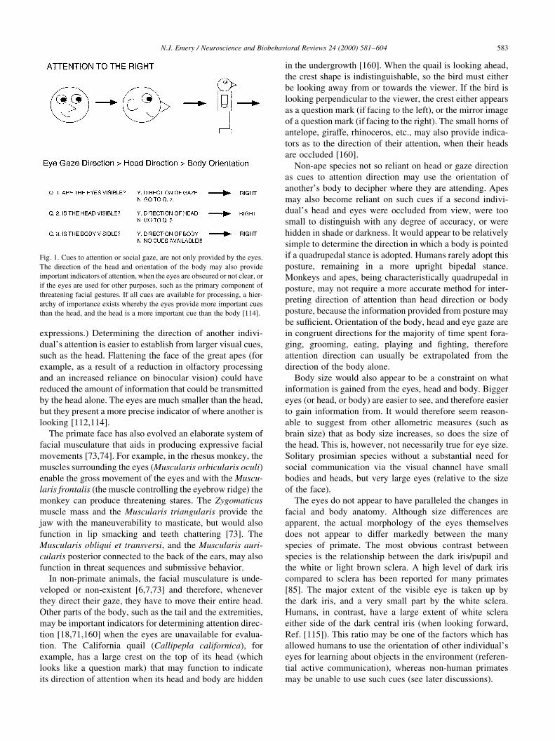
expressions.) Determining the direction of another indivi-
dual's attention is easier to establish from larger visual cues,
such as the head. Flattening the face of the great apes (for
example, as a result of a reduction in olfactory processing
and an increased reliance on binocular vision) could have
reduced the amount of information that could be transmitted
by the head alone. The eyes are much smaller than the head,
but they present a more precise indicator of where another is
looking [112,114].
The primate face has also evolved an elaborate system of
facial musculature that aids in producing expressive facial
movements [73,74]. For example, in the rhesus monkey, the
muscles surrounding the eyes (Muscularis orbicularis oculi)
enable the gross movement of the eyes and with the Muscu-
laris frontalis (the muscle controlling the eyebrow ridge) the
monkey can produce threatening stares. The Zygomaticus
muscle mass and the Muscularis triangularis provide the
jaw with the maneuverability to masticate, but would also
function in lip smacking and teeth chattering [73]. The
Muscularis obliqui et transversi, and the Muscularis auri-
cularis posterior connected to the back of the ears, may also
function in threat sequences and submissive behavior.
In non-primate animals, the facial musculature is unde-
veloped or non-existent [6,7,73] and therefore, whenever
they direct their gaze, they have to move their entire head.
Other parts of the body, such as the tail and the extremities,
may be important indicators for determining attention direc-
tion [18,71,160] when the eyes are unavailable for evalua-
tion. The California quail (Callipepla californica), for
example, has a large crest on the top of its head (which
looks like a question mark) that may function to indicate
its direction of attention when its head and body are hidden
in the undergrowth [160]. When the quail is looking ahead,
the crest shape is indistinguishable, so the bird must either
be looking away from or towards the viewer. If the bird is
looking perpendicular to the viewer, the crest either appears
as a question mark (if facing to the left), or the mirror image
of a question mark (if facing to the right). The small horns of
antelope, giraffe, rhinoceros, etc., may also provide indica-
tors as to the direction of their attention, when their heads
are occluded [160].
Non-ape species not so reliant on head or gaze direction
as cues to attention direction may use the orientation of
another's body to decipher where they are attending. Apes
may also become reliant on such cues if a second indivi-
dual's head and eyes were occluded from view, were too
small to distinguish with any degree of accuracy, or were
hidden in shade or darkness. It would appear to be relatively
simple to determine the direction in which a body is pointed
if a quadrupedal stance is adopted. Humans rarely adopt this
posture, remaining in a more upright bipedal stance.
Monkeys and apes, being characteristically quadrupedal in
posture, may not require a more accurate method for inter-
preting direction of attention than head direction or body
posture, because the information provided from posture may
be suf®cient. Orientation of the body, head and eye gaze are
in congruent directions for the majority of time spent fora-
ging, grooming, eating, playing and ®ghting, therefore
attention direction can usually be extrapolated from the
direction of the body alone.
Body size would also appear to be a constraint on what
information is gained from the eyes, head and body. Bigger
eyes (or head, or body) are easier to see, and therefore easier
to gain information from. It would therefore seem reason-
able to suggest from other allometric measures (such as
brain size) that as body size increases, so does the size of
the head. This is, however, not necessarily true for eye size.
Solitary prosimian species without a substantial need for
social communication via the visual channel have small
bodies and heads, but very large eyes (relative to the size
of the face).
The eyes do not appear to have paralleled the changes in
facial and body anatomy. Although size differences are
apparent, the actual morphology of the eyes themselves
does not appear to differ markedly between the many
species of primate. The most obvious contrast between
species is the relationship between the dark iris/pupil and
the white or light brown sclera. A high level of dark iris
compared to sclera has been reported for many primates
[85]. The major extent of the visible eye is taken up by
the dark iris, and a very small part by the white sclera.
Humans, in contrast, have a large extent of white sclera
either side of the dark central iris (when looking forward,
Ref. [115]). This ratio may be one of the factors which has
allowed humans to use the orientation of other individual's
eyes for learning about objects in the environment (referen-
tial active communication), whereas non-human primates
may be unable to use such cues (see later discussions).
N.J. Emery / Neuroscience and Biobehavioral Reviews 24 (2000) 581±604 583
Fig. 1. Cues to attention or social gaze, are not only provided by the eyes.
The direction of the head and orientation of the body may also provide
important indicators of attention, when the eyes are obscured or not clear, or
if the eyes are used for other purposes, such as the primary component of
threatening facial gestures. If all cues are available for processing, a hier-
archy of importance exists whereby the eyes provide more important cues
than the head, and the head is a more important cue than the body [114].

The morphology of the body, face and eyes provides a set
of constraints for primates' use of gaze in a communicative
capacity. A further set of constraints is provided by the
ecology of the environment in which the primates reside.
Those species living in an arboreal (tree-living) environ-
ment have a different sensory emphasis of perception and
communication from those species living in a terrestrial
(ground-dwelling) environment. A forest or jungle environ-
ment is perpetually dark; therefore vision would be an inef-
®cient means of communication. Species who live in this
environment therefore have to rely on other sensations as
their primary method of communication, such as olfactory
or auditory. Both auditory and olfactory communication
have two distinct disadvantages from visual communica-
tion: they are indirectly focused (anyone within range can
hear a call, or smell an odor, including predators), and are
limited to projecting simple information (such as mating
signals, ªI'm ready to mateº or spatial position, ªI'm over
hereº), and are less appropriate for projecting information
about objects in the environment (unless used in conjunction
with visual behavior, ªLook at that fruit treeº).
4. Phylogenic aspects of gaze perception
The eyes have long held special interest to humans; they
are said to be the window to the soul [10], they are often
used as symbols of a curse (evil eye) or as warning signals,
but are also one of the ®rst points of contact between infants
and their mothers [64]. Non-human vertebrates are also
attracted to the eyes, gather information from the eyes and
appear to use their own eyes for communication, such as
warning others of their emotional disposition (threatening or
submissive). This section discusses how a number of differ-
ent non-human vertebrate species utilize information
contained within the eyes (such as for defense against preda-
tors), and how this specialized form of visual social percep-
tion may have become utilized in non-human and
human primates for more complex communicative func-
tions.
Simple schematic representations of the eyes (two black
or colored circles) are perceived as eyes in a number of
species, and appear to form part of a rapid predator detection
system. Lesser mouse lemurs (Microcerbus murinus), for
example, display more gaze aversion when presented with
two schematic eyes presented in a horizontal orientation,
than when the same stimuli are presented in a vertical orien-
tation, or when more than two circles are presented or when
different stimuli which do not have ethological relevance,
such as squares, are presented [40]. A similar effect has also
been reported for jewel®sh [41], various species of birds
[79,134] and wild house mice [146] using similar stimuli.
Some species of butter¯ies and moths (Lepidoptera) have
evolved eye shaped patterns (eye spots) on their wings as an
effective method to deter potential predators (such as lemurs
and birds), as they resemble the eyes of other predators
(such as felines and birds of prey) that are the natural preda-
tors of small mammals and birds [49].
It would appear adaptive for all prey species to have the
ability to distinguish forward facing eyes (or simple repre-
sentations of the eyes) from other similar stimuli, as most
predators have eyes facing the front (binocular vision aids in
hunting). The view of one eye, or eyes too close together to
be facing forward, present information to the prey species
that the predator is not attending to them (see later sections)
and therefore may be eluded. Therefore, an important
further function of recognizing the presence of the eyes is
to determine whether they are looking at or away from you.
A large number of species appear to perform this discrimi-
nation. Black iguanas (Ctenosaurua similis), for example,
can differentiate the difference between whether an
approaching human is looking towards or away from them
[24]. Burger et al. suggested that iguanas perceive gaze
contact and direct approach as threatening and escape
quicker than when the same experimenter is approaching
directly, but looking away from them. Iguanas also appear
to be sensitive to the size of the approaching eyes, respond-
ing quicker when the eyes are larger [23].
Some species of birds can also perform this type of discri-
mination. Ristau [130] studied plovers' (Charadrius sp.)
reactions to human experimenters who walked past their
nests either looking at the nests in the dunes, or in the
opposite direction towards the ocean. Plovers incubating
their young remained off the nest longer under threatening
situations, such as when an experimenter was gazing
towards the plover's nest, compared to when the experimen-
ter was gazing towards the sea. Gallup, Cummings and Nash
[59] also looked at birds' responsivity to human gaze.
Chickens (Gallus gallus) respond to staring humans by
becoming rigid (tonic immobility). This is said to correlate
highly with fear [59]. The length of immobility was greatly
reduced when the eyes of an experimenter were directed
away from the chickens. The same phenomenon has also
been reported in some species of lizards (Anolis carolinen-
sis, Ref. [68]).
A related phenomenon to tonic immobility is death feign-
ing, as seen in hog-nosed snakes. This occurs in reaction to
eyes directed towards the snakes [25,26]. For example, hog-
nosed snakes not only react to the presence of a human
experimenter (as a predatory threat), but their response is
dependent on the orientation of the experimenter's gaze
(towards or away from the snake). Whether these actions
are innate behaviors or learnt through extensive experience
with predators is not known, although from an evolutionary
perspective it would be adaptive to possess this ability
innately. Scaife [133,134] suggested that the ability to
discriminate the eyes as a stimulus, which is part of the
face (a more primitive ability than determining whether
gaze is directed or averted), is an innate predisposition.
Other avian species have been tested for their responsiv-
ity to speci®c human head orientations and gaze directions.
House sparrows (Passer domesticus) were found to increase
N.J. Emery / Neuroscience and Biobehavioral Reviews 24 (2000) 581±604584

¯ight (an escape response) when a face was directed towards
them, but were unresponsive to the direction of the eyes,
whether pointed away or towards them [65]. The sparrows,
however, could determine the line of gaze from the number
of eyes visible. Therefore, the sparrows seemed to associate
the perception of direct eye gaze and head direction with a
subsequent aversive event, such as capture. Hampton did
not test the birds with other stimuli containing eyes or
eye-like shapes to determine whether the sparrows had
learnt that humans (or other animals) which were directed
towards them were to be avoided (producing a ¯ee response)
or whether this was a trait that was present early in devel-
opment.
Primates also possess a great interest in the eyes and the
region around them. How some primates may use the infor-
mation transmitted by the eyes will be discussed in the next
sections. Experimental studies of face recognition in
monkeys have revealed the interest some primate species
have in the eyes. Keating and Keating [83] studied the eye
movement responses of two rhesus monkeys whilst they
viewed different primate faces (rhesus monkey, chimpanzee
and human) with neutral expressions, and also to schematic
faces. The subjects showed an extreme bias of looking at the
eyes and the small region surrounding the eyes compared
with the nose and the mouth, for all four neutral face stimuli,
regardless of species. Nahm et al. [107] expanded on these
studies using implanted eye coils to record precisely the eye
movements of head-restrained monkeys when viewing
neutral and expressive faces. The subjects displayed a
predominant interest in the eyes and the mouth (the primary
components of facial expressions, see below). Baboons have
also been shown to look at the eye region more than other
parts of the face, and the eyes are essential to them for face
recognition. Kyes and Candland [89] presented baboon
subjects with slides of other baboon faces, and parts of
faces. Although the subjects looked longer at the slides of
full (complete) faces, they also inspected pictures which
contained eyes longer than slides containing just the nose,
or the mouth, or the nose and mouth. Therefore, the eyes
seem to be the most important components of the face to
these species of primates.
What is not known however, is whether this great interest
in the eye region is related to the eyes being a major compo-
nent of facial expressions, or whether the presence of the
eyes is essential for the recognition of individual faces. Can
non-human primates recognize faces by the eyes alone?
There is some evidence that humans can recognize and
remember faces when only presented with the eyes
[42,97]. Although some non-human primates can recognize
different individuals [142], it is not known what features
they use to perceive differences between individuals. It is
likely to be a multitude of features: eyes, facial con®gura-
tion, body size, color, behavior, personality, etc., which
provide clues to identity in non-human primates.
Color also performs an important function in highlighting
the eye region of some primate species. Kingdon [84]
described the facial patterns and coloration of different
species of guenon (Cercopithecus sp., an Old World
monkey). For example, C. mona and C. cephus cephus
have bright blue coloration around the eyes, but no colora-
tion of the genital area (white instead of the bright red color
of the majority of Cercopithecines, Ref. [60]). This may be
interpreted as an increased importance of the face and eyes,
and therefore used in facial expressions during sexual or
social signaling, or further enhancing the differentiation
between the face and the genital region. Cercopithecus
neglectus have a wide orange-colored brow-ridge, which
highlights the position of the eyes [84]. Species of guenon,
which do not have brightly colored facial features, usually
have brightly colored genitals. It may be interesting to note
that the brightly colored genitals are blue, and that the color
around the eyes of those species without the genital colora-
tion is blue, although the signi®cance of the color blue is not
known.
The eyes play a pivotal role in all primate facial
expressions [6,7,18,78,129,147]. Table 1 presents descrip-
tions of the role of the eyes, eyelids and eyebrows in each of
Van Hoof's thirteen universal facial expressions for catar-
rhine primates [147]. Any discussion of the role of the eyes
in primate emotional communication must, however,
mention the role of the whole face. The eyes are not
processed separately from the rest of the face, they are
analyzed in concert with other features, such as the nose
and mouth, and in particular the ocular muscles surrounding
and controlling the movements of the eyes. This is not to say
that the eyes alone are not important stimuli for the expres-
sion of emotion. In some primates, the effect of a stare with-
out the accompanying facial movements is very effective in
eliciting fear or ¯ight responses from conspeci®cs.
The majority of primates have very darkened eyes
compared to humans. Kobayashi and Kohshima [85]
found that of eighty-eight primate species, only humans
had eyes with a white sclera and a dark iris. The sclera of
most primate's eyes was found to be brown or light brown,
with two species (Old World macaques) having a pale
brown sclera and four species (Old and New World
monkeys) having a partially white sclera. The sclera of
macaque infants is less pigmented than adults. Perrett and
Mistlin [115] suggested that the reason why the sclera of
adult macaques may become darker compared to infants is
that adult monkeys could look out of the corner of their eye
without invoking threatening gestures usually associated
with eye contact from other conspeci®cs, whereas infant
macaques are less of a threat. Determining the precise direc-
tion of another's attention is dif®cult to assess when there is
no differentiation between the sclera and the iris. In humans,
interpreting gaze direction is made easier by the morphol-
ogy of the eyes. Gaze following can be performed using a
simple rule (dark in the center of the eye equals eye contact;
dark to the left of the eye equals looking left; dark to the
right of the eye equals looking right).
Primates have excellent abilities for discriminating
N.J. Emery / Neuroscience and Biobehavioral Reviews 24 (2000) 581±604 585

whether they are being looked at or whether another's gaze
is directed away from them. Keating and Keating [83] also
studied monkey subjects' eye movements in response to
viewing slides of rhesus monkeys with gesturing faces.
The expressive stimuli included a slide of a threatening
rhesus face with a direct stare, a slide of a rhesus grinning
with direct gaze, a slide of a rhesus grinning with averted
gaze and a slide of a rhesus with a neutral expression with
direct gaze. Both subjects looked at the eye region more
often than the nose and mouth and looked at the faces
with direct gaze (irrespective of facial expression) more
than the face with averted gaze. This pattern of results
was repeated when the subjects were presented with slides
of human faces. Only slides of two human gestures were
presented to the subjects, raised eyebrows and lowered
eyebrows. The eye region elicited a higher number of ®xa-
tions than the nose and mouth regions, and there were a
higher number of ®xations on the human faces with raised
eyebrows. (This does not suggest, however, that the
monkeys understood the meaning of the human facial
expressions.)
Perrett and Mistlin [115] reported the number of submis-
sive gestures (lip smacking and teeth chattering) that maca-
que subjects give in response to a human head, with the head
oriented towards or away from the viewer and the eyes
positioned in compatible or incompatible directions to the
head. The largest number of submissive gestures were made
by the subjects when the eyes were in contact with the
observing monkey, independent of the human's head posi-
tion. This experiment was repeated for elevation of the head
[102]. The raised head received fewer appeasement
(submissive) gestures compared to a level head position or
a head averted laterally by 458. Similar results were found
when Mistlin tested the emotional reactions of stumptailed
macaques to a life-sized model of an adult male macaque.
The model's head and eyes could be positioned to provide a
range of head and eyes orientations or elevation combina-
tions. The lowered head (similar to a threatening gesture)
received more appeasement gestures compared to the head
raised or level.
A number of different variables can in¯uence a primates'
reaction to eye contact from a human experimenter, and
presumably a conspeci®c. Thomsen [141] measured the
mean frequency of eye contact directed towards a human
staring at them; in differently reared rhesus monkeys; in
different primate species (talapoin, patas, and squirrel
monkeys and crab-eating, rhesus and stumptailed maca-
ques); and in rhesus macaques of different ages and sexes,
tested at two distances from the human experimenters.
Wild-born rhesus displayed more frequent eye contact
than the surrogate reared monkeys. Talapoin monkeys
displayed more frequent eye contact with the experimenter
N.J. Emery / Neuroscience and Biobehavioral Reviews 24 (2000) 581±604586
Table 1
Description of the contribution of the eyes, eyelids and eyebrows to different facial expressions in monkeys and apes (taken from Ref. [147])
Facial Expression Description
Relaxed face Eyes in a ªneutral positionº, ªupper eyelid is not lifted completely, the iris being only partly exposedº.
Alert face ªfully opened eyeº.
Tense-mouth face ªeyes may be opened rather widely and are staring ®xedly towards a partnerº, ªeyebrows are normal or lowered in a
frownº.
Staring open-mouth face ªeyes are staring ®xedly at the partnerº, ªeyebrows are lifted in a vary marked way¼the skin of the upper eyelid and
the region immediately above it¼is exposedº, ªthe eyebrows may be lowered in a frownº.
Staring bared-teeth scream face ªeyes in most cases are staring ®xedly at the opponentº, ªeyelids are fully apart, so that the eyes are fully openº,
ªeyebrows are fully lifted and the upper head skin is retractedº.
Frowning bared-teeth scream
face
ªeyes are closed or opened only to a small degree¼when not closed the eyes are never directed straight towards the
opponent; the animal looks awayº, ªeyebrows are lowered in a frownº.
Silent bared-teeth face ªeyes may be staring at the opponent in a ®xed way or be evasive (i.e. it may throw short glances towards the partner
out of the corners of its eyesº, ªeyebrows¼may be relaxed or in a lifted positionº, ªthe degree of opening of the eyes
varies; it may be maximum, or normal to slightly openedº.
Bared-teeth gecker face Same use of eyes as the silent bared-teeth face display.
Lip-smacking face ªeyelids are usually fully apart, so the eyes are completely openedº, ªeyes are usually staring in a ®xed way at the
partner. The head may also¼be turned sideways a little, so that the animal is looking out of the corner of its eyesº,
ªeyebrows are often liftedº.
Teeth-chattering face ªeyelids may be fully apart or in the normal positionº, ªeyes are either staring at the partner in a ®xed way or making
evasive movementsº, ªeyebrows¼may be retractedº.
Protruded-lips face ªeyelids may be either fully apart or in the normal positionº, ªeyes may either be staring at a partner in a ®xed way or
making evasive movementsº, ªeyebrows¼are lifted completelyº.
Pout face ªeyelids are either in the normal position or farther apartº, ªeyes may be directed towards a partnerº, ªeyebrows may
be lifted¼especially when the expression movement is directed towards a partnerº, ªsimultaneous contraction of the
m. depressor supercilli and perhaps the m. corrugator supercilli antagonistic to the m. frontalis responsible for the
lifting, may result in a curious slanting position of the eyebrowsº.
Relaxed open-mouth face ªeyelids are usually in the relaxed position or slightly drawn togetherº, ªouter corners of the eyes are always slightly
lifted due to a pressure from the zygomatic muscle, which draws the mouth-corners backwards and upwardsº, ªeyes
may occasionally be directed to the partner¼the gaze is¼less ®xedº, ªeyebrows¼are usually in the normal
position¼they may¼be lifted when the display is directed to a partner from a distanceº.

than patas, crab-eating, stump-tailed, squirrel and rhesus
monkeys. Young rhesus females displayed more frequent
eye contact than young males, and adult females and
males. In each case, the frequency of eye contact was less
at greater distances between the experimenter and monkey
subject. Frequency of eye contact, however, has a different
meaning to duration of eye contact. More frequent eye
contact suggest that the subject is checking the changing
state of the eye gaze of the experimenter, whereas a long
duration of eye contact would suggest a threat or af®liative
gesture (see below). Unfortunately, duration of eye contact
was not measured in this study. These results suggest vast
species differences in the importance of eye gaze. For exam-
ple, talapoin monkeys are a highly vigilant species,
compared to rhesus monkeys that avoid eye contact in
most social situations. Eye contact does not carry the
same relevance for young monkeys as adult monkeys,
which would suggest that it is a learnt response. Infant
monkeys do, however, respond differentially to a face
where the gaze is averted compared to a face with eye
contact, as early as three weeks of age [99].
Autonomic physiological changes in non-human primates
have been reported to accompany the detection of eye
contact, suggesting that eye gaze is an emotive stimulus.
Wada [149] studied the EEG responses from the brainstem
of macaques to electrical stimulation of the cortex. He found
that if he looked at the monkey subjects the EEG trace
would change dramatically (p. 41).
When the animal discovered it was being watched, the
response was depressed as long as the animal could
see the experimenter.... Such ¯attening regularly
occurred whenever the animal realized that the experi-
menter's gaze was ®xed on it.... The direct meaning of
the experimenter's gaze...suggests concentrated
focusing of discriminatory attention of a quality
necessary for self-preservation.
This physiological response may form part of a circuit
linking eye contact with a hormonal and/or motor response
during courtship and sexual behavior between male and
female monkeys. Linnankoski et al. [95] found that females
presenting their hindquarters to speci®c males (proceptivity)
caused the males to masturbate and ejaculate, but only when
eye contact was established between the male and female.
Other visual or olfactory cues, such as inspecting the
females perineal region, were not as effective initiators of
ejaculation as eye contact.
Monkeys do not appear to make the subtle distinctions
between direct staring and mutual eye contact that some
great apes and humans do. Direct staring is different from
mutual gaze or eye contact in a number of ways. Staring
involves the eyes, but also the eyebrows and brow ridges
being raised to increase the visibility of the eyes, the ears
being pulled back and the hair on the head standing up. The
timing of a stare may also be shorter than the duration of
mutual gaze. Face-to-face sex is an example of the way that
great apes such as bonobos [43], orangutans [58] and
humans [104] may use looking into each other's eyes as a
method for con®rming and strengthening the sexual and
af®liative bond [44]. Linnankoski's [95] study discussed
above would also suggest that monkeys might occasionally
use the eyes for this function.
5. Gaze following and joint attention
Determining the precise direction of another's attention
may be an important ability for non-human primates. Gaze
cues provide salient information about the location of
objects, but may also function in complex forms of social
cognition, such as visual perspective-taking, deception,
empathy and theory of mind (see Ref. [152,153,154]).
An important use for gaze following may be determining
the position of an individual in a dominance hierarchy.
Chance [36] called this social attention, where ªeach
individual [in a social group] accords and receives attention
as a function of his or her rankº. The most dominant animal
in a social hierarchy receives the highest number of glances
(attention from less dominant animals), and glances at other
animals the least. Chance stated that members of a social
group must have the capacity to determine 1) that the domi-
nant individual is the focus of the others' attention, 2) that
these glances total more than those directed towards less
dominant animals, and 3) that the group members extrapo-
late the information that the animal is dominant due to the
larger number of glances. There is very little quantitative
data to con®rm Chance's hypothesis, largely due to dif®cul-
ties in measuring gaze in the animal's natural environment.
A recent study by McNelis and Boatright-Horowitz [98],
however, found that social gaze could be measured accu-
rately in patas monkeys (Erythrocebus patas) and that
lower-ranking animals directed their gaze more towards
higher-ranking animals than vice versa.
Observational learning about speci®c objects in the world
would be impossible without gaze following (although other
forms of social learning may not require an appreciation of
gaze; Ref. [31]). In an interesting series of experiments,
Mineka et al. [101] attempted to determine whether fear
of snakes in rhesus macaques could be induced in young
monkeys through observational conditioning (a speci®c
form of social learning). Young laboratory-reared rhesus
monkeys (who were not previously fearful of snakes)
became fearful when they observed their wild-reared
parents showing fearful responses to real, toy and model
snakes. For the young monkeys to become fearful of the
snakes by observational learning, they must have used the
attention direction of their parents, coupled with their
parents' fearful facial expressions directed towards the
snakes. Individual X is producing a fearful response, and
individual X's attention is directed to the object Z on the
ground, therefore, they must be fearful of the object Z on the
ground. This form of observational learning would appear to
N.J. Emery / Neuroscience and Biobehavioral Reviews 24 (2000) 581±604 587

require information about another's direction of attention
and also so-called joint attention between the individual X
following the direction of conspeci®c Y's attention onto
object Z (an object of joint focus; see Fig. 2).
Shared attention differs from joint attention in a subtle
way (Ref. [53,112], see also Fig. 2). However, in the litera-
ture the two terms are used interchangeably. Joint attention
requires that two individuals (X and Y) are attending to the
same object (Z), based on one individual using the attention
cues of the second individual. Shared attention, however, is
a more complex form of communication that requires that
individuals X and Y each have knowledge of the directions
of the other individual's attention (or a method for checking
that what the other individual is looking at is the same as
what they are looking at). Baron-Cohen [9] proposed a
modular system for theory of mind (see later) that develops
in human infants, which contained components of the gaze
communication system. The four modules were an Eye
Direction Detector (EDD), an Intentionality Detector (ID),
a Shared Attention Mechanism (SAM) and a Theory of
Mind Mechanism (ToMM). The EDD module would repre-
sent gaze following and joint attention. Perrett and Emery
[112], in a commentary to Baron-Cohen [9], proposed a
Direction of Attention Detector (DAD) module for proces-
sing all potential attention cues (eyes, head or body) and a
Mutual Attention Mechanism (MAM) used for detecting
mutual gaze. Joint attention would only require activation
of the EDD or DAD modules, whereas SAM would require
activation of the EDD or DAD, and MAM modules.
Laboratory studies of gaze following and joint attention
per se are limited to a small number of species. The ability
to gaze follow has been demonstrated most successfully in
human infants. The age at which an infant ®rst follows
another's gaze is controversial, ranging from 6 to 18 months
of age [28,30,39,135]. These age differences may be due to
differences in methodology (variation in angle of gaze, the
use of an experimenter versus the infant's mother as a stimu-
lus) or in the de®nitions for gaze the experimenters utilized
(see above). Before 12 months old, human infants follow
their mother's gaze, but do not direct their attention to the
object of her attention. At around 12 months old, infants
begin to follow their mother's gaze towards particular
objects in their visual ®eld, and at around 18 months old
they can direct their attention to objects outside of their ®eld
of view (e.g. behind them; Ref. [27]).
Joint attention may be important for language learning in
human infants [47,106,144], and human enculturated and
language-trained bonobos [63] and parrots [111]. An early
stage in language development is the process of associating
a word (usually a noun) with the physical presence of an
object, such as pronouncing ªappleº whilst attending to an
apple. This stage of language learning is dif®cult to achieve
without the ability to follow gaze onto (or pointing) at speci-
®c objects.
The clearest evidence for the ability to follow gaze in
non-human primates comes from experimental work on
the great apes, in particular, studies with chimpanzees. Povi-
nelli and Eddy [121,122,123,125] in their experiments
concluded that chimpanzees can follow a human experi-
menter's gaze, but not use that information to learn about
objects in the world or the ªmental stateº of the individual
providing the gaze cues (see next section). Povinelli and
Eddy trained chimpanzee subjects to enter a room, and
respond for food from an experimenter by using a natural
begging gesture (placing a hand in front of the experimenter
for a food reward, this signi®ed the end of a trial). Once
trained to do this, the human experimenters performed
speci®c attention sequences for the subjects. The three
conditions were ®rst eyes and head, where the experimenter
shifted his head and eye gaze to behind and to the left or
right of the subject. The second condition was similar to the
®rst, but the experimenter only shifted the direction of his/
her eyes. The ®nal condition was no change in attention.
These three conditions were randomly assigned to test
sessions and each session was rewarded. In the eyes and
head condition, 50% of the trials (10 trials in each of 8
test sessions) elicited a gaze-following response from the
subjects to the correct side, and in the eyes-only condition,
30% of the trials elicited a gaze-following response to the
correct side. Povinelli and Eddy interpreted this as shared or
joint attention on the part of the chimpanzee subjects.
In a further series of experiments, Povinelli and Eddy
[123] obstructed the subjects' line of sight with an opaque
shield. The experimenters used their head and eyes to look
at an object out of sight of the subjects (on the same side as
the experimenters behind the shield). Subjects could follow
the experimenter's line of sight to the unseen object (i.e. the
subjects would look at the point on the opaque barrier where
the experimenter's line of sight hit the barrier). This ability
may be important when trying to extrapolate information
from other's attention, when the focus of attention is out
of sight (such as a predator or food hidden in the under-
growth).
Tomasello, Hare and Agnetta [145] replicated Povinelli
and Eddy's experiment with different types of barrier
(gutter, board, different rooms and a wall). They attempted
to determine whether the chimpanzees were using a simple
rule to follow gaze (i.e. stop gaze following when an inter-
esting object comes into the line of sight) or whether they
could actually use the experimenter's gaze to determine
geometrically what speci®c object the experimenter was
looking at. They replicated Povinelli and Eddy's results
(the subjects looked more around the barrier when the
experimenters had done so, than when the experimenters
looked in different directions). To further examine whether
the chimpanzees' gaze following was at a low or high level,
they provided an interesting distracter object for the experi-
menter to look at. The subjects were presented with three
conditions. First, the experimenter looked at a target loca-
tion on a wall. Second, the experimenter looked at an
animate object (distracter) which was located close to
him. Third, the experimenter looked at the target location
N.J. Emery / Neuroscience and Biobehavioral Reviews 24 (2000) 581±604588

whilst the distracter was in view. Tomasello et al. found that
the subjects looked at the distracter object, but continued to
follow the experimenter's gaze onto the target. This
suggests that chimpanzees do not just re¯exively follow
gaze onto the ®rst available object within view, but track
another individual's gaze geometrically to speci®c locations
and objects they attended to. Tomasello et al. suggest that
this is the ®rst study to show geometric gaze following in a
non-human primate, however, see the arguments and simi-
larities to these results below.
Other investigators have replicated the presence of gaze
following in chimpanzees and have described an absence in
Old and New World monkeys. Itakura [75] recently studied
the ability of eleven species of prosimians, monkeys and
apes to follow a human experimenter's gaze (eyes, head
and pointing in a corresponding direction). Only the oran-
gutan and chimpanzee subjects made greater than 70%
correct responses, with the orangutan making 100% correct
responses. This may be attributed to enculturation of the
orangutan as has been suggested for chimpanzees [35].
The non-ape subjects (brown lemur, black lemur, squirrel
monkey, brown capuchin, whiteface capuchin, stump-tailed
macaque, rhesus macaque, pig-tailed macaque, and tonkean
macaque) did not respond above chance level.
Anderson, Sallaberry, and Barbier [4] investigated the
ability of three capuchin monkeys (Cebus apella) to utilize
human attention cues to gain food rewards in a Wisconsin
General Testing Apparatus (WGTA). Anderson, Montant,
and Schmitt [3] also used the same paradigm with three
rhesus macaques. Both studies used a forced choice scenario
in which the monkey was allowed to choose one of two
wells in the WGTA, one covering a food reward. A
human experimenter would stand between and behind the
two covered food wells and demonstrate attention to the
baited food well using different cues; ªpointing onlyº,
ªgaze onlyº (which included both head and eye cues), and
ªgaze and pointingº (a combination of both these cues).
None of the macaque or capuchin subjects could be trained
to use the ªgaze onlyº cues to guide choice of food well.
Two subjects of each species could be trained to use either
ªpointing onlyº or ªgaze and pointingº. It is likely that the
success with the ªgaze and pointingº situation can be attrib-
uted to cues arising from pointing rather than the monitoring
of gaze direction cues (local enhancement; learning the
correct location of the food by using the proximity of the
hand to the correct well).
Pettigrew, Forsyth, and Perrett [119], using a similar
protocol (but without pointing), obtained equivalent results.
Despite training (.100 trials) they found that four out of six
rhesus monkeys failed to learn the rule that the head and
gaze direction of a human experimenter predicted the loca-
tion of food reward.
In a gaze paradigm similar to that of Anderson and collea-
gues [3,4], Itakura and Tanaka [77] found that chimpanzees,
an enculturated orangutan and human infants (3±4 years
old) could all use gaze (head and eyes) cues (near and
close to the baited food wells), pointing and glancing
(eyes only directed towards the food wells) to chose the
well baited with food. The responses of all subjects
appeared to be spontaneous, not requiring learning. Povi-
nelli and colleagues [120] have also used a similar paradigm
to test chimpanzees. They found that the chimpanzee
subjects could not determine which well was baited with
food when using the experimenter's eyes only as a cue.
When the experimenter's head and eyes were directed
towards the baited well or slightly above the well, the
subjects responded well above chance. The subjects could
also follow active gaze (head and eyes, not eyes only), i.e.
attention initially directed between the two wells then shift-
ing gaze to the baited well. The negative glancing (eyes
only) result reported by Povinelli et al. [120] could have
differed from the positive glancing result in the Itakura
and Tanaka [77] study due to the age, experimental experi-
ence and enculturation of the different groups of subjects.
Itakura and Anderson [76] reported successfully training
one juvenile capuchin monkey to use the experimenter's
head direction to choose between two presented objects.
Peignot and Anderson [110] also found positive results
when using captive lowland gorillas. All the subjects
appeared to be able to use, pointing and/or head plus gaze
cues to locate hidden food, but not use the ªeyes aloneº as a
cue.
As an aside to a study of apes' understanding of inten-
tional versus accidental actions, Call and Tomasello [32]
described the failure of ®ve chimpanzees and three orangu-
tans to follow a human experimenter's gaze (it is not known
whether eyes alone or head plus eyes were provided as cues)
onto one of two baited boxes. The subjects did display an
appreciation of intentional action versus accidental action,
so it is possible that the subjects did not ªtrustº the gaze cues
of certain experimenters. Also, the subjects results were
pooled together and included an enculturated orangutan,
which may have looked signi®cantly at the box attended
by the experimenter. Unfortunately, no individual data
was presented.
Call et al. [33] replicated these experiments with chim-
panzees, and found that they readily followed the experi-
menter's gaze to objects and locations out of their view. The
subjects were also tested on their levels of gaze following
when the type of occluder was manipulated. In the previous
experiments, the experimenters knew the location of the
hidden food, but could not see the food. Different abilities
may be important for solving these different problems.
When a bowl, which completely covered the food, was
used, the chimpanzees could not use gaze to locate the
food. When tubes or upturned boxes which allowed the
experimenter, but not the subject, to see the food were
used, the subjects could use gaze cues to locate the food.
This suggested that the chimpanzee subjects could under-
stand the physical properties of an occluder and that the
experimenter could see the food, but they could not. The
chimpanzees therefore appeared to understand that seeing
N.J. Emery / Neuroscience and Biobehavioral Reviews 24 (2000) 581±604 589

leads to knowing, but not that knowledge persists over time
(i.e. the experimenter saw where the food was located, but
not when it was covered). This result is surprising, as chim-
panzees (and a number of other species) have a concept of
object permanence [142]. Chimpanzees may have a concept
of object permanence, but do not appear to attribute this
concept to the minds of other individuals (see next section).
There are a number of potential reasons that New and Old
World monkeys may have failed in these tasks. One is the
use of human experimenters as demonstrators. In their
natural habitat, if non-human primates can utilize others'
gaze cues (as is suggested from ®eld research); a more
appropriate line of research would be to use conspeci®cs
as stimuli. A second potential reason may be related to the
use of operant tasks (using food rewards). Povinelli and
Eddy [122,123] suggested that following another indivi-
dual's gaze might be an automatic response and form part
of a primitive orienting re¯ex (POR). This would be more
adaptive than an evaluation system, especially when
confronted with the possibility of attack from predators or
losing a food or mating resource. The use of operant tasks to
test for gaze following, therefore, would fail to test the
presence of a POR compared to a more complex mechanism
involved in social cognition (such as theory of mind).
Monkeys may follow gaze, but do not learn how to use
gaze during an operant task. This can be assessed by record-
ing the natural, automatic behavioral responses of
subjects (such as head and body turns, eye movements
or facial expressions) in response to conspeci®c social
stimuli.
Emery et al. [53] utilized both of these techniques
(conspeci®cs as stimuli and eye movement recording) in
an attempt to evaluate the naturalistic-type responses of
rhesus monkey subjects to gaze stimuli. Brie¯y, the subjects
were head restrained and presented with videotaped
sequences of conspeci®c monkeys looking at a point in
space, monkeys looking at two identical moving objects
(created using video mirroring technology) or the two
objects with no conspeci®c monkey present. The subjects
consistently looked more at the region in space and the
object attended to by the conspeci®c. In the trials where
the conspeci®c was absent, the subjects looked equally at
the two identical objects. The subjects were also utilizing
the gaze cues from the conspeci®c and not making a random
response, as the largest amount of time (and number of
®xations) was directed towards the conspeci®c. These
results suggest that rhesus monkeys can follow the gaze
cues of other monkeys, and that this is an automatic POR
which is unrelated to the attribution of mental state (X is
looking at Y because of Z). If the subjects had continued
looking at the previously attended object after the conspe-
ci®c disappeared from the video display, this would have
suggested that the subjects learned that one object was
inherently more important than the other based on the beha-
vior of the previously viewed conspeci®c. This may have
been used to argue for a more complex role in mental state
attribution.
It is unclear which cues the observer monkeys were using
to direct their own attention, as body orientation, head direc-
tion and eye gaze direction were all available and all
presented in congruent directions. Lorincz, Baker and
Perrett [96] recorded the eye movements of monkeys to
the presentation of slides of head and body cues in either
comparable or con¯icting directions (i.e. head left, body
oriented right) to determine which cues the monkeys used
to orient their own attention. They also presented the
subjects with slides of heads only, with the head and eyes
in either comparable or con¯icting directions. They found
that the observing subjects used the information from the
head more readily than the body, and they also appeared to
follow eye gaze cues when the demonstrator's head was
oriented towards the observer monkey. This is therefore
the ®rst evidence for eye gaze following in a non-ape
species. Although it is possible that that observers were
N.J. Emery / Neuroscience and Biobehavioral Reviews 24 (2000) 581±604590
Fig. 2. Gaze direction provides a number of potential social cues which may
be utilized by an individual to learn about the external (other individuals,
objects, events, etc.) or internal (emotional and intentional) states. A.
Mutual gaze is where the attention of individuals A and B is directed to
one another. Averted gaze is where individual A is looking at B, but the
focus of their attention is elsewhere. B. Gaze following is where individual
A detects that B's gaze is not directed towards them, and follows the line of
sight of B onto a point in space. C. Joint Attention is the same as Gaze
Following except that there is a focus of attention (such as an object), so
individuals A and B are looking at the same object. D. Shared Attention is
a combination of Mutual Attention and Joint Attention, where the focus of
individual A and B's attention is on the object of joint focus and each other
(i.e. ªI know you're looking at X, and you know that I'm looking at Xº). E.
Mental state attribution or theory of mind, probably uses a combination
of the previous A-D attentional processes, and higher-order cognitive stra-
tegies (including experience and empathy) to determine that an individual is
attending to a particular stimulus because they intend to do something with
the object, or believe something about the object.

averting their gaze from the demonstrator monkey, the
direction in which they orient their own gaze is consistent
with the gaze direction of the demonstrator. When the head
and eye gaze were con¯icting, however, the observers
tended to follow head direction only.
In a study cited above [145], Tomasello and colleagues
suggested that they had found the ®rst evidence for
geometric gaze following in a non-human primate.
However, in the Emery et al. study [53], the monkeys obser-
ving the gaze cues of the monkeys on the video directed
their own gaze to very speci®c locations on the video screen.
When the demonstrator monkey only was present the
subjects looked signi®cantly more at the location in space
in which the demonstrator was attending than elsewhere on
screen (apart from the demonstrator monkey itself). A simi-
lar result was obtained when two objects were presented on
screen. This may suggest that the observer monkey was not
looking at the ®rst interesting object of attention, as there
was not a physical end-point to the demonstrator's gaze (i.e.
no object), and was therefore directing its gaze
geometrically. It remains to be tested whether monkeys
can ignore distracter objects which are located closer to
the target object, and not just presented on the opposite
side of the screen.
Tomasello, Call and Hare [143] con®rmed that some
species of primates could follow the gaze of conspeci®cs.
Tomasello et al. found that ®ve species of non-human
primate, rhesus, stumptailed and pigtailed macaques, sooty
mangabeys and chimpanzees, could follow the gaze cues of
conspeci®cs when semi-free±ranging in a large outdoor
enclosure. The experimenter waited for two conspeci®cs
to be proximal to one another, and then the experimenters
would attempt to gain the ®rst conspeci®c's attention by
holding up some food. The experimenter would then record
the second conspeci®c's (named the subject) behavior in
relation to the gaze behavior of the ®rst conspeci®c. The
experimental trials were analyzed as either questionable or
valid. The questionable trials were trials where the conspe-
ci®c looked at the food, but the subject did not notice the
conspeci®c's attention cue, but did look at the food. The
valid trials were designated as trials where the subject
looked at the conspeci®c (and hence gaze cues) and then
looked at the food. In the control trials, the food was
presented when only the conspeci®c was present. Tomasello
and colleagues found that in all species tested, the subjects
looked at the food on a highly signi®cant number of valid
trials (79±100%). Interestingly, the stumptailed macaque
subjects responded 100% correctly (signi®cantly better
than chimpanzees). Stumptails may use gaze cues more
often in af®liative gestures than other species. This may
be due to the gentle, relaxed manner with which stumptailed
macaques interact with conspeci®cs (such as alerting others
to danger, Ref. [44]).
In a recent report, Anderson and Mitchell [5] reported that
stumptailed macaques, but not black lemurs, could follow
the head turn of a human experimenter from eye contact to a
point in space at an angle 60±908 from center. The authors
do not describe how many sessions each subject received
(average six trials per session) and present the results as
absolute frequencies. The species differences therefore
may be due to the number of times in which the subjects
participated in the experiment, as there is some evidence
that lesser mouse lemurs can respond to low level aspects
of gaze (gaze aversion; Ref. [40]).
A sophisticated socio-cognitive ability may not be the
exclusive pre-requisite for gaze following, or learning to
use another's attention cues to locate a food reward. A
number of recent studies have proposed that domestication
may also effect gaze following abilities in dogs. Miklosi et
al. [100] examined whether assistant dogs (for the blind)
could utilize human attention cues (pointing, bowing,
nodding, head turning and glancing) to ®nd hidden food.
Potential olfactory biases were removed by keeping the
food reward wrapped in cloth, which would cover the test-
ing bowls. As with the Anderson [3,4] studies, the experi-
menter was positioned between the two food receptacles and
performed one of the attention cues towards one of the
bowls when the subject was paying attention. The subjects
responded to the pointing cues immediately, and learnt to
utilize the other cues over training. Only one of the
assistant dogs learned to use the glancing cue by the
end of training.
These results were replicated whilst pet dogs were tested
in their owners' homes with their owners providing the cues.
The performance was slightly better than the assistant dogs
for all the cues. When the experiment was replicated with
the experimenter providing the cues, the dogs showed the
same level of performance. Dogs can also appear to utilize
conspeci®c cues to locate food. Hare and Tomasello [66]
studied the response of dogs to either local enhancement
cues (i.e. proximity to the food as a cue) or gaze, body
orientation and pointing cues simultaneously, provided by
either humans or conspeci®cs. For the ten subjects tested, 8/
10 made the correct food choice when provided with
human-local enhancement cues; 5/10 made the correct
choice when provided with human-gaze and point cues; 6/
10 made the correct choice when provided with dog-local
enhancement cues and 4/10 made the correct choice when
provide with dog-gaze and point cues. Dogs may be able to
use conspeci®c cues as a throwback to their evolutionary
history as pack hunters. Hunting as part of a group requires a
degree of cooperation, which may rely on gaze or body
orientation monitoring [66].
It appears therefore that monkeys, apes (chimpanzees,
orangutans and gorillas) and dogs can follow another indi-
vidual's gaze, and that this ability is enhanced when the
gaze demonstrator is a conspeci®c. There is also evidence
that apes, and to a lesser extent monkeys, can follow gaze to
speci®c locations and objects in space and that gaze follow-
ing may be more than re¯exive and provide information
about an animal's behavioral (and possibly mental) inten-
tions. This is discussed in the following section.
N.J. Emery / Neuroscience and Biobehavioral Reviews 24 (2000) 581±604 591

6. Gaze and social attention as indicators of mental state
This section evaluates the role that analyzing another's
gaze (eye position, or head and body view) may play in
assessing what their intentions; dispositions and beliefs
are (see Fig. 2). Non-human primates, however, may not
be able to infer another's intentions from the presence or
direction of their eyes. The eyes may only be used to
communicate another's emotional state (such as anger
displayed as a threat; Section 4), or to communicate the
presence of objects within the immediate environment
(Section 5).
There are many instances in the primate literature which
suggest that some primates may use gaze to convey infor-
mation about their intentions, not just of emotions or refer-
ring to objects, events or individuals in the environment.
Primates may utilize gaze following for soliciting help
from conspeci®cs when challenged by or challenging
other group members. Soliciting for assistance or an
invitation to co-operate against a third party has been
described for baboons [87,109,150] and vervets [38]. Vervet
monkeys use quick, furtive glances between an aggressor
and a potential helper to gain support from the potential
helper. The aggressive monkey needs to be attended to,
both by the monkey soliciting help and the potential helper.
The helper may then determine the intentions of the solicit-
ing monkey. The object of attention needs to be known to a
high degree of accuracy in possibly volatile situations. Soli-
citing has been described for Papio anubis (olive baboons)
as (Ref. [109], p. 441):
A triadic interaction in which one individual, the
enlisting animal, repeatedly and rapidly turns his
head from a second individual, the solicited animal,
towards a third individual (opponent), while continu-
ously threatening the third.
Some individuals, during complex behavioral interac-
tions, may utilize the knowledge that other monkeys and
apes automatically follow gaze (see previous section). In a
database of naturally occurring instances of primate tactical
deception, Whiten and Byrne [155] describe an anecdote
where they observed a young baboon apparently using
gaze direction cues in a possibly deceptive communicative
role. Baboons and vervets usually stare and make calls at
predators in the distance [38,155]. In the following anec-
dote, the subadult male, ME, appeared to use this informa-
tion to his advantage (Ref. [155], p. 237).
Subadult male ME attacks one of the young juveniles
who screams. Adult male HL and several other adults
run over the hill into view, giving aggressive pant-
grunt calls; ME seeing them coming, stands on hind-
legs and stares into the distance across the valley. HL
and the other newcomers stop and look in this direc-
tion; they do not threaten or attack ME.
Whiten and Byrne suggested that subadult male ME
learned that his own attention was a salient cue, which
could be used to deter others from chasing him. In this
example, a predator (or other interesting object) was not
within the ®eld of view (or the object of ME's attention).
Attention appeared to be an automatically interesting cue,
which was distinguishable enough to disrupt the actions of
the pursuing animals. Whiten and Byrne [155,156] discuss
many instances where an individual manipulates another's
use of attention cues, as a form of deception.
How non-human primates use gaze in intentional
communication, and understand the meaning behind this
volitional use, has only started to be tested experimentally.
One recent study has highlighted one aspect of this use. One
gorilla has been shown to successfully use her gaze to refer
to objects and to direct humans' attention to objects and the
gorilla's behavior [61,62]. Gomez tested an infant gorilla
with a problem (similar to one of Kohler's problem-solving
experiments with chimpanzees; Ref. [86]) in which it had to
get out of a locked room using particular tools. Gomez
found that the gorilla not only used conventional objects
to solve the problem, but also used the experimenter as a
social object or agent.
An infant gorilla was placed into a locked room with a
latch to lock the door (out of reach of the gorilla), a box high
enough to reach the latch and a human experimenter. The
infant gorilla used four different strategies to attempt to
reach the latch. First, the gorilla dragged the box under
the latch and climbed onto the box. Second, the subject
dragged the experimenter under the latch, to climb on the
experimenter and reach the latch. Third, the gorilla gently
led the human experimenter to the door while looking
between the experimenter's eyes and the latch (the goal,
or object of attention). Finally, the gorilla would look
between the experimenter's eyes and the latch without lead-
ing the experimenter [61]. By looking at the eyes and face of
the experimenter, the gorilla could be said to be directing
the attention of the experimenter to the focus of the gorilla's
own attention, namely the latch. This may be similar to the
baboons described earlier that solicited help by looking
continuously between the goal of their attention (an oppo-
nent) and a solicited helper [109]. The gorilla may have
been checking to see that the experimenter was still attend-
ing to the latch and to their actions. The gorilla also
appeared to use eye contact to monitor if the human was
attending to the gorilla's request which the experimenter
acted upon [61].
Chimpanzees have also been described as attempting to
in¯uence the behavior of a human caregiver using their eyes
(i.e. providing evidence they understand that mental states
can be communicated between individuals using gaze).
Leavens et al. [91] evaluated the responses of a captive
male chimpanzee who alternated his gaze between dropped
food (during a matching-to-sample experiment; MTS) and
an experimenter. An initial observation of this behavior led
the experimenters to videotape the face and body gestures of
the chimpanzee during each MTS experiment to determine
N.J. Emery / Neuroscience and Biobehavioral Reviews 24 (2000) 581±604592

whether the chimpanzee was using gaze alternation and/or
pointing to direct the experimenters attention to the dropped
food. The chimpanzee's behavior was recorded when the
experimenter was either present or not present in the
room. Analysis of the videotapes over many years showed
that the subject appeared to use gaze alternation during 76%
of instances in which food was dropped.
Both the Gomez [61] and Leavens [91] studies can be
interpreted in ways that do not rely on mental state attribu-
tion. Many pet owners discuss the ability of cats and dogs to
look at the location of a food source or the door to outside,
then back to the pet owner, then back to the food or the door
(Emery, personal observations). This behavior is usually
associated with vocalizations. Although this behavior is
identical to that described above for chimpanzees and goril-
las, the domesticated pets would not be described as inten-
tionally communicating with their owners. The pets are
likely to have been looking at the location of the food or
outside at an earlier occasion, and vocalizing because of
motivation to gain the food or entry to outside. The pet
owner happened to initiate the required action (get food or
open the door) independent of the intentions of the cat or
dog. The pet, therefore, learnt through association that look-
ing and vocalizing at an object of interest leads to access to
that resource by the actions of the owner.
Distinguishing another individual's visual perspective
from one's own is thought to be an important step in inter-
preting their intentions and thoughts about the world.
Kummer et al. [88] attempted to train long-tailed macaques
(Macaca fascicularis) to take a juice reward, only if a
human experimenter was not in a position to observe them
taking the reward. The experimenters threatened the
monkeys if the monkeys took the reward whilst in their
view. The subjects, however, behaved equivalently, drink-
ing juice reward out of sight of the experimenter (hidden
behind an occluder), or in front of the experimenter in view.
Kummer and colleagues interpreted this as a lack of
perspective taking or a lack of ªexperiencingº another's
visual perception.
In an extensive series of experiments, Povinelli & Eddy
[124] tested chimpanzees' abilities to choose between an
experimenter who can see (and therefore has some knowl-
edge of an event) and an experimenter who cannot see (and
therefore does not have the same knowledge). The chimpan-
zees were trained to make a begging gesture to receive a
food reward. The subjects were then presented with a choice
of two experimenters: one for whom the eyes were open or
free from occlusion, and a second experimenter for whom
the eyes were covered with different objects, such as blind-
folds, buckets, tinted goggles. The subjects could not differ-
entiate between the experimenters when their eyes were
covered (i.e. did not consistently make begging gestures
towards the experimenter with the uncovered eyes). When
the whole head was covered, however, the subjects made the
correct discrimination. The chimpanzee subjects (all juve-
niles) could not make the inference that seeing (when using
the eyes) leads to knowing about an event (begging gesture),
but could when using the head as a cue. This series of
experiments has lead Povinelli and Eddy to state that chim-
panzees may use a sophisticated level of gaze following
(that is probably more sophisticated than that used by maca-
ques; Ref. [53]), but that their use of gaze is not within a
mentalistic framework (i.e. they do not have a concept of
another individual's mind from gaze cues).
Povinelli and colleagues have recently replicated and
expanded these experiments [128] to determine whether
the chimpanzee subjects' lack of sophisticated visual
perspective-taking in the Povinelli and Eddy [124] study
was due to developmental time-course (i.e. the subjects
were not old enough to appreciate gaze as a mental event,
ªseeingº). They found that there was no difference in perfor-
mance between ®ve and nine years of age, suggesting that
chimpanzees do not process another individual's gaze
within a mentalistic framework (or at least with respects
to this experimental design). Theall and Povinelli [140]
also provided evidence that the chimpanzees did not
respond preferentially to the experimenter who could see
them gesturing, which was not because of the arti®cial
begging response used to evaluate their performance. This
was achieved by recording the vocalizations and tactile
gestures directed towards each of the experimenters.
There was no difference in the number of non-begging
gestures directed to the experimenters independent of
whether the experimenter was attentive (eyes open and
head directed towards the subject) or inattentive (eyes
closed or above the subjects).
Santos and Hauser [132] recently utilized a technique
often employed in studies of pre-linguistic infants (the
expectancy violation paradigm) to determine whether a
New World monkey, cotton-top tamarins (Saguinus oedipus
oedipus), could predict the behavior of a human experimen-
ter based on their gaze cues (head plus eyes or eyes alone).
Santos and Hauser presented the tamarins with either an
expected or an unexpected event, based on the intention to
act related to gaze experience (i.e. an individual gazes at an
object he/she intends to interact with, and subsequent
actions will be with the attended object, not an unattended
object). In the expected trials, the experimenter looked
(either with the head plus eyes or the eyes alone) at one of
two novel, interesting objects (children's toys), then they
reached for the same toy that they were attending to. In
the unexpected trials, the experimenter looked at the
previously unattended toy, and then reached for the unat-
tended toy. Santos and Hauser reasoned that if the subjects
were linking seeing with intentional action, a violation of
this expectation (i.e. reaching for the unattended object)
would produce longer looking times. They found that this
was true when the experimenter looked at the object using
head plus eyes, but not the eyes alone. This result may
suggest that tamarins can predict another individual's beha-
vior based on their attention, however, there are some dif®-
culties with this technique.
N.J. Emery / Neuroscience and Biobehavioral Reviews 24 (2000) 581±604 593

Looking time does not always correlate with interest in an
object. Emery et al. [53] found that duration of looking in
their study was not signi®cant, however number of inspec-
tions (®xations) was signi®cant. A highly vigilant species
may be more likely to change their gaze than direct their
gaze for long lengths of time onto one area or object. It is
also possible that the tamarins were using a simple rule, and
that this rule was violated in the unexpected trials (direct
gaze at object A ! reach [use of speci®c motor pattern] for
object A). This was indeed possible, as the subjects received
three identical familiarization trials (expected sequence of
behavior) with one of the objects before the test trials. As
there was no evidence that the subjects used the experimen-
ter's eyes as a cue, this would also suggest that the tamarins
were not attributing mental states to the experimenter
(intention to reach for a speci®c object).
The experiments described above suggest that some apes
(and possibly some monkeys) may process another's gaze
within a behaviorist framework, but not within a framework
which requires a more cognitively based system of mental
state attribution (which probably requires some rudimentary
language skills). A behaviorist interpretation would state
that non-human primates respond to attention cues in
terms of reward, and learning about stimulus-response rela-
tionships [151]. For example, ªX follows the gaze of Y as X
has learnt that nutritious fruit attracts Y's gazeº. X will be
rewarded (i.e. will have access to fruit) if X responds appro-
priately after observing Y's gaze. This does suggest a level
of explicit knowledge of the outcome of another indivi-
dual's actions and behavioral responses.
Purposive behavior interpretations of the above experi-
ments require that the monkeys and apes understand that
other individuals of their own or other species perform
acts within an intentional framework (or with a ®nal goal
in mind). A leopard hunts monkeys because it has to eat; a
chimpanzee looks at a patch of fruit because he intends to
eat the fruit, etc. Dif®culties in interpretation arise, not just
when suggesting that others have purpose in their behavior,
but also when suggesting those individuals can interpret and
understand another's purpose from individual behavior
patterns.
The anecdotal and observational studies described above
of monkeys in their natural habitat, and experimental studies
of apes in the laboratory, suggest that they may understand
the social signi®cance of gaze and social attention cues of
other individuals. Studies of monkeys performed in the
laboratory, however, suggest a potential dichotomy between
the abilities of monkeys and apes in utilizing gaze cues as a
means for interpreting another individual's future behavior,
in relation to objects. Although the evidence is not entirely
convincing, chimpanzees, orangutans and to a lesser extent,
gorillas, appear to use direction of attention cues to interpret
something about another individual's intentions (as related
to objects in the world). There is no evidence that monkeys
in the laboratory use gaze cues for this purpose (and the
positive evidence provided by anecdotes could be inter-
preted in other ways). This dichotomy may have arisen
because the gaze cues presented in the monkey's natural
environment would have been provided by conspeci®cs,
and particularly by familiar or related conspeci®cs (see
arguments for monkey gaze following in previous section).
In the ape experiments, familiar human experimenters
provided the attention cues. (This also explains the positive
results for enculturated apes, Ref. [142].) Apes with experi-
ence of human experimenters (and particularly enculturated
apes [142]), but not wild apes, may have the ability to use
non-ape gaze cues (such as provided by humans). Wild
monkeys and apes may only utilize conspeci®c attention
direction cues. One potential experiment to test this would
be to provide laboratory apes with gaze cues from other
primate species (either other apes or monkeys) and test
their ability to utilize these cues within a mentalistic frame-
work.
There is very little evidence that any non-human primate
can use another individual's gaze cues to evaluate and
predict their intentions within a more complex mentalistic
framework. This lack of a theory of mind may be due to an
absence of language, consciousness or a sense of self in non-
human primates [69]. The evidence for the use of gaze cues
in humans is much stronger, and will be discussed in Section
7.3.
7. Neurobiology and psychopathology of social gaze
7.1. Neurons responsive to head and gaze direction
The rhesus monkey brain contains neurons that respond
preferentially to the sight of human or monkey faces
[22,46,117]. An important ®rst step in social interaction is
recognizing a conspeci®c. Cells with responses selective for
faces over other objects have been found in the upper and
lower banks and the fundus of the anterior superior temporal
sulcus (STS, see Fig. 3). This region contains cells that
respond to polymodal stimuli (visual, auditory and tactile,
Ref. [22]). Perrett et al. [117] recorded from cells in the
fundus of the STS (Fig. 3) and found that the responses of
face selective neurons were not affected by transformations
of the face (changing color, size, vertical orientation).
Scrambling the features of the face, however, did reduce
the cells' responses, suggesting speci®city in the cells'
response to facial stimuli. Dividing the face into its compo-
nent parts (eyes, nose, mouth, etc.) and presenting the sepa-
rate parts alone also reduced the response of some cells from
the initially strong response to the whole face. Some cells,
however, responded equally well when the eyes or the
mouth region were presented alone.
Rotating the face horizontally away from the front view
changes the response of a number of cells. Therefore, the
presence of the eyes and a speci®c head view are important
for the responses of a number of cells. For example, one cell
displayed an increased response when a face was presented
N.J. Emery / Neuroscience and Biobehavioral Reviews 24 (2000) 581±604594

in pro®le. Perrett et al. [118] found that changing head view
(horizontal orientation) had a dramatic effect on the
responses of face responsive neurons. Some cells were
tuned to the face, with a gradual decline in neural response
with increasing horizontal rotation from the front view of
the face (example of a narrow tuning curve). Other cells
produced opposite responses, where the cell was preferen-
tially responsive to a pro®le (e.g. left pro®le) with a decline
in response as the head was rotated towards the face view.
Perrett et al. [114,116] found that neurons responding to
all views of the head (broad-tuned, object-centered neurons)
were rare, whereas cells responding to speci®c views of the
head (narrow-tuned, viewer-centered cells) were numerous
in the anterior STS. A number of viewer-centered cells were
responsive to speci®c views which were not one of the four
characteristic views (face, back, left and right pro®les). This
suggested to Perrett et al. [116] that the function of these
neurons (and neurons with responses selective for one of the
four characteristic views) may be to determine the direction
of another's attention, as these cells were super¯uous to
requirements for recognizing faces as a distinct class of
objects, or for recognizing individual faces.
The breadth of tuning exhibited by some viewer-centered
neurons would not explain cell responses to vertical head
elevation. Perrett et al. [118] found that a number of cells
were unresponsive to changes in horizontal orientations of
the head (rotation), but were affected by vertical elevation.
For example, a cell responded to all frontal horizontal
views, but only when the head was directed upwards. The
cell had the same response to front head view directed
upwards, as it did to head right pro®le directed upwards
[54]. Other cells were dependent on the view and elevation
of the head. For example, a cell responded more strongly
when the front head view was directed upwards, compared
to the front head view directed downwards [54].
Perrett et al. [114] reported the activity of a cell respon-
sive to attention down. The cell was responsive when the
head was directed down, the eyes were directed down or the
head and body posture (quadrupedal) was directed down-
wards. Changing the elevation of the eyes in relation to the
head (e.g. eyes directed towards the viewer, head down-
wards) and the head in relation to the body (e.g. head direc-
ted at the viewer, but body posture downwards) changed the
direction of attention. Head elevation, therefore, is also an
important indicator of attention direction. For example,
attention to the left (horizontal, level head) is different to
attention up and left or attention down and left.
Eye gaze was also found to be important. Sixty-four
percent of cells selective for the face and pro®le were also
dependent on the position of the eyes [113,118]. The major-
ity of cells preferred that the head and eyes were presented
in compatible directions with respect to the viewer, such as
head left with eyes left. Some cells were selective for eye
position independent of the head view. For example, the
response to head left and eyes facing viewer would be the
same as head and eyes facing the viewer, or head right and
eyes facing the viewer. Eye gaze dependent cells often do
not require the presence of the rest of the head to respond
[158]. Other cells were only responsive when the eyes were
averted from the viewer. As discussed earlier, direct eye
contact is a threatening gesture for some non-human
primates. These selectively responsive neurons may form
part of a dedicated neural system for distinguishing threats
from non-threats. Some cells responsive to the face,
however, were insensitive to the position of the eyes; i.e.
there was no difference in response between eye contact and
eyes averted.
It is possible that if the eyes become occluded from view
(for example, under poor lighting conditions or occlusion),
information from the head becomes more important. In a
similar manner, cells responsive to information provided by
the body become important if the head is occluded from
view. Body orientation is used as a cue for spatial direction
(the direction another individual is moving to), especially by
hamadryas baboons during their daily marches in search for
food and water [136]. A quadrupedal stance is less ambig-
uous in providing information of attention direction than an
upright posture, and may negate the need to use subtler
forms of attention cues, such as eye gaze in some animals
(see earlier). Neurons in anterior STS have been found
which respond to speci®c views of bodies and body parts
in isolation [148]. Perrett et al. [114] proposed that the
information provided by eyes/face/body sensitive neurons
was part of a processing hierarchy (for attention direction or
social attention).
Emery [51] recorded from single units in the anterior STS
(STPa) and tested the response of head direction speci®c
neurons (such as responsive only to left pro®le) when an
object of focus was also present (see Fig. 4). The cells were
tested in response to the head alone, the head looking at the
object, the head looking away from the object, the head
looking at the object, but with a different, non-selective
orientation (such as right pro®le), and the object alone.
None of the cells tested �n � 4� were responsive selectively
to the head looking at the object (i.e. the cells responded
equally to the head alone, and the head looking at the
object). This suggests that either the number of cells tested
was insuf®cient or that a different brain region may code for
this sophisticated level of gaze processing (joint attention),
such as the amygdala. A number of neurons within this brain
area respond to another's body movements (walking or
reaching) in relation to objects [51,113]. Motion in relation
to an object may provide a more salient cue for neural
response, compared to the ambiguous relationship between
two static objects (head and object).
Neurophysiological responses to eye gaze have also been
found in other regions of the macaque brain. Brothers
[20,21] recorded from single cells in the amygdala (acces-
sory basal, lateral basal, medial basal, cortical and central
nuclei) and the perirhinal/entorhinal cortex. Brothers found
two cells which were responsive when a videotaped stimu-
lus of a stump-tailed macaque looked directly into the
N.J. Emery / Neuroscience and Biobehavioral Reviews 24 (2000) 581±604 595

camera (i.e. eyes directed towards the viewer), but not when
the stimulus monkey averted gaze away from the viewer.
The response of these neurons was not analyzed further, but
may function in emotive responses to eye contact, as
suggested earlier.
Electrophysiological methods have also been employed
in humans to determine responses to eye gaze. Bentin et al.
[17] recorded event related brain potential (ERP) responses
from the lateral temporal scalp of normal subjects and found
larger responses to isolated eyes compared to full faces. This
contrasts to ERP recordings made directly from electrodes
over the fusiform gyrus (ventral temporal lobe) which
displayed larger responses to full faces, compared to
isolated eyes [2]. The face-speci®c N170 component of an
ERP is larger to the eyes presented alone than to the entire
face [17]. Eimer [50], however, tested whether the N170
was dependent on the presence of the eyes in faces, and
found no affect on the amplitude of the N170 component;
but there was a delay in response to faces without the eyes
present. The eyes therefore appear to be as salient a cue for
humans as monkeys; however the speci®c electrophysiolo-
gical base for these responses requires further investigation.
Neuroimaging studies in normal human subjects have
also highlighted a role for the STS and amygdala in gaze
processing. Puce and colleagues [127], using functional
magnetic resonance imaging (fMRI), found signi®cant acti-
vation in the posterior STS to faces in which the eyes or
mouths were moving, but not to other moving facial parts
(such as facial muscles in the cheeks) and not to other
moving stimuli. Wicker and colleagues [157], using an
alternative neuroimaging technique (positron emission
tomography; PET), found signi®cant activation to mutual
and averted gaze in the fusiform gyrus (which also is acti-
vated in response to the whole faces; Ref. [126]), right
parietal lobe, right inferior temporal gyrus, and the middle
temporal gyrus.
Hoffman and Haxby [72] utilized fMRI to determine
whether the invariant (permanent and semi-permanent)
aspects of a face, such as identity (but this would also
include gender, race, age) were processed in different
parts of the brain from the changeable (moveable) aspects
of a face, such as expression and eye gaze. They found
signi®cant activation in the lateral fusiform gyrus to facial
identity (see also Ref. [126]), and signi®cant activation in
the STS to eye gaze. They also found signi®cant activation
in response to eye gaze in the intrapartietal sulcus (parietal
cortex). Gaze following, rather than detection of the eyes as
a facial component, may depend on the connectivity of the
STS with the intraparietal sulcus, as the geometric location
of objects determined from the direction of eye gaze should
require a sophisticated spatial cognition system. There is
some evidence for this in the monkey, as neurons responsive
to eye gaze in the STS connect reciprocally with neurons in
the intraparietal sulcus [67].
The amygdala has also been implicated in gaze proces-
sing in humans. Kawashima and colleagues [82] found
signi®cant activation in the amygdala using PET, during a
gaze discrimination task (eye contact versus no eye contact).
The activation was lateralized to the left amygdala during
both the eye contact and no eye-contact conditions, and to
the right amygdala during the eye-contact condition. This
may not indicate that the amygdala is essential for the discri-
mination of eye gaze per se, but that eye contact is an
emotional stimulus, important enough to activate the amyg-
dala.
7.2. Neuropsychology of gaze perception
Further evidence suggests that the STS, amygdala and
possibly orbitofrontal cortex form part of a system coding
the direction of another's visual attention (eye gaze direc-
tion, not just gaze away or towards a viewer) and the object
of another's focus. Campbell et al. [34] and Heywood and
Cowey [70] lesioned the STS (speci®cally the STPa) after
testing the limits of eye gaze discrimination in rhesus
monkeys. Pre-operatively, the monkeys had to perform a
two-choice picture task and to choose the picture with the
eyes averted. Differing degrees of eye gaze displacement
(head center view), from eye gaze aversion to eye contact,
were presented (5, 10 or 208 deviation from center view).
The eyes and head were varied independently (e.g. head
view 208 from center, eyes 108 from center), to determine
whether the monkeys were using eye gaze rather than head
position as a discriminative cue. Before surgery, the
monkeys were extremely good (75±90% correct) at discri-
minations of gaze directions, 10 and 208 from center, but
poor (chance) at 58 from center. After removal of the STS,
the monkeys' performance at discriminations of gaze direc-
tions, 10 and 208 from center, was reduced to chance level.
It would therefore appear that rhesus monkeys could discri-
minate gaze averted laterally from eye contact (10 and 208from center) and that the STS is important for these discri-
minations.
Eacott et al. [48] also tested the ability of rhesus monkeys
with lesions of the STS to discriminate direction of gaze.
Pairs of eyes (without the head) were presented towards the
viewing monkey subjects (center position) or averted by
deviations of 5, 10, 15 or 208 from the viewing monkey.
Although the head was occluded, the eyes were presented as
part of a head either directed towards the viewer, or averted
(208 to the left or right of the viewer). The subjects were
presented with a two-choice discrimination, between eyes
directed towards the viewer (incorrect stimulus) and eyes
averted (correct stimulus), irrespective of the position of the
head. The unoperated animals were better (overall) at discri-
minations of eye gaze deviation greater than 58 (10, 15 and
208), than the STS lesioned animals, but both groups of
subjects were equally impaired at discriminations of 58from the viewer (however the choices were signi®cantly
greater than chance). The percentage number of correct
choices increased with increasing deviation from the
viewer. Lesioned animals were also impaired on a novel
N.J. Emery / Neuroscience and Biobehavioral Reviews 24 (2000) 581±604596

two-choice discrimination task (between different groups of
ASCII characters). Eacott et al. [48] thus concluded that the
de®cits in processing differences in eye gaze were due to
de®cits in two-choice discrimination learning. Gaze direc-
ted towards the viewer may be an exemplar, which may be
compared with other instances of gaze. This may be why
there is no difference between operated and unoperated
subjects in discriminations of gaze deviations 58 from the
viewer. The eye position would not be suf®ciently different
from the exemplar to aid discrimination. Eacott [48] failed
to test the monkey subjects on eye gaze discrimination before
STS surgery. It is not known how the operated subjects would
have performed pre-operatively in the discrimination task.
Amygdala damage in humans has been shown to cause
de®cits in processing the direction of another's gaze. Patient
DR [159] sustained a bilateral ablation of the amygdaloid
complex after surgery for epilepsy, which caused profound
disturbances to the patient's processing of faces, such as
discrimination of facial identity and expression. DR was
also impaired in discriminating between eyes averted and
eye contact. A forced-choice paradigm was used where
pairs of photographs of faces were presented to the subject.
In one-third of the photographs, the head was directed to the
viewer, in one-third the head was directed 208 to the left and
in one-third the head was directed 208 to the right. Photo-
graphs in which the eyes were directed towards the viewer
were target faces. Non-target faces were faces in which the
eyes were looking away 5, 10 or 208 to the right or left.
There were therefore six directions of gaze (5, 10 and 208left and right) and three head views (08, 208 left and 208right), that produced 18 possible trials. Patient DR was de®-
cient in discriminating eye contact (13 out of 18 correct).
This was compared to a mean performance of 16.95 (out of
18 correct) for matched control subjects. DR did not make
any errors when the faces were deviated by 208 (6 out of 6
correct), but performance was poorer with smaller devia-
tions, 108 (3 out of 6 correct) and 58 (4 out of 6 correct).
The main question that now faces studies of the neuro-
biology of gaze processing is what are the different contri-
butions of the anterior STS and amygdala. The amygdala
has been attributed a role in emotion (particularly fear, Ref.
[92]) and social behavior [52]. The STS is likely to be
essential for recognizing the eyes, head and body as stimuli
used in social communication, whereas the amygdala is
likely to be essential for attaching socio-emotional signi®-
cance to these stimuli, such as a direct stare is a threat, or
glancing towards an object may provide information as to its
location or valence.
Responses of neurons within the brainstem [149] and
changes in the galvanic skin response (GSR, a method to
measure changes in the autonomic nervous system in rela-
tion to emotive stimuli; Ref. [108]) were dependent on the
perception of eye contact. The predominant (and possibly
only) route by which visual information about the eyes (and
the orientation of the eyes directed towards the viewer; see
above) could reach the brainstem, would be via the amyg-
dala. The amygdala may form part of a circuit that attaches
emotional signi®cance to the eyes (i.e. threat or embarrass-
ment) and then signals the brainstem to initiate an emotional
response (such as an increased GSR or blushing). This could
be achieved via a direct projection from the central nucleus
of the amygdala to the brainstem [80].
7.3. Psychopathology of gaze perception: autism and
schizophrenia
The development of gaze processing in normal human
infants follows a speci®c time course. The age at which
an infant ®rst follows another's gaze (usually its mother)
is controversial, ranging from 3 to 18 months
[28,29,30,39,45,135]. Infants as young as 5 months have
been shown to be sensitive to adult gaze shifts as low as
58 (i.e. from the eyes to the ears) when recording changes in
the infant's attention and smiling response [139]. During
normal development, the ability to focus on the object of
another's attention appears between 12 and 18 months of
age [103] and the ability to infer mental states from eye gaze
appears around 36±48 months [9,10].
Autistic individuals have problems with many forms of
social communication. Gaze processing is impaired at many
different levels, such as eye contact [81], gaze following
N.J. Emery / Neuroscience and Biobehavioral Reviews 24 (2000) 581±604 597
Fig. 3. Schematic representation of a lateral view of the rhesus macaque
brain displaying location of cells responsive to faces, eye gaze, head direc-
tion, body orientation, and facial expressions. See text for references. Cell
types are represented by schematic representations of stimuli, which elicit
responses. Left is anterior, right is posterior. Abbreviations: Superior
temporal sulcus (STS), amygdala (Amyg), inferotemporal cortex (IT), orbi-
tofrontal cortex (ORB), prefrontal cortex, and principal sulcus (A46).

[94], joint attention [37], and understanding gaze within a
mentalistic framework [11,15,93]. These de®cits are not
seen in children and adults with Down syndrome [11,15].
A major indication of autism is a lack of joint attention, such
as gaze following, protodeclarative pointing (informative
pointing), showing or other forms of ªreferential commu-
nicationº. Autistic individuals do not direct others attention
to objects and events in the environment [105] and they do
not use pointing gestures [10].
Two tests of joint attention skills (protodeclarative point-
ing and gaze monitoring) and a test of pretend play have
been used recently as part of a study of 16,000 18-month old
normal infants, in an attempt to evaluate the presence of
some of the symptoms of autism [12]. The infants were
determined to be at risk of being diagnosed as autistic if
they failed all three tests. Twelve infants failed all three
tests and subsequently ten of them were diagnosed as having
autism. After a 3.5-year follow up, all the diagnosed chil-
dren still displayed the autistic syndrome.
Gaze processing has also been tested in schizophrenics,
another syndrome which effects social cognition. The two
reported studies found different results. Rosse et al. [131]
tested 24 patients with schizophrenia and age-matched
controls using similar stimuli to Campbell et al. [34]. The
stimuli were slides of a human model with its head and eyes
positioned either in the same or incompatible directions at
different deviations from view 08. The subjects were asked
to choose the slides in which the model was looking directly
at them. The schizophrenic patients, in particular the para-
noid schizophrenics, were more likely to state that the
model was looking directly at them than the other subjects.
Franck et al. [57], however found that the performance of
schizophrenic patients, when presented with a forced choice
task of gaze discrimination was no different from control
subjects. The task employed was slightly different to that
used by Rosse et al. [131] in that the subjects had to state
whether the model's gaze was directed to the left or the right
and to respond using a keyboard. This task does not require
the subject to use themselves as a reference (mutual gaze),
which may be important for emotional processing of gaze,
but does require a gaze processing mechanism based on
spatial orientation (direction of gaze). The majority of
neurons in the macaque STS respond to mutual gaze, or
gaze averted, but not to speci®c directions of gaze. It may
be proposed that dysfunction of neurons in the STS respon-
sive to simple gaze (mutual or averted) may play a role in
schizophrenia, whereas neurons responsive to speci®c gaze
directions may be unimpaired.
8. Gaze processing, the amygdala and autism: ahypothesis
The potential link between the amygdala and autism has
N.J. Emery / Neuroscience and Biobehavioral Reviews 24 (2000) 581±604598
Fig. 4. Neuronal responses of a cell tested for selectivity to head view, and interaction with an object. Mean responses ( 1 /2 SEM, n � 5 each category) to
slides of two different views of a human head (left and right pro®le), with the head either looking towards and away from an object (hanging fruit), the head
without the presence of an object, or the object without the presence of the head, for one cell (S43_2498), located in the STS. There was a signi®cant effect of
condition, ANOVA: F�8; 36� � 3:12; p , 0:01: All views (in all combinations) were signi®cantly different from spontaneous activity; S/A (PLSD, post-hoc
comparison, p , 0:05�: There was a signi®cant main effect of view in a 2-way ANOVA, F�1; 8� � 9:73; p , 0:05; but no signi®cant effect of object presence,
F�2; 2� � 3:91; p � 0:2: There was also a non-signi®cant interaction between view and object presence, F�2; 16� � 0:21; p � 0:81: Unpublished data taken
from Ref. [51].

been suggested from neuropathological analyses of the
brains of autistic individuals (the neurons of particular
nuclei of the amygdala are more closely packed than in
normal brains; Ref. [16]), neuroimaging studies of the living
autistic brain [1], and studies of the effects of neonatal
lesions in rhesus monkeys [8]. A recent study by Baron-
Cohen and colleagues [14] tested normal and high-function-
ing autistic (Asberger's syndrome, AS) individuals on an
eye gaze task, where the subjects had to attribute mental
states to photographs containing only the eyes. Performance
was measured by the number of correct matches (from two
choices) of a mental state term (such as thoughtful) to the
appropriate picture. The subjects' brains were scanned using
fMRI during testing. Both sets of subjects displayed better
than chance performance, but the normal subjects were
signi®cantly better than the subjects with AS. In normal
subjects, the amygdala, prefrontal and superior temporal
cortices were activated when making mentalistic judgments
about the eyes, but only the prefrontal and superior temporal
cortices were activated in AS subjects. This would suggest
that the amygdala is important for some aspects of the attri-
bution of mental states to eye stimuli, but not all, as the AS
subjects' performance was greater than chance, and they
displayed signi®cant activation in fronto-temporal regions.
This suggestion has been strengthened by a recent study by
Stone et al. [138] who tested patients with amygdala lesions
(and patients with orbitofrontal lesions) with the same
mental state attribution task using eye stimuli as Baron-
Cohen et al. [14]. Both patient groups were signi®cantly
impaired on the task (controls 81.2% correct, amygdala-
lesioned 66% correct, orbitofrontal-lesioned 67% correct).
It is likely from anatomical and neuropsychological
studies in monkeys that the anterior temporal cortex, amyg-
dala and prefrontal cortex (speci®cally orbitofrontal cortex)
are all essential for sophisticated non-human primate social
abilities [52]. These regions may also contribute different
components to human mental state attribution [54], and
would therefore be affected or functionally absent in autistic
individuals. Evidence for the role of orbitofrontal cortex in
mental state attribution (theory of mind) in humans has been
provided by a number of neuroimaging studies. Single
photon emission computerized tomography (SPECT)
studies during recognition of mental state terms (which
autistic individuals fail) revealed signi®cant activation in
the right orbitofrontal cortex [13]. PET studies of story
comprehension, which also required attribution of mental
states, revealed signi®cant activation in the left medial fron-
tal gyrus, area 8 [56]. Finally, Stone et al. [137] tested
patients with damage to either the orbitofrontal cortex or
dorsolateral prefrontal cortex on theory of mind tasks
dependent on processing gaze, and found no de®cits in
theory of mind tasks in the dorsolateral group. The perfor-
mance of the orbitofrontal group was comparable to that of
patients with Asperger's syndrome. These studies suggest
different roles in gaze processing and mental state attribu-
tion from gaze cues, for the anterior temporal cortex, amyg-
dala and orbitofrontal cortex, however the different roles are
unknown.
9. Summary and conclusions
A number of different vertebrate species (reptiles, birds,
human and non-human mammals) appear to perceive eye
gaze and other attention cues in other individuals and use
gaze information in their social interactions. The use of eye
gaze as a social signal by human and non-human primates
may have become necessary due to morphological, environ-
mental and habitat changes throughout primate evolution.
This shift to visual processing, and gaze especially, as an
important means for signaling, may be related to the devel-
opment of mental state attribution (theory of mind) in
humans (and possibly the great apes).
Although the ability to perceive gaze during various
social contexts has been reported (in some form) for
monkeys and apes, the use of gaze within a mentalistic
context is limited to humans and possibly some of the
great apes (although the available evidence is thin and
very controversial; Ref. [69,124]). The capacity to follow
another individual's gaze onto speci®c objects, events and
individuals in the environment is accomplished by some
species of macaques and apes, suggesting that eye gaze
may be an important and complex component of learning
about the environment from conspeci®cs, which does not
require a more sophisticated `theory of mind' mechanism. A
summary table detailing the presence or absence of gaze
processing skills in different animal species, different stages
of human development and different pathological states in
humans is presented in Table 2.
The ability to learn about the world through another indi-
vidual's gaze appears to have a dedicated neural basis, and
in humans is compromised by a number of psychopatholo-
gical syndromes, such as autism and schizophrenia and by
speci®c brain lesions. The amygdala has been proposed as
an important brain region for processing another's gaze and
utilizing these cues as sources of information about the
external (objects, events, individuals) and internal
(emotions, beliefs, desires, intentions) worlds in human
and non-human primates. Future studies will determine
the precise role of the amygdala in gaze processing and
whether disruption of amygdala function is a major compo-
nent of autism.
Acknowledgements
I would like to thank John Capitanio, Nicky Clayton,
Bill Mason, Dave Perrett and an anonymous reviewer
for comments on different versions of this manuscript. I
would especially like to thank Nicky Clayton for stimu-
lating discussions and Dave Perrett for igniting my
interest in gaze processing. I would also like to thank
Mike Oram, Lisi Ashbridge and Chris Baker for
N.J. Emery / Neuroscience and Biobehavioral Reviews 24 (2000) 581±604 599
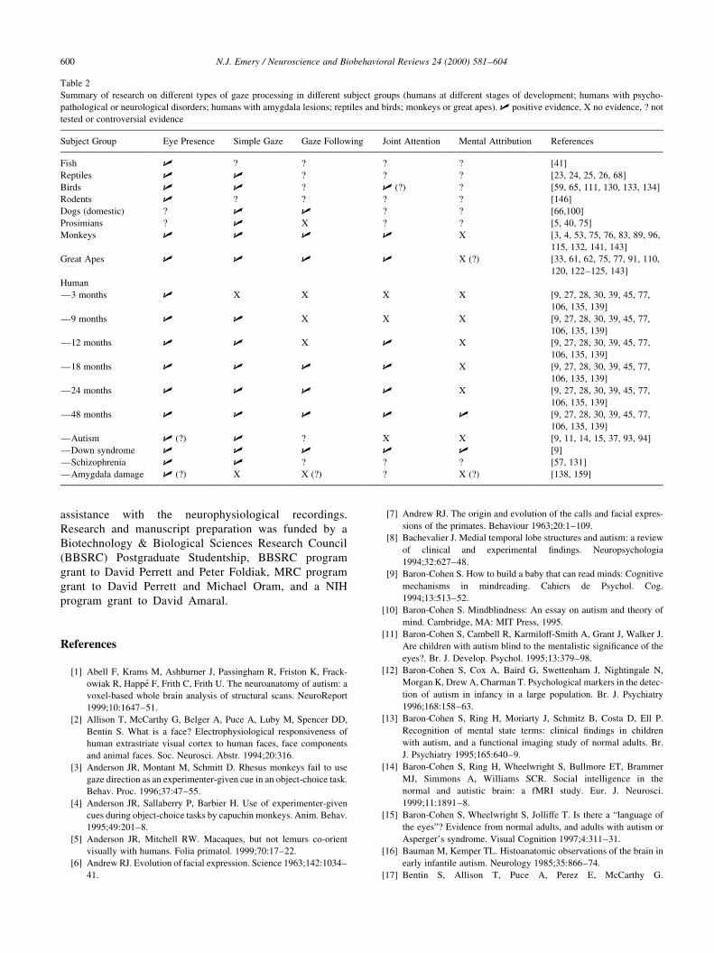
assistance with the neurophysiological recordings.
Research and manuscript preparation was funded by a
Biotechnology & Biological Sciences Research Council
(BBSRC) Postgraduate Studentship, BBSRC program
grant to David Perrett and Peter Foldiak, MRC program
grant to David Perrett and Michael Oram, and a NIH
program grant to David Amaral.
References
[1] Abell F, Krams M, Ashburner J, Passingham R, Friston K, Frack-
owiak R, Happe F, Frith C, Frith U. The neuroanatomy of autism: a
voxel-based whole brain analysis of structural scans. NeuroReport
1999;10:1647±51.
[2] Allison T, McCarthy G, Belger A, Puce A, Luby M, Spencer DD,
Bentin S. What is a face? Electrophysiological responsiveness of
human extrastriate visual cortex to human faces, face components
and animal faces. Soc. Neurosci. Abstr. 1994;20:316.
[3] Anderson JR, Montant M, Schmitt D. Rhesus monkeys fail to use
gaze direction as an experimenter-given cue in an object-choice task.
Behav. Proc. 1996;37:47±55.
[4] Anderson JR, Sallaberry P, Barbier H. Use of experimenter-given
cues during object-choice tasks by capuchin monkeys. Anim. Behav.
1995;49:201±8.
[5] Anderson JR, Mitchell RW. Macaques, but not lemurs co-orient
visually with humans. Folia primatol. 1999;70:17±22.
[6] Andrew RJ. Evolution of facial expression. Science 1963;142:1034±
41.
[7] Andrew RJ. The origin and evolution of the calls and facial expres-
sions of the primates. Behaviour 1963;20:1±109.
[8] Bachevalier J. Medial temporal lobe structures and autism: a review
of clinical and experimental ®ndings. Neuropsychologia
1994;32:627±48.
[9] Baron-Cohen S. How to build a baby that can read minds: Cognitive
mechanisms in mindreading. Cahiers de Psychol. Cog.
1994;13:513±52.
[10] Baron-Cohen S. Mindblindness: An essay on autism and theory of
mind. Cambridge, MA: MIT Press, 1995.
[11] Baron-Cohen S, Cambell R, Karmiloff-Smith A, Grant J, Walker J.
Are children with autism blind to the mentalistic signi®cance of the
eyes?. Br. J. Develop. Psychol. 1995;13:379±98.
[12] Baron-Cohen S, Cox A, Baird G, Swettenham J, Nightingale N,
Morgan K, Drew A, Charman T. Psychological markers in the detec-
tion of autism in infancy in a large population. Br. J. Psychiatry
1996;168:158±63.
[13] Baron-Cohen S, Ring H, Moriarty J, Schmitz B, Costa D, Ell P.
Recognition of mental state terms: clinical ®ndings in children
with autism, and a functional imaging study of normal adults. Br.
J. Psychiatry 1995;165:640±9.
[14] Baron-Cohen S, Ring H, Wheelwright S, Bullmore ET, Brammer
MJ, Simmons A, Williams SCR. Social intelligence in the
normal and autistic brain: a fMRI study. Eur. J. Neurosci.
1999;11:1891±8.
[15] Baron-Cohen S, Wheelwright S, Jolliffe T. Is there a ªlanguage of
the eyesº? Evidence from normal adults, and adults with autism or
Asperger's syndrome. Visual Cognition 1997;4:311±31.
[16] Bauman M, Kemper TL. Histoanatomic observations of the brain in
early infantile autism. Neurology 1985;35:866±74.
[17] Bentin S, Allison T, Puce A, Perez E, McCarthy G.
N.J. Emery / Neuroscience and Biobehavioral Reviews 24 (2000) 581±604600
Table 2
Summary of research on different types of gaze processing in different subject groups (humans at different stages of development; humans with psycho-
pathological or neurological disorders; humans with amygdala lesions; reptiles and birds; monkeys or great apes). U positive evidence, X no evidence, ? not
tested or controversial evidence
Subject Group Eye Presence Simple Gaze Gaze Following Joint Attention Mental Attribution References
Fish U ? ? ? ? [41]
Reptiles U U ? ? ? [23, 24, 25, 26, 68]
Birds U U ? U (?) ? [59, 65, 111, 130, 133, 134]
Rodents U ? ? ? ? [146]
Dogs (domestic) ? U U ? ? [66,100]
Prosimians ? U X ? ? [5, 40, 75]
Monkeys U U U U X [3, 4, 53, 75, 76, 83, 89, 96,
115, 132, 141, 143]
Great Apes U U U U X (?) [33, 61, 62, 75, 77, 91, 110,
120, 122±125, 143]
Human
Ð3 months U X X X X [9, 27, 28, 30, 39, 45, 77,
106, 135, 139]
Ð9 months U U X X X [9, 27, 28, 30, 39, 45, 77,
106, 135, 139]
Ð12 months U U X U X [9, 27, 28, 30, 39, 45, 77,
106, 135, 139]
Ð18 months U U U U X [9, 27, 28, 30, 39, 45, 77,
106, 135, 139]
Ð24 months U U U U X [9, 27, 28, 30, 39, 45, 77,
106, 135, 139]
Ð48 months U U U U U [9, 27, 28, 30, 39, 45, 77,
106, 135, 139]
ÐAutism U (?) U ? X X [9, 11, 14, 15, 37, 93, 94]
ÐDown syndrome U U U U U [9]
ÐSchizophrenia U U ? ? ? [57, 131]
ÐAmygdala damage U (?) X X (?) ? X (?) [138, 159]

Electrophysiological studies of face perception in humans. J.
Cog. Neurosci. 1997;8:551±65.
[18] Bertrand M. The behavioural repertoire of the stumptail macaque: a
descriptive and comparative study. Biblio. Primatol. 1969;11:1±
273.
[19] Bradbury JW, Vehrencamp SL. Principles of Animal Communica-
tion. Sunderland, MA: Sinauer Associates, Inc, 1998.
[20] Brothers L, Ring B. Medial temporal neurons in the macaque
monkey with responses selective for aspects of social stimuli.
Behav. Brain Res. 1993;57:53±61.
[21] Brothers L, Ring B, Kling A. Response of neurons in the macaque
amygdala to complex social stimuli. Behav. Brain Res.
1990;41:199±213.
[22] Bruce C, Desimone R, Gross CG. Visual properties of neurons in a
polysensory area in superior temporal sulcus of the macaque. J.
Neurophysiol. 1981;46:369±84.
[23] Burger J, Goch®eld M, Murray BG. Role of predator eye size in risk
perception by basking black iguanas (Ctenosaura similis). Anim.
Behav. 1991;42:471±6.
[24] Burger J, Gochfeld M, Murray BG. Risk discrimination of eye
contact and directness of approach in black iguanas (Ctenosaura
similis). J. Comp. Psych. 1992;106:97±101.
[25] Burghardt GM. Cognitive ethology and critical anthropomorphism:
a snake with two heads and hog-nosed snakes that play dead. In:
Ristau CA, editor. Cognitive Ethology: The minds of other animals,
Hillside, NJ: Lawrence Erlbaum, 1990.
[26] Burghardt GM, Greene HW. Predator simulation and duration of
death feigning in neonate hognose snakes. Anim. Behav.
1990;36:1842±4.
[27] Butterworth G. The ontogeny and phylogeny of joint visual atten-
tion. In: Whiten A, editor. Natural Theories of Mind, Oxford: Black-
well, 1991. p. 223±32.
[28] Butterworth G, Cochran E. Towards a mechanism of joint visual
attention in human infancy. Int. J. Behav. Develop. 1980;3:253±72.
[29] Butterworth G, Grover L. Joint visual attention, manual pointing and
preverbal communication in human infancy. In: Jeannerod M, editor.
Attention and Performance XIII, Hillside, NJ: Lawrence Erlbaum,
1989. p. 605±24.
[30] Butterworth G, Jarrett N. What minds have in common is space:
spatial mechanisms serving joint visual attention in infancy. Br. J.
Develop. Psychol. 1991;9:55±72.
[31] Call J, Tomasello M. The use of social information in the problem-
solving of orangutans (Pongo pygmaeus) and human children
(Homo sapiens). J. Comp. Psychol. 1995;109:308±20.
[32] Call J, Tomasello M. Distinguishing intentional from accidental
actions in orangutans (Pongo pygmaeus), chimpanzees (Pan troglo-
dytes) and human children (Homo sapiens). J. Comp. Psychol.
1998;112:192±6.
[33] Call J, Hare BA, Tomasello M. Chimpanzee gaze following in an
object-choice task. Anim. Cog. 1998;1:89±99.
[34] Campbell R, Heywood CA, Cowey A, Regard M, Landis T. Sensi-
tivity to eye gaze in prosopagnosic patients and monkeys with super-
ior temporal sulcus ablation. Neuropsychologia 1990;28:1123±42.
[35] Carpenter M, Tomasello M, Savage-Rumbaugh S. Joint attention
and imitative learning in children, chimpanzees and encultured
chimpanzees. Social Develop. 1995;4:217±37.
[36] Chance MRA. Attention structure as a basis of primate rank orders.
Man 1967;2:503±18.
[37] Charman T, Swettenham J, Baron-Cohen S, Cox A, Baird G, Drew
A. Infants with autism: An investigation of empathy, pretend play,
joint attention, and imitation. Develop. Psychol. 1997;33:781±9.
[38] Cheney DL, Seyfarth RM. How Monkeys See the World: Inside the
mind of another species. Chicago: University of Chicago Press,
1990.
[39] Corkum V, Moore C. The development of joint attention in infants.
In: Moore C, Dunham PJ, editors. Joint Attention: Its origins and
role in development, Hillsdale, NJ: Lawrence Erlbaum, 1995. p. 61±
83.
[40] Coss RG. Perceptual determinants of gaze aversion by the lesser
mouse lemur (Microcerbus murinus). The role of two facing eyes.
Behaviour 1978;64:248±67.
[41] Coss RG. Delayed plasticity of an instinct: recognition and avoid-
ance of 2 facing eyes by the jewel ®sh. Develop. Psychobiol.
1979;12:335±45.
[42] de Roiste A, Bonnet G. Maternal recognition of isolated newborn
physical features in the early post-partum period. J. Reprod. Infant
Psychol. 1996;14:293±8.
[43] de Waal FBM. Chimpanzee Politics: Power and sex among the apes.
Baltimore: John Hopkins University Press, 1982.
[44] de Waal FBM. Peacemaking Among Primates. Cambridge, MA:
Harvard University Press, 1989.
[45] D'Entremont B, Hains SMJ, Muir DW. A demonstration of gaze
following in 3- to 6-month-olds. Infant Behav. Develop.
1997;20:569±72.
[46] Desimone R, Albright TD, Gross CG, Bruce C. Stimulus-selective
properties of inferior temporal neurons in the macaque. J. Neurosci.
1984;4:2051±62.
[47] Dunham PJ, Dunham F, Curwin A. Joint attentional states and lexi-
cal acquisition at 18 months. Develop. Psychol. 1993;29:927±31.
[48] Eacott MJ, Heywood CA, Gross CG, Cowey A. Visual discrimina-
tion impairments following lesions of the superior temporal sulcus
are not speci®c for facial stimuli. Neuropsychologia 1993;31:609±
19.
[49] Edmunds M. Defence in Animals. Harlow, Essex: Longman, 1974.
[50] Eimer M. Does the face-speci®c N170 component re¯ect the activity
of a specialized eye processor?. NeuroReport 1998;9:2945±8.
[51] Emery, NJ. Neuroethological studies of primate social perception.
Unpublished doctoral dissertation, University of St Andrews, 1997.
[52] Emery NJ, Amaral DG. The role of the primate amygdala in social
cognition. In: Lane RD, Nadel L, editors. Cognitive Neuroscience of
Emotion, New York: Oxford University Press, 1999. p. 156±91.
[53] Emery NJ, Lorincz EN, Perrett DI, Oram MW, Baker CI. Gaze
following and joint attention in rhesus monkeys (Macaca mulatta).
J. Comp. Psychol. 1997;111:286±93.
[54] Emery NJ, Perrett DI. How can studies of the monkey brain help us
understand ªtheory of mindº and autism in humans?. In: Baron-
Cohen S, Tager-Flausberg H, Cohen DJ, editors. Understanding
Other Minds 2: Perspectives from developmental cognitive
neuroscience, Oxford: Oxford University Press, 1999. p. 279±310.
[55] Felleman DJ, Van Essen DC. Distributed hierarchical processing in
the primate cerebral cortex. Cerebral Cortex 1991;1:1±47.
[56] Fletcher PC, Happe F, Frith U, Baker SC, Dolan RJ, Frackowiak
RSJ, Frith CD. Other minds in the brain-a functional imaging study
of theory of mind in story comprehension. Cognition 1995;57:109±
28.
[57] Franck N, Daprati E, Michel F, Saoud M, DaleÂry J, Marie-Cardine
M, Georgieff N. Gaze discrimination is unimpaired in schizophrenia.
Psychiatry Res. 1998;81:67±75.
[58] Galdikas BMF. Re¯ections of Eden: My years with the orangutans
of Borneo. Boston: Little, Brown, 1995.
[59] Gallup GG, Cummings WH, Nash RF. The experimenter as an inde-
pendent variable in studies of animal hypnosis in chickens (Gallus
gallus) . Anim. Behav. 1972;20:166±9.
[60] Gautier J-P, Gautier A. Communication in Old World monkeys. In:
Sebeok TA, editor. How Animals Communicate, Bloomington: Indi-
ana University Press, 1977. p. 890±964.
[61] Gomez JC. The emergence of intentional communication as a
problem-solving strategy in the gorilla. In: Parker ST, Gibson KR,
editors. `Language' and Intelligence in Monkeys and Apes:
Comparative developmental approaches, Cambridge: Cambridge
University Press, 1990. p. 333±55.
[62] Gomez JC. Visual behavior as a window for reading the mind of
N.J. Emery / Neuroscience and Biobehavioral Reviews 24 (2000) 581±604 601

others in primates. In: Whiten A, editor. Natural Theories of Mind,
Oxford: Blackwell, 1991. p. 195±208.
[63] Green®eld PM, Savage-Rumbaugh ES. Grammatical combination in
Pan paniscus: processes of learning and invention in the evolution
and development of language. In: Parker ST, Gibson KR, editors.
`Language' and Intelligence in Monkeys and Apes: Comparative
development perspectives, New York & Cambridge: Cambridge
University Press, 1990. p. 540±78.
[64] Haith MM, Bergman T, Moore MJ. Eye contact and face scanning in
early infancy. Science 1977;198:853±5.
[65] Hampton RR. Sensitivity to information specifying the line of gaze
of humans in sparrows (Passer domesticus). Behaviour
1994;130:41±51.
[66] Hare B, Tomasello M. Domestic dogs (Canis familiaris) use human
and conspeci®c social cues to locate hidden food. J. Comp. Psychol.
1999;113:173±7.
[67] Harries MH, Perrett DI. Visual processing of faces in temporal
cortex: physiological evidence for a modular organization and possi-
ble anatomical correlates. J. Cog. Neurosci. 1991;3:9±24.
[68] Hennig CW. Effect of simulated predation on tonic immobility in
Anolis carolinensis: The role of eye contact. Bull. Psychon. Soc.
1977;9:239±42.
[69] Heyes CM. Theory of mind in nonhuman primates. Behav. Brain
Sci. 1998;21:101±48.
[70] Heywood CA, Cowey A. The role of the ªface-cellº area in the
discrimination and recognition of faces by monkeys. Phil. Trans.
Roy. Soc. Lond.: Biol. Sci. 1992;335:31±38.
[71] Hinde RA, Rowell TE. Communication by postures and facial
expression in the rhesus monkey (Macaca mulatta). Symp. Zool.
Soc. Lond. 1961;8:1±21.
[72] Hoffman EA, Haxby JV. Distinct representations of eye gaze and
identity in the distributed human neural system for face perception.
Nature Neurosci. 2000;3:80±84.
[73] Huber E. Evolution of Facial Musculature and Facial Expression.
Baltimore: John Hopkins Press, 1931.
[74] Huber E. The facial musculature and its innervation. In: Hartman
CG, Straus WL, editors. Anatomy of the Rhesus Monkey (Macaca
mulatta), New York: Hafner, 1961. p. 176±88.
[75] Itakura S. An exploratory study of gaze-monitoring in non-human
primates. Jap. Psychol. Res. 1996;38:174±80.
[76] Itakura S, Anderson JR. Learning to use experimenter-given cues
during an object-choice task by a capuchin monkey. Cahiers de
Psychol. Cog. 1996;15:103±12.
[77] Itakura S, Tanaka M. Use of experimenter-given cues during object-
choice tasks by chimpanzees (Pan troglodytes), an orangutan
(Pongo pygmaeus), and human infants (Homo sapiens). J. Comp.
Psych. 1998;112:119±26.
[78] Jolly A. The Evolution of Primate Behavior. New York: Macmillan,
1972.
[79] Jones RB. Reactions of male domestic chicks to two-dimensional
eye-like shapes. Anim. Behav. 1980;28:212±8.
[80] Jongen-Relo AL, Amaral DG. Evidence for a GABAergic projection
from the central nucleus of the amygdala to the brainstem of the
macaque monkey: a combined retrograde tracing and in situ hybri-
dization study. Eur. J. Neurosci. 1998;10:2924±33.
[81] Kanner L. Autistic disturbances of affective contact. Nervous Child
1943;2:217±50.
[82] Kawashima R, Sugiura M, Kato T, Nakamura A, Hatano K, Ito K,
Fukuda H, Kojima S, Nakamura K. The human amygdala plays and
important role in gaze monitoring: a PET study. Brain
1999;122:779±83.
[83] Keating CF, Keating EG. Visual scan patterns of rhesus monkeys
viewing faces. Perception 1982;11:211±9.
[84] Kingdon JS. The role of visual signals and face patterns in African
forest monkeys (guenons) of the genus Cercopithecus. Trans. Zool.
Soc. Lond. 1980;35:425±75.
[85] Kobayashi H, Kohshima S. Morphological uniqueness of human
eyes and its adaptive meaning. Nature 1997;387:767±8.
[86] Kohler W. The Mentality of Apes. London: Routledge & Kegan
Paul, 1925.
[87] Kummer H. Tripartite relations in hamadryas baboons. In: Altmann
S, editor. Social Communication Among Primates, Chicago: Univer-
sity of Chicago Press, 1967. p. 63±71.
[88] Kummer H, Anzenberger G, Hemlrijk CK. Hiding and perspective-
taking in long-tailed macaques (Macaca fascicularis). J. Comp.
Psychol. 1996;110:97±102.
[89] Kyes RC, Candland DK. Baboon (Papio hamadryas) visual prefer-
ences for regions of the face. J. Comp. Psychol. 1987;101:345±8.
[90] Langton SRH, Watt RJ, Bruce V. Do the eyes have it? Cues to the
direction of social attention. Trends Cog. Sci. 2000;4:50±59.
[91] Leavens DA, Hopkins WD, Bard KA. Indexical and referential
pointing in chimpanzees (Pan troglodytes). J. Comp. Psychol.
1996;110:346±53.
[92] LeDoux JE. The Emotional Brain: The mysterious underpinnings of
emotional life. New York: Simon & Schuster, 1996.
[93] Leekam S, Baron-Cohen S, Perrett DI, Milders M, Brown S. Eye-
direction detection: a dissociation between geometric and joint
attention skills in autism . Br. J. Develop. Psychol. 1997;15:77±95.
[94] Leekham SR, Hunnisett E, Moore C. Targets and cues: gaze follow-
ing in children with autism. J. Child Psychol. Psychiatry
1998;39:951±62.
[95] Linannkoski I, Gronroos M, Pertovaara A. Eye contact as a trigger of
male sexual arousal in stump-tailed macaques (Macaca arctoides).
Folia primatol. 1993;60:181±4.
[96] Lorincz EN, Baker CI, Perrett DI. Visual cues for attention following
in rhesus monkeys. Cahiers de Psychol. Cog. 1999;18:973±1003.
[97] McKelvie SJ. The role of eyes and mouth in the memory of a face.
Am. J. Psychol. 1976;89:311±23.
[98] McNelis NL, Boatright-Horowitz SL. Social monitoring in a primate
group: the relationship between visual attention and hierarchical
ranks. Anim. Cog. 1998;1:65±69.
[99] Mendelson MJ, Haith MM, Goldman-Rakic PS. Face scanning and
responsiveness to social cues in infant rhesus monkeys. Develop.
Psychol. 1982;18:222±8.
[100] Miklosi A, Polgardi R, Topal J, Csanyi V. Use of experimenter-
given cues in dogs. Anim. Cog. 1998;1:113±21.
[101] Mineka S, Davidson M, Cook M, Keir R. Observational condition-
ing of snake fear in rhesus monkeys . J. Abnorm. Psych.
1984;93:355±72.
[102] Mistlin, AJ. Neural mechanisms underlying the perception of
socially relevant stimuli in the macaque monkey. Unpublished
doctoral thesis, University of St Andrews, 1988.
[103] Morissette P, Ricard M, Decarie TG. Joint visual attention and point-
ing in infancy: a longitudinal study of comprehension. Br. J.
Develop. Psychol. 1995;13:163±75.
[104] Morris D. The Naked Ape. London: Triad Grafton Book, 1967.
[105] Mundy P, Sigman M, Ungerer J, Sherman T. De®ning the social
de®cits of autism: the contribution of non-verbal communication
measures. J. Child Psych. Psychiatry 1986;27:657±69.
[106] Mundy P, Gomes A. Individual differences in joint attention skill
development in the second year. Infant Behav. Develop.
1998;21:469±82.
[107] Nahm FKD, Perret A, Amaral DG, Albright TD. How do monkeys
look at faces?. J. Cog. Neurosci. 1997;9:611±23.
[108] Nichols K, Champness B. Eye gaze and the GSR. J. Exp. Soc.
Psychol. 1971;7:623±6.
[109] Packer C. Reciprocal altruism in Papio anubis. Nature
1977;265:441±3.
[110] Peignot P, Anderson JR. Use of experimenter-given manual and
facial cues by gorillas (Gorilla gorilla) in an object-choice task. J.
Comp. Psychol. 1999;113:253±60.
[111] Pepperberg IM, McLaughlin MA. Effect of avian-human joint
N.J. Emery / Neuroscience and Biobehavioral Reviews 24 (2000) 581±604602

attention in allospeci®c vocal learning by grey parrots (Psittacus
erithacus). J. Comp. Psychol. 1996;110:286±97.
[112] Perrett DI, Emery NJ. Understanding the intentions of others from
visual signals: neurophysiological evidence. Cahiers de Psychol.
Cog. 1994;13:683±94.
[113] Perrett DI, Harries MH, Mistlin AJ, Hietanen JK, Bevan R, Thomas
S, Ortega JO, Oram MW, Brierly K. Social signals analyzed at the
single cell level: someone's looking at me, something touched me,
something moved!. Int. J. Comp. Psychol. 1990;4:25±55.
[114] Perrett DI, Hietanen JK, Oram MW, Benson PJ. Organization and
functions of cells responsive to faces in the temporal cortex. Phil.
Trans. Roy. Soc. Lond.: Biol. Sci. 1992;335:23±30.
[115] Perrett DI, Mistlin AJ. Perception of facial characteristics by
monkeys. In: Stebbins WC, Berkley MA, editors. Comparative
Perception, New York: John Wiley, 1991. p. 187±215.
[116] Perrett DI, Oram MW, Harries MH, Bevan R, Hietanen JK, Benson
PJ, Thomas S. Viewer-centred and object-centred coding of heads in
the macaque temporal cortex. Exp. Brain Res. 1991;86:159±73.
[117] Perrett DI, Rolls ET, Caan W. Visual neurons responsive to faces in
the monkey temporal cortex. Exp. Brain Res. 1982;47:329±42.
[118] Perrett DI, Smith PAJ, Potter DD, Mistlin AJ, Head AS, Milner AD,
Jeeves MA. Visual cells in the temporal cortex sensitive to face view
and gaze direction. Proc. Roy. Soc. Lond.: Biol. Sci. 1985;223:293±
317.
[119] Pettigrew L, Forsyth H, Perrett DI. Training rhesus monkeys to use
human head and gaze direction in a visual discrimination task.
Unpublished manuscript, 1993.
[120] Povinelli DJ, Bierschwale DT, Reaux JE, Cech CG. Do juvenile
chimpanzees understand attention as a mental state? J. Comp.
Psychol. 1999 in press.
[121] Povinelli DJ, Eddy TJ. The eyes as a window: what young chim-
panzees see on the other side. Cahiers de Psychol. Cog.
1994;13:695±705.
[122] Povinelli DJ, Eddy TJ. Factors in¯uencing young chimpanzees'
(Pan troglodytes) recognition of attention. J. Comp. Psychol.
1996;110:336±45.
[123] Povinelli DJ, Eddy TJ. Chimpanzees: joint visual attention. Psychol.
Sci. 1996;7:129±35.
[124] Povinelli DJ, Eddy TJ. What young chimpanzees know about seeing.
Mono. Soc. Res. Child Develop. 1996;61:247.
[125] Povinelli DJ, Eddy TJ. Speci®city of gaze-following in young chim-
panzees. Br. J. Develop. Psychol. 1997;15:213±22.
[126] Puce A, Allison T, Gore JC, McCarthy G. Face-sensitive regions in
human extrastriate cortex studied by functional MRI. J. Neurophy-
siol. 1995;74:1192±9.
[127] Puce A, Allison T, Bentin S, Gore JC, McCarthy G. Temporal cortex
activation in humans viewing eye and mouth movements. J.
Neurosci. 1998;18:2188±99.
[128] Reaux JE, Theall LA, Povinelli DJ. A longitudinal investigation of
chimpanzee's understanding of visual perception. Child Develop.
1999;70:275±90.
[129] Redican WK. Facial expressions in nonhuman primates. In: Rosen-
blum LA, editor. Primate Behavior: Developments in ®eld and
laboratory research, New York: Academic Press, 1975. p. 104±94.
[130] Ristau CA. Before mindreading: attention, purposes and deception
in birds?. In: Whiten A, editor. Natural Theories of Mind, Oxford:
Blackwell, 1991. p. 209±22.
[131] Rosse RB, Kendrick K, Wyatt RJ, Isaac A, Deutsch SI. Gaze discri-
mination in patients with schizophrenia: preliminary report. Am. J.
Psychiatry 1994;151:919±21.
[132] Santos LR, Hauser MD. How monkeys see the eyes: cotton-top
tamarins' reaction to changes in visual attention and action. Anim.
Cog. 1999;2:131±9.
[133] Scaife M. The response to eye-like shapes by birds. I: the effect of
context: a predator and a strange bird. Anim. Behav. 1976;24:195±9.
[134] Scaife M. The response to eye-like shapes by birds. II: the impor-
tance of staring, pairedness and shape. Anim. Behav. 1976;24:200±
6.
[135] Scaife M, Bruner JS. The capacity for joint visual attention in the
infant. Nature 1975;253:265±6.
[136] Sigg H, Stolba A. Home range and daily march in a Hamadryas
baboon troop. Folia primatol. 1981;36:40±75.
[137] Stone VE, Baron-Cohen S, Knight RI. Frontal lobe contributions to
theory of mind. J. Cog. Neurosci. 1998;10:640±56.
[138] Stone VE, Baron-Cohen S, Young AW, Calder A, Keane J. Impair-
ments in social cognition following orbitofrontal or amygdala
damage. Soc. Neurosci. Abstr. 1998;24:1176.
[139] Symons LA, Hains SMJ, Muir DW. Look at me: Five-month-old
infants' sensitivity to very small deviations in eye-gaze during social
interactions. Infant Behav. Develop. 1998;21:531±6.
[140] Theall LA, Povinelli DJ. Do chimpanzees tailor their gestural signals
to ®t the attentional states of others?. Anim. Cog. 1999;2:207±14.
[141] Thomsen CE. Eye contact by non-human primates toward a human
observer. Anim. Behav. 1974;22:144±9.
[142] Tomasello M, Call J. Primate Cognition. New York: Oxford Univer-
sity Press, 1997.
[143] Tomasello M, Call J, Hare B. Five primate species follow the visual
gaze of conspeci®cs. Anim. Behav. 1997;55:1063±9.
[144] Tomasello M, Farrar MY. Joint attention and early language. Child
Develop. 1986;57:1454±63.
[145] Tomasello M, Hare B, Agnetta B. Chimpanzees, Pan troglodytes,
follow gaze direction geometrically. Anim. Behav. 1999;58:769±77.
[146] Topal J, Csanyi V. The effect of eye-like schema on shuttling activ-
ity of wild house mice (Mus musculus domesticus): Context-depen-
dent threatening aspects of the eyespot patterns. Anim. Learn.
Behav. 1994;22:96±102.
[147] van Hoof JARAM. The facial displays of the catarrhine monkeys
and apes. In: Morris D, editor. Primate Ethology, London: Weiden-
feld & Nicholson, 1967. p. 7±68.
[148] Wachsmuth E, Oram MW, Perrett DI. Recognition of objects and
their component parts: responses of single units in the temporal
cortex of the macaque. Cerebral Cortex 1994;5:509±22.
[149] Wada JA. Modi®cation of cortically induced responses in brain stem
of shift of attention in monkeys. Science 1961;133:40±2.
[150] Walters JR, Seyfarth RM. Con¯ict and co-operation. In: Smuts BB,
Cheney DL, Seyfarth RM, Wrangham RW, Struhsaker TT, editors.
Primate Societies, Chicago: University of Chicago Press, 1987. p.
306±17.
[151] Whiten A. When does smart behavior-reading become mind-read-
ing? In: Carruthers P, Smith PK, editors. Theories of Theories
of Mind, Cambridge: Cambridge University Press, 1996. p.
277±92.
[152] Whiten A. Imitation, pretence and mindreading: secondary repre-
sentation in comparative primatology and developmental psychol-
ogy?. In: Russon AE, Parker ST, Bard KA, editors. Reaching Into
Thought, Cambridge: Cambridge University Press, 1996. p. 300±24.
[153] Whiten A. Machiavellian Intelligence II: Evaluations and exten-
sions. In: Whiten A, Byrne RW, editors. Cambridge: Cambridge
University Press, 1997. p. 144±73.
[154] Whiten A. Evolutionary and developmental origins of the mindread-
ing system. In: Langer J, Killen M, editors. Piaget, Evolution and
Development, London: Lawrence Erlbaum, 1997.
[155] Whiten A, Byrne RW. The manipulation of attention in primate
tactical deception. In: Byrne RW, Whiten A, editors. Machiavellian
Intelligence: Social expertise and the evolution of intellect in
monkeys, apes and humans, Oxford: Oxford University Press,
1988. p. 211±23.
[156] Whiten A, Byrne RW. Tactical deception in primates. Behav. Brain
Sci. 1988;11:233±73.
[157] Wicker B, Michel F, Henaff M, Decety J. Brain regions
involved in the perception of gaze: A PET study. Neuroimage
1998;8:221±7.
[158] Yamane S, Kaji S, Kawano K. What facial features activate face
N.J. Emery / Neuroscience and Biobehavioral Reviews 24 (2000) 581±604 603

neurons in the inferotemporal cortex of the monkey?. Exp. Brain
Res. 1988;73:209±14.
[159] Young AW, Aggleton JP, Hellawell DJ, Johnson M, Broks P, Hanley
JR. Face processing impairments after amygdalectomy. Brain
1995;118:15±24.
[160] Zahavi A, Zahavi A. The Handicap Principle: A missing piece of
Darwin's puzzle. Oxford: Oxford University Press, 1997.
N.J. Emery / Neuroscience and Biobehavioral Reviews 24 (2000) 581±604604


![[PPT]PowerPoint Presentation - · Web view... (B – non-blue eyes, b – blue eyes) BB = non-blue eyes Bb = non-blue eyes bb = blue eyes Incomplete Dominance – Neither allele is](https://img.dokumen.tips/doc/110x75/5aae9c247f8b9a190d8c559f/pptpowerpoint-presentation-view-b-non-blue-eyes-b-blue-eyes-bb.jpg)