Embed Size (px)
Citation preview

The Extracellular Matrix ofXenopus laevis Eggs: A Quick-Freeze, Deep-Etch Analysis of Its Modification at Fertilization C a r o l y n A. Larabe l l a n d D o u g l a s E. C h a n d l e r
Department of Zoology, Arizona State University, Tempe, Arizona 85287
Abstract. Eggs of the amphibian, Xenopus laevis, were quick-frozen, deep-etched, and rotary-shadowed. The structure of the extracellular matrix surrounding these eggs, including the perivitelline space and the vitelline envelope (VE), was visualized in platinum replicas by electron microscopy. The perivitelline space contains an elaborate filamentous glycocalyx which connects microvillar tips to the plasma mem- brane, to adjacent microvilli, and to the overlying VE. The VE is comprised of two layers, the innermost of which is a thin network of horizontal fibrils lying on the tips of the microvilli. The outermost is a thicker layer of large, cable-like fibers which twist and turn
throughout the envelope. Upon fertilization, three dra- matic modifications of the matrix occur. A thin sheet of smooth material, termed the smooth layer, is depos- ited on the tips of the microviUi and separates the egg from the overlying envelopes. The VE above is trans- formed from a thick band of cable-like fibers to con- centric fibrous sheets, the altered VE. Finally, an ornate band of particles, corresponding to the fertiliza- tion layer in previous studies, is deposited at the al- tered VE/jelly interface. The altered VE and the fertil- ization layer comprise the fertilization envelope, which effects the structural block to polyspermy.
M ATURE eggs of many species exhibit a highly or- ganized extracellular matrix (ECM) ~ that is se- curely attached to the cell surface. These matrices
(e.g., the vitelline layer in sea urchin eggs and the zona pel- lucida in mammalian eggs) are filamentous in structure and contain glycoprotein sperm receptors which facilitate sperm binding (Glabe and Vacquier, 1978; Bleil and Wassarman, 1980; Rossignol et al., 1984). Upon fertilization, cortical granule secretions interact with these egg investments to pro- duce a chemically and structurally altered matrix which pre- vents further sperm entry and the possibility of abnormal de- velopment (Barros and Yanagimachi, 1971, 1972; Grey et al., 1974, 1976; Veron et al., 1977; Chandler and Heuser, 1980; Gerton and Hedrick, 1986). Thus, eggs represent an excellent model for analyzing ECM structural dynamics that are paralleled by a change in biological function.
The ECM of amphibians has been best studied in the South African clawed frog, Xenopus laevis. The unfertilized Xeno- pus egg is surrounded by the perivitelline space (PS) and vitelline envelope (VE). The PS, as seen in thin sections, is a 1-1xm-thick area between the cell surface and overlying VE and contains a glycocalyx-like, filamentous material as well
Carolyn A. Larabell's present address is Hopkins Marine Station, Stanford University, Pacific Grove, CA 93950.
1. Abbreviations used in thispaper: ECM, extracellular matrix; F layer, fer- tilization layer; Ji, innermost jelly coat; MV, microvilli; PS, perivitelline space; S layer, smooth layer; VE, vitelline envelope.
as clusters of small, membrane-bound vesicles (Grey et al., 1974). The VE, also ~ol lain thick, is a loose network of fila- ments ,,04-7 nm in diameter arranged in alternating layers or bundles (Grey et al., 1974). The VE consists of 85 % protein and 15 % sugar (Wolf et al., 1976), and gel electrophoresis reveals seven proteins with estimated molecular masses of 120, 112, 69, 64, 57, 41, and 37 kD, all of which are glycosy- lated except for the 57-kD protein (Gerton and Hedrick, 1986).
Fusion of a single sperm with the egg plasma membrane initiates a wave of cortical granule exocytosis which begins at the site of sperm entry and propagates around the entire egg (Grey et al., 1974; Hara and Tydeman, 1979). Contents of the cortical granules, including enzymes and structural proteins, are released into the PS where they interact with the ECM to produce a functionally altered fertilization enve- lope (Grey et al., 1974; Wolf, 1974; Gerton and Hedrick, 1986). These alterations are complete by ,,o15-20 min after sperm penetration and result in a structural block to poly- spermy (Grey et al., 1976).
Two major biochemical modifications in the ECM of Xenopus eggs at fertilization have been described. The first change is the formation of an electron-dense fertilization (F) layer at the interface of the VE and the innermost jelly coat (J1) (Grey et al., 1974; Wolf, 1974). This layer is formed from the calcium-dependent precipitation of a cortical gran- ule lectin with components of J1 (Wyrick et al., 1974; Greve and Hedrick, 1978). The second modification in the matrix
© The Rockefeller University Press, 0021-9525/88/08/731/I 1 $2.00 The Journal of Cell Biology, Volume 107, August 1988 731-741 731
on July 8, 2017jcb.rupress.org
Dow
nloaded from
is a decrease in molecular mass of two glycoproteins in the VE (69 to 66 kD and 64 to 61 kD) due to a limited hydrolysis reaction (Gerton and Hedrick, 1986). Thin-section EM, however, has been unable to visualize significant ultrastruc- tural changes accompanying this modification (Grey et al., 1974).
Here we report the three-dimensional organization of the ECM in unfertilized and fertilized Xenopus eggs as seen in platinum replicas of quick-frozen and deep-etched speci- mens. Using this technique, we demonstrate in great detail the multilayered nature of these extracellular investments and their structural modification at fertilization. In addition, we describe the structure of the smooth (S) layer, a complex surface coat surrounding the fertilized egg which has not been previously visualized.
Materials and Methods
Egg Procurement Adult female Xenopus were purchased from Nasco Biological Supply Co. (Fort Atkinson, Wl) and raised on a 12-h light/12-h dark cycle in recircu- lated, filtered 10% Holtfreter's solution. Females were injected with 900 IU human chorionic gonadotropin and eggs were collected 8-9 h later by strip-
ping them from the frog. Eggs were stored in 1.5x O-R2 buffer (123.8 mM NaCI, 3.8 mM KCI, 1.5 mM CaCI2, 1.5 mM MgCI2, 1.5 mM Na2HPO4, 5.7 mM NaOH, 7.5 mM Hepes, pH 7.8; Hollinger and Corton, 1980) until used (maximum storage time was I h). Males were killed and testes re- moved, washed, and then macerated in 1.5x O-R2. Sperm were collected and stored in fresh 1.5x O-R2 until needed (maximum storage time was 4 h). Fertilization was carried out by addition of 100 p.l of concentrated sperm to 6 ml of F-1 solution (41.25 mM NaCI, 1.25 mM KCI, 0.25 mM CaCI2, 0.06 mM MgC12, 0.5 mM Na2HPO4, 1.9 mM NaOH, 2.5 mM Hepes, pH 7.8; Hollinger and Corton, 1980) containing 20-40 eggs; the final sperm concentration was 105-106 cells/ml (Hollinger and Cotton, 1980). Success of fertilization was determined by allowing periodic samples to proceed to first cleavage.
Sample Preparation and EM Both fixed and unfixed samples were used for quick-freezing. Samples that were fixed were incubated overnight at 4°C in 2.5% glutaraldehyde (dejel- lied eggs) or 5% glutaraldehyde (jellied eggs) in 45 mM s-collidine buffer, pH 7.6. To compare our results with those obtained previously by thin sec- tioning, some glutaraldehyde-fixed samples were subsequently washed in buffer (three times) and fixed in 1% OsO4 in s-collidine buffer for 1 h. Unfixed and glutaraldehyde-fixed samples demonstrated comparable views, with only a slight decoration of filaments observed in fixed samples. Sam- ples fixed in both glutaraldehyde and OsO4 provided excellent views of the F layer but did not reveal the S layer. Presumably this layer must be osmium sensitive. All samples were dejellied before quick-freezing. Unfixed eggs and zygotes were chemically dejellied using 45 mM I~-mercaptoethanol in F-I, pH 9.3, for 3-5 min or until all jelly visible in the dissecting microscope
Figure 1. Electron micrograph of a platinum replica of an unfertilized Xenopus egg showing egg cortex, with large cortical granule (CG) just beneath the plasma membrane (PM), and overlying ECM. The ECM is composed of the PS and VE. The specimen was fixed in glutaraldehyde, mechanically dejellied, passed through distilled water, quick-frozen, freeze-fractured, deep-etched, and rotary-shadowed with plat inum-carbon. This figure and all subsequent figures have been photographically reversed; platinum deposits appear white. Bar, 0.2 ~m.
The Journal of Cell Biology, Volume 107, 1988 732
on July 8, 2017jcb.rupress.org
Dow
nloaded from
had been removed. Fixed samples were mechanically dejellied using Dumont No. 5 forceps. Excess salts were removed from fixed samples be- fore quick-freezing by rinsing samples in F-I solution (once) then in distilled H20 (three times). Unfixed samples were rinsed in 0.5x F-I to remove ex- cess salts without exposing the living samples to osmotic stress. All speci- mens were quick-frozen on a liquid helium-cooled copper block using the
apparatus designed by Heuser et al. (1979). Quick-frozen samples were then fractured in a Freeze-Etch Unit (model 400D, Balzers Corp., Hudson, NH) and allowed to etch at a temperature of -90"C for 8-12 min at a pressure of 10 -6 mbar. The specimen was then rotary-shadowed with platinum-car- bon applied from an electron gun positioned at 25 ° and the replica backed with carbon applied from an angle of 75". Replicas were cleaned in 5% so-
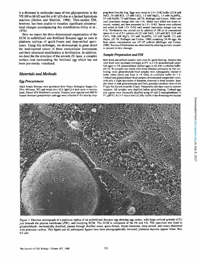
Figure 2. Oblique fracture through the PS of an unfertilized egg with exposed plasma membrane at the bottom of the micrograph. MV protruding from the plasma membrane have been fractured open at increasingly higher levels from bottom to top. Oblique fibers (arrows) can be seen extending from the MV to the egg surface as well as to adjacent MV; short, fine filaments (arrowheads) interconnect these fibers. Intermediate-sized fibrils (asterisks) are seen running between particles on the plasma membrane. Specimen was prepared as described in Fig. 1. Stereo pair shown. Bar, 0.1 pro.
Larabell and Chandler Extracellular Matrix of Xenopus Eggs 733
on July 8, 2017jcb.rupress.org
Dow
nloaded from
dium hypochlorite (60°C) for 1 h, followed by 70% sulfuric acid (60°C) for I h, and then 5 % sodium hypochlorite again (600C) for 1 h, With a rinse in distilled H20 between each transfer. Replicas were viewed at 80 kV in a microscope (model EM 300, Philips Electronic Instruments, Inc., Mahwah, NJ). Stereo images were obtained using a eucentric goniometer stage; the specimen was tilted +9 ° . All figures are presented as negative images so that platinum deposits appear white. Platinum replica thickness averaging 1.6 nm is included in the measurements of filament diameter. These are reported as means + SD in the results section.
Resu l t s
Replicas of quick-frozen, deep-etched Xenopus eggs provide a three-dimensional view of the ECM before and after fertil- ization. A cross fracture through the cortex of an unfertilized Xenopus egg illustrates the matrix, including the PS and VE, attached to the egg surface (Fig. 1). A large cortical granule lies just under the plasma membrane. The PS between the plasma membrane and the overlying VE is filled with a com- plex network of filaments. This network is visualized best in stereo micrographs of grazing fractures. In Fig. 2, an oblique fracture has exposed the external surface of the plasma mem- brane (at the bottom of the micrograph) and microvilli (MV) protruding into the PS. From the bottom of the figure to the top, MV have been cross fractured at increasingly higher lev- els revealing the network of filaments within. A complex glycocalyx lies within the extracellular space and intercon- nects these MV with at least three populations of fibers. The first and largest population consists of oblique fibers which connect microvillar tips with the plasma membrane below,
almost appearing like "guy wires" (arrow, Fig. 2). These fibers also run from one microvillar tip to another and join other guy wires midcourse. Oblique fibers are heteroge- neous in size, ranging from 6.5 to 16 nm in diameter (10.4 + 2.7 nm; mean + SD, n = 55). A second population of fibers in the PS consists of short, fine filaments (4.0 + 0.6 nm in diameter, n = 35) which interconnect the oblique fibers described above (arrowheads, Fig. 2). These are not as abundant, but are consistently observed. Finally, a third population of short fibrils (9.6 + 1 nm in diameter, n = 35) interconnects small particles located on the external surface of the plasma membrane (asterisk, Fig. 2). These three populations form an elaborate network which fills the en- tire PS.
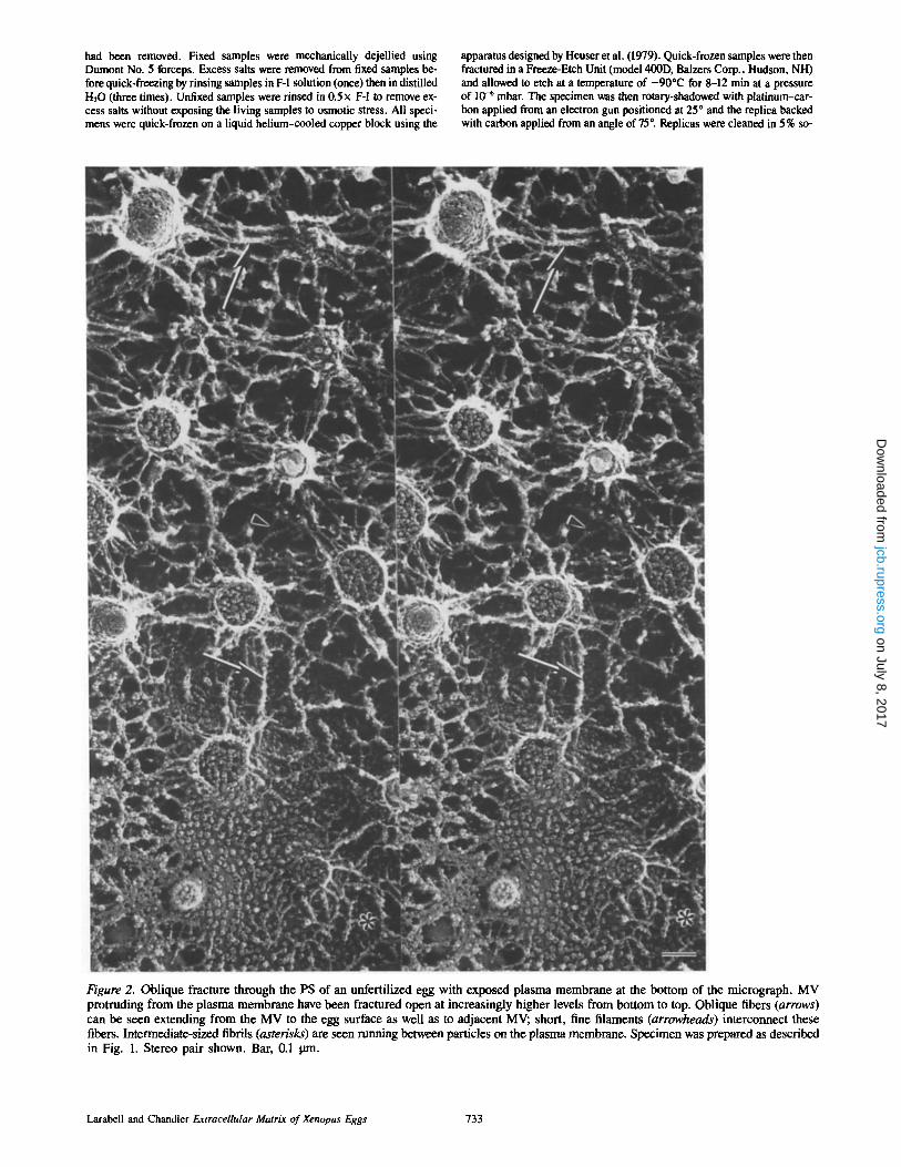
Numerous membrane-bound vesicles, ranging from 40 to 110 nm in diameter (60 5- 13 nm; mean + SD, n = 60), are also seen in the PS just under the VE (Fig. 3). Each vesi- cle is in contact with several of the fibrils coursing through the PS, making them an integral component of the filamen- tous network. Vesicles, when fractured, reveal that they are membrane bound and are filled with a globular material. The fibrils of the PS network, which interconnect the vesicles and MV, are continuous with the fibers of the overlying VE (Fig. 3).
The VE, at its innermost surface, is attached to the tips of the MV via a layer of horizontal filaments (HF, Fig, 3). The horizontal filaments that orbit the egg at the PS/VE interface represent an ultrastructurally distinct sublayer within the VE. This layer is composed of tightly packed fibrils (10 +
Figure 3. Oblique fracture o f the junct ion between the PS and overlying VE. The PS contains numerous membrane-bound vesicles (v), each of which is contacted by several fibrils (arrowheads). The inner port ion of the VE consists o f a dense meshwork o f horizontal illa- ments (HF) which are interconnected by short thin filaments (small arrows). The tips of MV (asterisks), seen just beneath the VE, are attached to the horizontal filament fibrils (large arrows). Specimen was prepared as described in Fig. 1. Bar, 0.1 ltm.
The Journal of Cell Biology, Volume 107, 1988 734
on July 8, 2017jcb.rupress.org
Dow
nloaded from

1.3 nm in diameter, n = 60) which are highly intercon- nected. Linking these fibrils are short, thin filaments ~4.6 ± 0.5 nm (n = 35) in diameter.
The remainder of the VE is of a clearly different architec- ture. Its structure is dominated by large, cable-like fibers (19 ± 1.6 nm in diameter, n = 35) which run throughout the envelope, often in a swirling fashion. Individual fibers can be followed for long distances as they twist and turn, often converging to form bundles (asterisk, Fig. 4) as well as bifur- cating into smaller fibrils. A second population of fibrils, in- termediate in size (12 + 1.6 nm in diameter, n = 40), are also observed coursing throughout the VE in a tangled fash- ion. These fibrils often bifurcate into two fibrils which then proceed in different directions. These two major sets of cable-like fibers are cross-linked by fine, short filaments (5.1 ± 0.7 nm in diameter, n = 35) at random locations.
The ECM of the unfertilized egg is dramatically altered af- ter fertilization in three distinct ways: formation of a smooth, multilayered envelope at the PS/VE interface, conversion of the VE to an altered VE, and formation of the F layer at the altered VE/J1 interface. All three modifications appear to re-
suit from interactions of the VE and jelly coat with enzymes and structural material released by cortical granule exocyto- sis. Two of the cortical granule enzymes are believed to be proteases responsible for the cleavage of the 69- and 64-kD components of the VE to the 66- and 61-kD components of the altered VE (Gerton and Hedrick, 1986). In addition, a large metalloglycoprotein (a 539/655-kD doublet) released from the cortical granules binds at the VE/J1 interface in a lectin/ligand reaction to form a band of precipitation, the F layer (Grey et al., 1974; Wolf, 1974; Wyrick, et al., 1974; Greve and Hedrick, 1978).
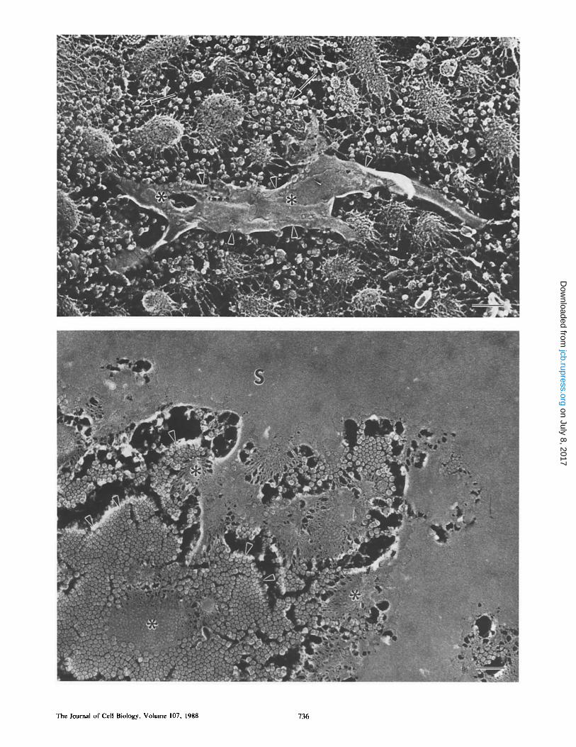
Deposition of an S layer of material on the tips of the MV at the PS/VE interface (Figs. 5-7) is seen within 10 min after insemination. This structure is deposited in a patchwork fashion at early times (Fig. 5) and by 15 rain after insemina- tion, the entire egg is surrounded by a very thin, multilami- nar envelope. The extremely smooth outer surface of this in- vestment suggests our designation: the S layer. The S layer is actually composed of two tightly apposed sheets which can be clearly distinguished at higher magnification. The inner- most sheet (FS, Fig. 7) manifests a fibrous nature and splays
Figure 4. The VE of the unfertilized egg (outer portion). Large cable-like fibers (thick arrows) run for long distances and occasionally form bundles (asterisk). Intermediate-sized fibrils also course throughout the VE, frequently bifurcating (thin arrows). Small, fine fila- ments interconnect the larger fibers and fibrils (arrowheads). Specimen was chemically dejellied, quick-frozen without fixation, and freeze- fractured as described in Fig. 1. Bar, 0.1 Ima.
Larabell and Chandler Extracellular Matrix of Xenopus Eggs 735
on July 8, 2017jcb.rupress.org
Dow
nloaded from
The Journal of Cell Biology, Volume I07, 1988 736
on July 8, 2017jcb.rupress.org
Dow
nloaded from
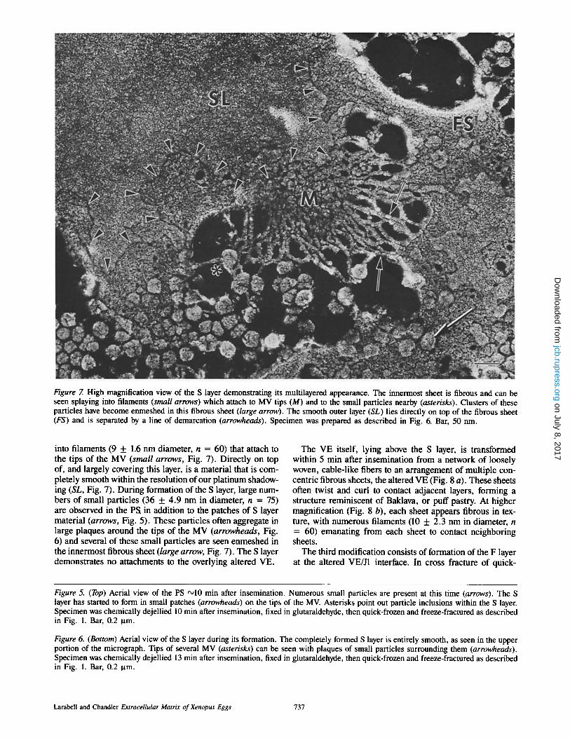
Figure 7. High magnification view of the S layer demonstrating its multilayered appearance. The innermost sheet is fibrous and can be seen splaying into filaments (small arrows) which attach to MV tips (M) and to the small particles nearby (asterisks). Clusters of these particles have become enmeshed in this fibrous sheet (large arrow). The smooth outer layer (SL) lies directly on top of the fibrous sheet (FS) and is separated by a line of demarcation (arrowheads). Specimen was prepared as described in Fig. 6. Bar, 50 nm.
into filaments (9 + 1.6 nm diameter, n = 60) that attach to the tips of the MV (small arrows, Fig. 7). Directly on top of, and largely covering this layer, is a material that is com- pletely smooth within the resolution of our platinum shadow- ing (SL, Fig. 7). During formation of the S layer, large num- bers of small particles (36 + 4.9 nm in diameter, n = 75) are observed in the PS in addition to the patches of S layer material (arrows, Fig. 5). These particles often aggregate in large plaques around the tips of the MV (arrowheads, Fig. 6) and several of these small particles are seen enmeshed in the innermost fibrous sheet (/arge arrow, Fig. 7). The S layer demonstrates no attachments to the overlying altered VE.
The VE itself, lying above the S layer, is transformed within 5 min after insemination from a network of loosely woven, cable-like fibers to an arrangement of multiple con- centric fibrous sheets, the altered VE (Fig. 8 a). These sheets often twist and curl to contact adjacent layers, forming a structure reminiscent of Baklava, or puff pastry. At higher magnification (Fig. 8 b), each sheet appears fibrous in tex- ture, with numerous filaments (10 + 2.3 nm in diameter, n = 60) emanating from each sheet to contact neighboring sheets.
The third modification consists of formation of the F layer at the altered VE/J1 interface. In cross fracture of quick-
Figure 5. (Top) Aerial view of the PS •10 min after insemination. Numerous small particles are present at this time (arrows). The S layer has started to form in small patches (arrowheads) on the tips of the MV. Asterisks point out particle inclusions within the S layer. Specimen was chemically dejellied 10 min after insemination, fixed in glutaraldehyde, then quick-frozen and freeze-fractured as described in Fig. 1. Bar, 0.2 ~tm.
Figure 6. (Bottom) Aerial view of the S layer during its formation. The completely formed S layer is entirely smooth, as seen in the upper portion of the micrograph. Tips of several MV (asterisks) can be seen with plaques of small particles surrounding them (arrowheads). Specimen was chemically dejellied 13 min after insemination, fixed in glutaraldehyde, then quick-frozen and freeze-fractured as described in Fig. 1. Bar, 0.2 ~tm.
Larabell and Chandler Extracellular Matrix of Xenopus Eggs 737
on July 8, 2017jcb.rupress.org
Dow
nloaded from
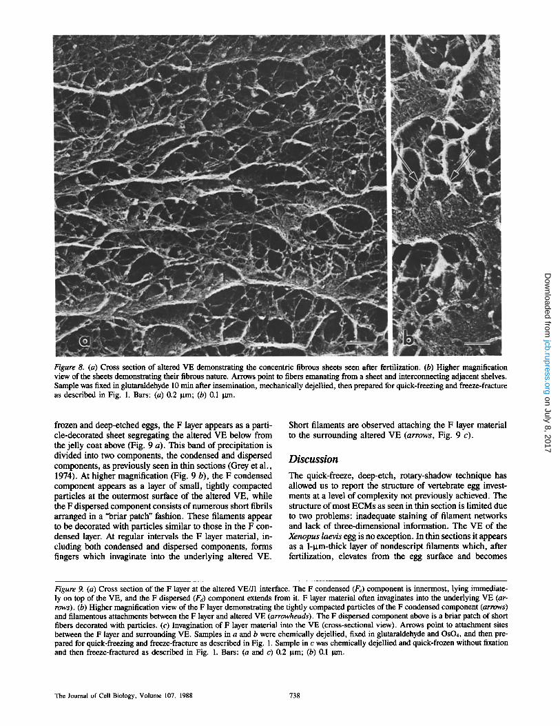
Figure 8. (a) Cross section of altered VE demonstrating the concentric fibrous sheets seen after fertilization. (b) Higher magnification view of the sheets demonstrating their fibrous nature. Arrows point to fibers emanating from a sheet and interconnecting adjacent shelves. Sample was fixed in glutaraldehyde 10 min after insemination, mechanically dejellied, then prepared for quick-freezing and freeze-fracture as described in Fig. 1. Bars: (a) 0.2 gm; (b) 0.1 0m.
frozen and deep-etched eggs, the F layer appears as a parti- cle-decorated sheet segregating the altered VE below from the jelly coat above (Fig. 9 a). This band of precipitation is divided into two components, the condensed and dispersed components, as previously seen in thin sections (Grey et al., 1974). At higher magnification (Fig. 9 b), the F condensed component appears as a layer of small, tightly compacted particles at the outermost surface of the altered VE, while the F dispersed component consists of numerous short fibrils arranged in a "briar patch" fashion. These filaments appear to be decorated with particles similar to those in the F con- densed layer. At regular intervals the F layer material, in- cluding both condensed and dispersed components, forms fingers which invaginate into the underlying altered VE.
Short filaments are observed attaching the F layer material to the surrounding altered VE (arrows, Fig. 9 c).
Discussion
The quick-freeze, deep-etch, rotary-shadow technique has allowed us to report the structure of vertebrate egg invest- ments at a level of complexity not previously achieved. The structure of most ECMs as seen in thin section is limited due to two problems: inadequate staining of filament networks and lack of three-dimensional information. The VE of the Xenopus laevis egg is no exception. In thin sections it appears as a 1-~tm-thick layer of nondescript filaments which, after fertilization, elevates from the egg surface and becomes
Figure 9. (a) Cross section of the F layer at the altered VE/J1 interface. The F condensed (Fc) component is innermost, lying immediate- ly on top of the VE, and the F dispersed (Fa) component extends from it. F layer material often invaginates into the underlying VE (ar- rows). (b) Higher magnification view of the F layer demonstrating the tightly compacted particles of the F condensed component (arrows) and filamentous attachments between the F layer and altered VE (arrowheads). The F dispersed component above is a briar patch of short fibers decorated with particles. (c) Invagination of F layer material into the VE (cross-sectional view). Arrows point to attachment sites between the F layer and surrounding VE. Samples in a and b were chemically dejellied, fixed in glutaraldehyde and OsO4, and then pre- pared for quick-freezing and freeze-fracture as described in Fig. 1. Sample in c was chemically dejellied and quick-frozen without fixation and then freeze-fractured as described in Fig. 1. Bars: (a and c) 0.2 gm; (b) 0.1 0an.
The Journal of Cell Biology, Volume 107, 1988 738
on July 8, 2017jcb.rupress.org
Dow
nloaded from

LarabeU and Chandler Extracellular Matrix of Xenopus Eggs 739
on July 8, 2017jcb.rupress.org
Dow
nloaded from

!
bigure 10. Diagram ot- the ECM of the X_enopus egg before and af- ter fertilization. The unfertilized egg (/eft s/de) with a cortical granule (CG) beneath the plasma membrane PM and MV extending into the PS. Filaments (f) connect MV to each other, to the plasma mem- brane, and to vesicles (v) seen in this region. The VE lies on the tips of the MV and is attached to the MV by a layer of horizontal filaments (hf) seen at the innermost portion of the VE. After fer- tilization (right side), the S layer (S) encircles the egg, lying on the tips of the MV. The VE has elevated and has been chemically and structurally altered (I/E*); the F layer (Fd and F<) has been deposited on the outer surface of the altered VE.
coated with an electron-dense band of precipitation, but otherwise appears to undergo little ultrastructural change.
In contrast, the deep-etching method we have used shows that the ECM of the unfertilized egg is a complex, mul- tilayered envelope whose structure and filament composition is summarized in Fig. 10 and Table I, respectively. The VE itself consists of two separate layers-an inner layer of horizontal filaments (hf, Fig. 10) and an outer, thicker layer of curling, cable-like fibers (VE, Fig. 10). These horizontal filaments are attached to the microvillar tips below and at numerous points become continuous with filaments in the PS. Beneath the VE is an elaborate glycocalyx (f, Fig. 10) that fills the PS. The glycocalyx is complex, but consists chiefly of oblique filaments that link microvillar surfaces with the plasma membrane below and numerous membrane- bound vesicles which are an integral component of this filamentous network (v, Fig. 10).
Unlike previous thin-section data, our work demonstrates that the VE structure is securely linked to PS filaments and the egg surface below as well as to the J1 above (data not shown). It is not surprising, then, that when these eggs are oviposited, the sticky jelly affixes to a substrate and conse- quently determines the orientation of the egg, which is often different than that expected based on gravity alone (i.e., heavier, vegetal hemisphere downward).
At fertilization, elaborate modification of the ECM is ob- served. Platinum replicas of deep-etched eggs show that the VE undergoes profound structural changes that cannot be seen in thin sections. The array of large cable-like fibers is transformed into concentric fibrous sheets; the fibers that emanate from these sheets to connect adjacent shelves are similar in diameter to the medium- and small-diameter illa- ments seen in the VE before fertilization (Table I). In addi- tion, the F layer (Fc and Fd, Fig. 10), previously seen in thin sections as an electron-dense band of material at the al- tered VE/J1 interface (Grey et al., 1974), is actually a plate
Table L Components of ECM around Xenopus Eggs before and after Fertilization
Unfertilized Diameter* Fertilized Diameter
n m n m
PS filaments PS filaments ND Oblique 10 + 2.7 On PM 10 + 1.2 S layer Small 4 + 0.6 Particles 36 + 4.9
HF filaments Filaments 9 + 1.6 Medium I0 + 1.3 Altered VE filaments Small 5 + 0.5 (between sheets)
VE filaments Medium 10 + 2.3 Large 19 _ 1.6 Small 4 + 0.8 Medium 12 + 1.6 Altered VE filaments Small 5 + 0.7 (below F layer)
Medium 10 + 3 Small 4 + 1
* Diameter measurements include platinum replica thickness; platinum thick- ness averaged 1.6 nm overall but may have varied locally. Measurements were made from replicas of three different samples.
of tightly compacted small particles. The F layer appears to be anchored to the altered VE by invaginations of this mate- rial as well as by numerous short fibers (Fig. 9, a and b); to- gether these structures constitute the fertilization envelope. These modifications could be those which convert the ECM from a structure which is receptive to sperm before fertiliza- tion to one which prevents additional sperm from contacting the egg surface after fertilization.
Our most striking discovery, however, is the presence of the S layer, a coating at the PS/VE interface that has not been previously identified. This extremely thin, multilayered en- velope, which completely encompasses the egg by 15 min af- ter insemination, orbits at the level of the microvillar tips. It is attached to the underlying MV by its innermost compo- nent, which is fibrous in nature, but is completely smooth at its outermost surface. No attachments are seen between the S layer and the overlying altered VE. This means that the filamentous connections seen between MV tips and overly- ing VE in the unfertilized egg have been severed. The fertil- ized egg is now completely unrestrained within the elevated altered VE. Soon after completion of the S layer, the egg ro- tates within the altered VE with respect to gravity, position- ing the animal pole upward (Palacek et al., 1978; Hara and Tydeman, 1979). Gravitational orientation is known to be important for proper dorsal-ventral axis formation; if such orientation is prevented, abnormal embryos develop (Ger- hart et al., 1981). Although not yet proven, formation of the S layer could act as a near-frictionless coating to facilitate ro- tation of the egg against the altered VE and jelly.
We believe the S layer, like the altered VE and F layer, is derived from contents of the cortical granules since its for- mation can be induced by the addition of cortical granule ex- udate to unfertilized eggs (Larabell, C. A., and D. E. Chand- ler, manuscript in preparation). The manner in which it is formed is not yet clear. One clue is that numerous small par- ticles '~35 nm in diameter are seen at the PS/VE interface coincident with the appearance of small patches of the S layer material. In addition, these particles aggregate in plaques around microvillar tips (Fig. 6) and are found embedded in
The Journal of Cell Biology, Volume 107, 1988 740
on July 8, 2017jcb.rupress.org
Dow
nloaded from
the innermost filamentous sheet of this structure (Fig. 7), suggesting that they play some role in S layer formation.
The S layer can now be visualized, but its role in early de- velopment remains to be assessed. This layer may play a part in allowing orientation of the egg after fertilization as sug- gested above. Alternatively, it may play a direct role in the block to polyspermy. Stewart-Savage and Grey (1987) have shown that fertilized eggs cannot be reinseminated even after the fertilization envelope is stripped away. This suggests that there may be an additional surface layer that is not stripped off which particpates in the block. Indeed, these authors have shown that there is such a layer at the tips of the MV that can be seen only after labeling with cationic ferritin (Grey, R. D., personal communication). Future experiments should show whether this layer is equivalent to the S layer we see in deep-etched and rotary-shadowed specimens. We thank Dr, Robert Grey for helpful discussions of his unpublished data.
This study was supported by grants from the National Science Founda- tion (DCB-8407152) and the National Institutes of Health (1KO4-HD00619).
Received for publication 24 February 1988, and in revised form 4 April 1988.
References
Barros, C., and R. Yanagimachi. 1971. Induction of zona reaction in golden hamster eggs by cortical granule material. Nature (Lond.). 233:268-269.
Barros, C., and R. Yanagimachi. 1972. Polyspermy-preventing mechanisms in the golden hamster egg. J. Exp. Zoo/. 180:251-266.
Bleil, J. D., and P. M. Wassarman. 1980. Mammalian sperm-egg interaction: identification of a glycoprotein in mouse egg zona pellucida possessing receptor activity for sperm. Cell. 20:873-882.
Chandler, D. E., and J. Heuser. 1980. The vitelline layer of the sea urchin egg and its modification during fertilization. J. Cell Biol. 84:618-632.
Gerhart, J., G. Ubbels, S. Black, K. Hara, and M. Kirschner. 1981. A reinves-
tigation of the role of the grey crescent in axis formation in Xenopus laevis. Nature (Lond.). 292:511-516.
Gerton, G. L., and J. L. Hedrick. 1986. The viteiline envelope to fertilization envelope conversion in eggs of Xenopus laevis. Dev. BIoL 116:1-7.
Glabe, C. G., and V. D. Vacquier. 1978. Egg surface glycoprot~in receptor for sea urchin sperm bindin. Proc. Natl. Acad. Sci. USA. 75:881-885.
Greve, L. C., and J. L. Hedrick. 1978. An immunocytochemical localization of the cortical granule lectin in fertilized and unfertilized eggs of Xenopus laevis. Gamete Res. !:13-18.
Grey, R. D., D. P. Wolf, and J. L. Hedrick. 1974. Formation and structure of the fertilization envelope in Xenopus laevis. Dev. Biol. 36:44-61.
Grey, R. D., P. K. Working, and J. L. Hedrick. 1976. Evidence that the fertil- ization envelope blocks sperm entry in eggs of Xenopus laevis: interaction of sperm with isolated envelopes. Dev. Biol. 54:52-60.
Hara, K., and P. Tydeman. 1979. Cinematographic observation of an "activa- tion wave ~ (AW) on the locally inseminated egg ofXenopus laevis. Wilhelm Roux's Arch. Dev. Biol. 186:91-94.
Heuser, J. E., T. S. Reese, M. J. Dennis, Y. Jan, L. Jan, and L. Evans. 1979. Synaptic vesicle exocytosis captured by quick-freezing and con'elated with qnantal transmitter release. J. Cell Biol. 81:275-300.
Hollinger, T. G., and G. L. Corton. 1980. Artificial fertilization of gametes from the South African clawed frog, Xenopus laevis. GameteRes. 3:45-57.
Palacek, J., G. A. Ubbels, and K. Rzehak. 1978. Changes of the external and internal pigment pattern upon fertilization in the egg of Xenopus laevis. J. Embryol. Exp. Morphol. 45:203-214.
Rossignol, D. P., B. J. Earles, G. L. Decker, andW. J. Lennarz. 1984. Charac- terization of the sperm receptor on the surface of eggs of Strongylocentrotus purpuratus. Dev. Biol. 104:308-321.
Stewart-Savage, J., and R. D. Grey. 1987. Loss of functional sperm entry into Xenopus eggs after activation correlates with a reduction in surface adhesiv- ity. Dev. Biol. 120:434-446.
Veron, M., C. Foerder, E. M. Eddy, and B. M. Shapiro. 1977. Sequential bio- chemical and morphological events during assembly of the fertilization mem- brane of the sea urchin. Cell. 10:321-328.
Wolf, D. P. 1974. The cortical granule reaction in living eggs of the toad, Xeno- pus laevis. Dev. Biol. 36:62-71.
Wolf, D. P., T. Nishihara, D. M. West, R. E. Wyrick, andJ. L. Hedrick. 1976. Isolation, physicochemical properties, and the macromolecular composition of the vitelline and fertilization envelopes from Xenopus laevis eggs. Bio- chemistry. 15:3671-3678.
Wyrick, R. E., T. Nishifiara, and J. L. Hedrick. 1974. Agglutination of jelly coat and cortical granule components and the block to polyspermy in the am- phibian Xenopus laevis. Proc. Natl. Acad. Sci. USA. 71:2067-2071.
Larabell and Chandler Extracellular Matrix of Xenopus Eggs 741
on July 8, 2017jcb.rupress.org
Dow
nloaded from